Article

Identification of Candidate Male-Reproduction-Related Genes from the Testis and Androgenic Gland of Macrobrachium nipponense, Regulated by PDHE1, through Transcriptome Profiling Analysis

1
Key Laboratory of Freshwater Fisheries and Germplasm Resources Utilization, Ministry of Agriculture and Rural Affairs, Freshwater Fisheries Research Center, Chinese Academy of Fishery Sciences, Wuxi 214081, China
2
Wuxi Fisheries College, Nanjing Agricultural University, Wuxi 214081, China
*
Authors to whom correspondence should be addressed.
Int. J. Mol. Sci. 2024, 25(3), 1940; https://doi.org/10.3390/ijms25031940
Submission received: 5 January 2024 / Revised: 27 January 2024 / Accepted: 31 January 2024 / Published: 5 February 2024
(This article belongs to the Section Molecular Biology)

Abstract

:
The previous publication identified that pyruvate dehydrogenase E1 (PDHE1) positively regulated the process of male reproduction in M. nipponense through affecting the expressions of insulin-like androgenic gland hormone. The present study aimed to identify the potential male-reproduction-related genes that were regulated by PDHE1 through performing the transcriptome profiling analysis in the testis and androgenic gland after the knockdown of the expressions of PDHE1 by the injection of dsPDHE1. Both RNA-Seq and qPCR analysis identified the significant decreases in PDHE1 expressions in the testis and androgenic gland in dsPDHE1-injected prawns compared to those in dsGFP-injected prawns, indicating the efficiency of dsPDHE1 in the present study. Transcriptome profiling analysis identified 56 and 127 differentially expressed genes (DEGs) in the testis and androgenic gland, respectively. KEGG analysis revealed that the energy-metabolism-related pathways represented the main enriched metabolic pathways of DEGs in both the testis and androgenic gland, including pyruvate metabolism, the Citrate cycle (TCA cycle), Glycolysis/Gluconeogenesis, and the Glucagon signaling pathway. Thus, it is predicted that these metabolic pathways and the DEGs from these metabolic pathways regulated by PDHE1 may be involved in the regulation of male reproduction in M. nipponense. Furthermore, four genes were found to be differentially expressed in both the testis and androgenic gland, of which ribosomal protein S3 was down-regulated and uncharacterized protein LOC113829596 was up-regulated in both the testis and androgenic gland in dsPDHE1-injected prawns. The present study provided valuable evidence for the establishment of an artificial technique to regulate the process of male reproduction in M. nipponense.

1. Introduction

Macrobrachium nipponenese, the oriental river prawn, is widely distributed through the whole of China [1]. It is the second culture freshwater prawn species in China. The annual production reached 220 thousand tons, producing huge economic benefits. The main culture regions in China include the Jiangsu, Jiangxi, Anhui, and Zhejiang provinces, where the annual productions are of over 20 thousand tons [2]. A previous publication has identified that both the testis and ovary of new hatchlings will mature within 45 days after hatching in the reproductive season. Rapid gonad maturation is the main problem, restricting the sustainable development of the M. nipponense industry. Rapid gonad maturation leads to inbreeding between the newborn prawns, resulting in having multiple generations in the same ponds, thus affecting the prawns in terms of market size [3]. Thus, it is urgently needed to establish an artificial technique to regulate the process of male reproduction in this species.
The glycolytic pathway is an important metabolic pathway to generate adenosine-triphosphate (ATP) in wide organisms. Glycolysis promotes the degradation and metabolism of glycogen or glucose under anaerobic conditions to produce ATP and is also a preparatory pathway for the aerobic oxidation of glucose in most organisms [4,5]. Pyruvate dehydrogenase (PDH) is an important enzyme involved in the glycolytic pathway. Glycolysis promotes the degradation of glucose to form pyruvate. PDH plays essential roles in the oxidation of glucose through catalyzing the oxidative decarboxylation of pyruvate to acetyl-CoA [6,7,8]. Furthermore, PDH has also been identified to link the glycolytic pathway with the tricarboxylic acid cycle (TCA cycle). Pyruvate dehydrogenase E1 (PDHE1) is a thiamine-diphosphate-dependent enzyme playing essential roles in catalyzing the oxidative decarboxylation of pyruvate. It is the rate-limiting step in the overall activity of PDH [9,10,11]. The absence of PDHE1 has negative effects on the conversion of pyruvate into acetyl-CoA, thus resulting in a decrease in ATP production through the TCA cycle [12]. Previous studies have identified the significant morphological difference in the testis and androgenic gland between the reproductive season and non-reproductive season [13,14]. PDHE1 was considered the candidate male-reproduction-related gene in M. nipponense because it was differentially expressed in the testis between the reproductive season and non-reproductive season [13]. PDHE1 was further identified to positively regulate the process of male reproduction in M. nipponense through performing qPCR, in situ hybridization, and RNAi analyses. qPCR analysis revealed that PDHE1 showed the highest expression level in the testis of M. nipponense. In situ hybridization analysis revealed that the signals of PDHE1 mRNA were observed in the spermatogonia of the testis in M. nipponense. The injection of double-standard RNA of PDHE1 (dsPDHE1) resulted in an over-95% decrease in PDHE1 expression, and the decrease in PDHE1 expression led to a 49% decrease in insulin-like androgenic gland hormone (IAG) expression. Histological observations revealed that less than 5% of cells were observed as sperm in the testis of dsPDHE1-injected prawns, which was significantly less than the percentage of sperm in the double-standard RNA of green fluorescent protein (dsGFP)-injected prawns (approximate 30%, in control). The knockdown of the expressions of PDHE1 by RNAi can delay the testis reproduction in this species [15].
Androgenic gland is a specific organ that only exists in male crustaceans. It produces hormones that contribute to male differentiation and the development of male secondary characteristics in crustacean species, especially in the regulation of testis reproduction [16,17]. The ablation of the androgenic gland from male Macrobrachium rosenbergii has resulted in sex reversal to “neo-female” [18,19]. The androgenic gland has been observed in many crustacean species, and similar functions in the regulation of male reproduction have been reported. Insulin-like androgenic gland hormone (IAG) is the most focused gene secreted by the androgenic gland. IAG plays essential roles in the regulation of male differentiation and reproduction in many crustacean species [18,19,20,21]. A previous publication identified that the knockdown of the expressions of IAG resulted in sex reversal in M. rosengergii [22]. The testis has also been reported to regulate the process of reproduction, sexual maturity, and sex differentiation in crustacean species [23,24,25].
Many previous publications have focused on the process of male differentiation and reproduction in M. nipponense, using the androgenic gland and testis as the target tissues. The transcriptome of the androgenic gland and the cDNA library have been constructed in this species in order to select candidate male-reproduction-related genes [26,27]. Transcriptome profiling analyses of the androgenic gland and testis have been further conducted after the ablation of eyestalks from male prawns [28,29], as have comparisons between the reproductive season and non-reproductive season [13,14]. RNAi has been employed to analyze the functions of candidate male-reproduction-related genes in M. nipponense, and Succinate dehydrogenase complex iron sulfur subunit B [30], Cyclin A [31], Cyclin B [32], and Cyclin B3 [33] have been identified to positively regulate the process of male reproduction in the species. Thus, it is hypothesized that the genes regulated by the PDHE1 in the testis and androgenic gland were predicted to be involved in the regulation of male reproduction in M. nipponense.
In the present study, we aimed to select the candidate male-reproduction-related genes regulated by PDHE1 through performing transcriptome profiling analysis in the androgenic gland and testis after the knockdown of the expressions of PDHE1 by RNAi. The present study will provide valuable evidence in the establishment of artificial techniques to regulate the process of male reproduction in M. nipponense.

2. Results

2.1. Measurement of the Efficiency of dsPDHE1

qPCR was used to verify the efficiency of dsPDHE1 in the present study. qPCR analysis revealed that the expressions of Mn-PDHE1 were significantly decreased in the testis and androgenic gland after the injection of dsPDHE1. The decreases reached 92.3% and 88.5% in the testis and androgenic gland after 5 days of dsPDHE1 injection, respectively compared to those of dsGFP-injected prawns (Figure 1).

2.2. Transcriptome Profiling Analysis in Testis

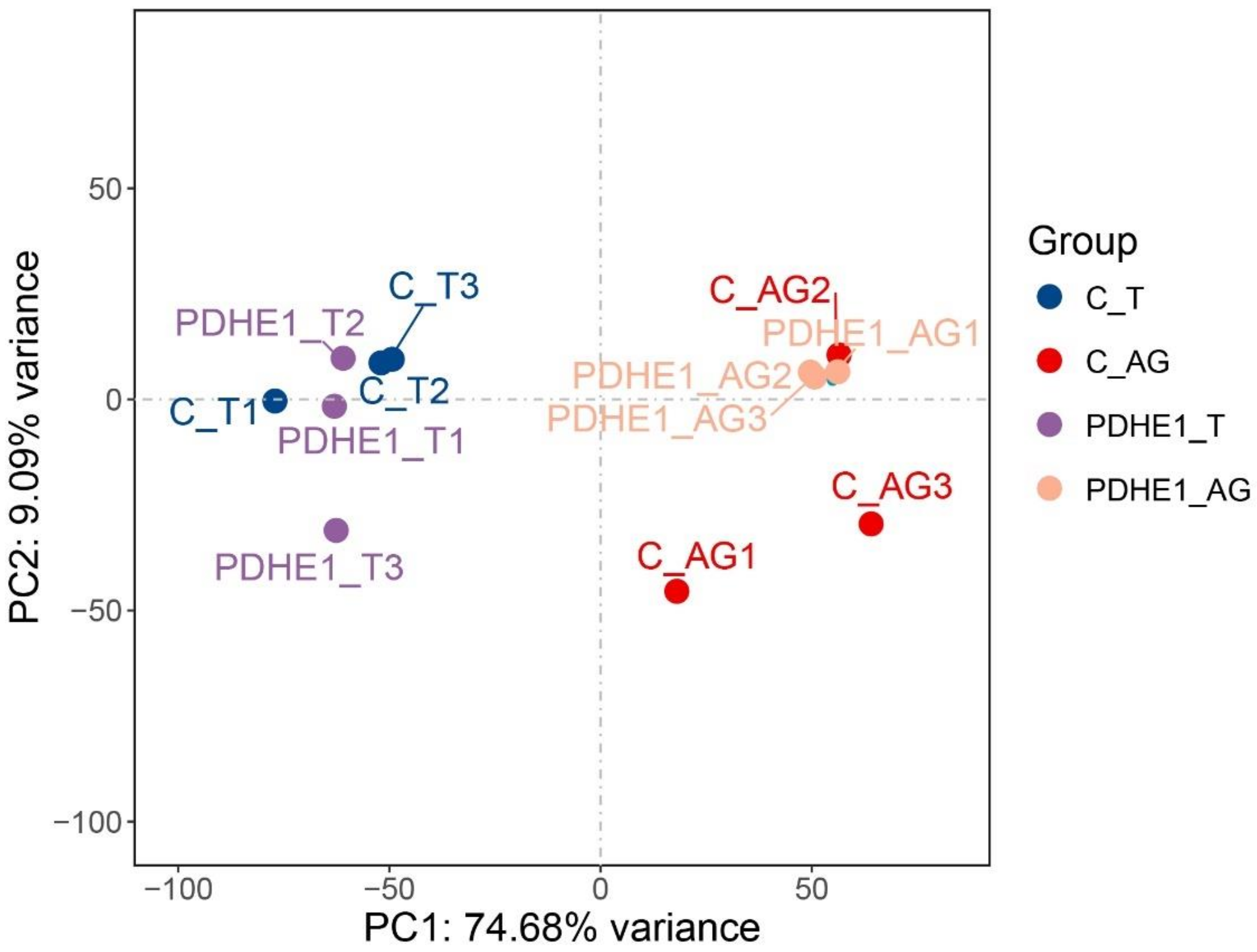
The principal component analysis was generated through the three biological replicates of the testis and androgenic gland transcriptome profiling analysis (Figure 2). Clear separations were observed between the samples of the testis and androgenic gland while the separations of the testis and androgenic gland between the dsPDHE1-infected prawns and dsGFP-injected prawns were not significant, indicating PDHE1 only regulated a limited number of gene expressions in the testis and androgenic gland, especially in the testis. In addition, PC1 and PC2 were responsible for 74.68% and 9.09% of variation sources, respectively, according to the extracted important variables.
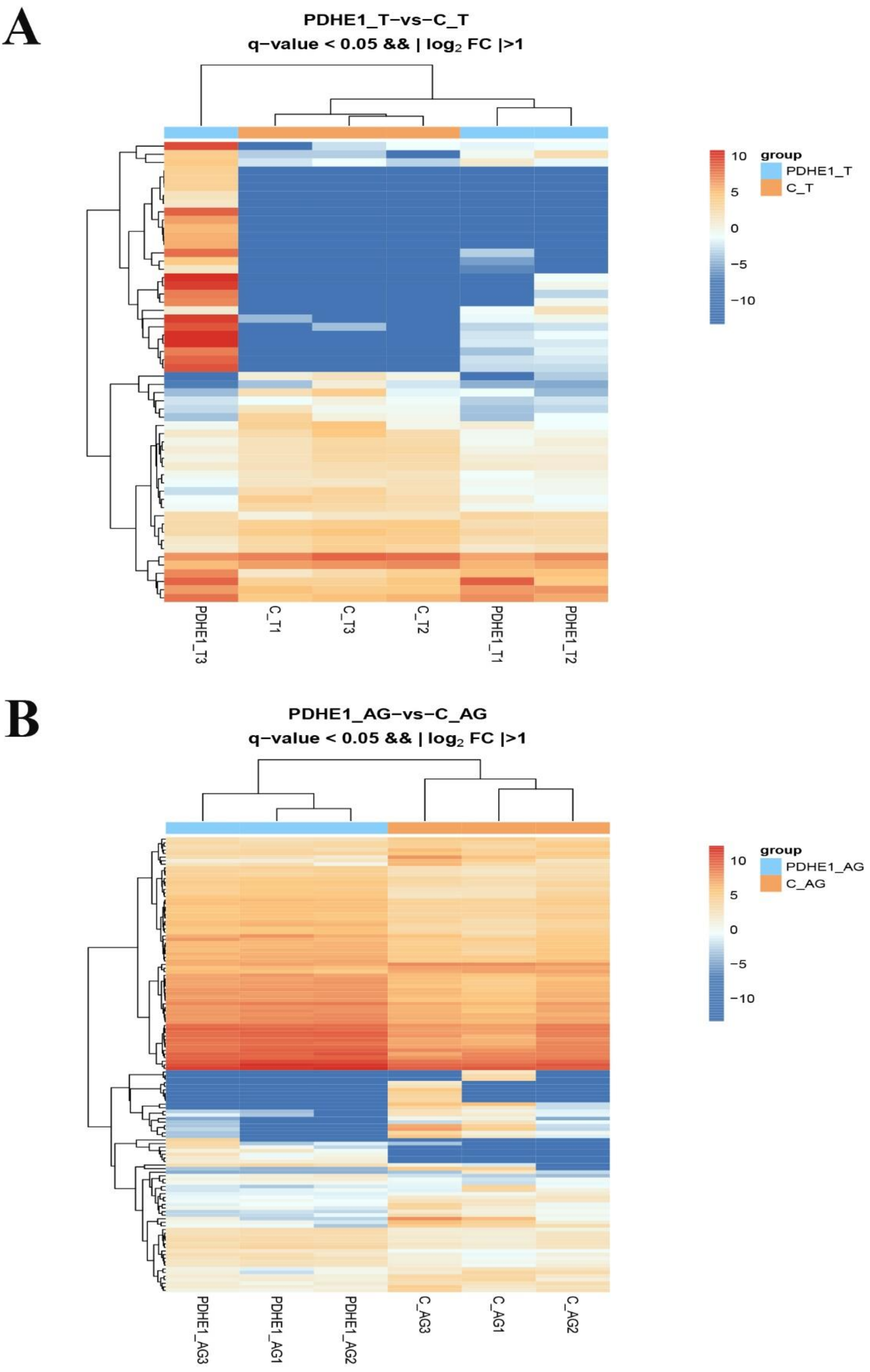
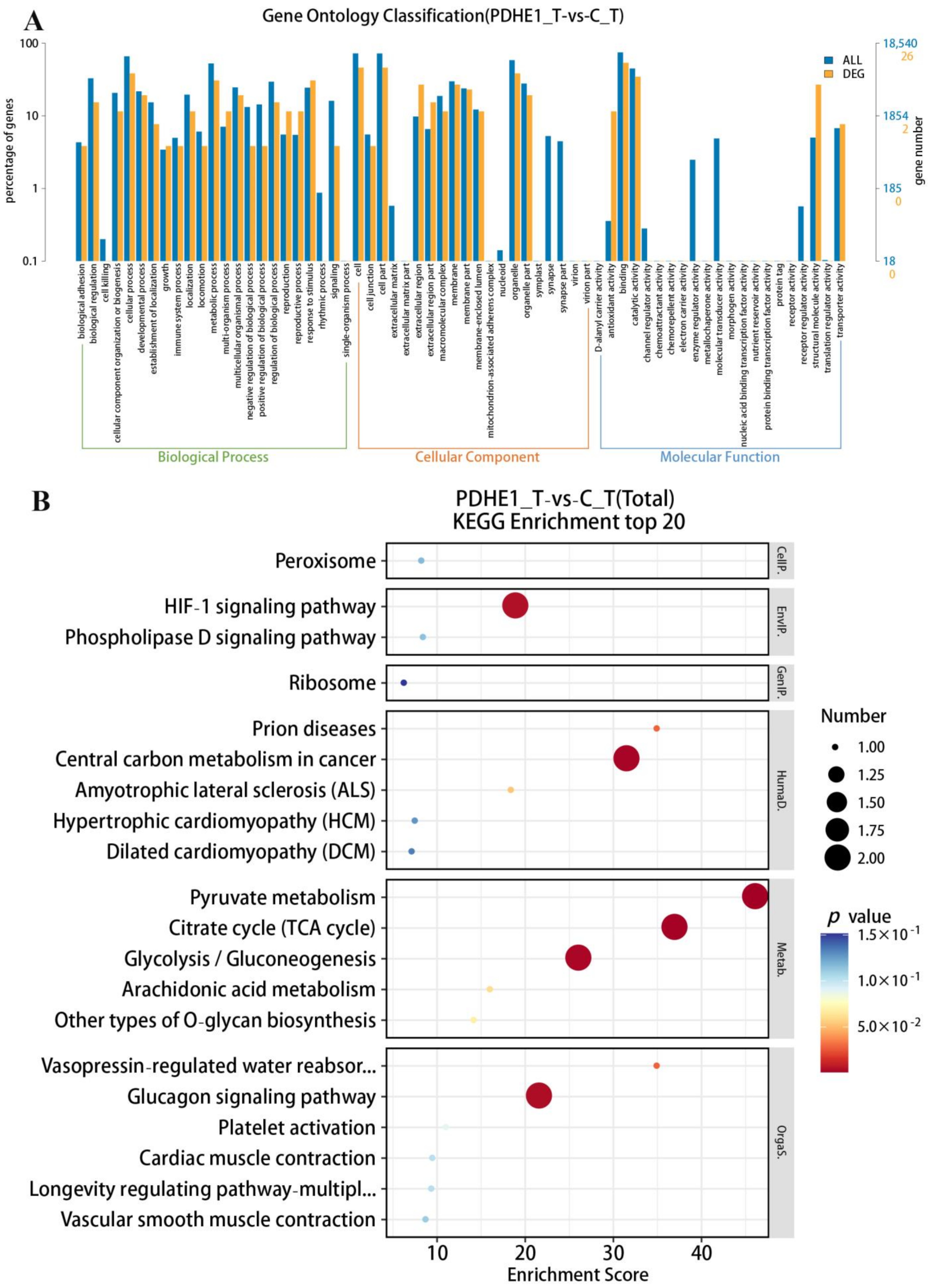
A total of 20,053 genes were matched to the known genes of the M. nipponense genome in the testis transcriptome. The DEGs were selected through performing the transcriptome profiling analysis in testis after the injection of dsPDHE1, using the criterion of > 2.0 as being up-regulated and < 0.5 as being down-regulated. Fifty-six genes were differentially expressed after the injection of dsPDHE1, including thirty-three up-regulated genes and twenty-three down-regulated genes (Figure 3A). All of the DEGs can be seen in Supplementary Table S1. A total of 26 DEGs and 8 DEGs were annotated in the GO and KEGG databases, respectively. GO analysis identified 36 functional groups in the present study ranging from 1 to 14. Binding, Cell, Cell Part, Cellular Process, and Organelle represented the main functional groups of DEGs wherein the numbers of DEGs were > 10 (Figure 4A). KEGG analysis consisted of 25 metabolic pathways. Pyruvate metabolism, the Citrate cycle (TCA cycle), Glycolysis/Gluconeogenesis, the Glucagon signaling pathway, and the HIF-1 signaling pathway represented the main metabolic pathways of DEGs wherein the number of DEGs was 2. The other metabolic pathways only had one DEG (Figure 4B).

2.3. Transcriptome Profiling Analysis in Androgenic Gland

A total of 17,035 genes were identified in the androgenic gland transcriptome that were highly matched to the known genes in the M. nipponense genome. A total 127 DEGs were selected in the androgenic gland after decreasing the expressions of PDHE1, including 76 up-regulated genes and 51 down-regulated genes (Figure 3B). All of the DEGs can be seen in Supplementary Table S2. A total of 63 DEGs and 18 DEGs were annotated in the GO and KEGG databases, respectively. A total of 40 functional groups were identified in the present study, according to the GO analysis. The number of DEGs in each functional group ranged from 1 to 46. Binding, Cell, Cell Part, Organelle, and Cellular Process represented the main functional groups of DEGs wherein the numbers of DEGs were > 30 (Figure 5A). KEGG analysis of DEGs identified 20 metabolic pathways ranging from 1 to 3. Phagosome represented the most enriched metabolic pathway of DEGs, and the other main metabolic pathways included pyruvate metabolism, the TCA cycle, Glycolysis/Gluconeogenesis, the Glucagon signaling pathway, the HIF-1 signaling pathway, Apoptosis, and Ribosome (Figure 5B).

2.4. Selection of Male-Reproduction-Related Genes

A total of 14 male-reproduction-related genes that were regulated by PDHE1 were selected in the present study and considered as the strong candidate genes involved in the process of male reproduction in M. nipponense. Four genes were found to be differentially expressed in both the testis and androgenic gland, including calcification-associated peptide (CAP), chitin-binding-like protein (CBP), ribosomal protein S3 (RPS3), and uncharacterized protein LOC113829596 (UP). The other main down-regulated genes were selected from the testis and androgenic gland and listed in Table 1. These genes, which were positively regulated by PDHE1, were considered the candidate male-reproduction-related genes in M. nipponense.

2.5. qPCR Analysis of DEGs

The accuracy of RNA-Seq was verified by using qPCR analysis (Figure 6). The expressions of DEGs, which were differentially expressed in both the testis and androgenic gland, were selected to perform the qPCR analyses. The qPCR analyses generally showed the same expression patterns with those found via RNA-Seq. The expressions of RPS3 were decreased by 62.3% and 55.7% in the testis and androgenic gland in dsPDHE1-injected prawns, respectively compared to those in dsGFP-injected prawns (p < 0.05) while UP expressions showed 512.3% and 713.5% increases in the testis and androgenic gland in dsPDHE1-injected prawns, respectively (p < 0.05). The expressions of CAP and CBP were increased in the testis in dsPDHE1-injected prawns while they were decreased in the androgenic gland in dsPDHE1-injected prawns (p < 0.05).

3. Materials and Methods

3.1. Sample Preparation

All of the prawns (M. nipponense) used for the present study were provided by Dapu M. nipponense Breeding Base in Wuxi, China (120°13′44″E, 31°28′ 22″N) in June 2022. All of the prawns were maintained in the lab condition for 3 days prior to the injection of dsPDHE1 and dsGFP. The dissolved oxygen was > 6.0 mg/L.

3.2. Synthesize of dsPDHE1 and dsGFP

Previous publication has identified the specific RNAi primer with a T7 promoter site, which can efficiently knockdown the expressions of PDHE1 in M. nipponense (Table 2) [15]. The dsPDHE1 and dsGFP were synthesized by using the Transcript Aid™ T7 High Yield Transcription kit (Fermentas, Inc., Waltham, MA, USA) in the present study following the manufacturer’s protocol. dsGFP was used as the negative control [34].

3.3. RNAi Analysis

A total of 300 healthy male prawns with body weights of 3.74–4.58 g were used in the present study, which were randomly divided into dsPDHE1-injected group and dsGFP-injected group. Each group contained 150 prawns, and the groups were maintained in three separate tanks. Previous publications have identified the suitable injected dose of double-stand RNA as 4 μg/g in M. nipponense [30,31,32,33]. In the present study, the injected doses of dsPDHE1 and dsGFP were both 4 μg/g, which was consistent with the previous studies. The dsPDHE1 and dsGFP were injected into the prawns through cavum pericardiale. The injected prawns were maintained in the lab condition with the dissolved oxygen of > 6.0 mg/L. Snails were fed twice every day with a body weight of 2%. Both the testis and androgenic glands were collected at 5 days after the injection of dsPDHE1 and dsGFP. Testis and androgenic gland samples were collected from fifteen individual prawns and pooled together to form a biological replicate for both transcriptome profiling analysis and qPCR analysis in order to provide sufficient tissues to extract RNA and eliminate the effects of individual differences. Three biological replicates were prepared for both transcriptome profiling analysis and qPCR analysis. The same biological replicates were used for the transcriptome profiling analysis and qPCR analysis.

3.4. qPCR Analysis

qPCR analysis was used to verify the efficiency of injected dsPDHE1 and dsGFP in the present study, as well as for the verification of differentially expressed gene (DEG) expressions. RNA isolation and cDNA synthesis were performed according to the detailed description in the previous studies [30,31,32,33]. Briefly, total RNA was firstly extracted from each tissue sample using the UNlQ-10 Column Trizol Total RNA Isolation Kit (Sangon, Shanghai, China) according to the manufacturer’s protocol. The integrity of total RNA was measured by using 1.2% agarose gel electrophoresis, and an ultraviolet spectrophotometer (Eppendorf, Hamburg, Germany) was used to determine the RNA concentrations for each tissue sample. A total of 1 μg total RNA from each tissue sample was used to synthesize the cDNA template by using the PrimeScript™ RT Reagent Kit (Takara Bio, Kusatsu, Japan) according to the descriptions of the manufacturer’s protocol. The qPCR analysis was determined by using the UltraSYBR Mixture (CWBIO, Beijing, China), and performed on the Bio-Rad iCycler iQ5 Real-Time PCR System (Bio-Rad, Hercules, CA, USA). All qPCR reactions were run using three technical replicates. The primers for qPCR analysis were listed in Table 2, including the Eukaryotic translation initiation factor 5A (EIF), which was used as the reference gene in the present study. EIF has been identified as a suitable reference gene for PCR analysis in M. nipponense that can stably be expressed under various conditions [35]. The relative mRNA expressions were calculated using the 2−∆∆CT method [36].

3.5. Transcriptome Profiling Analysis

The differentially expressed genes (DEGs) in the gills of M. nipponense, caused by the alkali treatment, were selected through performing the transcriptome profiling analysis. Previous publications have well described the detailed procedures for the RNA-Seq and analysis [13,14,27,28,29]. Briefly, the total RNA was extracted using RNAiso Plus Reagent (Takara Bio, Kusatsu, Japan) according to the manufacturer’s instructions. RNA integrity number (RIN) value of > 7.0 indicated the integrity of total RNA, measured by using A 2100 Bioanalyzer (Agilent Technologies, Inc., Santa Clara, CA, USA). RNA concentrations were measured using ultraviolet spectrophotometer (Eppendorf, Hamburg, Germany). A total of 4 µg of total RNA was used to construct the library. Illumina Hiseq-2500 sequencing platform was employed to perform the transcriptome profiling analysis under the parameter of PE150.
Fastp software (version 0.11.5) was employed to remove the low-quality raw reads with the default parameters [37]. The HISAT2 (version 2.2.1.0) software was then employed to map the obtained clean reads to the M. nipponense reference genome (Genbank access numbers: GCA_015110555.1 and GCA_015104395.1) [38]. Genes were annotated in the Gene Ontology (GO) (http://www.geneontology.org/, accessed on 15 August 2023) [39], Cluster of Orthologous Groups (COG) (http://www.ncbi.nlm.nih.gov/COG/, accessed on 15 August 2023) [40], and Kyoto Encyclopedia of Genes and Genomes (KEGG) databases (http://www.genome.jp/kegg/, accessed on 15 August 2023) [41], using FDR < 0.05. Gene expression was calculated using the FPKM method, where FPKM = cDNA fragments/mapped fragments (millions)/transcript length (kb), using HTSeq-count [42]. DESeq2 was used to perform the differential expression analysis [43]. The Benjamini–Hochberg correction method was used to calculate the false discovery rate (FDR) [44] with q-value < 0.05. Fold change > 2 was considered as marking up-regulated differentially expressed genes (DEGs), and fold change < 0.5 was considered as marking down-regulated DEGs.

3.6. Statistical Analysis

SPSS 23.0 was used to perform the statistical analyses in the present study, estimated using the Duncan’s multiple range test and LSD in one-way ANOVA. p < 0.05 and p < 0.01 indicated significant difference and extremely significant difference, respectively. Quantitative data were expressed as means ± SD.

4. Discussion

Rapid gonad maturation is the main problem affecting the sustainable development of the M. nipponense industry. Thus, the long-term goal of M. nipponense studies is aimed at establishing an artificial technique to regulate the process of gonad reproduction in this species. PDHE1 has been identified to positively regulate the process of male reproduction in M. nipponense [15]. In the present study, the genes regulated by PDHE1 were selected through performing transcriptome profiling analysis in the testis and androgenic glands of M. nipponense after the knockdown of the expressions of PDHE1 by injecting dsPDHE1. The selected genes were considered as being the strong candidate genes involved in the regulation of male reproduction in M. nipponense, providing valuable evidence for the establishment of an artificial technique to regulate the process of testis reproduction in this species. The expressions of PDHE1 decreased by 92.3% and 88.5% in the testis and androgenic gland in dsPDHE1-injected prawns after 5 days of dsPDHE1 injection, respectively compared to those in dsGFP-injected prawns. In addition, the transcriptome profiling analysis also identified 88.1% and 87.3% decreases in PDHE1 expression in the testis and androgenic gland in dsPDHE1-injected prawns, respectively, indicating that the injected dsPDHE1 had efficiently decreased the expressions of PDHE1 in the present study.
In the present study, a total of 56 and 127 DEGs were identified in the testis and androgenic gland, respectively according to the transcriptome profiling analyses after deceasing the expression of PDHE1 by injecting dsPDHE1. KEGG analysis revealed that pyruvate metabolism, the Citrate cycle (TCA cycle), Glycolysis/Gluconeogenesis, the Glucagon signaling pathway, and the HIF-1 signaling pathway were identified as the main enriched metabolic pathways of DEGs in both the testis and androgenic gland. In addition, Phagosome, Apoptosis, and Ribosome were also identified in the androgenic gland.
Pyruvate is produced from the glucose metabolism through glycolysis. Thus, pyruvate metabolism plays essential roles for energy production and has been identified to be involved in various metabolic pathways, depending on the cell’s demands [45]. The TCA cycle and Glycolysis/Gluconeogenesis are essential metabolic pathways to produce energy in wide organisms [46]. Pyruvate is produced from glucose through the catalysis of Glycolysis/Gluconeogenesis, promoting the generation of ATP from free energy [47]. The glucagon signaling pathway is an important pathway for the body to regulate blood glucose levels under hypoglycemic conditions [48]. These metabolic pathways are energy-metabolism-related pathways, which have been predicted to be participated in the process of male reproduction through producing ATP in M. nipponense [13,14,28,29]. PDHE1 is an energy-metabolism-related gene that has been identified to regulate the process of male reproduction in M. nipponense [15]. Thus, the DEGs from these metabolic pathways, which were regulated by PDHE1, were considered as the strong candidate genes that regulated the process of male reproduction by affecting the generation of ATP in M. nipponense. The potential functions of these DEGs in the process of male reproduction need further investigation.
Phagosomes are dynamic organelles formed by macrophages, playing essential roles in capturing and destroying microbial pathogens [49]. Apoptosis, which is regulated by a series of genes, is a process to capture and destroy damaged or aged cells in order to maintain a stable internal environment in order to better adapt to the living environment [50,51]. Ribosome exists in the cells of wild organisms. It promotes the process of correct translation from RNA to protein [52,53,54]. These metabolic pathways were also predicted to participate in the regulation of male reproduction through digesting the aged cells and ensuring corrected protein synthesis in our previous publications [13,14,28,29]. Thus, the DEGs from these metabolic pathways, which were regulated by PDHE1, were also predicted to regulate the process of male reproduction in M. nipponense.
Four DEGs were identified to be regulated by PDHE1 in both the testis and androgenic gland in the present study, predicting that these genes were involved in the regulation of male reproduction in M. nipponense. CAP contains the repeat sequence of aspartic acid in the C-terminus, which plays essential roles for calcium-binding activity [55,56]. Previous publications have revealed that CAP-1 is mainly involved in exoskeleton calcification in P. clarkia [55,56,57,58]. CAP has been also identified to play essential roles in the regulation of growth in crustacean species because it regulates the process of molting in crustaceans [59]. Chitin is mainly found in the exoskeletons of crustaceans and insects, playing essential roles in the stabilization of an arthropod’s body through acting as a physical defense against predators and desiccation [60]. Chitin biosynthesis plays essential roles in the regulation of the growth, development, and reproduction of insects and crustaceans [61]. The functions of RPS3 are identified to be involved in the regulation of translation initiation [62,63], ribosomal maturation [64,65], the induction of apoptosis [66,67,68], the suppression of tumors [69], the maintenance of genomic integrity [70], DNA repair [71,72], and the regulation of the cell cycle and gene transcription [73,74,75]. RPS3 is reported to be phosphorylated by cyclin-dependent kinase 2 (Cdc2) during the G2/M phase. Cdc2 has been also identified to regulate the process of male reproduction in M. nipponense by RNAi [76]. The knockdown of the expression of PDHE1 changes the process of male reproduction in M. nipponense. In the present study, the expressions of RPS3 were positively regulated by PDHE1, predicting that it may regulate male reproduction through maintaining genomic integrity and ribosomal maturation. Uncharacterized protein LOC113829596 was also differentially expressed in both the testis and androgenic gland in the present study. However, its function is still unknown. The potential functions of this gene in the regulation of male reproduction need further investigation.
The main genes from the testis that were regulated by PDHE1 included Glucoside xylosyltransferase 2 (GXYLT2), Cytochrome P450 3A (CYP3A), and Nesprin-1. These genes were also predicted to regulate the process of male reproduction in M. nipponense, based on the significant differences in gene expression between dsPDHE1 prawns and dsGFP prawns. GXYLT2 plays essential roles in the modification of the first xylose to the O-Glucose residue on epidermal growth factor (EGF) repeats of Notch receptors [77,78], which participate in the process of cell proliferation, migration, differentiation, and survival [79,80]. The CYP3A subfamily represents the largest portion of CYP proteins, which play essential roles in the metabolic clearance of wild chemically diverse compounds including environmental pollutants, pesticides, therapeutic drugs, dietary products, and steroids [81,82]. Nesprin-1 has been identified to regulate the cellular functions and diseases [83,84,85,86]. Abnormal cellular functions and subsequent disease pathogenesis may be caused because of an abnormal connection between the nucleus and F-actin through Nesprin-1, leading to insufficient force transmission to the nucleus.
The candidate male-reproduction-related genes from the androgenic gland included Cuticle protein AM1199, Cuticle protein 21, Pro-resilin, and Mucin-19, which were regulated by PDHE1. Previous publications have identified the functions of cuticle protein genes in response and adaption to environmental stresses including salinity, predators, and toxins [87,88,89]. Resilin, which is represented in specialized regions of the cuticle in most insects, has been identified to provide low stiffness, high strain, and efficient energy storage [90,91], thus playing important roles in insect flight [92,93], the jumping mechanism in fleas [94,95], and vocalization in cicadas [96]. Mucins are mainly divided into the cell-associated mucin and gel-forming mucin [97]. Many factors can stimulate the secretion of mucin, including toxins, proinflammatory cytokines, and neuropeptides. Thus, the expression of mucins will be regulated by the inflammatory condition [98,99].
In conclusion, PDHE1 is an energy-metabolism-related gene that has been identified to regulate the process of male reproduction in M. nipponense. The genes regulated by PDHE1 were selected from the testis and androgenic gland of M. nipponense through performing transcriptome profiling analysis, and these were predicted to be involved in the regulation of male reproduction in this species. The energy-metabolism-related metabolic pathways were identified as the main enriched metabolic pathways of DEGs in the present study, including pyruvate metabolism, the Citrate cycle (TCA cycle), Glycolysis/Gluconeogenesis, and the Glucagon signaling pathway, indicating that energy metabolism is an essential physiological process for male reproduction in M. nipponense. Thus, these energy-metabolism-related pathways and the DEGs from these metabolic pathways may play essential roles in the regulation of male reproduction in this species. Furthermore, four genes were found to be regulated by PDHE1 in both the testis and androgenic gland, predicting their potential roles in the regulation of male reproduction in M. nipponense. The present study identified novel candidate male-reproduction-related genes in M. nipponense, providing valuable evidence for studies on the mechanism of male reproduction as well as promoting the establishment of an artificial technique to regulate the process of gonad reproduction in this species.

Supplementary Materials

The following supporting information can be downloaded at: https://www.mdpi.com/article/10.3390/ijms25031940/s1.

Author Contributions

S.J. (Shubo Jin): conceptualization, writing—original draft. Y.W.: software. Y.X.: resources. W.Z. and H.Q.: validation, investigation, formal analysis. S.J. (Sufei Jiang) and H.F.: supervision, funding acquisition, writing—review & editing. All authors reviewed the manuscript. All authors have read and agreed to the published version of the manuscript.

Funding

This research was supported by grants from the Natural Science Foundation of Jiangsu Province (BK20221207); Central Public-Interest Scientific Institution Basal Research Fund, CAFS (2023JBFM04, 2023TD39); the seed industry revitalization project of Jiangsu Province (JBGS [2021] 118); Jiangsu Agricultural Industry Technology System; the earmarked fund for CARS-48-07; and the new cultivar breeding Major Project of Jiangsu Province (PZCZ201745).

Institutional Review Board Statement

We obtained the permissions from the Institutional Animal Care and Use Ethics Committee of the Freshwater Fisheries Research Center, Chinese Academy of Fishery Sciences (Wuxi, China) (Authorization no. 20220627074, 27 June 2022) to perform all of the experiment involved in the present study. Tissue collections were performed on an ice bath (2 °C).

Informed Consent Statement

We obtained the permissions from the Institutional Animal Care and Use Ethics Committee of the Freshwater Fisheries Research Center, Chinese Academy of Fishery Sciences (Wuxi, China) (Authorization no. 20220627074, 27 June 2022) to perform all of the experiment involved in the present study. Tissue collections were performed on an ice bath (2 °C).

Data Availability Statement

The raw data of the present study have been submitted to NCBI with the accession number of BioProject PRJNA863726.

Acknowledgments

We give thanks to the Jiangsu Province Platform for the Conservation and Utilization of Agricultural Germplasm.

Conflicts of Interest

The authors declare no conflicts of interest.

References

  1. Fu, H.T.; Jiang, S.F.; Xiong, Y.W. Current Status and Prospects of Farming the Giant River Prawn (Macrobrachium rosenbergii) and the oriental River Prawn (Macrobrachium nipponense) in china. Aquac. Res. 2012, 43, 993–998. [Google Scholar]
  2. Zhang, X.L.; Cui, L.F.; Li, S.M.; Liu, X.Z.; Han, X.; Jiang, K.Y.; Bureau of Fisheries, Ministry of Agriculture, P.R.C. Fisheries economic statistics. In China Fishery Yearbook; Beijing China Agricultural Press: Beijing, China, 2020; Volume 24. [Google Scholar]
  3. Jin, S.B.; Zhang, Y.; Guan, H.H.; Fu, H.T.; Jiang, S.F.; Xiong, Y.W.; Qiao, H.; Zhang, W.Y.; Gong, Y.S.; Wu, Y. Histological observation of gonadal development during post-larva in oriental river prawn, Macrobrachium nipponense. Chin. J. Fish. 2016, 29, 11–16. [Google Scholar]
  4. Metón, I.; Fernández, F.; Baanante, I.V. Short- and long-term effects of refeeding on key enzyme activities in glycolysis–gluconeogenesis in the liver of gilthead seabream (Sparus aurata). Aquaculture 2003, 225, 99–107. [Google Scholar] [CrossRef]
  5. Nordlie, R.C.; Foster, J.D.; Lange, A.J. Regulation OF glucose production BY the liver. Annu. Rev. Nutr. 1999, 19, 379–406. [Google Scholar] [CrossRef] [PubMed]
  6. Reed, L.J. A trail of research from lipoic acid to alpha-keto acid dehydrogenase complexes. J. Biol. Chem. 2001, 276, 38329–38336. [Google Scholar] [CrossRef]
  7. Perham, R.N. Domains, motifs, and linkers in 2-oxo acid dehydrogenase multienzyme complexes: A paradigm in the design of a multifunctional protein. Biochemistry 1991, 30, 8501–8512. [Google Scholar] [CrossRef] [PubMed]
  8. Patel, M.S.; Roche, T.E. Molecular biology and biochemistry of pyruvate dehydrogenase complexes. FASEB J. 1990, 4, 3224–3233. [Google Scholar] [CrossRef] [PubMed]
  9. Linn, T.C.; Pettit, F.H.; Reed, L.J. Alpha-keto acid dehydrogenase complexes. X. Regulation of the activity of the pyruvate dehydrogenase complex from beef kidney mitochondria by phosphorylation and dephosphorylation. Proc. Natl. Acad. Sci. USA 1969, 62, 234–241. [Google Scholar] [CrossRef]
  10. Linn, T.C.; Pettit, F.H.; Hucho, F.; Reed, L.J. Alpha-keto acid dehydrogenase complexes. XI. Comparative studies of regulatory properties of the pyruvate dehydrogenase complexes from kidney, heart, and liver mitochondria. Proc. Natl. Acad. Sci. USA 1969, 64, 227–234. [Google Scholar] [CrossRef]
  11. Patel, M.S.; Nemeria, N.S.; Furey, W.; Jordan, F. The pyruvate dehydrogenase complexes: Structure-based function and regulation. J. Biol. Chem. 2014, 289, 16615–16623. [Google Scholar] [CrossRef]
  12. Carlsson, J.; Kujala, U.; Edlund, M.J.K. Pyruvate Dehydrogenase Activity in Streptococcus mutans. Infect. Immun. 1985, 49, 674–678. [Google Scholar] [CrossRef] [PubMed]
  13. Jin, S.B.; Hu, Y.N.; Fu, H.T.; Sun, S.M.; Jiang, S.F.; Xiong, Y.W.; Qiao, H.; Zhang, W.; Gong, Y.; Wu, Y. Analysis of Testis Metabolome and Transcriptome from the Oriental River Prawn (Macrobrachium nipponense) in Response to Different Temperatures and Illumination Times. Comp. Biochem. Phys. D 2020, 34, 100662. [Google Scholar] [CrossRef] [PubMed]
  14. Jin, S.B.; Zhang, W.Y.; Xiong, Y.W.; Jiang, S.F.; Qiao, H.; Gong, Y.S.; Wu, Y.; Fu, H.T. Genetic regulation of male sexual development in the oriental river prawn Macrobrachium nipponense during reproductive vs. non-reproductive season. Aquacult. Int. 2022, 30, 2059–2079. [Google Scholar] [CrossRef]
  15. Jin, S.; Hu, Y.; Fu, H.; Jiang, S.; Xiong, Y.; Qiao, H.; Zhang, W.; Gong, Y.; Wu, Y. Identification and Characterization of the Pyruvate Dehydrogenase E1 Gene in the Oriental River Prawn, Macrobrachium nipponense. Front. Endocrinol. 2021, 12, 752501. [Google Scholar] [CrossRef] [PubMed]
  16. Sagi, A.; Cohen, D.; Wax, Y. Production of Macrobrachium rosenbergii in Momosex Population: Yield Characteristes Under Intensive Monoculture Conditions in Cages. Aquaculture 1986, 51, 265–275. [Google Scholar] [CrossRef]
  17. Sagi, A.; Cohen, D.; Milner, Y. Effect of Androgenic Gland Ablation on Morphotypic Differentiation and Sexual Characteristics of Male Freshwater Prawns, Macrobrachium rosenbergii. Gen. Comp. Endocr. 1990, 77, 15–22. [Google Scholar] [CrossRef] [PubMed]
  18. Ma, K.Y.; Li, J.L.; Qiu, G.F. Identification of putative regulatory region of insulin-like androgenic gland hormone gene (IAG) in the prawn Macrobrachium nipponense and proteins that interact with IAG by using yeast two-hybrid system. Gen. Comp. Endocr. 2016, 229, 112–118. [Google Scholar] [CrossRef]
  19. Rosen, O.; Manor, R.; Weil, S.; Gafni, O.; Linial, A.; Aflalo, E.D.; Ventura, T.; Sagi, A. A sexual shift induced by silencing of a single insulin-like gene in crayfish: Ovarian upregulation and testicular degeneration. PLoS ONE 2010, 5, e15281. [Google Scholar] [CrossRef]
  20. Ventura, T.; Manor, R.; Aflalo, E.D.; Weil, S.; Raviv, S.; Glazer, L.; Sagi, A. Temporal silencing of an androgenic gland-specific insulin-like gene affecting phenotypical gender differences and spermatogenesis. Endocrinology 2009, 150, 1278–1286. [Google Scholar] [CrossRef]
  21. Ventura, T.; Manor, R.; Aflalo, E.D.; Weil, S.; Sagi, A. Expression of an androgenic gland-specific insulin-like peptide during the course of prawn sexual and morpho-typic differentiation. ISRN Endocrinol. 2011, 2011, 476283. [Google Scholar] [CrossRef]
  22. Ventura, T.; Manor, R.; Aflalo, E.D.; Weil, S.; Rosen, O.; Sagi, A. Timing Sexual Differentiation: Full Functional Sex Reversal Achieved Through Silencing of a Single Insulin-Like Gene in the Prawn, Macrobrachium rosenbergii. Biol. Reprod. 2012, 86, 90. [Google Scholar] [CrossRef] [PubMed]
  23. Cao, J.X. The Molecular Mechanism in Male Reproductive Tract of the Prawn, Macrobrachium rosenbergii; Zhejiang University: Hangzhou, China, 2006. [Google Scholar]
  24. Guo, Z.H. Study on Proliferation and Differentiation of Spermatogenic Cells from Macrobrachium nipponense In Vitro; Hebei University: Baoding, China, 2007. [Google Scholar]
  25. Qiu, G.F.; Du, N.S.; Lai, W. Studies on the Male Reproductive System of the Freshwater Prawn, Macrobrachium nipponense. J. Shanghai Ocean Uni. 1995, 4, 107–111. [Google Scholar]
  26. Qiao, H.; Fu, H.T.; Jin, S.B.; Wu, Y.; Jiang, S.F.; Gong, Y.S.; Xiong, Y.W. Constructing and Random Sequencing Analysis of Normalized cDNA Library of Testis Tissue from Oriental River Prawn (Macrobrachium nipponense). Comp. Biochem. Phys. D 2012, 7, 268–276. [Google Scholar] [CrossRef] [PubMed]
  27. Jin, S.B.; Fu, H.T.; Zhou, Q.; Sun, S.M.; Jiang, S.F.; Xiong, Y.W.; Gong, Y.S.; Qiao, H.; Zhang, W.Y. Transcriptome analysis of androgenic gland for discovery of novel genes from the oriental river prawn, Macrobrachium nipponense, using Illumina Hiseq 2000. PLoS ONE 2013, 8, e76840. [Google Scholar] [CrossRef] [PubMed]
  28. Jin, S.B.; Fu, Y.; Hu, Y.N.; Fu, H.T.; Jiang, S.F.; Xiong, Y.W.; Qiao, H.; Zhang, W.Y.; Gong, Y.S.; Wu, Y. Transcriptome Profiling Analysis of the Testis After Eyestalk Ablation for Selection of the Candidate Genes Involved in the Male Sexual Development in Macrobrachium nipponense. Front. Genet. 2021, 12, 675928. [Google Scholar] [CrossRef] [PubMed]
  29. Jin, S.B.; Fu, Y.; Hu, Y.N.; Fu, H.T.; Jiang, S.F.; Xiong, Y.W.; Qiao, H.; Zhang, W.Y.; Gong, Y.S.; Wu, Y. Identification of candidate genes from androgenic gland in Macrobrachium nipponense regulated by eyestalk ablation. Sci. Rep. 2021, 11, 19855. [Google Scholar] [CrossRef] [PubMed]
  30. Jin, S.; Hu, Y.; Fu, H.; Jiang, S.; Xiong, Y.; Qiao, H.; Zhang, W.; Gong, Y.; Wu, Y. Identification and Characterization of the Succinate Dehydrogenase Complex Iron Sulfur Subunit B Gene in the Oriental River Prawn, Macrobrachium nipponense. Front. Genet. 2021, 12, 698318. [Google Scholar] [CrossRef]
  31. Zhang, W.; Xiong, Y.; Wang, P.; Chen, T.; Jiang, S.; Qiao, H.; Gong, Y.; Wu, Y.; Jin, S.; Fu, H. RNA interference analysis of potential functions of cyclin A in the reproductive development of male oriental river prawns (Macrobrachium nipponense). Front. Genet. 2022, 13, 1053826. [Google Scholar] [CrossRef]
  32. Zhang, W.; Wang, P.; Xiong, Y.; Chen, T.; Jiang, S.; Qiao, H.; Gong, Y.; Wu, Y.; Jin, S.; Fu, H. RNA Interference Analysis of the Functions of Cyclin B in Male Reproductive Development of the Oriental River Prawn (Macrobrachium nipponense). Genes 2022, 13, 2079. [Google Scholar] [CrossRef]
  33. Jin, S.; Zhou, Z.; Zhang, W.; Xiong, Y.; Qiao, H.; Gong, Y.; Wu, Y.; Jiang, S.; Fu, H. RNAi Analysis of Potential Functions of Cyclin B3 in Reproduction of Male Oriental River Prawns (Macrobrachium nipponense). Animals 2023, 13, 1703. [Google Scholar] [CrossRef]
  34. Zhang, S.B.; Jiang, P.; Wang, Z.Q.; Long, S.R.; Liu, R.D.; Zhang, X.; Yang, W.; Ren, H.J.; Cui, J. Dsrna-Mediated Silencing of Nudix Hydrolase in Trichinella spiralis Inhibits the Larval Invasion and Survival in Mice. Exp. Parasitol. 2016, 162, 35–42. [Google Scholar] [CrossRef]
  35. Hu, Y.N.; Fu, H.T.; Qiao, H.; Sun, S.M.; Zhang, W.Y.; Jin, S.B.; Jiang, S.F.; Gong, Y.S.; Xiong, Y.W.; Wu, Y. Validation and evaluation of reference genes for Quantitative real-time PCR in Macrobrachium nipponense. Int. J. Mol. Sci. 2018, 19, 2258. [Google Scholar] [CrossRef] [PubMed]
  36. Livak, K.J.; Schmittgen, T.D. Analysis of relative gene expression data using real-time quantitative PCR and the 2−∆∆CT method. Methods 2001, 25, 402–408. [Google Scholar] [CrossRef] [PubMed]
  37. Chen, S.; Zhou, Y.; Chen, Y.; Gu, J. Fastp: An ultra-fast all-in-one FASTQ preprocessor. Bioinformatics 2018, 34, i884–i890. [Google Scholar] [CrossRef]
  38. Kim, D.; Paggi, J.M.; Park, C.; Bennett, C.; Salzberg, S.L. Graph-based genome alignment and genotyping with HISAT2 and HISAT-genotype. Nat. Biotechnol. 2019, 37, 907–915. [Google Scholar] [CrossRef] [PubMed]
  39. Ashburner, M.; Ball, C.A.; Blake, J.A.; Botstein, D.; Butler, H.; Cherry, J.M.; Davis, A.l.P.; Dolinski, K.; Dwight, S.S.; Eppig, J.T.; et al. Gene ontology: Tool for the unification of biology. Nat. Genet. 2000, 25, 25–29. [Google Scholar] [CrossRef] [PubMed]
  40. Tatusov, R.L.; Fedorova, N.D.; Jackson, J.D.; Jacobs, A.R.; Kiryutin, B.; Koonin, E.V.; Krylov, D.M.; Mazumder, R.; Mekhedov, S.L.; Nikolskaya, A.N.; et al. The COG database: An updated version includes eukaryotes. BMC Bioinform. 2003, 4, 41. [Google Scholar] [CrossRef] [PubMed]
  41. Kanehisa, M.; Araki, M.; Goto, S.; Hattori, M.; Itoh, M. KEGG for linking genomes to life and the environment. Nucleic Acids Res. 2008, 36, 480–484. [Google Scholar] [CrossRef]
  42. Anders, S.; Pyl, P.T.; Huber, W. HTSeq-A Python framework to work with high-throughput sequencing data. Bioinformatics 2015, 31, 166–169. [Google Scholar] [CrossRef]
  43. Love, M.I.; Huber, W.; Anders, S. Moderated estimation of fold change and dispersion for RNA-seq data with DESeq2. Genome Biol. 2014, 15, 550. [Google Scholar] [CrossRef]
  44. Ferreira, J.A.; Zwinderman, A.H. On the Benjamini–Hochberg method. Ann. Statist. 2006, 34, 1827–1849. [Google Scholar] [CrossRef]
  45. Gray, L.R.; Tompkins, S.C.; Taylor, E.B. Regulation of pyruvate metabolism and human disease. Cell. Mol. Life Sci. 2014, 71, 2577–2604. [Google Scholar] [CrossRef] [PubMed]
  46. Shahin, A.Y.; Ismail, A.M.; Shaaban, O.M. Supplementation of clomiphene citrate cycles with Cimicifuga racemosa or ethinyl oestradiol-a randomized trial. Reprod. BioMed. Online 2009, 19, 501–507. [Google Scholar] [CrossRef] [PubMed]
  47. Lubert, S. Glycolysis. In Biochemistry, 4th ed.; W. H. Freeman and Company: New York, NY, USA, 1995; pp. 483–508. [Google Scholar]
  48. Yang, M.X.; Pan, M.Z.; Huang, D.; Liu, J.H.; Guo, Y.L.; Liu, Y.; Xiao, T.Y.; Mai, K.S.; Zhang, W.B. The uninhibited glucagon signaling pathway in Japanese flounder Paralichthys olivaceus: A link with the glucose intolerance? Aquaculture 2024, 579, 740172. [Google Scholar] [CrossRef]
  49. Westman, J.; Grinstein, S. Determinants of phagosomal pH during host-pathogen interactions. Front. Cell Dev. Biol. 2020, 8, 624958. [Google Scholar] [CrossRef] [PubMed]
  50. Ren, X.; Wang, S.; Zhang, C.; Hu, X.; Zhou, L.; Li, Y.; Xu, L. Selenium ameliorates cadmium-induced mouse leydig TM3 cell apoptosis via inhibiting the ROS/JNK/c-jun signaling pathway. Ecotoxicol. Environ. Saf. 2020, 192, 110266. [Google Scholar] [CrossRef] [PubMed]
  51. Zhou, L.; Wang, S.; Cao, L.; Ren, X.; Li, Y.; Shao, J.; Xu, L. Lead acetate induces apoptosis in Leydig cells by activating PPARgamma/caspase-3/PARP pathway. Int. J. Environ. Health Res. 2021, 31, 34–44. [Google Scholar] [CrossRef] [PubMed]
  52. Kraft, C.; Deplazes, A.; Sohrmann, M.; Peter, M. Mature ribosomes are selectively degraded upon starvation by an autophagy pathway requiring the Ubp3p/Bre5p ubiquitin protease. Nat. Cell Biol. 2008, 10, 602–610. [Google Scholar] [CrossRef]
  53. Wyant, G.A.; Abu-Remaileh, M.; Frenkel, E.M.; Laqtom, N.N.; Dharamdasani, V.; Lewis, C.A.; Chan, S.H.; Heinze, I.; Ori, A.; Sabatini, D.M. Nufip1 is a ribosome receptor for starvation-induced ribophagy. Science 2018, 360, 751–758. [Google Scholar] [CrossRef]
  54. An, H.; Harper, J.W. Systematic analysis of ribophagy in human cells reveals bystander flux during selective autophagy. Nat. Cell Biol. 2018, 20, 135–143. [Google Scholar] [CrossRef]
  55. Inoue, H.; Ozaki, N.; Nagasawa, H. Purification and structural determination of a phosphorylated peptide with anti-calcification and chitin-binding activities in the exoskeleton of the crayfish, Procambarus clarkia. Biosci. Biotechnol. Biochem. 2001, 65, 1840–1848. [Google Scholar] [CrossRef] [PubMed]
  56. Inoue, H.; Ohira, T.; Ozaki, N.; Nagasawa, H. Cloning and expression of a cDNA encoding a matrix peptide associated with calcification in the exoskeleton of the crayfish. Comp. Biochem. Physiol. B 2003, 136, 755–765. [Google Scholar] [CrossRef] [PubMed]
  57. Inoue, H.; Ohira, T.; Ozaki, N.; Nagasawa, H. A novel calcium-binding peptide from the cuticle of the crayfish, Procambarus clarkia. Biochem. Biophys. Res. Commun. 2004, 318, 649–654. [Google Scholar] [CrossRef] [PubMed]
  58. Inoue, H.; Ohira, T.; Nagasawa, H. Significance of the N- and C-terminal regions of CAP-1, a cuticle calcification-associated peptide from the exoskeleton of the crayfish, for calcification. Peptides 2007, 28, 566–573. [Google Scholar] [CrossRef] [PubMed]
  59. Nagasawa, H. The molecular mechanism of calcification in aquatic organisms. Biosci. Biotechnol. Biochem. 2013, 77, 1991–1996. [Google Scholar] [CrossRef]
  60. Boßelmann, F.; Romano, P.; Fabritius, H.; Raabe, D.; Epple, M. The composition of the exoskeleton of two crustacea: The American lobster Homarus americanus and the edible crab Cancer pagurus. Thermochim. Acta 2007, 463, 65–68. [Google Scholar] [CrossRef]
  61. Zhu, K.Y.; Merzendorfer, H.; Zhang, W.; Zhang, J.; Muthukrishnan, S. Biosynthesis, turnover, and functions of chitin in insects. Annu. Rev. Entomol. 2016, 61, 177–196. [Google Scholar] [CrossRef] [PubMed]
  62. Westermann, P.; Heumann, W.; Bommer, U.A.; Bielka, H.; Nygard, O.; Hultin, T. Crosslinking of initiation factor eIF-2 to proteins of the small subunit of rat liver ribosomes. FEBS Lett. 1979, 97, 101–104. [Google Scholar] [CrossRef]
  63. Tolan, D.R.; Hershey, J.W.; Traut, R.T. Crosslinking of eukaryotic initiation factor eIF3 to the 40S ribosomal subunit from rabbit reticulocytes. Biochimie 1983, 65, 427–436. [Google Scholar] [CrossRef]
  64. Schafer, T.; Maco, B.; Petfalski, E.; Tollervey, D.; Bottcher, B.; Aebi, U.; Hurt, E. Hrr25-dependent phosphorylation state regulates organization of the pre-40S subunit. Nature 2006, 441, 651–655. [Google Scholar] [CrossRef]
  65. Mitterer, V.; Murat, G.; Rety, S.; Blaud, M.; Delbos, L.; Stanborough, T.; Bergler, H.; Leulliot, N.; Kressler, D.; Pertschy, B. Sequential domain assembly of ribosomal protein S3 drives 40S subunit maturation. Nat. Commun. 2016, 7, 10336. [Google Scholar] [CrossRef]
  66. Jang, C.Y.; Lee, J.Y.; Kim, J. RpS3, a DNA repair endonuclease and ribosomal protein, is involved in apoptosis. FEBS Lett. 2004, 560, 81–85. [Google Scholar] [CrossRef] [PubMed]
  67. Jang, C.Y.; Kim, H.D.; Kim, J. Ribosomal protein S3 interacts with TRADD to induce apoptosis through caspase dependent JNK activation. Biochem. Biophys. Res. Commun. 2012, 42, 474–478. [Google Scholar] [CrossRef] [PubMed]
  68. Kim, W.; Youn, H.; Lee, S.; Kim, E.; Kim, D.; Lee, S.; Lee, J.M.; Youn, B.H. RNF138-mediated ubiquitination of rpS3 is required for resistance of glioblastoma cells to radiation-induced apoptosis. Exp. Mol. Med. 2018, 50, e434. [Google Scholar] [CrossRef]
  69. Kim, S.H.; Kim, J. Reduction of invasion in human fibrosarcoma cells by ribosomal protein S3 in conjunction with Nm23-H1 and ERK. Biochim. Biophys. Acta 2006, 1763, 823–832. [Google Scholar] [CrossRef] [PubMed]
  70. Yadavilli, S.; Mayo, L.D.; Higgins, M.; Lain, S.; Hegde, V.; Deutsch, W.A. Ribosomal protein S3: A multi-functional protein that interacts with both p53 and MDM2 through its KH domain. DNA Repair 2009, 8, 1215–1224. [Google Scholar] [CrossRef] [PubMed]
  71. Hegde, V.; Yadavilli, S.; McLaughlin, L.D.; Deutsch, W.A. DNA repair efficiency in transgenic mice over expressing ribosomal protein S3. Mutat. Res. 2009, 666, 16–22. [Google Scholar] [CrossRef]
  72. Kim, Y.; Kim, H.D.; Kim, J. Cytoplasmic ribosomal protein S3 (rpS3) plays a pivotal role in mitochondrial DNA damage surveillance. Biochim. Biophys. Acta 2013, 1833, 2943–2952. [Google Scholar] [CrossRef]
  73. Wan, F.; Anderson, D.E.; Barnitz, R.A.; Snow, A.; Bidere, N.; Zheng, L.; Hegde, V.; Lam, L.T.; Staudt, L.M.; Levens, D. Ribosomal protein S3: A KH domain subunit in NF-kappaB complexes that mediates selective gene regulation. Cell 2007, 131, 927–939. [Google Scholar] [CrossRef]
  74. Yoon, I.S.; Chung, J.H.; Hahm, S.H.; Park, M.J.; Lee, Y.R.; Ko, S.I.; Kang, L.W.; Kim, T.S.; Kim, J.; Han, Y.S. Ribosomal protein S3 is phosphorylated by Cdk1/cdc2 during G2/M phase. BMB Rep. 2011, 44, 529–534. [Google Scholar] [CrossRef]
  75. Jang, C.Y.; Kim, H.D.; Zhang, X.; Chang, J.S.; Kim, J. Ribosomal protein S3 localizes on the mitotic spindle and functions as a microtubule associated protein in mitosis. Biochem. Biophys. Res. Commun. 2012, 429, 57–62. [Google Scholar] [CrossRef] [PubMed]
  76. Wang, P.C.; Zhang, W.Y.; Xiong, Y.W.; Chen, T.Y.; Jiang, S.F.; Qiao, H.; Gong, Y.S.; Wu, Y.; Jin, S.B.; Fu, H.T. RNA interference analysis of the potential functions of cyclin-dependent kinase 2 in sexual reproduction of male oriental river prawns (Macrobrachium nipponense). Aquacult. Int. 2023, 31, 2849–2866. [Google Scholar] [CrossRef]
  77. Sethi, M.K.; Buettner, F.F.R.; Krylov, V.B.; Takeuchi, H.; Nifantiev, N.E.; Haltiwanger, R.S.; Gerardy-Schahn, R.; Bakker, H. Identification of glycosyltransferase 8 family members as xylosyltransferases acting on O-glucosylated notch epidermal growth factor repeats. J. Biol. Chem. 2010, 285, 1582–1586. [Google Scholar] [CrossRef] [PubMed]
  78. Lee, T.V.; Sethi, M.K.; Leonardi, J.; Rana, N.A.; Buettner, F.F.R.; Haltiwanger, R.S.; Bakker, H.; Jafar-Nejad, H. Negative regulation of notch signaling by xylose. PLoS Genet. 2013, 9, e1003547. [Google Scholar] [CrossRef] [PubMed]
  79. Artavanis-Tsakonas, S.; Rand, M.D.; Lake, R.J. Notch signaling: Cell fate control and signal integration in development. Science 1999, 284, 770–776. [Google Scholar] [CrossRef] [PubMed]
  80. Guruharsha, K.G.; Kankel, M.W.; Artavanis-Tsakonas, S. The Notch signalling system: Recent insights into the complexity of a conserved pathway. Nat. Rev. Genet. 2012, 13, 654–666. [Google Scholar] [CrossRef] [PubMed]
  81. Guengerich, F.P. Cytochrome P-450 3A4: Regulation and role in drug metabolism. Annu. Rev. Pharmacol. Toxicol. 1999, 39, 1–17. [Google Scholar] [CrossRef] [PubMed]
  82. Maurel, P. The CYP3 family. In Cytochrome P450 Metabolic and Toxicological Aspects; Ioannides, C., Ed.; CRC Press: Boca Raton, FL, USA, 1996; pp. 241–270. [Google Scholar]
  83. Chancellor, T.J.; Lee, J.; Thodeti, C.K.; Lele, T. Actomyosin tension exerted on the nucleus through nesprin-1 connections influences endothelial cell adhesion, migration, and cyclic strain-induced reorientation. Biophys. J. 2010, 99, 115–123. [Google Scholar] [CrossRef]
  84. Zhang, Q.; Bethmann, C.; Worth, N.F.; Davies, J.D.; Wasner, C.; Feuer, A.; Ragnauth, C.D.; Yi, Q.; Mellad, J.A.; Warren, D.T.; et al. Nesprin-1 and -2 are involved in the pathogenesis of Emery Dreifuss muscular dystrophy and are critical for nuclear envelope integrity. Hum. Mol. Genet. 2007, 16, 2816–2833. [Google Scholar] [CrossRef]
  85. Gros-Louis, F.; Dupré, N.; Dion, P.; Fox, M.A.; Laurent, S.; Verreault, S.; Sanes, J.R.; Bouchard, J.P.; Rouleau, G.A. Mutations in SYNE1 lead to a newly discovered form of autosomal recessive cerebellar ataxia. Nat. Genet. 2007, 39, 80–85. [Google Scholar] [CrossRef]
  86. Attali, R.; Warwar, N.; Israel, A.; Gurt, I.; McNally, E.; Puckelwartz, M.; Glick, B.; Nevo, Y.; Ben-Neriah, Z.; Melki, J. Mutation of SYNE-1, encoding an essential component of the nuclear lamina, is responsible for autosomal recessive arthrogryposis. Hum. Mol. Genet. 2009, 18, 3462–3469. [Google Scholar] [CrossRef] [PubMed]
  87. Ramasamy, R.; Thiruchenthooran, V.; Jayadas, T.T.P.; Eswaramohan, T.; Santhirasegaram, S.; Sivabalakrishnan, K.; Naguleswaran, A.; Uzest, M.; Cayrol, B.; Voisin, S.N.; et al. Transcriptomic, proteomic and ultrastructural studies on salinity-tolerant Aedes aegypti in the context of rising sea levels and arboviral disease epidemiology. BMC Genom. 2021, 22, 253. [Google Scholar] [CrossRef] [PubMed]
  88. Hu, L.; Gui, W.Y.; Chen, B.; Chen, L. Transcriptome profiling of maternal stress-induced wing dimorphism in pea aphids. Ecol. Evol. 2019, 9, 11848–11862. [Google Scholar] [CrossRef]
  89. Saavedra-Rodriguez, K.; Campbell, C.L.; Lozano, S.; Penilla-Navarro, P.; Lopez-Solis, A.; Solis-Santoyo, F.; Rodriguez, A.D.; Perera, R.; Black IV, W.C. Permethrin resistance in Aedes aegypti: Genomic variants that confer knockdown resistance, recovery, and death. PLoS Genet. 2021, 17, e1009606. [Google Scholar] [CrossRef] [PubMed]
  90. Andersen, S.O. The cross-links in resilin identified as dityrosine and trityrosine. Biochim. Biophys. Acta 1964, 93, 213–215. [Google Scholar] [CrossRef] [PubMed]
  91. Gosline, J.; Lillie, M.; Carrington, E.; Guerette, P.; Ortlepp, C.; Savage, K. Elastic proteins: Biological roles and mechanical properties. Philos. Trans. R. Soc. Lond. B Biol. Sci. 2002, 357, 121–132. [Google Scholar] [CrossRef] [PubMed]
  92. Weis-Fogh, T. A rubber-like protein in insect cuticle. J. Exp. Biol. 1960, 37, 887–907. [Google Scholar] [CrossRef]
  93. Gorb, S.N. Serial elastic elements in the damselfly wing: Mobile vein joints contain resilin. Naturwissenschaften 1999, 86, 552–555. [Google Scholar] [CrossRef]
  94. Neville, A.C.R.; Rothschild, M. Fleas-insects which fly with their legs. Proc. R. Entomol. Soc. Lond. 1967, 32, 9–10. [Google Scholar]
  95. Rothschild, M.; Schlein, J. The jumping mechanism of Xenopsylla cheopis. I. Exoskeletal structures and musculature. Philos. Trans. R. Soc. Lond. B Biol. Sci. 1975, 271, 457–490. [Google Scholar]
  96. Bennet-Clark, H. Resonators in insect sound production: How insects produce loud pure-tone songs. J. Exp. Biol. 1999, 202, 3347–3357. [Google Scholar] [CrossRef]
  97. Pontremoli, C.; Barbero, N.; Viscardi, G.; Visentin, S. Mucin–drugs interaction: The case of theophylline, prednisolone and cephalexin. Bioorg. Med. Chem. 2015, 23, 6581–6586. [Google Scholar] [CrossRef]
  98. Linden, S.K.; Sutton, P.; Karlsson, N.G.; Korolik, V.; McGuckin, M.A. Mucins in the mucosal barrier to infection. Mucosal Immunol. 2008, 1, 183–197. [Google Scholar] [CrossRef]
  99. Longman, R.J.; Poulsom, R.; Corfield, A.P.; Warren, B.F.; Wright, N.A.; Thomas, M.G. Alterations in the composition of the supramucosal defense barrier in relation to disease severity of ulcerative colitis. J. Histochem. Cytochem. 2006, 54, 1335–1348. [Google Scholar] [CrossRef]
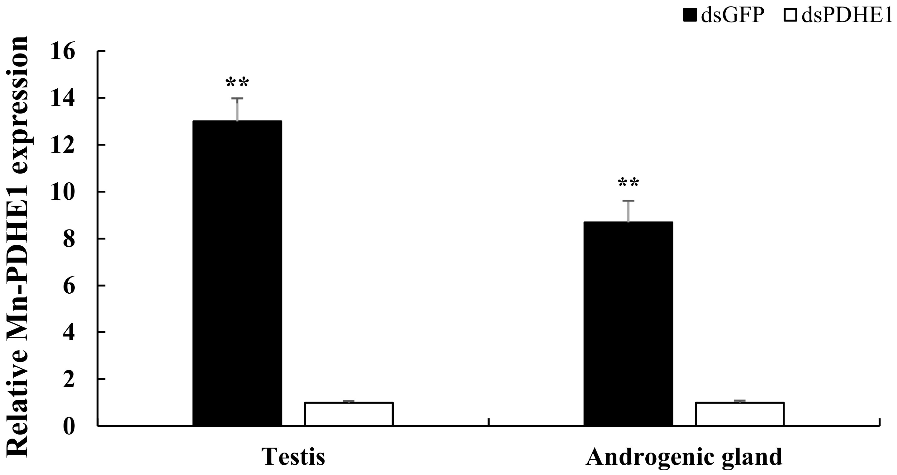
Figure 1. Verification of the efficiency of injected dsPDHE1. Data are shown as means ± SD (standard deviation) of tissues from three biological replicates. ** (p < 0.01) indicated the extremely significant difference of Mn-PDHE1 expressions in the testis and androgenic gland between the dsPDHE1-injected prawns and dsGFP-injected prawns.
Ijms 25 01940 g001
Figure 2. Principal component analysis for the testis and androgenic gland transcriptome profiling analysis using three biological replicates in M. nipponense after decreasing the expressions of PDHE1 by RNAi.
Ijms 25 01940 g002
Figure 3. Clustering diagram of DEGs. The horizontal coordinates represent the sample names and the clustering results of the samples while the vertical coordinates represent the DEGs and the clustering results of the DEGs. The colors represent the expression levels of DEGs in the samples. (A) indicates the clustering diagram comparing the testis between the dsPDHE1-injected prawns and dsGFP-injected prawns. (B) indicates the clustering diagram comparing the androgenic gland between the dsPDHE1-injected prawns and dsGFP-injected prawns.
Ijms 25 01940 g003
Figure 4. Gene Ontology (GO) analysis and Kyoto Encyclopedia of Genes and Genomes (KEGG) analysis of DEGs in the testis transcriptome after the injection of dsPDHE1. (A) indicates the GO analysis of DEGs. (B) indicates the KEGG analysis of DEGs.
Ijms 25 01940 g004
Figure 5. Gene Ontology (GO) analysis and Kyoto Encyclopedia of Genes and Genomes (KEGG) analysis of DEGs in the androgenic gland transcriptome after the injection of dsPDHE1. (A,B) indicate the GO analysis and KEGG analysis of DEGs, respectively.
Ijms 25 01940 g005
Figure 6. Verification of the expressions of DEGs in the testis and androgenic gland in the dsPDHE1-injected prawns and dsGFP-injected prawns. Data are shown as means ± SD (standard deviation) of tissues from three biological replicates. * (p < 0.05) and ** (p < 0.01) indicate significant difference and extremely significant difference in DEG expression between the testis and androgenic gland in the dsPDHE1-injected prawns and dsGFP-injected prawns.
Ijms 25 01940 g006
Table 1. The candidate male-reproduction-related genes regulated by PDHE1.
GeneAccession
Number
SpeciesE-ValueFold Change
(dsPDHE1 vs. dsGFP)
TestisAndrogenic Gland
Calcification-associated peptide (Cap)WGF13344.1Macrobrachium nipponense6.87 × 10−821.07−10.38
Chitin binding-like protein (CBP)ROT74831.1Penaeus vannamei3.79 × 10−53.25−2.53
Ribosomal protein S3 (RPS3)KAF6343595.1Pipistrellus kuhlii0.00011−3.12−2.78
Uncharacterized protein LOC113829596 (UP)XP_027238594.1Penaeus vannamei4.28 × 10−66.896.37
Glucoside xylosyltransferase 2XP_045611801.1Procambarus clarkii8.33 × 10−5−21.36
Skeletal muscle actin 6QPC96639.1Macrobrachium nipponense3.61 × 10−5−19.23
Polycystic kidney disease protein 1XP_047488675.1Penaeus chinensis4.12 × 10−5−13.26
Cytochrome P450 3A30XP_037798812.1Penaeus monodon4.11 × 10−5−12.37
Nesprin-1XP_045608233.1Procambarus clarkii5.18 × 10−5−10.39
Histidine-rich protein DDB_G0274557XP_047473847.1Penaeus chinensis6.77 × 10−9 −22.65
Cuticle protein AM1199 XP_042888222.1Penaeus japonicus8.87 × 10−9 −22.47
Cuticle protein 21XP_045617846.1Procambarus clarkii2.58 × 10−5 −21.50
Pro-resilin-likeXP_047479276.1Penaeus chinensis4.35 × 10−5 −19.85
Mucin-19-likeXP_037798457.1Penaeus monodon0.00012 −17.23
Note: E-value indicates the accuracy of the alignment and annotation in Nr database (p < 0.05).
Table 2. Primers used in the present study.
GenePrimer Sequence
dsPDHE1-FTAATACGACTCACTATAGGGGTGCTCTTAGCACTGGAGGC
dsPDHE1-RTAATACGACTCACTATAGGGCCAAGTAGTGGAAGGCAGGA
PDHE1-FTGACCTTAACGGCAACGAGG
PDHE1-RTGACCTTAACGGCAACGAGG
CAP-FTGGAAGCTCGCCGTAGTTTT
CAP-RCTTGACGAAGTGCTGGTTGC
CBP-FCAAGGGCGTCTTCGAGTTCT
CBP-RAAGTTGCCAGGTTCGGGAAT
RPS3-FATCATGGAGTCTGGCGCAAA
RPS3-RATACCCAGCACACCTTGACG
UP-FTGTTGGGCGGGAGTTGAAAT
UP-RGTCCTCCTCACCTTGCATCC
EIF-FCATGGATGTACCTGTGGTGAAAC
EIF-RCATGGATGTACCTGTGGTGAAAC
Disclaimer/Publisher’s Note: The statements, opinions and data contained in all publications are solely those of the individual author(s) and contributor(s) and not of MDPI and/or the editor(s). MDPI and/or the editor(s) disclaim responsibility for any injury to people or property resulting from any ideas, methods, instructions or products referred to in the content.