Article

Altered Protein Kinase A-Dependent Phosphorylation of Cav1.2 in Left Ventricular Myocardium from Cacna1c Haploinsufficient Rat Hearts

1
Institute of Pharmacology and Clinical Pharmacy, Faculty of Pharmacy, Biochemical and Pharmacological Center (BPC) Marburg, University of Marburg, 35032 Marburg, Germany
2
Center for Mind, Brain and Behavior (CMBB), University of Marburg, 35032 Marburg, Germany
3
Behavioral Neuroscience, Experimental and Biological Psychology, University of Marburg, 35032 Marburg, Germany
4
KU Leuven, Faculty of Psychology and Educational Sciences, Research Unit Brain and Cognition, Laboratory of Biological Psychology, Social and Affective Neuroscience Research Group, B-3000 Leuven, Belgium
5
KU Leuven, Leuven Brain Institute, B-3000 Leuven, Belgium
*
Author to whom correspondence should be addressed.
Int. J. Mol. Sci. 2024, 25(24), 13713; https://doi.org/10.3390/ijms252413713
Submission received: 6 November 2024 / Revised: 10 December 2024 / Accepted: 17 December 2024 / Published: 22 December 2024

Abstract

:
CACNA1C encodes the α1c subunit of the L-type Ca2+ channel, Cav1.2. Ventricular myocytes from haploinsufficient Cacna1c (Cacna1c+/−) rats exhibited reduced expression of Cav1.2 but an apparently normal sarcolemmal Ca2+ influx with an impaired response to sympathetic stress. We tested the hypothesis that the altered phosphorylation of Cav1.2 might underlie the sarcolemmal Ca2+ influx phenotype in Cacna1c+/− myocytes using immunoblotting of the left ventricular (LV) tissue from Cacna1c+/− versus wildtype (WT) hearts. Activation of cAMP-dependent protein kinase A (PKA) increases L-type Ca2+ current and phosphorylates Cav1.2 at serine-1928. Using an antibody directed against this phosphorylation site, we observed elevated phosphorylation of Cav1.2 at serine-1928 in LV myocardium from Cacna1c+/− rats under basal conditions (+110% versus WT). Sympathetic stress was simulated by isoprenaline (100 nM) in Langendorff-perfused hearts. Isoprenaline increased the phosphorylation of serine-1928 in Cacna1c+/− LV myocardium by ≈410%, but the increase was significantly smaller than in WT myocardium (≈650%). In conclusion, our study reveals altered PKA-dependent phosphorylation of Cav1.2 with elevated phosphorylation of serine-1928 under basal conditions and a diminished phosphorylation reserve during β-adrenergic stimulation. These alterations in the phosphorylation of Cav1.2 may explain the apparently normal sarcolemmal Ca2+ influx in Cacna1c+/− myocytes under basal conditions as well as the impaired response to sympathetic stimulation.

1. Introduction

L-type calcium (Ca2+) channels (LTCCs) play essential roles in neurons and cardiac myocytes, in particular the subtype Cav1.2, which is encoded by the CACNA1C gene [1]. Mutations and polymorphisms of CACNA1C are implicated in neuropsychiatric and cardiac diseases [1,2,3]. Cacna1c haploinsufficient (Cacna1c+/−) rats serve as a model to study the role of Cav1.2 in the aforementioned diseases. The animals exhibit both a behavioral as well as a cardiac phenotype. The behavioral phenotype includes deficits in pro-social communication and other behavioral alterations (e.g., increased self-grooming behavior) consistent with a putative role of Cav1.2 in several neuropsychiatric disorders [4,5]. The cardiac phenotype was unraveled only recently [6]. Isolated ventricular myocytes from Cacna1c+/− rats showed unaltered sarcolemmal Ca2+ influx, Ca2+ transients, and contractions under basal conditions, despite the altered expression or phosphorylation of major Ca2+-regulating proteins including Cav1.2, the sarco-/endoplasmic reticulum calcium ATPase (SERCA2a), the sodium/calcium exchanger (NCX), and the ryanodine receptor (RyR2). Following the stimulation of β-adrenergic receptors by isoprenaline, however, myocytes exhibited an attenuated ability to increase sarcolemmal Ca2+ influx, Ca2+ transients, and contractions. Thus, hearts and myocytes from Cacna1c+/− rats show an impaired sympathetic stress response. The role of Cav1.2 in this impaired sympathetic stress response in cardiac myocytes from Cacna1c+/− rats, however, remains elusive.
During sympathetic stimulation of the heart, β-adrenergic receptors become activated by norepinephrine. The resulting signaling cascade leads to an increase in cAMP concentration with subsequent activation of protein kinase A (PKA). PKA activation, in turn, leads to the phosphorylation of Ca2+ handling proteins mediating the positive-inotropic effect of sympathetic stimulation in ventricular myocardium [7,8]. A large increase in the L-type Ca2+ current (conducted by Cav1.2) is key to this positive-inotropic effect [9,10]. The increase in L-type Ca2+ current elicited by β-adrenergic stimulation is PKA-dependent [9,10,11]. PKA-dependent stimulation of the cardiac L-type current occurs mainly through the phosphorylation of Rad, a small GTPase protein associated with the channel complex [7]. PKA, however, also phosphorylates Cav1.2 directly. Several PKA phosphorylation sites have been identified in Cav1.2 [7,11]. The first one identified was serine-1927 (S1927; in mouse and guinea-pig) or serine-1928 (S1928; in rat, rabbit, and human) [11]. S1928 is heavily phosphorylated in rat ventricular myocytes upon β-adrenergic stimulation [12]. Moreover, phosphospecific antibodies targeted against this site are available, enabling direct monitoring of the PKA-dependent phosphorylation of Cav1.2. Here, we used such a phosphospecific antibody against S1928 in order to characterize the PKA-dependent phosphorylation of Cav1.2 and its potential role in the impaired sympathetic stress response of Cacna1c+/− rats. Our results revealed that ventricular myocardium from Cacna1c+/− rats (compared to wildtype (WT) rats) exhibited increased phosphorylation of S1928 under basal conditions but an attenuated increase in the phosphorylation of S1928 during β-adrenergic stimulation with isoprenaline. The results suggest that altered PKA-dependent regulation of Cav1.2 contributes to the normal sarcolemmal Ca2+ influx under basal conditions as well as to the impaired sympathetic stress response in Cacna1c+/− rats.

2. Results

2.1. Expression of Cav1.3 and Cavβ2 in Cacna1c+/− Left Ventricular Myocardium

Despite reduced expression of Cav1.2 in the left ventricular (LV) myocardium of Cacna1c+/− rats, basal L-type Ca2+ currents in ventricular myocytes were found to be unchanged [6]. This might be explained by several scenarios. For example, the reduced expression of Cav1.2 might have been compensated for by elevated expression of other voltage-dependent Ca2+ channels with similar properties. Alternatively, the regulation of Cav1.2 by phosphorylation could have been changed such that reduced expression of Cav1.2 was compensated for by higher basal phosphorylation levels.
First, we tested whether increased expression of the Cav1.3 channel, also found in the heart [13], might have compensated for the reduced expression of Cav1.2. As shown in Figure 1A,B, expression of Cav1.3 was found in LV myocardium from both WT (+/+) and Cacna1c+/− (+/−) rats. There was, however, no increase in Cav1.3 expression in LV myocardium of Cacna1c+/− rats, but rather an ≈25% decrease compared to WT controls. Moreover, the expression of the regulatory subunit of the Cav1.2 channel, Cavβ2, was unaltered (Figure 1C,D).

2.2. Phosphorylation of Cav1.2 at S1928

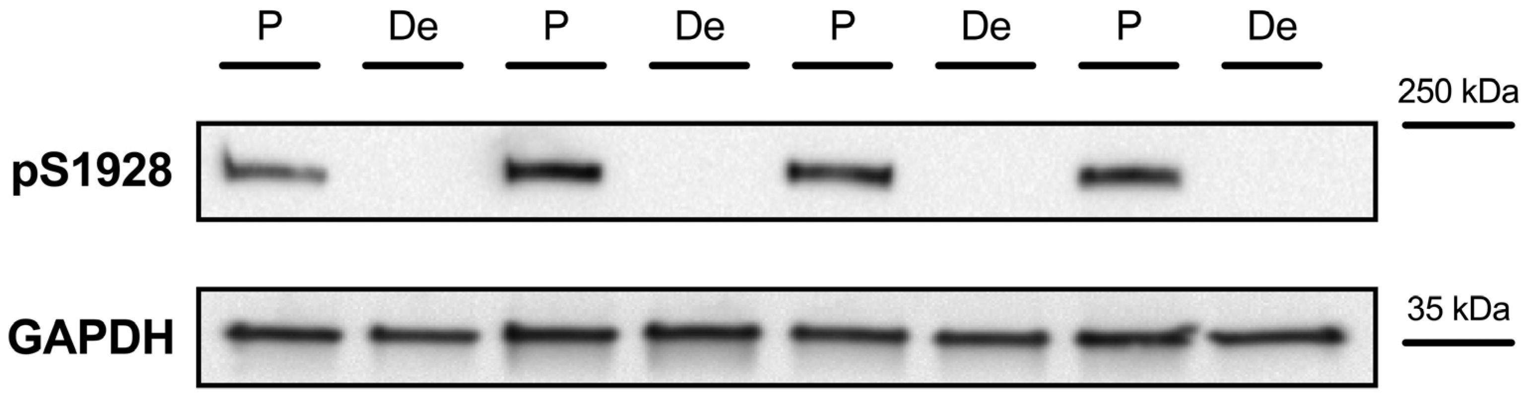
2.2.1. Detection of Cav1.2 Phosphorylation at S1928

Next, we aimed at evaluating whether the phosphorylation of Cav1.2 in LV myocardium of Cacna1c+/− rats was altered. Cav1.2 contains several phosphorylation sites. S1928 is a site primarily phosphorylated by PKA upon stimulation of β-adrenergic receptors. Before testing the phosphorylation of this site in Cacna1c+/− LV myocardium, we first evaluated the ability of the antibody used to reliably detect changes in the PKA-dependent phosphorylation of S1928. To this end, we employed Langendorff-perfused hearts from Sprague-Dawley rats treated with two different solutions. One solution was designed to induce high levels of PKA-dependent phosphorylation. It contained (1) isoprenaline, a β-adrenergic agonist, stimulating increases in cAMP concentration and PKA activity; (2) IBMX, an inhibitor of phosphodiesterases; as well as the phosphatase inhibitors (3) cantharidin and (4) cyclosporin A (phosphorylation or P solution). The other solution was designed to induce dephosphorylation. It contained (1) BDM and the protein kinase inhibitors (2) H-89 and (3) KN-62 (dephosphorylation or De solution). Four hearts each were treated with either a phosphorylation or dephosphorylation solution. LV homogenates of these hearts were subjected to standard Western blotting protocols using a rabbit polyclonal antibody directed against phosphorylated S1928 as previously characterized and validated [14].
As shown in Figure 2, LV homogenates from all four hearts treated with a phosphorylation solution (P) showed a clear strong band at a molecular weight of somewhat below 250 kDa. Conversely, there was an absence of any discernible signal for LV homogenates from all four hearts treated with the dephosphorylation solution (De). Thus, this experiment confirms the reliability of the antibody used to detect changes in the PKA-dependent phosphorylation of Cav1.2 at S1928.

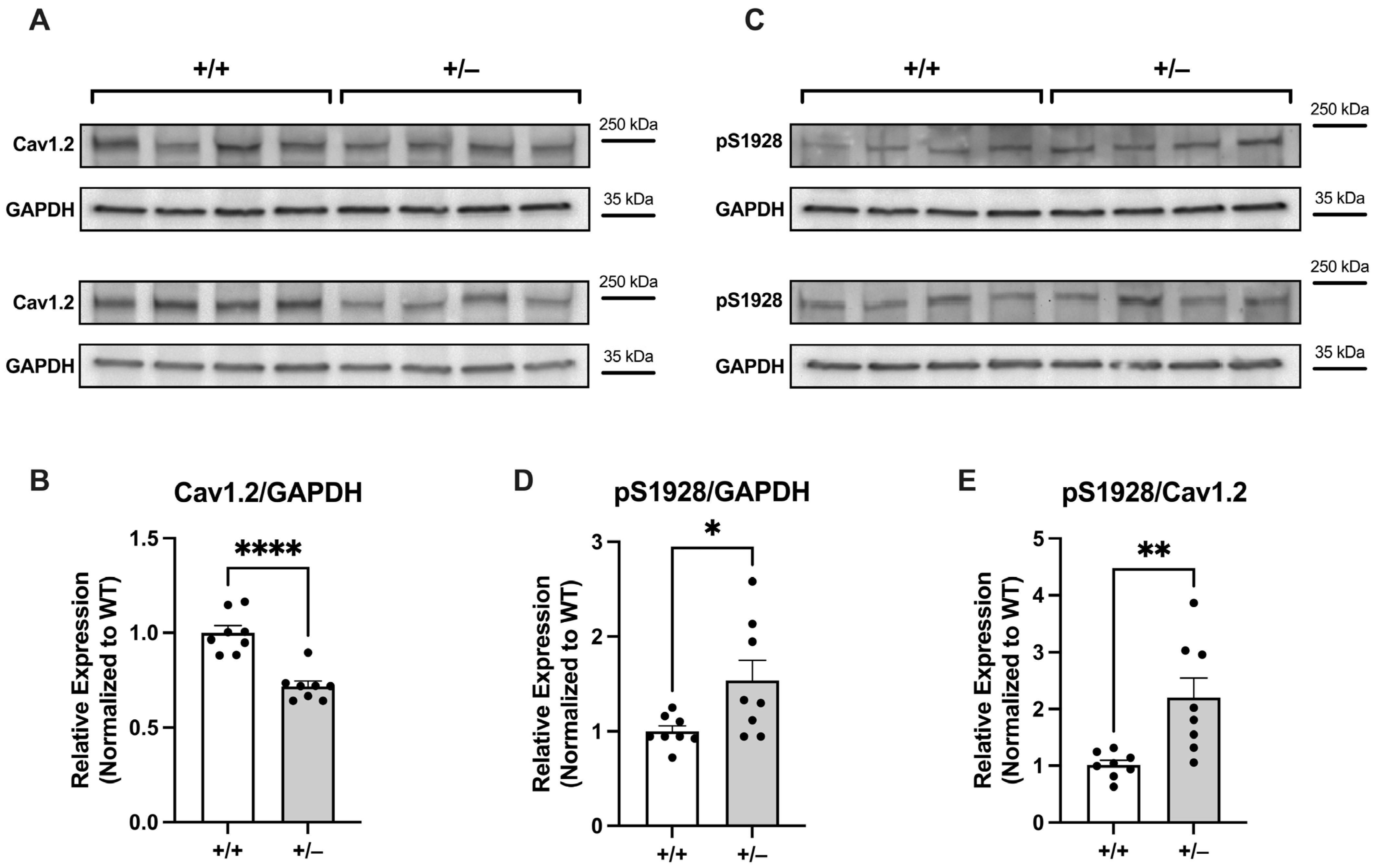
2.2.2. Increased Basal Phosphorylation of Cav1.2 at S1928 in Cacna1c+/− LV Myocardium

Having validated the antibody used, we next sought to examine the baseline phosphorylation level of S1928 on the Cav1.2 channel in LV myocardium from Cacna1c+/− rats. To accomplish this, we initially assessed the total expression of the Cav1.2 channel in Cacna1c+/− LV myocardium in comparison to WT (+/+) control. As shown in Figure 3A,B, there was a reduction by ≈30% in the total protein expression of Cav1.2 in Cacna1c+/− LV tissue when compared to the WT control group. This value is similar to the one reported previously [6]. We then determined the phosphorylation of Cav1.2 at S1928 using the same two sets of samples. As illustrated in Figure 3C, the phosphorylation of Cav1.2 at S1928 was increased in Cacna1c+/− LV tissue. When normalized to GAPDH expression, the phosphorylation levels of S1928 were elevated by ≈50% in the Cacna1c+/− group (Figure 3D). When normalizing to Cav1.2 expression (which is reduced in Cacna1c+/−), the phosphorylation of S1928 in LV myocardium from Cacna1c+/− was roughly doubled (+110%) compared to the WT controls (Figure 3E). These results reveal a greatly increased baseline phosphorylation of Cav1.2 at S1928 in LV myocardium from Cacna1c+/− rats.

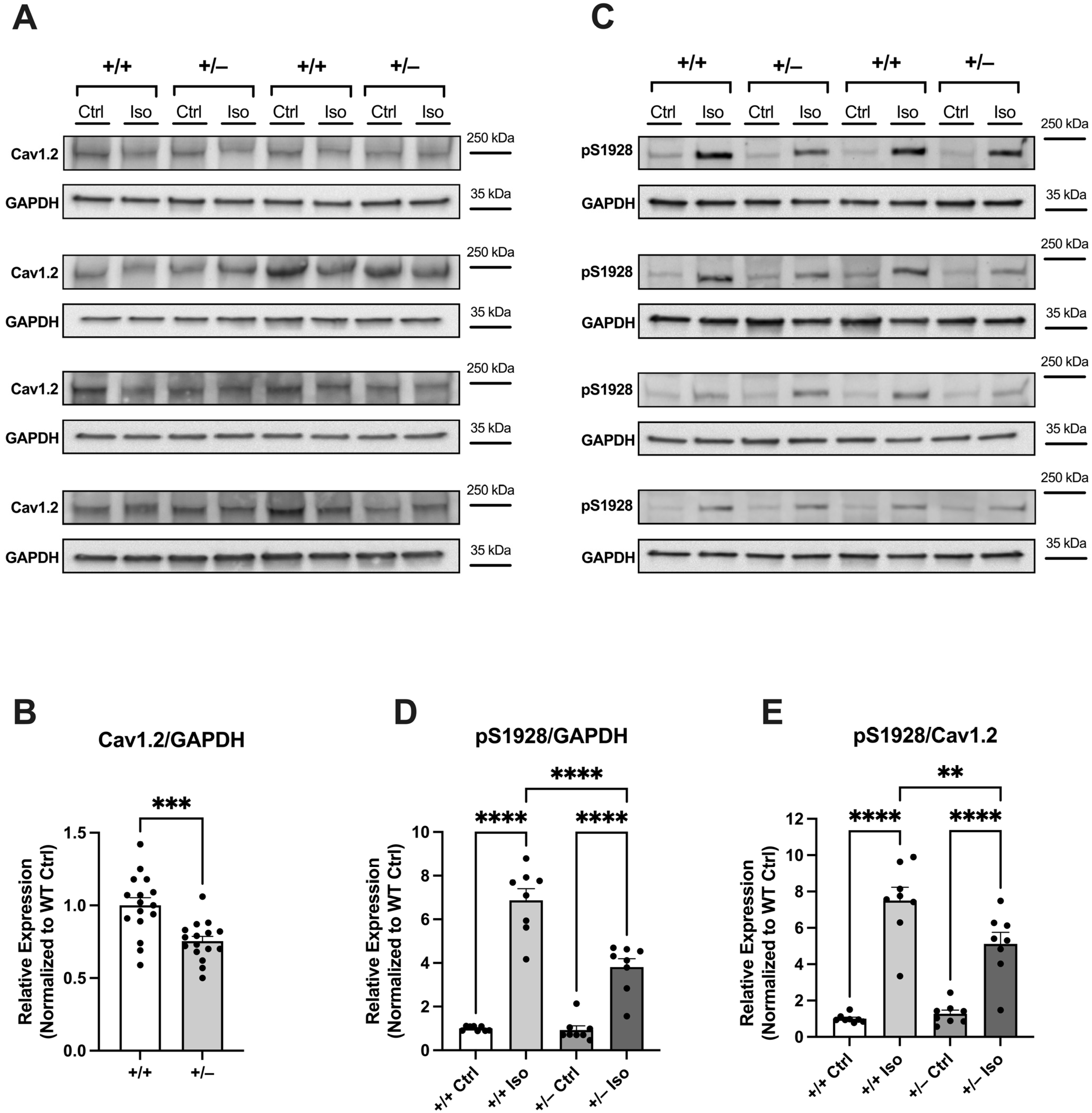
2.2.3. Attenuated Increase in Phosphorylation of Cav1.2 at S1928 in Cacna1c+/− LV Myocardium During Sympathetic Stress

To investigate the impact of sympathetic stress on the phosphorylation of Cav1.2 at S1928, we subjected Langendorff-perfused hearts to treatment with isoprenaline at a concentration of 100 nM, as described previously [6]. Subsequently, LV tissue homogenates from these hearts were tested for expression of Cav1.2 and phosphorylation at S1928 using Western blot analysis. The findings are presented in Figure 4.
A total of 32 hearts were used for this series: 16 WT (+/+) and 16 Cacna1c+/− (+/−) hearts. Each membrane contained eight samples: four WT (+/+) and four Cacna1c+/− (+/−) samples, which were applied in pairs of one untreated control (Ctrl) and one isoprenaline-treated (Iso) sample. This enabled direct comparison of the expression of Cav1.2 and the isoprenaline-induced increase in S1928 phosphorylation between WT and Cacna1c+/− on each membrane. Data were normalized to WT controls. Our initial step involved the assessment of total Cav1.2 expression levels (Figure 4A). Similar to our observations under basal conditions (Figure 3A,B), we noted a reduction in the expression of the Cav1.2 channel in Cacna1c+/− LV myocardium by ≈30% (Figure 4A,B).
Using the same set of samples and their respective positions on the membrane, we investigated the phosphorylation of S1928 on the Cav1.2 channel (Figure 4C). While no significant differences in phosphorylation were observed in the untreated control groups of both genotypes, there was a substantial disparity in the phosphorylation status of Serine-1928 in the isoprenaline-treated groups. When normalized to GAPDH expression, the isoprenaline-induced increase in S1928 phosphorylation amounted to ≈590% in the WT group, whereas in the Cacna1c+/− group, the increase was only ≈280% (Figure 4D). To account for the reduced expression of the Cav1.2 channel in the Cacna1c+/− rats, the phosphorylation status of S1928 was also normalized to Cav1.2 expression (Figure 4E). Again, there were no significant differences between the untreated control groups of both genotypes. Using this kind of analysis, isoprenaline increased S1928 phosphorylation by ≈650% in LV myocardium from WT rats. In contrast, the isoprenaline-induced effect was significantly attenuated in LV myocardium from Cacna1c+/− rats, where the phosphorylation at S1928 increased by only ≈410%.

3. Discussion

Haploinsufficient Cacna1c+/− rats have proven to be a unique animal model exhibiting both a behavioral and a cardiac phenotype, thus enabling studies of the potential role of CACNA1C/Cav1.2 in neuropsychiatric and cardiac diseases [4,5,6]. We have previously characterized the cardiac phenotype of Cacna1c+/− rats with respect to cellular Ca2+ handling and contraction [6]. We found substantial remodeling of the expression and phosphorylation of major Ca2+ handling proteins and an impaired response to sympathetic stress. The very role of Cav1.2 for this altered Ca2+ handling, however, remained enigmatic. Here, we show that the altered PKA-dependent phosphorylation of Cav1.2 is involved in this altered Ca2+ handling both under basal conditions and following sympathetic stress.

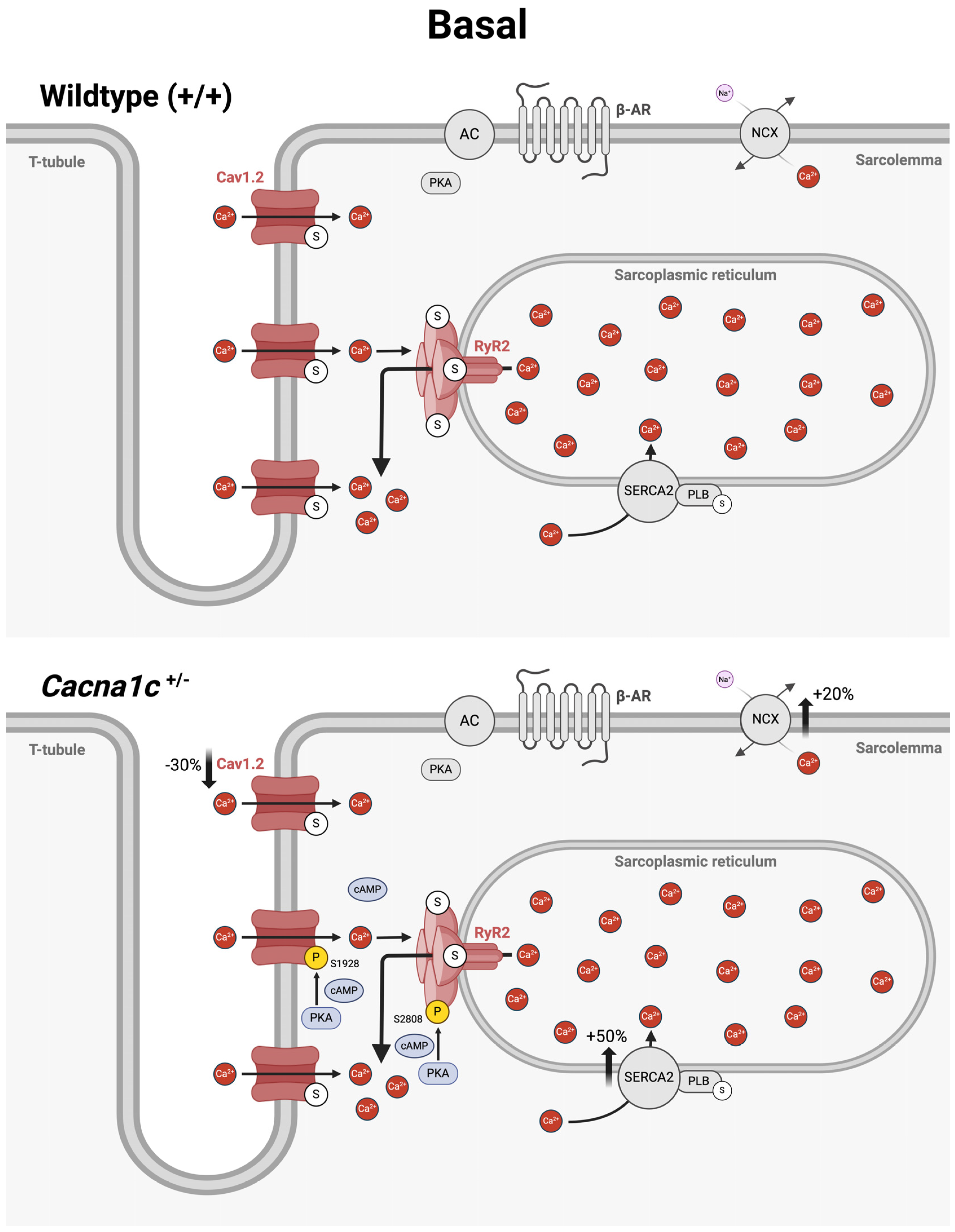
3.1. Potential Role of Altered Cav1.2 Phosphorylation for Basal Ca2+ Handling in Ventricular Myocardium from Cacna1c+/− Rats

Previous studies in mouse models with heterozygous knockout of Cacna1c in the heart have shown that, despite reduced expression of Cav1.2, the L-type Ca2+ current in the cardiac myocytes remains rather unaltered or is reduced much less than would be expected from the degree of downregulation of the mRNA or Cav1.2 protein [15,16]. This implies that cardiac myocytes are able to find ways to maintain a certain amount of functional Cav1.2 channels in the sarcolemma, which is required for proper function, i.e., Ca2+ transients and contractile activity. It is not always clear, though, how this is achieved.
In our initial cardiac characterization of the Cacna1c+/− rat model [6], we observed an ≈30% reduction in the expression of Cav1.2 in LV myocardium compared to WT littermates, and this is confirmed here (Figure 3 and Figure 4). Despite this substantial reduction in the protein amount of Cav1.2, however, the L-type Ca2+ current and the sarcolemmal Ca2+ influx (mainly carried by L-type Ca2+ channels) were essentially unchanged [6]. In order to unravel this conundrum, we considered two different scenarios here. First, we tested for a potential upregulation of auxiliary subunits or other Cav proteins, which might have compensated for the reduced expression of Cav1.2. However, neither the β2 subunit, Cavβ2, nor Cav1.3 was found to be upregulated in LV myocardium from Cacna1c+/− rats (Figure 1), making the first scenario rather unlikely. Second, we tested for altered PKA-dependent phosphorylation of Cav1.2. Using a well-characterized and validated antibody [14], which reliably and sensitively detects the phosphorylation of Cav1.2 at S1928 (Figure 2), a PKA-dependent phosphorylation site, we found that Cav1.2 exhibits substantially increased (roughly doubled) phosphorylation under baseline conditions in LV myocardium from Cacna1c+/− rats (Figure 3). Activation of the cAMP-PKA pathway causes a large increase in L-type Ca2+ current in ventricular myocytes, mainly by means of an increase in the open probability of individual L-type channels [7]. Thus, the elevated PKA-dependent phosphorylation of Cav1.2 observed in this study in LV myocardium from Cacna1c+/− rats could explain the unaltered L-type Ca2+ current and sarcolemmal Ca2+ influx in ventricular myocytes from Cacna1c+/− rats observed previously in the presence of a reduced expression of Cav1.2: In Cacna1c+/− ventricular myocytes, there are less Cav1.2 channels, but these are PKA “hyperphosphorylated” under baseline conditions, leading to a higher open probability of the individual channels and, thus, normalizing the global Cav1.2/L-type Ca2+ channel-mediated sarcolemmal Ca2+ influx. With normal sarcolemmal Ca2+ influx, there will be normal Ca2+ transients and contractions, as observed [6].

3.2. Potential Role of Altered Cav1.2 Phosphorylation for the Impaired Sympathetic Stress Response in Ventricular Myocardium from Cacna1c+/− Rats

Ventricular myocytes from Cacna1c+/− rats exhibit an impaired response to isoprenaline, i.e., sympathetic stimulation, such that the increases in sarcolemmal Ca2+ influx, the Ca2+ transient, and sarcomere shortenings upon isoprenaline stimulation are less pronounced than in WT myocytes [6]. The role of altered phosphorylation of Cav1.2 in this impaired response, however, remained elusive. The results of this study convincingly show that isoprenaline-induced, PKA-dependent phosphorylation of Cav1.2 in LV myocardium from Cacna1c+/− rats is attenuated compared with WT, thus explaining the attenuated increases in sarcolemmal Ca2+ influx. With less trigger Ca2+, there will be less RyR2-mediated Ca2+ release from the SR and, hence, a reduced Ca2+ transient and reduced sarcomere shortenings in Cacna1c+/− ventricular myocytes in the presence of sympathetic stimulation compared to WT myocytes. The impairments in SR Ca2+ release (caused by reduced trigger Ca2+) will be exacerbated by the impaired phosphorylation of RyR2 observed previously [6].

3.3. Altered Phosphorylation of Cav1.2 in Human Cardiac Disease

In ventricular myocytes isolated from human failing hearts (heart failure with reduced ejection fraction, HFrEF), most earlier studies reported an unchanged density of the L-type Ca2+ current [17,18,19,20,21], despite some evidence for robust reductions of channel mRNA and dihydropyridine binding sites [22]. Moreover, β-adrenergic stimulation of L-type Ca2+ current was blunted in both atrial and ventricular myocytes isolated from human failing hearts [23]. Multiple mechanisms may contribute to the impaired β-adrenergic signaling in human HFrEF including altered PKA-dependent phosphorylation of Cav1.2 (and other Ca2+-regulating proteins). Direct evidence from single-channel recordings suggests that under basal conditions, single L-type Ca2+ channels exhibited increased availability and open probability and kinetic properties similar to PKA-modified channels [24]. The channel properties resembled those caused by stimulation with a cAMP analogue and could not be further altered by inhibition of protein phosphatases [24]. Whole-cell recordings of L-type Ca2+ current revealed no differences in current density between failing and non-failing myocytes but a greatly attenuated stimulation by a cAMP analogue in failing myocytes [25]. Moreover, in failing myocytes, protein phosphatase 2A reduced L-type Ca2+ current, whereas the inhibition of protein phosphatases did not affect the current [25]. In addition, the L-type Ca2+ current from failing myocytes exhibited a blunted response to stimulation with Bay K8644, a dihydropyridine agonist, which could be normalized by pretreatment with acetylcholine [26]. Collectively, these studies provide strong evidence for the notion that, in ventricular myocytes from human end-stage failing hearts, there is a reduction in the number of L-type Ca2+ channels, which exhibit elevated phosphorylation by PKA to increase the open probability of individual channels, thereby normalizing the whole-cell current. Because of this PKA-dependent “hyperphosphorylation” under basal conditions, there is a diminished phosphorylation reserve of the channels during β-adrenergic stimulation, resulting in an attenuated response of the whole-cell L-type Ca2+ current. This situation in human HFrEF closely resembles the altered PKA-dependent regulation of Cav1.2 in ventricular myocytes from Cacna1c+/− rats reported here and in our previous study [6]: Cacna1c+/− myocytes exhibit a reduced expression of Cav1.2, which is “hyperphosphorylated” by PKA at S1928, presumably to increase the open probability and normalize the whole-cell current under basal conditions. Because of this “hyperphosphorylation” under basal conditions, there is a diminished phosphorylation reserve causing an attenuated increase in whole-cell current during β-adrenergic stimulation. Remarkably, many similarities also exist with regard to the PKA-dependent phosphorylation and regulation of RyR2 between human HFrEF and the Cacna1c+/− rats: in both cases, there is PKA-dependent “hyperphosphorylation” of RyR2 at S2808/S2809 under basal conditions, and this hyperphosphorylation increases the open probability of the channel [27].
Most recently, the reduced phosphorylation of Cav1.2 at S1928 has been shown to contribute to the reduction of L-type Ca2+ current in human atrial fibrillation [28]. The reduced phosphorylation of Cav1.2 in atrial fibrillation was shown to result from increased association of Cav1.2 with PDE8B2 and reduced local cAMP levels in the vicinity of the channel [28]. These findings are noteworthy in several respects: (1) they underscore that the regulation (of the phosphorylation) of Cav1.2 occurs in its immediate vicinity and that Cav1.2 is part of a macromolecular multi-protein complex, as also suggested for the channel in ventricular myocytes. (2) They show that local cAMP concentration and PKA activity in the microenvironment of Cav1.2 is an important regulator of L-type Ca2+ channel activity. (3) They demonstrate that altered PKA-dependent phosphorylation of Cav1.2 is involved not only in ventricular but also in atrial pathologies.

3.4. An Overview on Altered Cellular Ca2+ Handling in Ventricular Myocardium from Cacna1c+/− Rats Under Basal Conditions and During Sympathetic Stress

The results of this as well as our previous study [6] reveal altered cellular Ca2+ handling in ventricular myocytes from Cacna1c+/− rats both under basal conditions as well as during sympathetic stimulation (mimicked by the β-adrenergic agonist isoprenaline). These alterations are summarized and illustrated in Figure 5 for basal conditions and in Figure 6 for β-adrenergic stimulation.
Figure 5 illustrates the situation in a WT myocyte (top) and in a Cacna1c+/− myocyte (bottom). Ventricular myocytes from Cacna1c+/− rats exhibit a decreased expression of Cav1.2 (−30%) but increased expression of NCX (+20%) and SERCA2a (+50%). The latter two proteins are responsible mainly for Ca2+ extrusion from the cytosol following the systolic Ca2+ increase. NCX is distributed in the T-tubules and along the surface sarcolemma [29]. SERCA2a, in turn, localizes to the longitudinal SR [30]. The expression of other major Ca2+-regulating proteins is unaltered, including RyR2 and PLB. Major alterations with regard to phosphorylation occur in the dyadic cleft, where clusters of Cav1.2 in the T-tubular membrane and clusters of RyR2 in the junctional SR are localized vis-à-vis [31,32]. Both Cav1.2 and RyR2 are part of macromolecular complexes, which may include protein kinases (e.g., PKA), protein phosphatases (e.g., PP2A), phosphodiesterases (e.g., PDE4, PDE8), and scaffolding or adapter proteins (e.g., A-kinase anchoring proteins) [28,33,34,35]. This organization allows for a very local control of cAMP signaling in the immediate vicinity of the channels and, hence, local control of channel phosphorylation by PKA. In WT myocytes, the phosphorylation of Cav1.2 and RyR2 (as well as PLB) is very low, as depicted by the “empty”, i.e., non-phosphorylated serine (S) residues (white circles). By contrast, in Cacna1c+/− myocytes, in the absence of β-adrenergic stimulation, there is greatly increased phosphorylation of both Cav1.2 and RyR2 at PKA sites S1928 (+110%) and S2808 (+120%), respectively, as depicted by some phosphorylated (P) residues (yellow circles). The phosphorylation of PLB, which interacts with SERCA in the longitudinal SR [30], is equally low in WT and Cacna1c+/− myocytes. These findings imply that under basal conditions, local cAMP concentration and PKA activity in the dyadic cleft near Cav1.2 and RyR2 are elevated in Cacna1c+/− myocytes (note blue cAMP/PKA symbols here), whereas cAMP levels and PKA activity in the bulk cytosol are very low and do not differ between Cacna1c+/− and WT myocytes. Elevated PKA-dependent phosphorylation increases the open probability of both Cav1.2 and RyR2, thus augmenting Ca2+-induced Ca2+ release in Cacna1c+/− myocytes, and this mechanism is proposed to underlie the apparently normal sarcolemmal Ca2+ influx and Ca2+ transients in Cacna1c+/− myocytes despite the reduced expression of Cav1.2.
The situation is different, however, during sympathetic stress, as depicted in Figure 6. When β-adrenergic receptors (β-AR) are activated by isoprenaline (ISO), this results in the stimulation of adenylate cyclases (AC), a global increase in cAMP levels and activation of PKA throughout the cytosol. Thus, PKA phosphorylates PLB in the longitudinal SR (at S16) but also Cav1.2 (at S1928) and RyR2 (at S2808) in the dyadic cleft. The PKA phosphorylation of PLB at S16 during isoprenaline stimulation does not differ between Cacna1c+/− and WT myocytes, suggesting similar cAMP increases in the bulk cytosol in Cacna1c+/− and WT myocytes. In and near the dyadic cleft, however, isoprenaline-induced PKA-mediated phosphorylation of Cav1.2 and RyR2 differs between WT and Cacna1c+/− myocytes. In WT myocytes, isoprenaline elicits a full-blown response causing PKA-mediated phosphorylation (yellow circles) of (almost) all available S1928 and S2808 residues in Cav1.2 and RyR2. In Cacna1c+/− myocytes, on the other hand, isoprenaline also increases the phosphorylation of Cav1.2 and RyR2, i.e., on top of the already elevated PKA phosphorylation under basal conditions, but—importantly—this increase is less pronounced than in WT myocytes. Maximal PKA phosphorylation of Cav1.2 and RyR2 is reduced in Cacna1c+/− compared to WT myocytes, leaving “empty”, i.e., non-phosphorylated serine residues (S1928 and S2808) in Cav1.2 and RyR2 (see white circles) and suggesting—once again—altered local regulation of cAMP and PKA-dependent phosphorylation in the dyadic cleft of Cacna1c+/− myocytes. Thus, the increased phosphorylation of Cav1.2 and RyR2 in Cacna1c+/− myocytes under basal conditions comes at the expense of an attenuated response to β-adrenergic stimulation.

3.5. Conclusions and Perspectives

In summary, there is increased basal PKA-dependent phosphorylation and a reduced phosphorylation reserve during the sympathetic stress of Cav1.2 at S1928 in ventricular myocytes from Cacna1c+/− rats. In conjunction with the similarly altered phosphorylation of RyR2 at S2808 and the augmented expression of NCX and SERCA [6], these alterations in cellular Ca2+ handling may explain (1) the apparently normal sarcolemmal Ca2+ influx and excitation–contraction coupling in the face of reduced expression of Cav1.2 and (2) the impaired response to sympathetic stress in Cacna1c+/− ventricular myocytes. The PKA-dependent “hyperphosphorylation” of Cav1.2 and RyR2 observed under basal conditions in ventricular myocardium from apparently healthy Cacna1c+/− rats resembles the altered regulation of Cav1.2 and RyR2 found in human failing hearts. Therefore, we speculate that this kind of remodeling of cellular Ca2+ handling may increase the susceptibility of Cacna1c+/− rats to the development of cardiac disease during periods of physical or psychological stress.

4. Materials and Methods

4.1. Animals

This study was approved by local animal welfare authorities and was performed in accordance with the European Union Council Directive 2010/63/EU and the German Animal Welfare Act.
The generation of Cacna1c+/− (+/−) rats and wildtype (WT, +/+) littermates was performed as previously described [6]. Female rats at the age of 9–15 months were used in this study. For some experiments (validation of the anti-phospho-serine-1928 antibody, Figure 2), female Sprague-Dawley rats at the age of 6–8 months were used. These rats were obtained from Charles River (Cologne, Germany). All rats had free access to standard chow and water. For isolation of the hearts, the rats were anesthetized through exposure to isoflurane, and deep anesthesia was verified by the absence of pain reflexes. Subsequently, the rats were euthanized via decapitation using a guillotine. After the animals were sacrificed, their hearts were promptly removed and employed for either tissue processing or attachment to the Langendorff perfusion apparatus.

4.2. Left Ventricular (LV) Tissue Isolation

After excision of hearts or use of hearts on the Langendorff apparatus, the hearts were placed in an ice-cold solution. Left ventricles were separated, frozen in liquid nitrogen, and stored at −80 °C. LV tissue lysates were used for investigating protein expression or phosphorylation using immunoblotting (as described in Section 4.4).

4.3. Langendorff Perfusion and Treatment of Whole Hearts

To assess protein phosphorylation levels in Cacna1c+/− and WT samples, whole hearts were perfused on a Langendorff apparatus for 5 min with a 2,3 butanedione monoxime (BDM)-containing Tyrode’s solution composed of (mM) 130 NaCl, 5.4 KCl, 0.5 MgCl2, 0.15 CaCl2, 0.33 NaH2PO4, 25 HEPES, 22 glucose, pH 7.4 (with NaOH), and 1 mg/mL (≈9.9 mM) BDM. This initial perfusion with a BDM-containing solution served to depress contractile activity of the heart, thereby sparing cellular ATP, as BDM acts as an inhibitor of the actomyosin ATPase [36]. In addition, it served to induce a standard low level of phosphorylation of cardiac myocyte proteins, as BDM is a chemical phosphatase. Afterwards, hearts were perfused for another 5 min with either control solution (Tyrode’s solution without BDM) or with isoprenaline-containing Tyrode’s solution, from which BDM had been omitted and to which 100 nM isoprenaline had been added. Subsequently, the hearts were sectioned and frozen as described above.
To validate the anti-phospho-S1928 Cav1.2 antibody (phospho-Cav1.2 antibody), the hearts were perfused initially with a BDM-containing Tyrode’s solution as described above. Afterwards, the hearts were perfused either with a solution designed to induce maximal PKA-dependent phosphorylation levels (phosphorylation solution) or with a solution designed to induce maximal dephosphorylation (dephosphorylation solution). The phosphorylation solution was a Tyrode’s solution (without BDM) that contained, additionally, 100 nM isoprenaline, 100 µM 3-isobutyl-1-methylxanthine (IBMX), 30 µM cantharidin, and 1 µM cyclosporin A. The dephosphorylation solution, in turn, was a BDM-containing Tyrode’s solution additionally containing the protein kinase inhibitors H-89 (1 µM) and KN-62 (1 µM). Following perfusion with a phosphorylation (5 min) or dephosphorylation (10 min) solution, the hearts were sectioned and frozen as above.

4.4. Immunoblotting (Western Blotting)

Western blotting for the results shown in Figure 1 (expression of Cav1.3 and Cavβ2) was performed essentially as described previously [6]. For the remainder, i.e., the results shown in Figure 2, Figure 3 and Figure 4, we used slight modifications as described in detail in the following text: the left ventricular (LV) tissue was homogenized using micro tissue grinders (Wheaton, UK) with a homogenization buffer comprising a mixture of protease and phosphatase inhibitors (PhosStop™ and cOmplete™ Protease Inhibitor Cocktail, Merck, Darmstadt, Germany). Subsequently, protein concentrations were quantified through a BCA assay (Thermo-Fisher Scientific, Waltham, MA, USA) with a BSA standard curve. Protein expression and phosphorylation in homogenates were assessed using standard immunoblotting (Western blotting). Proteins were separated using SDS-PAGE with precast gradient gels (4–20% Mini-PROTEAN TGX, Bio-Rad, Neuried, Germany). The samples, each containing 30 μg of total protein, were prepared with Laemmli buffer containing 5% β-mercaptoethanol. In order to estimate the molecular weight of the analyzed proteins, 5 μL of PageRuler™ Plus Prestained Protein Ladder (Thermo-Fisher Scientific, Waltham, MA, USA) was loaded alongside the samples. The voltage was initially set at 90 V for 1 h and subsequently increased to 120 V until optimal separation was obtained. Separated proteins were transferred from the gel to a nitrocellulose membrane (0.45 μm, BioRad, Neuried, Germany) through the wet blotting procedure. Electrical current was set to 360 mA per gel for 2 h, and transfer took place in a cooling chamber at 4 °C. After the transfer, membranes were cut between the protein bands of interest, allowing for each segment of the membrane to be individually incubated with the respective antibody of interest. The membranes were subsequently washed with TBST buffer (3 times for 10 min each) on a rocking platform. Blocking was performed for 1 h at room temperature with 5% milk (i.e., skimmed milk powder, Sigma-Aldrich, Taufkirchen, Germany) in TBST (or 5% BSA in TBST, for phospho-Cav1.2). After that, the membranes were incubated with the primary antibody in 5% milk (or 5% BSA, for phospho-Cav1.2) in TBST overnight. The following primary antibodies were used (host; dilution; company, catalogue number): anti-Cav1.2 (CACNA1C) antibody (rabbit, 1:1,000; Alomone, Jerusalem, Israel, #ACC-003), phospho-Cav1.2 (Ser1927) polyclonal antibody (rabbit; 1:1,000; Invitrogen/Thermo-Fisher Scientific, Waltham, MA, USA, #PA5-64748), anti-Cav1.3 (CACNA1D) antibody (rabbit; 1:200; Alomone, #ACC-005), anti-CACNB2 antibody (rabbit; 1:800; Alomone, Jerusalem, Israel, #ACC-105), and anti-GAPDH mouse mAB (6c5) antibody (mouse; 1:50,000; Calbiochem, San Diego, CA, USA, #CB1001). On the following day, the membranes were washed (with TBST, 3 times for 10 min each), followed by incubation with the secondary antibody (in 5% milk (or 5% BSA, for phospho-Cav1.2) in TBST, 1 h, at room temperature) and another round of washing (with TBST, 3 times for 10 min each). The secondary antibodies used were either immunopure goat anti-mouse IgG, peroxidase conjugated (Thermo-Fisher Scientific, Waltham, MA, USA, #31430), or immunopure goat anti-rabbit IgG, peroxidase conjugated (Thermo-Fisher Scientific, Waltham, MA, USA, #31460), with a dilution of 1:5000 in each case. The chemiluminescence reaction was detected using a Chemidoc-XRS system (Bio-Rad, Neuried, Germany) with Quantity One Software (Version 4.6.5) after incubation of the membranes for 1 min with the reagent SuperSignalTM West Pico PLUS Chemiluminescent Substrate (Thermo-Fisher Scientific, Waltham, MA, USA) or the more sensitive SuperSignalTM West Femto Maximum Sensitivity Substrate (Thermo-Fisher Scientific, Waltham, MA, USA). Blots were analyzed using FIJI-2 (NIH, USA, Version 2.14.0/1.54f). GAPDH was used as a loading control. To compare the expression and phosphorylation of Ca2+ handling proteins between genotypes, the results were normalized to the averaged WT signal (in the absence of isoprenaline) on a given membrane (=100%).
All original Western blot images are shown in Supplementary Materials.

4.5. Statistics

Statistical analysis was performed with GraphPad Prism 9 (GraphPad Software, San Diego, CA, USA). Data are presented as scatter plots with bar graphs indicating mean ± SEM. The number of animals is provided as “N”. Data sets were compared by means of an unpaired, two-tailed Student’s t test and considered significant when p < 0.05. For multiple group comparisons, ANOVA was applied. Asterisks indicate the following levels of significance: * p < 0.05, ** p < 0.01, *** p < 0.001, **** p < 0.0001.

Supplementary Materials

The following supporting information can be downloaded at: https://www.mdpi.com/article/10.3390/ijms252413713/s1.

Author Contributions

Conceptualization, J.K., M.W., D.K. and H.F.; methodology, J.K., M.W. and T.M.K.; validation, all authors; formal analysis, D.K. and H.F.; investigation, D.K., H.F. and J.P.; interpretation of data, all authors; resources, M.W., T.M.K. and J.K.; writing—original draft preparation, D.K. and J.K.; writing—review and editing, all authors; visualization, D.K. and J.K.; project administration, J.K.; funding acquisition, J.K. and M.W. All authors have read and agreed to the published version of the manuscript.

Funding

This research was funded by the Flexi Funds Programme of Forschungscampus Mittelhessen (FCMH) [Projektnummer 2018_1_1_2 to J.K.]; the DFG-Forschergruppe “Neurobiology of affective disorders: Translational perspectives on brain structure and function” (FOR 2107) [DFG WO 1732/4-2 to M.W.]; and the DFG-Sonderforschungsbereich/Transregio “Trajectories of affective disorders: Cognitive-emotional mechanisms of symptom change” (SFB/TRR 393) [DFG-Projektnummer 521379614 to M.W.].

Institutional Review Board Statement

The experimental protocols involving animals were approved by RP Gießen and the Animal Welfare Officer of the University of Marburg (AZ MR 20/35 19/2014, AK-2-2018 and AK-12-2022).

Informed Consent Statement

Not applicable.

Data Availability Statement

All data are contained within the article or Supplementary Materials.

Acknowledgments

The authors would like to thank Ahmad Nawid Nasri and Marcel Rossol for critically reading the manuscript.

Conflicts of Interest

The authors declare no conflict of interest.

References

  1. Harrison, P.J.; Husain, S.M.; Lee, H.; Los Angeles, A.; Colbourne, L.; Mould, A.; Hall, N.A.L.; Haerty, W.; Tunbridge, E.M. CACNA1C (Ca(V)1.2) and other L-type calcium channels in the pathophysiology and treatment of psychiatric disorders: Advances from functional genomics and pharmacoepidemiology. Neuropharmacology 2022, 220, 109262. [Google Scholar] [CrossRef] [PubMed]
  2. Splawski, I.; Timothy, K.W.; Sharpe, L.M.; Decher, N.; Kumar, P.; Bloise, R.; Napolitano, C.; Schwartz, P.J.; Joseph, R.M.; Condouris, K.; et al. Ca(V)1.2 calcium channel dysfunction causes a multisystem disorder including arrhythmia and autism. Cell 2004, 119, 19–31. [Google Scholar] [CrossRef]
  3. Wemhoner, K.; Friedrich, C.; Stallmeyer, B.; Coffey, A.J.; Grace, A.; Zumhagen, S.; Seebohm, G.; Ortiz-Bonnin, B.; Rinne, S.; Sachse, F.B.; et al. Gain-of-function mutations in the calcium channel CACNA1C (Cav1.2) cause non-syndromic long-QT but not Timothy syndrome. J. Mol. Cell Cardiol. 2015, 80, 186–195. [Google Scholar] [CrossRef] [PubMed]
  4. Kisko, T.M.; Braun, M.D.; Michels, S.; Witt, S.H.; Rietschel, M.; Culmsee, C.; Schwarting, R.K.W.; Wohr, M. Cacna1c haploinsufficiency leads to pro-social 50-kHz ultrasonic communication deficits in rats. Dis. Model. Mech. 2018, 11, dmm034116. [Google Scholar] [CrossRef] [PubMed]
  5. Redecker, T.M.; Kisko, T.M.; Schwarting, R.K.W.; Wohr, M. Effects of Cacna1c haploinsufficiency on social interaction behavior and 50-kHz ultrasonic vocalizations in adult female rats. Behav. Brain Res. 2019, 367, 35–52. [Google Scholar] [CrossRef]
  6. Fender, H.; Walter, K.; Kiper, A.K.; Plackic, J.; Kisko, T.M.; Braun, M.D.; Schwarting, R.K.W.; Rohrbach, S.; Wohr, M.; Decher, N.; et al. Calcium Handling Remodeling Underlies Impaired Sympathetic Stress Response in Ventricular Myocardium from Cacna1c Haploinsufficient Rats. Int. J. Mol. Sci. 2023, 24, 9795. [Google Scholar] [CrossRef] [PubMed]
  7. Papa, A.; Kushner, J.; Marx, S.O. Adrenergic Regulation of Calcium Channels in the Heart. Annu. Rev. Physiol. 2022, 84, 285–306. [Google Scholar] [CrossRef]
  8. Bers, D.M. Cardiac excitation-contraction coupling. Nature 2002, 415, 198–205. [Google Scholar] [CrossRef] [PubMed]
  9. Reuter, H. Calcium channel modulation by neurotransmitters, enzymes and drugs. Nature 1983, 301, 569–574. [Google Scholar] [CrossRef]
  10. McDonald, T.F.; Pelzer, S.; Trautwein, W.; Pelzer, D.J. Regulation and modulation of calcium channels in cardiac, skeletal, and smooth muscle cells. Physiol. Rev. 1994, 74, 365–507. [Google Scholar] [CrossRef] [PubMed]
  11. van der Heyden, M.A.; Wijnhoven, T.J.; Opthof, T. Molecular aspects of adrenergic modulation of cardiac L-type Ca2+ channels. Cardiovasc. Res. 2005, 65, 28–39. [Google Scholar] [CrossRef] [PubMed]
  12. Hulme, J.T.; Westenbroek, R.E.; Scheuer, T.; Catterall, W.A. Phosphorylation of serine 1928 in the distal C-terminal domain of cardiac CaV1.2 channels during beta1-adrenergic regulation. Proc. Natl. Acad. Sci. USA 2006, 103, 16574–16579. [Google Scholar] [CrossRef]
  13. Filippini, L.; Ortner, N.J.; Kaserer, T.; Striessnig, J. Ca(v) 1.3-selective inhibitors of voltage-gated L-type Ca(2+) channels: Fact or (still) fiction? Br. J. Pharmacol. 2023, 180, 1289–1303. [Google Scholar] [CrossRef]
  14. Whitcomb, V.; Wauson, E.; Christian, D.; Clayton, S.; Giles, J.; Tran, Q.K. Regulation of beta adrenoceptor-mediated myocardial contraction and calcium dynamics by the G protein-coupled estrogen receptor 1. Biochem. Pharmacol. 2020, 171, 113727. [Google Scholar] [CrossRef]
  15. Rosati, B.; Yan, Q.; Lee, M.S.; Liou, S.R.; Ingalls, B.; Foell, J.; Kamp, T.J.; McKinnon, D. Robust L-type calcium current expression following heterozygous knockout of the Cav1.2 gene in adult mouse heart. J. Physiol. 2011, 589, 3275–3288. [Google Scholar] [CrossRef] [PubMed]
  16. Goonasekera, S.A.; Hammer, K.; Auger-Messier, M.; Bodi, I.; Chen, X.; Zhang, H.; Reiken, S.; Elrod, J.W.; Correll, R.N.; York, A.J.; et al. Decreased cardiac L-type Ca(2)(+) channel activity induces hypertrophy and heart failure in mice. J. Clin. Investig. 2012, 122, 280–290. [Google Scholar] [CrossRef] [PubMed]
  17. Beuckelmann, D.J.; Nabauer, M.; Erdmann, E. Intracellular calcium handling in isolated ventricular myocytes from patients with terminal heart failure. Circulation 1992, 85, 1046–1055. [Google Scholar] [CrossRef] [PubMed]
  18. Mewes, T.; Ravens, U. L-type calcium currents of human myocytes from ventricle of non-failing and failing hearts and from atrium. J. Mol. Cell Cardiol. 1994, 26, 1307–1320. [Google Scholar] [CrossRef] [PubMed]
  19. Beuckelmann, D.J. Contributions of Ca(2+)-influx via the L-type Ca(2+)-current and Ca(2+)-release from the sarcoplasmic reticulum to [Ca2+]i-transients in human myocytes. Basic. Res. Cardiol. 1997, 92 (Suppl. 1), 105–110. [Google Scholar] [CrossRef] [PubMed]
  20. Piacentino, V., 3rd; Weber, C.R.; Chen, X.; Weisser-Thomas, J.; Margulies, K.B.; Bers, D.M.; Houser, S.R. Cellular basis of abnormal calcium transients of failing human ventricular myocytes. Circ. Res. 2003, 92, 651–658. [Google Scholar] [CrossRef]
  21. Benitah, J.-P.; Alvarez, J.L.; Gómez, A.M. L-type Ca2+ current in ventricular cardiomyocytes. J. Mol. Cell. Cardiol. 2010, 48, 26–36. [Google Scholar] [CrossRef] [PubMed]
  22. Takahashi, T.; Allen, P.D.; Lacro, R.V.; Marks, A.R.; Dennis, A.R.; Schoen, F.J.; Grossman, W.; Marsh, J.D.; Izumo, S. Expression of dihydropyridine receptor (Ca2+ channel) and calsequestrin genes in the myocardium of patients with end-stage heart failure. J. Clin. Investig. 1992, 90, 927–935. [Google Scholar] [CrossRef]
  23. Ouadid, H.; Albat, B.; Nargeot, J. Calcium currents in diseased human cardiac cells. J. Cardiovasc. Pharmacol. 1995, 25, 282–291. [Google Scholar] [CrossRef]
  24. Schroder, F.; Handrock, R.; Beuckelmann, D.J.; Hirt, S.; Hullin, R.; Priebe, L.; Schwinger, R.H.; Weil, J.; Herzig, S. Increased availability and open probability of single L-type calcium channels from failing compared with nonfailing human ventricle. Circulation 1998, 98, 969–976. [Google Scholar] [CrossRef] [PubMed]
  25. Chen, X.; Piacentino, V., 3rd; Furukawa, S.; Goldman, B.; Margulies, K.B.; Houser, S.R. L-type Ca2+ channel density and regulation are altered in failing human ventricular myocytes and recover after support with mechanical assist devices. Circ. Res. 2002, 91, 517–524. [Google Scholar] [CrossRef]
  26. Chen, X.; Zhang, X.; Harris, D.M.; Piacentino, V., 3rd; Berretta, R.M.; Margulies, K.B.; Houser, S.R. Reduced effects of BAY K 8644 on L-type Ca2+ current in failing human cardiac myocytes are related to abnormal adrenergic regulation. Am. J. Physiol. Heart Circ. Physiol. 2008, 294, H2257–H2267. [Google Scholar] [CrossRef]
  27. Marx, S.O.; Reiken, S.; Hisamatsu, Y.; Jayaraman, T.; Burkhoff, D.; Rosemblit, N.; Marks, A.R. PKA phosphorylation dissociates FKBP12.6 from the calcium release channel (ryanodine receptor): Defective regulation in failing hearts. Cell 2000, 101, 365–376. [Google Scholar] [CrossRef] [PubMed]
  28. Grammatika Pavlidou, N.; Dobrev, S.; Beneke, K.; Reinhardt, F.; Pecha, S.; Jacquet, E.; Abu-Taha, I.H.; Schmidt, C.; Voigt, N.; Kamler, M.; et al. Phosphodiesterase 8 governs cAMP/PKA-dependent reduction of L-type calcium current in human atrial fibrillation: A novel arrhythmogenic mechanism. Eur. Heart J. 2023, 44, 2483–2494. [Google Scholar] [CrossRef] [PubMed]
  29. Despa, S.; Brette, F.; Orchard, C.H.; Bers, D.M. Na/Ca exchange and Na/K-ATPase function are equally concentrated in transverse tubules of rat ventricular myocytes. Biophys. J. 2003, 85, 3388–3396. [Google Scholar] [CrossRef]
  30. Kranias, E.G.; Hajjar, R.J. Modulation of cardiac contractility by the phospholamban/SERCA2a regulatome. Circ. Res. 2012, 110, 1646–1660. [Google Scholar] [CrossRef] [PubMed]
  31. Scriven, D.R.L.; Asghari, P.; Schulson, M.N.; Moore, E.D.W. Analysis of Cav1.2 and Ryanodine Receptor Clusters in Rat Ventricular Myocytes. Biophys. J. 2010, 99, 3923–3929. [Google Scholar] [CrossRef]
  32. Dixon, R.E. Nanoscale Organization, Regulation, and Dynamic Reorganization of Cardiac Calcium Channels. Front. Physiol. 2021, 12, 810408. [Google Scholar] [CrossRef] [PubMed]
  33. Man, K.N.M.; Bartels, P.; Horne, M.C.; Hell, J.W. Tissue-specific adrenergic regulation of the L-type Ca(2+) channel Ca(V)1.2. Sci. Signal 2020, 13, eabc6438. [Google Scholar] [CrossRef] [PubMed]
  34. Marx, S.O.; Reiken, S.; Hisamatsu, Y.; Gaburjakova, M.; Gaburjakova, J.; Yang, Y.M.; Rosemblit, N.; Marks, A.R. Phosphorylation-dependent regulation of ryanodine receptors: A novel role for leucine/isoleucine zippers. J. Cell Biol. 2001, 153, 699–708. [Google Scholar] [CrossRef]
  35. Lehnart, S.E.; Wehrens, X.H.; Reiken, S.; Warrier, S.; Belevych, A.E.; Harvey, R.D.; Richter, W.; Jin, S.L.; Conti, M.; Marks, A.R. Phosphodiesterase 4D deficiency in the ryanodine-receptor complex promotes heart failure and arrhythmias. Cell 2005, 123, 25–35. [Google Scholar] [CrossRef] [PubMed]
  36. Backx, P.H.; Gao, W.D.; Azan-Backx, M.D.; Marban, E. Mechanism of force inhibition by 2,3-butanedione monoxime in rat cardiac muscle: Roles of [Ca2+]i and cross-bridge kinetics. J. Physiol. 1994, 476, 487–500. [Google Scholar] [CrossRef] [PubMed]
Figure 1. Expression of Cav1.3 and Cavβ2 in LV myocardium from WT and Cacna1c+/− rats. (A) Expression of Cav1.3 in LV myocardium from WT and Cacna1c+/− rats. Values were normalized to the mean from WT. (B) Original Western blot images of 8 WT (+/+) and 8 Cacna1c+/− (+/−) samples derived from two membranes. The protein used for normalization (GAPDH) derived from the same membrane is shown below the protein of interest. (C) Expression of Cavβ2 in LV myocardium from WT and Cacna1c+/− rats. Values were normalized to the mean from WT. (D) Original Western blot images of 8 WT (+/+) and 8 Cacna1c+/− (+/−) samples derived from two membranes. The protein used for normalization (GAPDH) derived from the same membrane is shown below the protein of interest. Circles represent number of animals: N = 8 (WT); N = 8 (Cacna1c+/−); Student’s t-test, * p < 0.05; ns = not significant. All original Western blot images from this series are shown in Supplementary Materials.
Ijms 25 13713 g001
Figure 2. Phosphorylation of Cav1.2 at S1928 following treatment of rat hearts with a phosphorylation (P) or dephosphorylation (De) solution. Original Western blot images showing results of LV homogenates from 8 Sprague-Dawley rat hearts treated with either a phosphorylation (P) or dephosphorylation (De) solution. Top: results obtained with the antibody directed against phosphorylated S1928 of Cav1.2 (pS1928); Bottom: results obtained with the anti-GAPDH antibody. Both Western blots shown are derived from the same membrane.
Ijms 25 13713 g002
Figure 3. Expression of Cav1.2 and baseline phosphorylation at S1928 in LV myocardium from Cacna1c+/− and WT rats. (A) Original Western blot images of 8 WT (+/+) and 8 Cacna1c+/− (+/−) samples derived from two membranes probed with the anti-Cav1.2 antibody. The protein used for normalization (GAPDH) derived from the same membranes is shown below the protein of interest. (B) The expression of Cav1.2 (normalized to GAPDH; Cav1.2/GAPDH) is decreased by ≈30% in Cacna1c+/− (+/−). (C) Original Western blot images of 8 WT (+/+) and 8 Cacna1c+/− (+/−) samples derived from two membranes probed with the anti-pS1928 antibody. The protein used for normalization (GAPDH) derived from the same membranes is shown below. (D) Phosphorylation status of S1928 in Cacna1c+/− (+/−) versus WT (+/+) control samples when normalized to GAPDH (pS1928/GAPDH). Phosphorylation of S1928 is increased by ≈50% in Cacna1c+/− (+/−). (E) Phosphorylation status of S1928 in Cacna1c+/− (+/−) versus WT (+/+) control samples when normalized to Cav1.2 expression (pS1928/Cav1.2). The phosphorylation of S1928 is increased by ≈110% in Cacna1c+/− (+/−). Circles represent number of animals: N = 8 (WT); N = 8 (Cacna1c+/−); Student’s t-test, * p < 0.05, ** p < 0.01, **** p < 0.0001. Further information and all original Western blot images from this series are shown in Supplementary Materials.
Ijms 25 13713 g003
Figure 4. Isoprenaline-Induced Increase in Phosphorylation of Cav1.2 at S1928 in LV Myocardium from WT and Cacna1c+/− rats. (A) Original Western blot images of 16 WT (+/+) and 16 Cacna1c+/− (+/−) LV samples probed with anti-Cav1.2 antibody. The protein used for normalization (GAPDH) derived from the same membranes is shown below the protein of interest. Hearts were either left untreated (Ctrl) or treated with 100 nM isoprenaline (Iso). (B) The expression of Cav1.2 normalized to GAPDH (Cav1.2/GAPDH) was reduced in Cacna1c+/− (+/−) LV samples. (C) Original Western blot images of 16 WT (+/+) and 16 Cacna1c+/− (+/−) LV samples probed with anti-phospho-S1928 antibody. The protein used for normalization (GAPDH) derived from the same membranes is shown below the protein of interest. Hearts were either left untreated (Ctrl) or treated with 100 nM isoprenaline (Iso). Iso-treated samples exhibit larger phosphorylation of S1928. (D) Phosphorylation of S1928 normalized to GAPDH expression (pS1928/GAPDH) in the four groups: WT untreated control (+/+ Ctrl), WT treated with Iso (+/+ Iso), Cacna1c+/− untreated control (+/− Ctrl), and Cacna1c+/− treated with Iso (+/− Iso). N = 8 samples for each group. (E) Phosphorylation of S1928 normalized to Cav1.2 expression (pS1928/Cav1.2) in the four groups: WT untreated control (+/+ Ctrl), WT treated with Iso (+/+ Iso), Cacna1c+/− untreated control (+/− Ctrl), and Cacna1c+/− treated with Iso (+/− Iso). N = 8 samples for each group. Iso increased phosphorylation of S1928 and the Iso-induced increase was larger in WT than in Cacna1c+/− LV samples. Student’s t-test (B) or one-way ANOVA (D,E) were used for comparison of groups. ** p < 0.01, *** p < 0.001, **** p < 0.0001. All original Western blot images from this series are shown in Supplementary Materials.
Ijms 25 13713 g004
Figure 5. Proposed alterations of Ca2+ handling in ventricular myocytes from Cacna1c+/− rats under basal conditions. The scheme depicts part of a ventricular myocyte from a wildtype (top) and a Cacna1c+/− rat (bottom) with T-tubule, surface sarcolemma and the sarcoplasmic reticulum and the location of major Ca2+-regulating proteins: Cav1.2, RyR2, NCX, SERCA, and PLB. The space between the T-tubule (with Cav1.2) and the junctional SR (with RyR2) is termed dyadic cleft. In ventricular myocytes from Cacna1c+/− rats, the expression of Cav1.2 is reduced by 30%, whereas the expression of NCX and SERCA is elevated by +20% and +50%, respectively. β-adrenergic receptors (β-ARs) are not activated (grey), and cAMP levels in the bulk cytosol are very low. Hence, most serine (S) residues in Cav1.2, RyR2, and PLB, which are targets of PKA, are not phosphorylated, as indicated by the white circles attached to the respective proteins. In the dyadic cleft of Cacna1c+/− myocytes, however, Cav1.2 and RyR2 exhibit increased phosphorylation (P) of S1928 and S2808, respectively, as indicated by the yellow circles. The increased PKA-dependent phosphorylation of Cav1.2 and RyR2 is presumably caused by locally elevated cAMP concentration in the immediate vicinity of the channels, as depicted by the blue cAMP symbols. Abbreviations: AC, adenylate cyclase; β-AR, β-adrenergic receptor; P, phosphorylated serine residue; S, serine residue (non-phosphorylated). Created in BioRender. Königstein, D. (2024) https://BioRender.com/a97h526 (accessed on 17 December 2024).
Ijms 25 13713 g005
Figure 6. Proposed alterations of Ca2+ handling in ventricular myocytes from Cacna1c+/− rats during sympathetic stress. The scheme depicts part of a ventricular myocyte from a wildtype (top) and a Cacna1c+/− rat (bottom) with T-tubule, surface sarcolemma, and the sarcoplasmic reticulum and the location of major Ca2+-regulating proteins: Cav1.2, RyR2, NCX, SERCA, and PLB. The space between the T-tubule (with Cav1.2) and the junctional SR (with RyR2) is termed dyadic cleft. In ventricular myocytes from Cacna1c+/− rats, the expression of Cav1.2 is reduced by 30%, whereas the expression of NCX and SERCA is elevated by +20% and +50%, respectively. During the stimulation of β-AR with isoprenaline (ISO), mimicking sympathetic stress, cAMP levels increase throughout the cytosol and PKA phosphorylates target serine residues in Cav1.2 (S1928), RyR2 (S2808), and PLB (S16) in both wildtype and Cacna1c+/− myocytes. In wildtype myocytes, all available serine residues in Cav1.2 and RyR2 become phosphorylated by PKA (yellow circles). Note, however, that for Cacna1c+/− myocytes, not all available target serines (S) in Cav1.2 and RyR2 become phosphorylated by PKA, indicating a diminished phosphorylation reserve of these two channels. Abbreviations: AC, adenylate cyclase; β-AR, β-adrenergic receptor; ISO, isoprenaline; P, phosphorylated serine residue; S, serine residue (non-phosphorylated). Created in BioRender. Königstein, D. (2024) https://BioRender.com/o38g639 (accessed on 17 December 2024).
Ijms 25 13713 g006
Disclaimer/Publisher’s Note: The statements, opinions and data contained in all publications are solely those of the individual author(s) and contributor(s) and not of MDPI and/or the editor(s). MDPI and/or the editor(s) disclaim responsibility for any injury to people or property resulting from any ideas, methods, instructions or products referred to in the content.

