Article

Visualized Nucleic Acid Hybridization Lateral Flow Strip Integrating with Microneedle for the Point-of-Care Authentication of Ophiocordyceps sinensis

1
College of Life Sciences, North China University of Science and Technology, Tangshan 063200, China
2
College of Electrical Engineering, North China University of Science and Technology, Tangshan 063200, China
*
Authors to whom correspondence should be addressed.
Int. J. Mol. Sci. 2024, 25(24), 13599; https://doi.org/10.3390/ijms252413599
Submission received: 29 October 2024 / Revised: 11 December 2024 / Accepted: 13 December 2024 / Published: 19 December 2024

Abstract

:
Due to the price and demand of Ophiocordyceps sinensis having increased dramatically, adulteration with other fungi is a common problem. Thus, a reliable method of authentic O. sinensis identification is essential. In the present work, a rapid DNA extraction and double-tailed recombinase polymerase amplification (RPA) coupled with nucleic acid hybridization lateral flow strip (NAH-LFS) was developed to distinguish authentic O. sinensis ingredients from other fungi substitutes. In the presence of O. sinensis, the RPA amplicons with two ssDNA tails in the opposite ends, which could simultaneously bind with the SH-probes on gold nanoparticles (AuNPs) and capture the probe on the test line, formed visible red bands. RPA combined with NAH-LFS can efficiently detect O. sinensis DNA down to 1.4 ng/μL; meanwhile, the specificity test validated no cross reaction with common adulterants, including Cordyceps gunnii, Cordyceps cicadae, Cordyceps militaris, yungui Cordyceps, and Ophiocordyceps nutans. The whole RPA-NAH-LFS could be completed within 16 min. The RPA-NAH-LFS results in detecting 20 commercial O. sinensis samples are consistent with PCR-AGE and RT-PCR, confirming the feasibility of the RPA-NAH-LFS method. In conclusion, these results are expected to facilitate the application of RPA-NAH-LFS in the authentication detection of O. sinensis materials, providing a convenient and efficient method for O. sinensis quality control.

1. Introduction

Ophiocordyceps sinensis (O. sinensis), known as “dong chong xia cao” in Chinese, is a traditional Chinese medicinal material that is rarely encountered. Its use as a tonic food and herbal remedy has a history spanning thousands of years, which has contributed to its immense popularity worldwide [1]. O. sinensis is a species belong to fungi that includes stromata (family: Ophiocordycipitaceae) and shriveled worms (family: Hepialidae) as two parts [2,3]. The fungus invades the larva or caterpillar and takes over its body, eventually killing and growing out of the host’s head. O. sinensis requires specific environmental conditions to thrive, which are principally found in various high humidity, low oxygen levels, degrees of chilliness, and high-altitude regions (e.g., the snow mountain line or meadows of the Himalayas, Tibetan Plateau, Qinghai, etc.) [4]. As a traditional medicine, O. sinensis possesses various medicinal properties, including antioxidant and hypoglycemic properties, boosting the immune system, improving lung and kidney function [5,6]. Due to excessive harvest, habitat destruction, and huge market demand, the price of O. sinensis is comparable to gold, and high-quality O. sinensis is much more expensive than gold [2]. As a result, O. sinensis counterfeit events are becoming increasingly serious, affecting the safe use of O. sinensis and causing confusion in the marketplace. Fraudulent producers, lured by the prospect of profit, may substitute authentic O. sinensis with various counterfeits that possess a similar morphology, such as Cordyceps militaris, Cordyceps pruinosa, and Cordyceps gunnii, as well as other low-cost fungal species, and even dough, soybean flour, and some undeclared components [7]. However, O. sinensis is described as the only authentication recorded in the Chinese Pharmacopoeia. O. sinensis is often used as a raw material, but it is also ground into powder and used in health supplements and patented Chinese medicinal products, which makes the authentication of O. sinensis impossible based on its morphology. Therefore, it is imperative to establish a simple, cost-effective, and fast quality control method to authenticate O. sinensis from counterfeits.
Up to date, numerous analytical methods have been established for O. sinensis authentication based on the morphological characteristics, protein, DNA, and other effective components, including polysaccharides and nucleosides. The morphological characteristic-based method, relying on the professional’s subjective judgment and practical experience, has traditionally long been controversial on account of the lack of objective standards [8]. The methods based on protein analysis include enzyme-linked immunosorbent assay (ELISA) [9], ultra-performance liquid chromatography [10], and proteome [11]. Although these methods could effectively authenticate O. sinensis, the limitation lies in the reliance on the correct apparatus, technicians, and the presence of denatured proteins which are likely to go undetected in liquid health-care products under complicated processing conditions [10,12]. Among the polysaccharide- and nucleoside-based methods, high-performance liquid chromatography (HPLC), thin layer chromatography (TLC), and liquid chromatography–mass spectrometric detection (LC/MS) are outstanding and widely accepted due to their reliability. However, these methods can only detect a limited number of nucleosides with a relatively high content and are unlikely to enable the detection of trace nucleosides [13]. Furthermore, the content of nucleosides is susceptible to a variety of factors, such as different geographical sources, maturity, and storage. The major drawback of the above methods is that they require sophisticated and expensive instruments and a suitably equipped laboratory. At present, the detection of O. sinensis primarily relies on DNA-based assays. DNA exists in all cells of O. sinensis and is more conserved than other components. Furthermore, DNA exhibits better thermal stability and is more suitable for processed O. sinensis identification [14]. Sequencing, polymerase chain reaction (PCR), and its derived methods like quantitative PCR and digital PCR are usually used in the identification of O. sinensis product adulteration [15]. PCR-based methods have the characteristics of high sensitivity and specificity. Nevertheless, PCR-based methods are time-consuming, labor-intensive, and involve lengthy processing times, in addition to requiring thermal cycling equipment. These limitations restrict the application of PCR assays at the point-of-care in the field [16,17].
To avoid the use of thermal-cycling equipment, a variety of isothermal amplification methods were established, such as loop-mediated isothermal amplification (LAMP), recombinase polymerase amplification (RPA), and helicase-dependent amplification (HDA) [18]. RPA is the most widely used technology and has made rapid advancements in recent years. RPA is based on three enzymes (recombinant enzyme, polymerase, and single chain-binding protein) and a pair of primers to realize the amplification of DNA to detectable levels at a lower temperature (37–42 °C) within 20 min [19]. RPA is able to tolerate the impure samples which may inhibit PCR reactions and is available in a lyophilized form, facilitating transportation without requiring cold chain storage [20]. As a whole, RPA is an efficient amplification technique possessing many merits such as high specificity and sensitivity, short turnaround time, and constant incubation temperature [21,22]. However, the RPA amplicons are commonly analyzed using agarose gel electrophoresis (AGE) or fluorescence dye which is time-consuming and exposes analysts to toxic dyes.
Lateral flow strip (LFS) is a portable field diagnostic device, which when used to detect RPA and other isothermal or thermostatic amplification products has many merits, e.g., it is fast, sensitive, convenient, and cheap, and professional technicians and large equipment are not required. The testing results can be observed with naked eyes [23,24]. Combining RPA with LFS (RPA-LFS) is a promising approach because the advantages of both are amplified [25]. In previous LFS research, numerous lateral flow systems (LFSs) have been utilized for the detection of target amplicons by employing specific antigen–antibody binding. This involves modifying antigens to label primers and binding amplicons to gold nanoparticles (AuNPs) that are then captured by the antibodies on LFS. Furthermore, antigens and antibodies are expensive and unstable [26]. These shortcomings render conventional immunoassay LFS costly and time-consuming, thereby hindering their widespread adoption. Recently, nucleic acid hybridization LFS (NAH-LFS) has emerged as a popular alternative to immunological LFS. Apart from inheriting the benefits of traditional LFS, it is also characterized by its affordability, stability, and ease of storage.
In this study, a convenient detection platform was developed for authenticating O. sinensis by integrating rapid DNA extraction and double-tailed RPA with NAH-LFS for signal readout. This novel assay features advances over the previously reported methods. First, DNA from O. sinensis is extracted using a tungsten needle within just one minute, offering a quick and straightforward process that reduces dependence on specialized equipment and lowers costs. Second, we replaced the traditional antigen–antibody system with single-stranded oligonucleotides and tailing primers. The amplification primers are modified with ssDNA to enhance the binding of amplification products to nanoparticle-labeled reporter probes and capture probes on the lateral flow strip, utilizing base complementarity for precise binding. The optimal specific RPA primers, designed based on the highly conserved mitochondrial gene (RefSeq: KF696589.1) in O. sinensis, include a tag sequence and a specific binding region connected by spacer 9 (oxyethylene glycol monomers), which functions to inhibit further primer extension. RPA amplicons with two ssDNA tails bind to sulfhydryl-modified (SH) probes on gold nanoparticles (AuNPs) and capture probes immobilized on the test area (T line), generating visible red bands, while excess AuNPs-SH probes indicate the test’s validity in the control zone. We optimized the double-tailed RPA system and evaluated its sensitivity and specificity. The feasibility of the RPA-NAH-LFS method for O. sinensis authentication was tested using commercial O. sinensis and related products. This innovative detection tool offers numerous advantages, including low cost, portability, high efficiency, sensitivity, and specificity, making it an excellent choice for point-of-care O. sinensis authentication.

2. Results

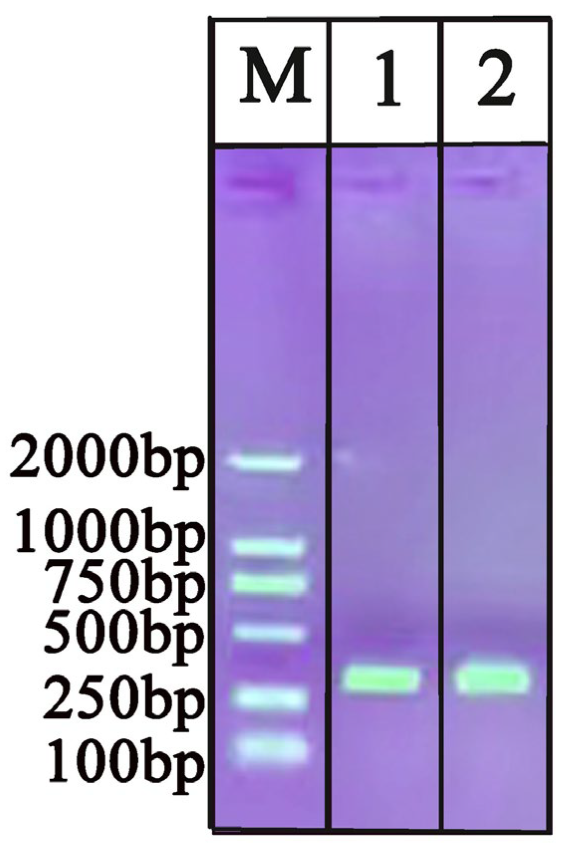
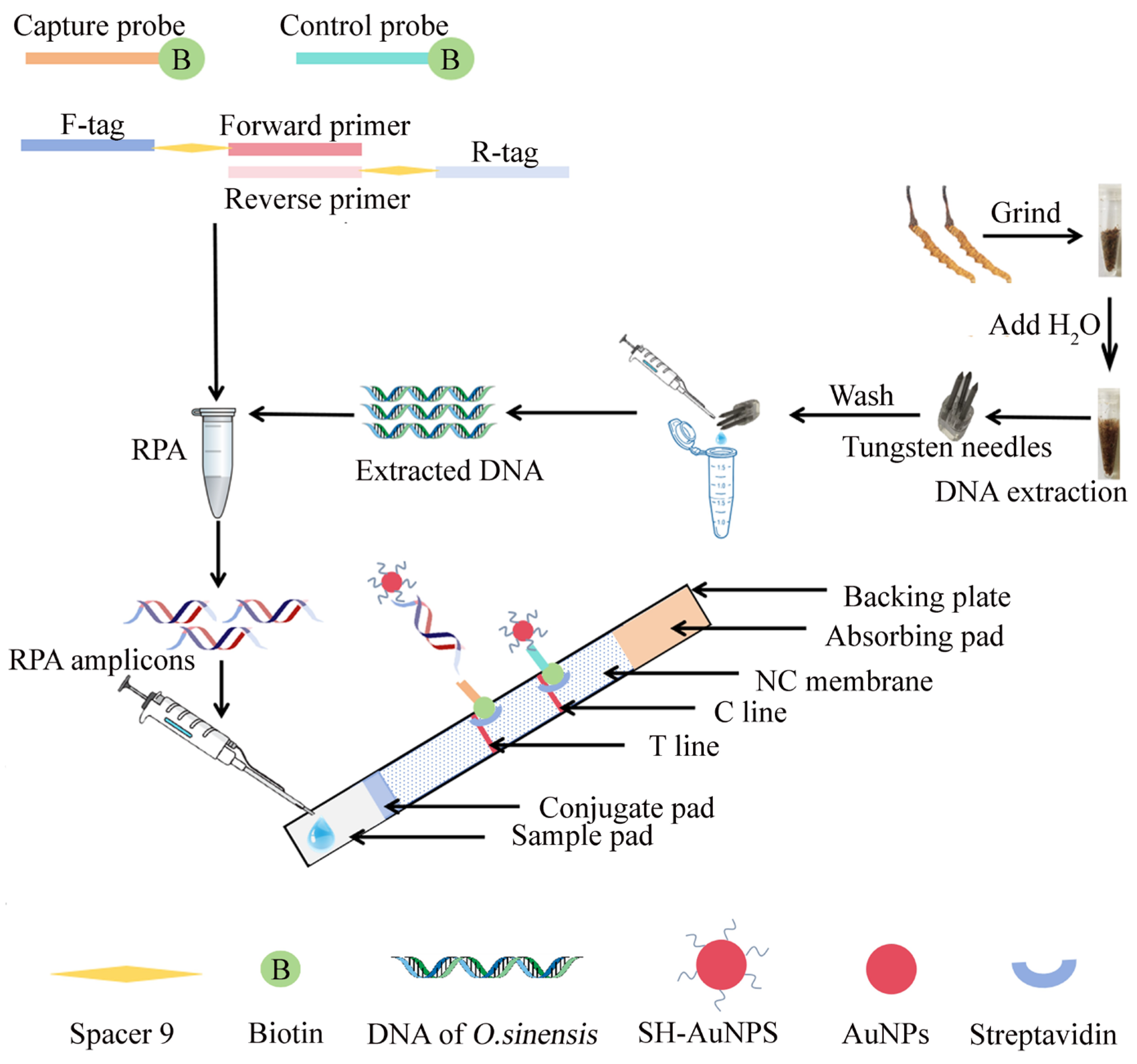
2.1. Evaluation of Extracted DNA by Commercial Kit and Tungsten

The process of DNA extraction using a tungsten needle is shown in Figure 1. The capacity of the tungsten needles and the commercial kit for DNA extraction was evaluated by comparing the absorbance ratio (A260/280 and A260/230) of nucleic acids, protein, and other organic contaminations (carbohydrates, EDTA, phenol); the results are shown in Table 1. The DNA extracted using a commercial kit exhibited a high concentration; however, the process was significantly more time-consuming and cumbersome compared to the tungsten method. Both the DNA extracted using tungsten and that from the commercial kit were subjected to PCR-AGE to compare the extraction efficiency. As shown in Figure 2, the PCR-AGE results verify that both the tungsten and commercial kit can obtain good DNA extraction effect.

2.2. Characterization of AuNPs and Verification the Formation of AuNPs-SH Probes

The obtained AuNPs were characterized by transmission electron microscope (TEM) and UV-Vis spectrophotometry (Figure 3A). The UV-Vis spectrophotometry showed that AuNPs exhibited an absorption maximum at around 520 nm. The inset shows a TEM image of the AuNPs, with the size of AuNPs being approximately 20 nm. The AuNPs synthesized in this study were burgundy in color. To verify the effect of pH on the coupling of AuNPs and SH probes, the conjunction of the SH probe with AuNPs was tested with the addition of 0.3 M NaCl. As shown in Figure 3B, the SH probes coupled with AuNPs at pH of 7.6 immediately turns colorless, indicating the failure conjugation of AuNPs and SH probes. When the pH is adjusted to 3.0, the SH probes and AuNPs conjunction remain burgundy with the 0.3 M NaCl addition, meaning the successful modification of AuNPs with the SH probes.

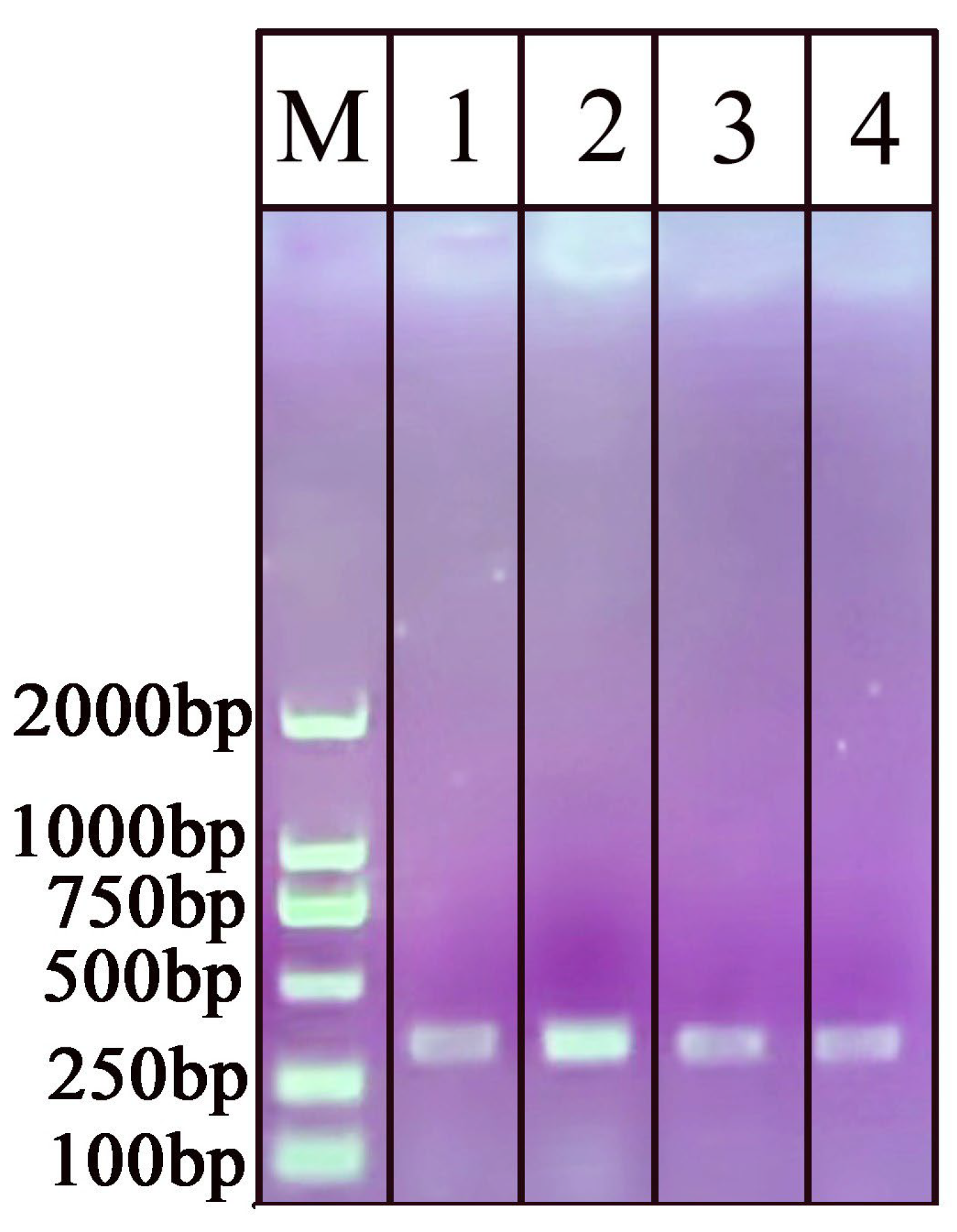
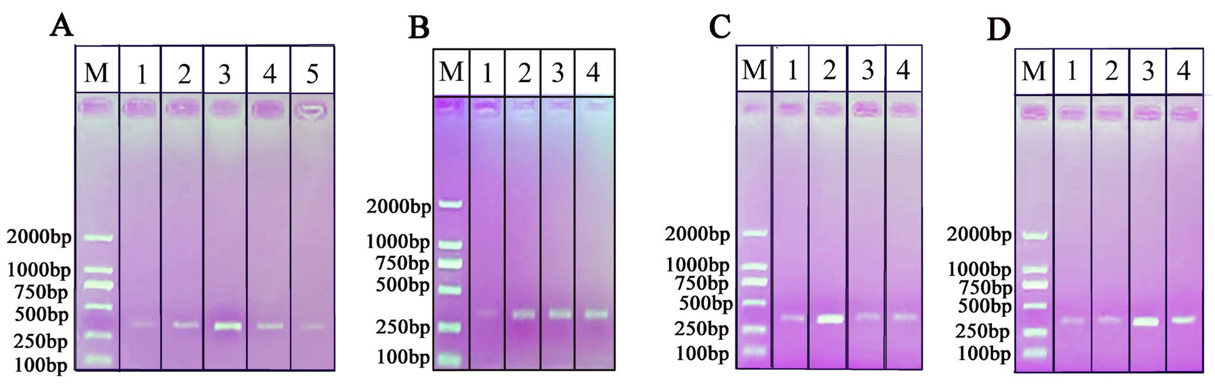
2.3. Primer Design and NAH-LFS Validation

Four pairs of primers (1, 2, 3, 4) (Table 2) were designed as candidates and subjected to RPA amplification using the genomic DNA of the O. sinensis as template. As expected, primer 2 produced a DNA band of 319 bp that was brighter than the bands generated by the other three primer sets (Figure 4). Therefore, primer 2 was selected as the appropriate primer pair for the amplification of O. sinensis. Then, the primer 2 was modified with spacer9 and tag sequence containing the following three functional regions: the first part of the forward/reverse primers must participate in the RPA amplicon located at the 3′ end of the primer, namely the specific binding region; the second part is a spacer region tag (spacer9) as a link to stop DNA polymerase amplification; the third part is a characteristic tag sequence located at the 5′ end of the primer that is maintained as single-stranded during amplification. The RPA products generated with the modified primer2 and O. sinensis DNA as the template resulted in the NAH-LFS displaying two red bands at both the C line and T line. In contrast, the negative control with double distilled H2O (ddH2O) as the template only showed a single red line at the control line, confirming a successful and valid test run of the NAH-LFS assay.

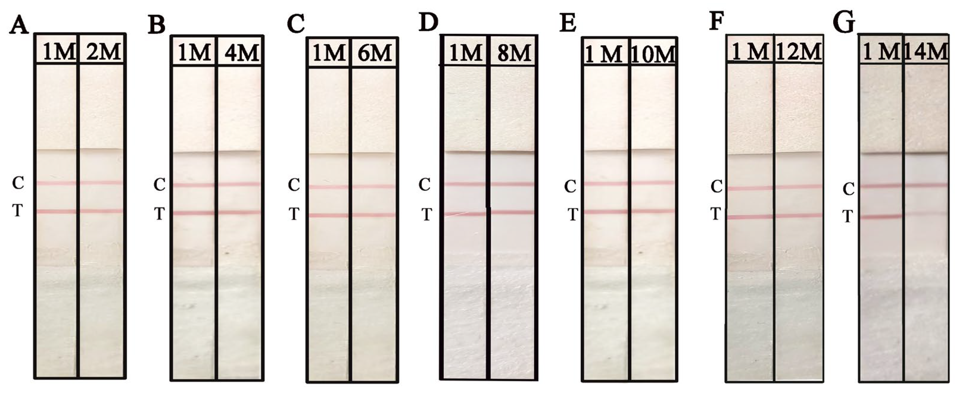
2.4. Optimization of RPA and the NAH-LFS Stability Under Room Temperature

To enhance the performance of RPA-NAH-LFS, it is essential to explore and optimize the crucial parameters. As the accuracy of RPA is crucial for the subsequent NAH-LFS, RPA amplifications were conducted under varying conditions, including different incubation times and temperatures, as well as the concentrations of Mg2+ and primer concentrations. As shown in Figure 5A, the incubation temperatures ranging from 38 °C to 40 °C produced single bands in AGE, with the optimal amplification temperature observed at 39 °C. In Figure 5B, the band brightness increased with the incubation time and stabilized at 10 min, as observed with the naked eyes. To streamline the process, a final incubation time of 10 min was selected. Figure 5C,D illustrate the optimization of primer and Mg2+ concentrations, revealing that Mg2+ concentrations of 2.5 μL (280 mM) and primer concentration of 1.8 μL (10 μM) produced the clearest bands. Therefore, the optimal RPA reaction conditions include an incubation temperature of 39 °C, an incubation time of 10 min, a Mg2+ amount of 2.5 μL (280 mM), and a primer amount of 1.8 μL (10 μM).
The working solution (WS) of NAH-LFS is another critical factor that can impact the sensitivity and reproducibility of NAH-LFS. In this study, WS1, WS2, WS3, and WS4 were employed to determine the optimal WS. From the results shown in Figure 6A, it is evident that WS2 yielded the best visual outcomes in terms of T/C line color intensity, distribution, and minimal background effects. Based on these findings, WS2 was used in subsequent experiments. Additionally, different types of NC membranes with varying pore sizes were evaluated. As depicted in Figure 6B, the flow rate (FR) of NC membrane on strip 1 (FR = 135 s/4 cm) demonstrated a superior performance as it facilitated the migration of RPA amplicons-AuNPs effectively. Therefore, the NC membrane (FR = 135 s/4 cm) was used for the subsequent experiments. RPA amplification using O. sinensis DNA (approximately 50 ng/μL) as template was subjected to NAH-LFS, which was stored for different months to evaluate the retention period. As shown in Figure 7 and Figure S1, the NAH-LFS can be stored at room temperature with a silica desiccant for up to 12 months without significant loss of activity. After more than 12 months, the color of the strips began to lighten.

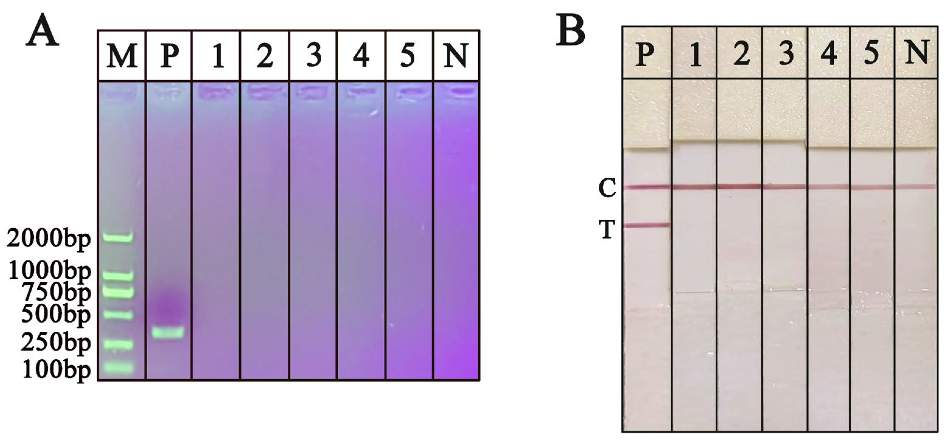
2.5. Specificity of the RPA-NAH-LFS Assay

To evaluate the specificity of the RPA-NAH-LFS, DNA extracted from O. sinensis and other fungal species using a tungsten needle was utilized as the PCR or RPA template. The PCR and RPA products were subsequently analyzed using AGE and NAH-LFS, respectively. As shown in Figure 8A, only the DNA from O. sinensis produced a bright band at the expected size of 319 bp by PCR-AGE, while DNA from the five non-target fungal species did not show any bands. In addition, as shown in Figure 8B, RPA products with O. sinensis DNA as the template produced clear red bands at both the T and C lines of the NAH-LFS, indicating the presence of O. sinensis. In contrast, the other five fungal species exhibited red band only at the C line, indicating the absence of O. sinensis. The results obtained from both PCR-AGE and RPA-NAH-LFS are consistent, demonstrating that the developed RPA-NAH-LFS assay exhibits high specificity and shows no interaction with other non-target fungal species.

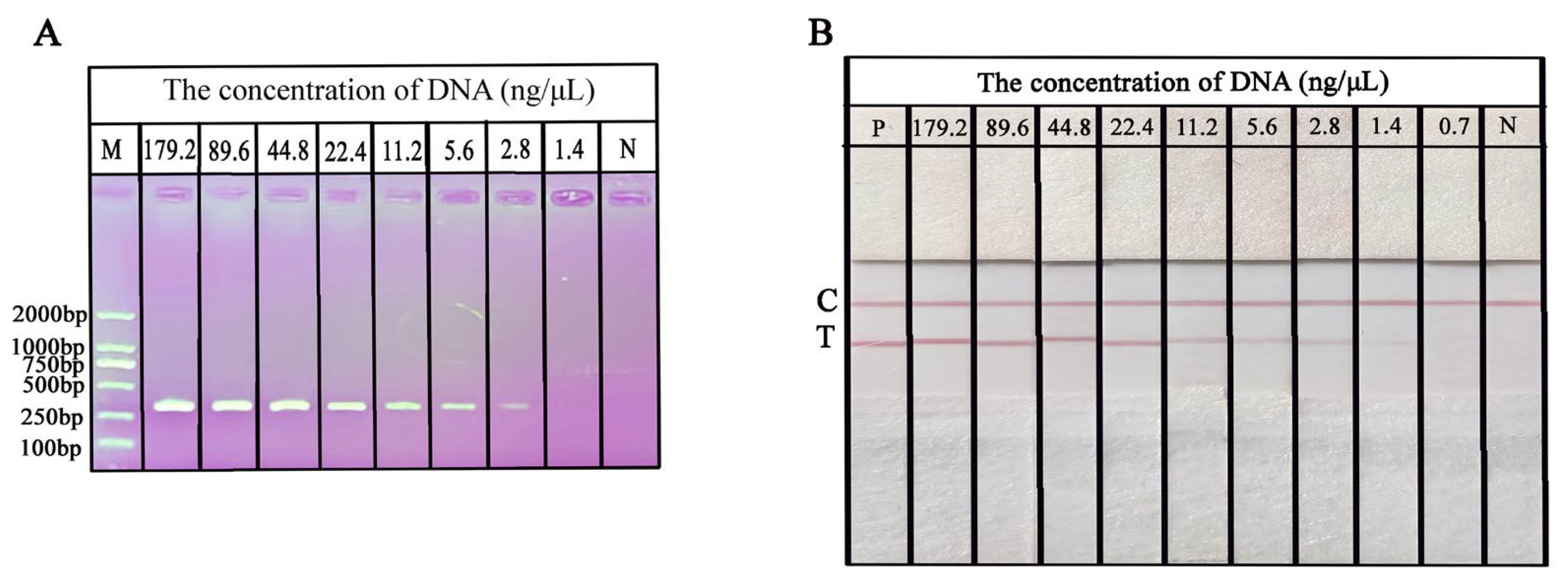
2.6. Sensitivity of the RPA-NAH-LFS Assay

The extracted DNA of O. sinensis with a continuous dilution (0.7–179.2 ng/μL) was prepared with ddH2O to evaluate the sensitivity of RPA-NAH-LFS. RPA was performed using various concentrations of DNA as templates to determine the limit of detection (LOD) of the RPA-NAH-LFS assay. As expressed in Figure 9A, analysis of PCR amplicons using AGE, the intensity of AGE bands gradually diminishes as the concentration of O. sinensis DNA is decreased from 179.2 ng/μL to 2.8 ng/μL. When the DNA concentration falls below 2.8 ng/μL, no distinct bands of a specific size are visible on the gel, suggesting that the LOD of the PCR-AGE for O. sinensis DNA is 2.8 ng/μL. Further analysis using RPA-NAH-LFS, as shown in Figure 9B, revealed that the T line gradually fades as the concentration of O. sinensis DNA decreases. When the concentration reaches 0.7 ng/μL, the T line on NAH-LFS does not exhibit any color. At the template concentration of 1.4 ng/μL, there is a visibly significant difference in color intensity from the negative control. Therefore, the detection sensitivity of the RPA-NAH-LFS for O. sinensis DNA is 1.4 ng/μL. The sensitivity of RPA-NAH-LFS is two times lower than that of the PCR-AGE.

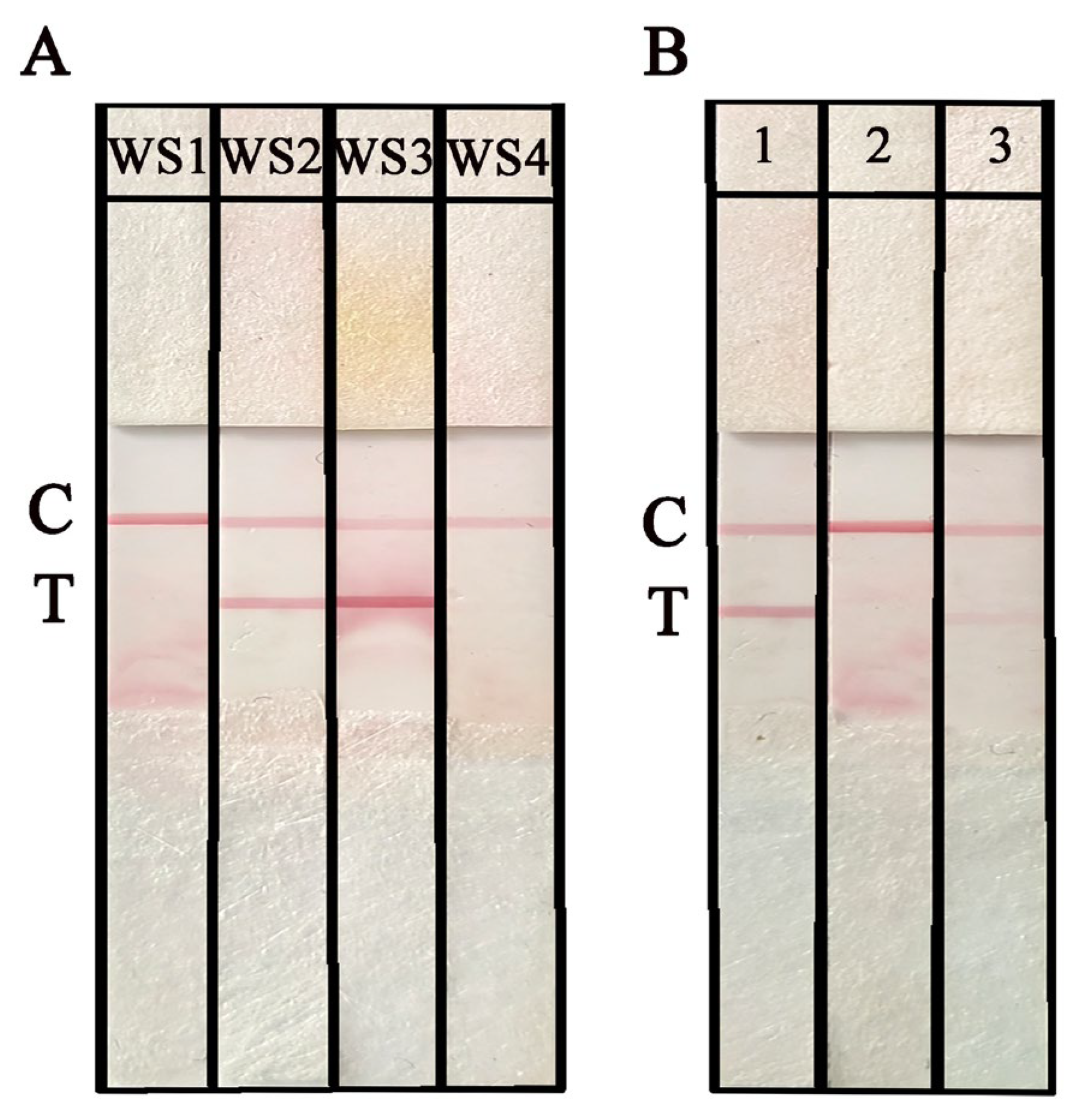
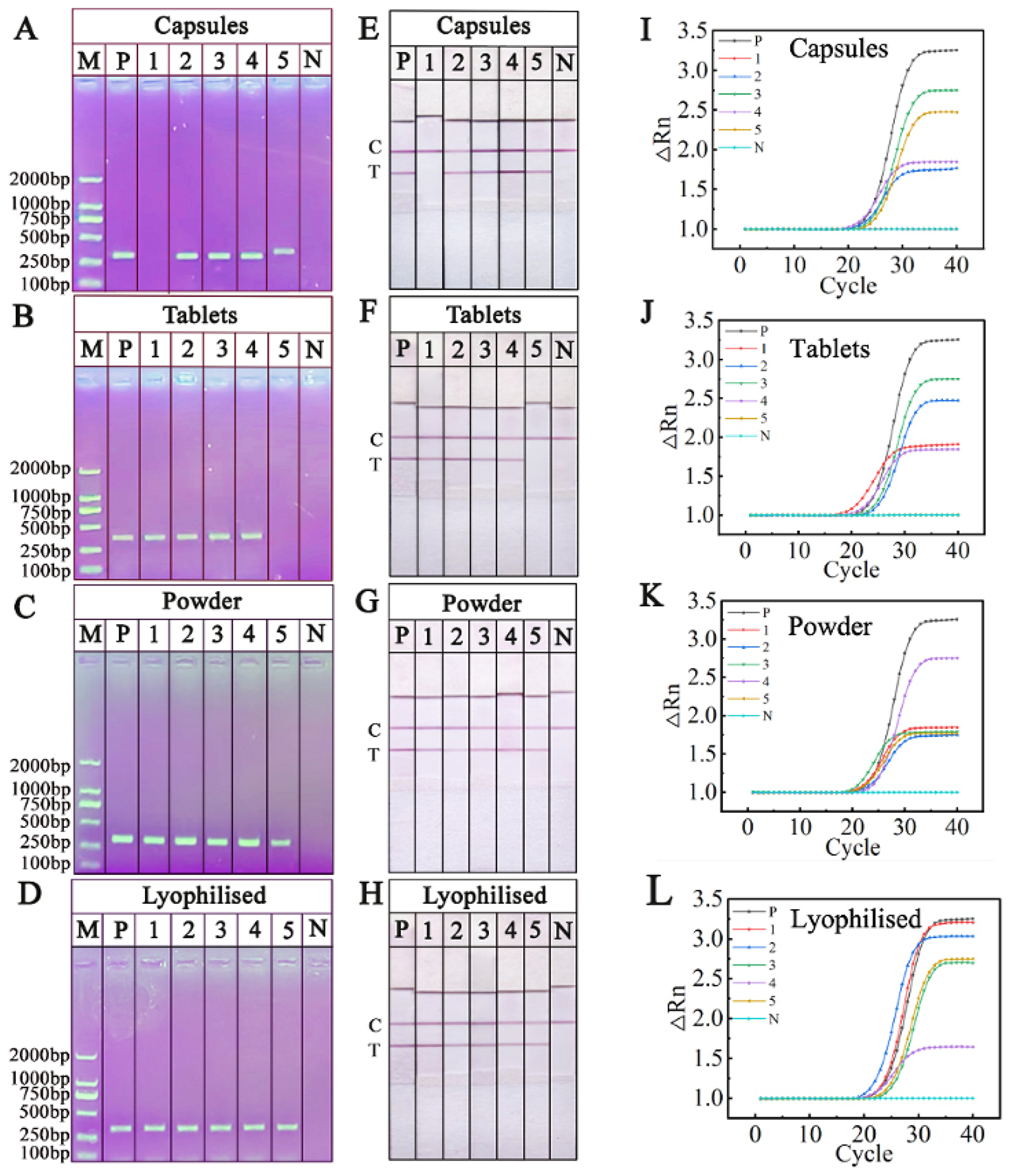
2.7. Detection Commercial O. sinensis Samples by RPA-NAH-LFS

In order to evaluate the effectiveness of RPA-NAH-LFS in analyzing commercial O. sinensis samples, 20 products labeled as authentic O. sinensis were collected and tested under optimized conditions. The results, illustrated in Figure 10, revealed that one out of five O. sinensis capsule products showed only a C line, indicating adulteration. One O. sinensis tablets products was found to lack genuine O. sinensis ingredients. Among the five O. sinensis powder products and five lyophilized O. sinensis products, displayed a distinct red color on the T line, indicating their authenticity. The results obtained from RPA-NAH-LFS (Figure 10A–D) was identical with PCR-AGE (Figure 10E–H) and RT-PCR (Figure 10I–L).

3. Discussion

O. sinensis a revered and rare edible fungus, is considered as superior tonic and herbal remedy, often compared to ginseng in terms of its benefits and commanding a higher price. The medicinal properties of O. sinensis include antioxidant effect, fatigue relief, anti-tumor potential, increased vitality, and cardiovascular benefits [27]. With the significant increase in price and demand for O. sinensis, other related fungi with lower mass have emerged as potential substitutes [28]. Reports indicate that while the annual production of authentic O. sinensis is around 185 tons, total market sales exceed 200 tons both domestically and internationally [29]. This disparity raises concerns about market disruptions, diminished consumer trust, and potential health risks associated with counterfeit products. Given these challenges, there is an urgent need for a reliable, point-of-care method to distinguish authentic O. sinensis from counterfeit products to protect consumers and ensure product authenticity.
DNA-based detection technology is increasingly being recognized for its precision and practicality in authenticating Chinese medicinal materials. In this study, wild O. sinensis within commercial products was identified using RPA coupled with NAH-LFS. Furthermore, the accuracy of the RPA-NAH-LFS results was confirmed through PCR-AGE analysis. The results indicated that RPA-NAH-LFS can accurately identify O. sinensis from counterfeit samples. The successful amplification and visualization of a 1.4 ng/µL DNA template using RPA-NAH-LFS suggest that its sensitivity is higher than that of PCR-AGE. Moreover, the entire RPA-NAH-LFS process can be completed in just 16 min (1 min for DNA extraction, 10 min for RPA, and 5 min for NAH-LFS). As shown in Table 3, this RPA-NAH-LFS demonstrates a greater speed, simplicity, and cost-effectiveness compared to PCR-AGE and other alternative methods [9,30]. Importantly, specificity testing confirmed no cross-reactivity with five other fungi, and the method is user-friendly for non-expert operators, requiring only a digital water bath and featuring straightforward operational steps.
In previous studies, various molecular biology methods have been utilized for the identification of O. sinensis and related products. For instance, Li et al. designed species-specific primers for PCR amplification of internal transcribed spacer sequences, with the LOD of 8 ng target DNA in 25 µL reaction volume [31]. Thanh et al. employed purpose-made capillary electrophoresis-based technology to determine the nucleosides of O. sinensis, achieving an LOD range of 11.2–22.0 μg/g [32]. Duan et al. introduced a multispectral imaging technique to enhance the efficiency of O. sinensis detection, achieving an impressive accuracy of 96.3% [33]. However, the existing studies have often faced challenges such as high equipment requirements, time-consuming procedures, the need for specialized expertise, and reliance on multiple statistical models and algorithms. The RPA-NAH-LFS method established in this study overcomes these limitations effectively.
AuNPs are prepared by the citrate reduction method and stabilized by interaction forces between citrate ions. Even micro-concentrations of NaCl (50 mM) will cause the AuNPs to aggregate, resulting in the solution becoming colorless [34]. However, when SH probes are adsorbed on the surface of AuNPs, the stability of AuNPs is significantly improved, even with the addition of 300 mM NaCl (Figure 3B). In this study, the solution remained burgundy after adding 300 mM NaCl, indicating that no aggregation of AuNPs occurred and proving that the SH probe was successfully coupled to the AuNP surface.
The NAH-LFS based on nucleic acid hybridization to catch target DNA in this study is ideal for rapid on-site testing and does not rely on costly antibodies. The stability of the NAH-LFS can be maintained for up to one year by ensuring dry storage conditions during transportation. The RPA-NAH-LFS method developed in this study has broader applications beyond O. sinensis authentication; it can be adapted to identify other traditional Chinese medicines by substituting the specific primer set. Liu et al. (2018) successfully utilized RPA-LFS to authenticate Ginkgo folium products, demonstrating superior accuracy compared to PCR-based methods [35]. Zhao et al. (2019) applied the RPA in combination with immunoassay LFS method to distinguish saffron from its adulterants, showcasing its potential versatility in analyzing processed products [36]. Meanwhile, the RPA-NAH-LFS method has been extensively employed in various fields, including meat product identification [37], virus detection [38], identification of pathogenic bacteria [39], and recognition of valuable botanical [40]. Compared to previous studies [37,41], the innovation lies in the fact that the entire O. sinensis detection process, from DNA extraction to the visualization of test results, does not require instruments or complex operations, and can be completed within 16 min. Additionally, there are limited studies in the literature on the utilization of RPA-NAH-LFS for O. sinensis identification. In summary, the RPA-NAH-LFS method enables on-site detection with limited resources across diverse areas to ensure food safety and prevent intentional contamination.

4. Materials and Methods

4.1. Materials

Chloroauric acid tetrahydrate (HAuCl4·4H2O), sodium chloride, agarose, sodium citrate, sodium dihydrogen phosphate, disodium hydrogen phosphate, hydrochloric acid, and tris (2-carboxyethyl) phenol hydrochloride (TCEP), were purchased from Aladdin Reagent (Shanghai, China). Streptomycin avidin, 2-[4-(2-hydroxyethyl)-1-piperazinyl] ethanesulfonic acid (HEPES), sucrose, Tween-20, and albumin bovine V (BSA) were obtained from Solebao Technology (Beijing, China). RPA basic amplification kit was obtained from TwistDxInc (Cambridge, UK). The Tiangen polysaccharide polyphenol plant genome DNA extraction kit was obtained from Tiangen Biotechnology (Beijing, China). Nitrocellulose (NC) membrane and glass fiber obtained from Jinbiao Biotechnology Co., Ltd. (Shanghai, China). The probes and primers were purchased from GENEWIZ (Suzhou, China). The 2×Taq PCR Mix plus were purchased from LabEAD (Beijing, China). Goldview nucleic acid dye was purchased from Biotopped (Beijing, China).

4.2. Sample Preparation and DNA Extraction

A total of six samples were purchased from various medicinal material markets in Tangshan, including O. sinensis, C. gunnii, C. cicadae, C. militaris, O. nutans, and yungui Cordyceps. These samples were visually identified by Beijing Tong Ren Tang before the experiment. Additionally, 20 commercial products of O. sinensis were purchased from online supermarket platforms and pharmacies; detailed information is provided in Table 4. All samples were dried for 24 h under a vacuum at 60 °C, and then ground into powder individually.
In this study, we compared the following two DNA extraction methods: (1) DNA extraction of all samples (0.02 g each) using a commercial kit (DP360, TIANGEN, Beijing, China) following the specified protocol; and (2) DNA extraction using tungsten with slight modifications [42,43]. Polymethyl methacrylate (PMMA), which features three holes, was utilized as a support for the fabrication of three tungsten needles. Each needle had a tip measuring 4 mm and a handle measuring 7 mm. After the three tungsten needles were affixed to the PMMA, a handle was attached to the opposite side of the PMMA (Figure 1). Specifically, approximately 100 μL of ddH2O was added to 0.02 g of O. sinensis powder and the needles were immersed in the solution for 1 min. The DNA was absorbed onto the surface of the needles, which were then washed with 100 μL of ddH2O. The extracted DNA was quantified using an ultra-micro-spectrophotometer K5800 (KAIAO, Beijing, China) by measuring the absorbance at A260/280 nm and A260/230. Subsequently, the extracted DNA from tungsten needles was subjected to PCR amplification and compared with the results obtained with commercial kits. The extracted DNA was diluted and stored at −20 °C until further use.

4.3. Synthesis of AuNPs and Conjunction with SH-Probes

AuNPs were synthesized following established protocols [41,44] to obtain magenta spherical AuNPs with an average diameter of 20 nm using the citrate reduction of HAuCl4. Briefly, 0.5 mL 1% HAuCl4 was added into a conical flask containing 49 mL of ultrapure water. The solution was stirred and heated to boiling. Subsequently, 1.125 mL 1% trisodium citrate was injected to the flask while continuing to heat and stir until it turned magenta, indicating that the AuNPs were obtained. For the combination of AuNPs and SH-probes, the following steps were carried out:
  • SH probes (10 μL 100 μM) were first reduced by adding them into TCEP (10 μL, 1 mM) and adjusted the solution pH to 3.0 with citrate–HCl buffer. The mixture was then incubated at room temperature for 1 h.
  • A total of 1330 μL AuNPs was injected into the above SH probe solution. After 10 min of reaction, citrate-HCl (30 μL, 10 mM) was added to AuNPs-SH probe solution to adjust the pH to 3.0. Then, NaCl (10 μL, 2.0 M) was added sequentially and allowed to react for additional 10 min.
  • The pH of the AuNPs-SH probe solution was adjusted to 7.5 by HEPES buffer (100 μL, 500 mM).
  • The AuNPs-SH probes were incubated for 1 h before being centrifuged at 2000× g for 10 min. The supernatant was removed, and the pellet was resuspended in 90 μL buffer (1 mM Tris-HCl, 10% sucrose, 0.5% Tween-20, and 1% BSA, pH of 7.6). The AuNPs-SH probe were sprayed onto the conjugate pad at 20 μL/cm.

4.4. Design of PCR/RPA Primers and NAH-LFS Probes

For the amplification of the O. sinensis DNA (RefSeq: KF696589.1), four primers were designed based on the specification of TwistAmp Basic kits. These primers were designed using Primer Premier 6.0 software and subsequently screened for accuracy through BLAST at NCBI and RPA amplification. The primer that produced the specific and brightest band on the AGE was selected as the optimum primer and used in the subsequent RPA-NAH-LFS.
In addition, three probes involved on NAH-LFS were as follows:
  • SH probes on AuNPs, which are reverse complementary to the tag sequence of forward primers;
  • biotin-labeled capture probe, which are reverse complementary to tag sequence of reverse primers; and
  • biotin-labeled control probe, which complementarily hybridize with SH probes.
The RPA amplicons have two single-strand tails (F-tag and R-tag) at the two opposite ends, which can bind to the reverse complementary capture probes at the T line of NAH-LFS and SH-AuNPs, respectively. Meanwhile, control probes were fixed on the control line (C line), which were reverse complementary to SH-AuNPs. The structure of the primer set is presented in Figure 1 and Table 2.

4.5. PCR and AGE Testing

Each PCR reaction included 10 μL 2×Taq PCR Mix plus, 1 μL 10 μM forward/reverse primers, 7 μL ddH2O, and 1 μL template DNA of O. sinensis. PCR was accomplished in a PCR instrument (Eppendorf, Germany) with the following conditions: a preheating step at 95 °C for 3 min, 30 cycles of 95 °C for 1 min, 54 °C for 40 s, 72 °C for 50 s, final extension at 72 °C for 5 min. The amplicons were detected by 1% AGE with 1 μL goldview nucleic acid dye.

4.6. Assembly and Application of the NAH-LFS

The NAH-LFS assembly involved attaching the sample pad, absorbing pad, NC membrane, and conjugate pad to the supporting backplane. To prepare the NC membrane, a complex consisting of biotinylated capture/control probes (5 μL, 10 μM) and streptavidin (2.5 μL, 1 mg/mL) was mixed with 11.6 μL deionized water and then sprayed on the T line/C line. The interaction between biotin and streptavidin enables the immobilization of probes on the NC membrane. The T line was positioned 1.5 cm from the top margin of the NC membrane and the C line was located at 1 cm from the top margin of the NC membrane. Both the NC membrane and conjugate pad were vacuum-dried at 38 °C for 4 h. Eventually, the NAH-LFS was assembled sequentially, and overlapped between each parts by 2 mm to ensure smooth capillary forces; then, the LFS was cut into 4 mm section using a cutter (Jinbiao Biotechnology Co., Ltd., Shanghai, China). The sample mixture was added to the sample pad, allowing it to migrate along the NAH-LFS. When the sample flows to the conjugate pad, the tag sequence of the forward primer was captured by SH-AuNPs and resulted in the RPA amplicons being labeled with AuNPs. Subsequently, the RPA amplicons–AuNPs continue to flow to the test line (T line), where the R-tag hybridized with capture probe preloaded on the T line, leading to the formation of a red line due to the colorimetric effect of the AuNPs. Finally, excessive SH-AuNPs diffuse to the C line, which were captured by control probes to form another red band indicating that the NAH-LFS is working properly. The working principle of NAH-LFS is shown in Figure 1.

4.7. Optimizing RPA Conditions and NAH-LFS Working Conditions

4.7.1. Optimizing RPA Conditions

The optimization of RPA conditions is essential for ensuring efficient amplification of the target DNA. Each RPA amplification reaction was carried out in a 50 μL reaction mixture containing primers, ddH2O, MgAcO, rehydration buffer, and lysozyme. Following amplification, the RPA products were detected using 1% AGE. The following parameters were optimized using the TwistAmp Basic Kit:
  • Reaction temperature and incubation time: RPA amplification reactions were conducted at temperatures ranging from 37 to 41 °C and varying incubation times between 5 and 20 min. These parameters were systematically varied to determine the optimal conditions for efficient amplification.
  • Primer concentration: The concentration of the primers in the RPA reaction mixture was optimized within the range of 1–1.6 μL (10 μM) to determine the ideal amount that promotes robust amplification.
  • Magnesium Acetate (MgAcO) concentration: The MgAcO concentration in the reaction mixture was varied from 2 μL (280 mM) to 3.5 μL (280 mM) to identify the optimal concentration that facilitates efficient amplification.
This comprehensive approach allowed for the confirmation of efficient amplification and successful detection of the target DNA.

4.7.2. Optimizing NAH-LFS Working Conditions

For improving performance of NAH-LFS, different types of WS and NC membrane with different pore size were compared to determine the optimal conditions. In the NAH-LFS phase, the RPA amplicon was diluted 1:10 with four types of WS (WS1: 0.01 M PBS, pH of 7.0; WS2: 0.01 M PBS with 0.05% Tween-20 and 1% BSA, pH of 7.0; WS3: 0.01M PBS, pH of 8.0; WS4: 0.01 M PBS, 0.05% Tween-20 and 1% BSA, pH of 8.0). Subsequently, 100 μL of the diluted solution was dropped onto the sample pad of NAH-LFS, and the result was observed within 5 min.
The flow rate (FR) and pore diameter of NC membranes play crucial roles in the performance of NAH-LFS. Therefore, three types of NC membranes with varying FR (FR = 33.75, 45.00, 22.50 cm/s) were evaluated to select the optimal NC membrane and working solution combination.
The WS and the NC membrane that produced the clearest red bands were selected as the suitable NAH-LFS working conditions. Positive results were indicated by the appearance of visible C and T lines on the NAH-LFS, while negative results showed only the C line. Other forms of coloration were invalid for the NAH-LFS assay.

4.8. Specificity and Sensitivity of the RPA-NAH-LFS Assay

To assess the specificity of the RPA-NAH-LFS assay, five non-target fungal species, namely C. gunnii; C. cicadae; C. militaris; O. nutans; and yungui Cordyceps, were selected. The genomic DNA of these non-target fungal species was used as templates for RPA reaction, and the resulting RPA products were examined by both NAH-LFS and AGE to confirm specificity. To determine the sensitivity of the RPA-NAH-LFS, the genomic DNA of O. sinensis was serially diluted with ddH2O to generate RPA templates. The resulting RPA products were then examined using both NAH-LFS and AGE to determine the limit of detection (LOD) of the assay. During the RPA amplification, deionized water served as a negative control to ensure the accuracy of the amplification process. The specificity and sensitivity of the RPA-NAH-LFS were evaluated through triplicate determinations to confirm repeatability and reliability of the results.

4.9. Detection of Commercial O. sinensis Products

To validate the practical application of the RPA-NAH-LFS assay under optimal conditions, 20 commercial O. sinensis products were detected by RPA-NAH-LFS assay. The products, including 5 O. sinensis capsules, 5 O. sinensis tablets, 5 O. sinensis powder, 5 lyophilized O. sinensis, were purchased from online supermarket platforms and pharmacies. The DNA of each product was extracted, followed by RPA amplification and NAH-LFS detection.
For the detection of commercial O. sinensis products, DNA was extracted with tungsten and screened-specific primers were employed for RPA amplification. After amplification, 10 μL of the RPA products were mixed with 90 μL of the WS in a centrifuge tube. And the mixture was then applied to the sample pad of the NAH-LFS and allowed to migrate along the strip. Results were visible within 5 min, providing a rapid and on-site detection for the presence of O. sinensis in the commercial products. The results were verified by PCR-AGE.

5. Conclusions

In this study, a visual RPA-NAH-LFS for the direct, rapid, and in situ detection of O. sinensis was established, utilizing AuNPs as labels to generate a distinctive red color signal. The detection process can be completed within 27 min using only a digital water bath. By incorporating spacer9 and a tag sequence into the RPA primer set for detecting O. sinensis DNA, the LFS relying on nucleic acid hybridization were established. The effectiveness of this RPA-NAH-LFS confirms the absence of nonspecific reactions between O. sinensis and five other fungi. The RPA-NAH-LFS can efficiently detects O. sinensis DNA concentrations as low as 1.4 ng/μL which is twice lower than PCR-AGE. In commercial product inspections, the results from RPA-NAH-LFS and PCR-AGE were consistent, revealing that the RPA-NAH-LFS is robust and accurate in detecting O. sinensis. Based on the advantages of time-saving, simplicity, and high levels of accuracy, specificity, and sensitivity, the RPA-NAH-LFS as a point-of-care detection method is expected to be widely adopted for authentic O. sinensis identification. Importantly, it also can be used to identify other forms of counterfeiting by substituting the specific primer set, indicating its potential for widespread use in vitro diagnostics.

Supplementary Materials

The following supporting information can be downloaded at: https://www.mdpi.com/article/10.3390/ijms252413599/s1.

Author Contributions

Conceptualization, H.L., X.S. and H.C.; methodology, X.W. and H.T.; validation, J.W. and Y.C.; investigation, Y.Y. and L.S.; writing—original draft preparation, H.L. and X.W.; funding acquisition, H.L. All authors have read and agreed to the published version of the manuscript.

Funding

This research was funded by the National Natural Science Foundation of China, grant number 32001791.

Institutional Review Board Statement

Not applicable.

Informed Consent Statement

Not applicable.

Data Availability Statement

All data were included in the manuscript.

Conflicts of Interest

The authors declare no conflicts of interest.

References

  1. Subedi, A.; Hamal, S.; Joshi, A. A Review on Ophiocordyceps sinensis. Science 2023, 7, 44–48. [Google Scholar] [CrossRef]
  2. Jin, G.S.; Wang, X.L.; Li, Y.; Wang, W.J.; Yang, R.H.; Ren, S.Y.; Yao, Y.J. Development of conventional and nested PCR assays for the detection of Ophiocordyceps sinensis. J. Basic. Microbiol. 2013, 53, 340–347. [Google Scholar] [CrossRef] [PubMed]
  3. Lee, S.; Lee, H.H.; Kim, J.; Jung, J.; Moon, A.; Jeong, C.S.; Kang, H.; Cho, H. Anti-tumor effect of Cordyceps militaris in HCV-infected human hepatocarcinoma 7.5 cells. J. Microbiol. 2015, 53, 468–474. [Google Scholar] [CrossRef] [PubMed]
  4. Guo, Y.; Zhao, Z.; Li, X. Moderate warming will expand the suitable habitat of Ophiocordyceps sinensis and expand the area of O. sinensis with high adenosine content. Sci. Total Environ. 2021, 787, 147605. [Google Scholar] [CrossRef]
  5. Xu, J.; Huang, Y.; Chen, X.X.; Zheng, S.C.; Chen, P.; Mo, M.H. The mechanisms of pharmacological activities of Ophiocordyceps sinensis fungi. Phytother. Res. 2016, 30, 1572–1583. [Google Scholar] [CrossRef] [PubMed]
  6. Wu, F.; Zhou, L.W.; Yang, Z.L.; Bau, T.; Li, T.H.; Dai, Y.C. Resource diversity of Chinese macrofungi: Edible, medicinal and poisonous species. Fungal Divers. 2019, 98, 1–76. [Google Scholar] [CrossRef]
  7. Guo, M.; Jiang, W.; Yu, J.; Pang, X. Investigating the authenticity of Ophiopogonis Radix and its Chinese patent medicines by using a nucleotide signature. J. Ethnopharmacol. 2020, 261, 113134. [Google Scholar] [CrossRef]
  8. Au, D.; Wang, L.; Yang, D.; Mok, D.K.; Chan, A.S.; Xu, H. Application of microscopy in authentication of valuable Chinese medicine I—Cordyceps sinensis, its counterfeits, and related products. Microsc. Res. Tech. 2012, 75, 54–64. [Google Scholar] [CrossRef] [PubMed]
  9. Tong, X.; Wang, Y.; Xue, Z.; Chen, L.; Qiu, Y.; Cao, J.; Peng, C.; Guo, J. Proteomic identification of marker proteins and its application to authenticate Ophiocordyceps sinensis. 3 Biotech 2018, 8, 1–8. [Google Scholar] [CrossRef] [PubMed]
  10. Zhang, P.; Li, S.; Li, J.; Wei, F.; Cheng, X.; Zhang, G.; Ma, S.; Liu, B.J.M. Identification of Ophiocordyceps sinensis and its artificially cultured Ophiocordyceps mycelia by ultra-performance liquid chromatography/Orbitrap fusion mass spectrometry and chemometrics. Int. J. Agric. Biol. Eng. 2018, 23, 1013. [Google Scholar] [CrossRef] [PubMed]
  11. Zhang, B.; Li, B.; Men, X.H.; Xu, Z.W.; Wu, H.; Qin, X.T.; Xu, F.; Teng, Y.; Yuan, S.J.; Jin, L.Q.; et al. Proteome sequencing and analysis of Ophiocordyceps sinensis at different culture periods. BMC Genom. 2020, 21, 886. [Google Scholar] [CrossRef] [PubMed]
  12. Zhang, F.L.; Yang, X.F.; Wang, D.; Lei, S.R.; Guo, L.A.; Liu, W.J.; Song, J. A simple and effective method to discern the true commercial Chinese cordyceps from counterfeits. Sci. Rep. 2020, 10, 2974. [Google Scholar] [CrossRef] [PubMed]
  13. Xie, J.W.; Huang, L.F.; Hu, W.; He, Y.B.; Wong, K.P. Analysis of the Main Nucleosides in Cordyceps sinensis by LC/ESI-MS. Molecules 2010, 15, 305–314. [Google Scholar] [CrossRef] [PubMed]
  14. Lo, Y.T.; Shaw, P.C.J.F.C. DNA-based techniques for authentication of processed food and food supplements. Food Chem. 2018, 240, 767–774. [Google Scholar] [CrossRef]
  15. Liu, Y.; Tang, Q.; Cheng, P.; Zhu, M.; Zhang, H.; Liu, J.; Zuo, M.; Huang, C.; Wu, C.; Sun, Z. Whole-genome sequencing and analysis of the Chinese herbal plant Gelsemium elegans. Acta Pharm. Sin. B 2020, 10, 374–382. [Google Scholar] [CrossRef]
  16. Xu, R.; Wei, S.; Zhou, G.; Ren, J.; Liu, Z.; Tang, S.; Cheung, P.C.; Wu, X. Multiplex TaqMan locked nucleic acid real-time PCR for the differential identification of various meat and meat products. Meat Sci. 2018, 137, 41–46. [Google Scholar] [CrossRef]
  17. Liu, H.; Cao, T.; Chen, H.; Zhang, J.; Li, W.; Zhang, Y.; Liu, H.J. Two-color lateral flow nucleic acid assay combined with double-tailed recombinase polymerase amplification for simultaneous detection of chicken and duck adulteration in mutton. J. Food Compos. Anal. 2023, 118, 105209. [Google Scholar] [CrossRef]
  18. Li, J.; Macdonald, J.J.B. Bioelectronics, Advances in isothermal amplification: Novel strategies inspired by biological processes. Biosens. Bioelectron. 2015, 64, 196–211. [Google Scholar] [CrossRef]
  19. Yusop, M.H.M.; Bakar, M. Review on halal forensic: A focus on DNA-based methods for pork authentication. Food Res. 2020, 4, 2347–2354. [Google Scholar] [CrossRef]
  20. Yeh, E.C.; Lee, L.P. One-step digital plasma separation for molecular diagnostics. In Proceedings of the 17th International Conference on Miniaturized Systems for Chemistry and Life Sciences, MicroTAS 2013, Freiburg, Germany, 27–31 October 2013; pp. 1323–1325. [Google Scholar]
  21. Daher, R.K.; Stewart, G.; Boissinot, M.; Bergeron, M.G. Recombinase polymerase amplification for diagnostic applications. Clin. Chem. 2016, 62, 947–958. [Google Scholar] [CrossRef]
  22. Li, J.; Macdonald, J.; Stetten, F. Correction: Review: A comprehensive summary of a decade development of the recombinase polymerase amplification. Analyst 2020, 145, 1950–1960. [Google Scholar] [CrossRef] [PubMed]
  23. Yusop, M.H.M.; Bakar, M.F.A.; Kamarudin, K.R.; Mokhtar, N.F.K.; Hossain, M.A.M.; Johan, M.R. Rapid Detection of Porcine DNA in Meatball Using Recombinase Polymerase Amplification Couple with Lateral Flow Immunoassay for Halal Authentication. Molecules 2022, 27, 8122. [Google Scholar] [CrossRef] [PubMed]
  24. Bougadi, E.T.; Kalogianni, D.P. Paper based DNA biosensor for food authenticity testing. Food Chem. 2020, 322, 126758. [Google Scholar] [CrossRef]
  25. Wang, J.; Wang, Y.; Hu, X.; Chen, Y.; Jiang, W.; Liu, X.; Liu, J.; Zhu, L.; Zeng, H.; Liu, H. A dual-RPA based lateral flow strip for sensitive, on-site detection of CP4-EPSPS and Cry1Ab/Ac genes in genetically modified crops. Food Sci. Hum. Wellness 2024, 13, 183–190. [Google Scholar] [CrossRef]
  26. Xu, Y.; Wei, Y.; Cheng, N.; Huang, K.; Wang, W.; Zhang, L.; Xu, W.; Luo, Y. Nucleic acid biosensor synthesis of an all-in-one universal blocking linker recombinase polymerase amplification with a peptide nucleic acid-based lateral flow device for ultrasensitive detection of food pathogens. Anal. Chem. 2018, 90, 708–715. [Google Scholar] [CrossRef]
  27. Chatnarin, S.; Thirabunyanon, M. Potential bioactivities via anticancer, antioxidant, and immunomodulatory properties of cultured mycelial enriched β-D-glucan polysaccharides from a novel fungus Ophiocordyceps sinensis OS8. Front. Immunol. 2023, 14, 1150287. [Google Scholar] [CrossRef] [PubMed]
  28. Cao, L.; Ye, Y.; Han, R. Fruiting body production of the medicinal Chinese caterpillar mushroom, Ophiocordyceps sinensis (Ascomycetes), in artificial medium. Int. J. Med. Mushrooms 2015, 17, 1107–1112. [Google Scholar] [CrossRef]
  29. Winkler, D. Caterpillar fungus (Ophiocordyceps sinensis) production and sustainability on the Tibetan Plateau and in the Himalayas. Asian Med. 2009, 5, 291–316. [Google Scholar] [CrossRef]
  30. Osathanunkul, M.; Osathanunkul, K.; Wongwanakul, S.; Osathanunkul, R.; Madesis, P. Multiuse of Bar-HRM for Ophiocordyceps sinensis identification and authentication. Sci. Rep. 2018, 8, 12770. [Google Scholar] [CrossRef]
  31. Liu, Y.; Wang, X.Y.; Gao, Z.T.; Han, J.P.; Xiang, L. Detection of Ophiocordyceps sinensis and its common adulterates using species-specific primers. Front. Microbiol. 2017, 8, 1179. [Google Scholar] [CrossRef]
  32. Nguyen, T.D.; Vu, M.T.; Nguyen, M.H.; Duong, H.A.; Mai, T.D.; Pham, H.V. A rapid and simple dual-channeled capillary electrophoresis with contactless conductivity detection method for the determination of adenosine, cordycepin, and inosine in Ophiocordyceps sinensis-based products. Food Anal. Methods 2021, 14, 1779–1787. [Google Scholar] [CrossRef]
  33. Duan, H.; Tong, X.; Cui, R.; Han, L.; Huang, G. On-site identification of Ophiocordyceps sinensis using multispectral imaging and chemometrics. Int. J. Agric. Biol. Eng. 2020, 13, 166–170. [Google Scholar] [CrossRef]
  34. Zhang, X.; Servos, M.R.; Liu, J. Instantaneous and quantitative functionalization of gold nanoparticles with thiolated DNA using a pH-assisted and surfactant-free route. J. Am. Chem. Soc. 2012, 134, 7266–7269. [Google Scholar] [CrossRef]
  35. Liu, Y.; Wang, X.Y.; Wei, X.M.; Gao, Z.T.; Han, J.P. Rapid authentication of Ginkgo biloba herbal products using the recombinase polymerase amplification assay. Sci. Rep. 2018, 8, 8002. [Google Scholar] [CrossRef]
  36. Zhao, M.; Wang, B.; Xiang, L.; Xiong, C.; Shi, Y.; Wu, L.; Meng, X.; Dong, G.; Xie, Y.; Sun, W. A novel onsite and visual molecular technique to authenticate saffron (Crocus sativus) and its adulterants based on recombinase polymerase amplification. Food Control 2019, 100, 117–121. [Google Scholar] [CrossRef]
  37. Liu, H.; Cao, T.; Wang, J.; Yuan, Y.; Li, H.; He, K.; Chen, H.; Wang, L. Analysis, Accurate and simultaneous detection of pork and horse meat adulteration by double tailed recombinase polymerase amplification integrated with SERS based two-color lateral flow nucleic acid hybridization strip. J. Food Compos. Anal. 2024, 134, 106562. [Google Scholar] [CrossRef]
  38. Ivanov, A.V.; Safenkova, I.V.; Zherdev, A.V.; Dzantiev, B.B. Multiplex assay of viruses integrating recombinase polymerase amplification, barcode—Anti-barcode pairs, blocking anti-primers, and lateral flow assay. Anal. Chem. 2021, 93, 13641–13650. [Google Scholar] [CrossRef]
  39. Batra, A.R.; Dike, C.C.; Mantri, N.; Ball, A.S. Recombinase polymerase amplification-lateral flow assay (RPA-LFA) as a rapid and sensitive test for Escherichia coli O157:H7 detection in food and beverage: A comparative study. Food Control 2024, 155, 110076. [Google Scholar] [CrossRef]
  40. Rodrigues, V.; Honrado, M.; Santos, J.; Pinto, M.A.; Amaral, J.S. Development of a loop-mediated isothermal amplification assay for the rapid detection of Styphnolobium japonicum (L.) Schott as an adulterant of Ginkgo biloba (L.). Phytomedicine 2024, 128, 155322. [Google Scholar] [CrossRef] [PubMed]
  41. Liu, H.; Cao, R.; Xu, W.; Ma, Y.; Li, W.; Zhang, Y.; Liu, H. A cost-effective method for the rapid detection of chicken adulteration in meat using recombinase polymerase amplification combined with nucleic acid hybridization lateral flow strip. J. Food Compos. Anal. 2022, 111, 104602. [Google Scholar] [CrossRef]
  42. Kim, Y.S.; Kim, J.; Na, W.; Sung, G.H.; Baek, S.K.; Kim, Y.K.; Kim, G.R.; Hu, H.J.; Park, J.H. Biotechnology, Development of a microneedle swab for acquisition of genomic DNA from buccal cells. Front. Bioeng. Biotech. 2022, 10, 829648. [Google Scholar] [CrossRef]
  43. Xiao, B.; Zhao, R.; Wang, N.; Zhang, J.; Sun, X.; Huang, F.; Chen, A. Integrating microneedle DNA extraction to hand-held microfluidic colorimetric LAMP chip system for meat adulteration detection. Food Chem. 2023, 411, 135508. [Google Scholar] [CrossRef] [PubMed]
  44. Liu, H.B.; Zang, Y.X.; Du, X.J.; Li, P.; Wang, S. Development of an isothermal amplification-based assay for the rapid visual detection of Salmonella bacteria. J. Dairy Sci. 2017, 100, 7016–7025. [Google Scholar] [CrossRef]
Figure 1. Scheme of the RPA-NAH-LFS for O. sinensis authentication detection. F-tag; forward primer; reverse primer; R-tag; control probe; capture probe in different colors means different nucleic acid sequence.
Ijms 25 13599 g001
Figure 2. Evaluation of extracted DNA by commercial kit and tungsten through PCR-AGE. 1: DNA extraction with commercial kit; 2: DNA extraction with tungsten; M: D2000 DNA marker.
Ijms 25 13599 g002
Figure 3. Characterization of AuNPs. (A) UV–visible spectrophotometry of AuNPs. Inset shows a TEM image of AuNPs. (B) Photographs of AuNP–SH probe solutions mixed with 0.3 M NaCl at pH of 3.0 and pH of 7.6.
Ijms 25 13599 g003
Figure 4. Select the optimum primers pair for O. sinensis. M: D2000 DNA marker, lanes 1, 2, 3, and 4 are RPA products with primer 1, 2, 3, and 4 (Table 2), respectively.
Ijms 25 13599 g004
Figure 5. Optimization of RPA conditions. (A) Incubation temperature. 1: 37 °C; 2: 38 °C; 3: 39 °C; 4: 40 °C; 5: 41 °C. (B) Incubation time. 1: 5 min; 2: 10 min; 3: 15 min; 4: 20 min. (C) The concentration of Mg2+. 1: 0.56 μM; 2: 0.7 μM; 3: 0.84 μM; 4: 0.98 μM. (D) The concentration of primers (10 μM). 1: 0.01 nM; 2: 0.012 nM; 3: 0.014 nM; 4: 0.016 nM. M: D2000 DNA marker.
Ijms 25 13599 g005
Figure 6. Optimization of NAH-LFS working conditions. (A) Optimization of WS. WS1: 0.01 M PBS, pH of 7.0; WS2: 0.01 M PBS with 0.05% Tween-20 and 1% BSA, pH of 7.0; WS3: 0.01 M PBS, pH of 8.0; WS4: 0.01 M PBS, 0.05% Tween-20 and 1% BSA, pH of 8.0. (B) Optimization the type of NC membranes. 1: FR = 135 s/4 cm; 2: FR = 180 s/4 cm; 3: 90 s/4 cm. C: control line, T: test line.
Ijms 25 13599 g006
Figure 7. The retention period of NAH-LFS for 2 months (A), 4 months (B), 6 months (C), 8 months (D), 10 months (E), 12 months (F), and 14 months (G). 1 M, 2 M, 4 M, 6 M, 8 M, 10 M means NAH-LFS stored for 1 month, 2 months, 4 months, 6 months, 8 months, 10 months, respectively. C: control line; T: test line.
Ijms 25 13599 g007
Figure 8. Specificity detection of RPA-AGE (A) and RPA-NAH-LFS (B). P: O. sinensis; 1: C. gunnii; 2: C. cicadae; 3: C. militaris; 4: O. nutans; 5: yungui Cordyceps; M: D2000 DNA marker; N: negative control; C: control line; T: test line.
Ijms 25 13599 g008
Figure 9. Sensitivity detection of PCR-AGE (A) and RPA-NAH-LFS (B). M: D2000 DNA marker; P: positive control; N: negative control; C: control line; T: test line.
Ijms 25 13599 g009
Figure 10. Testing results of PCR-AGE (AD), RPA-NAH-LFS (G,H), and RT-PCR (IL) for commercial O. sinensis samples. (A,E,I) O. sinensis capsule products; (B,F,J) O. sinensis tablet products; (C,G,K) O. sinensis powder products; (D,H,L) lyophilized O. sinensis products. P: positive control; N: negative control; C: control line; T: test line.
Ijms 25 13599 g010
Table 1. Comparison of DNA extraction by commercial kit and tungsten (n = 3).
MethodA260/230A260/280A260DNA ConcentrationTimeInstallation
Tungsten0.518 ± 0.031.323 ± 0.131.00450.2 ± 1.27 ng/µL1 min/
Commercial kit0.683 ± 0.061.741 ± 0.161.43071.5 ± 2.59 ng/µL50 minWater bath, Centrifuge, Vortex oscillator
Table 2. Primers and probes needed by RPA-NAH-LFS.
NameSequences (5′→3′)Tag Sequence (5′→3′)Function
Primer 1TTGTAGAAAACGGGGCAGGA/PCR
CCGCAGGGTCGAAAAATGAAG/
Primer 2TAGAAAACGGGGCAGGAACAATTTTTCACTGGGTTTATAGT-spacer9PCR/RPA
TTGTAGAAAACGGGGCAGGATCGAGTGACAGCTAATGTGTGATT-spacer9
Primer 3TTGGTGAACCAGCGGAGGGATCATT/PCR
GCTTGCTTCTTGACTGAGAGGTGCC/
Primer 4ATTAAGTCGTGGAAATG/PCR
GATCAGGAATAGTGGGA/
SH-probeACTATAAACCCAGTGAAAAAT-SHNAH-LFS
Control probeATTTTTCAGGGTTTTATAGTBiotinNAH-LFS
Capture probeAATCACACATTAGCTGTCACTCGABiotinNAH-LFS
Table 3. Comparison of RPA-NAH-LFS with other techniques.
MethodTimeCost
AmplificationDetectionEquipmentReagent
PCR-AGE120 min45 minThermal cycling equipment1.00 $
PCR-ICTS120 min5–15 minThermal cycling equipment1.50 $
RPA-ICTS10 min5 minWater bath6.92 $
RPA-NAH-LFS10 min5 minWater bath6.42 $
ICTS: immunochromatographic test strip.
Table 4. Detailed information and test results of 20 commercial O. sinensis samples.
No.SampleFromTest Results a
1O. sinensis capsule 1China Beijing Tang+
2O. sinensis capsule 2Mannings
3O. sinensis capsule 3Vitahealth+
4O. sinensis capsule 4Alibaba Health+
5O. sinensis capsule 5Wei shiya+
6O. sinensis tablet 1China Beijing Tang+
7O. sinensis tablet 2Mannings+
8O. sinensis tablet 3Vitahealth+
9O. sinensis tablet 4Alibaba Health+
10O. sinensis tablet 5Wei shiya
11O. sinensis powder 1China Beijing Tang+
12O. sinensis powder 2Mannings+
13O. sinensis powder 3Vitahealth+
14O. sinensis powder 4Alibaba Health+
15O. sinensis powder 5Wei shiya+
16lyophilized O. sinensis 1China Beijing Tang+
17lyophilized O. sinensis 2Mannings+
18lyophilized O. sinensis 3Vitahealth+
19lyophilized O. sinensis 4Alibaba Health+
20lyophilized O. sinensis 5Wei shiya+
a (+) positive result; (−) negative result.
Disclaimer/Publisher’s Note: The statements, opinions and data contained in all publications are solely those of the individual author(s) and contributor(s) and not of MDPI and/or the editor(s). MDPI and/or the editor(s) disclaim responsibility for any injury to people or property resulting from any ideas, methods, instructions or products referred to in the content.

