Review

Understanding Quorum-Sensing and Biofilm Forming in Anaerobic Bacterial Communities

Department of Medical Microbiology, Medical University of Warsaw, 5 Chalubinski Str., 02-004 Warsaw, Poland
*
Author to whom correspondence should be addressed.
Int. J. Mol. Sci. 2024, 25(23), 12808; https://doi.org/10.3390/ijms252312808
Submission received: 1 November 2024 / Revised: 22 November 2024 / Accepted: 25 November 2024 / Published: 28 November 2024

Abstract

Biofilms are complex, highly organized structures formed by microorganisms, with functional cell arrangements that allow for intricate communication. Severe clinical challenges occur when anaerobic bacterial species establish long-lasting infections, especially those involving biofilms. These infections can occur in device-related settings (e.g., implants) as well as in non-device-related conditions (e.g., inflammatory bowel disease). Within biofilms, bacterial cells communicate by producing and detecting extracellular signals, particularly through specific small signaling molecules known as autoinducers. These quorum-sensing signals are crucial in all steps of biofilm formation: initial adhesion, maturation, and dispersion, triggering gene expression that coordinates bacterial virulence factors, stimulates immune responses in host tissues, and contributes to antibiotic resistance development. Within anaerobic biofilms, bacteria communicate via quorum-sensing molecules such as N-Acyl homoserine lactones (AHLs), autoinducer-2 (AI-2), and antimicrobial molecules (autoinducing peptides, AIPs). To effectively combat pathogenic biofilms, understanding biofilm formation mechanisms and bacterial interactions is essential. The strategy to disrupt quorum sensing, termed quorum quenching, involves methods like inactivating or enzymatically degrading signaling molecules, competing with signaling molecules for binding sites, or noncompetitively binding to receptors, and blocking signal transduction pathways. In this review, we comprehensively analyzed the fundamental molecular mechanisms of quorum sensing in biofilms formed by anaerobic bacteria. We also highlight quorum quenching as a promising strategy to manage bacterial infections associated with anaerobic bacterial biofilms.

1. Introduction

The National Institutes of Health (NIH) has shown that 60–80% of all microbial infections are associated with biofilm formation [1]. The first description of surface-attached, structured microbial communities, later referred to as biofilm, came from the Dutch scientist Antoni van Leeuwenhoek, who in 1683 observed the aggregated bacteria in plaque on the teeth and, particles scraped from the tongue [2,3]. In the 1930s, biofilm was observed in the aquatic environment. In medicine, the first association between the etiology of chronic infection and aggregates of bacteria was evidenced during routine microscopic examination of Gram-stained sputum in the early 1970s by Nils Høiby. The sample was taken from cystic fibrosis patients with persistent Pseudomonas aeruginosa infection. In 1980, Costerton and co-workers published the image of P. aeruginosa microcolonies in a lung of a patient (post-mortem) with cystic fibrosis made by electron microscope [4]. The definition of biofilm has evolved over the last decades. The first medical report in which the word ‘biofilm’ was used was published in 1981 by dentists from the University of Lund, Sweden which showed that biofilm forms on all solid surfaces in the oral cavity [2,5,6]. The improved identification of anaerobic bacteria (by applying matrix-assisted laser desorption/ionization time-of-flight mass spectrometry; MALDI-TOF MS and 16S rRNA genes cluster sequencing) in clinical specimens has raised awareness of the fact that anaerobes are involved in infections in almost every part of the body [7,8,9]. It is now known that certain anaerobes can form mono-, dual- and even multi-species biofilms, although this property and its impact on infectious disease remain to be studied in more detail.
Under natural, physiological conditions, bacteria form biofilms on human tissues such as the skin, mucosal surfaces of the respiratory and digestive tracts, and the female genital tract. These biofilms have a protective role, preventing the emergence of infections. However, under certain circumstances, such as disruption of the continuity of the skin and mucous membranes due to trauma, diabetic foot or ulcerative colitis, peripheral vascular disease, or peripheral neuropathy, these niches can be overgrown by biofilm-forming pathogenic organisms. Biofilm-associated infections are usually chronic, can recur, and are, therefore, very difficult to control [10,11,12]. A thorough understanding of the clinical impact of biofilms formed by anaerobic organisms is not presented in the literature, most likely due to the difficulty and technical knowledge required to culture anaerobic organisms. Understanding the mechanism of biofilm formation and inter-bacterial interactions appears to be crucial for the development of strategies to combat pathogenic biofilms.
In this paper, we have reviewed the scientific literature with a primary focus on the involvement of anaerobic biofilms in infectious diseases. The main aim is to summarize the molecular mechanisms of cell-to-cell communication, known as quorum sensing (QS), within biofilms formed by anaerobic bacteria. This review also points to the inhibition of QS systems, called quorum quenching (QQ), which is now expected as a promising strategy to combat bacterial biofilm-associated infections.

2. Methods

The preliminary search was conducted to identify relevant articles to ensure the validity of the proposed idea and to ensure that we have enough sources to conduct the analysis. An electronic search was performed querying the following databases MEDLINE (accessed through PubMed), and EMBASE from January 1990 through October 2024. A manual search of publications consistent with the topic was also performed. The search strategy was constructed to include free-text terms in the title and abstract and any appropriate subject indexing (quorum sensing, quorum quenching, autoinducer, acyl homoserine lactone, signaling molecules, sensing molecule, Fusobacterium, Prevotella, Porphyromonas, Bacteroides, Clostridium, Clostridioides, Cutibacterium, Propionibacterium, anaerobic bacteria, biofilm) with Boolean operators. Full-text articles written in English were included in subsequent screening. For removing the duplicates, the automated systematic search deduplicator (ASySD) was used. The two researchers evaluated articles individually using the same searching criteria with a particular focus on relevance to the scope of this review. The search results were compared, and a list of eligible publications was compiled. The classified studies were thoroughly reviewed, and relevant data were extracted. The organization of the literature search is shown in the diagram (Figure 1).

3. Biofilms and Their Medical Impacts

Biofilms are sessile microbial communities and have been described as coherent clusters (microcolonies) of bacterial cells embedded in an extracellular polymeric substance (EPS). EPS can account for up to 80% of the total volume of biofilms and is composed of polysaccharides (e.g., alginate), proteins (e.g., fibrin), lipids, metal ions, and extracellular DNA (eDNA) [10]. Multispecies biofilm, composed of different microorganisms (e.g., dental biofilm which may consist of up to hundreds of species) is a common phenomenon [11]. Within the biofilm, bacteria alter their own metabolism and protein production, regulate gene expression which can lead to a reduced cell division rate, and as a result, adapt to environmental anoxia and nutrient limitation. Compared to bacteria in the planktonic (freely suspended) state, biofilm-forming bacteria exhibit much more virulent properties, are better adapted to environmental conditions, and can be highly resistant or tolerant to antimicrobials. Biofilms constitute an optimal environment for horizontal gene transfer thus promoting the spread of bacterial resistance to antimicrobial agents [12,13]. Biofilms that affect human health can be divided into three categories: formed on human tissues, formed on medical devices, and formed on surfaces outside the body. Surfaces in the mouth are easily colonized by bacteria that form a biofilm called dental plaque. The accumulation and maturation of dental plaque on tooth surfaces can lead to inflammatory disease, gingivitis, periodontitis, and irreversible destruction of tooth-supporting tissues, peri-implantitis [11,14]. Anaerobes colonize the oral cavity and have a potential tumor-promoting effect (oral squamous cell carcinoma). Currently, there are three periodontopathogens involved in carcinogenesis. Two of them are anaerobic bacteria, Porphyromonas gingivalis, and Fusobacterium nucleatum, the third is an aerobic organism Treponema denticola [15,16,17]. Approximately 10% to 40% of all sinusitis cases are the result of an odontogenic process. The understanding of the etiology and pathogenesis of chronic sinusitis is still incomplete. Correct diagnosis requires aspiration of maxillary sinus secretions and/or ethmoid sinuses by endoscopy, which is not a standard procedure. Failure to respond to first-line therapy may be associated not only with the emergence of resistant aerobic strains but also with anaerobic bacterial infections [18]. There is growing evidence that the oral pathogen F. nucleatum is involved in the progression of an increasing number of tumor types, including colorectal, pancreatic, esophageal, and breast cancers [19]. Moreover, anaerobic bacteria (mainly Bacteroides spp., Peptostreptococcus spp., Fusobacterium spp., Clostridium spp. as well as Cutibacterium acnes) can cause septic arthritis and osteomyelitis [20]. Commensal bacterium C. acnes can be responsible for up to 10% of bacterial prosthesis joint infections [21]. It is worth noting that anaerobes may accompany aerobic bacteria (mainly facultative streptococci and gram-negative bacilli) in mixed infections. Under such conditions, it is not clear which organism or organisms are the main infective agents of bone infection or whether there is a synergistic mechanism of infection [22]. Anaerobes are also involved in pelvic inflammatory diseases and endometritis [23,24]. Numerous studies have confirmed the contribution of Bacteroides fragilis in inflammatory bowel disease (IBD), Crohn’s disease, and colitis ulcerosa. Enterotoxigenic B. fragilis (ETBF) strains secrete a 20 kDa pro-inflammatory zinc-dependent metalloprotease that can stimulate high expression of host interleukin-17 and increase the permeability of intestinal epithelial cells, resulting in enhanced internalization of various intestinal bacteria [25]. In ulcerative colitis, some species classified as Bacteroidetes correlate with disease activity [26]. The locations of infections associated with biofilms formation are shown in Figure 2.
Anaerobic biofilm can be formed on medical devices, including the following:
  • orthopedic implants [21,27,28],
  • dental implants [11,27,29],
  • breast implants used in both postmastectomy breast reconstruction and cosmetic surgical procedures [27,30],
  • contact lenses [27,29],
  • intrauterine devices, e.g., as long-term contraception methods [27].
The biofilms can be formed also on surfaces outside the body, mainly in water distribution systems, and if not systematically removed, can be a source of infection, including nosocomial infections [31].
However, the contribution of anaerobes to biofilm formation is becoming better understood, and biological mechanisms of quorum-sensing signaling molecules have yet to be adequately analyzed. A comprehensive understanding of the microbial signaling cascade provides a basis for interpreting microbial behavior in infection-related diseases and, in perspective, for using molecular modeling as a step in the search for new compounds with potential antibiofilm activity.
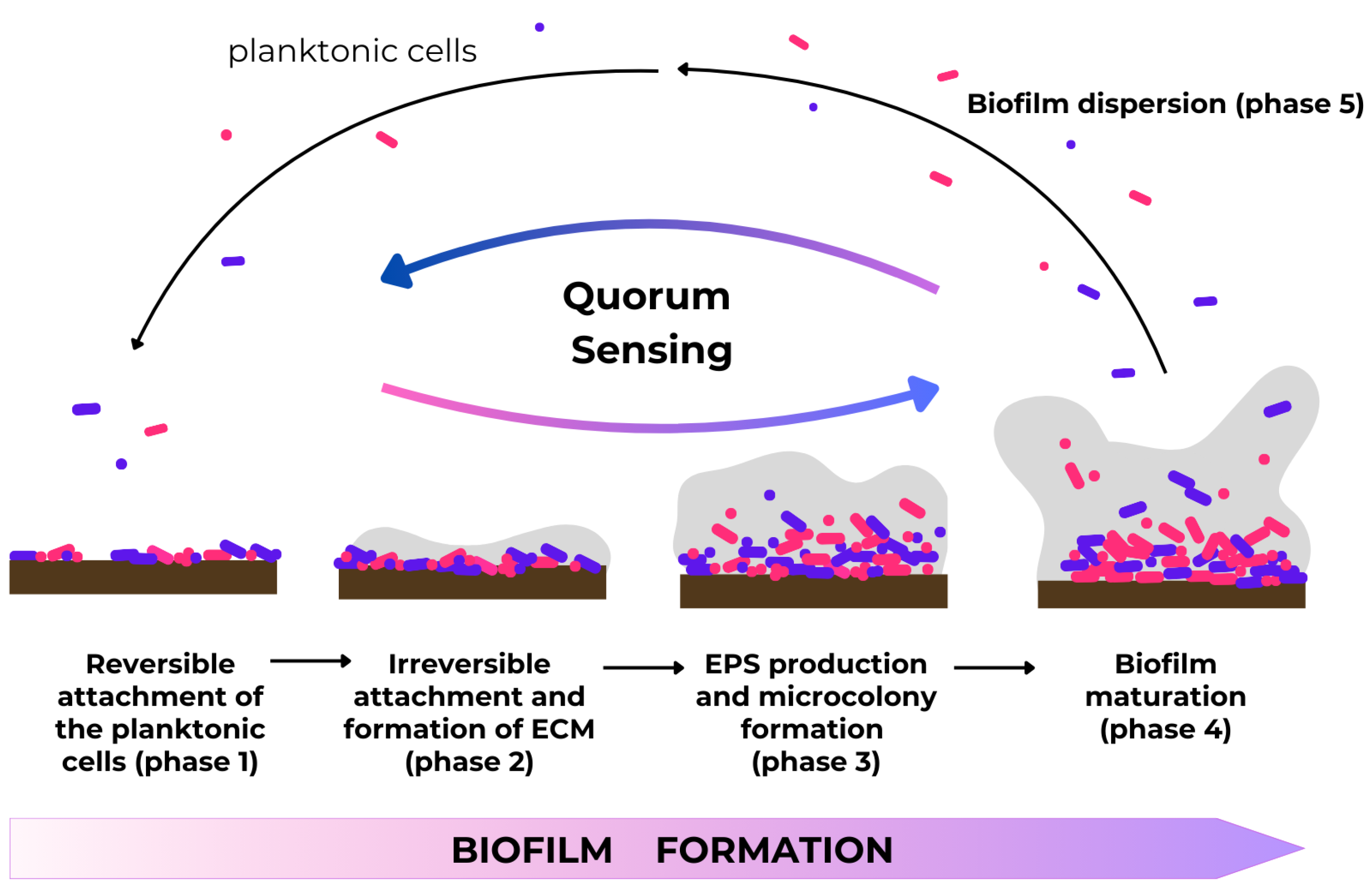
The formation of bacterial biofilms is a complex phenomenon, determined by many physical, chemical, and biological processes. Biofilm architecture is heterogeneous. It is constantly changing in both time and space due to external and internal factors. In the process of biofilm formation, phases such as reversible attachment of the planktonic cells (1), irreversible attachment and formation of extracellular matrix (ECM) (2), EPS production and microcolony formation (3), biofilm maturation (4), and final dispersion (5) have been identified (Figure 3) [32].
Phase 1 and Phase 2: Attachment. The initial phase is the reversible attachment of the microorganism to a solid surface using gravitational, electrostatic interactions, and van der Waals forces or bacterial structures such as flagella or pili. In this phase, cohesion also occurs. The next step is the irreversible binding of the bacteria to the surface and the production of an extracellular matrix. ECM promotes biochemical and physiological changes in the biofilm matrix which ensures structural integrity; stability; access to nutrients, metabolites, or signaling substances; and protection from adverse external factors (bactericides), desiccation, and host defense factors. During the irreversible attachment phase, bacterial cells interact with the surface using adhesins and lipopolysaccharide (LPS). This interaction causes changes in the surface properties including small damages and hydrophobicity decreasing. The binding intensity depends primarily on the species and the number of microorganisms adhering to the surface as well as the physicochemical characteristics of the surface. The speed and extent of biofilm growth depend, among other factors, on liquid flow rate, Brownian motion, nutrient availability, iron availability, pH, osmolality, oxygen concentration, concentration of antibacterial drugs, and ambient temperature. In such specific environment conditions (pH and oxygen gradients, co-adhesion), microorganisms synthesize, and release signaling molecules, communicate with each other, and change the surrounding microenvironment. The composition and structure of the biofilm matrix can evolve over time [32,33,34,35].
Phase 3: Microcolony formation. This phase is characterized by the synthesis of extracellular polymers (soluble and insoluble glucans, fructans, and heteropolymers). The matrix is biologically active, with channels retaining water, nutrients, and enzymes within the biofilm structure. In addition, the presence of certain bacteria creates an ecological niche for other microorganisms, enabling them to survive in the new favorable conditions [36,37].
Phase 4: Biofilm maturation and bacterial succession. The formation of a mature biofilm is associated with increased cell density and a reduction in the growth rate of specific bacteria. At this stage, interactions between microorganisms and host play the most important role not only in the formation of a mature biofilm structure but also in the separation of bacterial species/cells from such a formed structure. Bacteria can ‘sense’ specific environmental changes and induce the genes associated with active detachment [34]. One example is Prevotella loescheii (renamed as Hoylesella loescheii), which produces proteases that hydrolyze its adhesins, which are responsible for co-aggregation with Streptococcus mitis [36].
Phase 5: Dispersion. Finally, to avoid overgrowth, which would reduce the bacteria’s access to vital nutrients and lead to the accumulation of harmful waste products, bacterial cells detach from the biofilm and migrate as planktonic forms. In this way, microorganisms can spread throughout the human body. Therefore, a mature biofilm structure results from a balance between adhesion, growth, and microorganism removal [32,34,36].
Slow-growing cells in a biofilm phenotype are generally less sensitive or even resistant to antibiotics, antiseptics, and both innate and acquired immune responses, compared to planktonic cells. The extracellular matrix interacts with the environment, e.g., by attaching anaerobic or anaerobic-aerobic biofilms to human tissues (mucous membranes and damaged skin) or artificial materials. Within the biofilm, the individual bacteria communicate with each other by finely tuned molecular processes defined as quorum sensing [32,38].

4. Quorum Sensing and Quorum-Sensing Molecules (QSMs)

Biofilms are characterized by a high degree of structural and functional bacterial organization. Cell-to-cell communication within the biofilm is based on the generation and sensing of extracellular signals and the secretion of specific small signaling molecules called autoinducers (AIs). QS signals play a role in the biofilm development and dispersal. The concentration of signaling molecules is positively correlated with microbial population density.
When signaling molecule concentration reaches a certain threshold, they bind to intracellular receptors to activate target gene expression coordinating inter- and intra-population physiological behavior, including facilitating adaptation to the environment, bacterial motility, coordination of virulence factor expression, sporulation, and adhesion [39,40,41,42]. Quorum-sensing systems consist of a membrane-bound histidine sensor kinase and a cytoplasmic response regulator, which can function as a transcription factor [43]. In addition to their effects on the bacterial life cycle, quorum-sensing molecules (QSMs) also induce immune responses in host tissues [44,45] and have an impact on the development of antibiotic resistance [46,47].
In anaerobic bacteria, AIs are classified into three major classes:
  • N-Acyl homoserine lactones (AHL, autoinducer type-1, AI-1) specific to Gram-negative bacteria,
  • autoinducer-2 molecules (AI-2), consisting of 4,5-dihydroxy-2,3-pentandedione (DPD) derivatives in both Gram-negative and Gram-positive bacteria,
  • autoinducing peptides (AIPs), specific to Gram-positive bacteria [46,48].
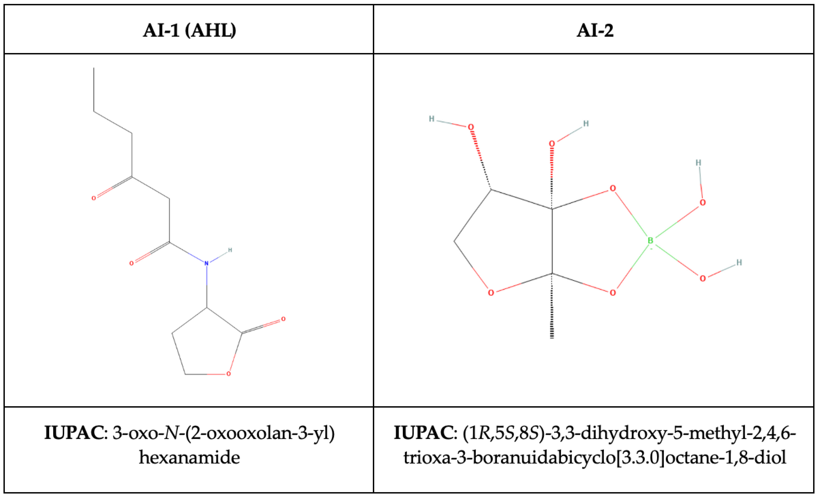
The chemical structure of the compounds AI-1 and AI-2 are shown in Figure 4.

4.1. N-Acyl-Homoserine Lactones (AHLs)

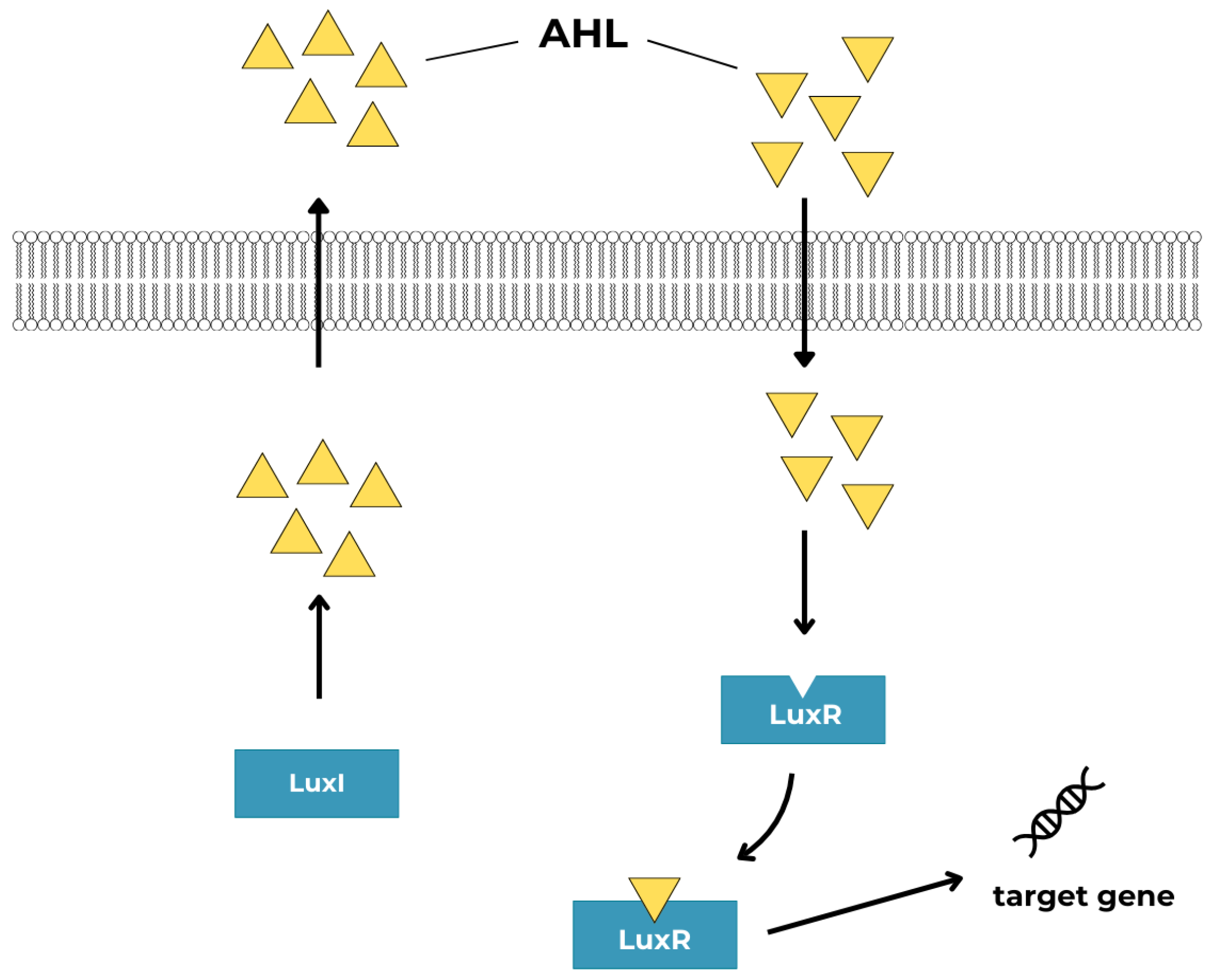
The type I autoinducers (AI-1) are represented by N-Acyl-homoserine lactones (AHLs) first described in the aerobic pathogen, Pseudomonas aeruginosa [50]. AHLs are a family of small diffusible signaling molecules, produced by a range of Gram-negative environmental and pathogenic bacteria [51]. AHLs consist of a hydrophilic homoserine lactone ring (S-adenosylmethionine) and hydrophobic acyl side chain (of varying length; short-chain AHLs with C4–C8 and long-chain AHLs with C10–C20). The degree of hydrophobicity increases with the length of the acyl side chain. The diversity of AHL molecular structures is caused by differences in the R group and the substituent group in the acyl chain [52]. AHLs are synthesized in the cell by acyl-homoserine lactone synthase enzyme (LuxI). Gene luxI is expressed at the basal level at low population density. The concentration of an AHL increases along with the growth of the bacterial cell population. AHL passively diffuses or passes with the support of transport proteins through the cell membrane down a gradient to the environment. When the threshold is reached, the AHL signal returns to the cell and binds to the cognate LuxR receptor. Signaling works through the LuxI/LuxR system [50,52,53,54]. The LuxR/AHL protein complex binds to promoter DNA regions and regulates the transcription of QS-regulated genes. Thus, it can regulate, e.g., carbon, nitrogen, and sulfur metabolism, allowing bacteria to quickly adapt to extreme environmental conditions [52]. Figure 5 shows a schematic presentation of an AHL-mediated QS system.
The production of AHLs can be determined by factors including pH, substrate concentration, and a carbon/nitrogen (C/N) ratio. AHLs have been detected in saliva and sputum samples [51], and in the human gastrointestinal tract [55]. The effect of AHL on infection processes involving anaerobic bacteria like Bacteroides spp. and Porphyromonas spp. has been described so far. Some bacteria, such as Bacteroides spp., have no capacity to synthesize AHLs due to the lack of LuxI homologs. Bacteria that have only LuxR homologs are called LuxR solo or LuxR orphans and, curiously, they can sense AHL from other bacteria present in the biofilm [52,56,57]. B. fragilis can respond to the presence of exogenous homoserine lactones and thereby modulate the bacterial society and regulate gene expression [52]. In the B. fragilis genome, LuxR orthologs (so far nine putative QS gene homologs of luxR) were identified. Pumbwe et al. reported an over-representation of luxR genes (luxR5-luxR9) in B. fragilis cells grown in a medium enriched with AHL (N-hexanoyl homoserine lactone C6-HSL) [58]. According to Grellier et al., the expression of luxR from Bacteroides spp. could be linked to inflammatory bowel disease-associated dysbiosis [59].
Interestingly, AHL regulates its own synthase and receptor genes in a positive feedback loop. This phenomenon has been described in other pathogens (e.g., aerobic bacteria, P. aeruginosa), while there is still a gap in understanding the role of commensal-derived AHL and their impact on the host, in particular on the gut ecosystem [50]. The results demonstrated by Muras and co-workers indicate a potential role of AHLs in the development of dysbiosis related to periodontal diseases. C6-HSL increases the abundance of Alloprevotella, Peptostreptococcus, and Prevotella species in periodontal biofilms. Some members of the Peptostreptococcus (P. micros) and Prevotella (P. intermedia, P. nigrescens) genera are included in, the so-called, orange-complex associated with periodontitis AHLs and seem to influence the growth and protein expression by a key periodontopathogen, P. gingivalis, a member of the red-complex [60,61].
It has been shown that AHLs can specifically affect lactic acid production, disrupt epithelial integrity by activating host cell protease secretion, and reduce the level of proteins responsible for the integrity of the epithelium (occludin and tricellulin). Occludin is one of the factors contributing to the inflammatory process associated with P. gingivalis. This is probably due to the activities of bacterial gingipains (involved in the degradation of cytokines) and epithelial matrix metalloproteinases (MMPs), which degrade components of the basal lamina, epithelial cell-cell junctions, and collagen [62]. Results of the study conducted by the Muras team revealed that P. gingivalis produced a small quantity of octanoyl-L-homoserine lactone OC8-HSL (0.3 ng/mL). The higher concentration of this molecule was observed in a dual-species biofilm formed with Streptococcus gordonii (0.83 ng/mL) or Streptococcus oralis (1.4 ng/mL) [51]. Multispecies biofilms composed of both aerobic and anaerobic bacteria are clinically relevant.
All these data strongly support the importance of AHL in the oral biofilm community. However, more studies are needed to identify the key players in AHL-mediated QS processes in plaque formation. The molecular phenomena of AHLs in biofilm formation processes involving anaerobes have not been thoroughly investigated. Most studies have been conducted with environmental bacteria or a small number of aerobically growing human pathogens. Considering the unquestioned role of anaerobes in the pathophysiology of infections, this gap needs to be filled.

4.2. Autoinducer-2 (AI-2)

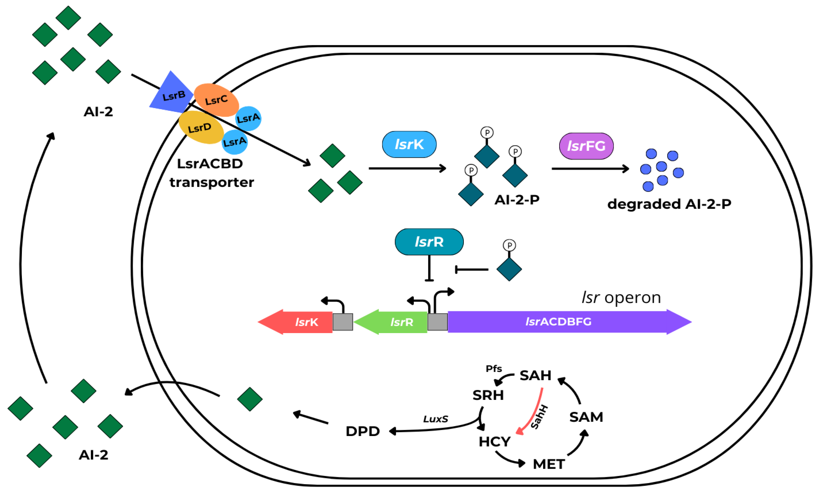
Autoinducer-2 molecules are universal, non-species-specific quorum-sensing signal compounds found in both Gram-negative and Gram-positive bacteria. AI-2’s chemical structure is the same for many bacteria species. AI-2 represents a group of (4S)-4,5-dihydroxy-2,3-pentanedione ([S]-DPD) derivatives produced by a complex process, in turn, involving LuxS (homodimeric metalloenzyme encoded by the gene luxS) [63]. In brief, S-adenosyl-L-methionine (SAM), a substrate for methyl transfers, is biosynthesized from L-methionine and ATP by methionine adenosyltransferase (MAT), the product of the metK gene. S-adenosyl-L-homocysteine (SAH) is produced from SAM through demethylation mediated by methyltransferases (MTases). SAH can be modified through two pathways: either by a single-step conversion to homocysteine (HCY), involving SAH-hydrolase or by a two-step process catalyzed by the Pfs and LuxS enzymes. Nucleosidase Pfs irreversibly cleaves SAH into adenine and SRH. LuxS converts SRH into 4,5-dihydroxy-2,3-pentanedione (DPD) and HCY. DPD undergoes further changes to form the active AI-2 molecule [64]. AI-2s are released from the cells and once AI-2 reaches a critical concentration outside the cell, it is transported back through the membrane channel via the LsrACBD transport system. In the cytoplasm, AI-2 is phosphorylated by bacterial kinase, LsrK. AI-2-P acts as a derepressor of the lsr operon by inactivation of LsrR. The AI-2-P degradation by LsrFG was conducted simultaneously.
In these conditions, the processing of DPD is accelerated, promoting the positive regulation of AI-2 QS [42,46,65]. The AI-2-mediated signaling system is shown in Figure 6.
The synthesis of AI-2 or the presence of the gene luxS has been detected in many anaerobic pathogenic and commensal rods, such as Actinomyces naeslundii, F. nucleatum, P. gingivalis, P. intermedia, C. acnes, Clostridioides difficile, and oral streptococci [46,66,67,68,69].
Although it is extensively studied, luxS-dependent AI-2 signaling remains not entirely understood. In the first decade of the 20th century, the impact of autoinducers on biofilm processes was mainly explored by phenotypic studies with luxS mutant strains [70]. Nowadays, transcriptomic [69,71] and proteomics analyses [67,72] are also used.
Inactivation of the luxS gene influences a variety of processes, such as type III secretion system, cell motility, biofilm formation, production and release of virulence factors, and antibiotic production [46,67,73]. From a clinical point of view, AI-2 may mediate bacterial colonization of the intestinal tract, altering the composition of intestinal bacteria and modulating the host immune response, leading to inflammatory bowel disease. AI-2 participates in the formation of the subgingival biofilm and affects the virulence of periodontal pathogens [46,65,66]. In P. gingivalis, which belongs to the red-complex periodontopathogens, LuxS/AI-2 signaling is involved in the regulation of hemin acquisition and growth under hemin-limited conditions, as well as the expression of proteases and stress-related genes [74,75,76,77,78]. AI-2 plays also a crucial role in the biofilm formation by orange-complex bacteria [79]. AI-2 secreted by F. nucleatum promotes co-aggregation and expression of adhesion molecules in P. gingivalis, T. denticola, and Tannerella forsythia and thereby the formation of mixed biofilms [47,70]. As demonstrated by Kolenbrander et al., the synthesis and activity of AI-2 are lower in commensal bacteria than in periodontal pathogens [80]. The accumulation of AI-2 could serve as evidence or contribute to the transition from a commensal to a pathogenic biofilm [54]. AI-2 synthetase can affect the host’s pro-inflammatory response [47]. Wu and coworkers showed that F. nucleatum AI-2 activates multiple signaling pathways in macrophages. The most significantly upregulated protein is the tumor necrosis factor ligand superfamily member 9 (TNFSF9), which participates in regulating immune cell infiltration in pancreatic adenocarcinoma. Thus, AI-2 may be a novel point of study for the association between bacteria and cancer [72,81]. AI-2 is also involved in signaling among C. difficile cells. C. difficile QS is involved in sporulation and toxin production, as well as activation of the flagella and colonization of the intestinal tract [42]. In a study conducted by Slater et al., a mutant defective in the luxS strain did not produce AI-2 and could not form a biofilm in vitro. In addition, it is hypothesized that C. difficile LuxS/AI-2 may use different mechanisms to mediate the formation of single-species and mixed-species communities [69].
In Clostridium perfringens, the AI-2 system enhances the expression of toxins in the mid-exponential period [82]. AI-2 is used by C. acnes to the regulation of virulence of biofilms and has an impact on the stimulation of the immune system and inflammation [83].
Table 1 shows the best-understood processes regulated by AI-2 in biofilm formed by anaerobic bacteria.

4.3. Autoinducing Peptides (AIPs)

Autoinducing peptides (AIPs), are cyclic oligopeptides that contain a thiolactone ring formed by condensation of the C-terminal carboxyl group and the sulfhydryl group of an internal cysteine. AIPs are synthesized in the form of pre-peptides which, after posttranslational modification, are released from the cell via the ABC-type transport system. After reaching the threshold concentration in the environment, the AIP molecules are bound by sensor proteins with kinase activity. The kinase is activated by a series of auto-phosphorylation steps followed by the transfer of the phosphate group to a response regulator which can act as a transcriptional regulator of target genes [91]. The best understood example of AIP-mediated QS is the accessory gene regulator (Agr) QS system in Staphylococcus aureus [92], which is also present in other Gram-positive bacteria. The autoinducing peptide-based Agr system has been evidenced in some anaerobic rods, as well [39,93,94]. The prototypical S. aureus Agr system is composed of a gene operon (agrACDB), two promoters P2 and P3, and RNAIII (Figure 7) agrACDB encoding proteins:
  • cytosolic response regulator acts as a transcription factor (AgrA),
  • histidine kinase cell surface receptor (AgrC),
  • ~45 residue pro-peptide precursor of the mature AIP (AgrD),
  • transmembrane cysteine endopeptidase (AgrB) [39].
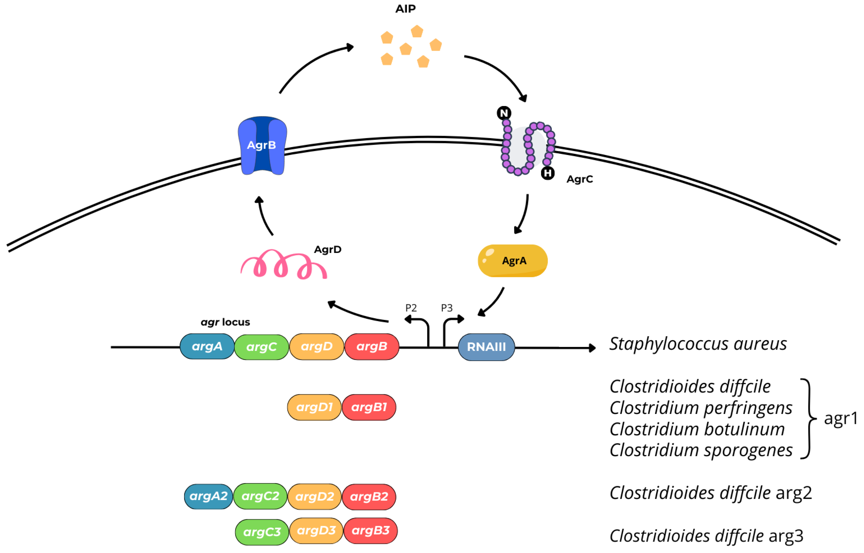
Figure 7. Genetic structure and organization of AIP-mediated signaling Agr system in S. aureus, C. difficile, and Clostridium spp.
Ijms 25 12808 g007
The Agr system in S. aureus plays a role in the cross-regulation of various proteins, such as immunoglobulin and fibronectin-binding proteins; exotoxins, including hemolysins and enterotoxins; leukocidins; and toxic shock syndrome toxin (TSST). It also regulates enzyme production and promotes the production of phenol soluble modulins (PSMs), amphipathic peptides exhibiting cytolytic activity similar to delta-toxin that participate in many infection-related processes, including the host cells cytolysis and the biofilms dissemination [95,96]. In vitro study conducted by Podkowik et al. demonstrated that the Agr system in S. aureus provides long-lived protection from the lethal activity of exogenous H2O2. The researchers noted that Agr systems found in other bacterial species differ from the S. aureus, albeit the relationship between Agr and pathogenesis in most organisms remains unclear [97]. Among anaerobes, Agr systems have been described in C. difficile, C. perfringens, Clostridium botulinum, and Clostridium tetani.
Virulence factors for C. difficile include sporulation, toxins (enterotoxin A; TcdA, and cytotoxin; TcdB) production, enzymes (collagenase, hyaluronidase, chondroitin sulfatase) releasing, and ability to biofilm formation. The capacity to form biofilms is a crucial factor linked with recurrent C. difficile infection (R-CDI) [86,98]. To date, three Agr systems—Agr1, Agr2, and Agr3—have been identified in C. difficile, each of which has a different type of gene organization [86]. C. difficile Agr2 is similar to S. aureus, with genes (agrACDB) in reverse order to those found in S. aureus [39]. Agr2 is a regulator of flagella formation (regulates bacterial flagella gene expression fliC, fliA/sidD, fliM), and TcdA production.
Agr1 system includes the genes for AgrB1 protease and AgrD1 peptide, only. The absence of AgrC1 and AgrA1 suggests that the receptor histidine kinase and transcription factor are encoded in other parts of the genome, or that Agr1 functions in a way that is not strictly dependent on these proteins [39,99]. AgrA or AgrC orthologues forming a two-component signal transduction system are not found in C. perfringens, C. botulinum, and Clostridium sporogenes, as well [39,100]. In the case of C. difficile, Agr1 acts as an AIP signaling system in sporulation and plays a role in toxins production, which was confirmed in vivo [101].
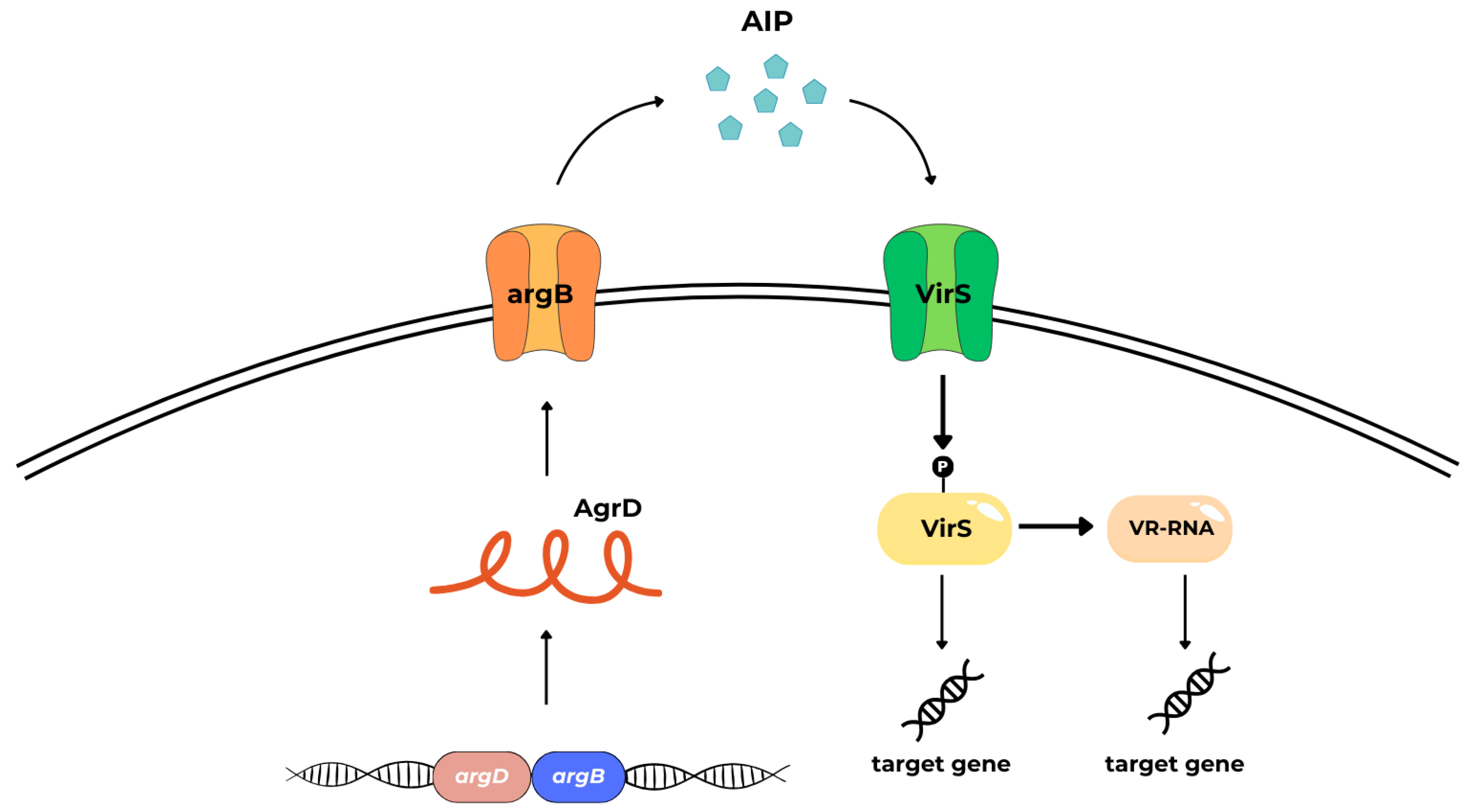
Agr3 system consists of AgrB3, AgrD3, and AgrC3 and appears to be encoded by a C. difficile bacteriophage (phiCDHM1), suggesting that may be highly mobile and transferred between different C. difficile strains. Its function and regulatory mechanisms are poorly understood [39,102]. In C. difficile and C. perfringens, two functional QS systems are described, the aforementioned LuxS-dependent AI-2 system, which involves the LuxS enzyme, and Agr-like systems [69,82,86,103]. The pathogenicity of C. perfringens, a bacterium that causes histotoxicity and enteritis in humans and other mammals, is linked to its ability to produce toxins (more than 20) and extracellular enzymes. Considering the set of toxins produced by C. perfringens, seven toxinotypes (from A to G) have been distinguished [104]. In C. perfringens, toxin production is controlled by the two-component VirS (membrane sensor protein histidine kinase)/VirR (a response regulator) signal transduction system (TCSTS) and the Agr system. The VirS/VirR system may function similarly to AgrA/AgrC (Figure 8) [105].
The VirS/VirR-VR-RNA cascade controls the pathogenesis by positively regulating virulence related and involved in energy metabolism genes. The study by Mehdizadeh Gohari et al. demonstrates that the VirS protein is a receptor for the AgrD-derived signaling peptide (SP) and that the second extracellular loop of VirS is critical for SP binding. VirS/R is activated by a signal that induces autophosphorylation of VirS, followed by phosphotransfer to VirR. Phosphorylated VirR directly regulates the expression of netB (encoding necrotic enteritis B-like toxin, NetB) and pfoA (encoding the perfringolysin O, PFO; theta-toxin). In addition, VirS/VirR TCSTS indirectly regulates cpb and cpa gene expression and thus controls the C. perfringens alpha (CPA) and beta toxin (CPB) indirectly by activating the production of a small regulatory RNA called VR-RNA. The VirS is proposed to be the signaling peptide receptor for the C. perfringens Agr-like QS system [104,106,107,108].
The Agr-like QS system, which encodes the AgrB and AgrD, is also a virulence regulator because it positively regulates the production of PFO, CPA, CPB, and NetB, sporulation, and C. perfringens enterotoxin (CPE) production (toxinotype F) [100,103,105,109,110]. Seven immunologically distinct botulin neurotoxins (BoNTs) designated by the letters from A to G and more than forty different subtypes are produced by six phylogenetically distinct clostridia (C. botulinum groups I–IV and some strains of C. baratii and C. butyricum). Toxigenic strains are associated with severe flaccid paralysis in vertebrates. Pathogenicity of C. botulinum is controlled by the QS system; however, regulation of botulinum neurotoxin gene (bont) expression and BoNT production are not fully understood [111]. The Agr system (Agr-1/Agr-2) has been evidenced in C. botulinum strains classified into group I. Agr-1 appears to be involved in sporulation, while Agr-2 is in neurotoxin production. Agr-1 and Agr-2 are homologs of AgrB, and AgrD proteins in C. perfringens [98].

5. Prevention of Biofilm Formation by Quorum Quenching (QQ) and Perspectives of QQ Application

In this review, we discuss the QS mechanisms that have been described so far in anaerobic bacteria to present potential solutions affecting the possibility of QS disruption. The process of interfering with microbial cell-to-cell communication is referred to as quorum quenching. QQ inhibition can be achieved in a variety of ways. Proposed QQ strategies in anaerobic bacteria include the following:
  • inactivation or enzymatic degradation of signaling molecules.
  • competition with signaling molecules; competing with inducers for the same binding site or binding the receptor noncompetitively.
  • blocking of signal transduction cascades.
The best studied QQ mechanism is signal degradation by enzymatic pathways or by means of analogs of signal molecules.

5.1. Inhibitors of AHL-Mediated Quorum Sensing

The chemical structure of AHLs suggests that the degradation of these molecules may be mediated by lactonases, acylases, decarboxylases, deaminases, and oxidoreductases [54].
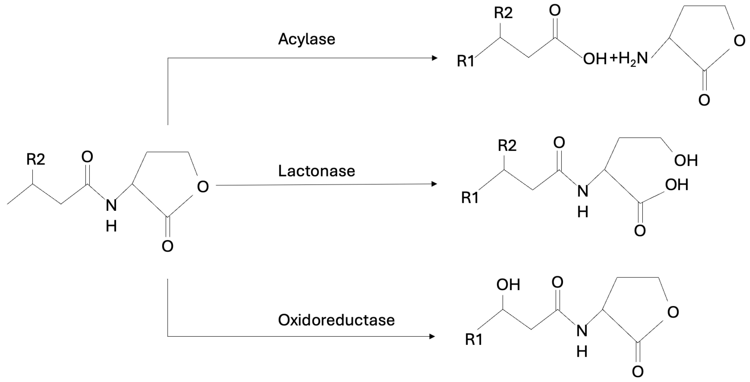
The most extensively studied groups of AHL-degrading enzymes are N-acyl-homoserine lactonases (AHL-lactonases) and N-acyl-homoserine lactone acylase (AHL-acylases). AHL-lactonases degrade AHL by hydrolyzing the lactone ring in the homoserine moiety of AHLs, while AHL-acylases hydrolyze the amide bond between the acyl side chain and the homoserine lactone in the AHL molecules producing the free fatty acid and the homoserine lactone [51,112]. One of the first described and best analyzed AHL lactonases is AiiA24B1, the enzyme produced by Bacillus sp. 24B1 aiiA gene [113]. Kinetic and substrate specificity analyses showed that AHL-lactonase had little or no residual activity towards non-acyl lactones and non-cyclic esters but showed strong enzyme activity towards many of the AHLs evaluated, regardless of the length and type of substitution at the C3 position of the acyl chain. AHL lactonases are expressed in most pathogens [113]. In the study conducted by Muras et al., the AHL-lactonase Aii20J caused a significant reduction in oral biofilms growing under aerobic and anaerobic conditions in vitro. Confocal microscopy analysis of in vitro multi-species oral biofilms formed by A. naeslundii, A. actinomycetemcomitans, F. nucleatum, P. gingivalis, S. oralis, and Veillonella parvula revealed a significant inhibition of biofilms formation when Aii20J was added to the culture media [51]. Parga and co-workers showed that Aii20J modulates polymicrobial biofilm formation without altering the microbiome structure of the biofilm [114]. Murugayah’s team using a fluorescamine-based assay demonstrated that AHL-acylase can degrade quorum-sensing molecules [51,61]. AHL acylase activity has been identified in several bacterial species. Their substrate specificity is based on the different acyl chain substitutions of AHLs. It has been proposed that acylases degrade AHL with a long side chain more than those with a short side chain [115].
There are also several known AHL oxidoreductases found in different bacteria that can reduce or oxidize the acyl chain of AHL, thereby inhibiting the specific binding of the autoinducer to its receptor. AHL oxidoreductases are modifying enzymes classified into two groups: reductases, which can convert 3-oxo-substituted AHLs to 3-hydroxyl AHLs, and cytochrome oxidases, which catalyze the oxidation of the acyl chain. The modified compounds can no longer function as signaling molecules without being degraded [115]. It has been evidenced that some bacteria produce substantial amounts of AHL-lactonase, acylase, or oxidoreductase enzymes. The enzymatic pathway of AHL-inhibitors is shown in Figure 9. Enzymatic QQ is the best studied QS inhibition strategy and is an interesting alternative to the problem of bacterial resistance to antibiotics. The potential of these enzymes depends on the quantity of other enzymes, their level of activity, biotechnological possibilities of synthesis, and stability [116].
Another strategy relevant to QQ is to inhibit the synthesis of the AHL signaling molecule through the use of signaling molecule analogs [62]. AHL-analogs modify not only the protein expression but also slow down the growth of P. gingivalis. This finding potentially opens new perspectives for the prevention or treatment of periodontal disease [51]. Blocking the LuxR/AHL interaction is most often caused by molecules that are AHL antagonists competing with signaling molecules for a binding site on the receptor [117].

5.2. Inhibitors of AI-2-Mediated Quorum Sensing

The quenching of signal molecule activity can be achieved by AI-2 quorum-sensing inhibitors (QSIs), which include AI-2 analogs. AI-2 mediates both intra- and inter-species communication and is considered notably a potential target for the control of periodontal diseases. It has been shown that monosaccharides, including D-ribose and D-galactose, reduce virulence gene expression and biofilm formation by blocking the AI-2 receptor, hence they could potentially be used for the prevention of the biofilm formation of periodontopathogens [54,65,70,79].
Ribose, as furanosyl borate diether, is structurally similar to AI-2 and has minimal toxic side effects. D-galactose significantly reduced the biofilm formation of F. nucleatum, P. gingivalis, and T. forsythia induced by AI-2 of F. nucleatum [65]. An et al. demonstrated that D-arabinose significantly reduces AI-2 activity and biofilm formation of oral bacteria (S. oralis, F. nucleatum, and P. gingivalis) on titanium discs. The researchers suggested that L-arabinose has initial anti-adhesive activity, as well [79].
Coumarins isolated from Coumarouna odorata (heterocycles, consisting of a benzene ring linked to a pyrone ring) are a group of compounds with potential antimicrobial utility as well. Coumarins are a class of bioactive compounds present in many plant sources, including beans, sweet clover, cinnamon oil, and lavender products. Their antiviral, antimicrobial, anti-inflammatory, antitumor, antioxidant, and anticoagulant activity are well known [13,118]. It has been shown that the incorporation of another heterocyclic group into the coumarin molecule can enrich the biological properties of the matrix compound. To date, coumarin derivatives have been reported to inhibit biofilm formation in S. aureus, Escherichia coli, and Chromobacterium violaceum [119]. He et al. demonstrated that coumarin at sub-MIC concentrations, without affecting bacterial growth, inhibited P. gingivalis biofilm formation. Anti-biofilm effects for the late-stage and pre-formed biofilm dispersion were also documented. After coumarin treatment, the biofilms became interspersed. Coumarin inhibited P. gingivalis biofilm formation through a QS system by interacting with the heme-binding protein (HmuY) that plays a leading role in P. gingivalis heme acquisition [13].
The study conducted by Marquis et al. confirmed that the growth of P. gingivalis was inhibited when exposed to the lacinartin compound, natural oxyprenylated coumarin present in the culture medium. Lacinartin also inhibited P. gingivalis biofilm formation, enhanced biofilm detachment, prevented P. gingivalis adherence to oral epithelial cells, inhibited P. gingivalis collagenase activity, reduced the secretion of cytokines (IL-8 and TNF-α) and matrix metalloproteinases (MMP-8 and MMP-9) by LPS-stimulated macrophages, and inhibited MMP-9 activity [120]. The molecular mechanism of lacinartin has not been explained.
Produced by the macroalga Delisea pulchra, brominated furanones are reported to inactivate (by covalently modifying) the LuxS enzyme, required for AI-2 synthesis, thereby inhibiting the QS activity of various bacterial species. The bromofuranone analog, 3-(dibromomethylene) isobenzofuran-1(3H)-one derivative demonstrated inhibitory activities against biofilm formation by periodontopathogens (F. nucleatum, P. gingivalis, and T. forsythia) without a bactericidal effect [70,121].
Heparinoids (glycosaminoglycans) are chemically and pharmacologically related to heparin, known for its anticoagulant activity. Heparinoids suppress C. acnes biofilm formation via AI-2 inhibition. The study conducted by Hamada et al. showed that heparinoids at low concentrations may lead to decreased lipase activity that is associated with irritation and inflammation specific to acne. The authors also demonstrated that heparinoids enhance isopropyl methylphenol (IPMP) bactericidal efficacy against C. acnes biofilms [122].
Phlorizin and phloretin polyphenols are commonly found in many types of human diets and possess anti-biofilm properties against P. gingivalis. Phloretin is a chalcone flavonoid naturally occurring in apple fruit, bark, and leaves. Phlorizin, a derivative of phloretin, has an additional glycoside in its structure [123].
Phloretin can inhibit the biofilm formation of the oral pathogen Streptococcus mutans and has the capability to suppress its QS by inhibition of glucosyltransferases GtfB and GtfC (enzymes that split sucrose into glucose and fructose and link the glucose moiety together via glycosidic bonds to form EPSs) [124]. In S. mutans, the signaling transduction system (VicRK) positively regulates the expression of gtfB/C genes by binding their promoter regions [125]. Genes encoding P. gingivalis glycosyltransferases involved in O-LPS and A-LPS biosynthesis, named gtfC, gtfD, gtfE, and gtfF, are identified by Shoji et al. [124]. Scanning electron microscopy showed that phloretin and phlorizin displayed a similar and remarkable destructive effect on P. gingivalis and the mixed biofilms [123]. This aligns with findings from transcriptome analysis conducted by Wu et al. confirmed that phlorizin and phloretin reduced AI-2 activity to 45.9% and 55.4%, which means that they can interfere with P. gingivalis’ intercellular communication. Furthermore, while other flavonoids with similar structures, such as naringenin, have been reported to inhibit the growth of P. gingivalis, F. nucleatum, and S. mitis, there are no studies specifically addressing their effects on biofilm inhibition [123].
Xu and co-workers have shown that reuterin (isolated from Lactobacillus reuteri LR 21) significantly suppressed the biofilm formation of C. perfringens. As mentioned earlier, toxin production and pathogenicity of C. perfringens depended on the Agr and LuxS quorum-sensing system. The authors hypothesized that the downregulation of agrB and luxS in C. perfringens treated with reuterin appears to be responsible for the decreased expression levels of cpa and pfo genes [126].

5.3. Inhibitors of Agr-like Quorum Sensing

It has been shown that in C. perfringens, an Agr-like QS system through a VirS/R two-component signal transduction system regulates the expression of virulence genes. Based on the structure-activity relationship (SAR) data on 5-residue thiolactone peptide AIP, two inhibitory peptides designed to target VirS, the receptor histidine kinase AIP, were found to attenuate Agr-mediated toxin production. One of them is a partial agonist (Z-AIPCp-L2A/T5A), the second is a partial antagonist (Z-AIPCp-F4A/T5S). To determine the agonist/antagonist activity of synthetic peptides, a transcriptional response was monitored by quantifying the level of pfoA after C. perfringens incubation with the tested peptides. In a virulent strain, both peptides significantly attenuated the transcription of the theta-toxin gene (pfoA) [127].
Inhibitors that have the potential to disrupt QS in anaerobic bacteria are demonstrated in Table 2.

6. Summary, Constraints, and Prospects

Biofilms are a critical survival strategy for pathogenic bacteria, serving as both non-specific virulence factors and non-specific mechanisms of antibiotic resistance. Quorum sensing, as a type of microbial cell-to-cell communication system, plays a key role in the biofilm formation process at every stage of development. The phenomenon of QS was discovered over five decades ago with the observation that bioluminescence in the marine bacterium Photobacterium fischeri only occurred at increased cell densities.
In anaerobic bacteria, QS rely on certain molecules known as autoinducers, such as AHLs (AI-1), AI-2s, or AIPs. Among these, the AI-2-dependent mechanism seems to be particularly important for this group of microorganisms. Several periodontal bacteria, such as P. gingivalis, T. denticola, P. intermedia, F. nucleatum, and C. difficile, associated with post-antibiotic diarrhea, have been studied for their QS capabilities, making them potential targets for quorum quenching strategies [46,86].
The quorum-quenching strategy may be a promising target for the development of a novel non-antibiotic therapy. Despite its evident potential, several challenges remain. The effectiveness of QQ strategies can vary significantly due to the diversity of microbial communities and the complexity of biofilm structures. Future research should focus on understanding the mechanisms of QQ in different environments, exploring its synergy with other antimicrobial strategies, and developing targeted QQ applications. Ongoing advances in biotechnology may pave the way for innovative solutions to biofilm-related problems, making QQ an exciting area of study in microbial ecology and applied sciences.
In conclusion, quorum quenching represents a novel approach to preventing biofilm formation. By disrupting bacterial communication, QQ strategies could transform the way we tackle the challenges posed by biofilms in medicine and across many other industries, leading to progress towards safer medical practices, better food preservation, and maintaining a cleaner environment.

Author Contributions

Conceptualization A.M. and K.M.; methodology A.M. and K.M.; validation A.M. and K.S.-M.; formal analysis H.P., A.M. and K.S.-M.; investigation K.M., A.M. and K.S.-M.; resources A.M. and K.M.; data curation K.M., K.S.-M. and A.M.; writing—original draft preparation K.M. and A.M.; writing—review and editing K.S.-M.; visualization K.M.; supervision H.P. and K.S.-M., project administration A.M.; funding acquisition A.M. All authors have read and agreed to the published version of the manuscript.

Funding

This research received no external funding.

Conflicts of Interest

The authors declare no conflicts of interest.

References

  1. Jamal, M.; Ahmad, W.; Andleeb, S.; Jalil, F.; Imran, M.; Nawaz, M.A.; Hussain, T.; Ali, M.; Rafiq, M.; Kamil, M.A. Bacterial biofilm and associated infections. J. Chin. Med. Assoc. 2018, 81, 7–11. [Google Scholar] [CrossRef] [PubMed]
  2. Hoiby, N. A short history of microbial biofilms and biofilm infections. APMIS 2017, 125, 272–275. [Google Scholar] [CrossRef] [PubMed]
  3. Harris, D.F. Anthony Van Leeuwenhoek, the First Bacteriologist. Sci. Mon. 1921, 12, 150–160. [Google Scholar]
  4. Lam, J.; Chan, R.; Lam, K.; Costerton, J.W. Production of mucoid microcolonies by Pseudomonas aeruginosa within infected lungs in cystic fibrosis. Infect. Immun. 1980, 28, 546–556. [Google Scholar] [CrossRef] [PubMed]
  5. Hoiby, N. A personal history of research on microbial biofilms and biofilm infections. Pathog. Dis. 2014, 70, 205–211. [Google Scholar] [CrossRef]
  6. Jendresen, M.D.; Glantz, P.O.; Baier, R.E.; Eick, J.D. Microtopography and clinical adhesiveness of an acid etched tooth surface. An in-vivo study. Acta Odontol. Scand. 1981, 39, 47–53. [Google Scholar] [CrossRef]
  7. Majewska, A.; Kierzkowska, M.; Kawecki, D. What we actually know about the pathogenicity of Bacteroides pyogenes. Med. Microbiol. Immunol. 2021, 210, 157–163. [Google Scholar] [CrossRef]
  8. Kierzkowska, M.; Majewska, A.; Kuthan, R.T.; Sawicka-Grzelak, A.; Mlynarczyk, G. A comparison of Api 20A vs MALDI-TOF MS for routine identification of clinically significant anaerobic bacterial strains to the species level. J. Microbiol. Methods 2013, 92, 209–212. [Google Scholar] [CrossRef] [PubMed]
  9. Nagy, E.; Boyanova, L.; Justesen, U.S.; ESCMID Study Group of Anaerobic Infections. How to isolate, identify and determine antimicrobial susceptibility of anaerobic bacteria in routine laboratories. Clin. Microbiol. Infect. 2018, 24, 1139–1148. [Google Scholar] [CrossRef]
  10. Percival, S.L.; Malone, M.; Mayer, D.; Salisbury, A.M.; Schultz, G. Role of anaerobes in polymicrobial communities and biofilms complicating diabetic foot ulcers. Int. Wound J. 2018, 15, 776–782. [Google Scholar] [CrossRef]
  11. Larsen, T.; Fiehn, N.E. Dental biofilm infections—An update. APMIS 2017, 125, 376–384. [Google Scholar] [CrossRef]
  12. Vestby, L.K.; Gronseth, T.; Simm, R.; Nesse, L.L. Bacterial Biofilm and its Role in the Pathogenesis of Disease. Antibiotics 2020, 9, 59. [Google Scholar] [CrossRef] [PubMed]
  13. He, Z.; Jiang, W.; Jiang, Y.; Dong, J.; Song, Z.; Xu, J.; Zhou, W. Anti-biofilm activities of coumarin as quorum sensing inhibitor for Porphyromonas gingivalis. J. Oral. Microbiol. 2022, 14, 2055523. [Google Scholar] [CrossRef]
  14. Huang, R.; Li, M.; Gregory, R.L. Bacterial interactions in dental biofilm. Virulence 2011, 2, 435–444. [Google Scholar] [CrossRef] [PubMed]
  15. Minarovits, J. Anaerobic bacterial communities associated with oral carcinoma: Intratumoral, surface-biofilm and salivary microbiota. Anaerobe 2021, 68, 102300. [Google Scholar] [CrossRef] [PubMed]
  16. Zhang, L.; Liu, Y.; Zheng, H.J.; Zhang, C.P. The Oral Microbiota May Have Influence on Oral Cancer. Front. Cell. Infect. Microbiol. 2019, 9, 476. [Google Scholar] [CrossRef] [PubMed]
  17. Binder Gallimidi, A.; Fischman, S.; Revach, B.; Bulvik, R.; Maliutina, A.; Rubinstein, A.M.; Nussbaum, G.; Elkin, M. Periodontal pathogens Porphyromonas gingivalis and Fusobacterium nucleatum promote tumor progression in an oral-specific chemical carcinogenesis model. Oncotarget 2015, 6, 22613–22623. [Google Scholar] [CrossRef] [PubMed]
  18. Urban, E.; Gajdacs, M.; Torkos, A. The incidence of anaerobic bacteria in adult patients with chronic sinusitis: A prospective, single-centre microbiological study. Eur. J. Microbiol. Immunol. (Bp) 2020, 10, 107–114. [Google Scholar] [CrossRef]
  19. Alon-Maimon, T.; Mandelboim, O.; Bachrach, G. Fusobacterium nucleatum and cancer. Periodontol. 2000 2022, 89, 166–180. [Google Scholar] [CrossRef] [PubMed]
  20. Brook, I. Microbiology and management of joint and bone infections due to anaerobic bacteria. J. Orthop. Sci. 2008, 13, 160–169. [Google Scholar] [CrossRef]
  21. Varin-Simon, J.; Colin, M.; Velard, F.; Tang-Fichaux, M.; Ohl, X.; Mongaret, C.; Gangloff, S.C.; Reffuveille, F. Cutibacterium acnes biofilm formation is influenced by bone microenvironment, implant surfaces and bacterial internalization. BMC Microbiol. 2024, 24, 270. [Google Scholar] [CrossRef] [PubMed]
  22. Lewis, R.P.; Sutter, V.L.; Finegold, S.M. Bone infections involving anaerobic bacteria. Medicine 1978, 57, 279–305. [Google Scholar] [CrossRef] [PubMed]
  23. Haggerty, C.L.; Hillier, S.L.; Bass, D.C.; Ness, R.B.; Evaluation, P.I.D.; Clinical Health study, i. Bacterial vaginosis and anaerobic bacteria are associated with endometritis. Clin. Infect. Dis. 2004, 39, 990–995. [Google Scholar] [CrossRef]
  24. Wiesenfeld, H.C.; Meyn, L.A.; Darville, T.; Macio, I.S.; Hillier, S.L. A Randomized Controlled Trial of Ceftriaxone and Doxycycline, With or Without Metronidazole, for the Treatment of Acute Pelvic Inflammatory Disease. Clin. Infect. Dis. 2021, 72, 1181–1189. [Google Scholar] [CrossRef] [PubMed]
  25. Zamani, S.; Hesam Shariati, S.; Zali, M.R.; Asadzadeh Aghdaei, H.; Sarabi Asiabar, A.; Bokaie, S.; Nomanpour, B.; Sechi, L.A.; Feizabadi, M.M. Detection of enterotoxigenic Bacteroides fragilis in patients with ulcerative colitis. Gut Pathog. 2017, 9, 53. [Google Scholar] [CrossRef]
  26. Nomura, K.; Ishikawa, D.; Okahara, K.; Ito, S.; Haga, K.; Takahashi, M.; Arakawa, A.; Shibuya, T.; Osada, T.; Kuwahara-Arai, K.; et al. Bacteroidetes Species Are Correlated with Disease Activity in Ulcerative Colitis. J. Clin. Med. 2021, 10, 1749. [Google Scholar] [CrossRef] [PubMed]
  27. Caldara, M.; Belgiovine, C.; Secchi, E.; Rusconi, R. Environmental, Microbiological, and Immunological Features of Bacterial Biofilms Associated with Implanted Medical Devices. Clin. Microbiol. Rev. 2022, 35, e0022120. [Google Scholar] [CrossRef] [PubMed]
  28. Lin, Z.X.; Steed, L.L.; Marculescu, C.E.; Slone, H.S.; Woolf, S.K. Cutibacterium acnes Infection in Orthopedics: Microbiology, Clinical Findings, Diagnostic Strategies, and Management. Orthopedics 2020, 43, 52–61. [Google Scholar] [CrossRef]
  29. Mirzaei, R.; Mohammadzadeh, R.; Alikhani, M.Y.; Shokri Moghadam, M.; Karampoor, S.; Kazemi, S.; Barfipoursalar, A.; Yousefimashouf, R. The biofilm-associated bacterial infections unrelated to indwelling devices. IUBMB Life 2020, 72, 1271–1285. [Google Scholar] [CrossRef]
  30. Kadirvelu, L.; Sivaramalingam, S.S.; Jothivel, D.; Chithiraiselvan, D.D.; Karaiyagowder Govindarajan, D.; Kandaswamy, K. A review on antimicrobial strategies in mitigating biofilm-associated infections on medical implants. Curr. Res. Microb. Sci. 2024, 6, 100231. [Google Scholar] [CrossRef]
  31. Hayward, C.; Brown, M.H.; Whiley, H. Hospital water as the source of healthcare-associated infection and antimicrobial-resistant organisms. Curr. Opin. Infect. Dis. 2022, 35, 339–345. [Google Scholar] [CrossRef] [PubMed]
  32. Schulze, A.; Mitterer, F.; Pombo, J.P.; Schild, S. Biofilms by bacterial human pathogens: Clinical relevance—Development, composition and regulation—Therapeutical strategies. Microb. Cell 2021, 8, 28–56. [Google Scholar] [CrossRef] [PubMed]
  33. Achinas, S.; Charalampogiannis, N.; Euverink, G.J.W. A Brief Recap of Microbial Adhesion and Biofilms. Appl. Sci. 2019, 9, 2801. [Google Scholar] [CrossRef]
  34. Muhammad, M.H.; Idris, A.L.; Fan, X.; Guo, Y.; Yu, Y.; Jin, X.; Qiu, J.; Guan, X.; Huang, T. Beyond Risk: Bacterial Biofilms and Their Regulating Approaches. Front. Microbiol. 2020, 11, 928. [Google Scholar] [CrossRef] [PubMed]
  35. Enigk, K.; Jentsch, H.; Rodloff, A.C.; Eschrich, K.; Stingu, C.S. Activity of five antimicrobial peptides against periodontal as well as non-periodontal pathogenic strains. J. Oral. Microbiol. 2020, 12, 1829405. [Google Scholar] [CrossRef]
  36. Krzyściak, W.; Jurczak, A.; Piątkowski, J. The Role of Human Oral Microbiome in Dental Biofilm Formation. In Microbial Biofilms; Dharumadurai, D., Nooruddin, T., Eds.; IntechOpen: Rijeka, Croatia, 2016; Chapter 16; pp. 329–382. [Google Scholar]
  37. Acemel, R.D.; Govantes, F.; Cuetos, A. Computer simulation study of early bacterial biofilm development. Sci. Rep. 2018, 8, 5340. [Google Scholar] [CrossRef]
  38. Okuda, K.I.; Nagahori, R.; Yamada, S.; Sugimoto, S.; Sato, C.; Sato, M.; Iwase, T.; Hashimoto, K.; Mizunoe, Y. The Composition and Structure of Biofilms Developed by Propionibacterium acnes Isolated from Cardiac Pacemaker Devices. Front. Microbiol. 2018, 9, 182. [Google Scholar] [CrossRef] [PubMed]
  39. Ahmed, U.K.B.; Ballard, J.D. Autoinducing peptide-based quorum signaling systems in Clostridioides difficile. Curr. Opin. Microbiol. 2022, 65, 81–86. [Google Scholar] [CrossRef]
  40. Muras, A.; Mallo, N.; Otero-Casal, P.; Pose-Rodriguez, J.M.; Otero, A. Quorum sensing systems as a new target to prevent biofilm-related oral diseases. Oral. Dis. 2022, 28, 307–313. [Google Scholar] [CrossRef]
  41. Abisado, R.G.; Benomar, S.; Klaus, J.R.; Dandekar, A.A.; Chandler, J.R. Bacterial Quorum Sensing and Microbial Community Interactions. mBio 2018, 9, e02331-17. [Google Scholar] [CrossRef]
  42. Gunaratnam, S.; Millette, M.; McFarland, L.V.; DuPont, H.L.; Lacroix, M. Potential role of probiotics in reducing Clostridioides difficile virulence: Interference with quorum sensing systems. Microb. Pathog. 2021, 153, 104798. [Google Scholar] [CrossRef] [PubMed]
  43. Papenfort, K.; Bassler, B.L. Quorum sensing signal-response systems in Gram-negative bacteria. Nat. Rev. Microbiol. 2016, 14, 576–588. [Google Scholar] [CrossRef] [PubMed]
  44. Fteita, D.; Kononen, E.; Gursoy, M.; Ma, X.; Sintim, H.O.; Gursoy, U.K. Quorum sensing molecules regulate epithelial cytokine response and biofilm-related virulence of three Prevotella species. Anaerobe 2018, 54, 128–135. [Google Scholar] [CrossRef] [PubMed]
  45. Paluch, E.; Rewak-Soroczynska, J.; Jedrusik, I.; Mazurkiewicz, E.; Jermakow, K. Prevention of biofilm formation by quorum quenching. Appl. Microbiol. Biotechnol. 2020, 104, 1871–1881. [Google Scholar] [CrossRef] [PubMed]
  46. Wright, P.P.; Ramachandra, S.S. Quorum Sensing and Quorum Quenching with a Focus on Cariogenic and Periodontopathic Oral Biofilms. Microorganisms 2022, 10, 1783. [Google Scholar] [CrossRef] [PubMed]
  47. Scheres, N.; Lamont, R.J.; Crielaard, W.; Krom, B.P. LuxS signaling in Porphyromonas gingivalis-host interactions. Anaerobe 2015, 35, 3–9. [Google Scholar] [CrossRef] [PubMed]
  48. Sintim, H.O.; Smith, J.A.; Wang, J.; Nakayama, S.; Yan, L. Paradigm shift in discovering next-generation anti-infective agents: Targeting quorum sensing, c-di-GMP signaling and biofilm formation in bacteria with small molecules. Future Med. Chem. 2010, 2, 1005–1035. [Google Scholar] [CrossRef]
  49. Kim, S.; Chen, J.; Cheng, T.; Gindulyte, A.; He, J.; He, S.; Li, Q.; Shoemaker, B.A.; Thiessen, P.A.; Yu, B.; et al. PubChem 2023 update. Nucleic Acids Res. 2023, 51, D1373–D1380. [Google Scholar] [CrossRef]
  50. Coquant, G.; Grill, J.P.; Seksik, P. Impact of N-Acyl-Homoserine Lactones, Quorum Sensing Molecules, on Gut Immunity. Front. Immunol. 2020, 11, 1827. [Google Scholar] [CrossRef]
  51. Muras, A.; Otero-Casal, P.; Blanc, V.; Otero, A. Acyl homoserine lactone-mediated quorum sensing in the oral cavity: A paradigm revisited. Sci. Rep. 2020, 10, 9800. [Google Scholar] [CrossRef]
  52. Liu, L.; Zeng, X.; Zheng, J.; Zou, Y.; Qiu, S.; Dai, Y. AHL-mediated quorum sensing to regulate bacterial substance and energy metabolism: A review. Microbiol. Res. 2022, 262, 127102. [Google Scholar] [CrossRef] [PubMed]
  53. Wang, Y.; Bian, Z.; Wang, Y. Biofilm formation and inhibition mediated by bacterial quorum sensing. Appl. Microbiol. Biotechnol. 2022, 106, 6365–6381. [Google Scholar] [CrossRef]
  54. Hernandez, P.; Sanchez, M.C.; Llama-Palacios, A.; Ciudad, M.J.; Collado, L. Strategies to Combat Caries by Maintaining the Integrity of Biofilm and Homeostasis during the Rapid Phase of Supragingival Plaque Formation. Antibiotics 2022, 11, 880. [Google Scholar] [CrossRef] [PubMed]
  55. Landman, C.; Grill, J.P.; Mallet, J.M.; Marteau, P.; Humbert, L.; Le Balc’h, E.; Maubert, M.A.; Perez, K.; Chaara, W.; Brot, L.; et al. Inter-kingdom effect on epithelial cells of the N-Acyl homoserine lactone 3-oxo-C12:2, a major quorum-sensing molecule from gut microbiota. PLoS ONE 2018, 13, e0202587. [Google Scholar] [CrossRef]
  56. Srinivasan, R.; Santhakumari, S.; Poonguzhali, P.; Geetha, M.; Dyavaiah, M.; Xiangmin, L. Bacterial Biofilm Inhibition: A Focused Review on Recent Therapeutic Strategies for Combating the Biofilm Mediated Infections. Front. Microbiol. 2021, 12, 676458. [Google Scholar] [CrossRef] [PubMed]
  57. Li, X.; Zhang, G.; Zhu, Y.; Bi, J.; Hao, H.; Hou, H. Effect of the luxI/R gene on AHL-signaling molecules and QS regulatory mechanism in Hafnia alvei H4. AMB Express 2019, 9, 197. [Google Scholar] [CrossRef] [PubMed]
  58. Pumbwe, L.; Skilbeck, C.A.; Wexler, H.M. Presence of quorum-sensing systems associated with multidrug resistance and biofilm formation in Bacteroides fragilis. Microb. Ecol. 2008, 56, 412–419. [Google Scholar] [CrossRef] [PubMed]
  59. Grellier, N.; Suzuki, M.T.; Brot, L.; Rodrigues, A.M.S.; Humbert, L.; Escoubeyrou, K.; Rainteau, D.; Grill, J.P.; Lami, R.; Seksik, P. Impact of IBD-Associated Dysbiosis on Bacterial Quorum Sensing Mediated by Acyl-Homoserine Lactone in Human Gut Microbiota. Int. J. Mol. Sci. 2022, 23, 15404. [Google Scholar] [CrossRef]
  60. Muras, A.; Mayer, C.; Otero-Casal, P.; Exterkate, R.A.M.; Brandt, B.W.; Crielaard, W.; Otero, A.; Krom, B.P. Short-Chain N-Acylhomoserine Lactone Quorum-Sensing Molecules Promote Periodontal Pathogens in In Vitro Oral Biofilms. Appl. Environ. Microbiol. 2020, 86, e01941-19. [Google Scholar] [CrossRef]
  61. Sikdar, R.; Beauclaire, M.V.; Lima, B.P.; Herzberg, M.C.; Elias, M.H. N-acyl homoserine lactone signaling modulates bacterial community associated with human dental plaque. bioRxiv 2024. [Google Scholar] [CrossRef]
  62. Elmanfi, S.; Ma, X.; Sintim, H.O.; Kononen, E.; Syrjanen, S.; Gursoy, U.K. Quorum-sensing molecule dihydroxy-2,3-pentanedione and its analogs as regulators of epithelial integrity. J. Periodontal Res. 2018, 53, 414–421. [Google Scholar] [CrossRef] [PubMed]
  63. Xu, Y.; Zeng, C.; Wen, H.; Shi, Q.; Zhao, X.; Meng, Q.; Li, X.; Xiao, J. Discovery of AI-2 Quorum Sensing Inhibitors Targeting the LsrK/HPr Protein-Protein Interaction Site by Molecular Dynamics Simulation, Virtual Screening, and Bioassay Evaluation. Pharmaceuticals 2023, 16, 737. [Google Scholar] [CrossRef] [PubMed]
  64. Zhang, J.; Zheng, Y.G. SAM/SAH Analogs as Versatile Tools for SAM-Dependent Methyltransferases. ACS Chem. Biol. 2016, 11, 583–597. [Google Scholar] [CrossRef] [PubMed]
  65. Ryu, E.J.; Sim, J.; Sim, J.; Lee, J.; Choi, B.K. D-Galactose as an autoinducer 2 inhibitor to control the biofilm formation of periodontopathogens. J. Microbiol. 2016, 54, 632–637. [Google Scholar] [CrossRef] [PubMed]
  66. Polizzi, A.; Donzella, M.; Nicolosi, G.; Santonocito, S.; Pesce, P.; Isola, G. Drugs for the Quorum Sensing Inhibition of Oral Biofilm: New Frontiers and Insights in the Treatment of Periodontitis. Pharmaceutics 2022, 14, 2740. [Google Scholar] [CrossRef]
  67. Wu, J.; Li, K.; Peng, W.; Li, H.; Li, Q.; Wang, X.; Peng, Y.; Tang, X.; Fu, X. Autoinducer-2 of Fusobacterium nucleatum promotes macrophage M1 polarization via TNFSF9/IL-1beta signaling. Int. Immunopharmacol. 2019, 74, 105724. [Google Scholar] [CrossRef]
  68. Jakubovics, N.S. Talk of the town: Interspecies communication in oral biofilms. Mol. Oral Microbiol. 2010, 25, 4–14. [Google Scholar] [CrossRef]
  69. Slater, R.T.; Frost, L.R.; Jossi, S.E.; Millard, A.D.; Unnikrishnan, M. Clostridioides difficile LuxS mediates inter-bacterial interactions within biofilms. Sci. Rep. 2019, 9, 9903. [Google Scholar] [CrossRef] [PubMed]
  70. Jang, Y.J.; Choi, Y.J.; Lee, S.H.; Jun, H.K.; Choi, B.K. Autoinducer 2 of Fusobacterium nucleatum as a target molecule to inhibit biofilm formation of periodontopathogens. Arch. Oral Biol. 2013, 58, 17–27. [Google Scholar] [CrossRef]
  71. Liu, Z.; Li, L.; Wang, Q.; Sadiq, F.A.; Lee, Y.; Zhao, J.; Zhang, H.; Chen, W.; Li, H.; Lu, W. Transcriptome Analysis Reveals the Genes Involved in Bifidobacterium Longum FGSZY16M3 Biofilm Formation. Microorganisms 2021, 9, 385. [Google Scholar] [CrossRef]
  72. Wu, J.; Wang, Y.; Jiang, Z. Immune induction identified by TMT proteomics analysis in Fusobacterium nucleatum autoinducer-2 treated macrophages. Expert. Rev. Proteom. 2020, 17, 175–185. [Google Scholar] [CrossRef] [PubMed]
  73. Shao, H.; Lamont, R.J.; Demuth, D.R. Autoinducer 2 is required for biofilm growth of Aggregatibacter (Actinobacillus) actinomycetemcomitans. Infect. Immun. 2007, 75, 4211–4218. [Google Scholar] [CrossRef] [PubMed]
  74. James, C.E.; Hasegawa, Y.; Park, Y.; Yeung, V.; Tribble, G.D.; Kuboniwa, M.; Demuth, D.R.; Lamont, R.J. LuxS involvement in the regulation of genes coding for hemin and iron acquisition systems in Porphyromonas gingivalis. Infect. Immun. 2006, 74, 3834–3844. [Google Scholar] [CrossRef] [PubMed]
  75. Burgess, N.A.; Kirke, D.F.; Williams, P.; Winzer, K.; Hardie, K.R.; Meyers, N.L.; Aduse-Opoku, J.; Curtis, M.A.; Camara, M. LuxS-dependent quorum sensing in Porphyromonas gingivalis modulates protease and haemagglutinin activities but is not essential for virulence. Microbiology 2002, 148 Pt 3, 763–772. [Google Scholar] [CrossRef] [PubMed]
  76. Hirano, T.; Beck, D.A.; Demuth, D.R.; Hackett, M.; Lamont, R.J. Deep sequencing of Porphyromonas gingivalis and comparative transcriptome analysis of a LuxS mutant. Front. Cell. Infect. Microbiol. 2012, 2, 79. [Google Scholar] [CrossRef] [PubMed]
  77. McNab, R.; Ford, S.K.; El-Sabaeny, A.; Barbieri, B.; Cook, G.S.; Lamont, R.J. LuxS-based signaling in Streptococcus gordonii: Autoinducer 2 controls carbohydrate metabolism and biofilm formation with Porphyromonas gingivalis. J. Bacteriol. 2003, 185, 274–284. [Google Scholar] [CrossRef]
  78. Yuan, L.; Hillman, J.D.; Progulske-Fox, A. Microarray analysis of quorum-sensing-regulated genes in Porphyromonas gingivalis. Infect. Immun. 2005, 73, 4146–4154. [Google Scholar] [CrossRef] [PubMed]
  79. An, S.J.; Namkung, J.U.; Ha, K.W.; Jun, H.K.; Kim, H.Y.; Choi, B.K. Inhibitory effect of d-arabinose on oral bacteria biofilm formation on titanium discs. Anaerobe 2022, 75, 102533. [Google Scholar] [CrossRef]
  80. Kolenbrander, P.E.; Palmer, R.J., Jr.; Rickard, A.H.; Jakubovics, N.S.; Chalmers, N.I.; Diaz, P.I. Bacterial interactions and successions during plaque development. Periodontol. 2000 2006, 42, 47–79. [Google Scholar] [CrossRef] [PubMed]
  81. Chattopadhyay, I.; Verma, M.; Panda, M. Role of Oral Microbiome Signatures in Diagnosis and Prognosis of Oral Cancer. Technol. Cancer Res. Treat. 2019, 18, 1533033819867354. [Google Scholar] [CrossRef]
  82. Ohtani, K.; Hayashi, H.; Shimizu, T. The luxS gene is involved in cell-cell signalling for toxin production in Clostridium perfringens. Mol. Microbiol. 2002, 44, 171–179. [Google Scholar] [CrossRef] [PubMed]
  83. Coenye, T.; Peeters, E.; Nelis, H.J. Biofilm formation by Propionibacterium acnes is associated with increased resistance to antimicrobial agents and increased production of putative virulence factors. Res. Microbiol. 2007, 158, 386–392. [Google Scholar] [CrossRef] [PubMed]
  84. Fong, K.P.; Chung, W.O.; Lamont, R.J.; Demuth, D.R. Intra- and interspecies regulation of gene expression by Actinobacillus actinomycetemcomitans LuxS. Infect. Immun. 2001, 69, 7625–7634. [Google Scholar] [CrossRef] [PubMed]
  85. Ethapa, T.; Leuzzi, R.; Ng, Y.K.; Baban, S.T.; Adamo, R.; Kuehne, S.A.; Scarselli, M.; Minton, N.P.; Serruto, D.; Unnikrishnan, M. Multiple factors modulate biofilm formation by the anaerobic pathogen Clostridium difficile. J. Bacteriol. 2013, 195, 545–555. [Google Scholar] [CrossRef]
  86. Rubio-Mendoza, D.; Martinez-Melendez, A.; Maldonado-Garza, H.J.; Cordova-Fletes, C.; Garza-Gonzalez, E. Review of the Impact of Biofilm Formation on Recurrent Clostridioides difficile Infection. Microorganisms 2023, 11, 2525. [Google Scholar] [CrossRef]
  87. Polkade, A.V.; Mantri, S.S.; Patwekar, U.J.; Jangid, K. Quorum Sensing: An Under-Explored Phenomenon in the Phylum Actinobacteria. Front. Microbiol. 2016, 7, 131. [Google Scholar] [CrossRef]
  88. Goldberg, E.; Amir, I.; Zafran, M.; Gophna, U.; Samra, Z.; Pitlik, S.; Bishara, J. The correlation between Clostridium-difficile infection and human gut concentrations of Bacteroidetes phylum and clostridial species. Eur. J. Clin. Microbiol. Infect. Dis. 2014, 33, 377–383. [Google Scholar] [CrossRef]
  89. Christiaen, S.E.; O’Connell Motherway, M.; Bottacini, F.; Lanigan, N.; Casey, P.G.; Huys, G.; Nelis, H.J.; van Sinderen, D.; Coenye, T. Autoinducer-2 plays a crucial role in gut colonization and probiotic functionality of Bifidobacterium breve UCC2003. PLoS ONE 2014, 9, e98111. [Google Scholar] [CrossRef] [PubMed]
  90. Lebeer, S.; Claes, I.J.; Verhoeven, T.L.; Shen, C.; Lambrichts, I.; Ceuppens, J.L.; Vanderleyden, J.; De Keersmaecker, S.C. Impact of luxS and suppressor mutations on the gastrointestinal transit of Lactobacillus rhamnosus GG. Appl. Environ. Microbiol. 2008, 74, 4711–4718. [Google Scholar] [CrossRef] [PubMed]
  91. Rutherford, S.T.; Bassler, B.L. Bacterial quorum sensing: Its role in virulence and possibilities for its control. Cold Spring Harb. Perspect. Med. 2012, 2, a012427. [Google Scholar] [CrossRef]
  92. Szymanek-Majchrzak, K.; Młynarczyk, A.; Młynarczyk, G. Regulatory systems of gene expression in Staphylococcus aureus. Adv. Microbiol. 2009, 48, 7–22. [Google Scholar]
  93. Williams, P.; Hill, P.; Bonev, B.; Chan, W.C. Quorum-sensing, intra- and inter-species competition in the staphylococci. Microbiology 2023, 169, 001381. [Google Scholar] [CrossRef] [PubMed]
  94. Navarro, M.A.; Li, J.; Beingesser, J.; McClane, B.A.; Uzal, F.A. The Agr-Like Quorum-Sensing System Is Important for Clostridium perfringens Type A Strain ATCC 3624 To Cause Gas Gangrene in a Mouse Model. mSphere 2020, 5, e00500-20. [Google Scholar] [CrossRef] [PubMed]
  95. Polaske, T.J.; West, K.H.J.; Zhao, K.; Widner, D.L.; York, J.T.; Blackwell, H.E. Chemical and biomolecular insights into the Staphylococcus aureus agr quorum sensing system: Current progress and ongoing challenges. Isr. J. Chem. 2023, 63, e202200096. [Google Scholar] [CrossRef]
  96. Cheung, G.Y.C.; Bae, J.S.; Otto, M. Pathogenicity and virulence of Staphylococcus aureus. Virulence 2021, 12, 547–569. [Google Scholar] [CrossRef]
  97. Podkowik, M.; Perault, A.I.; Putzel, G.; Pountain, A.; Kim, J.; DuMont, A.L.; Zwack, E.E.; Ulrich, R.J.; Karagounis, T.K.; Zhou, C.; et al. Quorum-sensing agr system of Staphylococcus aureus primes gene expression for protection from lethal oxidative stress. Elife 2024, 12, RP89098. [Google Scholar] [CrossRef]
  98. Popoff, M.R.; Bruggemann, H. Regulatory Networks Controlling Neurotoxin Synthesis in Clostridium botulinum and Clostridium tetani. Toxins 2022, 14, 364. [Google Scholar] [CrossRef]
  99. Okada, Y.; Okugawa, S.; Ikeda, M.; Kobayashi, T.; Saito, R.; Higurashi, Y.; Moriya, K. Genetic diversity and epidemiology of accessory gene regulator loci in Clostridioides difficile. Access Microbiol. 2020, 2, acmi000134. [Google Scholar] [CrossRef]
  100. Li, J.; Chen, J.; Vidal, J.E.; McClane, B.A. The Agr-like quorum-sensing system regulates sporulation and production of enterotoxin and beta2 toxin by Clostridium perfringens type A non-food-borne human gastrointestinal disease strain F5603. Infect. Immun. 2011, 79, 2451–2459. [Google Scholar] [CrossRef]
  101. Darkoh, C.; Odo, C.; DuPont, H.L. Accessory Gene Regulator-1 Locus Is Essential for Virulence and Pathogenesis of Clostridium difficile. mBio 2016, 7, e01237-16. [Google Scholar] [CrossRef]
  102. Hargreaves, K.R.; Kropinski, A.M.; Clokie, M.R. What does the talking?: Quorum sensing signalling genes discovered in a bacteriophage genome. PLoS ONE 2014, 9, e85131. [Google Scholar] [CrossRef] [PubMed]
  103. Vidal, J.E.; Ma, M.; Saputo, J.; Garcia, J.; Uzal, F.A.; McClane, B.A. Evidence that the Agr-like quorum sensing system regulates the toxin production, cytotoxicity and pathogenicity of Clostridium perfringens type C isolate CN3685. Mol. Microbiol. 2012, 83, 179–194. [Google Scholar] [CrossRef] [PubMed]
  104. Mehdizadeh Gohari, I.; Li, J.; McClane, B.A. Identifying the Basis for VirS/VirR Two-Component Regulatory System Control of Clostridium perfringens Beta-Toxin Production. J. Bacteriol. 2021, 203, e0027921. [Google Scholar] [CrossRef] [PubMed]
  105. Ohtani, K.; Yuan, Y.; Hassan, S.; Wang, R.; Wang, Y.; Shimizu, T. Virulence gene regulation by the agr system in Clostridium perfringens. J. Bacteriol. 2009, 191, 3919–3927. [Google Scholar] [CrossRef] [PubMed]
  106. Kawsar, H.I.; Ohtani, K.; Okumura, K.; Hayashi, H.; Shimizu, T. Organization and transcriptional regulation of myo-inositol operon in Clostridium perfringens. FEMS Microbiol. Lett. 2004, 235, 289–295. [Google Scholar] [CrossRef]
  107. Hassan, S.; Ohtani, K.; Wang, R.; Yuan, Y.; Wang, Y.; Yamaguchi, Y.; Shimizu, T. Transcriptional regulation of hemO encoding heme oxygenase in Clostridium perfringens. J. Microbiol. 2010, 48, 96–101. [Google Scholar] [CrossRef]
  108. Ohtani, K. Gene regulation by the VirS/VirR system in Clostridium perfringens. Anaerobe 2016, 41, 5–9. [Google Scholar] [CrossRef]
  109. Vidal, J.E.; Chen, J.; Li, J.; McClane, B.A. Use of an EZ-Tn5-based random mutagenesis system to identify a novel toxin regulatory locus in Clostridium perfringens strain 13. PLoS ONE 2009, 4, e6232. [Google Scholar] [CrossRef]
  110. Ma, M.; Li, J.; McClane, B.A. Structure-function analysis of peptide signaling in the Clostridium perfringens Agr-like quorum sensing system. J. Bacteriol. 2015, 197, 1807–1818. [Google Scholar] [CrossRef]
  111. Ihekwaba, A.E.; Mura, I.; Walshaw, J.; Peck, M.W.; Barker, G.C. An Integrative Approach to Computational Modelling of the Gene Regulatory Network Controlling Clostridium botulinum Type A1 Toxin Production. PLoS Comput. Biol. 2016, 12, e1005205. [Google Scholar] [CrossRef]
  112. Czajkowski, R.; Jafra, S. Quenching of acyl-homoserine lactone-dependent quorum sensing by enzymatic disruption of signal molecules. Acta Biochim. Pol. 2009, 56, 1–16. [Google Scholar] [CrossRef]
  113. Wang, L.H.; Weng, L.X.; Dong, Y.H.; Zhang, L.H. Specificity and enzyme kinetics of the quorum-quenching N-Acyl homoserine lactone lactonase (AHL-lactonase). J. Biol. Chem. 2004, 279, 13645–13651. [Google Scholar] [CrossRef]
  114. Parga, A.; Muras, A.; Otero-Casal, P.; Arredondo, A.; Soler-Olle, A.; Alvarez, G.; Alcaraz, L.D.; Mira, A.; Blanc, V.; Otero, A. The quorum quenching enzyme Aii20J modifies in vitro periodontal biofilm formation. Front. Cell. Infect. Microbiol. 2023, 13, 1118630. [Google Scholar] [CrossRef]
  115. D’Aquila, P.; De Rose, E.; Sena, G.; Scorza, A.; Cretella, B.; Passarino, G.; Bellizzi, D. Quorum Quenching Approaches against Bacterial-Biofilm-Induced Antibiotic Resistance. Antibiotics 2024, 13, 619. [Google Scholar] [CrossRef]
  116. Kameswaran, S.; Gujjala, S.; Zhang, S.; Kondeti, S.; Mahalingam, S.; Bangeppagari, M.; Bellemkonda, R. Quenching and quorum sensing in bacterial bio-films. Res. Microbiol. 2024, 175, 104085. [Google Scholar] [CrossRef]
  117. Huang, J.; Shi, Y.; Zeng, G.; Gu, Y.; Chen, G.; Shi, L.; Hu, Y.; Tang, B.; Zhou, J. Acyl-homoserine lactone-based quorum sensing and quorum quenching hold promise to determine the performance of biological wastewater treatments: An overview. Chemosphere 2016, 157, 137–151. [Google Scholar] [CrossRef] [PubMed]
  118. El-Sawy, E.R.; Abdel-Aziz, M.S.; Abdelmegeed, H.; Kirsch, G. Coumarins: Quorum Sensing and Biofilm Formation Inhibition. Molecules 2024, 29, 4534. [Google Scholar] [CrossRef]
  119. Ahmed, G.E.; Elshahid, Z.A.; El-Sawy, E.R.; Abdel-Aziz, M.S.; Abdel-Aziem, A. Synthesis, biofilm formation inhibitory, and inflammation inhibitory activities of new coumarin derivatives. Sci. Rep. 2024, 14, 9106. [Google Scholar] [CrossRef]
  120. Marquis, A.; Genovese, S.; Epifano, F.; Grenier, D. The plant coumarins auraptene and lacinartin as potential multifunctional therapeutic agents for treating periodontal disease. BMC Complement. Altern. Med. 2012, 12, 80. [Google Scholar] [CrossRef]
  121. Park, J.S.; Ryu, E.J.; Li, L.; Choi, B.K.; Kim, B.M. New bicyclic brominated furanones as potent autoinducer-2 quorum-sensing inhibitors against bacterial biofilm formation. Eur. J. Med. Chem. 2017, 137, 76–87. [Google Scholar] [CrossRef]
  122. Hamada, S.; Minami, S.; Gomi, M. Heparinoid enhances the efficacy of a bactericidal agent by preventing Cutibacterium acnes biofilm formation via quorum sensing inhibition. J. Microorg. Control 2024, 29, 27–31. [Google Scholar] [CrossRef] [PubMed]
  123. Wu, D.; Hao, L.; Liu, X.; Li, X.; Zhao, G. The Anti-Biofilm Properties of Phloretin and Its Analogs against Porphyromonas gingivalis and Its Complex Flora. Foods 2024, 13, 1994. [Google Scholar] [CrossRef] [PubMed]
  124. Shoji, M.; Sato, K.; Yukitake, H.; Kamaguchi, A.; Sasaki, Y.; Naito, M.; Nakayama, K. Identification of genes encoding glycosyltransferases involved in lipopolysaccharide synthesis in Porphyromonas gingivalis. Mol. Oral Microbiol. 2018, 33, 68–80. [Google Scholar] [CrossRef] [PubMed]
  125. Rudin, L.; Bornstein, M.M.; Shyp, V. Inhibition of biofilm formation and virulence factors of cariogenic oral pathogen Streptococcus mutans by natural flavonoid phloretin. J. Oral Microbiol. 2023, 15, 2230711. [Google Scholar] [CrossRef]
  126. Xu, Y.; Wang, Y.; Ding, X.; Wang, J.; Zhan, X. Inhibitory effects of reuterin on biofilm formation, quorum sensing and virulence genes of Clostridium perfringens. LWT 2022, 162, 113421. [Google Scholar] [CrossRef]
  127. Singh, R.P.; Okubo, K.; Ohtani, K.; Adachi, K.; Sonomoto, K.; Nakayama, J. Rationale design of quorum-quenching peptides that target the VirSR system of Clostridium perfringens. FEMS Microbiol. Lett. 2015, 362, fnv188. [Google Scholar] [CrossRef]
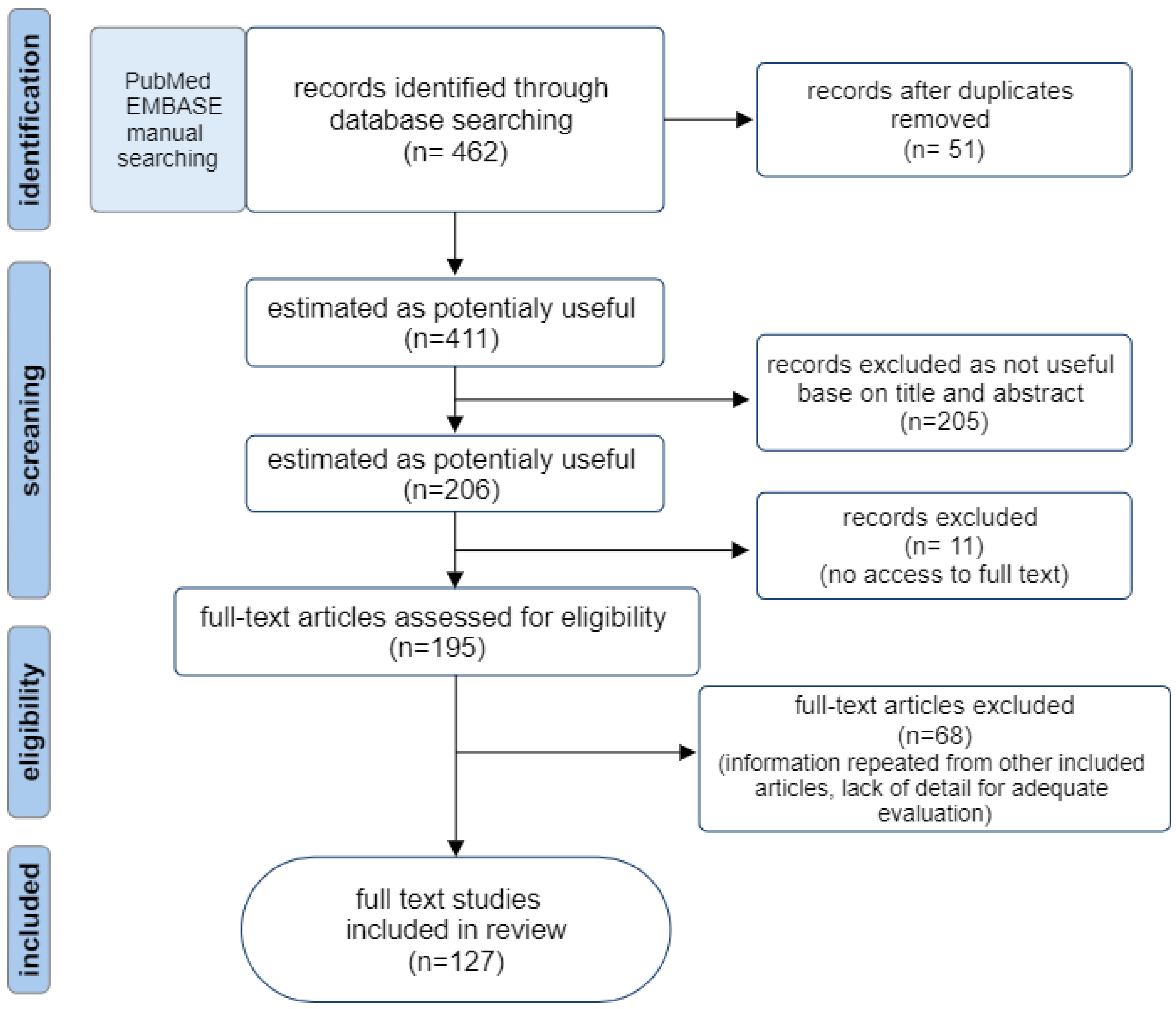
Figure 1. Flow diagram of the literature search strategy.
Ijms 25 12808 g001
Figure 2. Location of infections associated with biofilm formed by anaerobic bacteria.
Ijms 25 12808 g002
Figure 3. Schematic of the five main stages of biofilm formation.
Ijms 25 12808 g003
Figure 4. Chemical structure of the signaling molecules: type 1 (AI-1, AHL) and type 2 (AI-2) (found in Vibrio harveyi) [49].
Ijms 25 12808 g004
Figure 5. Schematic diagram of AHL signaling molecules regulating the QS system through the LuxI/LuxR pathway.
Ijms 25 12808 g005
Figure 6. Schematic diagram of AI-2 signaling molecules regulating the QS system.
Ijms 25 12808 g006
Figure 8. The molecular organization and co-transduction cascade of AgrBD and VirS/R in C. perfringens.
Ijms 25 12808 g008
Figure 9. AHL-degrading enzymes as an AHL-mediated inhibitor.
Ijms 25 12808 g009
Table 1. Influence of AI-2 signaling molecules on phenomena occurring in biofilms formed by anaerobic bacteria.
Biological Processes Associated with AI-2 in Biofilm Formed by Anaerobic BacteriaReferences
LuxS signaling in P. gingivalis plays a significant role in interaction with fibroblasts. This may be through the downregulation of the gene PGN_0482, which encodes a putative outer membrane immunoreactive protein.[47]
LuxS/AI-2 signaling mediates the interaction between P. gingivalis and periodontitis-associated species like Streptococcus gordonii.[77]
The luxS gene from A. actinomycetemcomitans can complement a luxS mutation in P. gingivalis.[84]
LuxS/AI-2 signaling in P. gingivalis regulates hemin acquisition, growth in hemin-limited conditions, and the expression of proteases and stress-related genes. In ΔluxS strains, genes encoding hemin binding protein (tlr) and (lysine-specific protease) kgp genes are downregulated, while gene hmuR encoding an outer-membrane hemin utilization receptor, fetB encoding heme-binding protein, feoB1 encoding ferrous iron transporter, and ftn ferritin-like proteins are upregulated.[74,76]
LuxS signaling is involved in promoting the survival of P. gingivalis in the host by regulating its response to host-induced stress factors (H2O2, and high pH). In the luxS mutant, genes related to stress response (htrA, clpB, groEL, dnaK, and ahpF), coding outer membrane efflux protein, and immunoreactive antigen are upregulated.[78]
In P. gingivalis, LuxS modulates protease levels and hemagglutination. luxS mutants exhibited a reduction in haemagglutinin titer and lower Rgp and Kgp proteases (gingipains) activity.[75]
AI-2 from F. nucleatum enhanced single species biofilm formation of F. nucleatum, P. gingivalis, T. denticola, and T. forsythia, and promoted co-aggregation with red complex species (P. gingivalis, T. denticola, T. forsythia). AI-2 induces mRNA synthesis of adhesion molecules like FadA, RgpA, Msp, and BspA (representative adhesion molecules of these bacteria).[70]
F. nucleatum AI-2 triggers inflammatory responses and promotes macrophage mobility and M1 polarization via the TNFSF9/TRAF1/p-AKT/IL-1β pathway.[67,72]
AI-2 may induce prophages in C. difficile biofilms, leading to phage-mediated cell lysis and eDNA release, enhancing biofilm growth.[69]
C. diffcile LuxS-mediated prophage induction. LuxS deficiency in C. difficile impairs prophage induction and biofilm formation in vitro.[85,86]
C. perfringens AI-2 regulates toxin production. luxS activates pfoA transcription and theta-toxin production and possibly influences post-transcriptional regulation of the production of alpha and kappa toxins.[82]
In C. acnes, AI-2 enhances virulence by increasing bacterial lipase activity.[83,87]
C. difficile AI-2 in co-culture triggers selective metabolic responses in B. fragilis, downregulating carbon metabolism genes; genes related to alanine, aspartate, and glutamate metabolism; and involved in amino acid biosynthesis.[69,88]
LuxS mutants of Lactobacillus rhamnosus GG and Bifidobacterium breve UCC2003 are less persistent in the murine gastrointestinal tract than wild strains, likely due to increased sensitivity to gastric fluid and impaired iron acquisition.[89,90]
Table 2. Review of proven QQ strategies in pathogenic anaerobic bacteria.
Potential QQ InhibitorsRefs.
Inhibitors of AHL-Mediated Quorum Sensing
AHL-degrading enzymes
AHL-degrading enzymes are lactonases, acylases, and oxidoreductases.
AHL-lactonase inhibits the QS process by hydrolyzing the lactone ring in the homoserine moiety of AHLs.
[51,62,112,115,116,117]
AHL-acylases hydrolyze the amide bond between the acyl side chain and the homoserine lactone in the AHL molecules producing the free fatty acid and the homoserine lactone.
AHL oxidoreductases reduce or oxidize the acyl chain of AHLs.
AHL analogs
AHL-analogs inhibit AHL synthesis, alter protein expression, and slow the growth of P. gingivalis, offering the potential for periodontal disease treatment.
AHL antagonists
AHL antagonists compete with natural signaling molecules for receptor binding by blocking the interaction between LuxR and AHL and disrupting quorum sensing.
Inhibitors of AI-2-mediated quorum sensing
AI-2 analogs
AI-2 analogs are D-ribose and D-galactose. These monosaccharides can block AI-2 receptors, reducing virulence gene expression and biofilm formation of periodontopathogens including A. actinomycetemcomitans, F. nucleatum, P. gingivalis, T. forsythia, and T. denticola. The production of bacterial adhesins was markedly reduced when the bacteria were grown in the presence of D-ribose.
D-galactose reduces biofilm formation of F. nucleatum, P. gingivalis, and T. forsythia by blocking the AI-2 receptor.
[65,70,79]
D-arabinose reduces biofilm formation by potentially competing with salivary receptors or bacterial adhesins.
Coumarin
Coumarin inhibits biofilm formation by P. gingivalis through interaction with the heme-binding protein HmuY and causes its interspersal and dispersion in pre-formed and late-stages.
[13,118,119]
Lacinartin
Lacinartin inhibits P. gingivalis growth, biofilm formation, and collagenase activity. Prevents adherence to oral epithelial cells and reduces inflammatory responses—secretion of IL-8 and TNF-α and matrix metalloproteinases (MMP-8 and MMP-9).
[120]
Brominated furanones
Brominated furanones inactivate the LuxS enzyme, required for AI-2 synthesis. The bromofuranone analog, 3-(dibromomethylene) isobenzofuran-1(3H)-one derivative inhibits biofilm formation by F. nucleatum, P. gingivalis, and T. forsythia without killing the bacteria.
[70,121]
Heparinoids
Glycosaminoglycans inhibit AI-2 activity, thereby reducing biofilm formation and lipase activity in C. acnes, additionally enhancing the bactericidal effects of isopropyl methylphenol on C. acnes biofilms.
[122]
Phlorizin and phloretin polyphenols
Phlorizin and phloretin disrupt biofilm forming by P. gingivalis and suppress its QS by inhibition of glucosyltransferases GtfB and GtfC.
[123,124,125]
Reuterin
Reuterin, produced by L. reuteri, suppresses biofilm formation of C. perfringens and downregulated QS-related genes like agrB and luxS, which decreases toxin production.
[126]
Inhibitors of Agr-like quorum sensing
In C. perfringens, a partial agonist (Z-AIPCp-L2A/T5A) and a partial antagonist (Z-AIPCp-F4A/T5S), significantly reduces transcription of the theta-toxin gene (pfoA).[127]
Disclaimer/Publisher’s Note: The statements, opinions and data contained in all publications are solely those of the individual author(s) and contributor(s) and not of MDPI and/or the editor(s). MDPI and/or the editor(s) disclaim responsibility for any injury to people or property resulting from any ideas, methods, instructions or products referred to in the content.