Article

Vicinal Diol Sesquiterpenes from Cinnamomum migao with Neuroprotective Effects in PC12 Cells

1
State Key Laboratory of Functions and Applications of Medicinal Plants, Guizhou Medical University, Guiyang 550014, China
2
Natural Products Research Center of Guizhou Province, Guiyang 550014, China
*
Authors to whom correspondence should be addressed.
Int. J. Mol. Sci. 2024, 25(23), 12693; https://doi.org/10.3390/ijms252312693
Submission received: 29 October 2024 / Revised: 16 November 2024 / Accepted: 22 November 2024 / Published: 26 November 2024
(This article belongs to the Section Molecular Neurobiology)

Abstract

In the ongoing search for new vicinal diol natural products, four new (Migaones A–D, 14) and four known (58) vicinal diol sesquiterpenoids were isolated from the branches and leaves of Cinnamomum migao. Their structures were unequivocally determined by comprehensive spectroscopic analyses (HRESIMS, 1D, and 2D NMR), single-crystal X-ray diffraction, electronic circular dichroism calculations, and comparison with existing literature data. All compounds isolated from C. migao possess vicinal diol structural units except compound 2. The newly isolated compounds (14) were evaluated for their neuroprotective activity using the PC12 cell injury model induced by N-methyl-daspartate acid (NMDA) and compounds 12 showing moderate neuroprotective activity against NMDA-induced neurotoxicity. Furthermore, molecular docking studies indicated that the most active compound 2 binds to the active site of the NMDA receptor via hydrogen bonds and hydrophobic interactions.

1. Introduction

Vicinal diol compounds are essential constituents of natural products, playing significant roles in the prevention and treatment of various diseases. Notable examples include the adrenergic drug epinephrine [1], antibiotic drug erythromycin [2], and the heart failure medication dapaglifozin [3]. In addition, vicinal diols are also crucial as synthetic intermediates. Thus, it is crucial to persistently investigate active vicinal diol natural products from plants.
Cinnamomum migao H. W. Li is an endemic medicinal plant, which is only distributed in the Guizhou, Yunnan, and Guangxi provinces of China [4]. Commonly known as Da-guo-mu-jiang-zi, it is a traditional remedy among Miao and Buyi ethnic minorities that is primarily used for treating arrhythmias, cerebrovascular diseases, and other ailments [5,6]. Additionally, C. migao is a key ingredient of Xinwei Zhitong Capsules and Liqi Huoxue Dropping Pills.
Sesquiterpenoids have demonstrated significant neuroprotective effects. For instance, atractylenolide III protects PC12 cells from corticosterone-induced injury [7], japonipene A-C exhibit neuroprotective effects against CoCl2-induced neuronal cell death in SH-SY5Y cells [8], flammuterpenols A–D protect against 6-hydroxydopamine-induced cell death in SH-SY5Y cells [9], and inderaggrols A−F show neuroprotective effects against erastin-induced ferroptosis in HT-22 cells [10]. Overall, sesquiterpenoids have considerable potential as neuroprotective agents.
Previous phytochemical investigations of C. migao have revealed that the plant is rich in guaiane-type sesquiterpenes, which exhibit notable anti-inflammatory activity [11,12]. Our earlier research identified a novel artemisinin-like sesquiterpenoid featuring an unprecedented tetracyclic 6/6/7/5 ring system, which demonstrated neuroprotective activity [13]. In this study, we report the isolation of four previously undescribed (14) and four known (58) sesquiterpenoids (Figure 1) from the branches and leaves of C. migao. Compounds 12 exhibited moderate neuroprotective activity against NMDA-induced neurotoxicity at 30 μM. Here, we report the isolation, structure elucidation, and neuroprotective activity of these undescribed compounds. This finding enriched the chemical composition of C. migao and provided a reference for the development and utilization of the plant resource in the future.

2. Results

2.1. Structure Elucidation

Migaone A (1) was isolated as a colorless needle crystal. The molecular formula, C15H24O3, was established by HR-ESI-MS ([M+Na]+ 275.1614, calcd for 275.1623), corresponding to four indices of hydrogen deficiency (IHDs). The 1H NMR spectrum (Table 1) of compound 1 revealed an olefinic proton [δH 6.87 (1H, s, H-6)] and four methyl groups [δH 1.07 (3H, d, J = 6.9 Hz, H3-12), 1.05 (3H, d, J = 6.9 Hz, H3-13), 1.23 (3H, s, H3-14), 1.37 (3H, s, H3-15). The 13C NMR and HSQC spectrum exhibited 15 carbon resonances, which were assigned as four methyls (δC 21.5, 22.1, 23.6, 26.7), four methylenes (δC 17.4, 33.0, 36.3, 51.2), two methines (δC 26.4, and an olefinic carbon δC 139.7), and five quaternary carbons (δC 41.3, two oxygenated carbons δC 74.4, 73.8, an olefinic carbon δC 148.5, and a ketone carbonyl δC 199.8). Taking into account the double bond and keto carbonyl, the two remaining IHDs suggest that compound 1 is a dicyclic sesquiterpene.
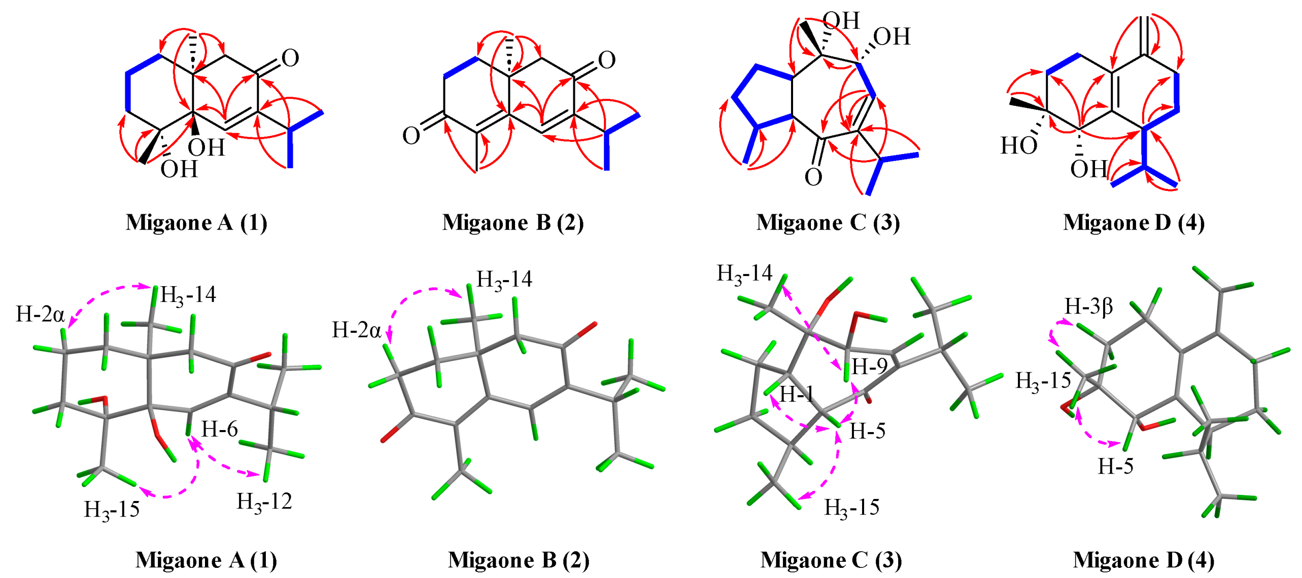
The planar structure of compound 1 was accomplished by 2D NMR spectroscopic analysis. The 1H−1H COSY correlations of H2-1/H2-2/H2-3 and H3-12/H-11/H3-13 revealed two key spin systems (Figure 2). The HMBC correlations (Figure 2) from H3-12 (δH 1.05)/H3-13 (δH 1.07) to C-7 (δC 148.5), C-11 (δC 26.4), from H-11 (δH 2.90) to C-6 (δC 139.7), C-7 (δC 148.5), C-8 (δC 199.8), C-12 (δC 22.1), C-13 (δC 21.5), from H3-14 (δH 1.23) to C-1 (δC 33.0), C-5 (δC 73.8), C-9 (δC 51.2), C-10 (δC 41.3), and from H3-15 (δH 1.37) to C-3 (δC 36.3), C-4 (δC 74.4), C-5 (δC 73.8) provided evidence for the presence of hydroxyl groups at C-4 and C-5, a double bond between C-6 and C-7, and a ketone carbonyl at C-8. Therefore, the planar structure of 1 was determined to be an eudesmane-type sesquiterpenoid with a 6/6 ring system.
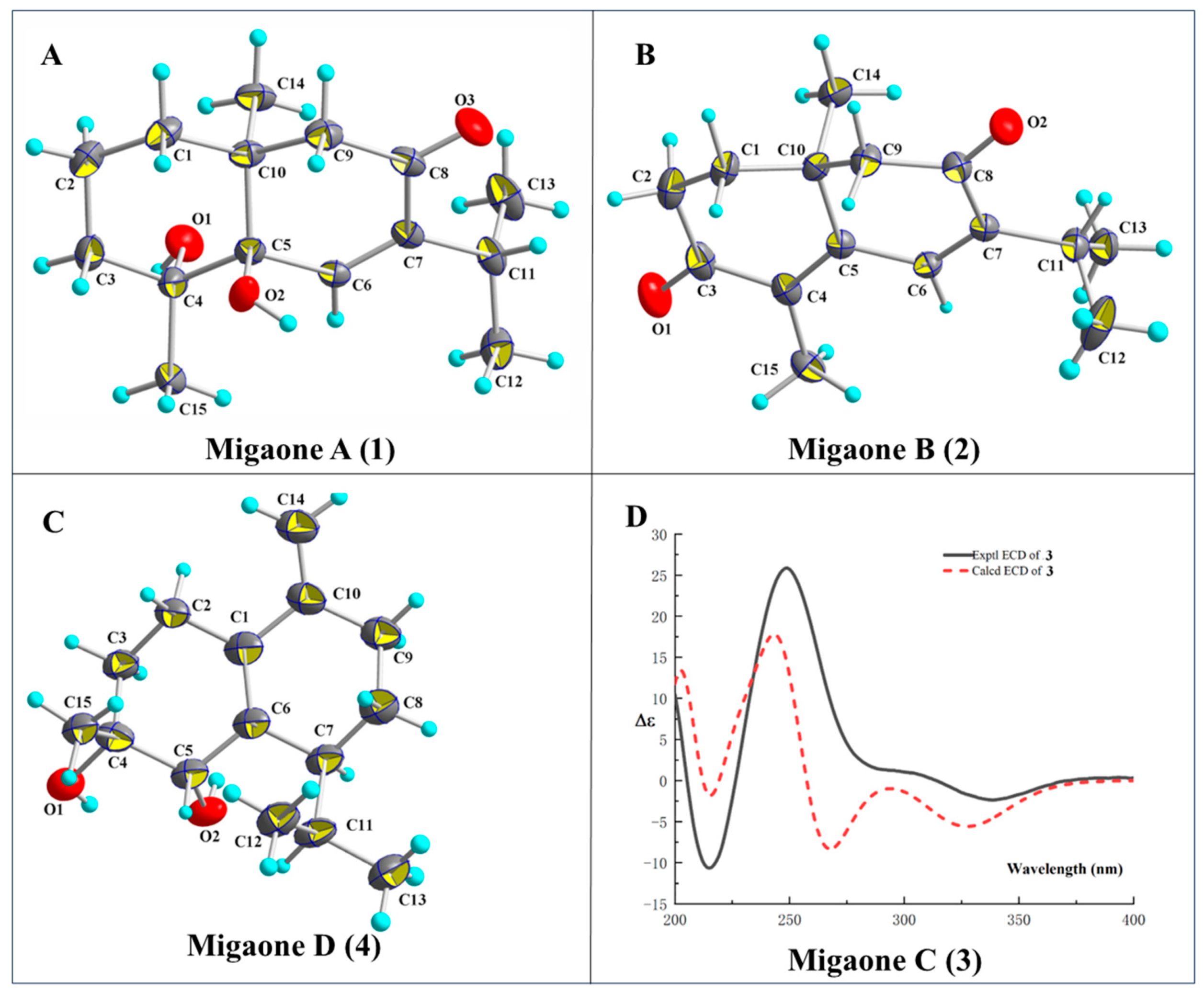
The relative configuration of compound 1 was assigned based on the analysis of Nuclear Overhauser Effect Spectroscopy (NOESY) cross-peaks involving H3-14/H-2α, H3-15/H-6/H3-12 (Figure 2). These data indicated that H3-14 is α-oriented and H3-15 is β-oriented. Finally, a single-crystal X-ray diffraction study was performed (CCDC number: 2377460) (Figure 3), confirming the above structural assignments and determining the absolute configuration as (4R, 5R, 10S), which was named Migaone A.
Migaone B (2) was obtained as a colorless needle crystal. Its molecular formula was determined to be C15H20O2 from HR-ESI-MS at m/z 233.1533 [M+H]+ (calcd for 233.1536). which corresponds to six IHDs. The 1H and HSQC spectrum revealed four methyl groups at δH 1.11 (3H, d, J = 6.9 Hz, H3-12), 1.14 (3H, d, J = 6.9 Hz, H3-13), 1.25 (3H, s, H3-14), and 1.97 (3H, s, H3-15) and an olefinic proton [δH 7.16 (1H, s, H-6)]. The 13C-NMR and HSQC spectra of compound 2 suggested the presence of 15 carbon resonances, including four methyls (δC 11.2, 21.8, 22.0, 23.9), three methylenes (δC 33.7, 35.9, 53.0), two methines (δC 27.2, a double carbon signal δC 133.6), and six quaternary carbons (δC 37.5, three double-bond carbons δC 133.3, 148.8, 152.1, two ketone carbonyl carbons δC 197.9, 198.6), accounting for four IHDs. Accordingly, the remaining two IHDs indicated that compound 2 has a dicyclic ring system.
In the 2D NMR data, two isolated spin systems were identified from the 1H−1H COSY spectrum, namely, H2-1/H2-2 and H3-12/H-11/H3-13 (Figure 2). The HMBC spectrum (Figure 2) revealed the following correlations, from H-6 (δH 7.16) to C-4 (δC 133.3), C-7 (δC 148.8), C-8 (δC 197.9) and C-10 (δC 37.5), from H-11 (δH 3.02) to C-6 (δC 133.6), C-7 (δC 148.8), C-8 (δC 197.9), C-12 (δC 22.0) and C-13 (δC 21.8), from H3-12 (δH 1.11)/H3-13 (δH 1.14) to C-7 (δC 148.8) and C-11 (δC 27.2), from H3-14 (δH 1.25) to C-1 (δC 35.9), C-5 (δC 152.1), C-9 (δC 53.0) and C-10 (δC 37.5), and from H3-15 (δH 1.97) to C-3 (δC 198.6), C-4 (δC 133.3), and C-5 (δC 152.1). These correlations suggested the presence of two double bonds at Δ4/5 and Δ6/7 and two ketone carbonyls located at C-3 and C-8.
The NOESY correlations between H3-14 and H-2α indicated that these protons were oriented on the same side (α-configurations). Its absolute configuration was confirmed to be (10 S) by X-ray diffraction analysis (CCDC number: 2377461), as shown in Figure 3, and it was named Migaone B.
Migaone C (3) was obtained as a colorless oil. Its HR-ESI-MS peak at m/z 275.1614 ([M+Na]+, calcd for 275.1617) and the 13C NMR data suggested a molecular formula of C15H24O3, indicating four IHDs. Analysis of its 1D and 2D NMR data suggested that compound 3 was structurally similar to (4R,10R)-9,10-dihydroxy-7-isopropyl-4,10-dimethyl-1,3,4,5,9,10-hexahydroazulen-6(1H)-one, as reported in the literature [14], with the exception of configurations differences. The relative configuration of 3 was established based on the NOESY data (Figure 2). The NOE correlations of H-9/H3-14 and H3-15/H-5/H-1 suggested that H-1, H-5, H-9 and H3-14 were arbitrarily assigned as β-oriented, while the correlations of H-4/H-2α indicated that H-4 was α-oriented. Finally, the absolute configuration of 3 (1S, 4S, 5R, 9R, 10S) was established through electronic circular dichroism (ECD) calculations with time-dependent density functional theory (TD-DFT), which were consistent with experimental data (Figure 3), and named Migaone C.
Migaone D (4) was obtained as a colorless needle crystal. Its molecular formula, C15H24O2, corresponding to four IHDs, was determined by positive HR-ESI-MS ([M+Na]+ 259.1664, calcd for 259.1669). The 1H and 13C NMR spectra of 4 resembled those of (4R, 5R)-muurol-1(6),10(14)-diene-4,5-diol [15] except for the configuration of the hydroxyl group at C-4 position; this was confirmed by the NOESY correlations between H-3β/H3-15/H-5, which indicated that the protons were oriented on the same side (β-configurations), and 4-OH was assigned as α-orientations. The absolute configuration of 4 was determined to be (4R, 5S, 7S) through X-ray diffraction analysis (CCDC number: 2377462), as shown in Figure 3, and it was named Migaone D.
The known compounds were identified based on their spectroscopic data analysis and comparisons with previously reported compounds in the literature (Figure 1), which were identified as trans-4,5-dihydroxycorocalane (5) [16], anomallenodiol (6) [17], oxyphyllenone A (7) [18], and stachytriol (8) [19].

2.2. Neuroprotective Activity

Pheochromocytoma (PC12) cells, derived from rat pheochromocytoma tumors, are widely used as neuronal cell lines in neurobiological studies [20]; they have the characteristics of nerve cells and are commonly employed in neuroprotective research. Previously, we isolated several undescribed guaiane-type sesquiterpenes with neuroprotective activity from the fruits of C. migao [9] as part of our ongoing exploration for structurally diverse sesquiterpenes with neuroprotective effects from C. migao. Compounds 14 were evaluated for their neuroprotective activity against NMDA-induced neurotoxicity in PC12 cells. Dizocilpine (MK801) was used as a positive control. The neuroprotective effects of compounds 14 are summarized in Figure 4. Compared to the model group (77.47 ± 0.68%), compounds 1 (81.85 ± 0.50%), 2 (96.21 ± 1.57%), and MK801 (97.33 ± 1.87%) demonstrated neuroprotective effects at 30 μM.

2.3. Molecular Docking Study

Molecular docking was employed to investigate the interaction between the active compounds and the NMDA receptor (NMDAR). Specifically, compounds 12 were docked into the glycine binding site of NMDAR (PDB ID: 4NF4) [21]. The docking results indicated that compounds 12 exhibited a favorable fit within the active pocket of NMDAR glycine antagonists. The calculated free energy change for compound 1 in its lowest energy conformation during molecular docking was −19.9995 kJ/mol. To facilitate a detailed analysis of the interactions between the ligand and NMDAR, Pymol software version 2.4 was used for visualization. Compound 1 established hydrogen bonds with amino acid residues GLN144 in the active pocket of NMDAR and also formed hydrophobic interactions with amino acid residues THR126, ASN128, GLN144, TYR184, and VAL267. The docking score for compound 2 was −24.2672 kJ/mol. Compound 2 also formed hydrogen bonds with GLN144 and ASN128. Additionally, it formed hydrophobic interactions with THR126, GLN144, LEU146, VAL181, and TYR184. The hydrogen bonding and hydrophobic interaction were key factors contributing to the binding affinity of these compounds to NMDAR. The results are shown in Figure 5.

3. Materials and Methods

3.1. General Experimental Procedures

X-ray crystallographic data were recorded on an XtaLAB AFC12 (RINC) using Cu Kα radiation. Optical rotation measurements were conducted at a wavelength of 589 nm using an MCP 500 apparatus at 25 °C. Melting points were determined on a MP30 melting point apparatus (Mettler Toledo, Zurich, Switzerland). Ultraviolet (UV) spectra were recorded on a Shimadzu-2600 spectrophotometer (Shimadzu, Tokyo, Japan). Electronic Circular Dichroism (ECD) spectra were obtained with a Chirascan spectrometer (Applied Photophysics Ltd., Leatherhead, UK). Infrared (IR) spectra (KBr) were measured using an IRAffinity-1 spectrometer; 1D and 2D spectra were collected on an AVANCE III-600 spectrometer with TMS as an internal standard (Bruker Corp, Bremen, Germany). HRESIMS was performed on an Agilent 6210 ESI/TOF mass spectrometer (Agilent Technologies Inc., Santa Clara, CA, USA). Analytical HPLC was conducted using an Agilent 1260 system equipped with a reversed-phase C18 column (5 μm, 250 mm × 4.60 mm, SHIMADZU shim-pack GIS, Shimadzu, Tokyo, Japan). Semi-preparative HPLC was performed using a Shimadzu LC-20A instrument equipped with a UV SPD-20A detector (Shimadzu, Tokyo, Japan) and a reversed-phase C18 column (5 μm, 250 mm × 10 mm, SHIMADZU shim-pack GIS, Shimadzu, Tokyo, Japan).

3.2. Plant Material

The branches and leaves of Cinnamomum migao H. W. Li were collected from Ceheng, Guizhou province, China, in October 2019. The plant was identified by Professor Qing-wen Sun. A voucher specimen (GZCH20191001) has been deposited at the Natural Products Research Center of Guizhou Province.

3.3. Extraction and Isolation

The branches and leaves of C. migao (21 kg) were extracted with 95% aqueous ethanol (4 × 30 L) under reflux three times (each time for 3 h) to yield a crude extract. The ethanol crude extract (1.0 kg) was suspended in water (10 L) and partitioned with petroleum ether (PE) and ethyl acetate (EtOAc), yielding PE (200 g) and EtOAc (300 g) layers, respectively.
The PE fraction (200 g) was subjected to silica gel column chromatography (CC) using a solvent gradient of PE–EtOAc (1:0 to 0:1, v/v), resulting in six fractions (Fr.A1–Fr.A6). Fr.A6 (300 mg) was further separated on an Rp-C18 column using a MeOH–H₂O gradient (60:40, 70:30, 80:20, 90:10, v/v), yielding four subfractions Fr.A6.1-Fr.A6.4. Fr.A6.1 (125 mg) was further purified by semi-preparative HPLC with CH3CN–H2O (65:35, v/v, 2 mL/min) to obtain compound 8 (65 mg, tR = 25.3 min).
The EtOAc layers (300 g) were chromatographed over a silica gel column using a PE–EtOAc gradient solvent system (10:1 to 0:1, v/v), resulting in seven fractions (Fr.B1–Fr.B7). Fr.B2 (300 mg) was further separated on silica gel with a PE–EtOAc solvent system (20:1, v/v), yielding compound 7 (20 mg). Fr.B4 (4 g) was chromatographed on an Rp-C18 CC, using MeOH−H2O (40:60, 60:40, 70:30, v/v) as the eluent solvent to give three subfractions (Fr.B4.1-Fr.B4.3). Fr.B4.2 (220 mg) was purified by semi-preparative HPLC eluted with CH3CN–H2O (45:55, v/v, 2 mL/min) to obtain compounds 6 (11 mg, tR = 25.4 min). Fr.B-5 (11 g) was separated on Rp-C18 gel with MeOH–H2O (40:60, 50:50, 60:40, 70:30, v/v) to yield four subfractions (Fr.B5.1-Fr.B5.4). Fr.B5.2 (231 mg) was separated by semi-preparative HPLC eluted with a MeOH–H2O (55:45, v/v, 2 mL/min) to afford 5 (16 mg, tR = 30.5 min). Fr.B5.2 (231 mg) was also separated by semi-preparative HPLC (MeOH–H2O, 70:30, v/v, 2 mL/min) to afford compound 4 (17 mg, tR = 23.6 min). Fr.B5.3 (60 mg) was separated on a silica gel column eluted with a PE−EtOAC gradient (3:1, v/v) to yield compound 1 (8 mg). Fr.B5.4 (72 mg) was purified by semi-preparative HPLC with MeOH–H2O (70:30, v/v, 2 mL/min) to afford 3 (4 mg, tR = 19.9 min) and 2 (4 mg, tR = 26.3 min).
Migaone A (1): colorless needle crystal (CHCl3–MeOH), [α ] D 25 + 23.7 (c 0.11, MeOH); m.p 130.9 °C. UV (MeOH) λmax (log ε): 235 (3.39) nm; IR (KBr) υmax 3441, 2962, 2926, 2870, 1656, 1458, 1373, 1020, 995, 970, 948 cm–1. For 1H and 13C NMR see Table 1. HR-ESI-MS m/z 275.1614 [M+Na]+(calcd for [C15H24O3Na]+, 275.1623). Supporting information: Figures S1–S9.
Migaone B (2): colorless needle crystal (CHCl3–MeOH); [α ] D 25 − 200 (c 0.12, MeOH). UV (MeOH) λmax (log ε): 310 (3.58) nm. IR (KBr) υmax 3446, 2960, 2926, 2872, 1660, 1300, 1199, 1022, 848 cm–1. 1H and 13C NMR, see Table 1. HR-ESI-MS m/z 233.1533 [M+H]+ (calculated for [C15H21O2]+, 233.1536). Supporting information: Figures S10–S18.
Migaone C (3): colorless oil. [α ] D 25 + 31.9 (c 0.23, MeOH); UV (MeOH) λmax (log ε): 201 (2.97), 241 (3.16) nm; ECD (MeOH) λ (Δε) 215 (−10.64), 249 (+25.87), 338 (−2.35) nm; IR (KBr) υmax 3381, 2956, 2870, 1660, 1462, 1373, 1049, 669 cm–1. For 1H and 13C NMR, see Table 1. HR-ESI-MS m/z 275.1614 [M+Na]+ (calculated for [C15H24O3Na]+, 275.1617). Supporting information: Figures S19–S27.
Migaone D (4): colorless needle crystal (CHCl3–MeOH). [α ] D 25 − 15.1 (c 0.20, MeOH). UV (MeOH) λmax (log ε): 242 (3.49). IR (KBr) υmax 3346, 2956, 2935, 1635, 1456, 1016, 667, 599, 555 cm–1. For 1H and 13C NMR, see Table 1. HR-ESI-MS m/z 259.1664 [M+Na]+ (calculated for [C15H24O2Na]+, 259.1669). Supporting information: Figures S28–S36.
Trans-4,5-dihydroxycorocalane (5): colorless oil, ESI-MS m/z 257 [M+Na]+, C15H22O2. 1H-NMR (600 MHz, CDCl3) δH: 2.67 (1H, dd, J = 16.6, 6.1 Hz, H-2a), 2.73 (1H, dd, J = 12.0, 6.4 Hz, H-2b), 1.75 (1H, ddt, J = 13.7, 6.6, 1.7 Hz, H-3a), 2.04 (1H, ddd, J = 13.7, 12.1, 6.8 Hz, H-3b), 4.47 (1H, s, H-5), 7.05 (1H, d, J = 7.9 Hz, H-8), 7.10 (1H, d, J = 7.9 Hz, H-9), 3.46 (1H, m, H-11), 1.23 (3H, d, J = 6.8 Hz, H-12), 1.29 (3H, d, J = 6.8 Hz, H-13), 2.20 (3H, s, H-14), 1.41 (3H, s, H-15); 13C-NMR (150 MHz, CDCl3) δC: 135.6 (C-1), 24.8 (C-2), 29.7 (C-3), 72.0 (C-4), 71.3 (C-5), 148.3 (C-6), 134.4 (C-7), 123.9 (C-8), 130.0 (C-9), 134.2 (C-10), 27.6 (C-11), 24.4 (C-12), 25.4 (C-13), 19.7 (C-14), 28.7 (C-15). Supporting information: Figures S37 and S38.
Anomallenodiol (6): colorless needle crystal (MeOH), ESI-MS m/z 261 [M+Na]+, C14H22O3. 1H-NMR (600 MHz, CD3OD) δ: 2.22 (1H, m, H-1a), 2.32 (1H, m, H-1b), 1.61 (1H, m, H-2a), 1.70 (1H, ddd, J = 13.5, 9.3, 6.1, H-2b), 3.90 (1H, s, H-4), 2.54 (1H, m, H-6), 2.01 (2H, m, H-7), 2.31 (1H, m, H-8a), 2.51 (1H, m, H-8b), 2.22 (1H, m, H-11), 0.94 (3H, d, J = 6.9 Hz, H-12), 1.09 (3H, d, J = 6.8 Hz, H-13), 1.25 (3H, s, H-15); 13C-NMR (150 MHz, CD3OD) δ: 21.3 (C-1), 31.1 (C-2), 72.0 (C-3), 74.3 (C-4), 160.4 (C-5), 42.7 (C-6), 23.7 (C-7), 36.2 (C-8), 202.4 (C-9), 133.7 (C-10), 30.8 (C-11), 22.1 (C-12), 19.7 (C-13), 25.1 (C-14). Supporting information: Figures S39 and S40.
Oxyphyllenone A (7): colorless oil, ESI-MS m/z 233 [M+Na]+, C12H18O3. 1H-NMR (400 MHz, CDCl3) δ 1.44 (1H, m, H-1a), 1.84 (1H, td, J = 13.8, 4.1 Hz, H-1b), 1.58 (1H, ddd, J = 13.9, 6.9, 3.6 Hz, H-2a), 2.42 (1H, tdd, J = 14.1, 4.1, 2.6 Hz, H-2b), 3.70 (1H, t, J = 3.0 Hz, H-3), 6.02 (3H, s, H-6), 2.33 (1H, ddd, J = 17.6, 3.4, 2.5Hz, H-8a), 2.64 (1H, ddd, J = 17.6, 15.2, 5.0 Hz, H-8b), 1.75 (1H, ddd, J = 13.2, 5.0, 2.3 Hz, H-9a), 1.93 (1H, m, H-9b), 1.43 (3H, s, H-14), 1.48 (3H, s, H-15); 13C-NMR (150 MHz, CDCl3) δC: 35.3 (C-1), 25.4 (C-2), 76.2 (C-3), 73.9 (C-4), 172.6 (C-5), 126.1 (C-6), 203.4 (C-7), 34.9 (C-8), 41.0 (C-9), 25.5 (C-14), 25.6 (C-15). Supporting information: Figures S41 and S42.
Stachytriol (8): cubic crystal (MeOH), ESI-MS m/z 279 [M+Na]+, C15H28O3. 1H NMR (600 MHz, CDCl3) δ: 1.33 (1H, m, H-2a), 2.41 (1H, m, H-2b), 1.39 (1H, m, H-3a), 1.88 (1H, m, H-3b), 2.55 (1H, m, H-4), 1.79 (1H, m, H-5), 1.48 (1H, m, H-6a), 1.57 (1H, m, H-6b), 1.79 (1H, m, H-7), 1.63 (1H, m, H-8a), 2.02 (1H, m, H-8b), 1.63 (1H, m, H-9a), 2.02 (1H, m, H-9b), 1.24 (3H, s, H-12), 1.14 (3H, s, H-13), 1.05 (3H, s, H-14), 0.89 (3H, d, J = 6.9 Hz, H-15); 13C NMR (150 MHz, CDCl3) δ: 90.7 (C-1), 32.3 (C-2), 29.6 (C-3), 35.4 (C-4), 47.9 (C-5), 24.5 (C-6), 36.5 (C-7), 19.3 (C-8), 26.2 (C-9), 75.8 (C-10), 73.7 (C-11), 29.4 (C-12), 29.2 (C-13), 26.8 (C-14), 15.1 (C-15). Supporting information: Figures S43 and S44.

3.4. X-Ray Crystallographic Analysis of Compounds 1, 2 and 4

Crystals of compounds 1, 2 and 4 were obtained by the slow evaporation of a CHCl3–MeOH (1:4) solution at room temperature over four days. These crystals were analyzed using a XtaLAB AFC12 (RINC) diffractometer (Rigaku Corporation, Tokyo, Japan) equipped with Cu Kα radiation during crystal data collection. Using Olex2 version 1.5 [22], the structure was solved with the SHELXT [23] structure solution program using intrinsic phasing and refined with the SHELXL [24] refinement package using least-squares minimization. The crystallographic data of 1, 2 and 4 have been deposited in the Cambridge Crystallographic Data Center (http://www.ccdc.cam.ac.uk, accessed on 15 September 2024).
Crystal data for Migaone A (1): C15H24O3 M = 252.34, orthorhombic, space group P212121 (no. 19), a = 18.0068 (3) Å, b = 10.4598 (2) Å, c = 7.3920 (2) Å, α = 90°, β = 90°, γ = 90°, V = 1392.26 (5) Å3, Z = 4, T = 150.3 (4) K, μ (Cu Kα) = 0.654 mm−1, 12,977 reflections measured (9.78° ≤ 2Θ ≤ 147.7°), 2733 unique (Rint = 0.0416, Rsigma = 0.0315), which were used in all calculations. The final R1 was 0.0328 (I > 2σ(I)) and wR2 was 0.0824 (all data). Flack parameter = −0.02(9). CCDC number: 2377460.
Crystal data for Migaone B (2): C15H20O2, M = 232.31, monoclinic, space group P21 (no. 4), a = 11.2012 (10) Å, b = 10.0306 (10) Å, c = 12.5157 (10) Å, α = 90°, β = 112.1760°, γ = 90°, V = 1302.18 (2) Å3, Z = 4, T = 99.98 (11) K, μ (Cu Kα) = 0.605 mm−1, 24,883 reflections measured (7.628° ≤ 2Θ ≤ 148.854°), 5208 unique (Rint = 0.0303, Rsigma = 0.0187) which were used in all calculations. The final R1 was 0.0309 (I > 2σ(I)) and wR2 was 0.0818 (all data). Flack parameter = 0.00 (5). CCDC number: 2377461.
Crystal data for Migaone D (4): C15H24O2, M = 236.34, monoclinic, space group P21 (no. 4), a = 6.6487 (11) Å, b = 13.674 (2) Å, c = 14.980 (4) Å, α = 90°, β = 95.739°, γ = 90°, V = 1355.1 (5) Å3, Z = 2, T = 99.98 (16) K, μ (Cu Kα) = 0.582 mm−1, 6393 reflections measured (5.93° ≤ 2Θ ≤ 149.358°), 3435 unique (Rint = 0.0647, Rsigma= 0.0898), which were used in all calculations. The final R1 was 0.0722 (I > 2σ(I)) and wR2 was 0.2144 (all data). Flack parameter = 0.00 (5). CCDC number: 2377462.

3.5. Theoretical ECD Calculation

The absolute configuration of compound 3 were determined using time-dependent density functional theory (TDDFT) calculations, which were performed with the Gaussian 16 program package. The stable conformers, devoid of imaginary frequencies, were subjected to ECD calculation using the TDDFT method at the B3LYP-SCRF (PCM)/6-31+G (d) levels with the CPCM model in methanol solvent. The final calculated ECD spectrum was obtained by Boltzmann averaging, and the calculated ECD of each conformer using SpecDis 1.64. The absolute configurations of compound 3 were determined by comparing the calculated ECD with the experimental ECD data [25].

3.6. Cell Viability Assay

Differentiated PC12 cells (purchased from Institute of Cell Biology, Chinese Academy of Sciences, Shanghai, China) were cultured in Dulbecco’s Modified Eagle Medium (DMEM) supplemented with 5% fetal bovine serum and 1% penicillin–streptomycin. The cells were incubated at 37 °C in a humidified environment containing 5% CO2 based on a previously reported method [21]. All tested compounds were dissolved in dimethyl sulfoxide (DMSO). Compounds 14 were evaluated for their neuroprotective effects against NMDA-induced neurotoxicity utilizing the MTT assay [26]. PC12 cells were plated at a density of 4000 cells/well in a 96-well plate. After 72 h of incubation, the cells were pretreated with MK-801 (positive control) and compounds 14 at 30 µM for 24 h, which was followed by treatment with NMDA (2 mM) for 6 h. Subsequently, 3-(4,5-dimethylthiazol-2-yl)-2,5-diphenyltetrazolium bromide (MTT) was added to the medium at a final concentration of 0.5 mg/mL. After 4 h incubation, the formazan crystals were dissolved in 100 µL DMSO per well, and the absorbance was measured at a wavelength of 490 nm using a Microplate Reader (Thermo Scientific Varioskan LUX Multimode Reader, Thermo Scientific Co., Ltd., Waltham, MA, USA). Data were processed using SPSS 27.0, and graphs were generated using GraphPad Prism 8.

3.7. Molecular Docking Study

The Autodock 4 version software package was used for the docking study between the ligand and protein. The NMDAR protein (PDB code: 4NF4) was downloaded from Protein Data Bank (https://www.rcsb.org/, accessed on 15 September 2024). For active site docking, a grid box of size 30 × 30 × 30 Å was defined with the center coordinates set at X = −13.559, Y = 7.812, Z = −37.745. The pose with the best score was selected for further analysis, which was performed visually using PyMoL version 2.4. Additionally, two-dimensional images were generated using the LIGPLOT+ Molecular Graphics System 2.2.5.

3.8. Statistical Analysis

Data are expressed as the mean ± standard deviation (SD). Multiple groups were compared using one-way analysis of variance (ANOVA), which was followed by the post hoc least significant difference (LSD) test. A p-value of less than 0.05 was considered statistically significant.

4. Conclusions

In conclusion, four new sesquiterpenes, Migaones A–D (14), along with four known compounds (58), were isolated from the branches and leaves of C. migao. Except for compound 2, all other isolated constituents possess vicinal diol structural units. Bioactivity assessments revealed that compounds 12 exhibited protective effects against NMDA-induced neurotoxicity in PC12 cells at a concentration of 30 μM. Furthermore, molecular docking analyses demonstrated that compounds 12 effectively interacted with the glycine binding pocket through hydrogen bonds and hydrophobic interactions with key amino acid residues. These molecular docking results were consistent with the observed biological activity.

Supplementary Materials

The supporting information can be downloaded at: https://www.mdpi.com/article/10.3390/ijms252312693/s1.

Author Contributions

Data curation, L.Z.; Funding acquisition, X.Y.; Investigation, M.P.; Methodology, F.C.; Project administration, X.Y.; Resources, X.P.; Software, H.L.; Supervision, X.Y.; Validation, L.Y. and J.Y.; Writing—original draft, L.Z.; Writing—review and editing, Q.L. All authors have read and agreed to the published version of the manuscript.

Funding

The work was funded by the National Natural Science Foundation of China (U1812403, 32160104 and 82160807), the construction of innovation capacity of scientific research institutions in Guizhou province (No. QJHFQ2024-005), Guizhou Provincial Natural Science Foundation (No. QKHCG2023-063), and Guizhou Provincial Basic Research Program (No. 2023-239).

Institutional Review Board Statement

Not applicable.

Informed Consent Statement

Not applicable.

Data Availability Statement

The data presented in this study are available on request from the corresponding author.

Conflicts of Interest

The authors declare no conflicts of interest.

References

  1. Xu, X.; Shonberg, J.; Kaindl, J.; Clark, M.J.; Stößel, A.; Maul, L.; Mayer, D.; Hübner, H.; Hirata, K.; Venkatakrishnan, A.J.; et al. Constrained catecholamines gain β2AR selectivity through allosteric effects on pocket dynamics. Nat. Commun. 2023, 14, 2138. [Google Scholar] [CrossRef] [PubMed]
  2. Luo, Z.W.; Yin, F.C.; Wang, X.B.; Kong, L.Y. Progress in approved drugs from natural product resources. Chin. J. Nat. Med. 2024, 22, 195–211. [Google Scholar] [CrossRef] [PubMed]
  3. Lamaida, N.; Cerciello, A. 852 Dapaglifozin in patients with cardiac heart failure. Eur. Heart J. Suppl. 2022, 24, suac121.150. [Google Scholar] [CrossRef]
  4. Editorial Committee of Chinese Flora. Flora of China; Science Press: Beijing, China, 1982; p. 176. [Google Scholar]
  5. Sun, X.X.; Xu, J.; Liu, J.; Guo, J.T.; Zhu, X.T. Optimization of the extraction process of Cinnamomum migao oil and its ameliorative effects on myocardial ischemia. J. Food. Saf. Qual. 2024, 15, 262–273. [Google Scholar]
  6. Sun, X.H.; Ye, J.Q. Effects of oil of Cinnamomum migao (CV-3) on isolated smooth muscles. Eur. J. Pharmacol. 1990, 183, 554. [Google Scholar] [CrossRef]
  7. Gong, W.X.; Zhou, Y.Z.; Qin, X.M.; Du, G.H. Involvement of mitochondrial apoptotic pathway and MAPKs/NF-κB inflammatory pathway in the neuroprotective effect of atractylenolide III in corticosterone-induced PC12 cells. Chin. J. Nat. Med. 2019, 17, 264–274. [Google Scholar] [CrossRef]
  8. Wang, S.N.; Jin, D.Q.; Xie, C.F.; Wang, H.; Wang, M.C.; Xu, J.; Guo, Y.Q. Isolation, characterization, and neuroprotective activities of sesquiterpenes from Petasites japonicus. Food Chem. 2013, 141, 2075–2082. [Google Scholar] [CrossRef]
  9. Wu, S.Y.; Chen, X.Q.; Ren, J.L.; Liu, P.L.; Yan, Q.; Chen, Z.M. Cuparene-type sesquiterpenes with neuroprotective activities from the edible mushroom Flammulina filiformis. Fitoterapia 2024, 179, 106235. [Google Scholar] [CrossRef]
  10. Ma, L.F.; Lou, S.Q.; Chen, H.Y.; Luo, D.; Guo, L.; Chen, N.Y.; Wu, R.; Fang, L.; Zhan, Z.J. Highly oxidized guaiane 12(8), 15(6)-dilactones with neuroprotective activities from the roots of Lindera aggregata (Sims) Kosterm. Phytochemistry 2024, 224, 114150. [Google Scholar] [CrossRef]
  11. Muhammad, I.; Hassan, S.S.U.; Xu, W.J.; Tu, G.L.; Yu, H.J.; Xiao, X.; Yan, K.; Jin, H.Z.; Bungau, S. An extensive pharmacological evaluation of novel anti-nociceptive and IL-6 targeted anti-inflammatory guaiane-type sesquiterpenoids from Cinnamomum migao H. W. Li through in-depth in-vitro, ADMET, and molecular docking studies. Biomed. Pharmacother. 2023, 164, 114946. [Google Scholar] [CrossRef]
  12. Muhammad, I.; Luo, W.; Shoaib, M.R.; Li, G.L.; Hassan, S.S.U.; Yang, Z.H.; Xiao, X.; Tu, G.L.; Yan, S.K.; Ma, X.P.; et al. Guaiane-type sesquiterpenoids from Cinnamomum migao H. W. Li: And their anti-inflammatory activities. Phytochemistry 2021, 190, 112850. [Google Scholar] [CrossRef] [PubMed]
  13. Zhou, L.; Yang, L.S.; Wang, L.; Liu, H.D.; Gao, M.; Chen, F.J.; Yang, J.; Li, Q.J.; Yang, X.S. Cinnamigones A–C, three highly oxidized guaiane-type sesquiterpenes with neuroprotective activity from Cinnamomum migao. Phytochemistry 2023, 212, 113728. [Google Scholar] [CrossRef]
  14. Morita, H.; Simizu, K.; Takizawa, H.; Aiyama, R.; Itokawa, H. Studies on Chemical Conversion of Alpinenone to Furopelargone B. Chem. Pharm. Bull. 1988, 36, 3156–3160. [Google Scholar] [CrossRef]
  15. Kiem, P.V.; Minh, C.V.; Nhiem, N.X.; Cuc, N.T.; Quang, N.V.; Anh, H.L.T.; Tai, B.H.; Yen, P.H.; Hoai, N.T.; Ho, K.Y.; et al. Muurolane-type sesquiterpenes from marine sponge Dysidea cinerea. Magn. Reson. Chem. 2014, 52, 51–56. [Google Scholar] [CrossRef] [PubMed]
  16. Kuo, Y.H.; Yu, M.T. Four New Sesquiterpenes from the Heartwood of Juniperus formosana var. concolor. Chem. Pharm. Bull. 1999, 47, 1017–1019. [Google Scholar] [CrossRef]
  17. Zan, K.; Shi, S.P.; Fu, Q.; Chen, X.Q.; Zhou, S.X.; Xiao, M.T.; Tu, P.F. New Sesquiterpenoids from Artemisia anomala. Helv. Chim. Acta. 2010, 93, 2000–2006. [Google Scholar] [CrossRef]
  18. Muraoka, O.; Fujimoto, M.; Tanabe, G.; Kubo, M.; Minematsu, T.; Matsuda, H.; Morikawa, T.; Toguchida, I.; Yoshikawa, M. Absolute stereostructures of novel norcadinane and trinoreudesmane-type sesquiterpenes with nitric oxide production inhibitory activity from Alpinia oxyphylla. Bioorg. Med. Chem. Lett. 2001, 11, 2217–2220. [Google Scholar] [CrossRef]
  19. Soliman, H.S.M.; El-Dib, R.; Shalaby, N.M.M.; Duddeck, H.; Simon, A.; Tóth, G. Isolation and structure determination of compounds from Stachys yemenensis hedge. Nat. Prod. Commun. 2007, 2, 977–980. [Google Scholar] [CrossRef]
  20. Mao, Q.Q.; Zhong, X.M.; Li, Z.Y.; Huang, Z. Paeoniflorin protects against NMDA-induced neurotoxicity in PC12 cells via Ca2+ antagonism. Phytother. Res. 2011, 25, 681–685. [Google Scholar] [CrossRef]
  21. Yang, L.S.; Yang, Q.; Wang, E.H.; Yang, J.; Li, Q.J.; Cao, J.F.; Wang, L.; Liao, X.; Yang, Y.; Yang, X.S. Synthesis of novel 1-phenyl-benzopyrrolizidin-3-one derivatives and evaluation of their cytoneuroprotective effects against NMDA-induced injury in PC12 cells. Bioorg. Med. Chem. 2022, 59, 116675. [Google Scholar] [CrossRef]
  22. Dolomanov, O.V.; Bourhis, L.J.; Gildea, R.J.; Howard, J.A.K.; Puschmann, H. OLEX2: A complete structure solution, refinement and analysis program. J. Appl. Cryst. 2009, 42, 339–341. [Google Scholar] [CrossRef]
  23. Sheldrick, G.M. SHELXL—Integrated space-group and crystal-structure determination. Acta Crystallogr. Sect. A Found. Adv. 2015, 71, 3–8. [Google Scholar] [CrossRef] [PubMed]
  24. Sheldrick, G.M. Crystal structure refinement with SHELXT. Acta Crystallogr. Sect. C Struct. Chem. 2015, 71, 3–8. [Google Scholar] [CrossRef]
  25. Lou, H.Y.; Chen, M.J.; Yi, P.; Jin, J.; Zeng, Y.R.; Gu, W.; Hu, Z.X.; Yang, J.; Hao, X.J.; Yuan, C.M. Hyperkouytones A—O, new polyprenylated acylphloroglucinols from Hypericum kouytchense with multidrug resistance reversal activity. Chin. J. Chem. 2024, 42, 3293–3307. [Google Scholar] [CrossRef]
  26. Reed, L.J.; Muench, H.A. A simple method of estimating 50 percent endpoints. Am. J. Hyg. 1938, 27, 493–497. [Google Scholar]
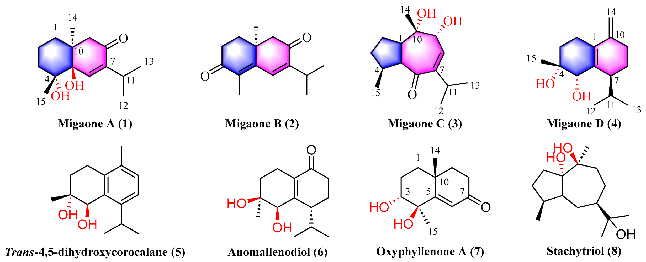
Figure 1. Structures of compounds 18.
Ijms 25 12693 g001
Figure 2. Key 2D NMR correlations of 14.
Ijms 25 12693 g002
Figure 3. (AC) X-ray of compounds of 1, 2, 4. (D) Experimental and calculated ECD of 3.
Ijms 25 12693 g003
Figure 4. The neuroprotective effects of compounds 14 against NMDA-induced injury in PC12 cells (NMDA: 2 mM; compounds 14: 30 μM; data are expressed as means ± SD, n = 3). ### p < 0.001 vs. control group, *** p < 0.001 vs. NMDA group.
Ijms 25 12693 g004
Figure 5. Molecular docking interaction of compounds 12 with NMDAR (PDB: 4NF4). Binding pose (a), detailed 3D (b) and 2D (c) protein interactions in the active site region of the molecules.
Ijms 25 12693 g005aIjms 25 12693 g005b
Table 1. NMR data for Migaone A−D (14).
No.Migaone A (1) aMigaone B (2) aMigaone C (3) bMigaone D (4) b
δCδH (J in Hz)δCδH (J in Hz)δCδH (J in Hz)δCδH (J in Hz)
133.01.85, m35.92.02, td (14.0, 5.3)51.52.15, m132.4
1.20, m1.82, ddd (13.3, 5.3, 2.3)
217.41.85, m33.72.62, ddd(17.8, 14.5, 5.2)26.21.78, m25.62.30, m
1.50, m2.55, ddd (17.9, 5.2, 2.3)1.70, m2.24, m
336.31.97, m198.6 35.11.70, m31.91.86, m
1.50, m1.26, m1.52, m
474.4 133.3 37.52.23, m71.4
573.8 152.1 62.01.93, dd (10.7, 7.8)73.23.78, s
6139.76.87, s133.67.16, s207.3 139.1
7148.5 148.8 147.6 43.12.43, m
8199.8 197.9 141.56.16, dd (4.6, 1.2)24.01.70, m
1.52, m
951.22.91, m53.02.45, m76.04.17, d (4.6)31.52.43, m
1.98, m 2.24, m
1041.3 37.5 76.0 146.0
1126.42.90, m27.23.02, p (6.9)30.12.82, m29.72.17, m
1222.11.05, d (6.9)22.01.11, d (6.9)22.11.04, d, (6.9)18.20.76, d (6.9)
1321.51.07, d (6.9)21.81.14, d (6.9)22.60.97, d, (6.9)21.90.97, d (6.9)
1423.61.23, s23.91.25, s19.91.24, s108.54.92, s
4.74, s
1526.71.37, s11.21.97, s21.31.09, d (6.7)24.71.15, s
a Spectra were measured in CDCl3 (1H, 600 MHz and 13C, 150 MHz); b Spectra were measured in CD3OD (1H, 600 MHz and 13C, 150 MHz).
Disclaimer/Publisher’s Note: The statements, opinions and data contained in all publications are solely those of the individual author(s) and contributor(s) and not of MDPI and/or the editor(s). MDPI and/or the editor(s) disclaim responsibility for any injury to people or property resulting from any ideas, methods, instructions or products referred to in the content.