Article

The Effect of Race/Ethnicity and MED12 Mutation on the Expression of Long Non-Coding RNAs in Uterine Leiomyoma and Myometrium

1
Department of Obstetrics and Gynecology, Harbor-UCLA Medical Center, Torrance, CA 90502, USA
2
The Lundquist Institute for Biomedical Innovation, Torrance, CA 90502, USA
3
Department of Obstetrics and Gynecology, David Geffen School of Medicine at University of California, Los Angeles, CA 90024, USA
*
Author to whom correspondence should be addressed.
Int. J. Mol. Sci. 2024, 25(2), 1307; https://doi.org/10.3390/ijms25021307
Submission received: 16 December 2023 / Revised: 18 January 2024 / Accepted: 18 January 2024 / Published: 21 January 2024
(This article belongs to the Section Molecular Genetics and Genomics)

Abstract

:
The objective of this study was to elucidate the expression of long non-coding RNA (lncRNA) in leiomyomas (Lyo) and paired myometrium (Myo) and explore the impact of race and MED12 mutation. Fold change analysis (Lyo/paired Myo) indicated the expression of 63 lncRNAs was significantly altered in the mutated group but not in the non-mutated Lyo. Additionally, 65 lncRNAs exhibited an over 1.5-fold change in the Black but not the White group. Fifteen differentially expressed lncRNAs identified with next-generation sequencing underwent qRT-PCR confirmation. Compared with Myo, the expression of TPTEP1, PART1, RPS10P7, MSC-AS1, SNHG12, CA3-AS1, LINC00337, LINC00536, LINC01436, LINC01449, LINC02433, and LINC02624 was significantly higher, while the expression of ZEB2-AS1, LINC00957, and LINC01186 was significantly lower. Comparison of normal Myo with diseased Myo showed significant differences in the expression of several lncRNAs. Analysis based on race and Lyo MED12 mutation status indicated a significantly higher expression of RPS10P7, SNHG12, LINC01449, LINC02433, and LINC02624 in Lyo from Black patients. The expression of TPTEP1, PART1, RPS10P7, MSC-AS1, LINC00337, LINC00536, LINC01436, LINC01449, LINC02433, and LINC02624 was higher, while LINC01186 was significantly lower in the MED12-mutated group. These results indicate that Lyo are characterized by aberrant lncRNA expression, which is further impacted by race and Lyo MED12 mutation status.

1. Introduction

Leiomyomas are benign fibrotic tumors characterized by excess accumulation of extracellular matrix (ECM), increased cell proliferation, and inflammation [1,2,3]. Ovarian steroids play a central role in stimulating their growth and progression [4], and Black women have a higher prevalence, size, number of tumors, and symptom severity as compared to White and Hispanic women [5,6,7]. Accumulated genomic studies in leiomyomas have indicated the existence of genetic heterogeneity in these tumors, including chromosomal rearrangements and gene mutation such as HMGA2 (high-mobility group AT-hook 2), COL4A5/6 (collagen, type IV, alpha 5 and alpha 6), MED12 (mediator complex subunit 12), and FH (fumarate hydratase), which may be related to leiomyoma development and progression [8,9,10]. Recent studies have focused on the role of MED12 mutation in the pathophysiology of uterine leiomyomas [11,12,13,14]. MED12 serves as a crucial element within the mediator complex, which, in humans, consists of 30 subunits. This complex plays a vital role in the transcription process, acting as a transcription coactivator for RNA polymerase II and participating in the regulation of a diverse set of genes [15,16]. Dysregulation of MED12 is found in many human cancers [16]. Somatic MED12 mutation in exon 2 occurs at a frequency of 60 to 80% in leiomyomas and has a functional role in their initiation and progression, potentially through its influence on many important pathways, including the Wnt/β-catenin signaling, hedgehog signaling, sex steroid receptor signaling, and transforming growth factor (TGF)-β receptor signaling pathways [12,13,14,17,18]. We have previously reported on the differential expression of protein-coding genes, some of which are influenced by race and MED12 mutation of the tumor [19,20].
Significant evidence has accumulated indicating a role for lncRNAs in tumorigenesis [21,22], and recent profiling studies using next-generation RNA sequencing have revealed the aberrant expression of non-coding RNA (ncRNA), including lncRNAs, in leiomyomas [23,24,25,26,27,28,29]. LncRNAs are single-strand RNA molecules >200 nucleotides in length and transcribed from different genomic loci representing bidirectional, intergenic, intronic, sense, or antisense regions of protein-coding genes [30,31]. LncRNAs can modulate gene expression through various mechanisms, including functioning as a “molecular sponge” or competing endogenous RNA (ceRNA), and through epigenetic mechanisms such as alterations in chromatin structure, recruitment of epigenetic enzymes, chromatin looping, recruitment of transcription factors or RNA polymerase II, and the release of transcriptional repressors [22,32,33,34,35]. In previous studies, we reported on the differential expression of lncRNAs and sncRNAs, which have 17–20 nucleotides and are further classified as microRNAs (miRNAs, 17–22 nucleotides), small nuclear/nucleolar RNA (snRNA and snoRNA, 70–120 nucleotides), and PIWI-interacting RNA (piRNA, 26–33 nucleotides) [25,36] in a small set of specimens [23,25]. Furthermore, we reported that a special class of long noncoding RNAs, the super-enhancer lncRNAs (SE-lncRNAs), which are transcribed from super-enhancer (SE) genomic loci forming an RNA:DNA:DNA triplex with multiple anchor DNA sites within their sequence, are differentially expressed in Lyo [29]. Functionally, in in vitro studies, we demonstrated the role of lncRNAs MIAT and XIST in Lyo extracellular matrix accumulation through their sponge effects on miR-29c- and miR-200c-, respectively [26,27]. The objective of this study was to elucidate the expression of lncRNA in a large sample set of Lyo and matched Myo specimens and to determine the role of any MED12 mutation of Lyo and race on their expression.

2. Results

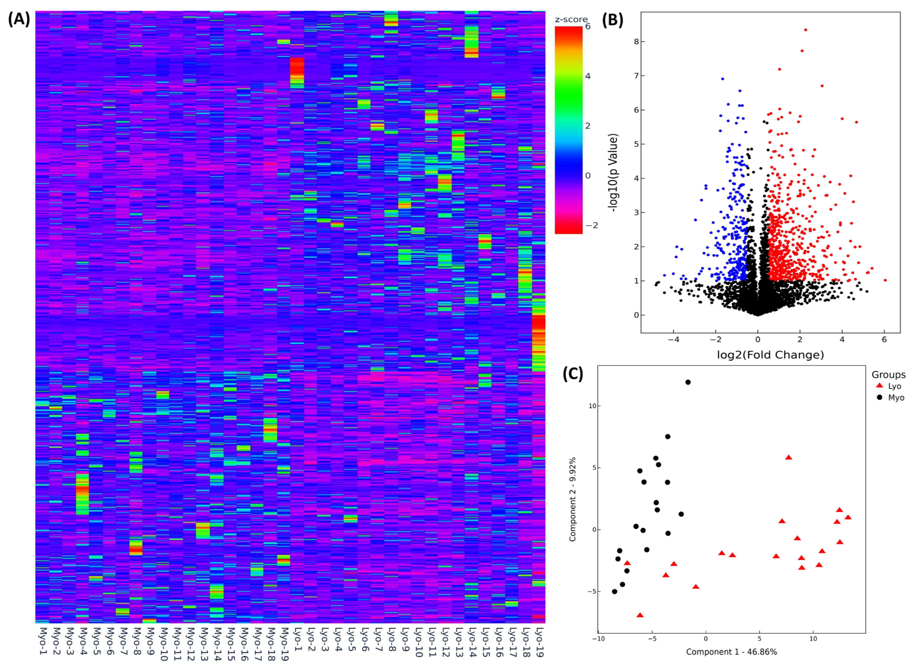
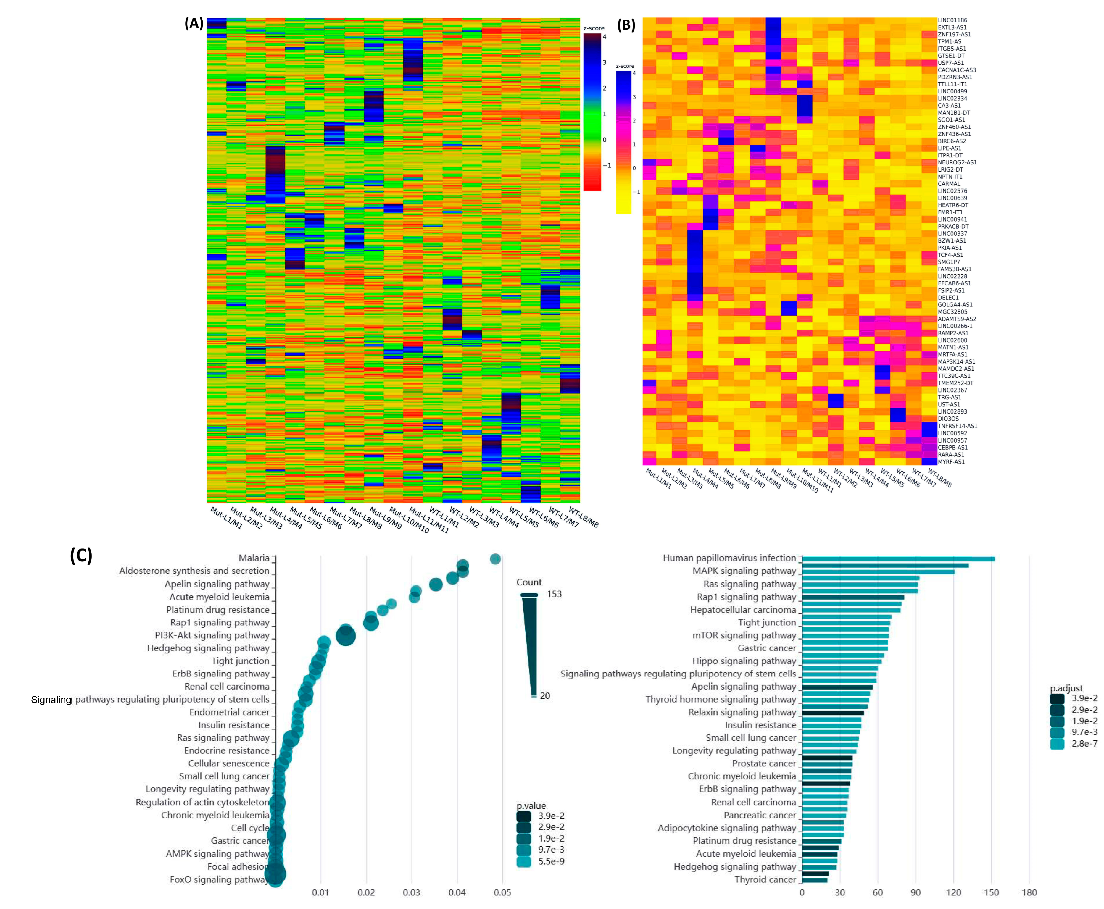
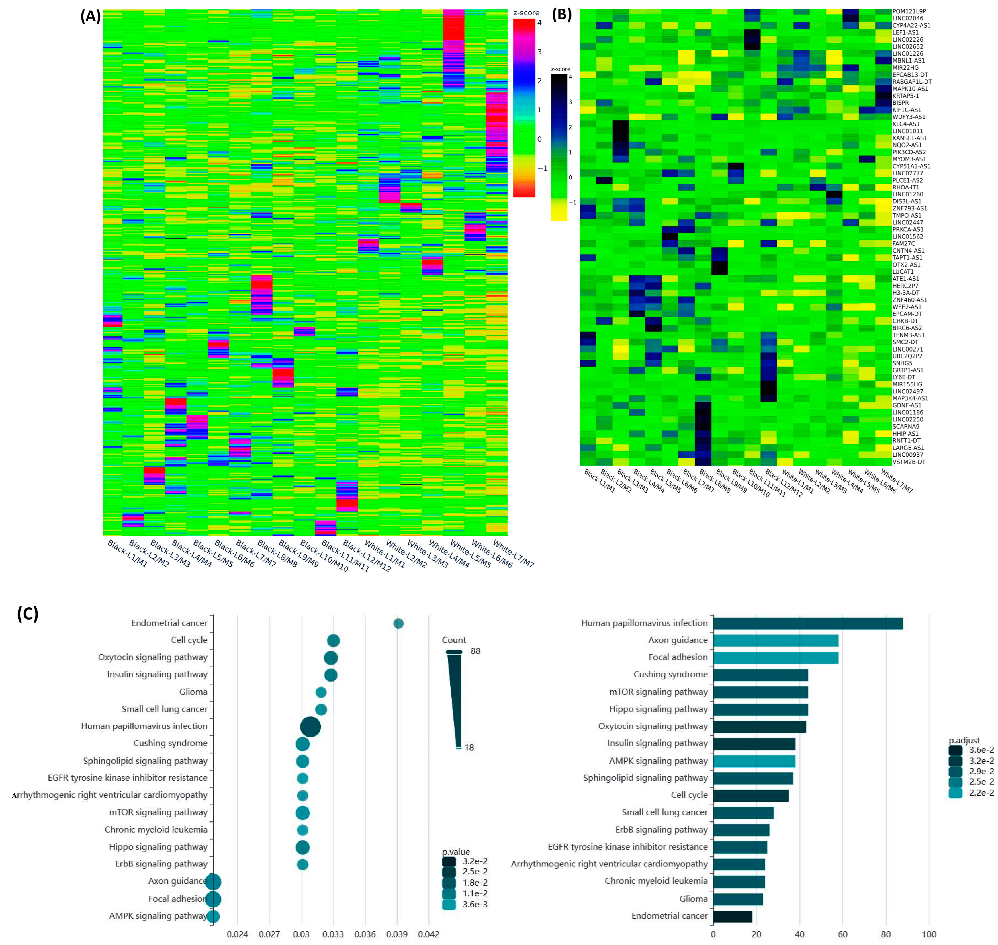
2.1. Differential Expression of Race/Ethnicity- and MED12 Mutation-Associated Long Non-Coding RNA Transcripts in Leiomyoma and Matched Myometrium

To elucidate the differential expression profile of lncRNAs in Lyo and paired Myo, we performed next-generation RNA sequencing (NGS) using RNA isolated from 19 paired leiomyomas. Following normalization, Hierarchical clustering and TreeView analysis resulted in identification of 4135 lncRNAs whose expression was altered, with increased expression of 1174 lncRNAs and decreased expression of 748 lncRNAs by 1.5-fold or greater in Lyo as compared to paired Myo (Figure 1A). After filtering through the volcano plot analysis, 1068 lncRNAs exhibited upregulation, while 718 lncRNAs displayed downregulation in leiomyomas as compared to their corresponding myometrium (fold change ≥ 1.5 and p < 0.05; Figure 1B). Following this analysis, principal component analysis (PCA) was conducted to assess the similarity in RNA transcript patterns between the two groups. The data’s high reliability was confirmed through k-mean analysis, which clustered the samples accordingly (Figure 1C).
We proceeded to investigate the variance in expression of lncRNAs between specimens with MED12 mutations and those without mutations. This analysis, performed as fold change (Leiomyoma/paired Myometrium), resulted in the identification of 3443 lncRNAs with altered expression, of which, the expression of 870 lncRNAs was increased, while the expression of 746 lncRNAs was decreased by 1.5-fold or greater in MED12-mutated specimens as compared with non-mutated specimens. Hierarchical clustering and TreeView analysis effectively categorized these transcripts into distinct groups (Figure 2A). Additionally, we identified 63 lncRNAs that exhibited more than 1.5-fold change (either up or down) specifically in the mutated specimens and with no change in the non-mutated specimens, as depicted in the heatmap (Figure 2B). The Gene Ontology (GO) and KEGG (Kyoto Encyclopedia of Genes and Genomes) pathway enrichment analysis for these 63 uniquely expressed lncRNAs in the mutated group indicated predominant involvement in cancer-associated cell signaling regulation (Figure 2C).
The comparison of the differential expression of lncRNAs based on race resulted in the identification of 3457 lncRNAs with altered expression, of which the expression of 851 lncRNAs was increased, while the expression of 876 lncRNAs was decreased by 1.5-fold or greater in the Black group as compared with the White group. Hierarchical clustering and TreeView analysis effectively categorized these lncRNAs into distinct groups (Figure 3A). We identified 65 lncRNAs that exhibited more than a 1.5-fold change (either up or down) specifically in the Black but not the White group, as illustrated in the heatmap (Figure 3B). The Gene Ontology (GO) and KEGG (Kyoto Encyclopedia of Genes and Genomes) pathway enrichment analysis for these 65 lncRNAs revealed that these lncRNAs were predominantly involved in the regulation of cell cycle and several cell-signaling pathways (Figure 3C).

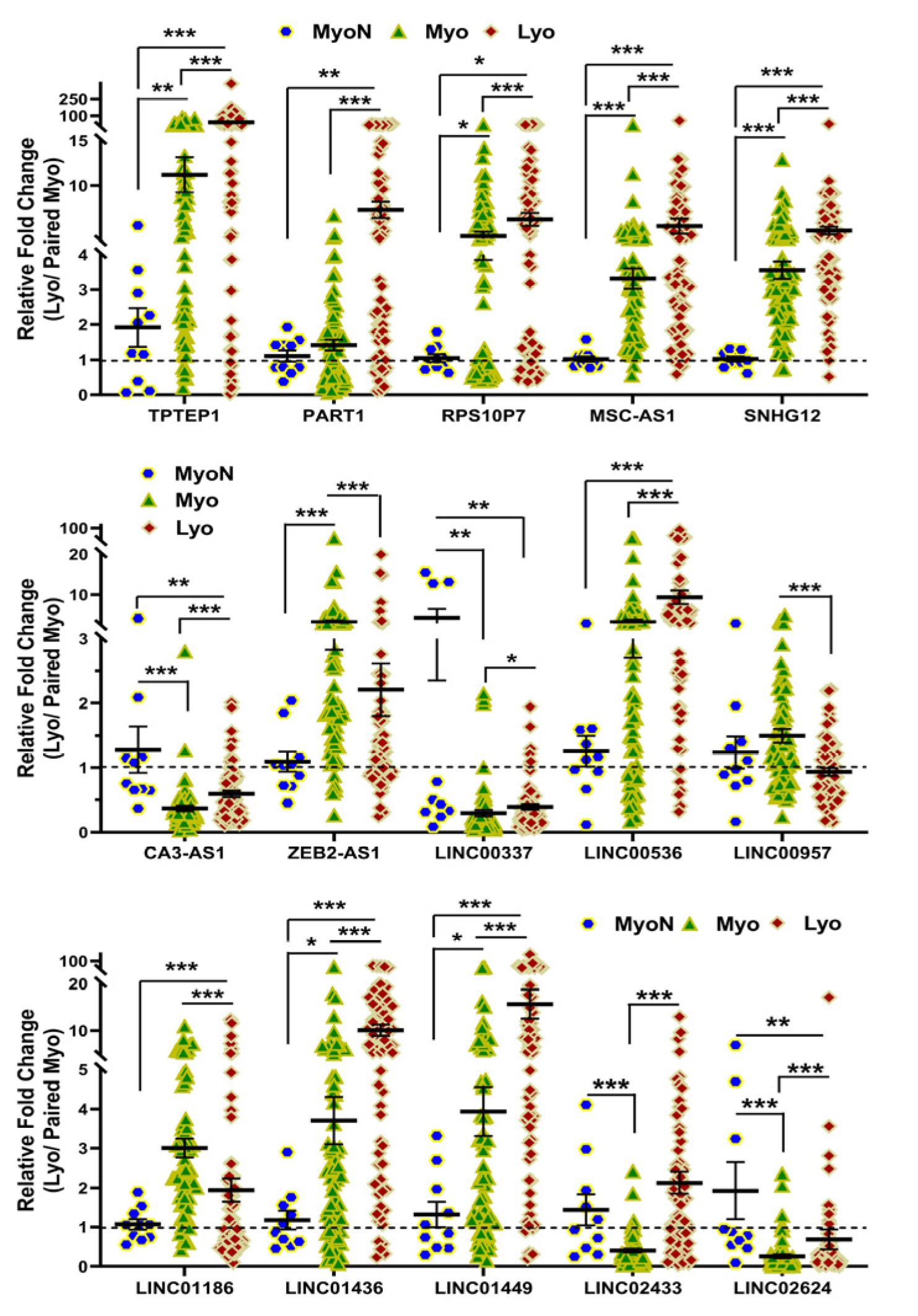
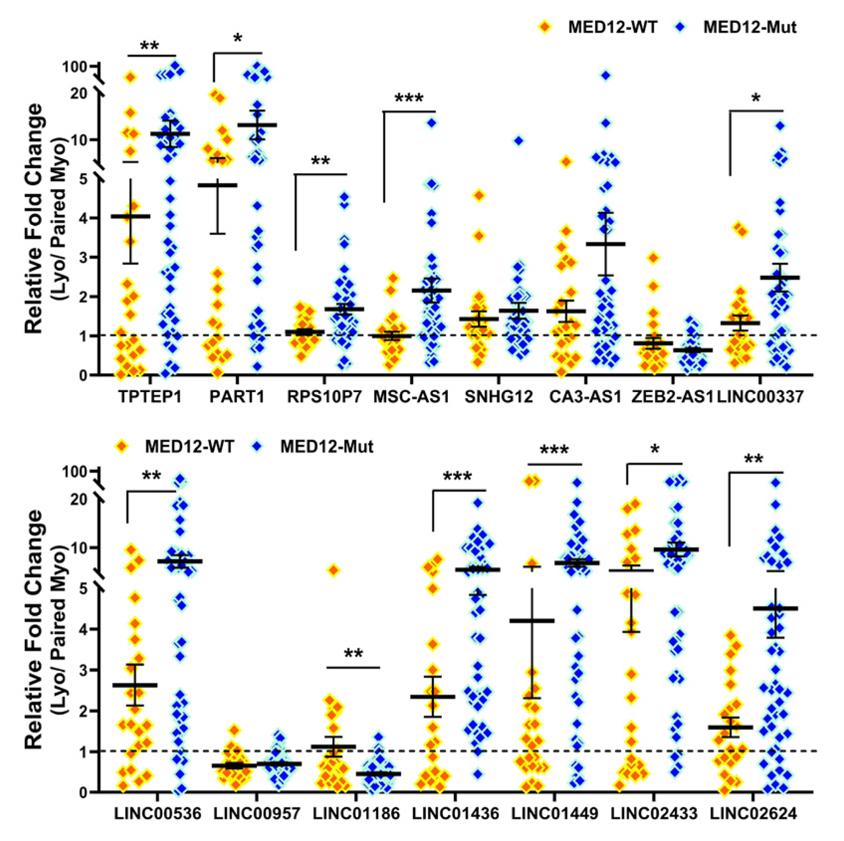
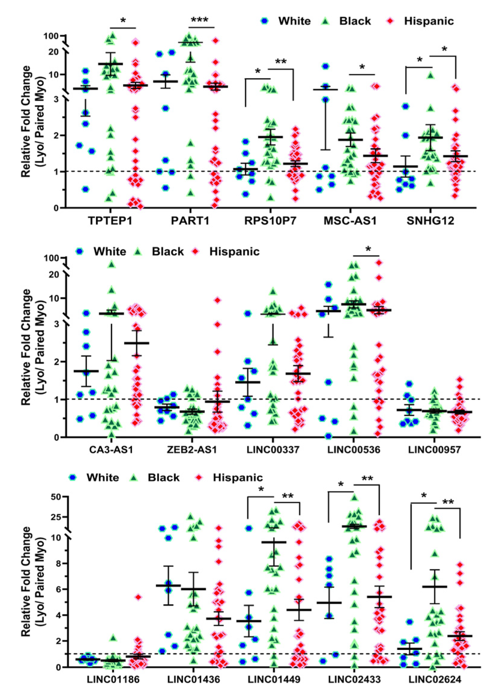
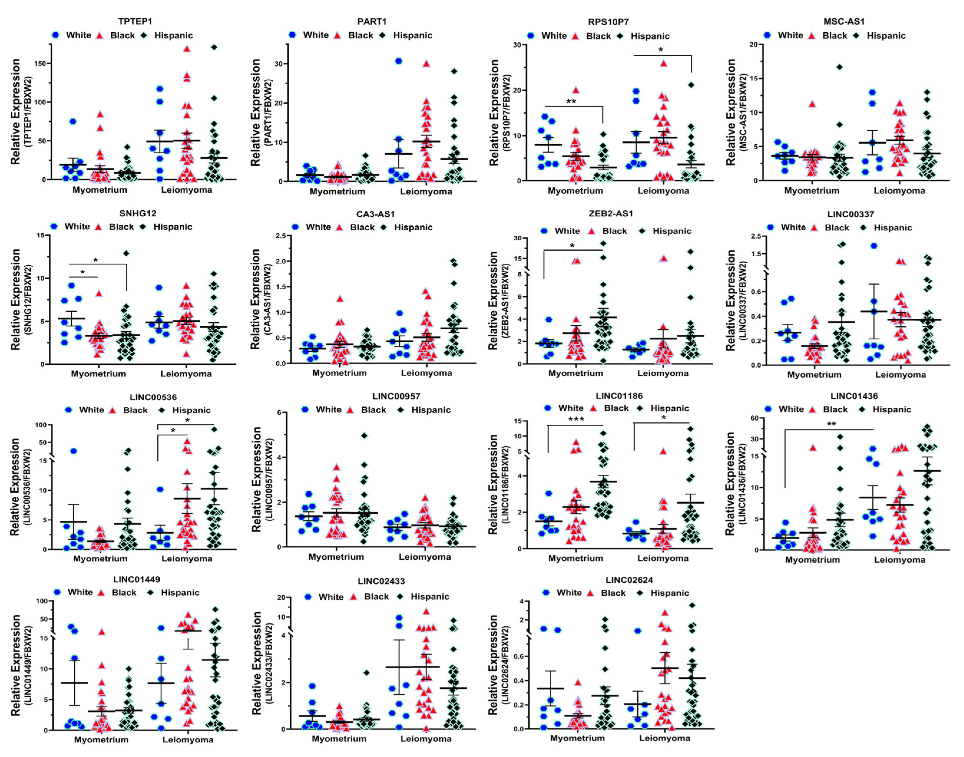
2.2. Validation of Race/Ethnicity- and MED12 Mutation-Associated Long Non-Coding RNA transcripts in Leiomyoma and Matched Myometrium

We next selected 15 novel lncRNAs from the NGS analysis and confirmed their expression by qRT-PCR in 10 non-diseased myometrial samples and in 69 Lyo with their matched Myo from premenopausal patients, including the same tissues that were used for RNAseq. Among these 15 lncRNAs, the expression of TPTEP1, PART1, RPS10P7, MSC-AS1, SNHG12, CA3-AS1, LINC00337, LINC00536, LINC01436, LINC01449, LINC02433 and LINC02624 was significantly higher, while the expression of ZEB2-AS1, LINC00957 and LINC01186 was significantly lower in Lyo as compared to matched Myo (Figure 4). When Lyo levels were compared with Myo from non-diseased uteri, the same pattern of change was noted, although the degree of overexpression was more exaggerated for TPTEP1, PART1, RPS10P7, MSC-AS1, SNHG12, LINC00536, LINC01436, and LINC01449 (Figure 4). The comparison of lncRNA levels in Myo from diseased uteri with Myo from non-diseased uteri revealed overexpression of TPTEP1, RPS10P7, MSC-AS1, SNHG12, ZEB2-AS1, LINC01436, and LINC01449 and reduced expression of CA3-AS1, LINC00337, LINC02433, and LINC02624 in the diseased Myo (Figure 4). When lncRNA levels were compared as fold change (Leiomyoma/paired Myometrium) in MED12-mutated and non-mutated specimens and Lyo from Black and White patients, the expression of TPTEP1, PART1, RPS10P7, MSC-AS1, LINC00337, LINC00536, LINC01436, LINC01449, LINC02433, and LINC02624 was found to be significantly higher, while the expression of LINC01186 was significantly lower in the MED12-mutated group (Figure 5). Moreover, RPS10P7, MSC-AS1, and LINC01186 expression were significantly altered in the MED12-mutated group but minimally changed in the non-mutated group (Figure 5). The expression analysis based on race revealed that the expression of RPS10P7, SNHG12, LINC01449, LINC02433, and LINC02624 was significantly higher in Lyo from Black women as compared with white women (Figure 6). Among these lncRNAs, the expression of RPS10P7 and SNHG12 in Lyo, as compared with their matched myometrium, was significantly higher in the Black group but minimally altered in the White group (Figure 6). Further comparisons among the three race/ethnicity groups were made based only on expression levels in the Lyo and in the Myo (Figure 7). A comparison of leiomyoma expression of lncRNAs revealed significantly higher levels of LINC00536 in the Black and Hispanic as compared to the White group and significantly higher levels of LINC01186 in the Hispanics as compared to the White group (Figure 7). A comparison of myometrial expression of lncRNAs revealed significantly lower levels of SNHG12 in the Black as compared to the White group and significantly higher ZEB2-AS1 and LINC01186 expressed in the Hispanics as compared to the White group (Figure 7). A summary of the analysis from Figure 4, Figure 5, Figure 6 and Figure 7 is shown in Table 1.
Based on their extensive interaction with miRNAs, we selected two lncRNAs, SNHG12 and LNC01449, for functional analysis using the Cytoscape software version 3.10.1 (Figure 8). This analysis was constructed based on ENCORI/starBase (Encyclopedia of RNA Interactomes) [37,38] and demonstrated the highlighted associations between target transcripts which were predominantly involved in the regulation of cell signaling and glycolysis. The analysis showed the involvement of many proteins such as MAP3K7, RPS6KB1, PPP2R2A, RPS6KB2, and HK2 in this interactive network, all of which are well established in leiomyoma pathogenesis [39,40,41,42,43], and other proteins such as ULK1, ELAVL1, SHC3, INSR, and IRS2, which are novel and require further investigation.

3. Discussion

Our present study indicates that lncRNAs are aberrantly expressed in Lyo, confirming our earlier study, which was based on a small sample set [23]. We identified a set of lncRNAs which were altered significantly in MED12-mutated Lyo and in Lyo from Black women, but minimally changed in non-mutated Lyo, and in Lyo from White women. We selected 15 lncRNAs (TPTEP1, PART1, RPS10P7, MSC-AS1, SNHG12, CA3-AS1, LINC00337, LINC00536, LINC01436, LINC01449, LINC02433, LINC02624, ZEB2-AS1, LINC00957, and LINC01186) from the NGS analysis and detected their expression with qRT-PCR in 10 non-diseased myometrial and 69 paired leiomyoma specimens. Our results confirmed that the expression of these lncRNAs was consistent with the NGS analysis, and that their expression was affected by the presence of MED12 mutation (TPTEP1, PART1, RPS10P7, MSC-AS1, LINC00337, LINC00536, LINC01436, LINC01449, LINC02433, LINC02624, and LINC01186) and race (RPS10P7, SNHG12, LINC01449, LINC02433, and LINC02624).
A strength of this study was that comparisons of lncRNA levels were made both with matched Myo from the diseased uteri and with non-diseased Myo in patients not using hormonal medications prior to surgery. A recent study [44] suggested the use of Myo from non-diseased uteri to compare with Lyo is more advantageous than a comparison made with diseased Myo because of potential effects of leiomyomas on the adjacent Myo. Our data showed that the comparison of gene expression between Lyo and adjacent Myo is similar to the comparison with non-diseased Myo, although for the upregulated lncRNAs, the magnitude of change was more pronounced for most lncRNAs when comparison was made with non-diseased Myo. This finding, along with the significant differences in expression of several lncRNAs in diseased versus non-diseased Myo, supports the notion that the presence of Lyo in and of itself could potentially affect the expression of lncRNA in the adjacent Myo, as previously suggested [44]. However, due to immense heterogeneity in fibroid tissues and our finding demonstrating a similar pattern of differential expression for most lncRNAs using diseased versus non-diseased Myo, the commonly used practice of comparing gene expression levels in Lyo with adjacent Myo is supported.
LncRNAs have been demonstrated to regulate gene expression at various levels through their interaction with miRNAs, mRNAs, DNA, and protein and by acting as epigenetic regulators in stem cell pluripotency and specific lineage commitment [45,46]. In a cell- and tissue-specific fashion, miRNAs and lncRNAs employ these mechanisms to regulate diverse cellular activities under physiological conditions. Additionally, they play a pivotal role in an extensive array of disorders, encompassing tumorigenesis, tumor progression, invasion, and metastasis, as well as cancer diagnosis and prognosis [33,45,46]. The functional role of lncRNAs in leiomyoma pathogenesis has recently been explored [28]. Cao et al. reported on the upregulation of the lncRNA H19 in leiomyomas and its association with elevated HMGA2 expression. Because of this linkage, the expression of several genes associated with cell proliferation, inflammation, and extracellular matrix (ECM) deposition was altered [47]. Our recent findings revealed increased expression levels of the lncRNAs XIST (X-inactive specific transcript) and MIAT (myocardial infarction-associated transcript) in leiomyomas [26,27]. These lncRNAs function as sponges for miR-29c and miR-200c, leading to an upregulation of their respective targets, which include COL3A1, COL1A1, FN1, and TGF-β3 [26,27]. In addition, the expression of MIAT correlated with the MED12 mutation status of the leiomyomas, and knock down of MIAT in leiomyoma smooth muscle cells’ spheroids blocked the effects of TGF-β3 on the induction of COL1A1 and COL3A1 expression [27].
Several of the lncRNAs identified in this study have been shown to be involved in tumorigenesis. Among these, TPTEP1 was reported to suppress diabetic retinopathy by reducing oxidative stress [48], inhibiting stemness and radio resistance [49], and it exhibited substantial antitumor effects in several cancers, including hepatocellular carcinoma (HCC) and non-small-cell lung cancer (NSCLC) [50,51]. In addition, recent studies indicated that the expression level of CA3-AS1 was decreased in gastric cancer and colorectal cancer [52,53]. The overexpression of CA3-AS1 suppressed the proliferation, invasion, and metastatic capabilities of both gastric cancer and colorectal cancer cells through modulation of the miR-93/BTG3/PTEN axis [52,53]. In contrast to the findings in malignant tumors, leiomyomas which are benign expressed higher levels of TPTEP1 and CA3-AS1. PART1, also known as prostate androgen-regulated transcript 1, was identified in exosomes and affected tumor progression via the miR-17-5p/SOCS6 axis and miR-302a-3p/CDC25A axis [54,55]. Another lncRNA, MSC-AS1, promoted cancer progression in gastric, colorectal, hepatocellular, melanoma, and ovarian cancer [56,57,58,59,60,61], and was shown to up-regulate CDK14 expression by acting as a molecular sponge for miR-29b-3p [62]. Our group and others have reported that leiomyomas, as compared with matched myometrium, have decreased expression of the miR-29 family, which plays essential roles in regulating the ECM and cell proliferation through epigenetic mechanisms [63,64,65]. SNHG12, also known by other names (LNC04080, ASLNC04080, C1orf79, LINC00100, PNAS-1230), was overexpressed in several cancers, including non-small-cell lung cancer, renal cell carcinoma, and prostate cancer, and its levels correlated with tumor size, progression, and metastasis [66]. This lncRNA was found to impair DNA damage repair, leading to lesional DNA damage, vascular senescence, and accelerated atherosclerosis [66]. The dysregulation of this lncRNA in Lyo might have relevance to its pathogenesis, as prior studies have shown the aberrant expression of DNA repair enzymes and genomic instability in Lyo [67].
Several lncRNAs LINC00337, LINC00536, and LINC01436, which were overexpressed in Lyo in a MED12 mutation-dependent manner, have been shown to have a role in tumorigenesis in other tissues. LINC00337 was upregulated in pancreatic ductal adenocarcinoma (PDAC) and facilitated PDAC cell proliferation, along with cell cycle transition, by acting as an E2F1 co-activator through its binding to E2F1 [68]. LINC00337 acts as an oncogenic lncRNA via its effect on EZH2, to repress p21 and promote gastric cancer cell proliferation [69], and through the miR-1285/YTHDF1 axis, to promote the progression of lung adenocarcinoma [70]. In both colorectal cancer (CRC) and non-small-cell lung cancer (NSCLC), this lncRNA plays a role in promoting tumorigenesis, angiogenesis, and tumor progression. It achieves this by recruiting DNMT1 to suppress the expression of CNN1 and TIMP2 [71,72]. Thus, LINC00337 may be involved in the stimulation of cell proliferation in leiomyomas, since leiomyomas express higher levels of E2F1, EZH2, and DNMT1 [65,73]. LINC00536 has been demonstrated to enhance the development and progression of breast cancer through the miR-214/ROCK1 axis and the miR-4282/CENPF axis [74,75]. Additionally, it plays a role in promoting hepatocellular carcinoma via the miR-203b/VEGFA axis and contributes to the progression of bladder cancer by modulating the Wnt3a/β-Catenin signaling pathway [76,77]. Leiomyomas are also characterized by activation of the WNT/β-catenin pathway, which is correlated with MED12 mutation status [13]. LINC01436 has been shown to be involved in gastric cancer cell proliferation, apoptosis, metastasis, and radio resistance through upregulation of MAPK1 and FBOX11 via sponging and epigenetically silencing miR-585 [78,79]. Thus, LINC01436 may be involved in leiomyoma progression. Our NGS analysis and in vitro confirmation showed the expression of ZEB2-AS1, LINC00957, and LINC01186 was significantly lower in Lyo as compared to paired Myo. The lncRNA, ZEB2-AS1, is a natural antisense transcript that corresponds to the 5’ UTR of zinc finger E-box binding homeobox 2 (ZEB2). It has been reported to promote tumorigenesis and cancer progression in numerous types of cancer, including gastric, lung, colon, pancreatic, and breast cancer [80]. In an in vivo mouse model of cardiac hypertrophy, the expression levels of ZEB2-AS1 were elevated, and they altered the progression of cardiac hypertrophy through downregulation of PTEN (phosphatase and tensin homolog) [81]. Our results showed the expression of LINC01186 was downregulated in leiomyomas. Recent studies also demonstrated a lower level of LINC01186 in lung cancer and papillary thyroid carcinoma (PTC). This lncRNA was involved in the regulation of EMT and cell proliferation, migration, and invasion in both cancer cells [82,83].
More recently, a role for MED12 in tumor initiation has been proposed, as evidenced by the development of leiomyoma-like tumors in transgenic mice with a uterine conditional mutation in MED12 [8,84]. Leiomyomas with a MED12 mutation are smaller in size and are frequently located subserously [85]. MED12 mutation-negative leiomyomas display copy number alterations in several other mediator complex subunits, such as MED18, MED8, CDK8, and the long non-coding RNA, RNA340 (CASC15). Mutation in the MED12 gene has been shown to disrupt its ability to activate cyclin C-dependent CDK8 [86] and its CDK19 stimulatory activity [87]. Furthermore, the silencing of MED12 in the immortalized human uterine fibroid cell line (HuLM) inhibited Wnt/β-catenin signaling and sex steroid receptor signaling, as well as proteins associated with cell cycle and fibrosis [13]. Our results demonstrated that the expression of several lncRNAs was affected by the presence of a MED12 mutation, which could be contributing to the differences in tumor size and progression in mutated leiomyomas as compared with non-mutated leiomyomas and merits further investigation.
Race plays an important role in leiomyoma severity and progression, with Black patients having larger-size and more numerous leiomyomas, a greater severity of symptoms, and earlier onset [2,88]. Previous research has presented evidence elucidating potential mechanisms contributing to the racial disparity in tumor development and progression, including a higher incidence of vitamin D deficiency in Black patients [89,90]; higher levels of aromatase (CYP19), progesterone receptor A (PR-A), and lower levels of retinoic acid receptor-α (RARA) [89,90]; and increased polymorphism of the genes ER (estrogen receptor), CYP17 (cytochrome P450C17α), and COMT, which are involved in estrogen synthesis, in tumors from Black women [89,90]. Recent reports from our laboratory have revealed a dysregulation of protein-coding and noncoding genes in a race-dependent manner in Lyo, including genes related to tryptophan metabolism [91,92] and SE-lncRNAs/mRNA pairs [29]. In addition, several miRNAs, including miR-200c, miR-21, miR-23b, and Let-7s, were dysregulated significantly in a race-specific manner in leiomyomas [93,94]. In the present study, we showed a race-dependent expression of several lncRNAs, which could be a contributing factor to differences in the expression of protein-coding genes and the racial disparity in fibroid symptomatology.
In summary, our data provide a comprehensive expression profile of lncRNAs in leiomyomas and demonstrate the influence of race/ethnicity and MED12 mutation on their expression, indicating their relevance to fibroid pathogenesis. Our findings also revealed that the differential expression pattern of lncRNAs was similar when comparing lncRNA expression levels in Lyo with adjacent matched Myo versus non-diseased Myo. The aberrant expression of lncRNAs in Lyo through various mechanisms such as interaction with miRNAs, mRNAs, DNA, and proteins could influence the expression of protein-coding genes associated with cell proliferation, ECM accumulation, and inflammation in Lyo.

4. Materials and Methods

4.1. Myometrium and Leiomyoma Tissues Collection

To reduce the variance among Lyo, tumors between 3 to 5 cm in diameter and with intramural location were obtained from premenopausal women (n = 79; aged 30–54) who underwent hysterectomy for symptomatic Lyo at Harbor-UCLA Medical Center. Normal myometrium was also obtained from surgical cases performed for non-Lyo indications such as prolapse and other urogynecologic disorders. Both gross, histologic and ultrasound studies did not show the presence of Lyo in these 10 specimens. Prior approval from the Institutional Review Board (18CR-31752-01R) at the Lundquist Institute was obtained. Informed consent was obtained from all the patients participating in the study who were not taking any hormonal medications for at least 3 months prior to surgery. The tissues were snap-frozen and stored in liquid nitrogen for further analysis as previously described [25,95]. The racial distribution for the 10 non-diseased myometrial specimens was White (N = 1) and Hispanic (N = 9), and for the 69 pairs was White (n = 8); Black (n = 24); Hispanic (n = 33), and Asian (n = 4).

4.2. MED12 Mutation Analysis

Genomic DNA from leiomyomas and paired myometrial specimens from premenopausal women (n = 69) was extracted from 100 mg of freshly frozen tissue using MagaZorb DNA Mini-Prep Kit (Promega, Madison, WI, USA) according to the manufacturer’s protocol. PCR amplification and Sanger sequencing (Laragen Inc. Culver City, CA, USA) was performed to investigate the MED12 exon 2 mutations using the primer sequences in the 5′–3′ direction: sense, GCCCTTTCACCTTGTTCCTT and antisense, TGTCCCTATAAGTCTTCCCAACC, as previously described [27]. The 19 pairs of tissues used for next-generation RNA sequencing were from 11 MED12 mutation-positive and 8 MED12 mutation-negative leiomyomas. The mutation analysis of the specimens (n = 69) indicated that 46 leiomyomas had the MED12 mutations (46/69 pairs; 66.7%) with no mutations in the myometrium. Missense mutations in MED12 exon 2 were the most frequent alteration (38/46 pairs), followed by in-frame insertion–deletion type mutations (8/46 pairs). The missense mutations in exon 2 included c.130G>C (p.Gly44Arg) (5/38 pairs), c.130G>A (p.Gly44Ser) (7/38 pairs), c.130G>T (p.Gly44Cys) (2/38 pairs), c.131G>C (p.Gly44Ala) (3/38 pairs), c.131G>A (p.Gly44Asp) (15/38 pairs), c.131G>T (p.Gly44Val) (5/38 pairs), and c.128A>C (p.Gln43Pro) (1/38 pairs). The MED12 mutation rate was 55.6% (5/9 pairs) in White patients, 82.6% in Black (19/23 pairs), and 60.6% in the Hispanic group (20/33 pairs).

4.3. RNA Sequencing and Bioinformatic Analysis

Total RNA was extracted from leiomyoma and matched myometrium (n = 19) using TRIzol (Thermo Fisher Scientific Inc., Waltham, MA, USA). RNA concentration and integrity was determined using a Nanodrop 2000c spectrophotometer (Thermo Scientific, Wilmington, DE, USA) and Agilent 2100 Bioanalyzer (Agilent Technologies, Santa Clara, CA, USA) as previously described [29,96]. Samples with RNA integrity numbers (RIN) greater than or equal to 9 were used for library preparation. One microgram of total RNA from each tissue was used to produce strand-specific cDNA libraries using the Truseq (Illumina, San Diego, CA, USA) according to manufacturer’s instructions. The RNA sequencing was carried out at Novogene Corporation Inc., (Sacramento, CA, USA) and bioinformatics analysis was performed as previously described [19,20]. To visualize the strength of differential gene expression, the Hierarchical clustering and TreeView graph, volcano plot, principal component analysis (PCA) plot, and the Gene Ontology (GO) and KEGG (Kyoto Encyclopedia of Genes and Genomes) Pathway Enrichment Analysis plot were made using Flaski and NcPath [97,98], and the interactive network of lncRNA-miRNA-mRNA was analyzed and made with the ENCORI/starBase (Encyclopedia of RNA Interactomes) and Cytoscape software Version 3.10.1 [37,38,99]. Overall, all the differential gene expressions were acceptable for subsequent statistical analysis. The RNA sequencing data is deposited in Gene Expression Omnibus (GEO) database with accession number (GSE224991).

4.4. Quantitative RT-PCR

Briefly, 2 μg of RNA was reverse transcribed using random primers for selected genes according to the manufacturer’s guidelines (Applied Biosystems, Carlsbad, CA, USA). Quantitative RT-PCR was carried out using SYBR gene expression master mix (Applied Biosystems) as previously described [25]. The expression levels of selected genes were quantified using Invitrogen StepOne System, with FBXW2 (F-box and WD repeat domain containing 2) used for normalization [100]. All reactions were run in triplicate, and relative mRNA expression was determined using the comparative cycle threshold method (2−ΔΔCq), as recommended by the supplier (Applied Biosystems). Values were expressed as fold change compared to the control group. The primer sequences in the 5′–3′ direction used are listed in Supplementary Table S1.

4.5. Statistical Analysis

Throughout the text, results are presented as mean ± SEM and analyzed with PRISM software (version 10.1.2, Graph-Pad, San Diego, CA, USA). Dataset normality was determined using the Kolmogorov–Smirnoff test. The data presented in this study was not normally distributed and therefore non-parametric tests were used. Comparisons involving two groups were analyzed using the Mann–Whitney test (Figure 5). Kruskal–Wallis test was used for comparisons involving multiple groups (Figure 4, Figure 6 and Figure 7). Statistical significance was established at p < 0.05.

Supplementary Materials

The following supporting information can be downloaded at: https://www.mdpi.com/article/10.3390/ijms25021307/s1.

Author Contributions

O.K. and T.-D.C. designed the research studies; T.-D.C., N.T., S.R. and D.B. conducted the experiments; T.-D.C. and N.T. analyzed the data; T.-D.C. and O.K. interpreted the data; T.-D.C. and O.K. wrote and edited the manuscript. No honorarium or other form of payment was given to anyone to produce the manuscript. All authors have read and agreed to the published version of the manuscript.

Funding

This study was supported in part by the National Institute of Health (HD100529, HD100529-02S1, HD101852, and HD109286).

Institutional Review Board Statement

The study was approved by The Lundquist Institute Institutional Review Board (18CR-31752-01R).

Informed Consent Statement

Written informed consent has been obtained from all participants in the study to publish this paper.

Data Availability Statement

Raw data were generated at The Lundquist Institute. Derived data supporting the findings of this study are available from the corresponding author O.K on request.

Conflicts of Interest

The authors declare no conflict of interest.

References

  1. Islam, M.S.; Ciavattini, A.; Petraglia, F.; Castellucci, M.; Ciarmela, P. Extracellular matrix in uterine leiomyoma pathogenesis: A potential target for future therapeutics. Hum. Reprod. Update 2018, 24, 59–85. [Google Scholar] [CrossRef] [PubMed]
  2. Taylor, D.K.; Holthouser, K.; Segars, J.H.; Leppert, P.C. Recent scientific advances in leiomyoma (uterine fibroids) research facilitates better understanding and management. F1000Res 2015, 4, 183. [Google Scholar] [CrossRef] [PubMed]
  3. Chegini, N. Proinflammatory and profibrotic mediators: Principal effectors of leiomyoma development as a fibrotic disorder. Semin. Reprod. Med. 2010, 28, 180–203. [Google Scholar] [CrossRef] [PubMed]
  4. Segars, J.H.; Parrott, E.C.; Nagel, J.D.; Guo, X.C.; Gao, X.; Birnbaum, L.S.; Pinn, V.W.; Dixon, D. Proceedings from the Third National Institutes of Health International Congress on Advances in Uterine Leiomyoma Research: Comprehensive review, conference summary and future recommendations. Hum. Reprod. Update 2014, 20, 309–333. [Google Scholar] [CrossRef]
  5. Stewart, E.A.; Cookson, C.L.; Gandolfo, R.A.; Schulze-Rath, R. Epidemiology of uterine fibroids: A systematic review. BJOG 2017, 124, 1501–1512. [Google Scholar] [CrossRef]
  6. Wise, L.A.; Laughlin-Tommaso, S.K. Epidemiology of Uterine Fibroids: From Menarche to Menopause. Clin. Obstet. Gynecol. 2016, 59, 2–24. [Google Scholar] [CrossRef]
  7. Marsh, E.E.; Ekpo, G.E.; Cardozo, E.R.; Brocks, M.; Dune, T.; Cohen, L.S. Racial differences in fibroid prevalence and ultrasound findings in asymptomatic young women (18–30 years old): A pilot study. Fertil. Steril. 2013, 99, 1951–1957. [Google Scholar] [CrossRef]
  8. Yatsenko, S.A.; Mittal, P.; Wood-Trageser, M.A.; Jones, M.W.; Surti, U.; Edwards, R.P.; Sood, A.K.; Rajkovic, A. Highly heterogeneous genomic landscape of uterine leiomyomas by whole exome sequencing and genome-wide arrays. Fertil. Steril. 2017, 107, 457–466.e9. [Google Scholar] [CrossRef]
  9. Gallagher, C.S.; Morton, C.C. Genetic Association Studies in Uterine Fibroids: Risk Alleles Presage the Path to Personalized Therapies. Semin. Reprod. Med. 2016, 34, 235–241. [Google Scholar] [CrossRef]
  10. Liegl-Atzwanger, B.; Heitzer, E.; Flicker, K.; Muller, S.; Ulz, P.; Saglam, O.; Tavassoli, F.; Devouassoux-Shisheboran, M.; Geigl, J.; Moinfar, F. Exploring chromosomal abnormalities and genetic changes in uterine smooth muscle tumors. Mod. Pathol. 2016, 29, 1262–1277. [Google Scholar] [CrossRef]
  11. Makinen, N.; Mehine, M.; Tolvanen, J.; Kaasinen, E.; Li, Y.; Lehtonen, H.J.; Gentile, M.; Yan, J.; Enge, M.; Taipale, M.; et al. MED12, the mediator complex subunit 12 gene, is mutated at high frequency in uterine leiomyomas. Science 2011, 334, 252–255. [Google Scholar] [CrossRef] [PubMed]
  12. Markowski, D.N.; Bartnitzke, S.; Löning, T.; Drieschner, N.; Helmke, B.M.; Bullerdiek, J. MED12 mutations in uterine fibroids--their relationship to cytogenetic subgroups. Int. J. Cancer 2012, 131, 1528–1536. [Google Scholar] [CrossRef]
  13. Al-Hendy, A.; Laknaur, A.; Diamond, M.P.; Ismail, N.; Boyer, T.G.; Halder, S.K. Silencing Med12 Gene Reduces Proliferation of Human Leiomyoma Cells Mediated via Wnt/beta-Catenin Signaling Pathway. Endocrinology 2017, 158, 592–603. [Google Scholar] [PubMed]
  14. El Andaloussi, A.; Al-Hendy, A.; Ismail, N.; Boyer, T.G.; Halder, S.K. Introduction of Somatic Mutation in MED12 Induces Wnt4/β-Catenin and Disrupts Autophagy in Human Uterine Myometrial Cell. Reprod. Sci. 2020, 27, 823–832. [Google Scholar] [CrossRef] [PubMed]
  15. Verger, A.; Monté, D.; Villeret, V. Twenty years of Mediator complex structural studies. Biochem. Soc. Trans. 2019, 47, 399–410. [Google Scholar] [CrossRef]
  16. Zhang, S.; O’Regan, R.; Xu, W. The emerging role of mediator complex subunit 12 in tumorigenesis and response to chemotherapeutics. Cancer 2020, 126, 939–948. [Google Scholar] [CrossRef] [PubMed]
  17. Zhou, H.; Kim, S.; Ishii, S.; Boyer, T.G. Mediator modulates Gli3-dependent Sonic hedgehog signaling. Mol. Cell. Biol. 2006, 26, 8667–8682. [Google Scholar] [CrossRef]
  18. Huang, S.; Holzel, M.; Knijnenburg, T.; Schlicker, A.; Roepman, P.; McDermott, U.; Garnett, M.; Grernrum, W.; Sun, C.; Prahallad, A.; et al. MED12 controls the response to multiple cancer drugs through regulation of TGF-beta receptor signaling. Cell 2012, 151, 937–950. [Google Scholar] [CrossRef]
  19. Chuang, T.D.; Ton, N.; Rysling, S.; Quintanilla, D.; Boos, D.; Gao, J.; McSwiggin, H.; Yan, W.; Khorram, O. The Influence of Race/Ethnicity on the Transcriptomic Landscape of Uterine Fibroids. Int. J. Mol. Sci. 2023, 24, 13441. [Google Scholar] [CrossRef]
  20. Chuang, T.D.; Gao, J.; Quintanilla, D.; McSwiggin, H.; Boos, D.; Yan, W.; Khorram, O. Differential Expression of MED12-Associated Coding RNA Transcripts in Uterine Leiomyomas. Int. J. Mol. Sci. 2023, 24, 3742. [Google Scholar] [CrossRef]
  21. Chi, Y.; Wang, D.; Wang, J.; Yu, W.; Yang, J. Long Non-Coding RNA in the Pathogenesis of Cancers. Cells 2019, 8, 1015. [Google Scholar] [CrossRef] [PubMed]
  22. Huang, W.; Li, H.; Yu, Q.; Xiao, W.; Wang, D.O. LncRNA-mediated DNA methylation: An emerging mechanism in cancer and beyond. J. Exp. Clin. Cancer Res. 2022, 41, 100. [Google Scholar] [CrossRef] [PubMed]
  23. Chuang, T.D.; Khorram, O. Expression Profiling of lncRNAs, miRNAs, and mRNAs and Their Differential Expression in Leiomyoma Using Next-Generation RNA Sequencing. Reprod. Sci. 2018, 25, 246–255. [Google Scholar] [CrossRef]
  24. Guo, H.; Zhang, X.; Dong, R.; Liu, X.; Li, Y.; Lu, S.; Xu, L.; Wang, Y.; Wang, X.; Hou, D.; et al. Integrated analysis of long noncoding RNAs and mRNAs reveals their potential roles in the pathogenesis of uterine leiomyomas. Oncotarget 2014, 5, 8625–8636. [Google Scholar] [CrossRef] [PubMed]
  25. Chuang, T.D.; Xie, Y.; Yan, W.; Khorram, O. Next-generation sequencing reveals differentially expressed small noncoding RNAs in uterine leiomyoma. Fertil. Steril. 2018, 109, 919–929. [Google Scholar] [CrossRef] [PubMed]
  26. Chuang, T.D.; Rehan, A.; Khorram, O. Functional role of the long noncoding RNA X-inactive specific transcript in leiomyoma pathogenesis. Fertil. Steril. 2021, 115, 238–247. [Google Scholar] [CrossRef] [PubMed]
  27. Chuang, T.D.; Quintanilla, D.; Boos, D.; Khorram, O. Long Noncoding RNA MIAT Modulates the Extracellular Matrix Deposition in Leiomyomas by Sponging MiR-29 Family. Endocrinology 2021, 162, bqab186. [Google Scholar] [CrossRef]
  28. Falahati, Z.; Mohseni-Dargah, M.; Mirfakhraie, R. Emerging Roles of Long Non-coding RNAs in Uterine Leiomyoma Pathogenesis: A Review. Reprod. Sci. 2022, 29, 1086–1101. [Google Scholar] [CrossRef]
  29. Chuang, T.D.; Quintanilla, D.; Boos, D.; Khorram, O. Differential Expression of Super-Enhancer-Associated Long Non-coding RNAs in Uterine Leiomyomas. Reprod. Sci. 2022, 29, 2960–2976. [Google Scholar] [CrossRef]
  30. Beermann, J.; Piccoli, M.-T.; Viereck, J.; Thum, T. Non-coding RNAs in Development and Disease: Background, Mechanisms, and Therapeutic Approaches. Physiol. Rev. 2016, 96, 1297–1325. [Google Scholar] [CrossRef]
  31. Dey, B.K.; Mueller, A.C.; Dutta, A. Long non-coding RNAs as emerging regulators of differentiation, development, and disease. Transcription 2014, 5, e944014. [Google Scholar] [CrossRef] [PubMed]
  32. Ergun, S.; Oztuzcu, S. Oncocers: ceRNA-mediated cross-talk by sponging miRNAs in oncogenic pathways. Tumor Biol. 2015, 36, 3129–3136. [Google Scholar] [CrossRef]
  33. Thomson, D.W.; Dinger, M.E. Endogenous microRNA sponges: Evidence and controversy. Nat. Rev. Genet. 2016, 17, 272–283. [Google Scholar] [CrossRef] [PubMed]
  34. Bradner, J.E.; Hnisz, D.; Young, R.A. Transcriptional Addiction in Cancer. Cell 2017, 168, 629–643. [Google Scholar] [CrossRef] [PubMed]
  35. Soibam, B. Super-lncRNAs: Identification of lncRNAs that target super-enhancers via RNA:DNA:DNA triplex formation. RNA 2017, 23, 1729–1742. [Google Scholar] [CrossRef]
  36. Kiss, T.; Fayet-Lebaron, E.; Jady, B.E. Box H/ACA small ribonucleoproteins. Mol. Cell 2010, 37, 597–606. [Google Scholar] [CrossRef]
  37. Li, J.H.; Liu, S.; Zhou, H.; Qu, L.H.; Yang, J.H. starBase v2.0: Decoding miRNA-ceRNA, miRNA-ncRNA and protein-RNA interaction networks from large-scale CLIP-Seq data. Nucleic Acids Res. 2014, 42, D92–D97. [Google Scholar] [CrossRef]
  38. Yang, J.H.; Li, J.H.; Shao, P.; Zhou, H.; Chen, Y.Q.; Qu, L.H. starBase: A database for exploring microRNA-mRNA interaction maps from Argonaute CLIP-Seq and Degradome-Seq data. Nucleic Acids Res. 2011, 39, D202–D209. [Google Scholar] [CrossRef]
  39. Aissani, B.; Zhang, K.; Wiener, H. Follow-up to genome-wide linkage and admixture mapping studies implicates components of the extracellular matrix in susceptibility to and size of uterine fibroids. Fertil. Steril. 2015, 103, 528–534.e13. [Google Scholar] [CrossRef]
  40. Chiang, S. Recent advances in smooth muscle tumors with PGR and PLAG1 gene fusions and myofibroblastic uterine neoplasms. Genes Chromosomes Cancer 2021, 60, 138–146. [Google Scholar] [CrossRef]
  41. Conconi, D.; Redaelli, S.; Lissoni, A.A.; Cilibrasi, C.; Perego, P.; Gautiero, E.; Sala, E.; Paderno, M.; Dalprà, L.; Landoni, F.; et al. Genomic and Epigenomic Profile of Uterine Smooth Muscle Tumors of Uncertain Malignant Potential (STUMPs) Revealed Similarities and Differences with Leiomyomas and Leiomyosarcomas. Int. J. Mol. Sci. 2021, 22, 1580. [Google Scholar] [CrossRef] [PubMed]
  42. Kerins, M.J.; Milligan, J.; Wohlschlegel, J.A.; Ooi, A. Fumarate hydratase inactivation in hereditary leiomyomatosis and renal cell cancer is synthetic lethal with ferroptosis induction. Cancer Sci. 2018, 109, 2757–2766. [Google Scholar] [CrossRef] [PubMed]
  43. Chuang, T.D.; Khorram, O. miR-200c Regulates IL8 Expression by Targeting IKBKB: A Potential Mediator of Inflammation in Leiomyoma Pathogenesis. PLoS ONE 2014, 9, e95370. [Google Scholar] [CrossRef] [PubMed]
  44. Paul, E.N.; Grey, J.A.; Carpenter, T.J.; Madaj, Z.B.; Lau, K.H.; Givan, S.A.; Burns, G.W.; Chandler, R.L.; Wegienka, G.R.; Shen, H.; et al. Transcriptome and DNA methylome analyses reveal underlying mechanisms for the racial disparity in uterine fibroids. JCI Insight 2022, 7, e160274. [Google Scholar] [CrossRef] [PubMed]
  45. Statello, L.; Guo, C.J.; Chen, L.L.; Huarte, M. Gene regulation by long non-coding RNAs and its biological functions. Nat. Rev. Mol. Cell. Biol. 2021, 22, 96–118. [Google Scholar] [CrossRef] [PubMed]
  46. Mattick, J.S.; Amaral, P.P.; Carninci, P.; Carpenter, S.; Chang, H.Y.; Chen, L.L.; Chen, R.; Dean, C.; Dinger, M.E.; Fitzgerald, K.A.; et al. Long non-coding RNAs: Definitions, functions, challenges and recommendations. Nat. Rev. Mol. Cell. Biol. 2023, 24, 430–447. [Google Scholar] [CrossRef] [PubMed]
  47. Cao, T.; Jiang, Y.; Wang, Z.; Zhang, N.; Al-Hendy, A.; Mamillapalli, R.; Kallen, A.N.; Kodaman, P.; Taylor, H.S.; Li, D.; et al. H19 lncRNA identified as a master regulator of genes that drive uterine leiomyomas. Oncogene 2019, 38, 5356–5366. [Google Scholar] [CrossRef]
  48. Wang, X.; Zhou, X.; Wang, F.; Zhang, N.; Zhang, Y.; Ao, Z.; He, F. Long noncoding RNA TPTEP1 suppresses diabetic retinopathy by reducing oxidative stress and targeting the miR-489-3p/NRF2 axis. Acta Biochim. Pol. 2023, 70, 45–50. [Google Scholar] [CrossRef]
  49. Tang, T.; Wang, L.X.; Yang, M.L.; Zhang, R.M. lncRNA TPTEP1 inhibits stemness and radioresistance of glioma through miR-106a-5p-mediated P38 MAPK signaling. Mol. Med. Rep. 2020, 22, 4857–4867. [Google Scholar] [CrossRef]
  50. Sun, X.; Qiu, X. LncRNA TPTEP1 inhibited the proliferation and metastasis of non-small cell lung cancer cells by targeting miR-761/LATS2 axis. Am. J. Transl. Res. 2021, 13, 8653–8669. [Google Scholar]
  51. Dong, Y.; Wang, Q.; Sun, J.; Liu, H.; Wang, H. Long non-coding RNA TPTEP1 exerts inhibitory effects on hepatocellular carcinoma by impairing microRNA-454-3p-mediated DLG5 downregulation. Dig. Liver Dis. 2022, 54, 268–279. [Google Scholar] [CrossRef] [PubMed]
  52. Zhang, X.Y.; Zhuang, H.W.; Wang, J.; Shen, Y.; Bu, Y.Z.; Guan, B.G.; Xu, F.; Dou, J. Long noncoding RNA CA3-AS1 suppresses gastric cancer migration and invasion by sponging miR-93-5p and targeting BTG3. Gene Ther. 2022, 29, 566–574. [Google Scholar] [CrossRef] [PubMed]
  53. Wei, H.; Yang, Z.; Lin, B. Overexpression of long non coding RNA CA3-AS1 suppresses proliferation, invasion and promotes apoptosis via miRNA-93/PTEN axis in colorectal cancer. Gene 2019, 687, 9–15. [Google Scholar] [CrossRef] [PubMed]
  54. Du, Y.; Shuai, Y.; Wang, H.; Li, H.; Li, Y. Exosome-mediated long noncoding RNA (lncRNA) PART1 suppresses malignant progression of oral squamous cell carcinoma via miR-17-5p/SOCS6 axis. Turk. J. Med. Sci. 2023, 53, 630–639. [Google Scholar] [CrossRef] [PubMed]
  55. Ding, N.; Song, X.; Yu, H.; Wang, J.; Huang, L.; Zhou, Y.; He, X. Mechanism of Exosomal LncRNA PART1 in Esophageal Cancer Angiogenesis by Targeting miR-302a-3p/CDC25A Axis. Technol. Cancer Res. Treat. 2023, 22, 15330338231184327. [Google Scholar] [CrossRef] [PubMed]
  56. Jin, X.; Qiao, L.; Fan, H.; Liao, C.; Zheng, J.; Wang, W.; Ma, X.; Yang, M.; Sun, X.; Zhao, W. Long non-coding RNA MSC-AS1 facilitates the proliferation and glycolysis of gastric cancer cells by regulating PFKFB3 expression. Int. J. Med. Sci. 2021, 18, 546–554. [Google Scholar] [CrossRef] [PubMed]
  57. He, C.; Wang, X.; Du, M.; Dong, Y. LncRNA MSC-AS1 Promotes Colorectal Cancer Progression by Regulating miR-325/TRIM14 Axis. J. Oncol. 2021, 2021, 9954214. [Google Scholar] [CrossRef]
  58. Cao, C.; Zhong, Q.; Lu, L.; Huang, B.; Li, J.; Meng, L.; Wei, H. Long noncoding RNA MSC-AS1 promotes hepatocellular carcinoma oncogenesis via inducing the expression of phosphoglycerate kinase 1. Cancer Med. 2020, 9, 5174–5184. [Google Scholar] [CrossRef]
  59. Kou, X.; Zhu, J.; Xie, X.; Hao, M.; Zhao, Y. Expression of lncRNA MSC-AS1 in hepatocellular carcinoma cell lines and its effect on proliferation, apoptosis, and migration. Turk. J. Gastroenterol. 2020, 31, 860–867. [Google Scholar] [CrossRef]
  60. Tian, T.; Luo, B.; Shen, G.; Ji, G. LncRNA MSC-AS1, as an oncogene in melanoma, promotes the proliferation and glutaminolysis by regulating the miR-330-3p/YAP1 axis. Anti-Cancer. Drugs 2022, 33, 1012–1023. [Google Scholar] [CrossRef]
  61. Zhao, Y.; Yuan, D.; Zhu, D.; Xu, T.; Huang, A.; Jiang, L.; Liu, C.; Qian, H.; Bu, X. LncRNA-MSC-AS1 inhibits the ovarian cancer progression by targeting miR-425-5p. J. Ovarian Res. 2021, 14, 109. [Google Scholar] [CrossRef] [PubMed]
  62. Sun, Y.; Wang, P.; Yang, W.; Shan, Y.; Zhang, Q.; Wu, H. The role of lncRNA MSC-AS1/miR-29b-3p axis-mediated CDK14 modulation in pancreatic cancer proliferation and Gemcitabine-induced apoptosis. Cancer Biol. Ther. 2019, 20, 729–739. [Google Scholar] [CrossRef] [PubMed]
  63. Chuang, T.D.; Khorram, O. Mechanisms underlying aberrant expression of miR-29c in uterine leiomyoma. Fertil. Steril. 2016, 105, 236–245.e1. [Google Scholar] [CrossRef] [PubMed]
  64. Chuang, T.-D.; Khorram, O. Cross-talk between miR-29c and transforming growth factor-β3 is mediated by an epigenetic mechanism in leiomyoma. Fertil. Steril. 2019, 112, 1180–1189. [Google Scholar] [CrossRef] [PubMed]
  65. Chuang, T.D.; Khorram, O. Regulation of Cell Cycle Regulatory Proteins by MicroRNAs in Uterine Leiomyoma. Reprod. Sci. 2019, 26, 250–258. [Google Scholar] [CrossRef] [PubMed]
  66. Ghafouri-Fard, S.; Shoorei, H.; Hussen, B.M.; Abdullah, S.R.; Poornajaf, Y.; Taheri, M.; Samsami, M. LncRNA SNHG12: A budding star in human diseases. Pathol. Res. Pract. 2023, 251, 154897. [Google Scholar] [CrossRef] [PubMed]
  67. Toprani, S.M.; Kelkar Mane, V. Role of DNA damage and repair mechanisms in uterine fibroid/leiomyomas: A review. Biol. Reprod. 2021, 104, 58–70. [Google Scholar] [CrossRef]
  68. Wang, H.; Yu, S.; Peng, H.; Shu, Y.; Zhang, W.; Zhu, Q.; Wu, Y.; Xu, Y.; Yan, J.; Xiang, H. Long noncoding RNA Linc00337 functions as an E2F1 co-activator and promotes cell proliferation in pancreatic ductal adenocarcinoma. J. Exp. Clin. Cancer Res. 2020, 39, 216. [Google Scholar] [CrossRef]
  69. Hu, B.; Wang, X.; Li, L. Long noncoding RNA LINC00337 promote gastric cancer proliferation through repressing p21 mediated by EZH2. Am. J. Transl. Res. 2019, 11, 3238–3245. [Google Scholar]
  70. Zhang, R.N.; Wu, D.M.; Wu, L.P.; Gao, G.W. LncRNA LINC00337 sponges mir-1285-3p to promote proliferation and metastasis of lung adenocarcinoma cells by upregulating YTHDF1. Cancer Cell Int. 2021, 21, 550. [Google Scholar] [CrossRef]
  71. Xu, X.; Nie, J.; Lu, L.; Du, C.; Meng, F.; Song, D. LINC00337 promotes tumor angiogenesis in colorectal cancer by recruiting DNMT1, which suppresses the expression of CNN1. Cancer Gene Ther. 2021, 28, 1285–1297. [Google Scholar] [CrossRef] [PubMed]
  72. Zhang, X.; Gong, J.; Lu, J.; Chen, J.; Zhou, Y.; Li, T.; Ding, L. Long noncoding RNA LINC00337 accelerates the non-small-cell lung cancer progression through inhibiting TIMP2 by recruiting DNMT1. Am. J. Transl. Res. 2019, 11, 6075–6083. [Google Scholar] [PubMed]
  73. Chuang, T.D.; Khorram, O. Tranilast Inhibits Genes Functionally Involved in Cell Proliferation, Fibrosis, and Epigenetic Regulation and Epigenetically Induces miR-29c Expression in Leiomyoma Cells. Reprod. Sci. 2017, 24, 1253–1263. [Google Scholar] [CrossRef] [PubMed]
  74. Hu, C.; Zhang, X.; Fang, K.; Guo, Z.; Li, L. LINC00536 Promotes Breast Cancer Progression by Regulating ROCK1 via Sponging of miR-214-5p. Biochem. Genet. 2023, 61, 1163–1184. [Google Scholar] [CrossRef] [PubMed]
  75. Zhou, M.F.; Wang, W.; Wang, L.; Tan, J.D. LINC00536 knockdown inhibits breast cancer cells proliferation, invasion, and migration through regulation of the miR-4282/centromere protein F axis. Kaohsiung J. Med. Sci. 2022, 38, 1037–1047. [Google Scholar] [CrossRef] [PubMed]
  76. Wang, L.G.; Song, H.Y.; Zhang, K.P.; Liu, L.M.; Jiang, Y.T.; Liu, S. LINC00536 promotes hepatocellular carcinoma progression via the miR-203b-5p/VEGFA axis. Neoplasma 2022, 69, 136–144. [Google Scholar] [CrossRef]
  77. Li, R.; Zhang, L.; Qin, Z.; Wei, Y.; Deng, Z.; Zhu, C.; Tang, J.; Ma, L. High LINC00536 expression promotes tumor progression and poor prognosis in bladder cancer. Exp. Cell Res. 2019, 378, 32–40. [Google Scholar] [CrossRef]
  78. Xu, Y.; Dong, M.; Wang, J.; Zhao, W.; Jiao, M. LINC01436 Inhibited miR-585-3p Expression and Upregulated MAPK1 Expression to Promote Gastric Cancer Progression. Dig. Dis. Sci. 2021, 66, 1885–1894. [Google Scholar] [CrossRef]
  79. Zhang, Y.; Yang, G.; He, X.; Chen, S.; Zhang, F.; Fang, X. LINC01436, regulating miR-585 and FBXO11, is an oncogenic lncRNA in the progression of gastric cancer. Cell Biol. Int. 2020, 44, 882–893. [Google Scholar] [CrossRef]
  80. Mahboobeh, Z.; Pegah, M.; Fatemeh, S.; Elham, K.; Hanieh, A.; Milad, R.; Mohammad, S. lncRNA ZEB2-AS1: A promising biomarker in human cancers. IUBMB Life 2020, 72, 1891–1899. [Google Scholar] [CrossRef]
  81. Cheng, Z.; Liu, L.; Li, Q. lncRNA ZEB2-AS1 stimulates cardiac hypertrophy by downregulating PTEN. Exp. Ther. Med. 2020, 20, 92. [Google Scholar] [CrossRef] [PubMed]
  82. Hao, Y.; Yang, X.; Zhang, D.; Luo, J.; Chen, R. Long noncoding RNA LINC01186, regulated by TGF-β/SMAD3, inhibits migration and invasion through Epithelial-Mesenchymal-Transition in lung cancer. Gene 2017, 608, 1–12. [Google Scholar] [CrossRef] [PubMed]
  83. Wang, N.; Duan, H.; Zhang, C.; Zhou, Y.; Gao, R. The LINC01186 suppresses cell proliferation and invasion ability in papillary thyroid carcinoma. Oncol. Lett. 2018, 16, 5639–5644. [Google Scholar] [CrossRef] [PubMed]
  84. Mittal, P.; Shin, Y.H.; Yatsenko, S.A.; Castro, C.A.; Surti, U.; Rajkovic, A. Med12 gain-of-function mutation causes leiomyomas and genomic instability. J. Clin. Investig. 2015, 125, 3280–3284. [Google Scholar] [CrossRef] [PubMed]
  85. Heinonen, H.R.; Pasanen, A.; Heikinheimo, O.; Tanskanen, T.; Palin, K.; Tolvanen, J.; Vahteristo, P.; Sjoberg, J.; Pitkanen, E.; Butzow, R.; et al. Multiple clinical characteristics separate MED12-mutation-positive and -negative uterine leiomyomas. Sci. Rep. 2017, 7, 1015. [Google Scholar] [CrossRef] [PubMed]
  86. Turunen, M.; Spaeth, J.M.; Keskitalo, S.; Park, M.J.; Kivioja, T.; Clark, A.D.; Makinen, N.; Gao, F.; Palin, K.; Nurkkala, H.; et al. Uterine leiomyoma-linked MED12 mutations disrupt mediator-associated CDK activity. Cell Rep. 2014, 7, 654–660. [Google Scholar] [CrossRef]
  87. Park, M.J.; Shen, H.; Spaeth, J.M.; Tolvanen, J.H.; Failor, C.; Knudtson, J.F.; McLaughlin, J.; Halder, S.K.; Yang, Q.; Bulun, S.E.; et al. Oncogenic exon 2 mutations in Mediator subunit MED12 disrupt allosteric activation of cyclin C-CDK8/19. J. Biol. Chem. 2018, 293, 4870–4882. [Google Scholar] [CrossRef]
  88. Bray, M.J.; Wellons, M.F.; Jones, S.H.; Torstenson, E.S.; Edwards, T.L.; Velez Edwards, D.R. Transethnic and race-stratified genome-wide association study of fibroid characteristics in African American and European American women. Fertil. Steril. 2018, 110, 737–745.e34. [Google Scholar] [CrossRef]
  89. Commandeur, A.E.; Styer, A.K.; Teixeira, J.M. Epidemiological and genetic clues for molecular mechanisms involved in uterine leiomyoma development and growth. Hum. Reprod. Update 2015, 21, 593–615. [Google Scholar] [CrossRef]
  90. Catherino, W.H.; Eltoukhi, H.M.; Al-Hendy, A. Racial and ethnic differences in the pathogenesis and clinical manifestations of uterine leiomyoma. Semin. Reprod. Med. 2013, 31, 370–379. [Google Scholar] [CrossRef]
  91. Chuang, T.D.; Quintanilla, D.; Boos, D.; Khorram, O. Tryptophan catabolism is dysregulated in leiomyomas. Fertil. Steril. 2021, 116, 1160–1171. [Google Scholar] [CrossRef] [PubMed]
  92. Chuang, T.D.; Quintanilla, D.; Boos, D.; Khorram, O. Further characterization of tryptophan metabolism and its dysregulation in fibroids. F S Sci. 2022, 3, 392–400. [Google Scholar] [CrossRef] [PubMed]
  93. Chuang, T.D.; Panda, H.; Luo, X.; Chegini, N. miR-200c is aberrantly expressed in leiomyomas in an ethnic-dependent manner and targets ZEBs, VEGFA, TIMP2, and FBLN5. Endocr. Relat. Cancer 2012, 19, 541–556. [Google Scholar] [CrossRef]
  94. Wang, T.; Zhang, X.; Obijuru, L.; Laser, J.; Aris, V.; Lee, P.; Mittal, K.; Soteropoulos, P.; Wei, J.J. A micro-RNA signature associated with race, tumor size, and target gene activity in human uterine leiomyomas. Genes Chromosomes Cancer 2007, 46, 336–347. [Google Scholar] [CrossRef] [PubMed]
  95. Chuang, T.D.; Ton, N.; Rysling, S.; Quintanilla, D.; Boos, D.; Khorram, O. Therapeutic Effects of In Vivo Administration of An Inhibitor of Tryptophan 2,3-dioxygenase (680C91) for the Treatment of Fibroids: A Preclinical Study. Fertil. Steril. 2023; online ahead of print. [Google Scholar]
  96. Chuang, T.D.; Munoz, L.; Quintanilla, D.; Boos, D.; Khorram, O. Therapeutic Effects of Long-Term Administration of Tranilast in an Animal Model for the Treatment of Fibroids. Int. J. Mol. Sci. 2023, 24, 10465. [Google Scholar] [CrossRef]
  97. Iqbal, A.; Duitama, C.; Metge, F.; Rosskopp, D.; Boucas, J. Flaski, Flaski (3.12.2); Zenodo: Geneva, Switzerland, 2021. [Google Scholar]
  98. Li, Z.; Zhang, Y.; Fang, J.; Xu, Z.; Zhang, H.; Mao, M.; Chen, Y.; Zhang, L.; Pian, C. NcPath: A novel platform for visualization and enrichment analysis of human non-coding RNA and KEGG signaling pathways. Bioinformatics 2023, 39, btac812. [Google Scholar] [CrossRef]
  99. Szklarczyk, D.; Franceschini, A.; Wyder, S.; Forslund, K.; Heller, D.; Huerta-Cepas, J.; Simonovic, M.; Roth, A.; Santos, A.; Tsafou, K.P.; et al. STRING v10: Protein-protein interaction networks, integrated over the tree of life. Nucleic Acids Res. 2015, 43, D447–D452. [Google Scholar] [CrossRef]
  100. Almeida, T.A.; Quispe-Ricalde, A.; Montes de Oca, F.; Foronda, P.; Hernandez, M.M. A high-throughput open-array qPCR gene panel to identify housekeeping genes suitable for myometrium and leiomyoma expression analysis. Gynecol. Oncol. 2014, 134, 138–143. [Google Scholar] [CrossRef]
Figure 1. Examination of the heterogeneity and transcriptomic changes of lncRNAs in leiomyomas compared to the myometrium. (A) Hierarchical clustered heatmap analysis was performed, illustrating the differentially expressed lncRNAs (fold change ≥ 1.5, p < 0.05) in 19 paired leiomyomas and their corresponding myometrium. The color gradient reflects gene expression as z-scores. (B) A volcano plot highlights upregulated (red; n = 1068) and downregulated genes (blue; n = 718) with a false discovery rate (FDR) p-value < 0.05. (C) Principal component analysis (PCA) plot of RNA-seq results from paired leiomyoma and myometrium samples (n = 19). Each dot represents a single sample, with myometrial samples (Myo) shown in black and leiomyoma samples (Lyo) in red.
Ijms 25 01307 g001
Figure 2. Examination of transcriptomic heterogeneity in lncRNAs within paired specimens based on MED12 mutation status (A) was performed utilizing Hierarchical clustered heatmap analysis; fold change (Leiomyoma/paired Myometrium) was assessed for MED12-mutated (n = 11) compared to non-mutated (n = 8) groups (fold change ≥ 1.5, p < 0.05). The color gradient reflects gene expression as z-scores. (B) Heatmap highlights the 63 enriched genes (Leiomyoma/paired Myometrium) present in the MED12-mutated (n = 11) but absent in the non-mutated (n = 8) groups (fold change ≥ 1.5, p < 0.05). The color gradient represents gene expression levels as z-scores. (C) Gene Ontology (GO) and KEGG (Kyoto Encyclopedia of Genes and Genomes) pathway enrichment analysis reveals the interactive set of pathways associated with these 63 lncRNAs.
Ijms 25 01307 g002
Figure 3. (A) Hierarchical clustered heatmap analysis, assessed as fold change (Leiomyoma/paired Myometrium) comparing the Black group (n = 12) with the White group (n = 7) (fold change ≥ 1.5, p < 0.05). The color gradient illustrates gene expression as z-scores. (B) Heatmap depicts the 65 enriched transcripts (Leiomyoma/paired Myometrium) found in the Black group (n = 12) but not in the White group (n = 7) (fold change ≥ 1.5, p < 0.05). The color gradient represents gene expression levels as z-scores. (C) Gene Ontology (GO) and KEGG (Kyoto Encyclopedia of Genes and Genomes) pathway enrichment analysis reveals the interactive set of pathways associated with these 65 lncRNAs.
Ijms 25 01307 g003
Figure 4. The expression of select lncRNAs in 10 non-diseased myometrial samples (MyoN) and 69 leiomyomas with their paired myometrium by qRT-PCR. The results are presented as mean ± SEM, with p values (* p < 0.05; ** p < 0.01; *** p < 0.001) indicated by corresponding lines.
Ijms 25 01307 g004
Figure 5. The expression of lncRNAs expressed as fold change (Lyo/paired Myo) in MED12-mutated (n = 46) and non-mutated (n = 23) specimens by qRT-PCR. The results are presented as mean ± SEM, with p values (* p < 0.05; ** p < 0.01; *** p < 0.001) as indicated by the corresponding lines.
Ijms 25 01307 g005
Figure 6. The expression of lncRNAs expressed as fold change (Lyo/paired Myo) in White (n = 8), Black (n = 24), and Hispanic groups (n = 33) by qRT-PCR. The results are presented as mean ± SEM, with p values (* p < 0.05; ** p < 0.01; *** p < 0.001) as indicated by the corresponding lines.
Ijms 25 01307 g006
Figure 7. The expression of lncRNAs expressed in the myometrium and leiomyomas in White (n = 8), Black (n = 24), and Hispanic groups (n = 33). The results are presented as mean ± SEM, with p values (* p < 0.05; ** p < 0.01; *** p < 0.001) as indicated by the corresponding lines.
Ijms 25 01307 g007
Figure 8. The interactive network (lncRNA-miRNA-mRNA) of lncRNAs SNHG12 and LINC01449. Different colors indicate different RNA molecules (green: mRNAs and blue: miRNAs).
Ijms 25 01307 g008
Table 1. LncRNAs selected based on the RNAseq analysis according to race and MED12 mutation status.
SymbolLyo vs. Myo MED12-Mut(Lyo/Myo) vs. MED12-WT(Lyo/Myo)Black(Lyo/Myo) vs. White(Lyo/Myo)Black(Myo) vs. White(Myo)Black(Lyo) vs. White(Lyo)
TPTEP1 Up (p < 0.001)Up (p < 0.01)No SignificanceNo SignificanceNo Significance
PART1 Up (p < 0.001)Up (p < 0.05)No SignificanceNo SignificanceNo Significance
RPS10P7 Up (p < 0.001)Up (p < 0.01)Up (p < 0.05)No SignificanceNo Significance
MSC-AS1 Up (p < 0.001)Up (p < 0.001)No SignificanceNo SignificanceNo Significance
SNHG12 Up (p < 0.001)No SignificanceUp (p < 0.05)Down (p < 0.05)No Significance
CA3-AS1 Up (p < 0.001)No SignificanceNo SignificanceNo SignificanceNo Significance
ZEB2-AS1 Down (p < 0.001)No SignificanceNo SignificanceNo SignificanceNo Significance
LINC00337 Up (p < 0.05)Up (p < 0.05)No SignificanceNo SignificanceNo Significance
LINC00536 Up (p < 0.001)Up (p < 0.01)No SignificanceNo SignificanceUp (p < 0.05)
LINC00957 Down (p < 0.001)No SignificanceNo SignificanceNo SignificanceNo Significance
LINC01186 Down (p < 0.001)Down (p < 0.01)No SignificanceNo SignificanceNo Significance
LINC01436 Up (p < 0.001)Up (p < 0.001)No SignificanceNo SignificanceNo Significance
LINC01449 Up (p < 0.001)Up (p < 0.001)Up (p < 0.05)No SignificanceNo Significance
LINC02433 Up (p < 0.001)Up (p < 0.05)Up (p < 0.05)No SignificanceNo Significance
LINC02624 Up (p < 0.01)Up (p < 0.01)Up (p < 0.05)No SignificanceNo Significance
Disclaimer/Publisher’s Note: The statements, opinions and data contained in all publications are solely those of the individual author(s) and contributor(s) and not of MDPI and/or the editor(s). MDPI and/or the editor(s) disclaim responsibility for any injury to people or property resulting from any ideas, methods, instructions or products referred to in the content.

