Article

Antioxidant and Emulsifying Activity of the Exopolymer Produced by Bacillus licheniformis

by
Sánchez-León Enrique
1,
Amils Ricardo
1,2 and
Abrusci Concepción
1,2,*
1
Departamento de Biología Molecular, Facultad de Ciencias, Universidad Autónoma de Madrid, UAM, Cantoblanco, 28049 Madrid, Spain
2
Centro de Biología Molecular Severo Ochoa, CSIC-UAM, 28049 Madrid, Spain
*
Author to whom correspondence should be addressed.
Int. J. Mol. Sci. 2024, 25(15), 8249; https://doi.org/10.3390/ijms25158249
Submission received: 16 June 2024 / Revised: 26 July 2024 / Accepted: 27 July 2024 / Published: 28 July 2024
(This article belongs to the Special Issue Biological Polysaccharides: Advances and Challenges)

Abstract

:
The exopolymer (ESPp) was obtained from Bacillus licheniformis IDN-EC, composed of a polyglutamic acid and polyglycerol phosphate chain O-substituted with αGal moieties (αGal/αGlcNH2 3:1 molar ratio) and with a 5000 Da molecular weight. The cytotoxicity activity of EPSp was determined by reducing the MTT (3-[4,5-dimethyl-thiazol-2-yl]-2,5-diphenyltetrazolium bromide) to formazan on HeLa cells. This EPS did not show cytotoxicity against the tested cell line. The ESPp presented great advantages as an antioxidant with free radical scavenging activities (1,1-diphenyl-2-picryl-hydrazyl radical (DPPH),hydroxyl radical (OH), and superoxide anion (O2−)) (65 ± 1.2%, 98.7 ± 1.9%, and 97 ± 1.7%), respectively. Moreover, EPSp increased the enzyme activity for catalase (CAT) and glutathione peroxidase (GSH-Px) in HeLa cells (CAT, 2.6 ± 0.24 U/mL; and GSH-Px, 0.75 ± 0.3 U/L). The presence of ESPp showed a significant protective effect against H2O2 in the cell line studied, showing great viability (91.8 ± 2.8, 89.9 ± 2.9, and 93.5 ± 3.6%). The EPSp presented good emulsifying activity, only for vegetable oils, olive oil (50 ± 2.1%) and sesame (72 ± 3%). Sesame was effective compared to commercials products, Triton X-100 (52.38 ± 1.6%), Tween 20 (14.29 ± 1.1%), and sodium dodecyl sulphate (SDS) (52.63 ± 1.6%). Furthermore, the EPS produced at 0.6 M has potential for environmental applications, such as the removal of hazardous materials by emulsification whilst resulting in positive health effects such as antioxidant activity and non-toxicity. EPSp is presented as a good exopolysaccharide for various applications.

Graphical Abstract

1. Introduction

The search for bacteria capable of producing exopolymers with biotechnological applications has focused in recent years on different environments [1,2,3]. The existence of hundreds of millions of small microbial factories, capable of synthesizing polymers with a high biotechnological potential, is enormously attractive. One of the most important aspects to consider in the search for biotechnological applications is the knowledge of the main components of the polymer. The composition of the polymer allows us to understand the possible mechanisms of action and, therefore, the most efficient applications at an industrial level. The polymers synthesized by bacteria are mainly exopolysaccharides (EPS). Cataloguing exopolysaccharides is complex, and their classification must consider their chemical nature.
Homopolysaccharides are composed of a single type of monosaccharide (α-d-glucans and β-d-glucans) and heteropolysaccharides are composed of more than one different type of monomer, such as fructans and polygalactan [4,5]. Polysaccharides can be composed of both organic and inorganic molecules. Common organic compounds are acetates, pyruvates, and amino acids, whilst the most widespread inorganic compounds are sulphates and phosphates. The advantages offered by bacterial exopolysaccharides make them an interesting alternative to synthetic compounds, such as a less complex purification compared to vegetable polymers, their low cost, their biodegradability, and their lack of toxicity [6]. They have potential in a wide spectrum of industrial and biotechnological applications, both from the environmental point of view, as emulsifiers, polyanionics, gelling agents, and pseudoplastics, and from the pharmaceutical and food industries’ perspective, as antioxidants, antimicrobials, and anticancer treatments [7,8,9,10,11]. However, the exploration of these exopolysaccharides produced by bacteria is focused on very specific niches, which can hinder a broader knowledge of their possible applications [12]. The search for micro-organisms with a wide spectrum in industrial applications opens up an interesting avenue for its exploration. Within the phylum Bacillota, the aerobic micro-organisms of the genus Bacillus are considered to be of interest due to its high level of diversity [13], and Bacillus licheniformis, being a well-characterized species of this genus [14]. This micro-organism is capable of colonizing very diverse environments, due to its wide range of adaptative parameters (temperature, oxygen, pH, ionic strength, etc.). In this sense, it is not surprising that this species can be a common organism in very diverse and disparate habitats such as soils, hot springs, plants, geothermal sediments, sea, and other very diverse sources [15]. For all these reasons, Bacillus licheniformis is a very attractive species to be tested for its use in different industrial processes [16,17]. In addition, the possible effect that the negative charges of the exopolymers can have in their different applications is an interesting hypothesis to test. The aim of this work was to evaluate the biotechnological possibilities of the purified exopolymer, EPSp, produced by B. licheniformis IDN-EC that contained polyglutamic acid and an acidic polysaccharide in its composition. This was formed by an O-substituted polyglycerol phosphate chain with αGal residues in terminal positions and further modified with αGlcNH2 with a molar ratio of 3:1, which were both negatively charged (Figure 1) [18]. For this, a series of investigations was carried out to determine the influence of this composition in cytotoxicity, antioxidant, and bioremediation applications.

2. Results and Discussion

The aim of this work was to investigate the biotechnological potential of the purified exopolymer, EPSp, produced by B. licheniformis IDN-EC. The ESPp was obtained from the procedure indicated in Section 3.2. The chemical composition of the EPSp was determined as described in a previous work [18].

2.1. Cytotoxicity

The cytotoxicity of EPSp in HeLa cells treated with the different concentrations of EPSp (200, 400, 600, 800, and 1000 μg/mL) is shown in Figure 2. The cell viability was above 80% very close to the control, and in no case was the cell proliferation compromised. These results demonstrated that the negative charges of the EPSp were not detrimental. The EPSp was less cytotoxic when compared to those obtained by other strains of the same species. This was the case for EPS-1 from B. licheniformis B3-15 with cytotoxicities of 29% at 500 μg/mL [19], whose EPS-1 was composed mainly of mannose, and from B. licheniformis PASS26, with a cytotoxicity of 46.2% at 800 μg/mL [20], composed of mainly mannose and galacturonic acid.

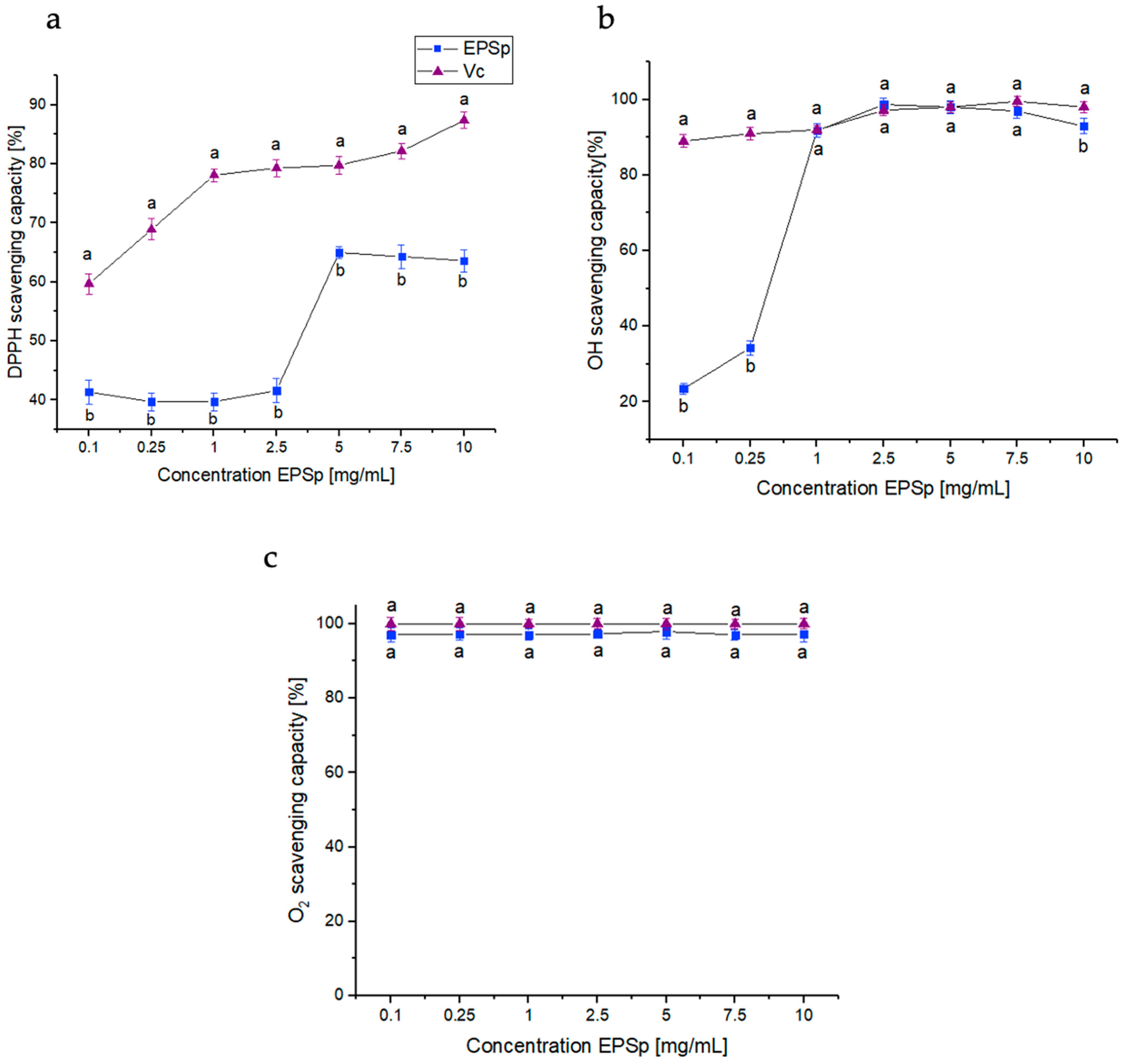
2.2. Free Radical Scavenging Activities

The EPSp free radical scavenging activities, 1,1-diphenyl-2-picryl hydrazyl radical (DPPH), hydroxyl radical (OH), and superoxide radicals (O2−), is shown in Figure 3. EPSp was analyzed in a range of 0.1 to 10 mg/mL, using ascorbic acid (Vc) as a positive control. The free radical scavenging activity of DPPH is shown in Figure 3a. The best activity was obtained between 5 and 10 mg/mL with a radical scavenging of 65 ± 1.2% and 63.6 ± 1.9%, respectively. This activity was higher than that presented by other B. licheniformis strains with an activity for its EPS20 of 35% at 10 mg/mL [21]. In the case of the OH scavenging activity (Figure 3b), the activity remained stable with a value of 90% in all concentrations, except at 2.5 mg/mL, which reached its maximum activity with 98.7 ± 1.7%. This activity was superior to that shown in B. licheniformis OSTK95 (50.9%), Bacillus velezensis SN-1 (58.9%), and Bacillus haynesii CamB6 (76.21%) [22,23,24]. In addition, ESPp showed excellent activity against O2− (Figure 3c), with 97% in all concentrations tested. The activity presented by EPSp was superior to other exopolymers of B. licheniformis OSTK95 (43.89%) [24] and B. licheniformis KS-20 (28%) [21]. The difference between the three antioxidant activities (DPPH; OH; and O2−) could be due to the chemical composition of the functional groups of EPSp. The availability to transfer electrons and to donate protons, or the physical structure of the EPSp molecule could contribute to these differences between antioxidant activities [25].

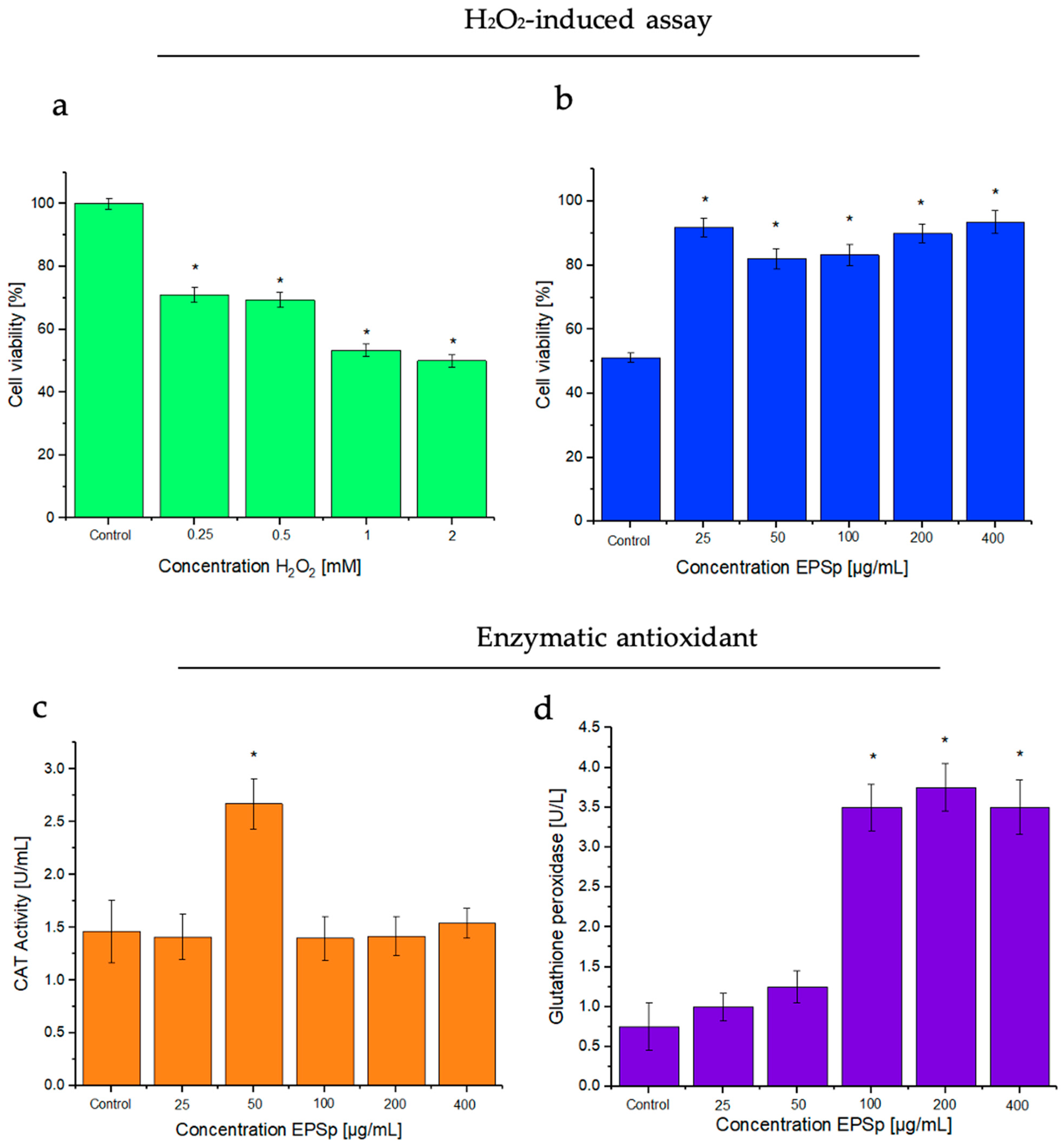
2.3. H2O2-Induced Assay, and Effects of the Exopolymer

The effects of the exopolymer against H2O2, and enzymatic antioxidant assays in HeLa cells are shown in Figure 4. The assay induced by H2O2 (Figure 4a) in HeLa cells had marked oxidative stress; at the 2 mM concentration, cell viability was reduced to 50%. From this reduction, the effects of the different concentrations of ESPp against H2O2 were tested (Figure 4b). The results indicated a significant protective effect of the EPSp at concentrations of 25, 200, and 400 μg/mL, showing a viability of 91.8 ± 2.8, 89.9 ± 2.9, and 93.5 ± 3.6%, respectively, with significant differences in relation to the control. Similar results were shown by the exopolysaccharide LPC-1 from Lactobacillus plantarum C88, in Caco-2 cells, showing protection with concentrations between 50 and 200 μg/mL, where a significant reduction in reactive oxygen species caused by H2O2 was observed [26]. This reduction in injuries could be due to the presence of negative charges in the exopolymer, which allowed for a significant increase in cell protection.

2.4. Enzymatic Antioxidant Assays: Catalase (CAT) and Glutathione Peroxidase (GSH-Px)

Assays for the enzymatic antioxidants CAT and GSH-Px are shown in Figure 4c,d. The presence of EPSp increased the enzyme activity in HeLa cells. In the case of CAT activity, the results showed a significant activity of 2.6 ± 0.24 U/mL in 50 μg/mL of EPSp, higher than that presented in the control (1.48 ± 0.3 U/mL). In the case of GSH-Px, the results showed an activity of 3.5 ± 0.29, 3.7 ± 0.3, and 3.5 ± 0.34 U/L in the 100, 200, and 400 μg/mL concentrations, respectively, of EPSp. These were significantly higher compared to control (0.75 ± 0.3 U/L). These results are more favorable than those presented by other species of the same genus such as Bacillus cereus SZ1 with a CAT activity of 1.5 U/mg at 300 μg EPS, compared to its control (0.9 U/mg) [27]. These results could demonstrate that the negative charges of the EPSp favored the increase in the enzymatic activity of CAT and GSH-Px with respect to their control. The increase in the antioxidant activity of EPSp makes it a candidate to cancel the toxic activity caused by hydrogen peroxide, maintaining the oxidative homeostasis of cells [28,29].

2.5. The Emulsifying Capacity of EPSp

The emulsifying capacity of EPSp (Figure 5) was evaluated to verify the bioremediation capacity of the exopolymer. The emulsifying capacity was tested with common natural oils (sunflower, olive, sesame, and coconut) and hydrocarbons (diesel, hexane, and toluene) used in the industry. In addition to this, a comparison was made between EPSp and commonly used commercial emulsifiers (Triton X-100, Tween 20, and SDS), at two different times (24 and 168 h), in different concentrations (0.5, 1, and 2 mg/mL), and at pH 7.0. Figure 5a shows that EPSp was not effective for natural oils and hydrocarbons at concentrations of 0.5 and 1 mg/mL, for any of the times tested. In the case of the 2 mg/mL concentration, the emulsifying activity of the EPS was significantly (p < 0.05) improved. The EPSp presented good emulsifying activity, equal to or greater than 50%, only for natural olive (50 ± 2.1%) and sesame (72 ± 3%) oils at 24 h, extending this emulsifying activity for sesame (70.6 ± 3.2%) to 168 h. On the other hand, EPSp did not show significant emulsifying activity compared to the commercial surfactants tested (Figure 5b). Only at a concentration of 2 mg/mL at 24 h did it present good activity, again with olive oil (50 ± 2.1%) and sesame (72 ± 3.2%). In addition, in the case of sesame (72 ± 3.2%), it could be considered very effective compared to the commercial products Triton X-100 (52.38 ± 1.6%), Tween 20 (14.29 ± 1.1%), and SDS (52.63 ± 1.6%). Similar results were found in the EPS1 of B. licheniformis MS3, for peanut oil (54%) [30]. However, in the B. licheniformis PASS26 strain, its EPS had emulsifying capacities for a greater number of natural oils (olive, sunflower, peanut, soybean, coconut, mustard, and rice) (70%) and for hydrocarbons (kerosene and hexane) (60%) [20]. These differences may be associated with the chemical composition of the polymer since the presence of negative charges in the EPSp could significantly affect the emulsifying capacity of the polymer [20,30,31,32].

3. Materials and Methods

3.1. Materials

The expolymer ESPp was obtained from Bacillus licheniformis IDN-EC (Accession #HM055601) [33]. The composition of the exopolymer was obtained following our previously described method [18].
Glucose, trypticase soy agar (TSA), DEAE-52 anion, 1,1-diphenyl-2-picryl hydrazyl radical (DPPH), hydrogen peroxide (H2O2), salicylic acid, pyrogallol, hydrochloric acid (HCl), potassium hydroxide (KOH), ferrous sulphate (FeSO4), phosphate-buffered saline (PBS), vitaminic C (Vc), catalase kit, gluthatione peroxidase, MTT (3-[4,5-dimethyl-thiazol-2-yl]-2,5-diphenyltetrazolium bromide) kit, vegetable oils, hydrocarbons, polyoxyethylene sorbitan monolaurate (Tween 20), 2-[4-(2,4,4-trimethylpentan-2-yl)phenoxy]ethanol (Triton X-100), and sodium dodecyl sulphate (SDS) were from Sigma-Aldrich, Madrid, Spain. The HeLa cells (human T-cell lymphoblast-like cell line) were obtained from the Centro de Biología Molecular Severo Ochoa (CBM-UAM), Universidad Autónoma de Madrid, Madrid, Spain. Dulbecco’s modified Eagle medium (H-DMEM), fetal bovine serum (FBS), L-glutamine, penicillin, and streptomycin were from Aldrich, Schnelldorf, Germany.

3.2. Production, Isolation, and Purification of the Exopolymer

The production, isolation, and purification of the exopolymer were carried out following our previously described method [18]. The strain was grown in a trypticase soy agar (TSA) medium and incubated at 45 °C for 24 h and moved into flasks of 100 mL filled with 20 mL of a minimal growth medium (MGM) [34]: g/L: K2HPO4 0.5, KH2PO4 0.04, NaCl 0.1, CaCl2 2H2O 0.002, (NH4) 2SO4 0.2, MgSO4 7H2O 0.02, and FeSO4 0.001, with glucose (4 g/L), and pH adjusted to 7.0. The flasks were incubated at 45 °C and 110 rpm for 24 h. Subsequently, and under the same conditions, 10 mL of this broth (concentration of 2.5 × 10⁷ cells/mL) were inoculated into flasks containing 1000 mL of MGM and glucose. Three independent trials were performed.
The cultures were centrifuged at 13,154× g for 30 min at 4 °C (Duppont-RC5). The exopolymer was precipitated with ethanol (−80 °C at three times the volume). The pellet was centrifuged at 13,154× g for 30 min at 4 °C and dialyzed with Milli-Q water for 48 h. For further purification, chromatography of the exopolymer (10 mL, 10 mg/mL) in a DEAE-52 anion exchange column (2.6 × 30 cm) eluted with deionized water, 0.05 and 0.3 M NaCl, at a flow rate of 1 mL/min rate was performed.

3.3. MTT Assay

The cytotoxicity activity was determined by reducing the MTT (3-[4,5-dimethyl-thiazol-2-yl]-2,5-diphenyltetrazolium bromide) to formazan on HeLa cells [35,36]. The HeLa were cultured on 24-well plates and reached a density of 5 × 10⁵ cells/per well and treated with different concentrations of EPSp (200, 400, 600, 800, and 1000 µg/mL) for 24 h. Salt solution (0.5 mg/mL) was added and incubated for 4 h under 5% CO2 atmosphere incubator and at 37 °C. Subsequently, 100 μL of SDS (10%) in 0.01 M of HCL was added to each well to dissolve the formazan crystals for 30 min. The absorbance was measured at 490 nm, and, finally, the cell viability was calculated.
C e l l   v i a b i l i t y   [ % ] = ( A 1 / A 2 ) × 100
A1 = cells treated with EPSp and MTT salt solution, and A2 = cells without any treatment with MTT salt solution.

3.4. Free Radical Scavenging Activities

3.4.1. DPPH (1,1-Diphenyl-2-picryl Hydrazyl Radical) Radical Scavenging Activity

The DPPH radical scavenging effect of the EPSp was measured using the method described in [8,37]. The sample solution was prepared in a final volume of 150 μL. This contained 50 μL of EPSp at different concentrations (0.1, 0.25, 0.8, 1.0, 2.5, 5.0, 7.5, and 10 mg/mL) and 100 μL DPPH (100 μM DPPH–ethanolic solution). The control solution was prepared by adding 50 μL Milli-Q water with 100 μL DPPH–ethanol solution. The positive control was vitamin C (Vc). Then, the mixtures were incubated in the dark for 30 min at room temperature. The absorbance of the DPPH radicals was determined at 525 nm. Percentage DPPH radical scavenging activity was calculated by the equation:
D P P H   s c a v e n g i n g   a c t i v i t y   [ % ] = [ 1 ( A   s a m p l e A   c o n t r o l ) / A   b l a n k ] × 100
A   b l a n k = 50   μ L   M i l l i   Q   w a t e r + 100   μ L   d e   e t h a n o l   96 %
A   c o n t r o l = 50   μ L   M i l l i   Q   w a t e r + 100   μ L   d e   D P P H e t h a n o l   s o l u t i o n
A   s a m p l e = 50   μ L   ( E P S   o r   V c ) + 100   μ L   d e   D P P H e t h a n o l   s o l u t i o n

3.4.2. Hydroxyl Radical (OH) Scavenging Activity

The hydroxyl radical scavenging activity effect of the EPSp was measured as described previously [8,38]. The sample solutions contained 40 μL of EPSp at different concentrations (0.1, 0.25, 0.8, 1.0, 2.5, 5.0, 7.5, and 10 mg/mL), 40 μL of salicylic acid (9 mM ethanol–salicylic acid solution), 40 μL FeSO4 solution (9 mM), and 40 μL of H2O2 (8.8 mM). The positive control was vitamin C (Vc). The mixtures were incubated for 30 min at 37 °C. The absorbance of the hydroxyl radical was determined at 510 nm. Percentage hydroxyl radical scavenging activity was calculated by the equation:
O H   s c a v e n g i n g   a c t i v i t y   [ % ] = [ 1 ( A   s a m p l e A   c o n t r o l ) / A   b l a n k ] × 100
A   b l a n k = 40   μ L   M i l l i   Q   w a t e r + 40   μ L   F e S O 4   s o l u t i o n + 40   μ L   d e   e t h a n o l   96 % + 40   μ L   o f   H 2 O 2
A   c o n t r o l = 40   μ L   M i l l i   Q   w a t e r + 40   μ L   F e S O 4   s o l u t i o n + 40   μ L   d e   e t h a n o l s a l i c y l i c   s o l u t i o n + 40   μ L   o f   H 2 O 2
A   s a m p l e = 40   μ L   ( E P S   o r   V c ) + 40   μ L   F e S O 4   s o l u t i o n + 40   μ L   d e   e t h a n o l s a l i c y l i c   s o l u t i o n + 40   μ L   o f   H 2 O 2

3.4.3. Superoxide Anion (O2−) Radical Scavenging Activity

The superoxide anion radical scavenging activity effect of the EPSp was measured as previously described [8,39]. The sample solutions contained 0.3 mL of EPSp at different concentrations (0.1, 0.25, 0.8, 1.0, 2.5, 5.0, 7.5, and 10 mg/mL), Then, 2.6 mL of phosphate buffer (50 mM, pH 8.2) and 90 μL of pyrogallol (3 mM) and HCl (10 mM) were added to the sample solutions. The positive control was vitamin C (Vc). The absorbance of superoxide anion radicals was determined at 325 nm. Percentage of the superoxide anion radical scavenging was calculated by the equation:
O 2   s c a v e n g i n g   a c t i v i t y   [ % ] = [ 1 ( A   s a m p l e   10 / A   c o n t r o l   10 ) ( A   s a m p l e   0 / A   c o n t r o l   0 ) ] × 100
A   c o n t r o l   ( 0   m i n   a n d   10   m i n ) = 0.3   m L   M i l l i   Q   w a t e r + 2.6   m L   f o s f a t e   B u f f e r + 90   m L   p y r o g a l l o l H C l
A   s a m p l e   ( 0   m i n   a n d   10   m i n ) = 0.3   m L   ( E P S   o r   V c ) + 2.6   m L   f o s f a t e   B u f f e r + 90   m L   p y r o g a l l o l H C l

3.5. H2O2-Induced Assay

3.5.1. H2O2-Induced Oxidative Stress

H2O2-induced oxidative stress was used to measure the damage of this non-radical on HeLa cells [8,40,41]. For the in vitro procedure, the cells were seeded in 96-well plates 5 × 10⁴ for 24 h, under 5% CO2 atmosphere incubator, and at 37 °C. Subsequently, the medium was replaced with 100 μL of different concentrations of H2O2 (0.25, 0.5, 1, and 2 mM) for 1 h at 37 °C. After this time, the medium was removed, and cell viability estimated by MTT method as described in Section 3.3.

3.5.2. Effects of the Exopolymer against H2O2

The effect of the EPSp against the non-radical H2O2 was evaluated on HeLa cells [8,40,41]. The cells were seeded in 96-well plates of 5 × 10⁴ for 24 h, under 5% CO2 atmosphere incubator and at 37 °C. After this time, the EPSp was diluted in a new medium with DMEM at different concentrations (25, 50, 100, 200, and 400 μg/mL) for 1 h. The positive control was vitamin C (Vc) (20 mg/mL). Cell viability was estimated by MTT method as described in Section 3.3.

3.6. Enzymatic Antioxidant Assays

Catalase (CAT) and Glutathione Peroxidase (GSH-Px) Assay

EPSp was investigated against oxidative stress on HeLa cells [41,42]. For it, 5 × 10⁵ cells were seeded in 24-well plates and incubated for 24 h. Cells were treated with EPS at various concentrations (50, 100, 200, and 400 μg/mL) for 1 h at 37 °C. The supernatants were collected and used for the determination of CAT and GSH-Px assay. The determination of the reactions was carried out using the kit (Sigma) established in the protocol.

3.7. Emulsifying Properties

The emulsifying properties of EPSp were evaluated using the method described by Meneghine et al. [43]. The assays were undertaken in transparent cylindrical 5 mL tubes that contained 1.5 mL of an oil phase and 1.5 mL of an aqueous phase, at two different times (24, and 168 h), in different concentrations (0.5, 1, and 2 mg/mL), and at pH 7.0. The oil phase contained vegetable oils (sunflower oil, olive oil, sesame oil, and coconut oil). For the aqueous phase, both commercial emulsifying compounds, such as polysorbate 20 (Tween 20) (Sigma), sodium dodecyl sulphate (SDS) (Sigma), and Triton X-100 (Sigma), and the obtained polymers were used for comparison purposes. All compounds used had a concentration of 3:2 v/v. The tubes were stirred in a vortex at 2400 rpm for 2 min. After 24 h and 168 h, the emulsification indices E24 and E168 were determined by the equation:
E   [ % ] = H E L / H T × 100
where HEL (mm) is the height of the emulsion layer and HT (mm) is the overall height.

3.8. Statistical Analysis

All experiments were carried out in triplicate. The data were recorded as mean ± standard deviation and analyzed by SPSS (version 21, SPSS® Inc., Chicago, IL, USA). One-way analysis of variance was performed by ANOVA procedures. Significant differences between means were determined by Duncan’s multiple-range tests. p < 0.05 was considered statistically significant.

4. Conclusions

The EPSp exhibited the absence of cytotoxicity and presented strong hydroxyl radical (OH), DPPH scavenging, and superoxide anion (O2−) activities. In addition, EPSp had significant antioxidant activity against H2O2-induced injury, protecting HeLa cells from oxidative stress. EPSp also stimulated the activities of the antioxidant enzymes Catalase (CAT) and Glutathione peroxidase (GSH-Px). The performance of EPSp as an emulsifier is notable in the case of sesame oil, as it had a higher performance than commercial emulsifiers. This study suggests that the EPSp negative charges of Bacillus licheniformis played an important role in shaping potential applications. EPSp’s performance in the evaluated assays suggests its versatility and suitability for various biotechnological applications.
The exploration of a broad spectrum of biotechnological activities of the EPS obtained by Bacillus licheniformis demonstrated excellent antioxidant activity, both non-enzymatic and enzymatic, protection against H2O2-induced oxidative stress, emulsifying properties, and, previously, antiviral capabilities. This type of deeper study allows us to have a more realistic vision of the application of exopolysaccharides produced by micro-organisms, and their competitive advantages at an industrial level.

Author Contributions

A.C.: conceptualization, formal analysis, investigation, data curation, funding acquisition, methodology, validation, resources, supervision, draft, and writing—review and editing; A.R.: funding acquisition, project administration, and manuscript review; S.-L.E.: formal analysis, investigation, and writing—original draft. All authors have read and agreed to the published version of the manuscript.

Funding

This research was funded by the Spanish Ministry of Science and Innovation for financial support (project PID2022-136607NB-I00) and FUAM, Universidad Autónoma de Madrid, Spain (project nº. 820053).

Institutional Review Board Statement

Not applicable.

Informed Consent Statement

Not applicable.

Data Availability Statement

No new data were created or analyzed in this study. Data sharing is not applicable to this article.

Conflicts of Interest

The authors declare no conflicts of interest.

References

  1. Kochhar, N.; Kavya, I.K.; Shrivastava, S.; Ghosh, A.; Rawat, V.S.; Sodhi, K.K.; Kumar, M. Perspectives on the Microorganism of Extreme Environments and Their Applications. Curr. Res. Microb. Sci. 2022, 3, 100134. [Google Scholar] [CrossRef]
  2. Gudiña, E.J.; Teixeira, J.A. Bacillus licheniformis: The Unexplored Alternative for the Anaerobic Production of Lipopeptide Biosurfactants? Biotechnol. Adv. 2022, 60, 108013. [Google Scholar] [CrossRef]
  3. Doan, C.T.; Tran, T.N.; Nguyen, T.T.; Tran, T.P.H.; Nguyen, V.B.; Tran, T.D.; Nguyen, A.D.; Wang, S.L. Production of Sucrolytic Enzyme by Bacillus licheniformis by the Bioconversion of Pomelo Albedo as a Carbon Source. Polymers 2021, 13, 1959. [Google Scholar] [CrossRef]
  4. Nabot, M.; Guérin, M.; Sivakumar, D.; Remize, F.; Garcia, C. Variability of Bacterial Homopolysaccharide Production and Properties during Food Processing. Biology 2022, 11, 171. [Google Scholar] [CrossRef]
  5. Oleńska, E.; Małek, W.; Kotowska, U.; Wydrych, J.; Polińska, W.; Swiecicka, I.; Thijs, S.; Vangronsveld, J. Exopolysaccharide Carbohydrate Structure and Biofilm Formation by Rhizobium leguminosarum Bv. Trifolii Strains Inhabiting Nodules of Trifolium Repens Growing on an Old Zn–Pb–Cd-polluted Waste Heap Area. Int. J. Mol. Sci. 2021, 22, 2808. [Google Scholar] [CrossRef]
  6. Wu, F.; Misra, M.; Mohanty, A.K. Challenges and New Opportunities on Barrier Performance of Biodegradable Polymers for Sustainable Packaging. Prog. Polym. Sci. 2021, 117, 101395. [Google Scholar] [CrossRef]
  7. Barcelos, M.C.S.; Vespermann, K.A.C.; Pelissari, F.M.; Molina, G. Current Status of Biotechnological Production and Applications of Microbial Exopolysaccharides. Crit. Rev. Food Sci. Nutr. 2020, 60, 1475–1495. [Google Scholar] [CrossRef] [PubMed]
  8. Sánchez-león, E.; Huang-lin, E.; Amils, R.; Abrusci, C. Production and Characterisation of an Exopolysaccharide by Bacillus amyloliquefaciens: Biotechnological Applications. Polymers 2023, 15, 1550. [Google Scholar] [CrossRef]
  9. Carrión, O.; Delgado, L.; Mercade, E. New Emulsifying and Cryoprotective Exopolysaccharide from Antarctic Pseudomonas sp. ID1. Carbohydr. Polym. 2015, 117, 1028–1034. [Google Scholar] [CrossRef]
  10. Du, R.; Yu, L.; Sun, M.; Ye, G.; Yang, Y.; Zhou, B.; Qian, Z.; Ling, H.; Ge, J. Characterization of Dextran Biosynthesized by Glucansucrase from Leuconostoc pseudomesenteroides and Their Potential Biotechnological Applications. Antioxidants 2023, 12, 275. [Google Scholar] [CrossRef]
  11. Revin, V.V.; Liyaskina, E.V.; Parchaykina, M.V.; Kurgaeva, I.V.; Efremova, K.V.; Novokuptsev, N.V. Production of Bacterial Exopolysaccharides: Xanthan and Bacterial Cellulose. Int. J. Mol. Sci. 2023, 24, 14608. [Google Scholar] [CrossRef]
  12. Netrusov, A.I.; Liyaskina, E.V.; Kurgaeva, I.V.; Liyaskina, A.U.; Yang, G.; Revin, V.V. Exopolysaccharides Producing Bacteria: A Review. Microorganisms 2023, 11, 1541. [Google Scholar] [CrossRef]
  13. Lee, H.; Chea, S.; Lee, K.; Cha, I.; Kim, D. A Report on Five Unrecorded Bacterial Species Belonging to the Phyla Actinomycetota, Bacillota and Pseudomonadota in Korea Isolated in 2020. J. Species Res. 2023, 12, 1–6. [Google Scholar] [CrossRef]
  14. Muras, A.; Romero, M.; Mayer, C.; Otero, A. Biotechnological Applications of Bacillus licheniformis. Crit. Rev. Biotechnol. 2021, 41, 609–627. [Google Scholar] [CrossRef]
  15. He, H.; Yu, Q.; Ding, Z.; Zhang, L.; Shi, G.; Li, Y. Biotechnological and Food Synthetic Biology Potential of Platform Strain: Bacillus licheniformis. Synth. Syst. Biotechnol. 2023, 8, 281–291. [Google Scholar] [CrossRef]
  16. Zammuto, V.; Spanò, A.; Nicolò, M.S.; Grillo, E.; Caccamo, M.T.; Magazù, S.; Cappello, S.; Gugliandolo, C. Thermophilic Hydrocarbon-Utilizing Bacilli from Marine Shallow Hydrothermal Vents as Producers of Biosurfactants. J. Mar. Sci. Eng. 2022, 10, 1077. [Google Scholar] [CrossRef]
  17. Zhang, Z.; Cai, R.; Zhang, W.; Fu, Y.; Jiao, N. A Novel Exopolysaccharide with Metal Adsorption Capacity Produced by a Marine Bacterium Alteromonas sp. JL2810. Mar. Drugs 2017, 15, 175. [Google Scholar] [CrossRef]
  18. Sánchez-León, E.; Bello-Morales, R.; López-Guerrero, J.A.; Poveda, A.; Jiménez-Barbero, J.; Gironès, N.; Abrusci, C. Isolation and Characterization of an Exopolymer Produced by Bacillus licheniformis: In Vitro Antiviral Activity against Enveloped Viruses. Carbohydr. Polym. 2020, 248, 116737. [Google Scholar] [CrossRef]
  19. Arena, A.; Maugeri, T.L.; Pavone, B.; Iannello, D.; Gugliandolo, C.; Bisignano, G. Antiviral and Immunoregulatory Effect of a Novel Exopolysaccharide from a Marine Thermotolerant Bacillus licheniformis. Int. Immunopharmacol. 2006, 6, 8–13. [Google Scholar] [CrossRef]
  20. Insulkar, P.; Kerkar, S.; Lele, S.S. Purification and Structural-Functional Characterization of an Exopolysaccharide from Bacillus licheniformis PASS26 with in-Vitro Antitumor and Wound Healing Activities. Int. J. Biol. Macromol. 2018, 120, 1441–1450. [Google Scholar] [CrossRef]
  21. Song, Y.R.; Song, N.E.; Kim, J.H.; Nho, Y.C.; Baik, S.H. Exopolysaccharide Produced by Bacillus licheniformis Strains Isolated from Kimchi. J. Gen. Appl. Microbiol. 2011, 57, 169–175. [Google Scholar] [CrossRef]
  22. Nguyen Vu, T.H.; Quach, N.T.; Nguyen, N.A.; Nguyen, H.T.; Ngo, C.C.; Nguyen, T.D.; Ho, P.H.; Hoang, H.; Chu, H.H.; Phi, Q.T. Genome Mining Associated with Analysis of Structure, Antioxidant Activity Reveals the Potential Production of Levan-Rich Exopolysaccharides by Food-Derived Bacillus velezensis Vtx20. Appl. Sci. 2021, 11, 7055. [Google Scholar] [CrossRef]
  23. Banerjee, A.; Mohammed Breig, S.J.; Gómez, A.; Sánchez-Arévalo, I.; González-Faune, P.; Sarkar, S.; Bandopadhyay, R.; Vuree, S.; Cornejo, J.; Tapia, J.; et al. Optimization and Characterization of a Novel Exopolysaccharide from Bacillus haynesii CamB6 for Food Applications. Biomolecules 2022, 12, 834. [Google Scholar] [CrossRef] [PubMed]
  24. Fang, Y.; Liu, S.; Lu, M.; Jiao, Y.; Wang, S. A Novel Method for Promoting Antioxidant Exopolysaccharidess Production of Bacillus licheniformis. Carbohydr. Polym. 2013, 92, 1172–1176. [Google Scholar] [CrossRef] [PubMed]
  25. Rahnama Vosough, P.; Habibi Najafi, M.B.; Edalatian Dovom, M.R.; Javadmanesh, A.; Mayo, B. Evaluation of Antioxidant, Antibacterial and Cytotoxicity Activities of Exopolysaccharide from Enterococcus Strains Isolated from Traditional Iranian Kishk. J. Food Meas. Charact. 2021, 15, 5221–5230. [Google Scholar] [CrossRef]
  26. Zhang, L.; Liu, C.; Li, D.; Zhao, Y.; Zhang, X.; Zeng, X.; Yang, Z.; Li, S. Antioxidant Activity of an Exopolysaccharide Isolated from Lactobacillus plantarum C88. Int. J. Biol. Macromol. 2013, 54, 270–275. [Google Scholar] [CrossRef]
  27. Zheng, L.P.; Zou, T.; Ma, Y.J.; Wang, J.W.; Zhang, Y.Q. Antioxidant and DNA Damage Protecting Activity of Exopolysaccharides from the Endophytic Bacterium Bacillus cereus SZ1. Molecules 2016, 21, 174. [Google Scholar] [CrossRef] [PubMed]
  28. Guo, S.; Guo, L. Unraveling the Multi-Enzyme-Like Activities of Iron Oxide Nanozyme via a First-Principles Microkinetic Study. J. Phys. Chem. C 2019, 123, 30318–30334. [Google Scholar] [CrossRef]
  29. Belinskaia, D.A.; Voronina, P.A.; Shmurak, V.I.; Vovk, M.A.; Batalova, A.A.; Jenkins, R.O.; Goncharov, N.V. The Universal Soldier: Enzymatic and Non-enzymatic Antioxidant Functions of Serum Albumin. Antioxidants 2020, 9, 966. [Google Scholar] [CrossRef] [PubMed]
  30. Asgher, M.; Urooj, Y.; Qamar, S.A.; Khalid, N. Improved Exopolysaccharide Production from Bacillus licheniformis MS3: Optimization and Structural/Functional Characterization. Int. J. Biol. Macromol. 2020, 151, 984–992. [Google Scholar] [CrossRef]
  31. Chowdhury, S.R.; Manna, S.; Saha, P.; Basak, R.K.; Sen, R.; Roy, D.; Adhikari, B. Composition Analysis and Material Characterization of an Emulsifying Extracellular Polysaccharide (EPS) Produced by Bacillus megaterium RB-05: A Hydrodynamic Sediment-Attached Isolate of Freshwater Origin. J. Appl. Microbiol. 2011, 111, 1381–1393. [Google Scholar] [CrossRef]
  32. Song, B.; Zhu, W.; Song, R.; Yan, F.; Wang, Y. Exopolysaccharide from Bacillus vallismortis WF4 as an Emulsifier for Antifungal and Antipruritic Peppermint Oil Emulsion. Int. J. Biol. Macromol. 2019, 125, 436–444. [Google Scholar] [CrossRef]
  33. Morro, A.; Catalina, F.; Sanchez-León, E.; Abrusci, C. Photodegradation and Biodegradation Under Thermophile Conditions of Mulching Films Based on Poly(Butylene Adipate-Co-Terephthalate) and Its Blend with Poly(Lactic Acid). J. Polym. Environ. 2019, 27, 352–363. [Google Scholar] [CrossRef]
  34. Abrusci, C.; Palomar, J.; Pablos, J.L.; Rodriguez, F.; Catalina, F. Efficient Biodegradation of Common Ionic Liquids by Sphingomonas paucimobilis Bacterium. Green Chem. 2011, 13, 709–717. [Google Scholar] [CrossRef]
  35. Morro, A.; Abrusci, C.; Pablos, J.L.; Marín, I.; García, F.C.; García, J.M. Inherent Antibacterial Activity and in Vitro Biocompatibility of Hydrophilic Polymer Film Containing Chemically Anchored Sulfadiazine Moieties. Eur. Polym. J. 2017, 91, 274–282. [Google Scholar] [CrossRef]
  36. Pérez-Blanco, C.; Huang-Lin, E.; Abrusci, C. Characterization, Biodegradation and Cytotoxicity of Thermoplastic Starch and Ethylene-Vinyl Alcohol Copolymer Blends. Carbohydr. Polym. 2022, 298, 120085. [Google Scholar] [CrossRef]
  37. Niknezhad, S.V.; Najafpour-Darzi, G.; Morowvat, M.H.; Ghasemi, Y. Eexopolysaccharide Production of Pantoea sp. BCCS 001 GH: Physical Characterizations, Emulsification, and Antioxidant Activities. Int. J. Biol. Macromol. 2018, 118, 1103–1111. [Google Scholar] [CrossRef] [PubMed]
  38. Sun, M.L.; Zhao, F.; Shi, M.; Zhang, X.Y.; Zhou, B.C.; Zhang, Y.Z.; Chen, X.L. Characterization and Biotechnological Potential Analysis of a New Exopolysaccharide from the Arctic Marine Bacterium Polaribacter sp. SM1127. Sci. Rep. 2015, 5, 18435. [Google Scholar] [CrossRef] [PubMed]
  39. Balakrishnan, B.; Prasad, B.; Rai, A.K.; Velappan, S.P.; Subbanna, M.N.; Narayan, B. In Vitro Antioxidant and Antibacterial Properties of Hydrolysed Proteins of Delimed Tannery Fleshings: Comparison of Acid Hydrolysis and Fermentation Methods. Biodegradation 2011, 22, 287–295. [Google Scholar] [CrossRef]
  40. Huang-Lin, E.; Sánchez-León, E.; Amils, R.; Abrusci, C. Potential Applications of an Exopolysaccharide Produced by Bacillus xiamenensis RT6 Isolated from an Acidic Environment. Polymers 2022, 14, 3918. [Google Scholar] [CrossRef]
  41. Abrusci, C.; Amils, R.; Sánchez-león, E. Biodegradation of Choline NTF2 by Pantoea agglomerans in Different Osmolarity. Characterization and Environmental Implications of the Produced Exopolysaccharide. Polymers 2023, 15, 3974. [Google Scholar] [CrossRef] [PubMed]
  42. Liu, Z.; Dong, L.; Jia, K.; Zhan, H.; Zhang, Z.; Shah, N.P.; Tao, X.; Wei, H. Sulfonation of Lactobacillus plantarum WLPL04 Exopolysaccharide Amplifies Its Antioxidant Activities in Vitro and in a Caco-2 Cell Model. J. Dairy Sci. 2019, 102, 5922–5932. [Google Scholar] [CrossRef] [PubMed]
  43. Meneghine, A.K.; Moretto, C.; Castellane, T.C.L.; Carareto Alves, L.M. Production, Characterization and Bioemulsifying Activity of an Exopolysaccharide Produced by Sphingomonas sp. Isolated from Freshwater. J. Polym. Environ. 2017, 25, 1080–1086. [Google Scholar] [CrossRef]
Figure 1. Structure of EPSp: (A) polyglutamic acid and (B) polyglycerol phosphate chain O-substituted with αGal moieties (αGal/αGlcNH2 3:1 molar ratio).
Ijms 25 08249 g001
Figure 2. Biocompatibility (MTT) of EPSp with HeLa cell. * Indicating significant differences between control and concentration of EPSp (p < 0.05).
Ijms 25 08249 g002
Figure 3. Non-enzymatic antioxidant radical scavenging activity: (a) 1,1-diphenyl-2-picryl hydrazyl radical (DPPH), (b) hydroxyl radical (OH), and (c) superoxide radicals (O2−). Different letters (a,b) indicate significant differences between concentration of Vc and EPSp (p < 0.05).
Ijms 25 08249 g003
Figure 4. H2O2-induced assay: (a) evaluation of the H2O2-damaged HeLa cells, and (b) evaluation of the protection of H2O2-damaged HeLa cells protection with EPSp. Enzymatic antioxidant activity: (c) catalase activity in the presence of EPSp, and (d) glutathione peroxidase activity in the presence of EPSp. * Indicates significant differences between control and concentration of EPSp (p < 0.05).
Ijms 25 08249 g004
Figure 5. Emulsifying activity of EPSp at concentrations of 0.5, 1, and 2 mg/mL: (a) emulsifying activity with the different natural oils and hydrocarbons used at 24 h (E24) and 168 h (E168) (* p < 0.05), (b) comparison of emulsifying activity at different concentrations of EPSp versus commercial emulsifiers (Triton X-100, Tween 20, and SDS) across different natural oils and hydrocarbons at 24 h (E24). Different letters (ad) represent the statistical difference between different emulsifiers for each natural oil and hydrocarbon (p < 0.05).
Ijms 25 08249 g005
Disclaimer/Publisher’s Note: The statements, opinions and data contained in all publications are solely those of the individual author(s) and contributor(s) and not of MDPI and/or the editor(s). MDPI and/or the editor(s) disclaim responsibility for any injury to people or property resulting from any ideas, methods, instructions or products referred to in the content.

