
Magnetic Fluorescent Quantum Dots Nanocomposites in Food Contaminants Analysis: Current Challenges and Opportunities

 ,
, 

Abstract
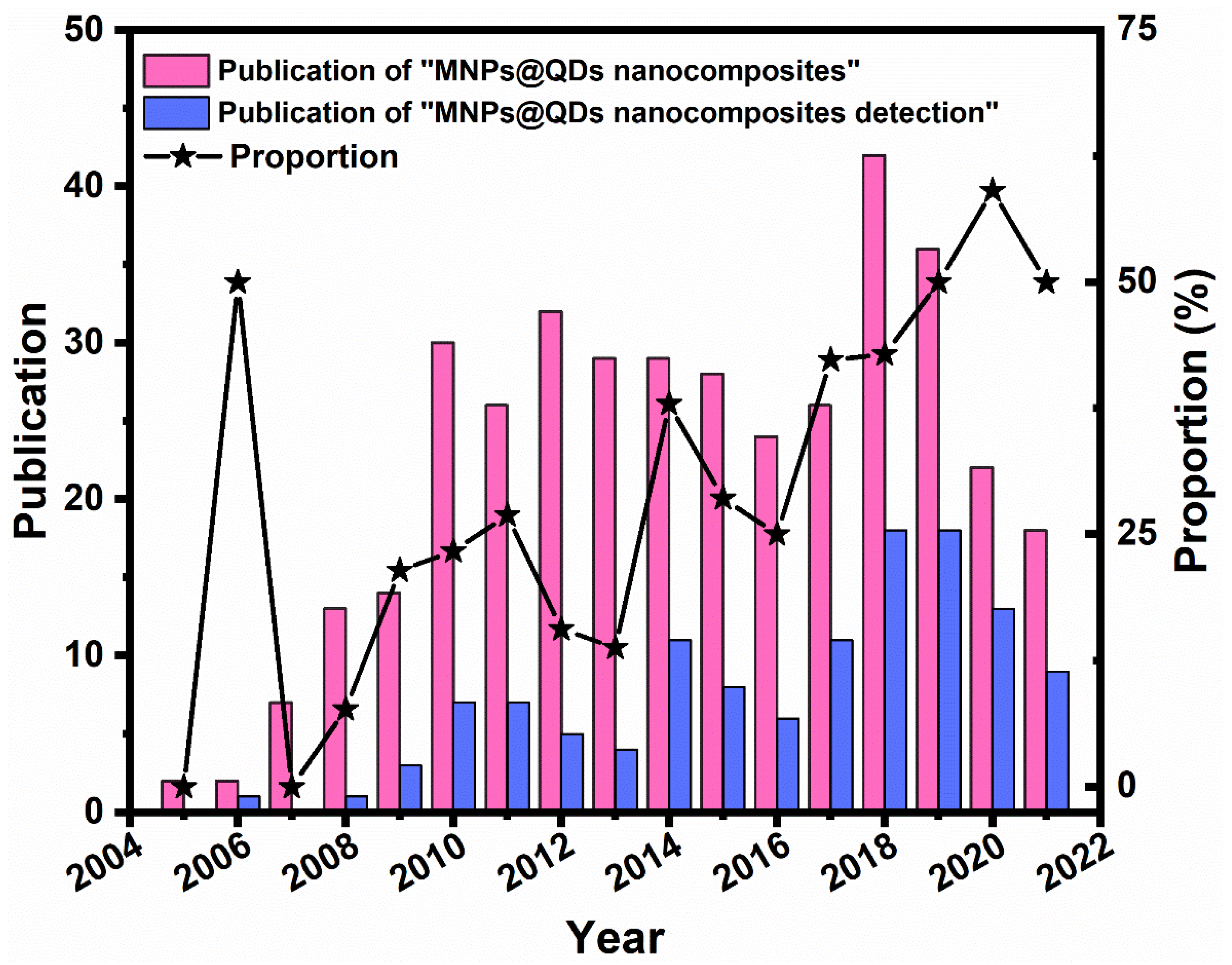
1. Introduction
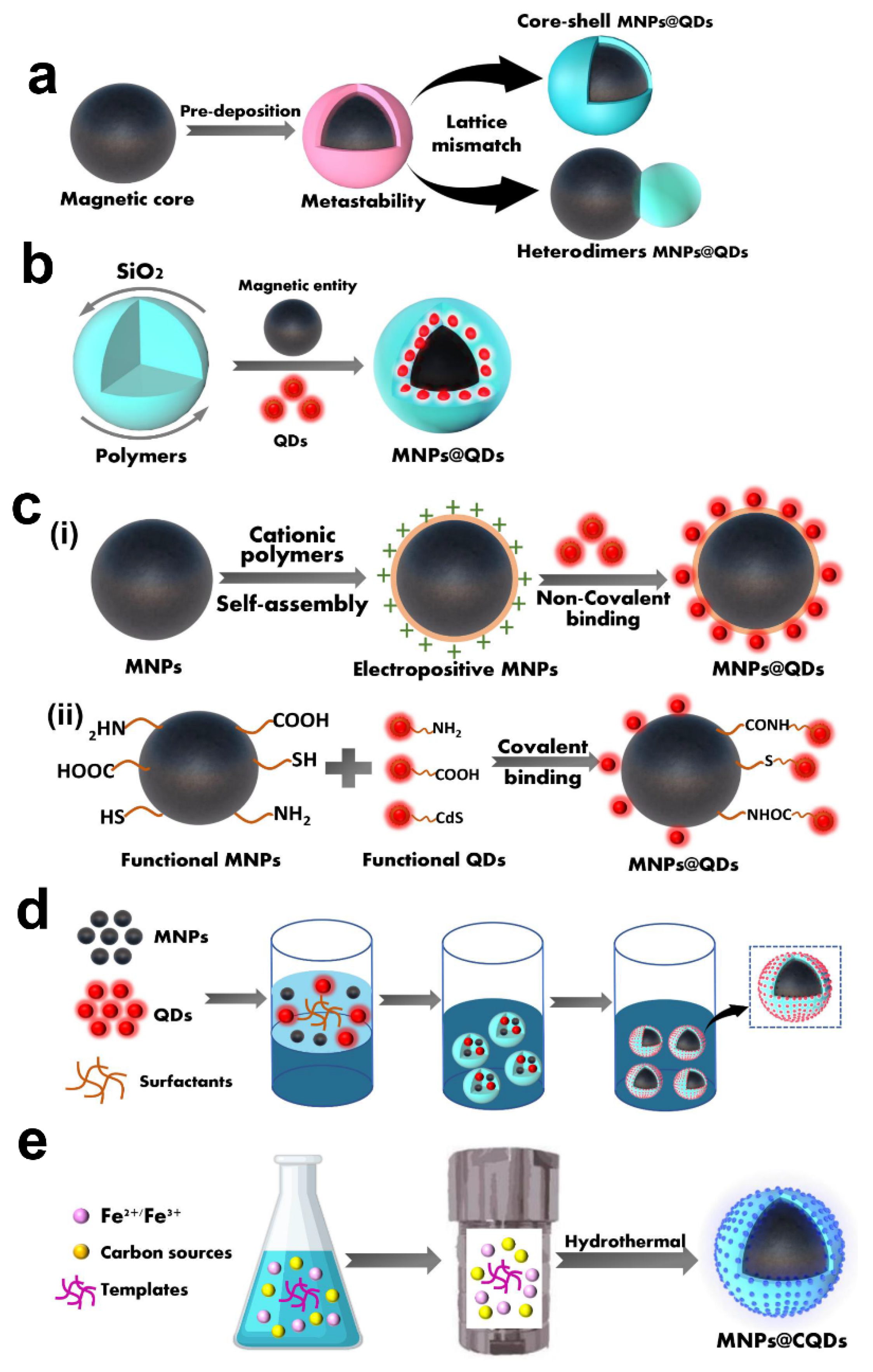
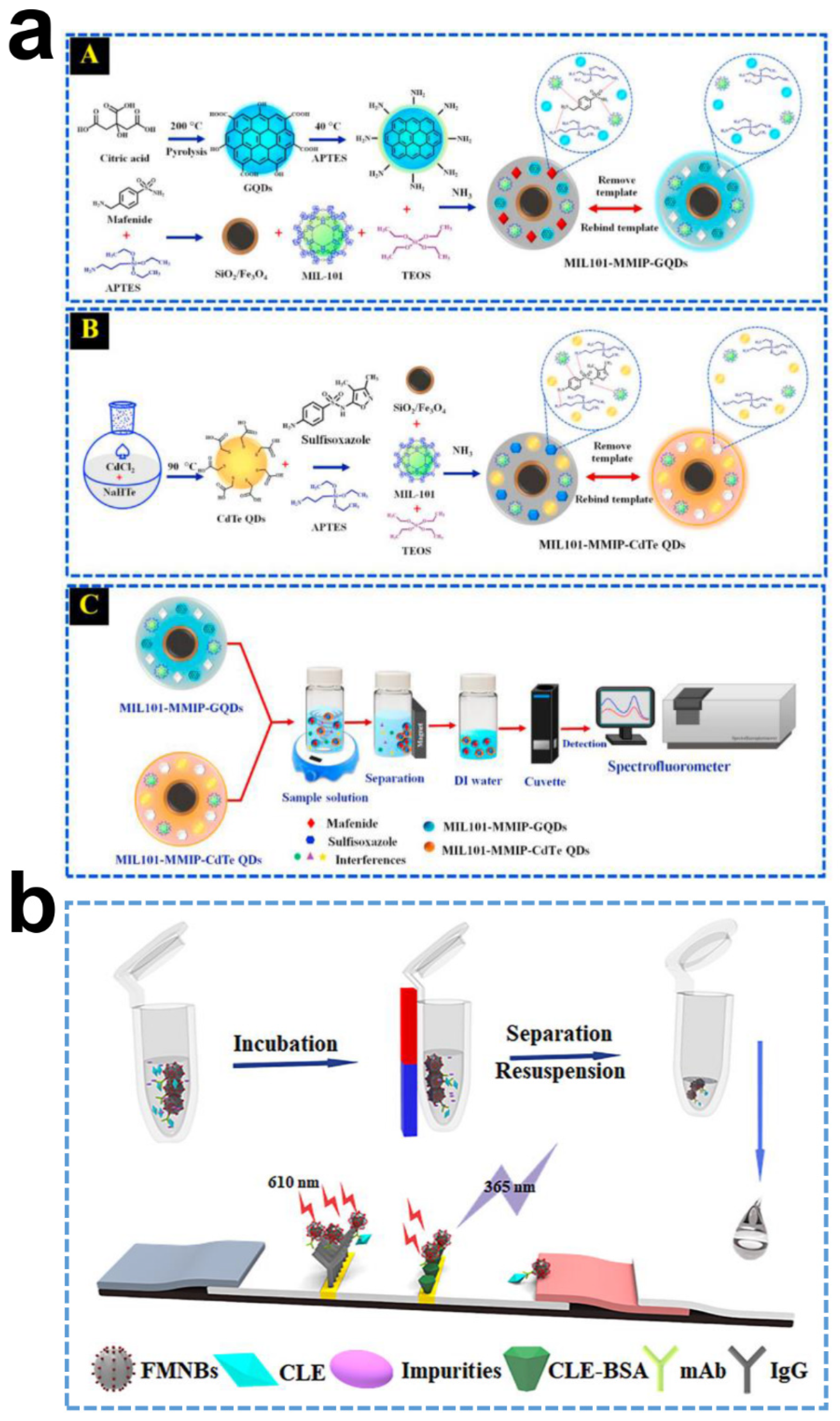
2. Preparation of MNPs@QDs
2.1. Hetero-Crystalline Growth
2.2. Template Embedding
2.3. Layer-by-Layer Assembly
2.4. Microemulsion Technique
2.5. One-Pot Method
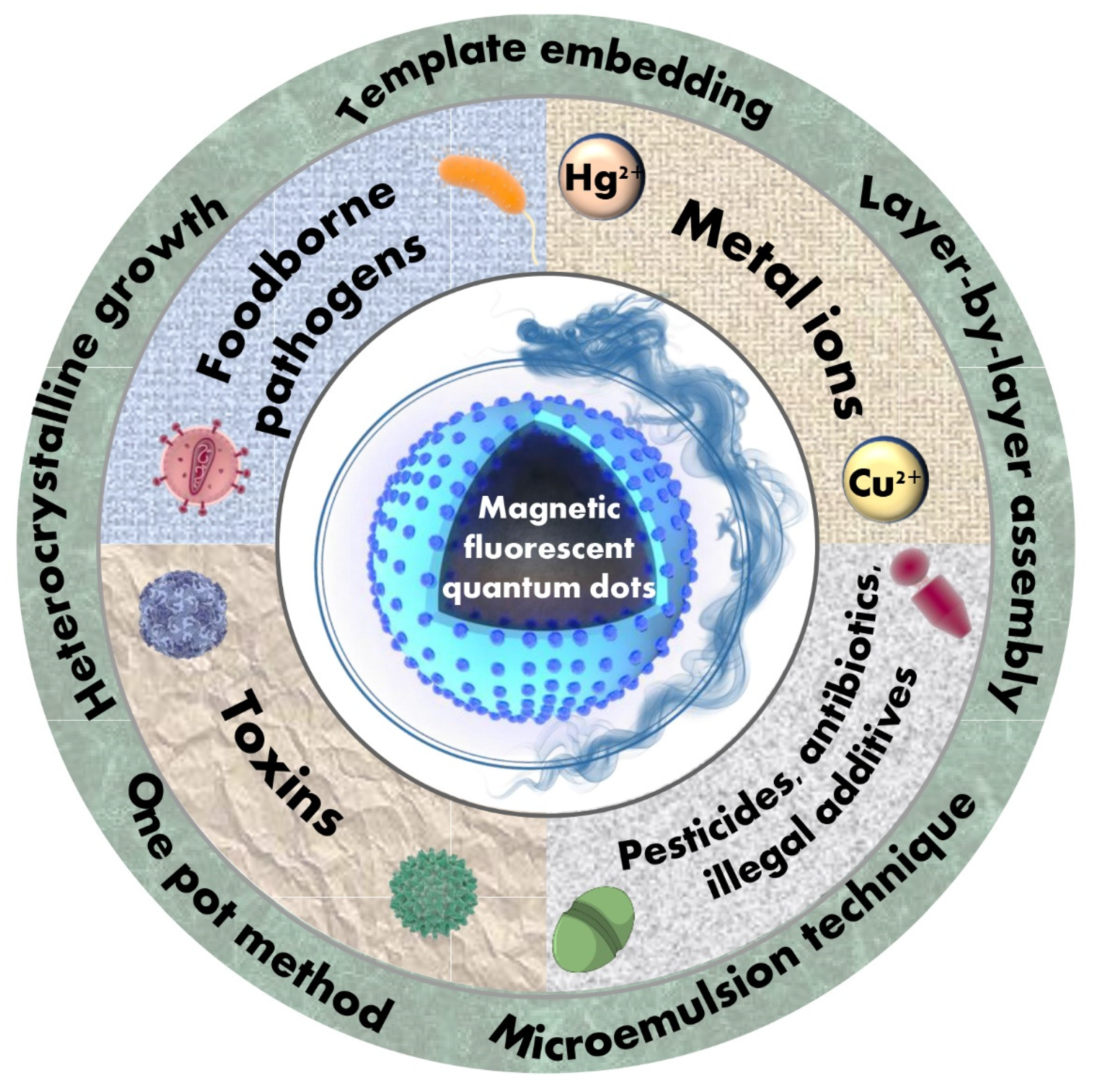
3. Applications to Food Contaminants Analysis
3.1. Metal Ions
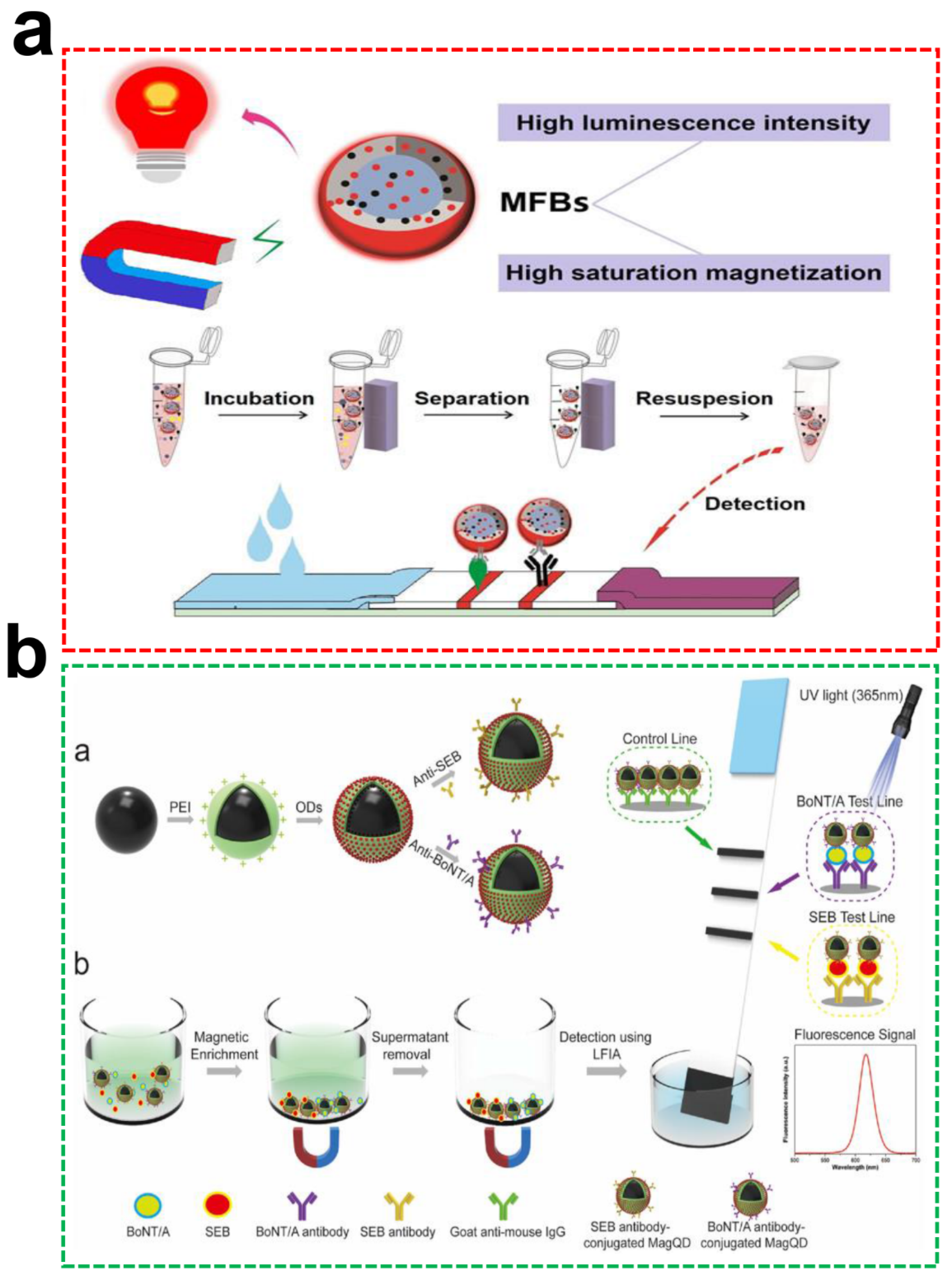
3.2. Foodborne Pathogens
3.3. Toxins
3.4. Pesticides, Antibiotics and Illegal Additives
4. Conclusions and Perspectives
- Pointing towards requirements of strong matrix tolerance and high QY, a universal method should be developed to simplify the preparation process and obtain a multifunctional MNPs@QDs probe.
- In-depth exploration of the adsorption, enrichment, and separation procedures between MNPs@QDs and food contaminants, to provide a theoretical basis for tailoring the appropriate pretreatment protocols with various characteristics of food samples.
- Combining high affinity, specific and stable recognition elements (such as MIPs, aptamers, and nanobodies) to construct rapid, sensitive, and high-throughput sensing platforms for food contaminants detections through different analytical forms.
- Miniaturized and portable equipment integrated with sensing platforms for immediate on-site detection to confront various food safety incidents. The smartphones and microfluidic technologies that belong to smart manufacturing also provide a future development direction for analysis devices.
Author Contributions
Funding
Institutional Review Board Statement
Informed Consent Statement
Data Availability Statement
Conflicts of Interest
References
- Bhavadharini, B.; Kavimughil, M.; Malini, B.; Vallath, A.; Prajapati, H.K.; Sunil, C.K. Recent Advances in Biosensors for Detection of Chemical Contaminants in Food—A Review. Food Anal. Methods 2022, 1–20. [Google Scholar] [CrossRef]
- Romero-Gonzalez, R. Food safety: How analytical chemists ensure it. Anal. Methods 2015, 7, 7193–7201. [Google Scholar] [CrossRef]
- Pena-Rosas, J.P.; De-Regil, L.M.; Rogers, L.M.; Bopardikar, A.; Panisset, U. Translating Research into Action: WHO Evidence-Informed Guidelines for Safe and Effective Micronutrient Interventions. J. Nutr. 2012, 142, 197–204. [Google Scholar] [CrossRef] [PubMed]
- Barzegar, F.; Kamankesh, M.; Mohammadi, A. Recent Development in Formation, Toxic Effects, Human Health and Analytical Techniques of Food Contaminants. Food Rev. Int. 2021, 1–27. [Google Scholar] [CrossRef]
- Mejia-Carmona, K.; Maciel, E.V.S.; Lancas, F.M. Miniaturized liquid chromatography applied to the analysis of residues and contaminants in food: A review. Electrophoresis 2020, 41, 1680–1693. [Google Scholar] [CrossRef]
- Xu, M.-L.; Gao, Y.; Wang, X.; Han, X.X.; Zhao, B. Comprehensive Strategy for Sample Preparation for the Analysis of Food Contaminants and Residues by GC-MS/MS: A Review of Recent Research Trends. Foods 2021, 10, 2473. [Google Scholar] [CrossRef]
- Masia, A.; Morales Suarez-Varela, M.; Llopis-Gonzalez, A.; Pico, Y. Determination of pesticides and veterinary drug residues in food by liquid chromatography-mass spectrometry: A review. Anal. Chim. Acta 2016, 936, 40–61. [Google Scholar] [CrossRef]
- Lv, M.; Liu, Y.; Geng, J.; Kou, X.; Xin, Z.; Yang, D. Engineering nanomaterials-based biosensors for food safety detection. Biosens. Bioelectron. 2018, 106, 122–128. [Google Scholar] [CrossRef]
- Bhardwaj, N.; Bhardwaj, S.K.; Nayak, M.K.; Mehta, J.; Kim, K.-H.; Deep, A. Fluorescent nanobiosensors for the targeted detection of foodborne bacteria. Trends Anal. Chem. 2017, 97, 120–135. [Google Scholar] [CrossRef]
- Bobrinetskiy, I.I.; Knezevic, N.Z. Graphene-based biosensors for on-site detection of contaminants in food. Anal. Methods 2018, 10, 5061–5070. [Google Scholar] [CrossRef]
- Sharma, R.; Ragavan, K.V.; Thakur, M.S.; Raghavarao, K.S.M.S. Recent advances in nanoparticle based aptasensors for food contaminants. Biosens. Bioelectron. 2015, 74, 612–627. [Google Scholar] [CrossRef] [PubMed]
- Marrubini, G.; Appelblad, P.; Maietta, M.; Papetti, A. Hydrophilic interaction chromatography in food matrices analysis: An updated review. Food Chem. 2018, 257, 53–66. [Google Scholar] [CrossRef] [PubMed]
- Barreiro, J.C.; Luiz, A.L.; Fernandes Maciel, S.C.; Soares Maciel, E.V.; Lancas, F.M. Recent approaches for on-line analysis of residues and contaminants in food matrices: A review. J. Sep. Sci. 2015, 38, 1721–1732. [Google Scholar] [CrossRef]
- Khan, R.; Rehman, A.; Hayat, A.; Andreescu, S. Magnetic Particles-Based Analytical Platforms for Food Safety Monitoring. Magnetochemistry 2019, 5, 63. [Google Scholar] [CrossRef]
- Gao, Q.; Luo, D.; Bai, M.; Chen, Z.-W.; Feng, Y.-Q. Rapid Determination of Estrogens in Milk Samples Based on Magnetite Nanoparticles/Polypyrrole Magnetic Solid-Phase Extraction Coupled with Liquid Chromatography-Tandem Mass Spectrometry. J. Agric. Food Chem. 2011, 59, 8543–8549. [Google Scholar] [CrossRef]
- Yu, X.; Zhong, T.; Zhang, Y.; Zhao, X.; Xiao, Y.; Wang, L.; Liu, X.; Zhang, X. Design, Preparation, and Application of Magnetic Nanoparticles for Food Safety Analysis: A Review of Recent Advances. J. Agric. Food Chem. 2021, 70, 46–62. [Google Scholar] [CrossRef]
- Mahmoudi, M.; Sant, S.; Wang, B.; Laurent, S.; Sen, T. Superparamagnetic iron oxide nanoparticles (SPIONs): Development, surface modification and applications in chemotherapy. Adv. Drug Deliv. Rev. 2011, 63, 24–46. [Google Scholar] [CrossRef]
- Tan, J.; Wang, T.; Li, Y.; Xu, S.; Chen, S.; Hao, H. Review on functionalized magnetic nanoparticles for the pretreatment of organophosphorus pesticides. Green Process. Synth. 2021, 10, 485–498. [Google Scholar] [CrossRef]
- Peltomaa, R.; Barderas, R.; Benito-Pena, E.; Moreno-Bondi, M.C. Recombinant antibodies and their use for food immunoanalysis. Anal. Bioanal. Chem. 2022, 414, 193–217. [Google Scholar] [CrossRef]
- Yan, M.; Li, H.; Li, M.; Cao, X.; She, Y.; Chen, Z. Advances in Surface-Enhanced Raman Scattering-Based Aptasensors for Food Safety Detection. J. Agric. Food Chem. 2021, 69, 14049–14064. [Google Scholar] [CrossRef]
- Villa, C.C.; Sanchez, L.T.; Valencia, G.A.; Ahmed, S.; Gutierrez, T.J. Molecularly imprinted polymers for food applications: A review. Trends Food Sci. Technol. 2021, 111, 642–669. [Google Scholar] [CrossRef]
- Chen, L.; Cheng, Z.; Luo, M.; Wang, T.; Zhang, L.; Wei, J.; Wang, Y.; Li, P. Fluorescent noble metal nanoclusters for contaminants analysis in food matrix. Crit. Rev. Food Sci. Nutr. 2021, 1–19. [Google Scholar] [CrossRef] [PubMed]
- He, H.; Sun, D.; Wu, Z.; Pu, H.; Wei, Q. On-off-on fluorescent nanosensing: Materials, detection strategies and recent food applications. Trends Food Sci. Technol. 2022, 119, 243–256. [Google Scholar] [CrossRef]
- Li, T.; Li, Z.; Huang, T.; Tian, L. Carbon quantum dot-based sensors for food safety. Sens. Actuators A 2021, 331, 113003. [Google Scholar] [CrossRef]
- Wu, Y.; Sun, J.; Huang, X.; Lai, W.; Xiong, Y. Ensuring food safety using fluorescent nanoparticles-based immunochromatographic test strips. Trends Food Sci. Technol. 2021, 118, 658–678. [Google Scholar] [CrossRef]
- Ledentsov, N.N.; Ustinov, V.M.; Shchukin, V.A.; Kop’ev, P.S.; Alferov, Z.I.; Bimberg, D. Quantum dot heterostructures: Fabrication, properties, lasers (Review). Semiconductors 1998, 32, 343–365. [Google Scholar] [CrossRef]
- Galstyan, V. “Quantum dots: Perspectives in next-generation chemical gas sensors”—A review. Anal. Chim. Acta 2021, 1152, 238192. [Google Scholar] [CrossRef]
- Kaur, M.; Kaur, M.; Sharma, V.K. Nitrogen-doped graphene and graphene quantum dots: A review onsynthesis and applications in energy, sensors and environment. Adv. Colloid Interface Sci. 2018, 259, 44–64. [Google Scholar] [CrossRef]
- Khan, Z.G.; Patil, P.O. A comprehensive review on carbon dots and graphene quantum dots based fluorescent sensor for biothiols. Microchem. J. 2020, 157, 105011. [Google Scholar] [CrossRef]
- Molaei, M.J. Principles, mechanisms, and application of carbon quantum dots in sensors: A review. Anal. Methods 2020, 12, 1266–1287. [Google Scholar] [CrossRef]
- Wang, X.; Cheng, L. Multifunctional two-dimensional nanocomposites for photothermal-based combined cancer therapy. Nanoscale 2019, 11, 15685–15708. [Google Scholar] [CrossRef] [PubMed]
- Kaewsaneha, C.; Tangboriboonrat, P.; Polpanich, D.; Elaissari, A. Multifunctional Fluorescent-Magnetic Polymeric Colloidal Particles: Preparations and Bioanalytical Applications. ACS Appl. Mater. Interfaces 2015, 7, 23373–23386. [Google Scholar] [CrossRef] [PubMed]
- Koole, R.; Mulder, W.J.M.; van Schooneveld, M.M.; Strijkers, G.J.; Meijerink, A.; Nicolay, K. Magnetic quantum dots for multimodal imaging. Wiley Interdiscip. Rev. Nanomed. Nanobiotechnol. 2009, 1, 475–491. [Google Scholar] [CrossRef] [PubMed]
- Mahajan, K.D.; Fan, Q.; Dorcena, J.; Ruan, G.; Winter, J.O. Magnetic quantum dots in biotechnology–synthesis and applications. Biotechnol. J. 2013, 8, 1424–1434. [Google Scholar] [CrossRef]
- Roullier, V.; Marchi-Artzner, V.; Cador, O.; Dorson, F.; Aubert, T.; Cordier, S.; Molard, Y.; Grasset, F.; Mornet, S.; Haneda, H. Synthesis and characterisation of magnetic-luminescent composite colloidal nanostructures. Int. J. Nanotechnol. 2010, 7, 46–57. [Google Scholar] [CrossRef]
- Xia, H.; Tong, R.; Song, Y.; Xiong, F.; Li, J.; Wang, S.; Fu, H.; Wen, J.; Li, D.; Zeng, Y.; et al. Synthesis and bio-applications of targeted magnetic-fluorescent composite nanoparticles. J. Nanopart. Res. 2017, 19, 149. [Google Scholar] [CrossRef]
- Acharya, A. Luminescent Magnetic Quantum Dots for In Vitro/In Vivo Imaging and Applications in Therapeutics. J. Nanosci. Nanotechnol. 2013, 13, 3753–3768. [Google Scholar] [CrossRef]
- Jing, L.; Ding, K.; Kershaw, S.V.; Kempson, I.M.; Rogach, A.L.; Gao, M. Magnetically Engineered Semiconductor Quantum Dots as Multimodal Imaging Probes. Adv. Mater. 2014, 26, 6367–6386. [Google Scholar] [CrossRef]
- Tufani, A.; Qureshi, A.; Niazi, J.H. Iron oxide nanoparticles based magnetic luminescent quantum dots (MQDs) synthesis and biomedical/biological applications: A review. Mater. Sci. Eng. C 2021, 118, 111545. [Google Scholar] [CrossRef]
- Kim, H.; Achermann, M.; Balet, L.P.; Hollingsworth, J.A.; Klimov, V.I. Synthesis and characterization of Co/CdSe core/shell nanocomposites: Bifunctional magnetic-optical nanocrystals. J. Am. Chem. Soc. 2005, 127, 544–546. [Google Scholar] [CrossRef]
- Zhou, S.; Chen, Q.; Hu, X.; Zhao, T. Bifunctional luminescent superparamagnetic nanocomposites of CdSe/CdS-Fe3O4 synthesized via a facile method. J. Mater. Chem. 2012, 22, 8263–8270. [Google Scholar] [CrossRef]
- Bhandari, S.; Khandelia, R.; Pan, U.N.; Chattopadhyay, A. Surface Complexation-Based Biocompatible Magnetofluorescent Nanoprobe for Targeted Cellular Imaging. ACS Appl. Mater. Interfaces 2015, 7, 17552–17557. [Google Scholar] [CrossRef] [PubMed]
- Wu, Y.; Zou, H.; Zhang, Y.; Mou, M.; Niu, Q.; Yan, Z.; Liao, S. The Loading of Luminescent Magnetic Nanocomposites Fe3O4@Polyaniline/Carbon Dots for Methotrexate and Its Release Behavior In Vitro. J. Nanosci. Nanotechnol. 2020, 20, 701–708. [Google Scholar] [CrossRef] [PubMed]
- Lee, J.-S.; Bodnarchuk, M.I.; Shevchenko, E.V.; Talapin, D.V. “Magnet-in-the-Semiconductor” FePt-PbS and FePt-PbSe Nanostructures: Magnetic Properties, Charge Transport, and Magnetoresistance. J. Am. Chem. Soc. 2010, 132, 6382–6391. [Google Scholar] [CrossRef]
- Gu, H.W.; Zheng, R.K.; Zhang, X.X.; Xu, B. Facile one-pot synthesis of bifunctional heterodimers of nanoparticles: A conjugate of quantum dot and magnetic nanoparticles. J. Am. Chem. Soc. 2004, 126, 5664–5665. [Google Scholar] [CrossRef]
- Selvan, S.T.; Patra, P.K.; Ang, C.Y.; Ying, J.Y. Synthesis of silica-coated semiconductor and magnetic quantum dots and their use in the imaging of live cells. Angew. Chem. Int. Ed. 2007, 46, 2448–2452. [Google Scholar] [CrossRef]
- Lin, A.W.H.; Ang, C.Y.; Patra, P.K.; Han, Y.; Gu, H.; Le Breton, J.-M.; Juraszek, J.; Chiron, H.; Papaefthymiou, G.C.; Selvan, S.T.; et al. Seed-mediated synthesis, properties and application of gamma-Fe2O3-CdSe magnetic quantum dots. J. Solid State Chem. 2011, 184, 2150–2158. [Google Scholar] [CrossRef]
- McDaniel, H.; Shim, M. Size and Growth Rate Dependent Structural Diversification of Fe3O4/CdS Anisotropic Nanocrystal Heterostructures. ACS Nano 2009, 3, 434–440. [Google Scholar] [CrossRef]
- Gao, J.; Zhang, W.; Huang, P.; Zhang, B.; Zhang, X.; Xu, B. Intracellular spatial control of fluorescent magnetic nanoparticles. J. Am. Chem. Soc. 2008, 130, 3710–3711. [Google Scholar] [CrossRef]
- Du, G.H.; Liu, Z.L.; Lu, Q.H.; Xia, X.; Jia, L.H.; Yao, K.L.; Chu, Q.; Zhang, S.M. Fe3O4/CdSe/ZnS magnetic fluorescent bifunctional nanocomposites. Nanotechnology 2006, 17, 2850–2854. [Google Scholar] [CrossRef]
- Hines, M.A.; Guyot-Sionnest, P. Synthesis and characterization of strongly luminescing ZnS-Capped CdSe nanocrystals. J. Phys. Chem. 1996, 100, 468–471. [Google Scholar] [CrossRef]
- Deng, S.; Ruan, G.; Han, N.; Winter, J.O. Interactions in fluorescent-magnetic heterodimer nanocomposites. Nanotechnology 2010, 21, 145605. [Google Scholar] [CrossRef] [PubMed]
- Beaune, G.; Dubertret, B.; Clement, O.; Vayssettes, C.; Cabuil, V.; Menager, C. Giant vesicles containing magnetic nanoparticles and quantum dots: Feasibility and tracking by fiber confocal fluorescence microscopy. Angew. Chem. Int. Ed. 2007, 46, 5421–5424. [Google Scholar] [CrossRef] [PubMed]
- Ruan, G.; Vieira, G.; Henighan, T.; Chen, A.; Thakur, D.; Sooryakumar, R.; Winter, J.O. Simultaneous Magnetic Manipulation and Fluorescent Tracking of Multiple Individual Hybrid Nanostructures. Nano Lett. 2010, 10, 2220–2224. [Google Scholar] [CrossRef]
- Park, J.-H.; von Maltzahn, G.; Ruoslahti, E.; Bhatia, S.N.; Sailor, M.J. Micellar hybrid nanoparticles for simultaneous magnetofluorescent imaging and drug delivery. Angew. Chem. Int. Ed. 2008, 47, 7284–7288. [Google Scholar] [CrossRef]
- Kim, J.; Lee, J.E.; Lee, J.; Yu, J.H.; Kim, B.C.; An, K.; Hwang, Y.; Shin, C.H.; Park, J.G.; Kim, J.; et al. Magnetic fluorescent delivery vehicle using uniform mesoporous silica spheres embedded with monodisperse magnetic and semiconductor nanocrystals. J. Am. Chem. Soc. 2006, 128, 688–689. [Google Scholar] [CrossRef]
- Sun, L.; Zang, Y.; Sun, M.D.; Wang, H.G.; Zhu, X.J.; Xu, S.F.; Yang, Q.B.; Li, Y.X.; Shan, Y.M. Synthesis of magnetic and fluorescent multifunctional hollow silica nanocomposites for live cell imaging. J. Colloid Interface Sci. 2010, 350, 90–98. [Google Scholar] [CrossRef]
- Xiao, Q.; Xiao, C. Preparation and Characterization of Silica-Coated Magnetic-Fluorescent Bifunctional Microspheres. Nanoscale Res. Lett. 2009, 4, 1078–1084. [Google Scholar] [CrossRef]
- Yi, D.K.; Selvan, S.T.; Lee, S.S.; Papaefthymiou, G.C.; Kundaliya, D.; Ying, J.Y. Silica-coated nanocomposites of magnetic nanoparticles and quantum dots. J. Am. Chem. Soc. 2005, 127, 4990–4991. [Google Scholar] [CrossRef]
- Ruan, J.; Wang, K.; Song, H.; Xu, X.; Ji, J.J.; Cui, D.X. Biocompatibility of hydrophilic silica-coated CdTe quantum dots and magnetic nanoparticles. Nanoscale Res. Lett. 2011, 6, 13. [Google Scholar] [CrossRef]
- Kyeong, S.; Jeong, C.; Kim, H.Y.; Hwang, D.W.; Kang, H.; Yang, J.-K.; Lee, D.S.; Jun, B.-H.; Lee, Y.-S. Fabrication of mono-dispersed silica-coated quantum dot-assembled magnetic nanoparticles. RSC Adv. 2015, 5, 32072–32077. [Google Scholar] [CrossRef]
- Huang, L.; Zhang, Y.; Liao, T.; Xu, K.; Jiang, C.; Zhuo, D.; Wang, Y.; Wen, H.-M.; Wang, J.; Ao, L.; et al. Compact Magneto-Fluorescent Colloids by Hierarchical Assembly of Dual-Components in Radial Channels for Sensitive Point-of-Care Immunoassay. Small 2021, 17, 2100862. [Google Scholar] [CrossRef] [PubMed]
- Tan, Y.F.; Chandrasekharan, P.; Maity, D.; Yong, C.X.; Chuang, K.-H.; Zhao, Y.; Wang, S.; Ding, J.; Feng, S.-S. Multimodal tumor imaging by iron oxides and quantum dots formulated in poly (lactic acid)-D-alpha-tocopheryl polyethylene glycol 1000 succinate nanoparticles. Biomaterials 2011, 32, 2969–2978. [Google Scholar] [CrossRef] [PubMed]
- Xie, H.Y.; Zuo, C.; Liu, Y.; Zhang, Z.L.; Pang, D.W.; Li, X.L.; Gong, J.P.; Dickinson, C.; Zhou, W.Z. Cell-targeting multifunctional nanospheres with both fluorescence and magnetism. Small 2005, 1, 506–509. [Google Scholar] [CrossRef] [PubMed]
- Li, Y.-H.; Song, T.; Liu, J.-Q.; Zhu, S.-J.; Chang, J. An efficient method for preparing high-performance multifunctional polymer beads simultaneously incorporated with magnetic nanoparticles and quantum dots. J. Mater. Chem. 2011, 21, 12520–12528. [Google Scholar] [CrossRef]
- Wilson, R.; Spiller, D.G.; Prior, I.A.; Veltkamp, K.J.; Hutchinson, A. A Simple Method for Preparing Spectrally Encoded Magnetic Beads for Multiplexed Detection. ACS Nano 2007, 1, 487–493. [Google Scholar] [CrossRef] [PubMed]
- Wang, C.; Wang, L.; Yang, W. Preparation and characterization of functional inorganic/organic composite microspheres via electrostatic interaction. J. Colloid Interface Sci. 2009, 333, 749–756. [Google Scholar] [CrossRef]
- Li, L.; Choo, E.S.G.; Liu, Z.; Ding, J.; Xue, J. Double-layer silica core-shell nanospheres with superparamagnetic and fluorescent functionalities. Chem. Phys. Lett. 2008, 461, 114–117. [Google Scholar] [CrossRef]
- Yin, N.; Wang, X.; Yang, T.; Ding, Y.; Li, L.; Zhao, S.; Li, P.; Xu, X.; Zhu, L. Multifunctional Fe3O4 cluster@ quantum dot-embedded mesoporous SiO2 nanoplatform probe for cancer cell fluorescence-labelling detection and photothermal therapy. Ceram. Int. 2021, 47, 8271–8278. [Google Scholar] [CrossRef]
- Yoo, J.H.; Kim, J.S. The Preparation of Core-Shell Magnetic Silica Nanospheres for Enhancing Magnetism and Fluorescence Intensity. J. Nanosci. Nanotechnol. 2013, 13, 7615–7619. [Google Scholar] [CrossRef]
- Sathe, T.R.; Agrawal, A.; Nie, S. Mesoporous silica beads embedded with semiconductor quantum dots and iron oxide nanocrystals: Dual-function microcarriers for optical encoding and magnetic separation. Anal. Chem. 2006, 78, 5627–5632. [Google Scholar] [CrossRef] [PubMed]
- Xie, H.-Y.; Xie, M.; Zhang, Z.-L.; Long, Y.-M.; Liu, X.; Tang, M.-L.; Pang, D.-W.; Tan, Z.; Dickinson, C.; Zhou, W. Wheat germ agglutinin-modified trifunctional nanospheres for cell recognition. Bioconjugate Chem. 2007, 18, 1749–1755. [Google Scholar] [CrossRef] [PubMed]
- Zebli, B.; Susha, A.S.; Sukhorukov, G.B.; Rogach, A.L.; Parak, W.J. Magnetic Targeting and Cellular Uptake of Polymer Microcapsules Simultaneously Functionalized with Magnetic and Luminescent Nanocrystals. Langmuir 2005, 21, 4262–4265. [Google Scholar] [CrossRef] [PubMed]
- Li, Z.; Wang, G.; Shen, Y.; Guo, N.; Ma, N. DNA-Templated Magnetic Nanoparticle-Quantum Dot Polymers for Ultrasensitive Capture and Detection of Circulating Tumor Cells. Adv. Funct. Mater. 2018, 28, 1707152. [Google Scholar] [CrossRef]
- Zou, W.-S.; Yang, J.; Yang, T.-T.; Hu, X.; Lian, H.-Z. Magnetic-room temperature phosphorescent multifunctional nanocomposites as chemosensor for detection and photo-driven enzyme mimetics for degradation of 2,4,6-trinitrotoluene. J. Mater. Chem. 2012, 22, 4720–4727. [Google Scholar] [CrossRef]
- Ortgies, D.H.; de la Cueva, L.; del Rosal, B.; Sanz-Rodríguez, F.; Fernández, N.; Iglesias-de la Cruz, M.C.; Salas, G.; Cabrera, D.; Teran, F.J.; Jaque, D.; et al. In Vivo Deep Tissue Fluorescence and Magnetic Imaging Employing Hybrid Nanostructures. ACS Appl. Mater. Interfaces 2016, 8, 1406–1414. [Google Scholar] [CrossRef]
- Xiao, L.-H.; Wang, T.; Zhao, T.-Y.; Zheng, X.; Sun, L.-Y.; Li, P.; Liu, F.-Q.; Gao, G.; Dong, A. Fabrication of Magnetic-Antimicrobial-Fluorescent Multifunctional Hybrid Microspheres and Their Properties. Int. J. Mol. Sci. 2013, 14, 7391–7404. [Google Scholar] [CrossRef]
- Fan, H.-M.; Olivo, M.; Shuter, B.; Yi, J.-B.; Bhuvaneswari, R.; Tan, H.-R.; Xing, G.-C.; Ng, C.-T.; Liu, L.; Lucky, S.S.; et al. Quantum Dot Capped Magnetite Nanorings as High Performance Nanoprobe for Multiphoton Fluorescence and Magnetic Resonance Imaging. J. Am. Chem. Soc. 2010, 132, 14803–14811. [Google Scholar] [CrossRef]
- Hong, X.; Li, J.; Wang, M.J.; Xu, J.J.; Guo, W.; Li, J.H.; Bai, Y.B.; Li, T.J. Fabrication of magnetic luminescent nanocomposites by a layer-by-layer self-assembly approach. Chem. Mater. 2004, 16, 4022–4027. [Google Scholar] [CrossRef]
- Wang, C.W.; Shen, W.Z.; Rong, Z.; Liu, X.X.; Gu, B.; Xiao, R.; Wang, S.Q. Layer-by-layer assembly of magnetic-core dual quantum dot-shell nanocomposites for fluorescence lateral flow detection of bacteria. Nanoscale 2020, 12, 795–807. [Google Scholar] [CrossRef]
- Chen, B.; Zhang, H.; Zhai, C.; Du, N.; Sun, C.; Xue, J.; Yang, D.; Huang, H.; Zhang, B.; Xie, Q.; et al. Carbon nanotube-based magnetic-fluorescent nanohybrids as highly efficient contrast agents for multimodal cellular imaging. J. Mater. Chem. 2010, 20, 9895–9902. [Google Scholar] [CrossRef]
- Sun, X.; Ding, K.; Hou, Y.; Gao, Z.; Yang, W.; Jing, L.; Gao, M. Bifunctional Superparticles Achieved by Assembling Fluorescent CuInS2@ZnS Quantum Dots and Amphibious Fe3O4 Nanocrystals. J. Phys. Chem. C 2013, 117, 21014–21020. [Google Scholar] [CrossRef]
- Wang, D.; He, J.; Rosenzweig, N.; Rosenzweig, Z. Superparamagnetic Fe2O3 Beads−CdSe/ZnS Quantum Dots Core−Shell Nanocomposite Particles for Cell Separation. Nano Lett. 2004, 4, 409–413. [Google Scholar] [CrossRef]
- Nikitin, M.P.; Zdobnova, T.A.; Lukash, S.V.; Stremovskiy, O.A.; Deyev, S.M. Protein-assisted self-assembly of multifunctional nanoparticles. Proc. Natl. Acad. Sci. USA 2010, 107, 5827. [Google Scholar] [CrossRef] [PubMed]
- Shibu, E.S.; Ono, K.; Sugino, S.; Nishioka, A.; Yasuda, A.; Shigeri, Y.; Wakida, S.-i.; Sawada, M.; Biju, V. Photouncaging Nanoparticles for MRI and Fluorescence Imaging in Vitro and in Vivo. ACS Nano 2013, 7, 9851–9859. [Google Scholar] [CrossRef] [PubMed]
- Song, E.; Han, W.; Xu, H.; Jiang, Y.; Cheng, D.; Song, Y.; Swihart, M.T. Magnetically Encoded Luminescent Composite Nanoparticles through Layer-by-Layer Self-Assembly. Chem.—Eur. J. 2014, 20, 14642–14649. [Google Scholar] [CrossRef] [PubMed]
- Rong, Z.; Bai, Z.K.; Li, J.N.; Tang, H.; Shen, T.Y.; Wang, Q.; Wang, C.W.; Xiao, R.; Wang, S.Q. Dual-color magnetic-quantum dot nanobeads as versatile fluorescent probes in test strip for simultaneous point-of-care detection of free and complexed prostate-specific antigen. Biosens. Bioelectron. 2019, 145, 8. [Google Scholar] [CrossRef]
- Wang, C.; Cheng, X.; Liu, L.; Zhang, X.; Yang, X.; Zheng, S.; Rong, Z.; Wang, S. Ultrasensitive and Simultaneous Detection of Two Specific SARS-CoV-2 Antigens in Human Specimens Using Direct/Enrichment Dual-Mode Fluorescence Lateral Flow Immunoassay. ACS Appl. Mater. Interfaces 2021, 13, 40342–40353. [Google Scholar] [CrossRef]
- Dong, S.; Wang, S.; Wang, X.; Zhai, L. Superparamagnetic nanocomposite Fe3O4@SiO2-NH2/CQDs as fluorescent probe for copper (II) detection. Mater. Lett. 2020, 278, 128404. [Google Scholar] [CrossRef]
- Zhang, X.; Tang, B.; Li, Y.; Liu, C.; Jiao, P.; Wei, Y. Molecularly Imprinted Magnetic Fluorescent Nanocomposite-Based Sensor for Selective Detection of Lysozyme. Nanomaterials 2021, 11, 1575. [Google Scholar] [CrossRef]
- Li, C.-L.; Huang, B.-R.; Chang, J.-Y.; Chen, J.-K. Bifunctional superparamagnetic-luminescent core-shell-satellite structured microspheres: Preparation, characterization, and magnetodisplay application. J. Mater. Chem. C 2015, 3, 4603–4615. [Google Scholar] [CrossRef]
- Koc, K.; Karakus, B.; Rajar, K.; Alveroglu, E. Synthesis and characterization. of ZnS@Fe3O4 fluorescent-magnetic bifunctional nanospheres. Superlattices Microstruct. 2017, 110, 198–204. [Google Scholar] [CrossRef]
- Wang, K.; Xu, X.; Li, Y.; Rong, M.; Wang, L.; Lu, L.; Wang, J.; Zhao, F.; Sun, B.; Jiang, Y. Preparation Fe3O4@chitosan-graphene quantum dots nanocomposites for fluorescence and magnetic resonance imaging. Chem. Phys. Lett. 2021, 783, 139060. [Google Scholar] [CrossRef]
- Wang, Z.; Jiang, X.; Liu, W.; Lu, G.; Huang, X. A rapid and operator-safe powder approach for latent fingerprint detection using hydrophilic Fe3O4@SiO2-CdTe nanoparticles. Sci. China Chem. 2019, 62, 889–896. [Google Scholar] [CrossRef]
- Wang, M.; Fei, X.F.; Lv, S.W.; Sheng, Y.; Zou, H.F.; Song, Y.H.; Yan, F.; Zhu, Q.L.; Zheng, K.Y. Synthesis and characterization of a flexible fluorescent magnetic Fe3O4@SiO2/CdTe-NH2 nanoprobe. J. Inorg. Biochem. 2018, 186, 307–316. [Google Scholar] [CrossRef] [PubMed]
- Zhang, Y.; Wang, S.-N.; Ma, S.; Guan, J.-J.; Li, D.; Zhang, X.-D.; Zhang, Z.-D. Self-assembly multifunctional nanocomposites with Fe3O4 magnetic core and CdSe/ZnS quantum dots shell. J. Biomed. Mater. Res. Part A 2008, 85A, 840–846. [Google Scholar] [CrossRef]
- Chen, O.; Riedemann, L.; Etoc, F.; Herrmann, H.; Coppey, M.; Barch, M.; Farrar, C.T.; Zhao, J.; Bruns, O.T.; Wei, H.; et al. Magneto-fluorescent core-shell supernanoparticles. Nat. Commun. 2014, 5, 5093. [Google Scholar] [CrossRef]
- Piao, Y.; Burns, A.; Kim, J.; Wiesner, U.; Hyeon, T. Designed Fabrication of Silica-Based Nanostructured Particle Systems for Nanomedicine Applications. Adv. Funct. Mater. 2008, 18, 3745–3758. [Google Scholar] [CrossRef]
- Guo, L.; Shao, Y.; Duan, H.; Ma, W.; Leng, Y.; Huang, X.; Xiong, Y. Magnetic Quantum Dot Nanobead-Based Fluorescent Immunochromatographic Assay for the Highly Sensitive Detection of Aflatoxin B1 in Dark Soy Sauce. Anal. Chem. 2019, 91, 4727–4734. [Google Scholar] [CrossRef]
- Guo, S.; Chen, Y.-Q.; Lu, N.-N.; Wang, X.-Y.; Xie, M.; Sui, W.-P. Ultrasonication-assisted one-step self-assembly preparation of biocompatible fluorescent-magnetic nanobeads for rare cancer cell detection. Nanotechnology 2014, 25, 505603. [Google Scholar] [CrossRef]
- Kim, J.; Lee, J.E.; Lee, S.H.; Yu, J.H.; Lee, J.H.; Park, T.G.; Hyeon, T. Designed fabrication of a multifunctional polymer nanomedical platform for simultaneous cancer-targeted imaging and magnetically guided drug delivery. Adv. Mater. 2008, 20, 478–483. [Google Scholar] [CrossRef]
- Leng, Y.; Wu, W.; Li, L.; Lin, K.; Sun, K.; Chen, X.; Li, W. Magnetic/Fluorescent Barcodes Based on Cadmium-Free Near-Infrared-Emitting Quantum Dots for Multiplexed Detection. Adv. Funct. Mater. 2016, 26, 7581–7589. [Google Scholar] [CrossRef]
- Lan, J.; Chen, J.; Li, N.; Ji, X.; Yu, M.; He, Z. Microfluidic generation of magnetic-fluorescent Janus microparticles for biomolecular detection. Talanta 2016, 151, 126–131. [Google Scholar] [CrossRef] [PubMed]
- Zhou, C.; Wang, Z.; Xia, J.; Via, B.K.; Zhang, F.; Xia, Y.; Li, Y. A simplistic one-pot method to produce magnetic graphene-CdS nanocomposites. Comptes Rendus Chim. 2012, 15, 714–718. [Google Scholar] [CrossRef]
- Maleki, S.; Madrakian, T.; Afkhami, A. Magnetic solid-phase extraction of codeine in a biological sample utilizing Fe3O4/CDs/Lys nanocomposite as an efficient adsorbent. J. Iran. Chem. Soc. 2019, 16, 2111–2121. [Google Scholar] [CrossRef]
- Liu, X.; Jiang, H.; Ye, J.; Zhao, C.; Gao, S.; Wu, C.; Li, C.; Li, J.; Wang, X. Nitrogen-Doped Carbon Quantum Dot Stabilized Magnetic Iron Oxide Nanoprobe for Fluorescence, Magnetic Resonance, and Computed Tomography Triple-Modal In Vivo Bioimaging. Adv. Funct. Mater. 2016, 26, 8694–8706. [Google Scholar] [CrossRef]
- Li, B.; Wang, X.; Guo, Y.; Iqbal, A.; Dong, Y.; Li, W.; Liu, W.; Qin, W.; Chen, S.; Zhou, X.; et al. One-pot synthesis of polyamines improved magnetism and fluorescence Fe3O4-carbon dots hybrid NPs for dual modal imaging. Dalton Trans. 2016, 45, 5484–5491. [Google Scholar] [CrossRef][Green Version]
- Irmania, N.; Dehvari, K.; Gedda, G.; Tseng, P.-J.; Chang, J.-Y. Manganese-doped green tea-derived carbon quantum dots as a targeted dual imaging and photodynamic therapy platform. J. Biomed. Mater. Res. Part B 2020, 108, 1616–1625. [Google Scholar] [CrossRef]
- Ji, Z.; Ai, P.; Shao, C.; Wang, T.; Yan, C.; Ye, L.; Gu, W. Manganese-Doped Carbon Dots for Magnetic Resonance/Optical Dual-Modal Imaging of Tiny Brain Glioma. ACS Biomater. Sci. Eng. 2018, 4, 2089–2094. [Google Scholar] [CrossRef]
- Yao, Y.-Y.; Gedda, G.; Girma, W.M.; Yen, C.-L.; Ling, Y.-C.; Chang, J.-Y. Magnetofluorescent Carbon Dots Derived from Crab Shell for Targeted Dual-Modality Bioimaging and Drug Delivery. ACS Appl. Mater. Interfaces 2017, 9, 13887–13899. [Google Scholar] [CrossRef]
- Liao, H.; Wang, Z.; Chen, S.; Wu, H.; Ma, X.; Tan, M. One-pot synthesis of gadolinium(III) doped carbon dots for fluorescence/magnetic resonance bimodal imaging. RSC Adv. 2015, 5, 66575–66581. [Google Scholar] [CrossRef]
- Yu, C.; Xuan, T.; Chen, Y.; Zhao, Z.; Liu, X.; Lian, G.; Li, H. Gadolinium-doped carbon dots with high quantum yield as an effective fluorescence and magnetic resonance bimodal imaging probe. J. Alloys Compd. 2016, 688, 611–619. [Google Scholar] [CrossRef]
- Wu, F.; Yue, L.; Yang, L.; Wang, K.; Liu, G.; Luo, X.; Zhu, X. Ln(III) chelates-functionalized carbon quantum dots: Synthesis, optical studies and multimodal bioimaging applications. Colloids Surf. B 2019, 175, 272–280. [Google Scholar] [CrossRef] [PubMed]
- Moro, L.; Turemis, M.; Marini, B.; Ippodrino, R.; Giardi, M.T. Better together: Strategies based on magnetic particles and quantum dots for improved biosensing. Biotechnol. Adv. 2017, 35, 51–63. [Google Scholar] [CrossRef]
- Li, Y.; Chen, Y.; Yu, H.; Tian, L.; Wang, Z. Portable and smart devices for monitoring heavy metal ions integrated with nanomaterials. TrAC Trends Anal. Chem. 2018, 98, 190–200. [Google Scholar] [CrossRef]
- Liu, L.; Xiao, L.; Zhu, H.; Shi, X. Preparation of magnetic and fluorescent bifunctional chitosan nanoparticles for optical determination of copper ion. Microchim. Acta 2012, 178, 413–419. [Google Scholar] [CrossRef]
- Xie, C.; Xiao, L.; Peng, S.; Shi, X. Preparation of novel magnetic and fluorescent CS-Fe3O4@CdSeS nanoparticles for simultaneous removal and optical determination of trace copper ions. New J. Chem. 2014, 38, 6095–6102. [Google Scholar] [CrossRef]
- Kumar, A.; Chowdhuri, A.R.; Laha, D.; Chandra, S.; Karmakar, P.; Sahu, S.K. One-pot synthesis of carbon dot-entrenched chitosan-modified magnetic nanoparticles for fluorescence-based Cu2+ ion sensing and cell imaging. RSC Adv. 2016, 6, 58979–58987. [Google Scholar] [CrossRef]
- Rafiee, F.; Tajfar, N.; Mohammadnejad, M. The synthesis and efficiency investigation of a boronic acid-modified magnetic chitosan quantum dot nanocomposite in the detection of Cu2+ ions. Int. J. Biol. Macromol. 2021, 189, 477–482. [Google Scholar] [CrossRef]
- Wang, H.; Sun, L.; Li, Y.; Fei, X.; Sun, M.; Zhang, C.; Li, Y.; Yang, Q. Layer-by-Layer Assembled Fe3O4@C@CdTe Core/Shell Microspheres as Separable Luminescent Probe for Sensitive Sensing of Cu2+ Ions. Langmuir 2011, 27, 11609–11615. [Google Scholar] [CrossRef]
- Li, L.; Jia, C.; Wang, F.J.; Fan, H.L.; Jiao, W.Z.; Shao, Z.Q. Facile synthesis of magnetic fluorescent nanoparticles: Adsorption and selective detection of Hg(II) in water. J. Mater. Chem. C 2018, 6, 2360–2369. [Google Scholar] [CrossRef]
- Yang, C.H.; Ding, Y.L.; Qian, J. Design of magnetic-fluorescent based nanosensor for highly sensitive determination and removal of HG2+. Ceram. Int. 2018, 44, 9746–9752. [Google Scholar] [CrossRef]
- Zhai, X.; Gong, Y.; Yang, W.; Kang, H.; Zhang, X. Mn-doped CdS/ZnS/CdS QD-based fluorescent nanosensor for rapid, selective, and ultrasensitive detection of copper(ii) ion. RSC Adv. 2015, 5, 63458–63464. [Google Scholar] [CrossRef]
- Yang, P.; Zhu, B.; Zhao, J.; Yu, H.; Yan, L.; Wei, Q.; Du, B. A novel reusable glutathione-modified magnetic fluorescent nanosensor for highly sensitive determination and removal of Cu2+. Inorg. Chim. Acta 2013, 408, 120–125. [Google Scholar] [CrossRef]
- Zhu, B.; Yang, P.; Yu, H.; Yan, L.; Wei, Q.; Du, B. Development of a novel water-soluble magnetic fluorescent nanoparticle for the selective detection and removal of Cu2+. Nanotechnology 2013, 24, 495502. [Google Scholar] [CrossRef] [PubMed]
- Kurshanov, D.A.; Khavlyuk, P.D.; Baranov, M.A.; Dubavik, A.; Rybin, A.V.; Fedorov, A.V.; Baranov, A.V. Magneto-Fluorescent Hybrid Sensor CaCO3-Fe3O4-AgInS2/ZnS for the Detection of Heavy Metal Ions in Aqueous Media. Materials 2020, 13, 4373. [Google Scholar] [CrossRef]
- Xie, R.; Qu, Y.; Tang, M.; Zhao, J.; Chua, S.; Li, T.; Zhang, F.; Wheatley, A.E.H.; Chai, F. Carbon dots-magnetic nanocomposites for the detection and removal of Hg2+. Food Chem. 2021, 364, 130366. [Google Scholar] [CrossRef]
- Xu, J.; Wang, Y.; Hu, S. Nanocomposites of graphene and graphene oxides: Synthesis, molecular functionalization and application in electrochemical sensors and biosensors. A review. Microchim. Acta 2017, 184, 1–44. [Google Scholar] [CrossRef]
- Alvand, M.; Shemirani, F. A Fe3O4@SiO2@graphene quantum dot core-shell structured nanomaterial as a fluorescent probe and for magnetic removal of mercury(II) ion. Microchim. Acta 2017, 184, 1621–1629. [Google Scholar] [CrossRef]
- Wang, Y.; Tang, M.; Shen, H.; Che, G.; Qiao, Y.; Liu, B.; Wang, L. Recyclable Multifunctional Magnetic Mesoporous Silica Nanocomposite for Ratiometric Detection, Rapid Adsorption, and Efficient Removal of Hg(II). ACS Sustain. Chem. Eng. 2018, 6, 1744–1752. [Google Scholar] [CrossRef]
- Ahmadian-Fard-Fini, S.; Ghanbari, D.; Salavati-Niasari, M. Photoluminescence carbon dot as a sensor for detecting of Pseudomonas aeruginosa bacteria: Hydrothermal synthesis of magnetic hollow NiFe2O4-carbon dots nanocomposite material. Compos. Part B 2019, 161, 564–577. [Google Scholar] [CrossRef]
- Ahmadian-Fard-Fini, S.; Ghanbari, D.; Amiri, O.; Salavati-Niasari, M. Green sonochemistry assisted synthesis of hollow magnetic and photoluminescent MgFe2O4-carbon dot nanocomposite as a sensor for toxic Ni(ii), Cd(ii) and Hg(ii) ions and bacteria. RSC Adv. 2021, 11, 22805–22811. [Google Scholar] [CrossRef]
- Yue, L.; Li, H.; Liu, Q.; Guo, D.; Chen, J.; Sun, Q.; Xu, Y.; Wu, F. Manganese-doped carbon quantum dots for fluorometric and magnetic resonance (dual mode) bioimaging and biosensing. Microchim. Acta 2019, 186, 315. [Google Scholar] [CrossRef] [PubMed]
- Wu, N.; Liu, X.; Zeng, M.; Gao, J.; Lu, X.; Zeng, Z.; Zheng, Y. Controllable synthesis of novel luminescent CuFeS2 quantum dots with magnetic properties and cation sensing features. J. Nanopart. Res. 2019, 21, 268. [Google Scholar] [CrossRef]
- Zhao, X.; Lin, C.W.; Wang, J.; Oh, D.H. Advances in Rapid Detection Methods for Foodborne Pathogens. J. Microbiol. Biotechnol. 2014, 24, 297–312. [Google Scholar] [CrossRef]
- Al-Tayyar, N.A.; Youssef, A.M.; Al-hindi, R. Antimicrobial food packaging based on sustainable Bio-based materials for reducing foodborne Pathogens: A review. Food Chem. 2020, 310, 125915. [Google Scholar] [CrossRef]
- Cho, I.-H.; Ku, S. Current Technical Approaches for the Early Detection of Foodborne Pathogens: Challenges and Opportunities. Int. J. Mol. Sci. 2017, 18, 2078. [Google Scholar] [CrossRef]
- Xiong, J.; He, S.; Wang, Z.; Xu, Y.; Zhang, L.; Zhang, H.; Jiang, H. Dual-readout fluorescence quenching immunochromatographic test strips for highly sensitive simultaneous detection of chloramphenicol and amantadine based on gold nanoparticle-triggered photoluminescent nanoswitch control. J. Hazard. Mater. 2022, 429, 128316. [Google Scholar] [CrossRef]
- Huang, Z.; Peng, J.; Han, J.; Zhang, G.; Huang, Y.; Duan, M.; Liu, D.; Xiong, Y.; Xia, S.; Lai, W. A novel method based on fluorescent magnetic nanobeads for rapid detection of Escherichia coli O157:H7. Food Chem. 2019, 276, 333–341. [Google Scholar] [CrossRef]
- Hu, J.; Jiang, Y.-Z.; Tang, M.; Wu, L.-L.; Xie, H.-y.; Zhang, Z.-L.; Pang, D.-W. Colorimetric-Fluorescent-Magnetic Nanosphere-Based Multimodal Assay Platform for Salmonella Detection. Anal. Chem. 2019, 91, 1178–1184. [Google Scholar] [CrossRef]
- Ghasemi, R.; Mirahmadi-zare, S.Z.; Nasr-Esfahani, M.H.; Allafchian, A.; Behmanesh, M. Optical biosensing of Streptococcus agalactiae based on core/shell magnetic nanoparticle-quantum dot. Anal. Bioanal. Chem. 2019, 411, 6733–6743. [Google Scholar] [CrossRef] [PubMed]
- Amaya-González, S.; De-los-Santos-Álvarez, N.; Miranda-Ordieres, A.J.; Lobo-Castañón, M.J. Aptamer-Based Analysis: A Promising Alternative for Food Safety Control. Sensors 2013, 13, 16292–16311. [Google Scholar] [CrossRef] [PubMed]
- Du, Y.; Li, B.; Wang, E. “Fitting” Makes “Sensing” Simple: Label-Free Detection Strategies Based on Nucleic Acid Aptamers. Acc. Chem. Res. 2013, 46, 203–213. [Google Scholar] [CrossRef] [PubMed]
- Guo, Z.; Huang, X.; Li, Z.; Shi, J.; Zhai, X.; Hu, X.; Liang, N.; Zou, X. Rapid and highly sensitive detection of Salmonella typhimurium in lettuce by using magnetic fluorescent nanoparticles. Anal. Methods 2020, 12, 5861–5868. [Google Scholar] [CrossRef]
- Li, L.; Li, Q.; Liao, Z.; Sun, Y.; Cheng, Q.; Song, Y.; Song, E.; Tan, W. Magnetism-Resolved Separation and Fluorescence Quantification for Near-Simultaneous Detection of Multiple Pathogens. Anal. Chem. 2018, 90, 9621–9628. [Google Scholar] [CrossRef]
- Ahmadian-Fard-Fini, S.; Salavati-Niasari, M.; Ghanbari, D. Hydrothermal green synthesis of magnetic Fe3O4-carbon dots by lemon and grape fruit extracts and as a photoluminescence sensor for detecting of E. coli bacteria. Spectrochim. Acta Part B 2018, 203, 481–493. [Google Scholar] [CrossRef]
- Wang, Z.; Li, X.; Zhao, Y.; Yuan, Y.; Cai, R.; Yue, T. Synthesis of multifunctional fluorescent magnetic nanoparticles for the detection of Alicyclobacillus spp. in apple juice. Food Res. Int. 2018, 114, 104–113. [Google Scholar] [CrossRef]
- Cui, W.; Xu, L.; Shi, Y.; Dong, N.; Chen, P. Rapid Detection of Salmonella via Off-on Composite Fluorescent Probe Based on Fluorescence Resonance Energy Transfer. Chin. J. Anal. Chem. 2019, 47, 1973–1980. [Google Scholar]
- Wall-Martinez, H.A.; Ramirez-Martinez, A.; Wesolek, N.; Brabet, C.; Durand, N.; Rodriguez-Jimenes, G.C.; Garcia-Alvarado, M.A.; Salgado-Cervantes, M.A.; Robles-Olvera, V.J.; Roudot, A.C. Risk assessment of exposure to mycotoxins (aflatoxins and fumonisins) through corn tortilla intake in Veracruz City (Mexico). Food Addit. Contam. Part A 2019, 36, 929–939. [Google Scholar] [CrossRef]
- Tolosa, J.; Rodriguez-Carrasco, Y.; Ruiz, M.J.; Vila-Donat, P. Multi-mycotoxin occurrence in feed, metabolism and carry-over to animal-derived food products: A review. Food Chem. Toxicol. 2021, 158, 112661. [Google Scholar] [CrossRef]
- Duracova, M.; Klimentova, J.; Fucikova, A.; Dresler, J. Proteomic Methods of Detection and Quantification of Protein Toxins. Toxins 2018, 10, 99. [Google Scholar] [CrossRef] [PubMed]
- Wang, C.; Xiao, R.; Wang, S.; Yang, X.; Bai, Z.; Li, X.; Rong, Z.; Shen, B.; Wang, S. Magnetic quantum dot based lateral flow assay biosensor for multiplex and sensitive detection of protein toxins in food samples. Biosens. Bioelectron. 2019, 146, 111754. [Google Scholar] [CrossRef] [PubMed]
- Shenashen, M.A.; Emran, M.Y.; El Sabagh, A.; Selim, M.M.; Elmarakbi, A.; El-Safty, S.A. Progress in sensory devices of pesticides, pathogens, coronavirus, and chemical additives and hazards in food assessment: Food safety concerns. Prog. Mater. Sci. 2022, 124, 100866. [Google Scholar] [CrossRef]
- Li, D.; Qiao, X.; Lu, J.; Xu, Z. Synthesis and Evaluation of a Magnetic Molecularly Imprinted Polymer Sorbent for Determination of Trace Trichlorfon Residue in Vegetables by Capillary Electrophoresis. Adv. Polym. Technol. 2018, 37, 968–976. [Google Scholar] [CrossRef]
- Zhu, W.; Zhou, Y.; Liu, S.; Luo, M.; Du, J.; Fan, J.; Xiong, H.; Peng, H. A novel magnetic fluorescent molecularly imprinted sensor for highly selective and sensitive detection of 4-nitrophenol in food samples through a dual-recognition mechanism. Food Chem. 2021, 348, 129126. [Google Scholar] [CrossRef] [PubMed]
- Hu, Y.; Liu, J.; Li, J.; Chen, T.; Wu, M. Dual-functional imprinted magnetic nanoprobes for fluorescence detection of N-nitrosodiphenylamine. Anal. Methods 2018, 10, 2384–2389. [Google Scholar] [CrossRef]
- Shao, Y.; Wang, Y.; Yuan, Y.; Xie, Y. A systematic review on antibiotics misuse in livestock and aquaculture and regulation implications in China. Sci. Total Environ. 2021, 798, 149205. [Google Scholar] [CrossRef]
- Lammie, S.L.; Hughes, J.M. Antimicrobial Resistance, Food Safety, and One Health: The Need for Convergence. Annu. Rev. Food Sci. Technol. 2016, 7, 287–312. [Google Scholar] [CrossRef]
- Li, X.; Li, C.; Chen, L. Preparation of multifunctional magnetic–fluorescent nanocomposites for analysis of tetracycline hydrochloride. New J. Chem. 2015, 39, 9976–9982. [Google Scholar] [CrossRef]
- Chen, S.; Su, X.; Yuan, C.; Jia, C.Q.; Qiao, Y.; Li, Y.; He, L.; Zou, L.; Ao, X.; Liu, A.; et al. A magnetic phosphorescence molecularly imprinted polymers probe based on manganese-doped ZnS quantum dots for rapid detection of trace norfloxacin residual in food. Spectrochim. Acta Part A 2021, 253, 119577. [Google Scholar] [CrossRef]
- Bunkoed, O.; Raksawong, P.; Chaowana, R.; Nurerk, P. A nanocomposite probe of graphene quantum dots and magnetite nanoparticles embedded in a selective polymer for the enrichment and detection of ceftazidime. Talanta 2020, 218, 121168. [Google Scholar] [CrossRef] [PubMed]
- Chansud, N.; Longnapa, N.; Bunkoed, O. A nanohybrid magnetic sensing probe for levofloxacin determination integrates porous graphene, selective polymer and graphene quantum dots. J. Pharm. Biomed. Anal. 2021, 205, 114316. [Google Scholar] [CrossRef] [PubMed]
- Chaitong, N.; Chansud, N.; Orachorn, N.; Limbut, W.; Bunkoed, O. A magnetic nanocomposite optosensing probe based on porous graphene, selective polymer and quantum dots for the detection of cefoperazone in milk. Microchem. J. 2021, 171, 106838. [Google Scholar] [CrossRef]
- Orachorn, N.; Bunkoed, O. Nanohybrid magnetic composite optosensing probes for the enrichment and ultra-trace detection of mafenide and sulfisoxazole. Talanta 2021, 228, 122237. [Google Scholar] [CrossRef]
- Xiao, D.; Jiang, Y.; Bi, Y. Molecularly imprinted polymers for the detection of illegal drugs and additives: A review. Microchim. Acta 2018, 185, 247. [Google Scholar] [CrossRef]
- Huang, Z.; Xiong, Z.; Chen, Y.; Hu, S.; Lai, W. Sensitive and Matrix-Tolerant Lateral Flow Immunoassay Based on Fluorescent Magnetic Nanobeads for the Detection of Clenbuterol in Swine Urine. J. Agric. Food Chem. 2019, 67, 3028–3036. [Google Scholar] [CrossRef]
- Wu, L.; Lin, Z.-Z.; Zeng, J.; Zhong, H.-P.; Chen, X.-M.; Huang, Z.-Y. Detection of malachite green in fish based on magnetic fluorescent probe of CdTe QDs/nano-Fe3O4@MIPs. Spectrochim. Acta Part B 2018, 196, 117–122. [Google Scholar] [CrossRef]
- Shi, J.; Zhang, X.; Zhang, Q.; Yang, P. Ultrasensitive and Highly Selective Detection of Bisphenol a Using Core-Shell Magnetic Molecularly Imprinted Quantum Dots Electrochemiluminescent Probe. Bull. Environ. Contam. Toxicol. 2021, 108, 379–385. [Google Scholar] [CrossRef]


| Analytes | Nanocomposites | Synthetic Strategy | Samples | LOD | Linear Range | Remarks | Reference |
|---|---|---|---|---|---|---|---|
| Cu2+ | Fe3O4@SiO2-NH2/CQDs | LBL assembly | Water | 0.16 μM | 0–80 μM | Enhanced selectivity and sensitivity; eco-friendly | [89] |
| Cu2+ | Fe3O4-CS@CdSeS QDs | LBL assembly | Tap/spring water | 0.022 ng/mL | 0.073–80 ng/mL | Simultaneous removal and optical detection; high saturation adsorption capacity; good sensitivity and selectivity | [117] |
| Cu2+ | Fe3O4@OCMC@CQDs | One-pot method | Water | 0.56 μM | 0.01–200 μM | Enhanced sensitivity and selectivity | [118] |
| Cu2+ | Fe3O4@C@CdTe QDs | LBL assembly | Water | ND | 1–10 μM | Highly adsorptive; simple removal and detection; recyclable; non-specificity | [120] |
| Hg2+ | Fe3O4@CQDs | One-pot method | Lake/tap water and drinks | 0.3 nM | 0.003–0.01 μM | Multifunctional separation, enrichment, detection and removal; ultra-sensitive; good recoveries and reproducibility | [127] |
| Hg2+ | Fe3O4@SiO2@GQDs | LBL assembly | Tap/well/river water | 30 nM | 0.1–70 μM | Favorable sensitivity and selectivity; strong affinity and fast response; recyclable | [129] |
| Hg2+ | Fe3O4@SiO2@CdTe QDs-Rh6G | Template embedding | Deionized/tap water | 2.5 nM | 7–900 nM | Highly selective, sensitive, and regenerative ratiometric fluorescent sensing | [130] |
| Cu2+, Fe3+ | CuFeS2 QDs | One-pot method | Water | 1.98, 2.15 μM | 0–30 μM; 0–45 μM | High throughput detection; non-specificity | [134] |
| Co2+, Ni2+, Pb2+ | CaCO3-Fe3O4-AgInS2/ZnS QDs | LBL assembly | Water | 10, 100, 100 nM | ND | Quick fluorescence response; high-throughput detection; non-specificity | [126] |
| Analytes | Nanocomposites | Synthetic Strategy | Samples | LOD (CFU/mL) | Linear Range (CFU/mL) | Remarks | Reference |
|---|---|---|---|---|---|---|---|
| E. coli | Fe3O4@SiO2@QDs | LBL assembly | Milk | 2.39 × 10² | 2.5 × 10²–5 × 10⁵ | Simple and rapid; increased sensitivity; good anti-interference property | [139] |
| S. typhi | Fe3O4@PEI@CdSe/ZnS QDs | LBL assembly | Water, milk | 3.75 × 10³ | 1.88 × 10⁴–1.88 × 10⁷ | Multifunctional target separation and enrichment; multi-signal readout; double formats of quantitation; good anti-interference property | [140] |
| S. typhi | Fe3O4@CS@CQDs | LBL assembly | Lettuce | 1.38 × 10² | 103–106 | Favorable sensitivity and selectivity; rapid and simple; inexpensive and eco-friendly | [144] |
| S. typhi | Fe3O4@SiO2@CdTe/ZnS QDs | LBL assembly | Milk | 1.7 × 10² | ND | Excellent sensitivity, selectivity, stability, and reproducibility; more time-consumption | [148] |
| E. coli, S. typhi | Fe2O3@SiO2@CdSe/ZnS QDs | LBL assembly | Milk | 16; 25 | 40–108; 63–108 | Magnetic encoded for high throughput detection; excellent sensitivity and stability; controllable | [145] |
| S. agalactiae | Fe3O4@SiO2@CdTe QDs | LBL assembly | Milk | 102 | ND | Good sensitivity and selectivity; distinguished by naked-eye; complicated operations and insufficient | [141] |
| Alicyclobacillus spp. | Fe3O4@SiO2@CdSe/ZnS QDs | LBL assembly | Apple juice | 104 | 104–107 | Good selectivity; more time-consuming | [147] |
| Analytes | Nanocomposites | Synthetic Strategy | Samples | LOD | Linear Range | Remarks | Reference |
|---|---|---|---|---|---|---|---|
| AFB1 | PMMA-PMAO (OA-MNPs@OC-QDs) | Microemulsion | Dark soy sauce | 3 pg/mL | 5–150 pg/mL | Enhanced sensitivity and accuracy; rapid and low cost; good anti-interference ability | [99] |
| BoNT/A, SEB | Fe3O4@PEI@CdSe/Zn QDs | LBL assembly | Milk, grape juice | BoNT/A:2.52 pg/mL SEB:2.86 pg/mL | Both 1–100 pg/mL | Sensitive and high-throughput; favorable selectivity and reproducibility; time-saving | [152] |
| Analytes | Nanocomposites | Synthetic Strategy | Samples | LOD | Linear Range | Remarks | Reference |
|---|---|---|---|---|---|---|---|
| Pesticides | |||||||
| Trichlorfon | Fe3O4@SiO2@CdTe QDs-MIPs | LBL assembly | Rape | 30 ng/g | ND | High adsorption capacity; good selectivity and reproducibility; low sensitivity | [154] |
| 4-nitrophenol | Fe3O4@SiO2@CQDs | Microemulsion | Water/Fish | 23.45 nM | 0.08–10 μM | High selectivity and sensitivity; excellent stability and reusability; good anti-interference | [155] |
| N-Nitrosodiphenylamine | Fe3O4@SiO2@Mn-ZnS QDs-MIPs | Microemulsion | Tap water/Seawater | 0.69 μM | 0–120 μM | High selectivity; complicated operations | [156] |
| Antibiotics | |||||||
| Fe3O4@SiO2@Mn-ZnS QDs-MIPs | LBL assembly | Fish/Milk | 0.8 ng/mL | 1–90 ng/mL | Easy operation; quick response; rapid detection and cost-effective | [160] | |
| Cefoperazone | PGr/CdTe QDs/Fe3O4@SiO2/MIPs | LBL assembly | Milk | 0.09 ng/mL | 0.1–25 ng/mL | Ultra-sensitive, selective, and rapid; cost-effective and user-friendly | [163] |
| Mafenide, sulfisoxazole | MIL101-MMIP-GQDs; MIL101-MMIP-CdTe QDs | LBL assembly | Milk | Both 0.1 ng/mL | Both 0.1–25 ng/mL | Excellent selectivity and sensitivity; high-throughput; time-saving | [164] |
| Illegal additives | |||||||
| Clenbuterol | Fe3O4@SiO2@CdSe/ZnS QDs | LBL assembly | Swine urine | 0.22 ng/mL | 0.25–5 ng/mL | High sensitivity, accuracy, and specificity; good matrix tolerance; rapid and portable | [166] |
| Malachite green | CdTe QDs/nano-Fe3O4@MIPs | Microemulsion | Fish | 0.014 μM | 0.025–1.5 μM | Good sensitivity and reproducibility; non-specificity | [167] |
| Bisphenol A | Fe3O4@SiO2@CdSe QDs | Template embedding | Water | 0.34 nM | 10−9–10−4M | High sensitivity and stability; more time-consumption | [168] |
Publisher’s Note: MDPI stays neutral with regard to jurisdictional claims in published maps and institutional affiliations. |
© 2022 by the authors. Licensee MDPI, Basel, Switzerland. This article is an open access article distributed under the terms and conditions of the Creative Commons Attribution (CC BY) license (https://creativecommons.org/licenses/by/4.0/).
Share and Cite
Xiong, J.; Zhang, H.; Qin, L.; Zhang, S.; Cao, J.; Jiang, H. Magnetic Fluorescent Quantum Dots Nanocomposites in Food Contaminants Analysis: Current Challenges and Opportunities. Int. J. Mol. Sci. 2022, 23, 4088. https://doi.org/10.3390/ijms23084088
Xiong J, Zhang H, Qin L, Zhang S, Cao J, Jiang H. Magnetic Fluorescent Quantum Dots Nanocomposites in Food Contaminants Analysis: Current Challenges and Opportunities. International Journal of Molecular Sciences. 2022; 23(8):4088. https://doi.org/10.3390/ijms23084088
Chicago/Turabian StyleXiong, Jincheng, Huixia Zhang, Linqian Qin, Shuai Zhang, Jiyue Cao, and Haiyang Jiang. 2022. "Magnetic Fluorescent Quantum Dots Nanocomposites in Food Contaminants Analysis: Current Challenges and Opportunities" International Journal of Molecular Sciences 23, no. 8: 4088. https://doi.org/10.3390/ijms23084088
APA StyleXiong, J., Zhang, H., Qin, L., Zhang, S., Cao, J., & Jiang, H. (2022). Magnetic Fluorescent Quantum Dots Nanocomposites in Food Contaminants Analysis: Current Challenges and Opportunities. International Journal of Molecular Sciences, 23(8), 4088. https://doi.org/10.3390/ijms23084088