Review

Recent Advances of Microneedles and Their Application in Disease Treatment

1
Center for Translational Medicine Research and Development, Institute of Biomedical and Health Engineering, Shenzhen Institute of Advanced Technology, Chinese Academy of Sciences, Shenzhen 518055, China
2
University of Chinese Academy of Sciences, Beijing 100049, China
3
Musculoskeletal Research Laboratory, Department of Orthopedics and Traumatology, The Chinese University of Hong Kong, Hong Kong 999077, China
4
CAS-HK Joint Lab of Biomaterials, Shenzhen 518055, China
5
Key Laboratory of Health Informatics, Shenzhen Institute of Advanced Technology, Chinese Academy of Sciences, Shenzhen 518055, China
*
Author to whom correspondence should be addressed.
Int. J. Mol. Sci. 2022, 23(5), 2401; https://doi.org/10.3390/ijms23052401
Submission received: 22 December 2021 / Revised: 4 February 2022 / Accepted: 7 February 2022 / Published: 22 February 2022

Abstract

:
For decades, scientists have been doing a lot of research and exploration to find effective long-term analgesic and/or disease-modifying treatments. Microneedles (MNs) are a simple, effective, and painless transdermal drug delivery technology that has emerged in recent years, and exhibits great promise for realizing intelligent drug delivery. With the development of materials science and fabrication technology, the MN transdermal drug delivery technology has been applied and popularized in more and more fields, including chronic illnesses such as arthritis or diabetes, cancer, dermatocosmetology, family planning, and epidemic disease prevention, and has made fruitful achievements. This paper mainly reviews the latest research status of MNs and their fabrication methodology, and summarizes the application of MNs in the treatment of various diseases, as well as the potential to use nanotechnology to develop more intelligent MNs-based drug delivery systems.

1. Introduction

Syringes and hypodermic needles have been used to deliver drugs to patients for more than 160 years and are still the most routine and effective means [1]. However, there are some obvious limitations of this traditional syringe injection or needle delivery method in clinical application. First of all, there is obvious pain resulting from needle insertion, and a large proportion of patients, especially children, show needle phobia and poor compliance, causing patients to psychologically reject the treatment, seriously affecting the effectiveness of the drugs. Secondly, needle reuse errors may lead to risk of infection with blood-borne pathogens, posing an incalculable risk to patients. In addition, drug administration requires practitioners with a foundation of professional training to perform drug administration, which is very inconvenient, especially for patients requiring frequent drug administration. Therefore, the cost and risk should be taken into consideration. Moreover, the needles and syringes used by patients are mostly disposable medical supplies, resulting in the production of a lot of sharp waste [2]. In addition to percutaneous administration, there are also obvious problems with other conventional administration methods. For example, oral administration cannot be used for many drugs due to the limitation of enzyme metabolism in the gastrointestinal tract and liver, leading to low bioavailability of drugs [3]. Medicated baths are a traditional percutaneous administration method, but due to skin obstruction, the medicinal effect is greatly degraded, while the medication is also inconvenient. Topical ointment administration involves the penetration of the drug through the skin into the body, which is limited to lipophilic small molecule drugs [2,4]. Hypodermic injection is still an irreplaceable method of drug delivery, as it can enable the drug to reach the lesion site more quickly and effectively. Therefore, developing hypodermic delivery methods that can alleviate pain and infection is particularly important for effective drug delivery.
The skin is the largest organ, and is a strong biological barrier, preventing infectious diseases and harmful substances from entering the body. However, the skin is also a major barrier to the effective delivery of drugs to lesions via percutaneous administration, while meanwhile also being susceptible to pain [5]. The skin consists of three morphologically distinct layers: the cuticle, the living epidermis, and the dermis [6]. The skin acts as a barrier to drug absorption largely by means of the outermost stratum corneum (10–20 μm) [2,7,8,9]. The protective function of the skin is mainly provided by the stratum corneum. The stratum corneum consists of dead keratinocytes and intercellular matrix (commonly referred to as brick and mortar, respectively). The latter is composed primarily of cholesterol, triglycerides, and ceramides, forming a dense structure with lipophilic properties (1.4 g/cm3). Due to the dense structure, it is almost impossible for drugs with molecular weight >500 Da and Log P in the 1–3 range to penetrate the cuticle [10]. Drugs with low molecular weight (<500 Da), high lipophilicity, and requiring doses as low as a milligram are allowed to pass through the cuticle [9,11]. Below the cuticle is a living epidermis (50–100 μm), which is responsible for the formation of the cuticle. The living epidermis is composed of different layers—basal layer, spinous layer, and granular layer from inside to outside, respectively—and without neural networks [6,12,13]. The dermis (1–2 mm) includes a wide range of nerve endings and blood vessels, so it can feel pain [13].
Microneedles (MNs), as the name implies, are a drug delivery tool with a pointer size in the micron scale, and consists of an MN patch in the form of many MN arrays with a needle length of 25–1000 μm that can only penetrate the active cuticle and live epidermis without reaching the nerve endings and blood vessels. Because of the microscopic size of MNs, the use of MNs does not stimulate nerves in the dermis or damage blood vessels, and thus patients do not feel intense pain during the process [14,15]. MNs could also overcome patients’ fear of needles, and avoid the liver and adverse metabolic and gastrointestinal side effects of oral administration, while penetrating the barrier of the cuticle without producing obvious pain. In addition, through the unique material selection and design, it can achieve the sustained long release of drugs under specific conditions [16,17,18,19]. In this review, we firstly introduce the background and the typical variety of MN drug delivery methods, including solid MNs, coated MNs, hollow MNs, degradable or soluble MNs, swelling MNs and porous MNs. Then we summarize the typical MN fabrication techniques and recent progress. However, in real application, the complete process route for making target MNs usually requires a combination of multiple techniques. Next, we review the latest research status of MNs and summarize the application of MNs in the treatment of various diseases, including chronic illnesses such as arthritis, diabetes, cancer, dermatology, cosmetics, family planning, and epidemic disease prevention. Finally, this paper gives an overview of the functional design of MN drug delivery systems using nano-engineering technology, which could be used to realize intelligent control of drug release amount, release kinetics, or stimuli-responsive delivery. In addition, we also present a research prospect for combining nanotechnology with MNs technology to achieve intelligent drug delivery.

2. Types of MNs

The development of a safe, fast, effective, and convenient drug delivery method is the main purpose of MNs research. According to different drug delivery methods, MNs can be divided into solid MNs, coated MNs, hollow MNs, degradable or soluble MNs, swelling MNs, and porous MNs [2,20,21]. Figure 1 shows the typical variety of drug delivery methods that use MNs.

2.1. Solid MNs

Solid MNs are made of silicon or metal without carrying drugs, which can be used for skin pretreatment. The MNs are removed after the punctured epidermis forms microchannels, and the drug preparation is applied to the puncture site and then diffuses into the body through the channels [22,28]. The microchannels gradually shrink as the skin heals over several hours until they close completely. This kind of MN administration process is divided into two tedious steps, and the number of drugs entering the body cannot be accurately measured. Recently, Howells et al. [22] presented simple single wet etch step fabricated solid MNs with different geometries to determine the optimal MN length and width for effective penetration and minimally invasive drug delivery (Figure 1A). In addition, silicon or metal materials have good mechanical force, but not enough bending force, so there will be a high risk of “breaking” during insertion, resulting in residual needle bodies being left behind.

2.2. Coated MNs

Coated MNs are surface drug-loaded MNs. The drug is attached to the surface of the MNs by dipping, spraying, or coating. The drug dissolves when the needle is inserted into the skin and is absorbed by the body. This method is mainly applicable to water-soluble drugs, which have the advantages of rapid drug release, high use rate, and easy dose control, as shown in Figure 1B [23]. However, the amount of drug-coated on the MNs is usually small, and the application scope is limited to just those cases where the drug effect is strong and the amount of drug required is small, such as vaccines [23,29]. Zeng et al. [30] used MN arrays coated with immune-polyelectrolyte multilayers to expand melanoma-specific T cells in vivo. The coating itself affects the sharpness of the needle, and friction may cause some of the drug to remain on the skin during penetration. Methods for enhancing the stability of the drug coating and reducing the loss rate of the drug under the premise of ensuring the efficient release of the drug require more research in the future. In addition, it causes waste, as the used needles need to be discarded.

2.3. Hollow MNs

With hollow MNs, the drug is preloaded into the needle body with a hole in the tip and then injected into the skin using a syringe or micropump to pressurize it (Figure 1C). Among the various MN types, hollow MNs have the largest amount of one-time infusion and the most accurate dosage, and the speed can be freely adjusted (similar to injection). However, the manufacturing process of hollow MNs is precise and the manufacturing cost is high. The disadvantage is that the pinhole is easily blocked by the skin tissue, and the angle of the needle wall is not designed properly, which can lead to the drug spilling out of the skin during injection; furthermore, the physical strength of the needle is low, and it can easily break and remain in the skin [31,32].

2.4. Soluble or Degradable MNs

Soluble or degradable MNs are a kind of MN that is being studied a lot at present. The MN body can be made of biodegradable or water-soluble polymers (polylactic acid, polycarbonate, polyglycolic acid, polylactide ethyl lactide, etc.). After entering the skin, the needle body is degraded or dissolved within minutes or hours, whereby the needle body separates from the substrate and releases the internal drug after degradation or dissolution [29]. The molecules are encapsulated in MNs, which can be dissolved in the skin for bolus or sustained delivery without leaving biohazardous sharp medical waste (Figure 1D) [25,33,34]. Soluble MNs with different degradation rates can be prepared by changing the material and shape of the needle body according to the treatment requirements. Since they are not used repeatedly, this method reduces the risk of cross-infection, and medical waste is more easily disposed of without sharp waste left after use.

2.5. Swellable MNs

Swellable MNs are a relatively new kind of MN, and are prepared from a cross-linked hydrogel and can expand but not dissolve after absorbing interstitial fluid (IF). This kind of hydrogel MNs can be loaded in two ways: one is to pre-position the drug at the base of the MNs, and after piercing into the skin, the hydrogel absorbs the intercellular fluid to expand, forming a gel channel [32]. The other is that the hydrogel MN substrate and the needle body are both prepared by a mixture of drugs and polymers [26]. After penetrating the skin, the fluid penetrates, the needle swells, and the drug is released (Figure 1E). In addition, the application of swellable MNs is not limited to drug delivery, but can also be used to extract IF for subsequent analysis [35].

2.6. Porous MNs

Porous MNs have recently been researched owing to their distinctive and unique characteristics, where porous structures inside MNs with continuous nano- or micro-sized pores can transport drugs or biofluids by capillary action [21]. However, they are inherently fragile, due to the large number of pores and require hard inorganic materials to make porous MNs [36,37,38]. As a result, porous MNs have received relatively little attention in the past due to their complex manufacturing process and the ease with which they fracture. In recent years, porous MNs have shown promise for drug/vaccine delivery and ISF extraction/biosensing. In addition, as low-invasive partial breaking of the stratum corneum and the wholly interconnected micropores, porous MNs have been successfully applied for efficient drug delivery (penetration) and analysis of ISF (extraction) (Figure 1F) [27,39].

3. Fabrication of MNs

3.1. Materials and Properties of MNs

Compared with traditional syringe and needle transdermal delivery, the requirements of MNs are higher in terms of material selection and design, depending on its purpose. The materials from which MNs are constructed mainly include silicon, glass, ceramic, metal, hydrogel, polymer, sugar, etc. Common matrix materials for degradable or soluble MNs include sodium carboxymethyl cellulose (CMC), polyvinylpyrrolidone (PVP), hyaluronic acid (HA), polylactic acid (PLA), chitosan, etc. Advanced biodegradable metals have excellent biocompatibility and sufficient mechanical properties, and are also a potential kind of matrix material [40,41]. The design materials and characteristics of MNs vary greatly according to the application and administration requirements.
Ideal products must meet the requirements of good mechanical performance and insertion performance, which are closely linked to their materials and design. The transdermal properties of some typical recent MNs are listed in Table 1.

3.2. Design of MNs

MNs have been investigated for decades; in this review, the construction idea of MNs is divided into two main categories on the basis of MN design concept with respect to their precursors: either the substrate material is formed by physically mixed stacking casting [54], or by forming chemical bonds between various materials to increase the strength of the MNs [33]. It can be a single material [55] or a multilayer combination [16]. Physical mixing is relatively traditional, and is the most common method of MN preparation. With the deepening of materials science research, the design of base materials is becoming increasingly diverse. The most promising approach is to design functional and targeted materials to meet the demands of specific diseases or biological microenvironments. The earliest solid MNs break the skin corneum layer by puncturing the skin to produce channels, with most using non-biodegradable materials such as silicon and metal, with the MNs molded from a single material. However, nowadays, an increasing number of biologically safe materials that are soluble or biodegradable are being used to build MNs with multilayer structures but different properties in drug delivery. For example, an MN patch was designed with effervescent backing, and when the MN patch was applied on the skin, MNs were rapidly delivered through the fast dissolution of the effervescent backing [16].

3.3. Fabrication Technology of MNs

Manufacturing technology is one of the important factors affecting the development and application of MN transdermal drug delivery technology. Based on the substrate material and the characteristics of the loaded drug (such as the stability of the sensitivity to temperature and light), different manufacturing processes need to be selected. In general, MN production can take place through either a mold-based process or a mold-free process. The mold-based process is currently the simplest and most popular one. A mold made by copying the metal or stainless-steel master structures is required first, and the mold usually used is polydimethylsiloxane (PDMS) mold [29]. Typical MNs and mold fabrication techniques are listed Table 2. MN masters are usually produced using these techniques. Then, after mold fabrication, microneedles can be replicated on it. Here, as presented in Figure 2A, composite MNs consisting of a sodium hyaluronate tip and chitosan were developed using this mold-based approach for the rapid and continuous release of bipolar antigen [56]. Among mold-free technologies, mainly for polymer MN fabrication, additive manufacturing, or 3D printing, has proved to be an emerging field in MN structure fabrication. Three-dimensional (3D) printing is a manufacturing process for constructing objects from computer-aided design models [57,58]. In the field of drug delivery, 3D printing has already gained visibility and has been recognized by the respective regulatory bodies (e.g., FDA) [59,60]. Compared to other microscale manufacturing methods, 3D printing easily overcomes challenges in the fabrication of MNs with complex geometric shapes and multifunctional performance. As shown in Figure 2B,C, a magnetic-field-assisted 3D printing (MF-3DP) process has been used to build MNs, using aligned iron oxide nanoparticles (aIOs) and polymer matrix materials, as well as a novel 3D-printed hollow microneedle microelectromechanical system combining two cutting-edge technologies—3D printing of MNs and Microelectromechanical System (MEMS)—to create a 3DMNMEMs device [60,61]. However, it remains a challenge to use 3D printing technologies to fabricate microscale MNs with fine detail that meets the necessary degree of precision and strong mechanical performance; this is, therefore, a bottleneck that hinders application in the clinic. To date, researchers in this field have developed and optimized many MN fabrication methods. In practice, the complete process route for making target MNs usually requires a combination of multiple techniques [62].

4. Biomedical Applications and Latest Research Progress of MNs

With the rapid development of manufacturing technology, the application of MNs has been extended to many fields, such as immunobiology, disease diagnosis, and cosmetology. By searching the keyword “microneedle” on PubMed, 3,179 results were retrieved (as shown in Figure 3). Among these, the earliest report on MNs was in 1952, when REAUME of Yale University proposed the use of hydrofluoric acid in making glass MNs [89]. The early MNs emerged as a useful tool mainly for laboratory research in fields such as experimental embryology, tissue culture, and electron microscopy [90,91,92,93,94,95]. Applications of MNs increased significantly in the 1980s, but were largely limited to the delivery of proteins, DNA, and more in the field of laboratory cell engineering [96,97]. It is said that MNs were first proposed in 1976, and a US patent for transdermal MNs administration was issued simultaneously [11,20]. Although MNs were first proposed in the 1970s, the technology for making micron-sized needles was not widely used until a little over two decades ago. MNs have attracted wide attention in the field of medicine. The manufacture and design of MNs have been studied and innovated continuously, and they have entered the clinical trial stage [98].

4.1. The Treatment of Osteoarthritis

Osteoarthritis (OA) is the most common degenerative joint disease, affecting more than 250 million people worldwide [99]. Since osteoarthritis is the most common type of arthritis and is a major cause of disability, there is a great need for disease treatment and symptomatic treatment [100]. However, the current treatment methods are limited, and there is still a lack of effective treatment methods for OA. MN percutaneous administration, as a non-invasive route of administration, is an attractive alternative to parenteral administration. At the same time, percutaneous administration can avoid the enzyme degradation caused by the gastrointestinal tract and liver. The use of MNs offers several potential advantages for the treatment of OA, including avoidance of liver metabolism, reduction of the severe adverse reactions of oral drugs to the gastrointestinal tract, achievement of long-term and sustained drug release, and ease of terminating drug delivery in the event of toxicity.
Multi-functional bionic MNs allow drugs to be delivered to parts of the body with high flexibility sustainably and over a long period. After MNs loaded with glucocorticoid were used to treat knee osteoarthritis in rats, the degree of swelling and inflammation in the knee joints of rats was greatly reduced (as shown in Figure 4) [54]. Meloxicam is a drug with poor water solubility. As with other NSAIDs, the occurrence of gastrointestinal side effects results in poor patient compliance. In addition, because OA is a chronic disease, drugs for treating arthritis are often taken for a long time. Amodwala et al. used polyvinyl alcohol and polyvinyl alcohol pyrrolidone to encapsulate meloxicam with MNs, and in vivo pharmacodynamics experiments in rats showed anti-inflammatory activity comparable to that of approved oral tablets [18]. The safety, high efficiency, and stability of the MN patch were proved. At the same time, Chen et al. designed rapidly produced two-layer MNs, composed of a soluble needle body and an insoluble substrate, and using water-soluble polymer to encapsulate meloxicam at the tip of the MNs [14]. The entire process of making the MNs took only about an hour. The results showed that the MNs had the advantages of rapid drug release (91.72% within 30 min), effective skin administration (79.18%), no obvious skin irritation, good relative bioavailability (122.3%), strong anti-inflammatory and analgesic effects, etc. As a transdermal drug delivery approach, MNs are able to solve the problems of the low oral utilization rate of some drugs and poor patient compliance. However, few MNs have been reported for the treatment of osteoarthritis.

4.2. The Treatment of Rheumatoid Arthritis

In addition to meloxicam used for OA, some other drugs have also been combined with MNs technology for the treatment of rheumatoid arthritis, such as methotrexate [101,102,103], artemether [104,105], alkaloids [106,107], capsaicin [108], neurotoxin [109], triptolide [110], paeoniflorin [111], sinomenine [112] and Etanercept (EN) [113]. MN transdermal delivery thus offers a promising route of administration for arthritis. It is used to control the release rate, reduce the dosage, and avoid the risk of drug side effects with minimally invasive drug delivery. It is noteworthy that the nano-lipid carrier was used as the drug carrier, and the drug was encapsulated with the carrier until it was delivered by soluble MNs, successfully solving the limitation of drug solubility [107]. As shown in Figure 5A, the nanostructured lipid carriers (NLCs) eliminated the hepatic first-pass metabolism and toxicity, while MNs technology significantly enhanced drug delivery, forming cutaneous depots to promote the sustained release of the drug [107]. In addition, Dangol et al. [108] studied innovative polymeric system (IPS) for solvent-free powered lipophilic drug transdermal delivery via dissolving MNs (DMNs). The IPS facilitated phase transition of micron-sized crystal lipophilic drug into a transdermal diffusible nano-sized amorphous form and fabricated the DMN backbone for transdermal delivery, as shown in Figure 5B. The results demonstrated that the IPS-based DMN with powder capsaicin was a therapeutically efficient means of inflammation treatment in rheumatic arthritis.

4.3. The Treatment of Dermatology Dermatosis

The skin is an important line of defense in the human body, and skin-related diseases are one of the main research directions of MNs at present. After skin injury, mesh with the function of avoiding wound infection and promoting tissue remodeling has important value in wound healing. Typical patches have the characteristics of simple microstructure and slow drug delivery, which limits the repair effect and further medical application of the patch. Yet MNs have porous microstructures and could use antibacterial multifunctional polymer materials to encapsulate skin growth factor or drugs to achieve sustained release or controlled release. For example, chitosan (CS) is a unique alkaline polysaccharide in nature that has prominent characteristics of biodegradation, non-toxicity, biocompatibility, natural antibacterial, hemostasis, etc., and is widely used to develop multi-functional smart micron array (CSMNA) patches for wound healing [48]. Furthermore, Yang et al. [19] combined the drug inducing hair follicle activation with exosomes derived from mesenchymal stem cells (MSC) and small-molecule drug UK5099 in the MN patch for drug administration, and selected keratin, a natural protein derived from hair, as the matrix material for MNs to regulate hair follicle cycle transition and promote hair regeneration. The MN combined drug delivery method not only overcomes the disadvantage of low transdermal efficiency when the drug is applied externally by UK5099, but also solves the problem of short drug residence time and frequent injection when the exosome is administered by subcutaneous injection.

4.4. The Delivery of Vaccine

MNs used in vaccine delivery comprise an earlier field of study, making it a more mature subject. In particular, MN vaccine can be stored at room temperature and transported in solid form, and can be used for the packaging or encapsulation of DNA vaccine, subunit antigen, or inactivated or live virus vaccine [114]. It also offers a painless, non-invasive, convenient method for the management and transmission of reagents, without requiring a cold chain for storage and transport, and reducing sharp medical waste. In addition, it can reduce damage and the spread of infectious blood-borne diseases in developing countries or rural areas without good medical conditions and management [115]. Recently, coronaviruses have posed a serious threat to global health, and there is an urgent need for a safe vaccine that can quickly produce an effective and long-lasting virus-specific immune response against these pathogens. In the study of Kim et al. [116], the researchers delivered MERS-CoV vaccine and SARS-CoV-2 vaccine to mice by routine subcutaneous needle injection or by percutaneous administration of dissolved MN arrays (MNA), and performed a comprehensive preclinical immunogenicity analysis of the MERS-CoV vaccine in mice. The mice produced anti-SARS-CoV-2 antibodies within two weeks. Mice immunized with the MERS-CoV vaccine produced antibody levels sufficient to neutralize the virus for at least a year, and those immunized for SARS-CoV-2 seemed to follow the same trend. In addition, MNA delivery of these vaccines was shown to produce a stronger immune response than traditional hypodermic needle injections. This suggests that MNA vaccination is a promising immunization strategy for coronavirus infection. Importantly, the novel coronavirus MN vaccine remains effective even after being thoroughly sterilized with gamma rays. This is a critical step in the development of a vaccine suitable for humans. MNA delivery has the potential to speed up the vaccine production process and significantly reduce costs by reducing the required vaccine dose, and the development and advances in MNs technology are expected to enable faster responses to emerging pandemics [117].

4.5. The Treatment of Cancer

MNs have been employed to deliver anticancer drugs (chemotherapy), as well as photodynamic therapy (PDT) and photothermal therapy (PTT) agents, to skin tumor sites in a minimally invasive fashion and with improved therapeutic efficacy. In mice with subcutaneous melanoma tumors, the delivery of ovalbumin-pulsed dendritic cells via cryomicroneedles elicited higher antigen-specific immune responses and led to slower tumor growth than intravenous and subcutaneous injections of the cells. Cryomicroneedles used for delivery cells could retain their viability and proliferative capability. Biocompatible cryomicroneedles may facilitate minimally invasive cell delivery for a range of cell therapies [118]. The combination of MN percutaneous delivery with cancer immunotherapy to improve the efficacy of cancer treatment is also an attractive direction. Duong et al. [119] proposed soluble MN combination delivery for the release of vaccines and adjuvants in skin tissues. Compared with subcutaneous injection of the MN-free nanocomposite, subepithelial implantation of MNs combination induced stronger antigen-specific antibody response. Compared with traditional vaccination, the soluble MN combination enhanced the antibodies’ memory. The combination of photothermal therapy and chemotherapy has great potential to improve the efficacy of cancer treatment and even promote its eradication, preventing cancer recurrence caused by residual cancer cells [46,51]. Moreira et al. [51] prepared layered polyvinylpyrrolidone microneedles coated with chitosan loaded with adriamycin and polyvinyl alcohol rich in AuMSS to mediate the transmission of adriamycin and AuMSS nanorod (Dox@MicroN) to cancer cells. The Dox@MicroN patch was proved to be a simple large-scale delivery device, and improved the therapeutic effect of tumors in combination with chemotherapy–photothermal therapy. This microneedle delivery system can be used to mediate the local delivery of the new drug-photothermal combination, avoiding all the problems associated with the systemic delivery of anticancer drugs.

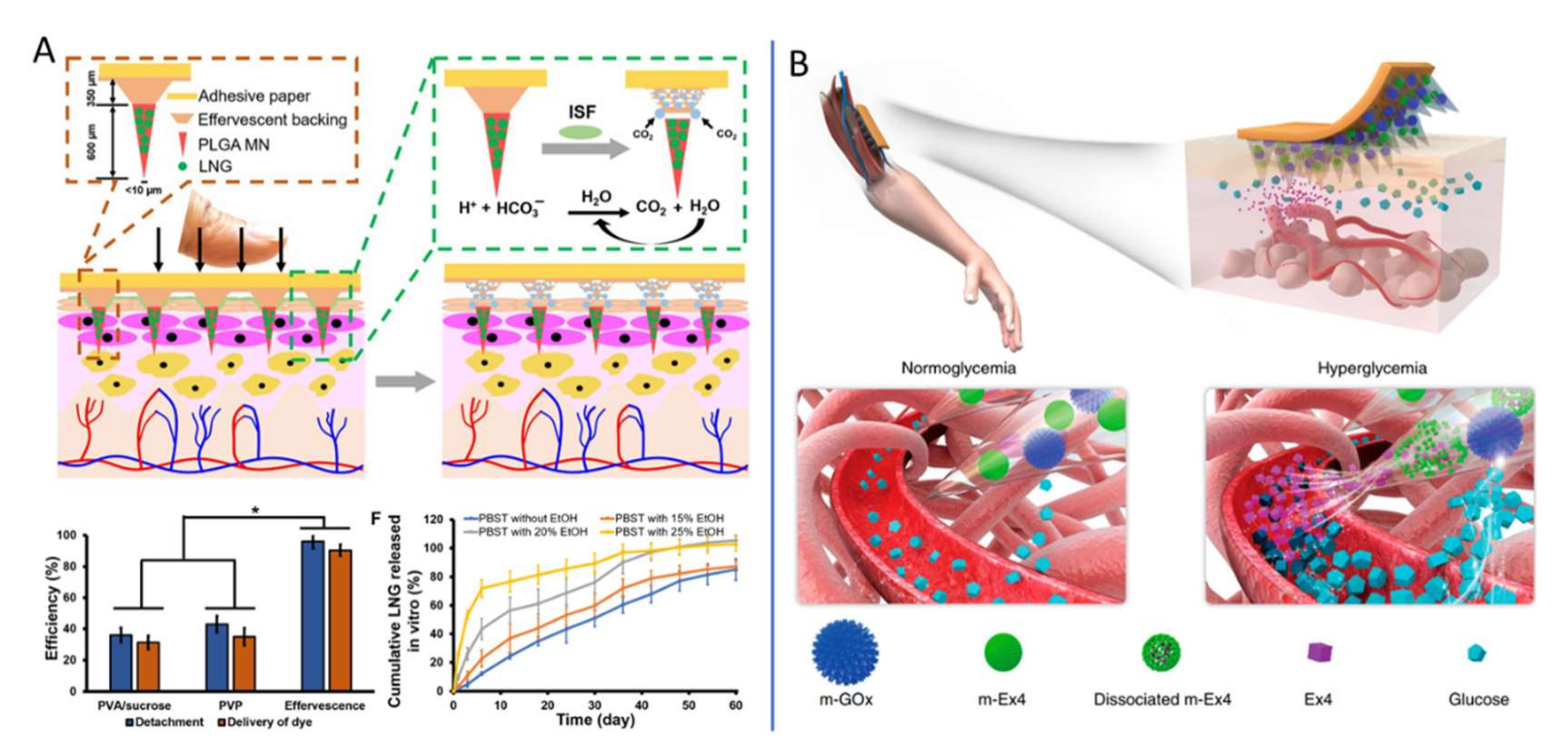
4.6. Usage in the Contraception Field

Since MN patch technology was proposed, it has been applied in many biomedical fields, showing great application prospects. Li et al. [16] extended the application of the MN patch to the field of contraception. Effective contraception is important for women’s health. To increase long-term contraception, they developed a long-acting contraceptive MN patch, which is easy to use and does not produce harmful sharp waste. As shown in Figure 6A, biodegradable polymer PLGA was loaded with contraceptive hormone (levonorgestrel, LNG) to form a microneedle body that releases LNG over a duration of about 1 month through the slow degradation of PLGA in the body. These studies showed that effervescent microneedle patches can promote greater long-acting contraception [120].

4.7. The Treatment of Diabetes

Insulin is a natural hormone secreted by pancreatic islet cells in the pancreas to help the body regulate blood sugar. Long-term insulin injections are often necessary for diabetics, whether they are type 1 (which cannot produce insulin itself) or type 2 (which cannot respond to normal levels of insulin). However, there is difficulty in controlling blood sugar with insulin. When the dose is wrong, there may be a risk of hypoglycemia, which in severe cases can lead to seizures, coma, and even death. As a result, diabetics need to constantly monitor their blood sugar levels while taking insulin [122]. Insulin delivery by MNs is one of the most advanced and important directions of MN application. In 2015, Zhen Gu’s research group first reported the concept and prototype of the “Smart Insulin Patch”, and founded the Zenomics company to promote its clinical transformation. The basic idea is to combine a preparation that responds to blood sugar to release insulin with an MN array made of a polymer material (HA). The use of MNs less than 1 mm in size can reduce the pain caused by injection and improve the quality of life of patients [123]. The team continued to improve the technology of the new “smart insulin patch” for blood glucose response and has completed several stages of progress [50,122,124]. In addition to responding to glucose, as shown in Figure 6B, Chen et al. [121] combined m-GOx and m-Ex4 to design a smart pH-responsive Ex4 MNs delivery patch based on alginate for the treatment of type 2 diabetes. Metformin as a hypoglycemic agent is also embedded in separable MNs composed of polyvinyl alcohol and sucrose (PVA/Suc), which is released as needed under NIR irradiation [125].

4.8. Other Applications

Initially, MNs were used to break barriers and enhance transdermal efficiency. Now, the use of microneedles has expanded to many other fields. The prominent studies of biomedical application of MNs were list in Table 3. In addition to the treatment or drug delivery of various diseases, as described above, in the skin, MNs have also been used to treat the eye, cells, blood vessels, and so on [2]. In particular, hollow microneedles and porous microneedles are mostly used in biosensor research, and good results have been obtained using them. Takeuchi et al. [36] interfaced porous MNs with a microfluidic chip consisting of a capillary pump for the continuous sampling of ISF, which will lead to minimally invasive and continuous biosampling for long-term healthcare monitoring. With increasing crossover between disciplines, microneedles will be able to take on more functions. Chen et al. combined physical (MNs) and nonphysical (enhancer) modes of drug delivery enhancement for a macromolecule in a large animal model [126]. These systems could potentially enable the delivery of a range of drugs through the generation and maintenance of a privileged region in the gastrointestinal tract.
MN drug delivery technology is a new transdermal drug delivery method based on the dual drug release characteristics of subcutaneous injection and transdermal patch technology. Research into the drug treatment of chronic illnesses such as arthritis or diabetes, cancer, dermatocosmetology, family planning, genes, and epidemic disease prevention demonstrates a diversification of development, including the use of small molecule chemistry medicine, biological macromolecule polypeptides, protein and gene medicine, insulin, and so on. Local or systemic administration is also carried out according to disease type and drug performance, and drugs with long-term or short-term release have been designed for use with MNs.

5. Outlook: Nanotechnology Potentiates MNs in Biomedicine

On the basis of the previous studies, it can be seen that the functional design of MN drug delivery systems using nano-engineering technology could make it possible to realize intelligent control of drug release amount, release kinetics, or stimuli-responsive delivery. The main advantage of nanomaterials is that they can be designed for specific functions, and are responsive to specific microenvironments [5]. There are special microenvironments and landmark substances at the lesion site that could be extensively harnessed for the design of functional nanomaterials incorporating MNs for different diseases therapy. It has been reported that high-density avascular chondrocyte extracellular matrix (ECM) impeded drug penetration, but nanoparticles (NPs) (<96 nm) can enter the dense ECM to extend drug retention [127,128,129]. These characteristics provide a starting point for the design of functional nanomaterials. For example, the upregulation of MMP-13 is a key step in OA pathogenesis, leading to the degradation of cartilage ECM. Chen et al. [127] designed novel biocompatibility targeting cartilage and MMP-13/pH response ferritin nanocage (CMFn) containing an anti-inflammatory drug (Hydroxychloroquine, HCQ) for OA imaging and treatment. CMFn@HCQ nanocages can target cartilage and continuously release HCQ in OA joints under acidic pH conditions, thereby extending drug retention time to 14 days and significantly reducing synovitis in OA joints.
Since the emergence of MNs as a promising drug delivery technology, MNs have developed into a broad and rapidly growing research field [10,114]. The rapid development of nanotechnology and materials science has provided a great boost to the research and innovation of MNs, providing new ideas for the treatment of disease and better treatment possibilities. Nanotechnology can be used for engineering biomaterials or drugs to integrate more functions that can achieve intelligent drug delivery in the targeted lesion site. MNs are a very suitable carrier for bioactive nanoparticles. Combining nanotechnology with MNs technology could bring new hope for the treatment of patients.

Author Contributions

Conceptualization, W.Z. (Wenjing Zhang) and Y.L.; investigation, W.Z. (Wenjing Zhang); resources, W.Z. (Wei Zhang), L.Q. and Y.L.; writing—original draft preparation, W.Z. (Wenjing Zhang); writing—review and editing, W.Z. (Wei Zhang), C.L., J.Z., L.Q. and Y.L.; supervision, Y.L.; funding acquisition, W.Z. (Wei Zhang), L.Q. and Y.L. All authors have read and agreed to the published version of the manuscript.

Funding

This research was funded by National Key R&D Program of China (2021YFE0202600), the National Natural Science Foundation of China (82022045, 22007098), CAS Interdisciplinary Innovation Team (JCTD-2020-19), Shenzhen Double Chain Project for Innovation and Development Industry supported by the Bureau of Industry and Information Technology of Shenzhen (201908141541). Shenzhen Fundamental Research Foundation (JCYJ20190807154807663).

Conflicts of Interest

The authors declare no conflict of interest.

References

  1. McAllister, D.V.; Allen, M.G.; Prausnitz, M.R. Microfabricated Microneedles for Gene and Drug Delivery. Annu. Rev. Biomed. Eng. 2000, 2, 289–313. [Google Scholar] [CrossRef] [PubMed] [Green Version]
  2. Kim, Y.; Park, J.; Prausnitz, M.R. Microneedles for drug and vaccine delivery. Adv. Drug Deliver. Rev. 2012, 64, 1547–1568. [Google Scholar] [CrossRef] [PubMed] [Green Version]
  3. Escobar-Chavez, J.J.; Bonilla-Martinez, D.; Villegas-Gonzalez, M.A.; Molina-Trinidad, E.; Casas-Alancaster, N.; Revilla-Vazquez, A.L. Microneedles: A valuable physical enhancer to increase transdermal drug delivery. J. Clin. Pharmacol. 2011, 51, 964–977. [Google Scholar] [CrossRef] [PubMed]
  4. Kalluri, H.; Banga, A.K. Microneedles and transdermal drug delivery. J. Drug Deliv. Sci. Tec. 2009, 19, 303–310. [Google Scholar] [CrossRef]
  5. Yan, L.; Alba, M.; Tabassum, N.; Voelcker, N.H. Micro-and Nanosystems for Advanced Transdermal Delivery. Adv. Therap. 2019, 2, 1900141. [Google Scholar] [CrossRef]
  6. Bouwstra, J.A.; Ponec, M. The skin barrier in healthy and diseased state. Biochim. Biophys. Acta 2006, 1758, 2080–2095. [Google Scholar] [CrossRef] [PubMed] [Green Version]
  7. Morales, J.O.; Fathe, K.R.; Brunaugh, A.; Ferrati, S.; Li, S.; Montenegro-Nicolini, M.; Mousavikhamene, Z.; McConville, J.T.; Prausnitz, M.R.; Smyth, H.D.C. Challenges and Future Prospects for the Delivery of Biologics: Oral Mucosal, Pulmonary, and Transdermal Routes. AAPS J. 2017, 19, 652–668. [Google Scholar] [CrossRef]
  8. Hegde, N.R.; Kaveri, S.V.; Bayry, J. Recent advances in the administration of vaccines for infectious diseases: Microneedles as painless delivery devices for mass vaccination. Drug Discov. Today 2011, 16, 1061–1068. [Google Scholar] [CrossRef]
  9. Teo, A.L.; Shearwood, C.; Ng, K.C.; Lu, J.; Moochhala, S. Transdermal microneedles for drug delivery applications. Mater. Sci. Eng. B 2006, 132, 151–154. [Google Scholar] [CrossRef]
  10. Rzhevskiy, A.S.; Singh, T.R.R.; Donnelly, R.F.; Anissimov, Y.G. Microneedles as the technique of drug delivery enhancement in diverse organs and tissues. J. Control. Release 2018, 270, 184–202. [Google Scholar] [CrossRef] [Green Version]
  11. Prausnitz, M.R.; Mitragotri, S.; Langer, R. Current status and future potential of transdermal drug delivery. Nat. Rev. Drug Discov. 2004, 3, 115–124. [Google Scholar] [CrossRef] [PubMed]
  12. Lambert, P.H.; Laurent, P.E. Intradermal vaccine delivery: Will new delivery systems transform vaccine administration? Vaccine 2008, 26, 3197–3208. [Google Scholar] [CrossRef] [PubMed]
  13. Gniadecka, M.; Jemec, G.B. Quantitative evaluation of chronological ageing and photoageing in vivo: Studies on skin echogenicity and thickness. Br. J. Dermatol. 1998, 139, 815–821. [Google Scholar] [CrossRef] [PubMed]
  14. Chen, J.; Huang, W.; Huang, Z.; Liu, S.; Ye, Y.; Li, Q.; Huang, M. Fabrication of Tip-Dissolving Microneedles for Transdermal Drug Delivery of Meloxicam. AAPS Pharmscitech. 2017, 19, 1141–1151. [Google Scholar] [CrossRef]
  15. Donnelly, R.F.; Garland, M.J.; Morrow, D.I.; Migalska, K.; Singh, T.R.; Majithiya, R.; Woolfson, A.D. Optical coherence tomography is a valuable tool in the study of the effects of microneedle geometry on skin penetration characteristics and in-skin dissolution. J. Control. Release 2010, 147, 333–941. [Google Scholar] [CrossRef]
  16. Li, W.; Tang, J.; Terry, R.N.; Li, S.; Brunie, A.; Callahan, R.L.; Noel, R.K.; Rodríguez, C.A.; Schwendeman, S.P.; Prausnitz, M.R. Long-acting reversible contraception by effervescent microneedle patch. Sci. Adv. 2019, 5, eaaw8145. [Google Scholar] [CrossRef] [Green Version]
  17. Singh, P.; Carrier, A.; Chen, Y.; Lin, S.; Wang, J.; Cui, S.; Zhang, X. Polymeric microneedles for controlled transdermal drug delivery. J. Control. Release 2019, 315, 97–113. [Google Scholar] [CrossRef]
  18. Amodwala, S.; Kumar, P.; Thakkar, H.P. Statistically optimized fast dissolving microneedle transdermal patch of meloxicam: A patient friendly approach to manage arthritis. Eur. J. Pharm. Sci. 2017, 104, 114–123. [Google Scholar] [CrossRef]
  19. Yang, G.; Chen, Q.; Wen, D.; Chen, Z.; Wang, J.; Chen, G.; Wang, Z.; Zhang, X.; Zhang, Y.; Hu, Q.; et al. A Therapeutic Microneedle Patch Made from Hair-Derived Keratin for Promoting Hair Regrowth. ACS Nano 2019, 13, 4354–4360. [Google Scholar] [CrossRef]
  20. Yang, J.; Liu, X.; Fu, Y.; Song, Y. Recent advances of microneedles for biomedical applications: Drug delivery and beyond. Acta Pharm. Sin. B 2019, 9, 469–483. [Google Scholar] [CrossRef]
  21. Bao, L.; Park, J.; Bonfante, G.; Kim, B. Recent advances in porous microneedles: Materials, fabrication, and transdermal applications. Drug Deliv. Transl. Res. 2022, 12, 395–414. [Google Scholar] [CrossRef] [PubMed]
  22. Howells, O.; Blayney, G.J.; Gualeni, B.; Birchall, J.C.; Eng, P.F.; Ashraf, H.; Sharma, S.; Guy, O.J. Design, fabrication, and characterisation of a silicon microneedle array for transdermal therapeutic delivery using a single step wet etch process. Eur. J. Pharm. Biopharm. 2021, 171, 19–28. [Google Scholar] [CrossRef] [PubMed]
  23. Zhou, Z.; Zhang, S.; Yang, G.; Gao, Y. Enhanced delivery efficiency and sustained release of biopharmaceuticals by complexation-based gel encapsulated coated microneedles: rhIFNα-1β example. Asian J. Pharm. Sci. 2021, 16, 612–622. [Google Scholar] [CrossRef] [PubMed]
  24. Mishra, R.K.; Goud, K.Y.; Li, Z.; Moonla, C.; Mohamed, M.A.; Tehrani, F.; Teymourian, H.; Wang, J. Continuous Opioid Monitoring along with Nerve Agents on a Wearable Microneedle Sensor Array. J. Am. Chem. Soc. 2020, 142, 5991–5995. [Google Scholar] [CrossRef]
  25. Lee, J.W.; Park, J.; Prausnitz, M.R. Dissolving microneedles for transdermal drug delivery. Biomaterials 2008, 29, 2113–2124. [Google Scholar] [CrossRef] [Green Version]
  26. Seong, K.; Seo, M.; Hwang, D.Y.; O’Cearbhaill, E.D.; Sreenan, S.; Karp, J.M.; Yang, S.Y. A self-adherent, bullet-shaped microneedle patch for controlled transdermal delivery of insulin. J. Control. Release 2017, 265, 48–56. [Google Scholar] [CrossRef]
  27. Yi, K.; Wang, Y.; Shi, K.; Chi, J.; Lyu, J.; Zhao, Y. Aptamer-decorated porous microneedles arrays for extraction and detection of skin interstitial fluid biomarkers. Biosens. Bioelectron. 2021, 190, 113404. [Google Scholar] [CrossRef]
  28. Lee, K.; Goudie, M.J.; Tebon, P.; Sun, W.; Luo, Z.; Lee, J.; Zhang, S.; Fetah, K.; Kim, H.; Xue, Y.; et al. Non-transdermal microneedles for advanced drug delivery. Adv. Drug Deliver. Rev. 2019, 165–166, 41–59. [Google Scholar] [CrossRef]
  29. Jamaledin, R.; Di Natale, C.; Onesto, V.; Taraghdari, Z.B.; Zare, E.N.; Makvandi, P.; Vecchione, R.; Netti, P.A. Progress in Microneedle-Mediated Protein Delivery. J. Clin. Med. 2020, 9, 542. [Google Scholar] [CrossRef] [Green Version]
  30. Zeng, Q.; Gammon, J.M.; Tostanoski, L.H.; Chiu, Y.; Jewell, C.M. In Vivo Expansion of Melanoma-Specific T Cells Using Microneedle Arrays Coated with Immune-Polyelectrolyte Multilayers. ACS Biomater. Sci. Eng. 2016, 3, 195–205. [Google Scholar] [CrossRef]
  31. Garland, M.J.; Migalska, K.; Mahmood, T.M.; Singh, T.R.; Woolfson, A.D.; Donnelly, R.F. Microneedle arrays as medical devices for enhanced transdermal drug delivery. Expert Rev. Med. Devices 2011, 8, 459–482. [Google Scholar] [CrossRef] [PubMed]
  32. Donnelly, R.F.; Singh, T.R.; Garland, M.J.; Migalska, K.; Majithiya, R.; McCrudden, C.M.; Kole, P.L.; Mahmood, T.M.; McCarthy, H.O.; Woolfson, A.D. Hydrogel-Forming Microneedle Arrays for Enhanced Transdermal Drug Delivery. Adv. Funct. Mater. 2012, 22, 4879–4890. [Google Scholar] [CrossRef] [PubMed] [Green Version]
  33. Chen, M.; Ling, M.; Lai, K.; Pramudityo, E. Chitosan Microneedle Patches for Sustained Transdermal Delivery of Macromolecules. Biomacromolecules 2012, 13, 4022–4031. [Google Scholar] [CrossRef] [PubMed]
  34. DeMuth, P.C.; Garcia-Beltran, W.F.; Ai-Ling, M.L.; Hammond, P.T.; Irvine, D.J. Composite Dissolving Microneedles for Coordinated Control of Antigen and Adjuvant Delivery Kinetics in Transcutaneous Vaccination. Adv. Funct. Mater. 2013, 23, 161–172. [Google Scholar] [CrossRef] [Green Version]
  35. Chang, H.; Zheng, M.; Yu, X.; Than, A.; Seeni, R.Z.; Kang, R.; Tian, J.; Khanh, D.P.; Liu, L.; Chen, P.; et al. A Swellable Microneedle Patch to Rapidly Extract Skin Interstitial Fluid for Timely Metabolic Analysis. Adv. Mater. 2017, 29, 1702243. [Google Scholar] [CrossRef]
  36. Takeuchi, K.; Takama, N.; Sharma, K.; Paul, O.; Ruther, P.; Suga, T.; Kim, B. Microfluidic chip connected to porous microneedle array for continuous ISF sampling. Drug Deliv. Transl. Res. 2022, 12, 435–443. [Google Scholar] [CrossRef]
  37. Liu, L.; Kai, H.; Nagamine, K.; Ogawa, Y.; Nishizawa, M. Porous polymer microneedles with interconnecting microchannels for rapid fluid transport. RSC Adv. 2016, 6, 48630–48635. [Google Scholar] [CrossRef]
  38. Li, J.; Liu, B.; Zhou, Y.; Chen, Z.; Jiang, L.; Yuan, W.; Liang, L. Fabrication of a Ti porous microneedle array by metal injection molding for transdermal drug delivery. PLoS ONE 2017, 12, e0172043. [Google Scholar] [CrossRef] [Green Version]
  39. Kusama, S.; Sato, K.; Matsui, Y.; Kimura, N.; Abe, H.; Yoshida, S.; Nishizawa, M. Transdermal electroosmotic flow generated by a porous microneedle array patch. Nat. Commun. 2021, 12, 658. [Google Scholar] [CrossRef]
  40. Yang, H.; Lin, W.; Zheng, Y. Advances and perspective on the translational medicine of biodegradable metals. Biomater. Transl. 2021, 2, 177–187. [Google Scholar]
  41. Long, J.; Teng, B.; Zhang, W.; Li, L.; Zhang, M.; Chen, Y.; Yao, Z.; Meng, X.; Wang, X.; Qin, L.; et al. Preclinical evaluation of acute systemic toxicity of magnesium incorporated poly(lactic-co-glycolic acid) porous scaffolds by three-dimensional printing. Biomater. Transl. 2021, 2, 272–284. [Google Scholar]
  42. Larraneta, E.; Moore, J.; Vicente-Perez, E.M.; Gonzalez-Vazquez, P.; Lutton, R.; Woolfson, A.D.; Donnelly, R.F. A proposed model membrane and test method for microneedle insertion studies. Int. J. Pharm. 2014, 472, 65–73. [Google Scholar] [CrossRef] [PubMed] [Green Version]
  43. Permana, A.D.; Mir, M.; Utomo, E.; Donnelly, R.F. Bacterially sensitive nanoparticle-based dissolving microneedles of doxycycline for enhanced treatment of bacterial biofilm skin infection: A proof of concept study. Int. J. Pharm. X 2020, 2, 100047. [Google Scholar] [CrossRef] [PubMed]
  44. Chen, Y.; Yang, Y.; Xian, Y.; Singh, P.; Feng, J.; Cui, S.; Carrier, A.; Oakes, K.; Luan, T.; Zhang, X. Multifunctional Graphene-Oxide-Reinforced Dissolvable Polymeric Microneedles for Transdermal Drug Delivery. ACS Appl. Mater Interfaces 2020, 12, 352–360. [Google Scholar] [CrossRef]
  45. Li, Z.; He, Y.; Deng, L.; Zhang, Z.; Lin, Y. A fast-dissolving microneedle array loaded with chitosan nanoparticles to evoke systemic immune responses in mice. J. Mater. Chem. B. 2020, 8, 216–225. [Google Scholar] [CrossRef]
  46. Mao, J.; Wang, H.; Xie, Y.; Fu, Y.; Li, Y.; Liu, P.; Du, H.; Zhu, J.; Dong, L.; Hussain, M.; et al. Transdermal delivery of rapamycin with poor water-solubility by dissolving polymeric microneedles for anti-angiogenesis. J. Mater. Chem. B 2020, 8, 928–934. [Google Scholar] [CrossRef]
  47. Derakhshandeh, H.; Aghabaglou, F.; McCarthy, A.; Mostafavi, A.; Wiseman, C.; Bonick, Z.; Ghanavati, I.; Harris, S.; Kreikemeier Bower, C.; Moosavi Basri, S.M.; et al. A Wirelessly Controlled Smart Bandage with 3D-Printed Miniaturized Needle Arrays. Adv. Funct. Mater. 2020, 30, 1905544. [Google Scholar] [CrossRef]
  48. Chi, J.; Zhang, X.; Chen, C.; Shao, C.; Zhao, Y.; Wang, Y. Antibacterial and angiogenic chitosan microneedle array patch for promoting wound healing. Bioact. Mater. 2020, 5, 253–259. [Google Scholar] [CrossRef]
  49. Yang, X.; Feng, P.; Cao, J.; Liu, W.; Tang, Y. Composition-Engineered Metal–Organic Framework-Based Microneedles for Glucose-Mediated Transdermal Insulin Delivery. ACS Appl. Mater. Inter. 2020, 12, 13613–13621. [Google Scholar] [CrossRef]
  50. Yu, J.; Wang, J.; Zhang, Y.; Chen, G.; Mao, W.; Ye, Y.; Kahkoska, A.R.; Buse, J.B.; Langer, R.; Gu, Z. Glucose-responsive insulin patch for the regulation of blood glucose in mice and minipigs. Nat. Biomed. Eng. 2020, 4, 499–506. [Google Scholar] [CrossRef]
  51. Moreira, A.F.; Rodrigues, C.F.; Jacinto, T.A.; Miguel, S.P.; Costa, E.C.; Correia, I.J. Poly (vinyl alcohol)/chitosan layer-by-layer microneedles for cancer chemo-photothermal therapy. Int. J. Pharm. 2020, 576, 118907. [Google Scholar] [CrossRef] [PubMed]
  52. Lim, S.H.; Tiew, W.J.; Zhang, J.; Ho, P.C.; Kachouie, N.N.; Kang, L. Geometrical optimisation of a personalised microneedle eye patch for transdermal delivery of anti-wrinkle small peptide. Biofabrication 2020, 12, 035003. [Google Scholar] [CrossRef] [PubMed]
  53. Vora, L.K.; Courtenay, A.J.; Tekko, I.A.; Larraneta, E.; Donnelly, R.F. Pullulan-based dissolving microneedle arrays for enhanced transdermal delivery of small and large biomolecules. Int. J. Biol. Macromol. 2019, 146, 290–298. [Google Scholar] [CrossRef] [PubMed]
  54. Zhang, X.; Chen, G.; Yu, Y.; Sun, L.; Zhao, Y. Bioinspired Adhesive and Antibacterial Microneedles for Versatile Transdermal Drug Delivery. Research 2020, 2020, 3672120. [Google Scholar] [CrossRef]
  55. Vecchione, R.; Coppola, S.; Esposito, E.; Casale, C.; Vespini, V.; Grilli, S.; Ferraro, P.; Netti, P.A. Electro-Drawn Drug-Loaded Biodegradable Polymer Microneedles as a Viable Route to Hypodermic Injection. Adv. Funct. Mater. 2014, 24, 3515–3523. [Google Scholar] [CrossRef]
  56. Chiu, Y.; Chen, M.; Wan, S. Sodium Hyaluronate/Chitosan Composite Microneedles as a Single-Dose Intradermal Immunization System. Biomacromolecules 2018, 19, 2278–2285. [Google Scholar] [CrossRef]
  57. Zheng, Z.; Eglin, D.; Alini, M.; Richards, G.R.; Qin, L.; Lai, Y. Visible Light-Induced 3D Bioprinting Technologies and Corresponding Bioink Materials for Tissue Engineering: A Review. Engineering 2021, 7, 966–978. [Google Scholar] [CrossRef]
  58. Qiu, G.; Ding, W.; Tian, W.; Qin, L.; Zhao, Y.; Zhang, L.; Lu, J.; Chen, D.; Yuan, G.; Wu, C.; et al. Medical Additive Manufacturing: From a Frontier Technology to the Research and Development of Products. Engineering 2020, 6, 1217–1221. [Google Scholar] [CrossRef]
  59. Di Prima, M.; Coburn, J.; Hwang, D.; Kelly, J.; Khairuzzaman, A.; Ricles, L. Additively manufactured medical products–the FDA perspective. 3D Print. Med. 2016, 2, 1. [Google Scholar] [CrossRef] [Green Version]
  60. Economidou, S.N.; Uddin, M.J.; Marques, M.J.; Douroumis, D.; Sow, W.T.; Li, H.; Reid, A.; Windmill, J.F.C.; Podoleanu, A. A novel 3D printed hollow microneedle microelectromechanical system for controlled, personalized transdermal drug delivery. Addit. Manuf. 2021, 38, 101815. [Google Scholar] [CrossRef]
  61. Li, X.; Shan, W.; Yang, Y.; Joralmon, D.; Zhu, Y.; Chen, Y.; Yuan, Y.; Xu, H.; Rong, J.; Dai, R.; et al. Limpet Tooth-Inspired Painless Microneedles Fabricated by Magnetic Field-Assisted 3D Printing. Adv. Funct. Mater. 2021, 31, 2003725. [Google Scholar] [CrossRef]
  62. Qiu, Y.; Gao, Y.; Hu, K.; Li, F. Enhancement of skin permeation of docetaxel: A novel approach combining microneedle and elastic liposomes. J. Control. Release 2008, 129, 144–150. [Google Scholar] [CrossRef] [PubMed]
  63. Sullivan, S.P.; Murthy, N.; Prausnitz, M.R. Minimally Invasive Protein Delivery with Rapidly Dissolving Polymer Microneedles. Adv. Mater. 2008, 20, 933–938. [Google Scholar] [CrossRef] [PubMed] [Green Version]
  64. Henry, S.; McAllister, D.V.; Allen, M.G.; Prausnitz, M.R. Microfabricated microneedles: A novel approach to transdermal drug delivery. J. Pharm. Sci. 1998, 87, 922–925. [Google Scholar] [CrossRef] [PubMed]
  65. Yan, G.; Warner, K.S.; Zhang, J.; Sharma, S.; Gale, B.K. Evaluation needle length and density of microneedle arrays in the pretreatment of skin for transdermal drug delivery. Int. J. Pharmaceut. 2010, 391, 7–12. [Google Scholar] [CrossRef] [PubMed]
  66. Li, W.Z.; Huo, M.R.; Zhou, J.P.; Zhou, Y.Q.; Hao, B.H.; Liu, T.; Zhang, Y. Super-short solid silicon microneedles for transdermal drug delivery applications. Int. J. Pharm. 2010, 389, 122–129. [Google Scholar]
  67. Gill, H.S.; Prausnitz, M.R. Coated microneedles for transdermal delivery. J. Control. Release 2007, 117, 227–237. [Google Scholar] [CrossRef] [Green Version]
  68. Fomani, A.A.; Mansour, R.R. Fabrication and characterization of the flexible neural microprobes with improved structural design. Sens. Actuators A Phys. 2011, 168, 233–241. [Google Scholar] [CrossRef]
  69. Kim, M.J.; Park, S.C.; Rizal, B.; Guanes, G.; Baek, S.K.; Park, J.H.; Betz, A.R.; Choi, S.O. Fabrication of Circular Obelisk-Type Multilayer Microneedles Using Micro-Milling and Spray Deposition. Front. Bioeng. Biotechnol. 2018, 6, 54. [Google Scholar] [CrossRef] [Green Version]
  70. Demir, Y.K.; Akan, Z.; Kerimoglu, O. Characterization of polymeric microneedle arrays for transdermal drug delivery. PLoS ONE 2013, 8, e77289. [Google Scholar] [CrossRef] [Green Version]
  71. Lee, K.; Lee, H.C.; Lee, D.S.; Jung, H. Drawing lithography: Three-dimensional fabrication of an ultrahigh-aspect-ratio microneedle. Adv. Mater. 2010, 22, 483–486. [Google Scholar] [CrossRef] [PubMed]
  72. Choi, C.K.; Lee, K.J.; Youn, Y.N.; Jang, E.H.; Kim, W.; Min, B.K.; Ryu, W. Spatially discrete thermal drawing of biodegradable microneedles for vascular drug delivery. Eur. J. Pharm. Biopharm. 2013, 83, 224–233. [Google Scholar] [CrossRef] [PubMed]
  73. Ruggiero, F.; Vecchione, R.; Bhowmick, S.; Coppola, G.; Coppola, S.; Esposito, E.; Lettera, V.; Ferraro, P.; Netti, P.A. Electro-drawn polymer microneedle arrays with controlled shape and dimension. Sens. Actuators B Chem. 2018, 255, 1553–1560. [Google Scholar] [CrossRef]
  74. Chen, Z.; Ren, L.; Li, J.; Yao, L.; Chen, Y.; Liu, B.; Jiang, L. Rapid fabrication of microneedles using magnetorheological drawing lithography. Acta Biomater. 2018, 65, 283–291. [Google Scholar] [CrossRef] [PubMed]
  75. Kim, J.D.; Kim, M.; Yang, H.; Lee, K.; Jung, H. Droplet-born air blowing: Novel dissolving microneedle fabrication. J. Control. Release 2013, 170, 430–436. [Google Scholar] [CrossRef]
  76. Han, M.; Hyun, D.; Park, H.; Lee, S.S.; Kim, C.; Kim, C. A novel fabrication process for out-of-plane microneedle sheets of biocompatible polymer. J. Micromech. Microeng. 2007, 17, 1184–1191. [Google Scholar] [CrossRef] [Green Version]
  77. Moon, S.-J.; Lee, S.S. Fabrication of microneedle array using inclined LIGA Process. In TRANSDUCERS ’03. 12th International Conference on Solid-State Sensors, Actuators and Microsystems; Digest of Technical Papers (Cat. No.03TH8664); IEEE: Boston, MA, USA, 2003; Volume 2, pp. 1546–1549. [Google Scholar]
  78. Pere, C.; Economidou, S.N.; Lall, G.; Ziraud, C.; Boateng, J.S.; Alexander, B.D.; Lamprou, D.A.; Douroumis, D. 3D printed microneedles for insulin skin delivery. Int. J. Pharm. 2018, 544, 425–432. [Google Scholar] [CrossRef] [Green Version]
  79. Luzuriaga, M.A.; Berry, D.R.; Reagan, J.C.; Smaldone, R.A.; Gassensmith, J.J. Biodegradable 3D printed polymer microneedles for transdermal drug delivery. Front. Bioeng. Biotechnol. 2018, 18, 1223–1230. [Google Scholar] [CrossRef]
  80. Lim, S.H.; Ng, J.Y.; Kang, L. Three-dimensional printing of a microneedle array on personalized curved surfaces for dual-pronged treatment of trigger finger. Biofabrication 2017, 9, 015010. [Google Scholar] [CrossRef]
  81. Moussi, K.; Bukhamsin, A.; Hidalgo, T.; Kosel, J. Biocompatible 3D Printed Microneedles for Transdermal, Intradermal, and Percutaneous Applications. Adv. Eng. Mater. 2019, 22, 1901358. [Google Scholar] [CrossRef] [Green Version]
  82. Harvey, E.C.; Rumsby, P.T. Fabrication techniques and their application to produce novel micromachined structures and devices using excimer laser projection. In Proceedings of the SPIE1997, Austin, TX, USA, 5 September 1997; Volume 3223, pp. 26–33. [Google Scholar]
  83. Li, J.; Zhou, Y.; Yang, J.; Ye, R.; Gao, J.; Ren, L.; Liu, B.; Liang, L.; Jiang, L. Fabrication of gradient porous microneedle array by modified hot embossing for transdermal drug delivery. Mater. Sci. Eng. C Mater. Biol. Appl. 2019, 96, 576–582. [Google Scholar] [CrossRef] [PubMed]
  84. Janphuang, P.; Laebua, M.; Sriphung, C.; Taweewat, P.; Sirichalarmkul, A.; Sukjantha, K.; Promsawat, N.; Leuasoongnoen, P.; Suphachiaraphan, S.; Phimol, K.; et al. Polymer based microneedle patch fabricated using microinjection moulding. In Proceedings of the 4th International Conference on Engineering, Applied Sciences and Technology (ICEAST 2018), Phuket, Thailand, 4–7 July 2018; Volume 192, p. 01039. [Google Scholar]
  85. Yung, K.L.; Xu, Y.; Kang, C.; Liu, H.; Tam, K.F.; Ko, S.M.; Kwan, F.Y.; Lee, T.M.H. Sharp tipped plastic hollow microneedle array by microinjection moulding. J. Micromech. Microeng. 2012, 22, 015016. [Google Scholar] [CrossRef]
  86. Duong, H.; Yin, Y.; Thambi, T.; Nguyen, T.L.; Giang, P.V.; Lee, M.S.; Lee, J.E.; Kim, J.; Jeong, J.H.; Lee, D.S. Smart vaccine delivery based on microneedle arrays decorated with ultra-pH-responsive copolymers for cancer immunotherapy. Biomaterials 2018, 185, 13–24. [Google Scholar] [CrossRef] [PubMed]
  87. McGrath, M.G.; Vucen, S.; Vrdoljak, A.; Kelly, A.; O’Mahony, C.; Crean, A.M.; Moore, A. Production of dissolvable microneedles using an atomised spray process: Effect of microneedle composition on skin penetration. Eur. J. Pharm. Biopharm. 2014, 86, 200–211. [Google Scholar] [CrossRef]
  88. Gill, H.S.; Prausnitz, M.R. Coating formulations for microneedles. Pharm. Res. 2007, 24, 1369–1380. [Google Scholar] [CrossRef]
  89. REAUME, S.E. The use of hydrofluoric acid in making glass microneedles. Science 1952, 116, 641. [Google Scholar] [CrossRef]
  90. Robinson, N. Enzyme response of traumatized tissue after intracortical injection into 5 day old rat brain. J. Neurol. Neurosurg. Psychiatry 1972, 35, 865–872. [Google Scholar] [CrossRef] [Green Version]
  91. Szubinska, B. “New membrane” formation in Amoeba proteus upon injury of individual cells. Electron microscope observations. J. Cell Biol. 1971, 49, 747–772. [Google Scholar] [CrossRef]
  92. Joel, D.D.; Hess, M.W.; Cottier, H. Magnitude and pattern of thymic lymphocyte migration in neonatal mice. J. Exp. Med. 1972, 135, 907–923. [Google Scholar] [CrossRef] [Green Version]
  93. DOSSEL, W.E. Preparation of tungsten micro-needles for use in embryologic research. Lab. Invest. 1958, 7, 171–173. [Google Scholar]
  94. De Jonge, A.J.; Vermeulen, W.; Keijzer, W.; Hoeijmakers, J.H.; Bootsma, D. Microinjection of Micrococcus luteus UV-endonuclease restores UV-induced unscheduled DNA synthesis in cells of 9 xeroderma pigmentosum complementation groups. Mutat. Res. 1985, 150, 99–105. [Google Scholar] [CrossRef] [Green Version]
  95. Edds, K.T. Motility in Echinosphaerium nucleofilum. I. An analysis of particle motions in the axopodia and a direct test of the involvement of the axoneme. J. Cell Biol. 1975, 66, 145–155. [Google Scholar] [CrossRef] [PubMed] [Green Version]
  96. Hoeijmakers, J.H. Characterization of genes and proteins involved in excision repair of human cells. J. Cell Sci. Suppl. 1987, 6, 111–125. [Google Scholar] [CrossRef] [Green Version]
  97. Riedel, H.; Kondor-Koch, C.; Garoff, H. Cell surface expression of fusogenic vesicular stomatitis virus G protein from cloned cDNA. EMBO J. 1984, 3, 1477–1483. [Google Scholar] [CrossRef]
  98. Poirier, D.; Renaud, F.; Dewar, V.; Strodiot, L.; Wauters, F.; Janimak, J.; Shimada, T.; Nomura, T.; Kabata, K.; Kuruma, K.; et al. Hepatitis B surface antigen incorporated in dissolvable microneedle array patch is antigenic and thermostable. Biomaterials 2017, 145, 256–265. [Google Scholar] [CrossRef] [PubMed]
  99. Liang, R.; Zhao, J.; Li, B.; Cai, P.; Loh, X.J.; Xu, C.; Chen, P.; Kai, D.; Zheng, L. Implantable and degradable antioxidant poly(epsilon-caprolactone)-lignin nanofiber membrane for effective osteoarthritis treatment. Biomaterials 2020, 230, 119601. [Google Scholar] [CrossRef] [PubMed]
  100. Gerwin, N.; Hops, C.; Lucke, A. Intraarticular drug delivery in osteoarthritis. Adv. Drug Deliver. Rev. 2006, 58, 226–242. [Google Scholar] [CrossRef] [PubMed]
  101. Prasad, R.; Koul, V. Transdermal delivery of methotrexate: Past, present and future prospects. Ther. Deliv. 2012, 3, 315–325. [Google Scholar] [CrossRef]
  102. Abla, M.J.; Chaturvedula, A.; O Mahony, C.; Banga, A.K. Transdermal delivery of methotrexate for pediatrics using silicon microneedles. Ther. Deliv. 2013, 4, 543–551. [Google Scholar] [CrossRef]
  103. Shende, P.; Salunke, M. Transepidermal microneedles for co-administration of folic acid with methotrexate in the treatment of rheumatoid arthritis. Biomed. Phys. Eng. Express 2019, 5, 025023. [Google Scholar] [CrossRef]
  104. Qiu, Y.; Li, C.; Zhang, S.; Yang, G.; He, M.; Gao, Y. Systemic delivery of artemether by dissolving microneedles. Int. J. Pharm. 2016, 508, 1–9. [Google Scholar] [CrossRef] [PubMed]
  105. Zhang, Y.; Hu, H.; Jing, Q.; Wang, Z.; He, Z.; Wu, T.; Feng, N.P. Improved Biosafety and Transdermal Delivery of Aconitine via Diethylene Glycol Monoethyl Ether-Mediated Microemulsion Assisted with Microneedles. Pharmaceutics 2020, 12, 163. [Google Scholar] [CrossRef] [PubMed] [Green Version]
  106. Guo, T.; Zhang, Y.; Li, Z.; Zhao, J.; Feng, N. Microneedle-mediated transdermal delivery of nanostructured lipid carriers for alkaloids from Aconitum sinomontanum. Artif. Cells Nanomed. Biotechnol. 2018, 46, 1541–1551. [Google Scholar] [CrossRef] [PubMed]
  107. Guo, T.; Cheng, N.; Zhao, J.; Hou, X.; Zhang, Y.; Feng, N. Novel nanostructured lipid carriers-loaded dissolving microneedles for controlled local administration of aconitine. Int. J. Pharm. 2019, 572, 118741. [Google Scholar] [CrossRef] [PubMed]
  108. Dangol, M.; Yang, H.; Li, C.G.; Lahiji, S.F.; Kim, S.; Ma, Y.; Jung, H. Innovative polymeric system (IPS) for solvent-free lipophilic drug transdermal delivery via dissolving microneedles. J. Control. Release 2016, 223, 118–125. [Google Scholar] [CrossRef]
  109. Yao, W.; Tao, C.; Zou, J.; Zheng, H.; Zhu, J.; Zhu, Z.; Zhu, J.; Liu, L.; Li, F.; Song, X. Flexible two-layer dissolving and safing microneedle transdermal of neurotoxin: A biocomfortable attempt to treat Rheumatoid Arthritis. Int. J. Pharm. 2019, 563, 91–100. [Google Scholar] [CrossRef]
  110. Chen, G.; Hao, B.; Ju, D.; Liu, M.; Zhao, H.; Du, Z.; Xia, J. Pharmacokinetic and pharmacodynamic study of triptolide-loaded liposome hydrogel patch under microneedles on rats with collagen-induced arthritis. Acta Pharm. Sin. B 2015, 5, 569–576. [Google Scholar] [CrossRef] [Green Version]
  111. Cui, Y.; Mo, Y.; Zhang, Q.; Tian, W.; Xue, Y.; Bai, J.; Du, S. Microneedle-Assisted Percutaneous Delivery of Paeoniflorin-Loaded Ethosomes. Molecules 2018, 23, 3371. [Google Scholar] [CrossRef] [Green Version]
  112. Wu, X.; Chen, Y.; Gui, S.; Wu, X.; Chen, L.; Cao, Y.; Yin, D.; Ma, P. Sinomenine hydrochloride-loaded dissolving microneedles enhanced its absorption in rabbits. Pharm. Dev. Technol. 2016, 21, 787–793. [Google Scholar] [CrossRef]
  113. Cao, J.; Zhang, N.; Wang, Z.; Su, J.; Yang, J.; Han, J.; Zhao, Y. Microneedle-Assisted Transdermal Delivery of Etanercept for Rheumatoid Arthritis Treatment. Pharmaceutics 2019, 11, 235. [Google Scholar] [CrossRef] [Green Version]
  114. Ali, A.A.; McCrudden, C.M.; McCaffrey, J.; McBride, J.W.; Cole, G.; Dunne, N.J.; Robson, T.; Kissenpfennig, A.; Donnelly, R.F.; McCarthy, H.O. DNA vaccination for cervical cancer; a novel technology platform of RALA mediated gene delivery via polymeric microneedles. Nanomed. UK 2017, 13, 921–932. [Google Scholar] [CrossRef] [PubMed] [Green Version]
  115. Li, J.; Zeng, M.; Shan, H.; Tong, C. Microneedle Patches as Drug and Vaccine Delivery Platform. Curr. Med. Chem. 2017, 24, 2413–2422. [Google Scholar] [CrossRef] [PubMed]
  116. Kim, E.; Erdos, G.; Huang, S.; Kenniston, T.W.; Balmert, S.C.; Carey, C.D.; Raj, V.S.; Epperly, M.W.; Klimstra, W.B.; Haagmans, B.L.; et al. Microneedle array delivered recombinant coronavirus vaccines: Immunogenicity and rapid translational development. EBioMedicine 2020, 55, 102743. [Google Scholar] [CrossRef] [PubMed]
  117. Arya, J.; Prausnitz, M.R. Microneedle patches for vaccination in developing countries. J. Control. Release 2016, 240, 135–141. [Google Scholar] [CrossRef] [PubMed] [Green Version]
  118. Chang, H.; Chew, S.W.T.; Zheng, M.; Lio, D.C.S.; Wiraja, C.; Mei, Y.; Ning, X.; Cui, M.; Than, A.; Shi, P.; et al. Cryomicroneedles for transdermal cell delivery. Nat. Biomed. Eng. 2021, 5, 1008–1018. [Google Scholar] [CrossRef] [PubMed]
  119. Duong, H.; Yin, Y.; Thambi, T.; Kim, B.S.; Jeong, J.H.; Lee, D.S. Highly potent intradermal vaccination by an array of dissolving microneedle polypeptide cocktails for cancer immunotherapy. J. Mater. Chem. B 2020, 8, 1171–1181. [Google Scholar] [CrossRef] [PubMed]
  120. Lee, Y.; Li, W.; Tang, J.; Schwendeman, S.P.; Prausnitz, M.R. Immediate detachment of microneedles by interfacial fracture for sustained delivery of a contraceptive hormone in the skin. J. Control. Release 2021, 337, 676–685. [Google Scholar] [CrossRef]
  121. Chen, W.; Tian, R.; Xu, C.; Yung, B.C.; Wang, G.; Liu, Y.; Ni, Q.; Zhang, F.; Zhou, Z.; Wang, J.; et al. Microneedle-array patches loaded with dual mineralized protein/peptide particles for type 2 diabetes therapy. Nat. Commun. 2017, 8, 1777. [Google Scholar] [CrossRef]
  122. Wang, J.; Yu, J.; Zhang, Y.; Kahkoska, A.R.; Wang, Z.; Fang, J.; Whitelegge, J.P.; Li, S.; Buse, J.B.; Gu, Z. Glucose transporter inhibitor-conjugated insulin mitigates hypoglycemia. Proc. Natl. Acad. Sci. USA 2019, 116, 10744–10748. [Google Scholar] [CrossRef] [Green Version]
  123. Yu, J.; Zhang, Y.; Ye, Y.; DiSanto, R.; Sun, W.; Ranson, D.; Ligler, F.S.; Buse, J.B.; Gu, Z. Microneedle-array patches loaded with hypoxia-sensitive vesicles provide fast glucose-responsive insulin delivery. Proc. Natl. Acad. Sci. USA 2015, 112, 8260–8265. [Google Scholar] [CrossRef] [Green Version]
  124. Ye, Y.; Yu, J.; Wang, C.; Nguyen, N.Y.; Walker, G.M.; Buse, J.B.; Gu, Z. Microneedles Integrated with Pancreatic Cells and Synthetic Glucose-Signal Amplifiers for Smart Insulin Delivery. Adv. Mater. 2016, 28, 3115–3121. [Google Scholar] [CrossRef] [PubMed] [Green Version]
  125. Liu, T.; Jiang, G.; Song, G.; Zhu, J.; Yang, Y. Fabrication of separable microneedles with phase change coating for NIR-triggered transdermal delivery of metformin on diabetic rats. Biomed. Microdevices 2020, 22, 12. [Google Scholar] [CrossRef] [PubMed]
  126. Chen, W.; Wainer, J.; Ryoo, S.W.; Qi, X.; Chang, R.; Li, J.; Lee, S.H.; Min, S.; Wentworth, A.; Collins, J.E.; et al. Dynamic omnidirectional adhesive microneedle system for oral macromolecular drug delivery. Sci. Adv. 2022, 8, eabk1792. [Google Scholar] [CrossRef]
  127. Chen, H.; Qin, Z.; Zhao, J.; He, Y.; Ren, E.; Zhu, Y.; Liu, G.; Mao, C.; Zheng, L. Cartilage-targeting and dual MMP-13/pH responsive theranostic nanoprobes for osteoarthritis imaging and precision therapy. Biomaterials 2019, 225, 119520. [Google Scholar] [CrossRef] [PubMed]
  128. Kavanaugh, T.E.; Werfel, T.A.; Cho, H.; Hasty, K.A.; Duvall, C.L. Particle-based technologies for osteoarthritis detection and therapy. Drug. Deliv. Transl. Res. 2016, 6, 132–147. [Google Scholar] [CrossRef] [PubMed] [Green Version]
  129. Rothenfluh, D.A.; Bermudez, H.; O’Neil, C.P.; Hubbell, J.A. Biofunctional polymer nanoparticles for intra-articular targeting and retention in cartilage. Nat. Mater. 2008, 7, 248–254. [Google Scholar] [CrossRef] [PubMed]
Figure 1. The typical variety of drug delivery methods using MNs. (A) Solid MNs. Adapted with permission from Ref. [22]. (B) Coated MNs. Adapted from Ref. [23]. (C) Hollow MNs. Adapted with permission from Ref. [24]. (D) Degradable or soluble MNs (a) before insertion, and (b) 10 s, (c) 1 min, (d) 15 min, and (e) 1 h after insertion into pig cadaver skin. Adapted with permission from Ref. [25]. (E) Swelling MNs. Adapted with permission from Ref. [26]. (F) Porous MNs. Adapted with permission from Ref. [27].
Ijms 23 02401 g001
Figure 2. (A) The fabrication process for the HA/Chitosan MNs with polyvinyl alcohol/polyvinyl pyrrolidone (PVA/PVP) supporting structures. OVA: ovalbumin; PLA: poly(L-lactide-co-D, L-lactide). Adapted with permission from Ref. [56]. (B) Limpet tooth-inspired painless microneedles fabricated by magnetic field-assisted 3D printing. Adapted with permission from Ref. [61]. (C) A novel 3D-printed hollow microneedle microelectromechanical system. Adapted with permission from Ref. [60].
Ijms 23 02401 g002
Figure 3. Statistical analysis of previously published papers obtained by searching the keyword “microneedle” on PubMed.
Ijms 23 02401 g003
Figure 4. MNs loaded with glucocorticoid were used to treat knee osteoarthritis in rats. (A) Fabrication and characterization of the bioinspired multifunctional MNs. (B) Measurement of adhesion ability. (C) Evaluation of the multifunctional MNs on treatment for the KOA rat model. Adapted with from Ref. [54].
Ijms 23 02401 g004
Figure 5. Nanocarriers combined with MNs for drug delivery to treat rheumatoid arthritis (RA). (A) Schematic representation for the ACO-NLCs-loaded dissolving MNs (ACO-NLCs-MNs) fabrication procedure. Adapted with permission from Ref. [107]. (B) Schematic representation for innovative polymeric system (IPS) for solvent-free lipophilic drug transdermal delivery via dissolving MNs, and the Scanning Electron Microscopy (SEM) image. Adapted with permission from Ref. [108].
Ijms 23 02401 g005
Figure 6. MNs are used in other biomedical applications. (A) Long-acting reversible contraception by effervescent MN patch. Adapted from Ref. [16]. (B) Glucose-responsive Ex4 delivery MN-array patches. Adapted with permission from Ref. [121].
Ijms 23 02401 g006
Table 1. Transdermal properties of MN composites.
MaterialsApplicationMorphologyMechanical StrengthInsertion CapabilityReference
PVP, PVABacterial biofilm skin infectionNeedle density of 16 × 16, pyramidal needles; 850 μm height [600 μm pyramidal tip, 250 μm base column] and 300 μm width at the base and 300 μm interspacingAfter the application of 32 N/MN array, the reductions in MN height were found to be 12.36 ± 3.12%, 13.03 ± 2.71%, 12.65 ± 3.22%, 12.98 ± 2.09%, 13.16 ± 3.10%, 12.91 ± 2.98% and 13.21 ± 2.11%, respectively. The percentage of height reduction of MNs after the application of 32 N/MN arrays, equivalent to human manual compression pressure [42].In the full-thickness porcine skin, the penetration depth of 503.65 ± 12.43 μm, 500.43 ± 10.12 μm, 506.43 ± 21.11 μm, 498.43 ± 10.41 μm, 502.11 ± 10.03 μm and 499.87 ± 10.03 μm.2020 [43]
PVP (30,000 Da), CMC (250,000 Da), HA (200,000–300,000 Da)Melanoma10 × 10 square pyramidal MNs, and geometry, that is, 350 μm base width, 700 μm height, about 15 μm tip width, and 500 μm needle center-to-center spacingTransdermal applications (threshold, 0.15 N). Go (500 μg/mL) increasing CMC failure force the up to about 0.2 NComplete insertion (98–100%) of PVP MNs by incorporating 500 μg/mL GO2020 [44]
PVP(K29/32)ImmuneAn 8 mm × 8 mm array, and uniformly distributed with 225 (15 × 15) square pyramid needles having a bottom edge of 200 μm and a height of 600 μm The depths of the holes created by MN needles were about 150 μm.2020 [45]
PVP(10 kDa)Anti-angiogenesis, skin tumors and vascular anomalies (vas)Had a uniform structure of pyramidal shape and contained 100 needles (i.e., a 10 × 10 array) with a length of 800 ± 15 μm.The compression force increased to 0.6 N/needle when the displacement increased to 0.6 mm.Deliver RAPA to a depth of 200 μm.2020 [46]
resinchronic woundsWith different needle spacings (1.5–3 mm), needle lengths (0.8–3 mm), base sizes (0.5–1.5 mm), and opening diameters (0.2–0.5 mm).
a length of about 2 mm was selected
Mechanical tester: the MNAs did not break and were only bent under a compressive force of about 78 N.
Pigskin: no deformation or breakage was observed upon penetration and removal from the pigskin.
The majority of the MNAs penetrated fresh pigskin with less than 2N of force and full penetration was achieved with about 7N.2020 [47]
Chitosan (180 kDa)Wound healingA 20 × 20 mn array and each MN possessed a conical shape with a tip diameter of 5 μm, a height of 600 μm, and a base diameter of 300 μm 2020 [48]
PVA (Mw: 31,000–50,000, 87–89% hydrolyzed)Diabetes15 × 15 array, 650 μm needle length, the total area of 11 × 11 mm2.
A base of 315 μm and a height of 650 μm
0.71 N/needle, which is efficient for skin penetration 2020 [49]
N-vinylpyrrolidone (NVP)Insulina 20 × 20 array; each needle had a pyramidal shape, with a width of 400 µm at the base and a height of 900 µmThe fracture force of the MNs to be 0.90 ± 0.35 N/per needle using a tensile compression machineThe MN patch could effectively penetrate the skin of the minipig.2020 [50]
PVA (Mw: 31,000 g/mol), PVP (Mw: 360,000 g/mol), chitosan (Mw: 50,000–190,000 Da)CancerA 16 (4 by 4) MN array, a bevel-like structure with 425 µm, 1420 µm, and 1740 µm of width, height, and tip-to-tip distance, respectively. 2020 [51]
resinAnti-wrinkleThe MN illustrated here are that of MN height 800 μm; MN tip diameter 100 μm; MN base diameter of 400 μm and MN interspacing of 800 μm.able to withstand breakage from a typical thumb force of about 20NThe approximate depth of penetration for the intact skin is about 220 μm; FMNP about 480 μm and PMNP about 750 μm.2020 [52]
pullulan (viscosity: 133 mm2/s, 10% w/w,
Ubbelohde type viscometer, average Mw 200 kDa)
Small molecule drugs and biomoleculesDisplayed well-formed DMNs with sharp tips, a complete array of needles.An insertion force of 0.089 N per needle for 30 s may be suitable for penetrating the skin403 ± 35.8 μm inserted out of the total height of 504 ± 6.4 μm which is 80 ± 7.2% insertion.2019 [53]
MeHATimely metabolic analysisThe obtained MN patch displayed a height of about 800 µm with a base width of about 250 µm and inter needle spacing of about 450 µm.A thumb press (about 1.5 N) could easily penetrate the MNs into porcine skin.The efficient penetration depth of MN was about 300 µm.2017 [35]
PVP: poly(vinylpyrrolidone); PVA: polyvinyl alcohol, vinylalcohol polymer; CMC: carboxymethyl cellulose; HA: sodium hyaluronate; NVP: N-vinylpyrrolidone; MeHA: methacrylated hyaluronic acid.
Table 2. Typical MN fabrication techniques.
Fabrication MethodsReference
Mold-free methods/master structuresPhotolithography 2008 [63]
Dry etch1998 [64], 2010 [65]
Wet etch2010 [66]
Laser cutting2007 [67]
Electroplating or electroless-plating 2011 [68]
Micro milling and micro grinding2018 [69], 2013 [70]
Drawing lithography2010 [71], 2013 [72]
Electro-drawing2018 [73], 2014 [55]
Magnetorheological drawing lithography2018 [74]
Droplet-born air blowing (DAB) method2013 [75]
Lithography, electroplating, and molding (LIGA) technique 2007 [76], 2003 [77]
Stereolithography (STL)2018 [78]
Fused deposition modeling (FDM)2018 [79]
Micro-stereolithographic 3D printing2017 [80],2021 [60]
A two-photon polymerization (TPP) 3D printing methodology2019 [81]
magnetic field-assisted 3D printing (MF-3DP)2021 [61]
Laser ablation and cutting1997 [82]
Mold-based methodsHot embossing2018 [83]
Injection molding2018 [84], 2012 [85]
Solvent casting2012 [33], 2018 [56,86]
Coated methodSpraying2014 [87]
Dipping2007 [88]
Method for forming porous structureElectrochemical anodization2022 [21]
Wet etching
Mild micro-molding
Sintering process
Porogen leaching
Hot embossing
Phase separation
Emulsion and bonding
Table 3. The prominent studies of biomedical application and latest research progress of MNs.
Biomedical ApplicationLatest Research
OsteoarthritisGlucocorticoid [54],
Meloxicam [14,18]
Rheumatoid arthritisMethotrexate [101,102,103]
Artemether [104,105]
Alkaloids [106,107]
Capsaicin [108]
Neurotoxin [109]
Triptolide [110]
Paeoniflorin [111]
Sinomenine [112]
Etanercept (EN) [113]
DermatosisExosomes and small molecule drug UK5099 and keratin [19]
Delivery of vaccineMERS-CoV vaccine and SARS-CoV-2 vaccine [117]
CancerDelivery of ovalbumin-pulsed dendritic cells for subcutaneous melanoma tumors [119]
Bioresorbable polypeptide matrix with a nanopolyplex, loaded ovalbumin-expressing plasmid OVA (pOVA) and immunostimulant-polyinosinic:polycytidylic acid (poly(I:C)), for B16/OVA melanoma tumors [120]
Mediating the delivery of doxorubicin and aumss nanorods (Dox@micron) to cancer cells [51]
Loaded rapamycin for skin tumors and vascular anomalies [51]
ContraceptionLevonorgestrel [16]
DiabeticsInsulin delivery [50,122,123,124]
Exendin-4 (Ex4) and glucose oxidase (GOx) delivery [125]
Publisher’s Note: MDPI stays neutral with regard to jurisdictional claims in published maps and institutional affiliations.

