Review

Straight to the Point—The Novel Strategies to Cure Pediatric AML

1
Laboratory of Genetic Diagnostics, II Faculty of Pediatrics, Medical University of Lublin, A. Gębali 6, 20-093 Lublin, Poland
2
Student Scientific Society, Laboratory of Genetic Diagnostics, II Faculty of Pediatrics, Medical University of Lublin, A. Gębali 6, 20-093 Lublin, Poland
*
Author to whom correspondence should be addressed.
Int. J. Mol. Sci. 2022, 23(4), 1968; https://doi.org/10.3390/ijms23041968
Submission received: 30 December 2021 / Revised: 4 February 2022 / Accepted: 7 February 2022 / Published: 10 February 2022
(This article belongs to the Special Issue Translational Research on Leukemia)

Abstract

:
Although the outcome has improved over the past decades, due to improved supportive care, a better understanding of risk factors, and intensified chemotherapy, pediatric acute myeloid leukemia remains a life-threatening disease, and overall survival (OS) remains near 70%. According to French-American-British (FAB) classification, AML is divided into eight subtypes (M0–M7), and each is characterized by a different pathogenesis and response to treatment. However, the curability of AML is due to the intensification of standard chemotherapy, more precise risk classification, improvements in supportive care, and the use of minimal residual disease to monitor response to therapy. The treatment of childhood AML continues to be based primarily on intensive, conventional chemotherapy. Therefore, it is essential to identify new, more precise molecules that are targeted to the specific abnormalities of each leukemia subtype. Here, we review abnormalities that are potential therapeutic targets for the treatment of AML in the pediatric population.

1. Introduction

Acute myeloid leukemia (AML) is a relatively rare heterogeneous group of hematologic malignancies in children, but it causes disproportionate mortality [1]. AML is associated with various molecular alterations in myeloid stem cells leading to rapid and uncontrolled growth and differentiation arrest of leukemic cells in bone marrow (BM) [2]. Despite the fact that outcome has improved over the past decades due to improved supportive care, a better understanding of risk factors, and intensified chemotherapy, pediatric AML remains a life-threatening disease, and overall survival (OS) remains near 70%. The outcome depends mainly on molecular and cytogenetic aberrations, and thus on the initial response to treatment. The majority of AML cases represent de novo entities, but there is also evidence of AML as a secondary malignancy [3]. The incidence rate of AML in infants, individuals aged 1–4, and individuals aged 5–9 is respectively 1.5, 0.9, and 0.4 per 100,000 individuals per year, while in adulthood it gradually increases up to an incidence of 6.12 per 100,000 elderly individuals (over 80 years old) [4]. Epidemiological studies indicate that AML is one in five leukemias occurring in children and one in three leukemias occurring in adolescents and young adults. Therefore, age is considered as a very strong prognostic factor, independent of other risk factors [5,6]. Pediatric AML diagnosis schema includes methods such as cytochemistry, immunophenotyping, morphology, and molecular genetics [4]. The curability of AML is due to the intensification of standard chemotherapy, more precise risk classification, improvements in supportive care, and the use of minimal residual disease to monitor response to therapy. Chemotherapy offers a good chance of recovery in acute promyelocytic leukemia and in a form with specific favorable molecular features. In other forms of acute myeloid leukemia (AML), the cure rate with chemotherapy alone is very low (10–15%). The use of very intensive chemotherapy with auto-HCT increases the chance of a cure to about >40%, and allo-HCT to about >60% of patients. Nevertheless, due to the high proportion of patients with unfavorable prognostic factors, the treatment results in the entire group of unfavorable AML risk are still poor. Therefore, it is essential to identify new, more precise molecules that are targeted to the specific abnormalities of each leukemia subtype [7]. Recent research shows potential for on-target/off-tumor immunotherapeutic toxicity due to target antigen expression on non-malignant cells [8].

2. Genetic Subtypes and Characterization

The development of genetics, whole genome, exome, and RNA sequencing in recent years has significantly contributed to a better understanding of the molecular landscape of AML. Although many of these studies were conducted using adult AML cases, the range of mutations observed in childhood AML is similar, but differences are observed in the frequency of these changes in childhood cases [9,10]. According to the type of cell from which the leukemia has developed and its degree of maturity, AML is divided into eight subtypes (M0–M7), which are the French-American-British (FAB) classification. FAB classification takes into account the appearance of the malignant cells using light microscopy and/or cytogenetics to characterize underlying chromosomal abnormalities and thus immunophenotype [5]. As previously mentioned, age is an important prognostic factor, because the frequency of cytogenetic subgroups of AML is associated with age, and different subtypes vary in responses to therapy. There is evidence of an association of age with the cytogenetic profile of AML; increasing age is associated with a decrease in favorable and an increase in unfavorable cytogenetic changes (Table 1) [11,12]. Differences in adult and pediatric AML genetics are shown in Table 2.

3. Genetics Influence the Success of Treatment

3.1. Low-Risk Genetics

Alterations associated with a favorable prognosis occur about once in ten cases of childhood AML, such as translocation (8;21), chromosome 16 inversion, NPM1 and CEBPA gene mutations [24].

3.1.1. RUNX1-RUNX1T1

Non-random chromosomal aberration t(8;21)(q22;q22) is one of the best-known mutations; it usually correlates with the AML M2 subtype and results in RUNX1::RUNX1T1 fusion gene formation, which was one of the first fusion genes to be used for minimal residual disease (MRD) monitoring [65]. t(8;21) occurs in approximately 10–12% of childhood AML cases [13]. Morphologically, this aberration is commonly associated with a relatively low WBC count (10,000/μL) and the presence of large blasts with abundant basophilic cytoplasm containing plenty of azurophilic granules and Auer rods. There have also been cases of blasts with very large granules, which could indicate a fusion of these cells. In contrast, smaller blasts containing t(8;21) aberration could be found in the peripheral blood and promyelocytes, myelocytes, and mature granulocytes with abnormal nuclear segmentation and/or distinctive homogenous pink colored cytoplasm could be found in the bone marrow. In addition to this, in the cohort with t(8;21)(q22;q22) and inv(16)(p13.1;q22), infants are extremely rare and 5-year OS is roughly at the 80–90% level [13,14]. Treatment of AML patients with mutation t(8;21) is based mainly on the use of anthracyclines and cytarabine, followed by 2–4 cycles of cytarabine. Therapy can be assisted by the addition of gemtuzumab-ozogamicin (GO), which is an anti-CD33 antibody [66]. Recently, the Japanese Paediatric Leukaemia/Lymphoma Study Group (JPLSG) conducted a trial (protocol number AML-05) on a group of 100 RUNX1::RUNX1T1–positive pediatric acute myeloid leukemia (AML) patients to determine risk factors for relapse. The results of the study indicate that CD19 negativity might be a distinct characteristic of the poor prognostic subgroup of RUNX1::RUNX1T1–positive AML patients. CD19-negative patients showed inferior RFS. Moreover, the results of a recent preclinical study suggest that the proteasome inhibitor bortezomib and the BCL-2 inhibitor ABT-737 can induce apoptosis and inhibit cell proliferation of RUNX1::RUNX1T1 cells in vitro and in vivo [67].

3.1.2. CBFβ::MYH11

Chromosome inv(16)(p13;q22)or t(16;16)(p13.1;q22), which in the majority of cases is associated with M4 subtype of pediatric AML with a specific abnormal eosinophil component (M4Eo), is a chromosome aberration that leads to a fusion gene CBFB-MYH11 formation. MYH11 and CBFB encode the smooth muscle myosin heavy chain (SMMHC) and β-subunit of core binding factor (CBF), respectively, and the fusion protein resulting from inv(16)(p13;q22) contains 1–5 CBFβ exons and C-terminal region of SMMHC. It is known that oncogenic fusion protein CBFβ::SMMHC blocks the differentiation of myeloid cells and, therefore, is a potent inhibitor of leukemogenesis [14,68]. Patients with inv(16)(p13;q22) show a high WBC level (46,000/μL), excess of monocytes and characteristic abnormal eosinophil component, and are thus commonly called the M4Eo AML subgroup. Bone marrow contains eosinophils at all stages of maturation without significant maturation arrest; most striking is the fact that at the promyelocyte and myelocyte stages, these cells contain immature eosinophilic granules. The differences between immature and mature granules are that immature ones are often larger, purple-violet in color, and in some of cells so dense that they obscure the cell morphology [69]. AML with t(8;21) or inv(16) are usually reported together as core binding factor AML (CBF-AML) [49]. CBF-AML accounts for approximately 25% of pediatric and 15% of adult de novo AML patients, and it is considered as the most common cytogenetic subtype of AML [70]. OS for pediatric patients with CBF-AML compared to AML with normal cytogenetics is slightly better, but a subset of these patients has a poor prognosis, suggesting heterogeneity in this patient population and indicating that additional mutational changes may influence disease pathogenesis [71,72]. Ro5-3335 is a benzodiazepine that blocked the interaction between CBFβ and RHD in preclinical studies in mice. Ro5-3335 binds both RUNX1 and CBFβ and inhibits the activity of RUNX1. It is possible that Ro5-3335 causes a conformational change in either RUNX1 or CBFβ that does not block heterodimerization but alters the complex’s DNA binding specificity or its ability to activate and/or repress transcription. In leukemic mice, Ro5-3335 reduces disease burden and increases survival. Extended treatment of wildtype mice with Ro5-3335 causes minor changes in platelet and red blood cells [68].

3.1.3. Mutated NPM1 without FLT3/ITD

Cytogenetically normal AML (CN-AML) aberrations can also be classified as alterations associated with a favorable prognosis, and these include NPM1 and CEBPA gene mutations [73]. The NPM1 gene, which is localized at chromosome 5q35, encodes nucleophosmin, which is predominantly localized in the nucleoli but shuttles rapidly between the nucleus and cytoplasm, and its function differs depending on cellular processes. Nucleophosmin is involved in centrosome duplication through cyclin E/cyclin-dependent kinase 2 (CDK2) phosphorylation, ribosome biogenesis, maintenance of genomic integrity, preventing aggregation of proteins in the nucleolus, and also in the ARF-p53 tumor-suppressor pathway [16,74,75]. Mutations in exon 12 of NPM1 occur much more frequently in adult compared to pediatric AML, or 50–60% to about 10% of cases, respectively [24,58,76,77]. There are several NPM1 mutation variants, but all of them consist of an insertion in the C-terminal region, causing aberrant movement of the protein to the cytoplasm (NPMc+), which is present in approximately one-third of adult AML cases [78]. AML with NPMc+ is highly associated with FLT3/ITD (internal tandem duplication) mutations, high WBC, a higher percentage of blasts, and most importantly, with improved response to chemotherapy induction, but better long-term outcomes and OS were observed in NPM1 mutations without FLT3/ITD mutation, also in the pediatric population (5-year OS of 85%) [16,17,79,80]. The standard of care for adult patients with NPM1 mutations is chemotherapy. A specific feature of AML cells with NPM1 mutation is high expression of CD33; therefore, such cases can be treated with gemtuzumab ozogamicin. Adding this agent to chemotherapy improved EFS and OS. In clinical practice, allo-HSCT transplant is recommended in patients with NPM1 mutation with FLT-3 and not recommended for patients with NPM1 mutation without FLT-3. Inducing nucleolar stress could be therapeutically effective in NPM1-mutated AML. Actino-mycin D, which triggers nucleolar stress by inhibiting RNA polymerase I, induces CR in NPM1-mutated AML. Another therapeutic approach that may be effective in treating AML is blocking NPM1 oligomerization, because pentameric/decameric NPM1wt is required for proper nucleolus formation. According to other studies, targeting HOX expression may turn out to be effective in this AML [81,82,83].

3.1.4. CEBPA

Mutations in the CEBPA gene, which encodes CCAAT/enhancer binding protein alpha (C/EBPα), occurs in about 2.4% and 5.6% of childhood AML patients (for single and double mutants, respectively) and shows favorable prognosis with 80% 5-year OS for double mutants vs. 25% for single mutants [18,24]. C/EBPα is one of the essential transcription factors responsible for myeloid cell development; therefore, CEBPA gene mutations cause a selective early block of granulocyte differentiation [84]. Moreover, CEBPA promoter’s hypermethylation, resulting in CEBPA silencing, is one of the mechanisms that give an effect similar to that of mutation [85]. It has also been shown that C/EBPα function can be disrupted by post-transcriptional or post-translational inhibition by several oncogenes, for example, FLT3/ITD, AML1-ETO and CBF-MYH11 [86,87]. According to scientific research, the AML subtypes with the CEBPA mutation show high sensitivity to the effects of treatment targeting MLL1 histone-methyltransferase complex. CEBPA-mutated hematopoietic progenitor cells are hypersensitive to pharmacological targeting of the MLL1 complex. Furthermore, the use of CRISPR/Cas9 to induce mutagenesis results in proliferation arrest and myeloid differentiation. The identification of CEBPAdm status in AML has major clinical importance, allowing relapse risk to be stratified properly for post-remission treatment. [88,89].

3.1.5. PML::RARA

Translocation t(15;17)(q24.1;q21.2) resulting in PML::RARA fusion gene formation is the most common mutation driving the development of acute promyelocytic leukemia (APL), which is classified as FAB-M3 AML [15,90]. APL accounts for only 5–10% of pediatric AML cases and is found in about 2% of infants with AML [11]. RARA and PML encode retinoic acid (RA) receptor alpha and promyelocytic leukemia protein, respectively [91]. Nowadays, due to enormous improvement of therapy, APL outcomes are significantly better than a few decades ago, with an average OS near 95% and EFS of 90% [92,93]. The current standard of care for children with APL remains ATRA (all-trans retinoic acid) plus ATO (arsenic trioxide) in combination with chemotherapy. ATRA was introduced into treatment about 30 years ago. ATRA’s mechanism relies on binding to PML::RARα and induces a conformational change leading to the degradation of the fusion protein. Combination of ATRA with chemotherapy and ATO induces apoptosis in APL cells, yielding good results [90]. Analysis of treatment results of 20 pediatric patients treated in Australia showed 100% molecular remission (MR). Analysis of outcome (median follow up = 2.5 years) showed an OS of 63% and 73%, with and without abandonment, respectively. By risk group, the high-risk (HR) group (38% and 50% with and without abandonment, respectively) and standard-risk (SR) group (82% and 88% with and without abandonment, respectively) showed better outcomes [94]. However, clinical trials currently under way aim to reduce the use of chemotherapy. Treatments for relapsed patients mainly include GO, an anti-CD33 antibody. Recently, much attention has been directed to tamibarotene. Tamibarotene is a synthetic retinoid that inhibits proliferation and induces differentiation of malignant cells by binding to the retinoic acid receptor α/β, which shows a higher binding affinity for PML::RARα. In 2018, the final results of a prospective randomized JALSG-APL204 study were published in Nature. In this study, the authors compared tamibarotene with all-trans retinoic acid (ATRA) in the maintenance therapy for newly diagnosed acute promyelocytic leukemia (APL). Additionally, they reported the results of this study with a median follow-up of 7.3 years. A total of 269 patients in molecular remission who had received ATRA and chemotherapy were randomized into two groups: 135 to ATRA (45mg/m2 daily), and 134 to tamibarotene (6mg/m2 daily) for 14 days every 3 months for 2 years. The 7-year RFS was 84% in the ATRA arm and 93% in the tamibarotene arm (p = 0.027, HR = 0.44, 95% CI, 0.21 to 0.93). Tamibarotene has been shown to be more effective than ATRA in reducing relapses in high-risk patients [95].

3.2. High Risk Genetic

In addition to the genetic changes that are associated with a good prognosis and, therefore, a good response to treatment, there are also chromosome aberrations and gene mutations that lead to a poor outcome. Some of these are FLT3/ITD mutation, 11q23 rearrangements, t(5;11) (leads to NUP98::NSD1) or inv(16)(p13.3q24.3) (leads to CBFA2T3::GLIS2) [24]. In the case of 11q23 rearrangements, outcome depends mainly on partner genes that form fusion genes together with KMT2A. MLLT3 and MLLT11 together with KMT2A are associated with a good prognosis in pediatric AML, whereas the predicted outcome for AFDN, MLLT10 and ABI1 as partner genes is poorer [28].

3.2.1. FLT3/ITD Mutation

FLT3 proto-oncogene is an FMS-like tyrosine kinase 3 located in the chromosomal region 13q12.2. FLT3 is involved in the proliferation, differentiation, and survival of cells during hematopoietic processes mainly in lymphohematopoietic organs—for example, BM, lymph nodes, liver, thymus and spleen, where it has the highest expression [96]. The major form of FLT3 mutation is an ITD (internal tandem duplication) in exons 14 and 15, which leads to ligand-independent auto-phosphorylation and activation of the receptor [97]. FLT3/ITD mutation occurs relatively often in childhood AML; it occurs in about 10–20% of pediatric AML cases, but the frequency increases with age, from 1.5% in infants to 7% in children aged 1–5 years to nearly 17% in adolescents and young adults. In contrast to alterations with a favorable prognosis, the 5-year OS is 30–40% for patients with high allelic ratios, which means a high mutant to normal allelic ratio [24,25]. Additionally, FLT3/ITD is an important therapeutic target, and despite that mutation of this gene leads to a poor prognosis. Rapid FLT3/ITD diagnosis allows early intervention with targeted therapies [82]. Sorafenib is a multikinase inhibitor that targets FLT3/ITD mutations and has a key role in tumor cell signaling, proliferation, and angiogenesis [98]. Early-phase clinical trials in children with relapsed AML demonstrated that sorafenib was tolerable and effective when given with chemotherapy [99]. Another report from the Children’s Oncology Group (Protocol AAML1031) showed that Sorafenib improved rates of induction II CR, as well as 3-year EFS and reduced RR from CR, compared to historical controls [100]. The effectiveness of sorafenib also has been confirmed in clinical trials involving adult AML patients. The efficacy of FLT3/ITD inhibitor has been studied at 15 centers in Germany and Austria. Authors reported data from a randomized, placebo-controlled, double-blind phase II trial (SORMAIN). Adult patients with FLT3/ITD–positive AML (n = 83) in complete hematologic remission after HCT were randomly assigned to receive for 24 months either the multitargeted and FLT3-kinase inhibitor sorafenib (n = 43) or placebo (n = 40). The results showed that sorafenib maintenance therapy reduces the risk of relapse and death after HCT for patients with confirmed FLT3/ITD mutation [101].

3.2.2. 11q23/KMT2A Rearrangements

Some of the most frequent chromosome aberrations in AML are KMT2A gene (also known as the MLL gene) rearrangements, which are located in the chromosomal region 11q23 [102]. The KMT2A gene, which encodes a histone 3 lysine 4 methyltransferase, involved in the regulation of transcription and epigenetic modulations, has at least 77 fusion partners, and most of the rearrangements lead to the formation of fusion proteins, and because of that, the prognostic impact depends on the specific recombination [29]. 11q23/KMT2A rearrangements occur in 20% of childhood AML cases and most frequently in infants [24]. The most frequent 11q23/KMT2A abnormality, occurring in 6–9% of pediatric patients, is t(9;11)(p22;q23), resulting in fusion of KMT2A with the MLLT3 gene, which presents a better outcome compared with any other 11q23/KMT2A rearrangement both in adult and pediatric AML. MLLT3 is the most common partner, representing approximately 50% of all pediatric AML cases with KMT2A rearrangements, and this subtype is associated with an intermediate prognosis [27,28,102]. There are also other subtypes representing intermediate prognosis, such as t(11;19)(q23;p13) either with the ELL gene (19p13.1) or MLLT1 (ENL) gene (19p13.3), which account for 1–2% of all AML pediatric patients [29,30]. Translocation t(1:11)(q21;q23), resulting in KMT2A-MLLT11 fusion, also leads to favorable clinical outcomes, but there are some 11q23/KMT2A rearrangements that, independently of other prognostic factors, result in a poor prognosis, such as t(10;11)(p12;q23) and t(6;11)(q27;q23), which involve MLLT10 and MLLT4 as fusion partners for KMT2A, respectively. MLLT10 is more common among infants (2–3% of all pediatric AML cases), whereas MLLT4 is prevalent in older children (1–2% of all pediatric AML cases) [30]. There is also t(10;11)(p12;q14), which leads to PICALM::MLLT10 fusion, a rare (<1% of all pediatric AML cases) abnormality assigned to an intermediate risk group and that can closely resemble t(10;11)(p12;q23) in the FISH analysis [21]. Another fusion partner for KMT2A is AFF1 (AF4/FMR2 family member 1), which encodes a member of the AF4/lymphoid nuclear proteins related to the Fragile X E syndrome (FRAXE) family of proteins. KMT2A::AFF1 fusion is rarely found in AML, but it is also known to be a molecular marker of infant acute lymphoblastic leukemia (ALL) [103]. In addition, KMT2A fusion proteins also recruit the DOT1L histone 3 lysine 79 (H3K79me) methyltransferase that positively regulates the expression of critical target genes. The histone methyltransferase DOT1L is involved in supporting the proliferation of MLL-r cells, for which a target inhibitor, Pinometostat, has been evaluated in a clinical trial recruiting pediatric MLL-r leukemic patients. Targeting DOT1L with Pinometostat sensitizes pediatric AML cells to further treatment with the multi-kinase inhibitor Sorafenib. It causes an increase in apoptosis and growth suppression of both AML cell lines and primary pediatric AML cells with diverse genotypes [104].

3.2.3. 11p15/NUP98::NSD1 Rearrangements

Another chromosomal aberration associated with unfavorable outcomes in childhood AML is t(5;11)(q35;p15), occurring in approximately 3–4% of cases [24,31]. This translocation results in NUP98::NSD1 fusion gene creation, where NUP98 is nucleoporin 98-kDa and NSD1 is nuclear receptor-binding SET-domain protein 1 [105]. Translocation t(5;11) is characterized by 4-year event free survival (EFS) below 10% [32]. It is well-known that this aberration is frequently found along with deletion of the long arm of chromosome 5; moreover, NUP98::NSD1 also has a strong association with previously mentioned FLT3/ITD mutation [31,33]. NUP98::NSD1-positive AML is often associated with other mutations similar to FLT3/ITD, NRASG12D, or MYC. Translocation t(11;12)(p15;p13) is another cytogenic abnormality involving the NUP98 gene, which fuses with the KDM5A gene. This rearrangement, initially described in M7 pediatric AML, occurs in 2% of all childhood cases, and it is associated with a poor prognosis with 5-year OS rate of around 33% [34,35,36]. Sagarajit Mohanty et al. [106] analyzed the synergistic effect of NUP98::NSD1 and NRASG12D using an in vivo mouse model. To demonstrate the leukemic potential of NRASG12D, NUP98::NSD1, and NUP98::NSD1 + NRASG12D in vivo, they transplanted transduced mouse bone marrow cells into mice. The authors reported that mice transplanted with NRASG12D transduced cells did not show any engraftment over 11 months. NUP98::NSD1 mice showed very low engraftment, while NUP98::NSD1 + NRASG12D mice showed rapidly increasing engraftment over 12 weeks. Moreover, mice that had been transplanted with NUP98::NSD1 cells developed leukemia with long latency, and mice transplanted with NUP98::NSD1+NRASG12D cells developed aggressive leukemia with short latency. The median survival was 251 vs. 54 days after transplantation, p = 0.001. Reported data might suggest that targeted inhibition of the fusion might be a potential treatment in NUP98::NSD1-positive AML patients. This appears to be a promising area for further clinical research [106]. In other preclinical studies, Johannes Schmoeller et al. [107] investigated the effectiveness of CDK4/CDK6 inhibition in in vivo and in vitro models of NUP98-fusion AML. The authors revealed CDK6 as a highly expressed, directly regulated target of NUP98-fusion proteins. Palbociclib, an inhibitor of CDK4/CDK6 that is approved for breast cancer therapy, was tested on both NUP98-rearranged cell lines and mice transplanted with NUP98::NSD1 leukemia cells. Monotherapy using a patient-derived xenograft model of NUP98::NSD1-rearranged AML caused a highly significant prolongation of survival compared to the vehicle-treated control cohort (median survival 152 vs. 103 days). Collected data show that NUP98-rearranged AML is sensitive towards CDK4/CDK6 inhibition in vivo and in vitro [107].

3.2.4. MNX::ETV6

One of the most common chromosome aberrations in pediatric AML is t(7;12)(q36;p13); it is present in about 30% of infant AML cases and is associated with a poor outcome [108]. This translocation involves heterogenous breakpoints at 7q36 of the MNX1 gene (previously named HLXB9) and 12p13 of the ETV6 gene, which is a motor neuron and pancreas homeobox 1, and ETS variant transcription factor 6, respectively [108,109,110]. The majority of t(7;12) cases have been reported in high association together with coexisting aberrations, e.g., additional copies of chromosomes 8, 19 and/or 22 [111]. Due to specific translocation breakpoints at the chromosomal level (breakpoints affecting the terminal regions of both chromosomes 7 and 12), t(7;12)(q36;p13) is considered as a cryptic rearrangement, and the specific transcript is present in approximately 50–60% of patients [109]. It is reported that 3-year EFS for pediatric patients with t(7;12) is below 24% [24,37].

3.2.5. Aberrations Involving GATA2 and MECOM (EVI1)

Inversion inv(3)(q21q26.2) or translocation t(3;3)(q21;q26.2) are abnormalities that share the same chromosomal breakpoints; both involve GATA2 and MECOM genes, and both result in relocation of GATA2’s enhancer to the vicinity of MECOM. As a result of these abnormalities, GATA2 is silenced, and MECOM is overexpressed [112]. In adult AML these aberrations are well-known, but in childhood AML they occur in 1–2% of cases as a poor prognosis lesion (long-term OS < 10%), with a median age of 3 years. There are several coexisting secondary abnormalities associated, such as monosomy 7, dysmorphic platelets and megakaryocytes, and high platelet count [11,40,41].

3.3. Intermediate Risk

Among many chromosomal and genetic abnormalities in pediatric AML, there is still a subgroup of alterations whose presence, on the one hand, for example, may cause significant regression of the disease, but, on the other hand, may negatively impact response to therapy.

3.3.1. MYST3::CREBBP

One of the examples of alterations of uncertain significance is t(8;16)(p11;p13), whereby MYST3 gene encoding a histone acetyltransferase is fused with CREB-binding protein (CREBBP) gene encoding nuclear receptor coactivator. Both proteins are involved in transcriptional regulation and cell cycle control [46,113]. The mentioned translocation is observed in 10% of childhood AML cases [24]. This change is interesting because several cases of spontaneous remission in AML patients with t(8;16) have been observed and described in the literature. Characteristic features of patients with this aberration were bluish papular rash at the time of birth and an increased leukemic blast count in BM [45,46]. t(8;16) patients’ morphology meets the criteria for myelomonocytic (M4) or monocytic (M5) FAB type AML [114]. One case showed spontaneous remission 4 months after initial diagnosis, and complete remission at least 11 months long [45].

3.3.2. Trisomy 21

Children with Down syndrome (DS) have a substantially increased risk of multiple health conditions. They have a particularly elevated risk (estimated 150-fold) of developing AML before age 5. AML-affected children develop a unique type of malignancy, referred to as myeloid leukemia of Down syndrome (ML-DS), which is recognized as a separate entity in the actual World Health Organization (WHO) classification of leukemia. Approximately 15% of pediatric AML cases occur in DS children. ML-DS demonstrates unique characteristics such as the predominance of FAB M7, an age predilection during the first 4 years of life, and higher sensitivity to chemotherapeutic agents, which translate into a good treatment response as well as increased treatment-related toxicities. [22]. AML in DS children is associated with several unique features. There is a high prevalence of the acute megakaryocytic leukemia (AMKL) phenotype 2. Moreover, a mutation in the gene for the X-linked transcription factor GATA1 occur in almost all DS patients. The most frequent imbalances in ML-DS are duplications in 1q (16%), or deletions in 7p (10%) and/or 16 (7.4%) [23]. The cytogenetic profiles of ML-DS cases differ significantly from non-DS patients with AML [16,18,19]. ML-DS children show more frequently acquired trisomies of chromosomes 8, 11, and 19, dup(1p), del(6q), del(7p), dup(7q), and del(16q). Typically, the favorable translocations associated with non-DS AML (e.g., t(8;21); t(15;17); inv(16), 11q23 rearrangements) are rarely seen in ML-DS patients. For ML-DS children older than 4 years, cytogenetic features, molecular biology findings and response to therapy significantly diverge from younger patients [115]. Among the secondary molecular abnormalities, we can distinguish mutations in cohesion complex genes: STAG2, RAD21, MPL, RAS, JAK2, JAK3 [116].

3.3.3. KIT Mutations

KIT is a protooncogene that encodes transmembrane glycoprotein, which is one of the type III receptor tyrosine kinase family members. KIT, upon binding with a stem cell factor, activates signaling pathways affecting proliferation, differentiation, and survival of hematopoietic stem cells. Mutations in exons 8, 10, 11 and 17, which encode extracellular, transmembrane, juxtamembrane domains and activation loop of the tyrosine kinase domain, respectively, lead to ligand-independent activation of KIT [111]. These mutations are considered as a prognostic factor in the adult CBF-AML population and may be associated with worse clinical outcomes [47,117]. The clinical significance in the pediatric CBF-AML population is less clear; however, it is estimated that incidence is at the level of 5%, and 25% of patients have a favorable prognosis, but on the other hand, KIT mutations may negatively impact response to therapy; therefore, KIT remains a factor of uncertain significance [47,48].

3.3.4. FLT3/TKD

Mutations among tyrosine kinase domain of FLT3 gene (FLT3/TKD) have an incidence of 7% in childhood inv(16) AML, which is significantly less than in adult patients with the aforementioned inversion, where it accounts for 28% of cases [24,49]. In the study conducted by N. Duployez et al., the FLT3/TKD mutations in CBF-AML patients with a mutant allelic ratio of 10% or greater were associated with a higher cumulative incidence of relapse (CIR) compared with a lower ratio and non-mutated patients. The 5-year CIR was 58.8%, 20.0% and 31.5%, respectively [49]. Findings of KIT and FLT3/TKD mutations highlight the multiclonality of CBF-AML and encourage investigators to delve deeper into the topic and advance the science in this area so that better identification of risk in AML patients will be possible.

3.3.5. BCR::ABL1

BCR::ABL1 fusion gene, resulting from Philadelphia chromosome formation, is one of the most characteristic features of chronic myeloid leukemia (CML), and it has also been found in AML [113]. In 2016, WHO published the WHO classification of myeloid neoplasms and acute leukemia, and BCR::ABL + AML was listed there as a provisional entity. For AML to be classified as BCR::ABL + AML, patients with de novo AML must not show any evidence of underlying CML and aberrations such as mutated NPM1 or CEBPA, t(9;11)(p21.3;q23.3), t(8;21)(q22;q22.1), or inv(16), and inv(3) must not be present, as in this case leukemia would be classified as “AML with recurrent genetic abnormalities” [43,114]. According to European Leukemia Net (ELN) [40] and current National Comprehensive Cancer Network guidelines [115] BCR::ABL + AML is classified as a disease with a poor outcome [40,115]. Therefore, we believe that BCR::ABL + AML is classified as a high-risk disease because if the previously mentioned aberrations coexisted, the disease could not be so classified. It seems that the prediction of BCR::ABL + AML prognosis is much more complicated and depends mainly on specific genetic background, such as coexisting aberrations, rather than BCR::ABL itself.

3.3.6. NPM1::MLF1

Another aberration with uncertain significance for prognosis is t(3;5)(q25;q35), which results in the formation of chimeric gene NPM1::MLF1, where NPM1 and MLF1 encode nucleophosmin and myelodysplasia/myeloid leukemia factor 1, respectively [42,116]. This aberration is mainly described in young adults, and in the case of pediatric patients (mainly M2, M4 and M6), its frequency is below 0.5%; thus, prognosis prediction is complicated and risk at diagnosis remains intermediate [42,53].

3.3.7. t(16;21)

There are two distinct abnormalities important for pediatric AML that are related to translocation between 16 and 21 chromosomes: t(16;21)(p11;q22) and t(16;21)(q24;q22), which produce fusion proteins FUS::ERG and RUNX1::CBFA2T3, respectively. t(16;21)(p11;q22) occurs in about 0.4%, and the second one in 0.2% of pediatric AML cases; therefore, prognosis prediction is complicated, but the I-BFM Study Group indicated that the prognosis for FUS::ERG is poor with 4-year EFS of 7%, and outcome for RUNX1::CBFA2T3 is significantly better with 4-year EFS of 77% [20,21].

3.3.8. RBM15::MKL1

Another chromosome aberration that shows an intermediate outcome is translocation t(1;22)(p13;q13). This translocation occurs only in infants and young children, with a median age of 0.7 years, and in general in 0.3% of all pediatric AML cases, mainly in the AMKL cohort [35,50,51]. This abnormality leads to fusion of the RBM15 and MKL1 genes, and clinically it manifests as abnormal megakaryopoiesis, as this is characteristic for the FAB M7 subtype of AML [35]. Patients carrying this entity have intermediate outcome with a 5-year EFS of 54.5% and 5-year OS of 58.2% [50].

3.3.9. Trisomy 8

Trisomy 8 in pediatric AML may occur either as a sole cytogenetic change or it can be associated with another chromosomal aberration, and thus frequency significantly differs—trisomy 8 is generally present in about 10–14% of pediatric AML patients, but only in 3% of cases as a sole abnormality and mainly over the age of 10 [13,54]. The most frequent co-existing abnormalities are FLT3/ITD, KMT2A rearrangements and trisomy 19, 6 and 21. Prognostically, trisomy of chromosome 8 seems to be associated with intermediate or poor prognosis, but no molecular data were provided, and a poor prognostic impact seems to be mainly dependent on the coexisting aberrations; thus, we categorize it as an intermediate/discussed aberration (5-year EFS ~25%) [54,55].

3.3.10. Monosomy 7/5 or Del(5q)

Abnormalities of chromosome 5q, primarily deletions, both in adult and pediatric AML cohorts, lead to a higher WBC and blast count at diagnosis [38,118]. The prevalence of monosomy 5 or del(5q) accounts for 1.2% of pediatric AML cases, of which 61.5% are male patients and the median age is 12.5 years. The 5-year OS and 5-year EFS are 27% and 23%, respectively [38]. Monosomy 7 occurs at a rate of 4% of AML cases in children with a median age of 5.5 years. Patients with monosomy 7 show an inferior outcome with 10-year OS of 32% and 10-year EFS of 29% [13].

3.3.11. Hyperdiploid and Complex Karyotypes

A hyperdiploid karyotype can be defined as three or more numerical gains of chromosomes. According to the NOPHO-AML trial, which included 596 pediatric patients with AML, 11% were hyperdiploid cases with 48–65 chromosomes. The most frequent solely numerical gains were trisomies of 6, 8, 21 and 19. Clinically, these cases were not shown to have a poor prognosis, but on the other hand, were strongly associated with AMKL and lower WBC count [56,57]. Complex karyotype, as described in BFM98 trial analysis, can be defined as three or more chromosomal aberrations, including at least one structural chromosomal aberration and excluding favorable cytogenetics and KMT2Ar, and was found in 8% of pediatric AML cases as a poor risk factor [14]. In another study, namely the MRC trial, the definition of complex karyotype did not exclude KMT2Ar, but it was detected in 15% of pediatric AML cases and showed an intermediate prognosis [13].

3.4. Mutations That May Significantly Affect Prognosis

Among the variety of genetic changes outlined above, there are some somatic mutations with well-established prognostic relevance, such as NPM1, FLT3/ITD, CEBPAdm and WT1mt, that can significantly affect the prognosis of pediatric AML patients when they coexist with other mutations and aberrations. These anomalies were presented in Table 3. The presence of these mutations can improve a patient’s prognosis or, independently of other genetic factors, significantly worsen it.
NPM1 encodes nucleophosmin and is translocated or mutated in several hematologic malignancies, forming a variety of fusion proteins, such as NPM::ALK, NPM::RARa, NPM::MLF1, or NPM mutant products [118]. Nucleophosmin plays several key roles in the cell life cycle; it is involved in ribosome biogenesis, apoptotic response to stress and oncogenic stimuli. It maintains genomic stability by controlling DNA repair mechanisms and stabilizes the oncosuppressor ARF and determines its subcellular localization, which leads to growth pathway suppression [52,119,120,121,122]. NPM1 along with CEBPAdm are mainly found in normal karyotype cases, assigning them to a low-risk category. It is also worth noting that the coexistence of FLT3/ITD with NPM1 mutations counteracts its negative influence on prognosis [12,16,123]. CEBPAdm is significantly associated with GATA2 mutations, FLT3/ITD, and CBFB::MYH11, and shows a positive impact on OS [110,123].
FLT3/ITD, as mentioned above, is the major form of FLT3 gene mutation [90]. Because of its frequent occurrence with childhood AML, FLT3/ITD co-occurs with a normal karyotype and a variety of genetic aberrations, both those associated with good and poor prognosis, but also those for which the prognosis is intermediate. Examples of secondary cytogenetic abnormalities for FLT3/ITD are as follows: t(15;17)(q24.1;q21.2), t(8;21)(q22;q22), inv(16)(p13.1q22) or t(16;16)(p13.1;q22), t(5;11)(q35;p15), t(6;9)(p22;q34), which lead to formation of PML::RARA, RUNX1::RUNX1T1, CBFB::MYH11, NUP98-NSD1, and DEK-NUP214, respectively, and furthermore trisomy 8, CEBPAdm and mutated WT1 [13,14,54]. In most cases, FLT3/ITD is a bad prognostic factor, and it worsens the outcome, which is equal to poorer OS and/or EFS [123].
The WT1 gene is known to be overexpressed among leukemias including childhood AML. Mutated WT1 is often found to be a secondary aberration along with CEBPA and NPM1 gene mutations, N-RAS, and K-RAS, and it has a strong association with NUP98::NSD1 and FLT3/ITD [12,123]. WT1mt, similar to FLT3/ITD, worsens the outcome [110].
Table 3. Significant modifiers of prognosis in pediatric AML.
Molecular AlterationMost Common Secondary Cytogenetic FactorsInfluence on PrognosisReferences
NPM1 gene mutationsFLT3/ITD
Normal karyotype
Improve the prognosis[12,16,123]
CEBPA gene
mutations
FLT3/ITD
GATA2 mutations
CBFB::MYH11
Normal karyotype
Improve the prognosis[12,16,110,123]
FLT3/ITDNormal karyotype
PML::RARA
RUNX1::RUNX1T1
CBFB::MYH11
NUP98::NSD1
DEK::NUP214
trisomy 8
CEBPAdm
mutated WT1
Generally, worsens the prognosis with a few exceptions (e.g., NPM1)[13,14,54,123]
Mutated WT1NUP98::NSD1
FLT3/ITD
CEBPA
NPM1
N-RAS
K-RAS
Worsens the prognosis[12,110,123]
Abbreviations: AML: acute myeloid leukemia; ITD: internal tandem duplication.

4. New Therapeutic Achievements
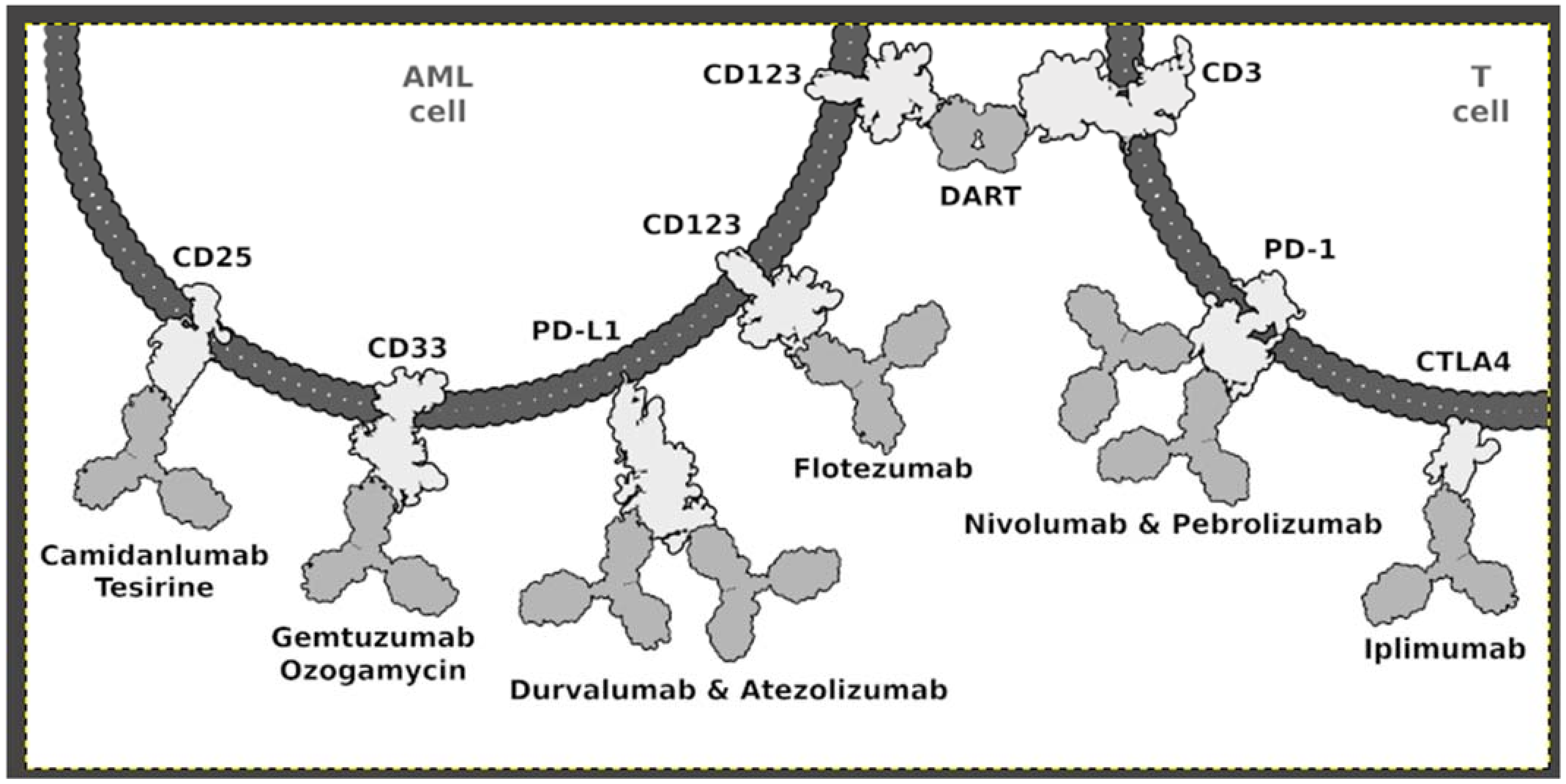
4.1. Immunotherapy

The development of immunotherapy for the treatment of AML in the pediatric population faces many barriers. The main one is the lack of an antigen specific only to cancer cells. Furthermore, AML blasts create an immunosuppressive microenvironment. Due to the fact that the majority of surface proteins that define malignant myeloid blasts are also expressed on normal progenitors, potential therapeutic targets are mainly seen in dysregulated gene expression [8]. However, it should be noted that the impaired expression of immunotargets significantly differs between adults and children. For example, immunotargets of adult AML, such as IL3RA, were not overexpressed in pediatric AML. The best described antigen AML tumor associated antigen (TAA) is the sialic acid-binding immunoglobulin lectin (SIGLEC) CD33 [7,124]. It has been proven that this antigen is found on most AML and progenitor cells. GO is a humanized anti-CD33 antibody that shows activity in pediatric and adult patients with AML [125]. A recently conducted single-center, phase III, double-arm trial (AAML0531) enrolled 1022 children, adolescents, and young adults aged 0 to 29 years with newly diagnosed AML. Patients were randomly assigned to either standard five-course chemotherapy alone (Arm A) or to the same chemotherapy with two doses of GO (3 mg/m2/dose) administered once in induction course 1 and once in intensification course 2 (Arm B). Data obtained showed GO significantly improved EFS (3 years: 53.1% vs. 46.9%; hazard ratio [HzR], 0.83; 95% CI, 0.70 to 0.99; p = 0.04) but not OS (3 years: 69.4% vs. 65.4%; HzR, 0.91; 95% CI, 0.74 to 1.13; p = 0.39) [126]. Collected data showed that GO added to chemotherapy improved EFS through a reduction in RR for children and adolescents with AML. JL1 is a CD43 epitope and cell surface glycoprotein of the mucin family, which is expressed during lymphoid maturation but is not expressed on mature blood cells. Recent studies reported that JL1 antigen is expressed on leukemic T, B, and myeloid lineage cells in >80% of acute leukemia patients and thus could serve as a potential candidate for immunotherapy. In a recent clinical trial conducted in Korea, authors included 78 patients diagnosed as having de novo pediatric acute leukemia (52 ALL and 26 AML) with a median age of 96 months (range: 2–216 months) and a median follow-up period of 424 days (range: 79–753 days). JL1 expression assessment was performed by flow cytometry, and positive JL1 expression was defined as ≥ 20% expression among the gated leukemic blasts. The study demonstrated that de novo pediatric AML patients with positive JL1 expression have higher CD13 and lower CD65 and CD15 expressions than patient without JL1 expression. Moreover, it was noted that de novo pediatric AML patients with positive JL1 expression presented with RUNX1::RUNX1T1, CBFB::MYH11, and PML::RARA rearrangements, which lead to chromosomal aberrations. These results suggest that JL1 may be a potential therapeutic target in immunotherapy for pediatric AML patients [127]. In the context of AML immunotherapy, it is worth approximating the results of a clinical trial with the use of flotetuzumab. Flotetuzumab (MGD006) is an investigational bispecific antibody-based molecule to CD3e and CD123 engineered in a DART format. CD3-engaging molecules work by stimulating the effector cells of the immune system in order to inactivate cancer cells. Knowing that high CD123 expression is also associated with a poor prognosis, flotetuzumab targeting of CD123 represents an interesting treatment option [128].
In recent years, research has identified the engagement of immune checkpoint receptors as a mechanism of tumor evasion. T-cell checkpoint receptors such as CTLA-4 and PD-1 relay inhibitory signals that modulate T-cell activation. In acute myeloid leukemia, PD1 expression is observed on T-cell subpopulations, including CD4+ effector T cells, CD4+ Treg, and CD8+ T cells, both in untreated patients and in relapses. Increased PD1 expression on CD8+ T lymphocytes may be one of the factors leading to the dysfunction of this group of immune cells and a reduction in the immune response to the progressive course of AML. Blocking signaling through checkpoint receptors results in increased T-cell activation, with effector T-cell proliferation and increased cytotoxicity toward cancer cells [129]. Inhibitors of PD-1 (nivolumab) and CTLA-4 (ipilimumab) have shown promise for the treatment of advanced melanoma and relapsed Hodgkin’s lymphoma with response rates ranging from 7 to 40%. In vitro studies have shown that AML may utilize the PD-1/PD-L1 axis to evade an anticancer immune response. For adult cancer, inhibitors of PD-1 (nivolumab) and CTLA-4 (ipilimumab) have shown promise with response rates ranging from 7 to 40%. PD-1 and/or PD-L1 are expressed in AML cells, and their blockade coupled with the depletion of regulatory T cells showed potent anti-leukemic activity in preclinical models. Several monoclonal antibodies (e.g., Nivolumab, Prembrolizumab, Durvalumab, and Ipilimumab) are currently studied for their anti-leukemic potential in refractory/relapsed AML patients [103]. Nivolumab is a human anti-PD1 IgG4 monoclonal antibody that blocks its interaction with PDL1 and PDL2 [8]. Pembrolizumab, also known as MK3475, is a humanized IgG4 monoclonal antibody that binds to PD1, blocking its interaction with PDL1 and PD L2 ligands (Figure 1) [7].

4.2. CART-T

Therapy with T cells expressing chimeric antigen receptors that are specific for tumor antigens turned out to be a success in the treatment of patients with B-cell ALL [130]. That is why CAR-T therapy remains a highly promising strategy also for AML patients. The key to the success of this therapy is the identification of specific antigens for the cancer cells. The ideal antigen target should play a key role in cell differentiation and survival. In practice, determining such a therapeutic target is extremely difficult. AML cells express a variety of stem cell and myeloid differentiation antigens on the cell membrane, such as CD33, CD34, CD123, CD135. However, the same antigens are expressed on healthy bone marrow cells, causing normal hematopoiesis to be affected during treatment [131]. CD33 is expressed on about 85–90% of AML blast cells, making it a promising therapeutic target. Data obtained from preclinical studies support the effectiveness of an anti-CD33 CAR-T therapy for AML in mice [132,133,134]. Kim et al. [135] demonstrated an approach to prevent damage to physiological hematopoiesis. They produced CD33 knockout human hematopoietic stem cells and progenitor cells (HSPCs) that have been successfully implanted in immunodeficient mice. Edited donor allogeneic hematopoietic stem cells are not eliminated by anti-CD33 CART, which would efficiently eliminate leukemia cells without marrow toxicity. CD123 is expressed at the levels both of leukemic stem cells (LSCs) and more differentiated leukemic blasts [135,136]. Numerous preclinical studies have confirmed the efficacy of anti-CD123 CART in vivo and in vitro [137,138,139]. A novel approach to the subject was demonstrated by Simon Loff et al. [140], who presented data from the preclinical and translational development of a UniCAR-based treatment of acute leukemia. They showed efficient tumor reactivity in vitro and in vivo using T cells that were engineered to express a UniCAR construct optimized for clinical applications and redirected against CD123+ leukemia cells. UniCar technology has been designed so that T cells do not express any characteristic antigen. Instead, they express the universal CAR (UniCAR-T) that recognizes a small linear peptide derived from the nuclear human La/SS-B protein (UniCAR epitope (UCE)). UniCART-T remains inactive until it connects with targeting modules (TMs) consisting of the UCE linked to an appropriate binding domain. The UniCAR-Ts, in combination with TM123 effectiveness and safety, will be assessed in a clinical trial [140]. The use of CD123-targeting T cells could be an encouraging strategy for the potential treatment of AML patients. Currently (referring to the clinicaltrials.gov database [141]), one anti-CD123 CAR-T trial is being conducted. This is a phase 1 study. Subjects will receive CART123 cells via a single IV infusion at a dose of 2 × 10⁶ CART123 cells/kg, following lymphodepleting chemotherapy. The total dose administered to each subject will be based on the subject’s body weight obtained at the time of apheresis. The minimum acceptable dose for infusion is 1 × 10⁵ CART123 cells/kg [141]. In conclusion, the main problem of potent, antigen-specific immunotherapy for AML is the absence of truly AML-specific surface antigens, which pose a high toxicity risk. MGD006 is a bispecific CD3 × CD123 dual-affinity re-targeting (DART) molecule that binds T lymphocytes and cells expressing CD123, an antigen up-regulated in several hematological malignancies including AML. MGD006 mediates blast killing in AML samples, together with concomitant activation and expansion of residual T cells. In a preclinical study, Gurunadh R. et al. [142] provided preclinical activity, safety, pharmacokinetic, and pharmacodynamic data supporting MGD006, a CD3 × CD123 bispecific DART capable of redirecting host T cells to kill CD123+ targets, as a potential therapeutic agent for the treatment of CD123+ hematological malignancies [142].

4.3. Other Therapeutical Achievements

4.3.1. CPX-351 (Vyxeos®)

CPX-351 (Vyxeos®) is a dual-drug liposomal encapsulation of cytarabine and daunorubicin that was rationally designed to improve efficacy over the traditional 7 + 3 cytarabine/daunorubicin chemotherapy regimen for patients with acute myeloid leukemia (AML). The CPX-351 liposome protects cytarabine and daunorubicin from metabolism and elimination. Thanks to this solution, the difference in the pharmacokinetics of both compounds is cancelled, and they can act simultaneously. In clinical studies, these liposome properties markedly increased the elimination half-life of CPX-351 versus free cytarabine and daunorubicin and maintained a synergistic drug ratio for over 24 h after administration. The use of a liposome allows for less exposure to tissues that are off-target tissues. CPX-351 shows high efficiency in patients with newly diagnosed high-risk/secondary AML [143].

4.3.2. HDAC Inhibitors

HDACs catalyze the removal of acetyl functional groups from the lysine residues of the histones. HDACs may also play a role in the regulation of the immune system by targeting the transcriptional regulator STAT3. HDACs are important proteins; they directly regulate gene expression and control cellular activity by reversing the state of histone acetylation. If the chromatin structure is altered through (de)acetylation of histones, this may result in decreased or increased gene transcription, altering gene expression levels. Numerous scientific studies indicate that HDAC deregulation may lead to the development of neoplasms, including hematological neoplasms [144]. HDAC inhibitors can be classified most commonly into five groups: hydroxamates, benzamides, cyclic tetrapeptides, aliphatic acids, and electrophilic ketones. These inhibitors have shown the ability to induce differentiation, cell cycle arrest, and apoptosis in AML. However, preliminary preclinical studies suggest that HDAC inhibitors could be effective in combination therapy and not as monotherapy. Preclinical studies conducted on leukemic cell lines have shown that JAK2/HDAC dual inhibitors have therapeutic potential in treating AML [145].

5. NGS—Predisposition in Pediatric AML

An important approach in medicine is not only the treatment of a disease entity, but also the assessment of the predisposition for the development of the disease, even before its occurrence. The World Health Organization’s latest leukemia classification scheme has included germline predisposition to myeloid malignancies as a provisional category. Predictive testing has become possible since the widespread use of next-generation sequencing (NGS). The development of NGS techniques, commercially available since 2006, allowed for cost- and time-effective sequencing [146]. The data presented by A. Andersson et al. demonstrate that mutations in isocitrate dehydrogenase 1 (IDH1) and 2 (IDH2) are exceedingly rare in pediatric ALL, but are more common in pediatric AMLs, occurring in 3.5% of cases overall, and in 9.8% of pediatric AMLs with a normal karyotype. In their pediatric cohort, they could not demonstrate any significant statistical association of IDH1/IDH2 mutations with overall survival or event-free survival, although the power of this analysis is influenced by the low overall frequency of IDH1/IDH2 mutations [147]. In myeloid cancers, IDH1/2 mutation has been identified as an induction event. However, IDH1 mutations are mainly involved in early occurrences of AML. Mondesir et al. reported that IDH1 modifications are found in about 10% of AML patients and are associated with worse outcomes in patients undergoing thorough chemotherapy [148]. Drazer et al. used NGS-targeted panels including genes associated with hereditary hematopoietic malignancies (HHMs) to identify pathogenic germline variants in malignant cells, thereby identifying patients at risk for HHMs. In total, pathogenic or likely pathogenic variants in ANKRD26, CEBPA, DDX41, ETV6, GATA2, RUNX1, or TP53 were identified in 74 (21%) of 360 patients. Three DDX41 variants, 2 GATA2 variants, and a TP53 variant previously implicated in Li-Fraumeni syndrome were of germline origin [149]. According to the latest research, IDH2 mutations may occur in the early stages of AML leukemia development in children; their presence makes cells more susceptible to oncogenic activities of FLT3 activating mutations. It makes them a potential gene that can be studied by NGS. Germinal mutations are one of the factors conditioning the development of hematological neoplasms that gives 100% certainty of tumor development regardless of environmental conditions. An example of one such mutation is a germline 5′-end CEBPA mutation. In addition, variants identified in leukemia cells should be considered as likely germline genes that can be mutated in germline or somatic tissues, including TP53, CEBPA, RUNX1 and DDX41 [150].

6. Conclusions

In recent years, the intensification of standard chemotherapy, more precise risk classification, improvements in supportive care, and the use of minimal residual disease to monitor response to therapy contributed to the improvement of the curability of AML patients in low-risk groups. Despite this, the curability of patients with high-risk AML in the pediatric population is still low. Many children become refractory or relapse even after successful therapy. A major challenge is the lack of specific antigens on the surfaces of tumor cells. Modern medicine has a wide range of treatment protocols, but only extending them with new therapeutic targets can provide a chance to improve the cure rate in high-risk groups. The increasing number of emerging clinical trials offers hope for new therapeutic solutions in the near future and improvement of the cure rate in the pediatric AML population.

Author Contributions

Conceptualization, M.L., I.D. and M.J.; writing—original draft preparation, I.D. and M.J.; writing—review and editing, M.L.; visualization, M.L.; supervision, M.L.; project administration, M.L. All authors have read and agreed to the published version of the manuscript.

Funding

This research received no external funding.

Institutional Review Board Statement

Not applicable.

Informed Consent Statement

Not applicable.

Data Availability Statement

No new data were created or analyzed in this study. Data sharing is not applicable to this article.

Conflicts of Interest

The authors declare no conflict of interest.

References

  1. Ksiazek, T.; Czogala, M.; Kaczowka, P.; Sadowska, B.; Pawinska-Wasikowska, K.; Bik-Multanowski, M.; Sikorska-Fic, B.; Matysiak, M.; Skalska-Sadowska, J.; Wachowiak, J.; et al. High Frequency of Fusion Gene Transcript Resulting From t(10;11)(P12;Q23) Translocation in Pediatric Acute Myeloid Leukemia in Poland. Front. Pediatrics 2020, 8, 278. [Google Scholar] [CrossRef] [PubMed]
  2. Song, M.K.; Park, B.B.; Uhm, J.E. Targeted Therapeutic Approach Based on Understanding of Aberrant Molecular Pathways Leading to Leukemic Proliferation in Patients with Acute Myeloid Leukemia. Int. J. Mol. Sci. 2021, 22, 5789. [Google Scholar] [CrossRef] [PubMed]
  3. Pui, C.H.; Carroll, W.L.; Meshinchi, S.; Arceci, R.J. Biology, Risk Stratification, and Therapy of Pediatric Acute Leukemias: An Update. J. Clin. Oncol. 2011, 29, 551–565. [Google Scholar] [CrossRef] [PubMed] [Green Version]
  4. de Rooij, J.; Zwaan, C.; van den Heuvel-Eibrink, M. Pediatric AML: From Biology to Clinical Management. J. Clin. Med. 2015, 4, 127–149. [Google Scholar] [CrossRef] [PubMed]
  5. Creutzig, U.; Kutny, M.A.; Barr, R.; Schlenk, R.F.; Ribeiro, R.C. Acute Myelogenous Leukemia in Adolescents and Young Adults. Pediatric Blood Cancer 2018, 65, e27089. [Google Scholar] [CrossRef] [PubMed]
  6. Creutzig, U.; Büchner, T.; Sauerland, M.C.; Zimmermann, M.; Reinhardt, D.; Döhner, H.; Schlenk, R.F. Significance of Age in Acute Myeloid Leukemia Patients Younger than 30 Years: A Common Analysis of the Pediatric Trials AML-BFM 93/98 and the Adult Trials AMLCG 92/99 and AMLSG HD93/98A. Cancer 2008, 112, 562–571. [Google Scholar] [CrossRef] [PubMed]
  7. Bonifant, C.L.; Velasquez, M.P.; Gottschalk, S. Advances in Immunotherapy for Pediatric Acute Myeloid Leukemia. Expert Opin. Biol. Ther. 2018, 18, 51–63. [Google Scholar] [CrossRef] [PubMed]
  8. Lamble, A.J.; Tasian, S.K. Opportunities for Immunotherapy in Childhood Acute Myeloid Leukemia. Blood Adv. 2019, 3, 3750–3758. [Google Scholar] [CrossRef]
  9. Grimwade, D.; Ivey, A.; Huntly, B.J.P. Molecular landscape of acute myeloid leukemia in younger adults and its clinical relevance. Blood J. Am. Soc. Hematol. 2016, 127, 29–41. [Google Scholar] [CrossRef] [Green Version]
  10. Papaemmanuil, E.; Gerstung, M.; Bullinger, L.; Gaidzik, V.I.; Paschka, P.; Roberts, N.D.; Potter, N.E.; Heuser, M.; Thol, F.; Bolli, N.; et al. Genomic Classification and Prognosis in Acute Myeloid Leukemia. N. Engl. J. Med. 2016, 374, 2209–2221. [Google Scholar] [CrossRef]
  11. Creutzig, U.; Zimmermann, M.; Reinhardt, D.; Rasche, M.; von Neuhoff, C.; Alpermann, T.; Dworzak, M.; Perglerová, K.; Zemanova, Z.; Tchinda, J.; et al. Changes in Cytogenetics and Molecular Genetics in Acute Myeloid Leukemia from Childhood to Adult Age Groups. Cancer 2016, 122, 3821–3830. [Google Scholar] [CrossRef] [PubMed]
  12. Bolouri, H.; Farrar, J.E.; Triche, T.; Ries, R.E.; Lim, E.L.; Alonzo, T.A.; Ma, Y.; Moore, R.; Mungall, A.J.; Marra, M.A.; et al. The Molecular Landscape of Pediatric Acute Myeloid Leukemia Reveals Recurrent Structural Alterations and Age-Specific Mutational Interactions. Nat. Med. 2018, 24, 103–112. [Google Scholar] [CrossRef] [PubMed] [Green Version]
  13. Harrison, C.J.; Hills, R.K.; Moorman, A.v.; Grimwade, D.J.; Hann, I.; Webb, D.K.H.; Wheatley, K.; de Graaf, S.S.N.; van den Berg, E.; Burnett, A.K.; et al. Cytogenetics of Childhood Acute Myeloid Leukemia: United Kingdom Medical Research Council Treatment Trials AML 10 and 12. J. Clin. Oncol. 2010, 28, 2674–2681. [Google Scholar] [CrossRef] [PubMed]
  14. Von Neuhoff, C.; Reinhardt, D.; Sander, A.; Zimmermann, M.; Bradtke, J.; Betts, D.R.; Zemanova, Z.; Stary, J.; Bourquin, J.P.; Haas, O.A.; et al. Prognostic Impact of Specific Chromosomal Aberrations in a Large Group of Pediatric Patients with Acute Myeloid Leukemia Treated Uniformly According to Trial AML-BFM 98. J. Clin. Oncol. 2010, 28, 2682–2689. [Google Scholar] [CrossRef]
  15. Kutny, M.A.; Alonzo, T.A.; Gerbing, R.B.; Wang, Y.C.; Raimondi, S.C.; Hirsch, B.A.; Fu, C.H.; Meshinchi, S.; Gamis, A.S.; Feusner, J.H.; et al. Arsenic Trioxide Consolidation Allows Anthracycline Dose Reduction for Pediatric Patients with Acute Promyelocytic Leukemia: Report from the Children’s Oncology Group Phase III Historically Controlled Trial AAML0631. J. Clin. Oncol. 2017, 35, 3021–3029. [Google Scholar] [CrossRef] [PubMed]
  16. Hollink, I.H.I.M.; Zwaan, C.M.; Zimmermann, M.; Arentsen-Peters, T.C.J.M.; Pieters, R.; Cloos, J.; Kaspers, G.J.L.; de Graaf, S.S.N.; Harbott, J.; Creutzig, U.; et al. Favorable Prognostic Impact of NPM1 Gene Mutations in Childhood Acute Myeloid Leukemia, with Emphasis on Cytogenetically Normal AML. Leukemia 2009, 23, 262–270. [Google Scholar] [CrossRef]
  17. Verhaak, R.G.W.; Goudswaard, C.S.; Van Putten, W.; Bijl, M.A.; Sanders, M.A.; Hugens, W.; Uitterlinden, A.G.; Erpelinck, C.A.J.; Delwel, R.; Löwenberg, B.; et al. Mutations in Nucleophosmin (NPM1) in Acute Myeloid Leukemia (AML): Association with Other Gene Abnormalities and Previously Established Gene Expression Signatures and Their Favorable Prognostic Significance. Blood 2005, 106, 3747–3754. [Google Scholar] [CrossRef]
  18. Hollink, I.H.I.M.; van den Heuvel-Eibrink, M.M.; Arentsen-Peters, S.T.C.J.M.; Zimmermann, M.; Peeters, J.K.; Valk, P.J.M.; Balgobind, B.v.; Sonneveld, E.; Kaspers, G.J.L.; de Bont, E.S.J.M.; et al. Characterization of CEBPA Mutations and Promoter Hypermethylation in Pediatric Acute Myeloid Leukemia. Haematologica 2011, 96, 384–392. [Google Scholar] [CrossRef] [Green Version]
  19. Fasan, A.; Haferlach, C.; Alpermann, T.; Jeromin, S.; Grossmann, V.; Eder, C.; Weissmann, S.; Dicker, F.; Kohlmann, A.; Schindela, S.; et al. The Role of Different Genetic Subtypes of CEBPA Mutated AML. Leukemia 2014, 28, 794–803. [Google Scholar] [CrossRef] [PubMed]
  20. Noort, S.; Zimmermann, M.; Reinhardt, D.; Cuccuini, W.; Pigazzi, M.; Smith, J.; Ries, R.E.; Alonzo, T.A.; Hirsch, B.; Tomizawa, D.; et al. Prognostic Impact of t(16;21)(P11;Q22) and t(16;21)(Q24;Q22) in Pediatric AML: A Retrospective Study by the I-BFM Study Group. Blood 2018, 132, 1584–1592. [Google Scholar] [CrossRef] [PubMed] [Green Version]
  21. Shiba, N.; Yoshida, K.; Hara, Y.; Yamato, G.; Shiraishi, Y.; Matsuo, H.; Okuno, Y.; Chiba, K.; Tanaka, H.; Kaburagi, T.; et al. Transcriptome Analysis Offers a Comprehensive Illustration of the Genetic Background of Pediatric Acute Myeloid Leukemia. Blood Adv. 2019, 3, 3157–3169. [Google Scholar] [CrossRef] [PubMed] [Green Version]
  22. Taga, T. Treatment strategy for myeloid leukemia with Down syndrome. Rinsho ketsueki. Jpn. J. Clin. Hematol. 2020, 61, 1382–1387. [Google Scholar] [CrossRef]
  23. Forestier, E.; Izraeli, S.; Beverloo, B.; Haas, O.; Pession, A.; Michalová, K.; Stark, B.; Harrison, C.J.; Teigler-Schlegel, A.; Johansson, B. Cytogenetic Features of Acute Lymphoblastic and Myeloid Leukemias in Pediatric. Patients with Down Syndrome: An IBFM-SG Study. Blood 2008, 111, 1575–1583. [Google Scholar] [CrossRef] [PubMed] [Green Version]
  24. Elgarten, C.W.; Aplenc, R. Pediatric Acute Myeloid Leukemia: Updates on Biology, Risk Stratification, and Therapy. Curr. Opin. Pediatrics 2020, 32, 57–66. [Google Scholar] [CrossRef] [PubMed]
  25. Meshinchi, S.; Alonzo, T.A.; Stirewalt, D.L.; Zwaan, M.; Zimmerman, M.; Reinhardt, D.; Kaspers, G.J.L.; Heerema, N.A.; Gerbing, R.; Lange, B.J.; et al. Clinical Implications of FLT3 Mutations in Pediatric AML. Blood 2006, 108, 3654–3661. [Google Scholar] [CrossRef] [Green Version]
  26. Coenen, E.; Harbott, J.; Zwaan, C.; Raimondi, S.; van den Heuvel-Eibrink, M. 11Q23 Rearrangements in De Novo Childhood Acute Myeloid Leukemia. Atlas Genet. Cytogenet. Oncol. Haematol. 2012, 16, 574–581. [Google Scholar] [CrossRef] [Green Version]
  27. Chen, Y.; Kantarjian, H.; Pierce, S.; Faderl, S.; O’Brien, S.; Qiao, W.; Abruzzo, L.; De Lima, M.; Kebriaei, P.; Jabbour, E.; et al. Prognostic Significance of 11q23 Aberrations in Adult Acute Myeloid Leukemia and the Role of Allogeneic Stem Cell Transplantation. Leukemia 2013, 27, 836–842. [Google Scholar] [CrossRef] [PubMed]
  28. Martinez-Climent, J.A.; Espinosa, R., III; Thirman, M.J.; Le Beau, M.M.; Rowley, J.D. Abnormalities of Chromosome Band 11q23 and the MLL Gene in Pediatric Myelomonocytic and Monoblastic Leukemias. Identification of the t(9;11) as an Indicator of Long Survival. J. Pediatric Hematol./Oncol. 1995, 17, 277–283. [Google Scholar] [CrossRef]
  29. Meyer, C.; Burmeister, T.; Gröger, D.; Tsaur, G.; Fechina, L.; Renneville, A.; Sutton, R.; Venn, N.C.; Emerenciano, M.; Pombo-De-Oliveira, M.S.; et al. The MLL Recombinome of Acute Leukemias in 2017. Leukemia 2018, 32, 273–284. [Google Scholar] [CrossRef]
  30. Balgobind, B.V.; Raimondi, S.C.; Harbott, J.; Zimmermann, M.; Alonzo, T.A.; Auvrignon, A.; Beverloo, H.B.; Chang, M.; Creutzig, U.; Dworzak, M.N.; et al. Novel Prognostic Subgroups in Childhood 11q23/MLL-Rearranged Acute Myeloid Leukemia: Results of an International Retrospective Study. Blood 2009, 114, 2489–2496. [Google Scholar] [CrossRef] [Green Version]
  31. Akiki, S.; Dyer, S.A.; Grimwade, D.; Ivey, A.; Abou-Zeid, N.; Borrow, J.; Jeffries, S.; Caddick, J.; Newell, H.; Begum, S.; et al. NUP98-NSD1 Fusion in Association with FLT3-ITD Mutation Identifies a Prognostically Relevant Subgroup of Pediatric Acute Myeloid Leukemia Patients Suitable for Monitoring by Real Time Quantitative PCR. Genes Chromosomes Cancer 2013, 52, 1053–1064. [Google Scholar] [CrossRef] [PubMed]
  32. Balgobind, B.V.; Hollink, I.H.I.M.; Arentsen-Peters, S.T.C.J.M.; Zimmermann, M.; Harbott, J.; Berna Beverloo, H.; von Bergh, A.R.M.; Cloos, J.; Kaspers, G.J.L.; de Haas, V.; et al. Integrative Analysis of Type-I and Type-II Aberrations Underscores the Genetic Heterogeneity of Pediatric Acute Myeloid Leukemia. Haematologica 2011, 96, 1478–1487. [Google Scholar] [CrossRef] [Green Version]
  33. Radtke, I.; Mullighan, C.G.; Ishii, M.; Su, X.; Cheng, J.; Ma, J.; Ganti, R.; Cai, Z.; Goorha, S.; Pounds, S.B.; et al. Genomic Analysis Reveals Few Genetic Alterations in Pediatric Acute Myeloid Leukemia. Proc. Natl. Acad. Sci. USA 2009, 106, 12944–12949. [Google Scholar] [CrossRef] [PubMed] [Green Version]
  34. Struski, S.; Lagarde, S.; Bories, P.; Puiseux, C.; Prade, N.; Cuccuini, W.; Pages, M.P.; Bidet, A.; Gervais, C.; Lafage-Pochitaloff, M.; et al. NUP98 Is Rearranged in 3.8% of Pediatric AML Forming a Clinical and Molecular Homogenous Group with a Poor Prognosis. Leukemia 2017, 31, 565–572. [Google Scholar] [CrossRef]
  35. Hara, Y.; Shiba, N.; Ohki, K.; Tabuchi, K.; Yamato, G.; Park, M.J.; Tomizawa, D.; Kinoshita, A.; Shimada, A.; Arakawa, H.; et al. Prognostic Impact of Specific Molecular Profiles in Pediatric Acute Megakaryoblastic Leukemia in Non-Down Syndrome. Genes Chromosomes Cancer 2017, 56, 394–404. [Google Scholar] [CrossRef]
  36. Noort, S.; Wander, P.; Alonzo, T.A.; Smith, J.; Ries, R.E.; Gerbing, R.B.; Dolman, E.M.M.; Locatelli, F.; Reinhardt, D.; Baruchel, A.; et al. The Clinical and Biological Characteristics of NUP98-KDM5A Pediatric Acute Myeloid Leukemia. Haematologica 2021, 106, 630–634. [Google Scholar] [CrossRef] [PubMed]
  37. Espersen, A.D.L.; Noren-Nyström, U.; Abrahamsson, J.; Ha, S.Y.; Pronk, C.J.; Jahnukainen, K.; Jónsson, Ó.G.; Lausen, B.; Palle, J.; Zeller, B.; et al. Acute Myeloid Leukemia (AML) with t(7;12)(Q36;P13) Is Associated with Infancy and Trisomy 19: Data from Nordic Society for Pediatric Hematology and Oncology (NOPHO-AML) and Review of the Literature. Genes Chromosomes Cancer 2018, 57, 359–365. [Google Scholar] [CrossRef] [PubMed]
  38. Johnston, D.; Alonzo, T.A.; Gerbing, R.B.; Hirsch, B.; Heerema, N.A.; Ravindranath, Y.; Woods, W.G.; Lange, B.J.; Gamis, A.S.; Raimondi, S.C. Outcome of Pediatric Patients with Acute Myeloid Leukemia (AML) and -5/5q- Abnormalities from Five Pediatric AML Treatment Protocols: A Report from the Children’s Oncology Group. Pediatric Blood Cancer 2013, 60, 2073–2078. [Google Scholar] [CrossRef] [PubMed]
  39. Masetti, R.; Pigazzi, M.; Togni, M.; Astolfi, A.; Indio, V.; Manara, E.; Casadio, R.; Pession, A.; Basso, G.; Locatelli, F. CBFA2T3-GLIS2 Fusion Transcript Is a Novel Common Feature in Pediatric, Cytogenetically Normal AML, Not Restricted to FAB M7 Subtype. Blood 2013, 121, 3469–3472. [Google Scholar] [CrossRef] [Green Version]
  40. Creutzig, U.; Van Den Heuvel-Eibrink, M.M.; Gibson, B.; Dworzak, M.N.; Adachi, S.; De Bont, E.; Harbott, J.; Hasle, H.; Johnston, D.; Kinoshita, A.; et al. Diagnosis and Management of Acute Myeloid Leukemia in Children and Adolescents: Recommendations from an International Expert Panel. Blood 2012, 120, 3167–3205. [Google Scholar] [CrossRef]
  41. Lugthart, S.; Gröschel, S.; Beverloo, H.B.; Kayser, S.; Valk, P.J.M.; Van Zelderen-Bhola, S.L.; Ossenkoppele, G.J.; Vellenga, E.; Van Den Berg-De Ruiter, E.; Schanz, U.; et al. Clinical, Molecular, and Prognostic Significance of WHO Type Inv(3)(Q21q26.2)/t(3;3)(Q21;Q26.2) and Various Other 3q Abnormalities in Acute Myeloid Leukemia. J. Clin. Oncol. 2010, 28, 3890–3898. [Google Scholar] [CrossRef] [PubMed]
  42. Grimwade, D.; Hills, R.K.; Moorman, A.V.; Walker, H.; Chatters, S.; Goldstone, A.H.; Wheatley, K.; Harrison, C.J.; Burnett, A.K. Refinement of Cytogenetic Classification in Acute Myeloid Leukemia: Determination of Prognostic Significance of Rare Recurring Chromosomal Abnormalities among 5876 Younger Adult Patients Treated in the United Kingdom Medical Research Council Trials. Blood 2010, 116, 354–365. [Google Scholar] [CrossRef] [PubMed] [Green Version]
  43. Neuendorff, N.R.; Hemmati, P.; Arnold, R.; Ihlow, J.; Dörken, B.; Müller-Tidow, C.; Westermann, J. BCR-ABL1 Acute Myeloid Leukemia: Are We Always Dealing with a High-Risk Disease? Blood Adv. 2018, 2, 1409–1411. [Google Scholar] [CrossRef] [PubMed] [Green Version]
  44. Borel, C.; Dastugue, N.; Cances-Lauwers, V.; Mozziconacci, M.J.; Prebet, T.; Vey, N.; Pigneux, A.; Lippert, E.; Visanica, S.; Legrand, F.; et al. PICALM-MLLT10 Acute Myeloid Leukemia: A French Cohort of 18 Patients. Leuk. Res. 2012, 36, 1365–1369. [Google Scholar] [CrossRef] [PubMed]
  45. Arbor, A. Spontaneous Remission of Congenital Acute Myeloid Leukemia. Pediatric Blood Cancer 2011, 331–332. [Google Scholar] [CrossRef]
  46. Wong, K.; Frcp, H.Y.; Siu, L.L.P.; Pang, A.; Kwong, Y. T(8;16)(P11;P13) Predisposes to a Transient but Potentially Recurring Neonatal Leukemia. Hum. Pathol. 2008, 39, 1702–1707. [Google Scholar] [CrossRef]
  47. Boissel, N.; Leroy, H.; Brethon, B.; Philippe, N.; De Botton, S.; Auvrignon, A.; Raffoux, E.; Leblanc, T.; Thomas, X.; Hermine, O. Incidence and Prognostic Impact of C-Kit, FLT3, and Ras Gene Mutations in Core Binding Factor Acute Myeloid Leukemia (CBF-AML). Leukemia 2006, 965–970. [Google Scholar] [CrossRef] [Green Version]
  48. Pollard, J.A.; Alonzo, T.A.; Gerbing, R.B.; Ho, P.A.; Zeng, R.; Ravindranath, Y.; Dahl, G.; Lacayo, N.J.; Becton, D.; Chang, M.; et al. Prevalence and Prognostic Significance of KIT Mutations in Pediatric Patients with Core Binding Factor AML Enrolled on Serial Pediatric Cooperative Trials for de Novo AML. Blood 2016, 115, 2372–2380. [Google Scholar] [CrossRef] [Green Version]
  49. Duployez, N.; Marceau-Renaut, A.; Boissel, N.; Petit, A.; Bucci, M.; Geffroy, S.; Lapillonne, H.; Renneville, A.; Ragu, C.; Figeac, M.; et al. Comprehensive Mutational Profiling of Core Binding Factor Acute Myeloid Leukemia. Blood 2016, 127, 2451–2459. [Google Scholar] [CrossRef]
  50. Inaba, H.; Zhou, Y.; Abla, O.; Adachi, S.; Auvrignon, A.; Beverloo, H.B.; De Bont, E.; Chang, T.T.; Creutzig, U.; Dworzak, M.; et al. Heterogeneous Cytogenetic Subgroups and Outcomes in Childhood Acute Megakaryoblastic Leukemia: A Retrospective International Study. Blood 2015, 126, 1575–1584. [Google Scholar] [CrossRef]
  51. De Rooij, J.D.E.; Masetti, R.; Van Den Heuvel-Eibrink, M.M.; Cayuela, J.M.; Trka, J.; Reinhardt, D.; Rasche, M.; Sonneveld, E.; Alonzo, T.A.; Fornerod, M.; et al. Recurrent Abnormalities Can Be Used for Risk Group Stratification in Pediatric AMKL: A Retrospective Intergroup Study. Blood 2016, 127, 3424–3430. [Google Scholar] [CrossRef] [PubMed]
  52. Falini, B.; Nicoletti, I.; Bolli, N.; Paola Martelli, M.; Liso, A.; Gorello, P.; Mandelli, F.; Mecucci, C.; Fabrizio Martelli, M.; Sapienza, L.; et al. Translocations and Mutations Involving the Nucleophosmin (NPM1) Gene in Lymphomas and Leukemias. Haematology 2007, 92, 519–532. [Google Scholar] [CrossRef] [Green Version]
  53. Lim, G.; Choi, J.R.; Kim, M.J.; Kim, S.Y.; Lee, H.J.; Suh, J.T.; Yoon, H.J.; Lee, J.; Lee, S.; Lee, W.I.; et al. Detection of t(3;5) and NPM1/MLF1 Rearrangement in an Elderly Patient with Acute Myeloid Leukemia: Clinical and Laboratory Study with Review of the Literature. Cancer Genet. Cytogenet. 2010, 199, 101–109. [Google Scholar] [CrossRef] [PubMed] [Green Version]
  54. Laursen, A.C.L.; Sandahl, J.D.; Kjeldsen, E.; Abrahamsson, J.; Asdahl, P.; Ha, S.Y.; Heldrup, J.; Jahnukainen, K. Trisomy 8 in Pediatric Acute Myeloid Leukemia: A NOPHO-AML Study. Genes Chromosomes Cancer 2016, 55, 719–726. [Google Scholar] [CrossRef] [PubMed]
  55. Rasche, M.; von Neuhoff, C.; Dworzak, M.; Bourquin, J.P.; Bradtke, J.; Göhring, G.; Escherich, G.; Fleischhack, G.; Graf, N.; Gruhn, B.; et al. Genotype-Outcome Correlations in Pediatric AML: The Impact of a Monosomal Karyotype in Trial AML-BFM 2004. Leukemia 2017, 31, 2807–2814. [Google Scholar] [CrossRef] [PubMed] [Green Version]
  56. Sandahl, J.D.; Kjeldsen, E.; Abrahamsson, J.; Ha, S.Y.; Heldrup, J.; Jahnukainen, K.; Jonsson, O.G.; Lausen, B.; Palle, J.; Zeller, B.; et al. Ploidy and Clinical Characteristics of Childhood Acute Myeloid Leukemia: A NOPHO-AML Study Julie. Genes Chromosomes Cancer 2014, 53, 667–675. [Google Scholar] [CrossRef] [PubMed]
  57. Chilton, L.; Hills, R.K.; Harrison, C.J.; Burnett, A.K.; Grimwade, D.; Moorman, A.V. Hyperdiploidy with 49–65 Chromosomes Represents a Heterogeneous Cytogenetic Subgroup of Acute Myeloid Leukemia with Differential Outcome. Leukemia 2014, 28, 321–328. [Google Scholar] [CrossRef] [PubMed]
  58. Falini, B.; Nicoletti, I.; Martelli, M.F.; Mecucci, C. Acute Myeloid Leukemia Carrying Cytoplasmic/Mutated Nucleophosmin (NPMc+ AML): Biologic and Clinical Features. Blood 2007, 109, 874–885. [Google Scholar] [CrossRef] [PubMed] [Green Version]
  59. Dufour, A.; Schneider, F.; Metzeler, K.H.; Hoster, E.; Schneider, S.; Zellmeier, E.; Benthaus, T.; Sauerland, M.-C.; Berdel, W.E.; Büchner, T.; et al. Acute Myeloid Leukemia with Biallelic CEBPA Gene Mutations and Normal Karyotype Represents a Distinct Genetic Entity Associated with a Favorable Clinical Outcome. J. Clin. Oncol. Off. J. Am. Soc. Clin. Oncol. 2010, 28, 570–577. [Google Scholar] [CrossRef]
  60. Wouters, B.J.; Löwenberg, B.; Erpelinck-Verschueren, C.A.J.; van Putten, W.L.J.; Valk, P.J.M.; Delwel, R. Double CEBPA Mutations, but Not Single CEBPA Mutations, Define a Subgroup of Acute Myeloid Leukemia with a Distinctive Gene Expression Profile That Is Uniquely Associated with a Favorable Outcome. Blood 2009, 113, 3088–3091. [Google Scholar] [CrossRef] [Green Version]
  61. Kottaridis, P.D.; Gale, R.E.; Frew, M.E.; Harrison, G.; Langabeer, S.E.; Belton, A.A.; Walker, H.; Wheatley, K.; Bowen, D.T.; Burnett, A.K.; et al. The Presence of a FLT3 Internal Tandem Duplication in Patients with Acute Myeloid Leukemia (AML) Adds Important Prognostic Information to Cytogenetic Risk Group and Response to the First Cycle of Chemotherapy: Analysis of 854 Patients from the United King. Blood 2001, 98, 1752–1759. [Google Scholar] [CrossRef] [PubMed]
  62. Bill, M.; Mrózek, K.; Kohlschmidt, J.; Eisfeld, A.K.; Walker, C.J.; Nicolet, D.; Papaioannou, D.; Blachly, J.S.; Orwick, S.; Carroll, A.J.; et al. Mutational Landscape and Clinical Outcome of Patients with de Novo Acute Myeloid Leukemia and Rearrangements Involving 11q23/KMT2A. Proc. Natl. Acad. Sci. USA 2020, 117, 26340–26346. [Google Scholar] [CrossRef] [PubMed]
  63. Smol, T.; Collonge-Rame, M.-A. t(8;16)(P11;P13) KAT6A/CREBBP. Atlas Genet. Cytogenet. Oncol. Haematol. 2017, 11–16. [Google Scholar] [CrossRef] [Green Version]
  64. Ayatollahi, H.; Shajiei, A.; Sadeghian, M.H.; Sheikhi, M.; Yazdandoust, E.; Ghazanfarpour, M.; Shams, S.F.; Shakeri, S. Prognostic Importance of C-KIT Mutations in Core Binding Factor Acute Myeloid Leukemia: A Systematic Review. Hematol./Oncol. Stem Cell Ther. 2017, 10, 1–7. [Google Scholar] [CrossRef] [Green Version]
  65. Allan, J.M. Genetic Susceptibility to Breast Cancer in Lymphoma Survivors. Blood 2019, 133, 1004–1006. [Google Scholar] [CrossRef]
  66. Al-Harbi, S.; Aljurf, M.; Mohty, M.; Almohareb, F.; Ahmed, S.O.A. An Update on the Molecular Pathogenesis and Potential Therapeutic Targeting of AML with t(8;21)(Q22;Q22.1);RUNX1-RUNX1T1. Blood Adv. 2020, 4, 229–238. [Google Scholar] [CrossRef] [Green Version]
  67. Sakamoto, K.; Shiba, N.; Deguchi, T.; Kiyokawa, N.; Hashii, Y.; Moriya-Saito, A.; Tomizawa, D.; Taga, T.; Adachi, S.; Horibe, K.; et al. Negative CD19 Expression Is Associated with Inferior Relapse-Free Survival in Children with RUNX1-RUNX1T1–Positive Acute Myeloid Leukaemia: Results from the Japanese Paediatric Leukaemia/Lymphoma Study Group AML-05 Study. Br. J. Haematol. 2019, 187, 372–376. [Google Scholar] [CrossRef] [PubMed]
  68. Kundu, M.; Liu, P.P. Function of the Inv(16) Fusion Gene CBFB-MYH11. Curr. Opin. Hematol. 2001, 8, 201–205. [Google Scholar] [CrossRef] [PubMed]
  69. Huret, J. Inv(16)(P13Q22)-T(16;16)(P13;Q22)-Del(16)(Q22). Atlas Genet. Cytogenet. Oncol. Haematol. 2011, 3, 147–149. [Google Scholar] [CrossRef] [Green Version]
  70. Schoch, C.; Kern, W.; Schnittger, S.; Büchner, T.; Hiddemann, W.; Haferlach, T. The Influence of Age on Prognosis of de Novo Acute Myeloid Leukemia Differs According to Cytogenetic Subgroups. Haematologica 2004, 89, 1082–1090. [Google Scholar]
  71. Hann, I.M.; Webb, D.K.W.; Gibson, B.E.S.; Harrison, C.J. MRC Trials in Childhood Acute Myeloid Leukaemia. Ann. Hematol. 2004, 83 (Suppl. 1), S108–S112. [Google Scholar] [CrossRef] [PubMed]
  72. Paschka, P.; Marcucci, G.; Ruppert, A.S.; Mrózek, K.; Chen, H.; Kittles, R.A.; Vukosavljevic, T.; Perrotti, D.; Vardiman, J.W.; Carroll, A.J.; et al. Adverse Prognostic Significance of KIT Mutations in Adult Acute Myeloid Leukemia with Inv(16) and t(8;21): A Cancer and Leukemia Group B Study. J. Clin. Oncol. 2006, 24, 3904–3911. [Google Scholar] [CrossRef] [PubMed]
  73. Hollink, I.H.I.M.; Van Den Heuvel-Eibrink, M.M.; Zimmermann, M.; Balgobind, B.V.; Arentsen-Peters, S.T.C.J.M.; Alders, M.; Willasch, A.; Kaspers, G.J.L.; Trka, J.; Baruchel, A.; et al. Clinical Relevance of Wilms Tumor 1 Gene Mutations in Childhood Acute Myeloid Leukemia. Blood 2009, 113, 5951–5960. [Google Scholar] [CrossRef] [PubMed] [Green Version]
  74. Colombo, E.; Marine, J.C.; Danovi, D.; Falini, B.; Pelicci, P.G. Nucleophosmin Regulates the Stability and Transcriptional Activity of P53. Nat. Cell Biol. 2002, 4, 529–533. [Google Scholar] [CrossRef] [PubMed]
  75. Brady, S.N.; Yu, Y.; Maggi, L.B.; Weber, J.D. ARF Impedes NPM/B23 Shuttling in an Mdm2-Sensitive Tumor Suppressor Pathway. Mol. Cell. Biol. 2004, 24, 9327–9338. [Google Scholar] [CrossRef] [Green Version]
  76. Brown, P.; McIntyre, E.; Rau, R.; Meshinchi, S.; Lacayo, N.; Dahl, G.; Alonzo, T.A.; Chang, M.; Arceci, R.J.; Small, D. The Incidence and Clinical Significance of Nucleophosmin Mutations in Childhood AML. Blood 2007, 110, 979–985. [Google Scholar] [CrossRef] [Green Version]
  77. Cazzaniga, G.; Dell’Oro, M.G.; Mecucci, C.; Giarin, E.; Masetti, R.; Rossi, V.; Locatelli, F.; Martelli, M.F.; Basso, G.; Pession, A.; et al. Nucleophosmin Mutations in Childhood Acute Myelogenous Leukemia with Normal Karyotype. Blood 2005, 106, 1419–1422. [Google Scholar] [CrossRef] [Green Version]
  78. Falini, B.; Mecucci, C.; Tiacci, E.; Alcalay, M.; Rosati, R.; Pasqualucci, L.; La Starza, R.; Diverio, D.; Colombo, E.; Santucci, A.; et al. Cytoplasmic Nucleophosmin in Acute Myelogenous Leukemia with a Normal Karyotype. N. Engl. J. Med. 2005, 352, 254–266. [Google Scholar] [CrossRef]
  79. Schnittger, S.; Schoch, C.; Kern, W.; Mecucci, C.; Tschulik, C.; Martelli, M.F.; Haferlach, T.; Hiddemann, W.; Falini, B. Nucleophosmin Gene Mutations Are Predictors of Favorable Prognosis in Acute Myelogenous Leukemia with a Normal Karyotype. Blood 2005, 106, 3733–3739. [Google Scholar] [CrossRef] [Green Version]
  80. Thiede, C.; Koch, S.; Creutzig, E.; Steudel, C.; Illmer, T.; Schaich, M.; Ehninger, G. Prevalence and Prognostic Impact of NPM1 Mutations in 1485 Adult Patients with Acute Myeloid Leukemia (AML). Blood 2006, 107, 4011–4020. [Google Scholar] [CrossRef] [Green Version]
  81. Falini, B.; Brunetti, L.; Sportoletti, P.; Paola Martelli, M. NPM1-Mutated Acute Myeloid Leukemia: From Bench to Bedside. Blood 2020, 136, 1707–1721. [Google Scholar] [CrossRef]
  82. Daver, N.; Schlenk, R.F.; Russell, N.H.; Levis, M.J. Targeting FLT3 Mutations in AML: Review of Current Knowledge and Evidence. Leukemia 2019, 33, 299–312. [Google Scholar] [CrossRef] [PubMed] [Green Version]
  83. Falini, B.; Sciabolacci, S.; Falini, L.; Brunetti, L.; Martelli, M.P. Diagnostic and Therapeutic Pitfalls in NPM1-Mutated AML: Notes from the Field. Leukemia 2021, 35, 3113–3126. [Google Scholar] [CrossRef] [PubMed]
  84. Zhang, P.; Iwasaki-Arai, J.; Iwasaki, H.; Fenyus, M.L.; Dayaram, T.; Owens, B.M.; Shigematsu, H.; Levantini, E.; Huettner, C.S.; Lekstrom-Himes, J.A.; et al. Enhancement of Hematopoietic Stem Cell Repopulating Capacity and Self-Renewal in the Absence of the Transcription Factor C/EBPα. Immunity 2004, 21, 853–863. [Google Scholar] [CrossRef] [Green Version]
  85. Wouters, B.J.; Jordà, M.A.; Keeshan, K.; Louwers, I.; Erpelinck-Verschueren, C.A.J.; Tielemans, D.; Langerak, A.W.; He, Y.; Yashiro-Ohtani, Y.; Zhang, P.; et al. Distinct Gene Expression Profiles of Acute Myeloid/T-Lymphoid Leukemia with Silenced CEBPA and Mutations in NOTCH1. Blood 2007, 110, 3706–3714. [Google Scholar] [CrossRef]
  86. Radomska, H.S.; Bassères, D.S.; Zheng, R.; Zhang, P.; Dayaram, T.; Yamamoto, Y.; Sternberg, D.W.; Lokker, N.; Giese, N.A.; Bohlander, S.K.; et al. Block of C/EBPα Function by Phosphorylation in Acute Myeloid Leukemia with FLT3 Activating Mutations. J. Exp. Med. 2006, 203, 371–381. [Google Scholar] [CrossRef] [PubMed]
  87. Helbling, D.; Mueller, B.U.; Timchenko, N.A.; Schardt, J.; Eyer, M.; Betts, D.R.; Jotterand, M.; Meyer-Monard, S.; Fey, M.F.; Pabst, T. CBFB-SMMHC Is Correlated with Increased Calreticulin Expression and Suppresses the Granulocytic Differentiation Factor CEBPA in AML with Inv(16). Blood 2005, 106, 1369–1375. [Google Scholar] [CrossRef] [PubMed] [Green Version]
  88. Schmidt, L.; Heyes, E.; Scheiblecker, L.; Eder, T.; Volpe, G.; Frampton, J.; Nerlov, C.; Valent, P.; Grembecka, J.; Grebien, F. CEBPA-Mutated Leukemia Is Sensitive to Genetic and Pharmacological Targeting of the MLL1 Complex. Leukemia 2019, 33, 1608–1619. [Google Scholar] [CrossRef]
  89. Mannelli, F.; Ponziani, V.; Bencini, S.; Bonetti, M.I.; Benelli, M.; Cutini, I.; Gianfaldoni, G.; Scappini, B.; Pancani, F.; Piccini, M.; et al. CEBPA–Double-Mutated Acute Myeloid Leukemia Displays a Unique Phenotypic Profile: A Reliable Screening Method and Insight into Biological Features. Haematologica 2017, 102, 529–540. [Google Scholar] [CrossRef] [PubMed] [Green Version]
  90. Conneely, S.E.; Stevens, A.M. Advances in Pediatric Acute Promyelocytic Leukemia. Children 2020, 7, 11. [Google Scholar] [CrossRef] [Green Version]
  91. De Braekeleer, E.; Douet-Guilbert, N.; De Braekeleer, M. RARA Fusion Genes in Acute Promyelocytic Leukemia: A Review. Expert Rev. Hematol. 2014, 7, 347–357. [Google Scholar] [CrossRef] [PubMed]
  92. Taga, T.; Tomizawa, D.; Takahashi, H.; Adachi, S. Acute Myeloid Leukemia in Children: Current Status and Future Directions. Pediatrics Int. 2016, 58, 71–80. [Google Scholar] [CrossRef]
  93. Iland, H.J.; Collins, M.; Bradstock, K.; Supple, S.G.; Catalano, A.; Hertzberg, M.; Browett, P.; Grigg, A.; Firkin, F.; Campbell, L.J.; et al. Use of Arsenic Trioxide in Remission Induction and Consolidation Therapy for Acute Promyelocytic Leukaemia in the Australasian Leukaemia and Lymphoma Group (ALLG) APML4 Study: A Non-Randomised Phase 2 Trial. Lancet Haematol. 2015, 2, e357–e366. [Google Scholar] [CrossRef]
  94. Jabbar, N.; Khayyam, N.; Arshad, U.; Maqsood, S.; Hamid, S.A.; Mansoor, N. An Outcome Analysis of Childhood Acute Promyelocytic Leukemia Treated with Atra and Arsenic Trioxide, and Limited Dose Anthracycline. Indian J. Hematol. Blood Transfus. 2021, 37, 569–575. [Google Scholar] [CrossRef] [PubMed]
  95. Takeshita, A.; Asou, N.; Atsuta, Y.; Sakura, T.; Ueda, Y.; Sawa, M.; Dobashi, N.; Taniguchi, Y.; Suzuki, R.; Nakagawa, M.; et al. Tamibarotene Maintenance Improved Relapse-Free Survival of Acute Promyelocytic Leukemia: A Final Result of Prospective, Randomized, JALSG-APL204 Study. Leukemia 2019, 33, 358–370. [Google Scholar] [CrossRef]
  96. Lagunas-Rangel, F.A.; Chávez-Valencia, V. FLT3–ITD and Its Current Role in Acute Myeloid Leukaemia. Med. Oncol. 2017, 34. [Google Scholar] [CrossRef]
  97. Janke, H.; Pastore, F.; Schumacher, D.; Herold, T.; Hopfner, K.P.; Schneider, S.; Berdel, W.E.; Büchner, T.; Woermann, B.J.; Subklewe, M.; et al. Activating FLT3 Mutants Show Distinct Gain-of-Function Phenotypes in Vitro and a Characteristic Signaling Pathway Profile Associated with Prognosis in Acute Myeloid Leukemia. PLoS ONE 2014, 9, e89560. [Google Scholar] [CrossRef] [Green Version]
  98. Haghi, A.; Salami, M.; Kian, M.M.; Nikbakht, M.; Mohammadi, S.; Chahardouli, B.; Rostami, S.; Malekzadeh, K. Effects of Sorafenib and Arsenic Trioxide on U937 and KG-1 Cell Lines: Apoptosis or Autophagy? Cell J. 2020, 22, 253–262. [Google Scholar] [CrossRef] [PubMed]
  99. Inaba, H.; Rubnitz, J.E.; Coustan-Smith, E.; Li, L.; Furmanski, B.D.; Mascara, G.P.; Heym, K.M.; Christensen, R.; Onciu, M.; Shurtleff, S.A.; et al. Phase I Pharmacokinetic and Pharmacodynamic Study of the Multikinase Inhibitor Sorafenib in Combination with Clofarabine and Cytarabine in Pediatric Relapsed/Refractory Leukemia. J. Clin. Oncol. 2011, 29, 3293–3300. [Google Scholar] [CrossRef] [PubMed] [Green Version]
  100. Pollard, J.A.; Alonzo, T.A.; Brown, P.A.; Gerbing, R.B.; Fox, E.; Choi, J.K.; Fisher, B.T.; Hirsch, B.A.; Kahwash, S.; Levine, J.E.; et al. Sorafenib in Combination with Standard Chemotherapy for Children with High Allelic Ratio FLT3/ITD+ AML Improves Event-Free Survival and Reduces Relapse Risk: A Report from the Children’s Oncology Group Protocol AAML1031. Blood 2019, 134, 292. [Google Scholar] [CrossRef]
  101. Burchert, A.; Bug, G.; Fritz, L.V.; Finke, J.; Stelljes, M.; Röllig, C.; Wollmer, E.; Wäsch, R.; Bornhäuser, M.; Berg, T.; et al. Sorafenib Maintenance after Allogeneic Hematopoietic Stem Cell Transplantation for Acute Myeloid Leukemia with FLT3-Internal Tandem Duplication Mutation (SORMAIN). J. Clin. Oncol. 2020, 38, 2993–3002. [Google Scholar] [CrossRef] [PubMed]
  102. Coenen, E.A.; Raimondi, S.C.; Harbott, J.; Zimmermann, M.; Alonzo, T.A.; Auvrignon, A.; Beverloo, H.B.; Chang, M.; Creutzig, U.; Dworzak, M.N.; et al. Prognostic Significance of Additional Cytogenetic Aberrations in 733 de Novo Pediatric 11q23/MLL-Rearranged AML Patients: Results of an International Study. Blood 2011, 117, 7102–7111. [Google Scholar] [CrossRef] [PubMed]
  103. Mercher, T.; Schwaller, J. Pediatric Acute Myeloid Leukemia (AML): From Genes to Models Toward Targeted Therapeutic Intervention. Front. Pediatrics 2019, 7, 401. [Google Scholar] [CrossRef] [PubMed] [Green Version]
  104. Lonetti, A.; Indio, V.; Laginestra, M.A.; Tarantino, G.; Chiarini, F.; Astolfi, A.; Bertuccio, S.N.; Martelli, A.M.; Locatelli, F.; Pession, A.; et al. Inhibition of Methyltransferase Dot1l Sensitizes to Sorafenib Treatment Aml Cells Irrespective of Mll-Rearrangements: A Novel Therapeutic Strategy for Pediatric Aml. Cancers 2020, 12, 1972. [Google Scholar] [CrossRef] [PubMed]
  105. Shiba, N.; Ichikawa, H.; Taki, T.; Park, M.-J.; Jo, A.; Mitani, S.; Kobayashi, T.; Shimada, A.; Sotomatsu, M.; Arakawa, H.; et al. NUP98-NSD1 Gene Fusion and Its Related Gene Expression Signature Are Strongly Associated with a Poor Prognosis in Pediatric Acute Myeloid Leukemia. Genes Chromosomes Cancer 2013, 52, 683–693. [Google Scholar] [CrossRef] [PubMed]
  106. Mohanty, S.; Jyotsana, N.; Sharma, A.; Othman, B.; Kloos, A.; Mandhania, M.; Schottmann, R.; Ramsay, E.; Vornlocher, H.-P.; Ganser, A.; et al. Targeted Inhibition of the NUP98-NSD1 Fusion Oncogene in AML. Blood 2019, 134, 2545. [Google Scholar] [CrossRef]
  107. Schmoellerl, J.; Barbosa, I.A.M.; Eder, T.; Brandstoetter, T.; Schmidt, L.; Maurer, B.; Troester, S.; Pham, H.T.T.; Sagarajit, M.; Ebner, J.; et al. CDK6 Is an Essential Direct Target of NUP98 Fusion Proteins in Acute Myeloid Leukemia. Blood 2020, 136, 387–400. [Google Scholar] [CrossRef] [PubMed]
  108. Naiel, A.; Vetter, M.; Plekhanova, O.; Fleischman, E.; Sokova, O.; Tsaur, G.; Harbott, J.; Tosi, S. A Novel Three-Colour Fluorescence in Situ Hybridization Approach for the Detection of t(7;12)(Q36;P13) in Acute Myeloid Leukaemia Reveals New Cryptic Three Way Translocation t(7;12;16). Cancers 2013, 5, 281–295. [Google Scholar] [CrossRef]
  109. Tosi, S.; Mostafa Kamel, Y.; Owoka, T.; Federico, C.; Truong, T.H.; Saccone, S. Paediatric Acute Myeloid Leukaemia with the t(7;12)(Q36;P13) Rearrangement: A Review of the Biological and Clinical Management Aspects. Biomark. Res. 2015, 3, 1–11. [Google Scholar] [CrossRef] [Green Version]
  110. Ballabio, E.; Cantarella, C.D.; Federico, C.; Di Mare, P.; Hall, G.; Harbott, J.; Hughes, J.; Saccone, S.; Tosi, S. Ectopic Expression of the HLXB9 Gene Is Associated with an Altered Nuclear Position in t(7;12) Leukaemias. Leukemia 2009, 23, 1179–1182. [Google Scholar] [CrossRef]
  111. von Bergh, A.R.M.; van Drunen, E.; van Wering, E.R.; van Zutven, L.J.C.M.; Hainmann, I.; Lo¨nnerholm, G.; Meijerink, J.P.; Pieters, R.; Beverloo, H.B. High Incidence of t(7;12)(Q36;P13) in Infant AML but Not in Infant ALL, with a Dismal Outcome and Ectopic Expression of HLXB9. Genes Chromosomes Cancer 2006, 45, 731–739. [Google Scholar] [CrossRef] [PubMed]
  112. Gröschel, S.; Sanders, M.A.; Hoogenboezem, R.; De Wit, E.; Bouwman, B.A.M.; Erpelinck, C.; Van Der Velden, V.H.J.; Havermans, M.; Avellino, R.; Van Lom, K.; et al. A Single Oncogenic Enhancer Rearrangement Causes Concomitant EVI1 and GATA2 Deregulation in Leukemia. Cell 2014, 157, 369–381. [Google Scholar] [CrossRef] [Green Version]
  113. Gervais, C.; Murati, A.; Helias, C.; Struski, S.; Eischen, A.; Lippert, E.; Tigaud, I.; Penther, D.; Bastard, C.; Mugneret, F.; et al. Acute Myeloid Leukaemia with 8p11 (MYST3) Rearrangement: An Integrated Cytologic, Cytogenetic and Molecular Study by the Groupe Francophone de Cytogénétique Hématologique. Leukemia 2008, 11, 1567–1575. [Google Scholar] [CrossRef] [PubMed] [Green Version]
  114. Coenen, E.A.; Zwaan, C.M.; Reinhardt, D.; Harrison, C.J.; Haas, O.A.; De Haas, V.; Mih, V.; De Moerloose, B.; Jeison, M.; Rubnitz, J.E.; et al. Pediatric Acute Myeloid Leukemia with t(8;16)(P11;P13), a Distinct Clinical and Biological Entity: A Collaborative Study by the AML-Study Group. Blood J. Am. Soc. Hematol. 2016, 122, 2704–2713. [Google Scholar] [CrossRef] [Green Version]
  115. Gamis, A.S.; Woods, W.G.; Alonzo, T.A.; Buxton, A.; Lange, B.; Barnard, D.R.; Gold, S.; Smith, F.O. Increased Age at Diagnosis Has a Significantly Negative Effect on Outcome in Children with Down Syndrome and Acute Myeloid Leukemia: A Report from the Children’s Cancer Group Study 2891. J. Clin. Oncol. Off. J. Am. Soc. Clin. Oncol. 2003, 21, 3415–3422. [Google Scholar] [CrossRef] [PubMed] [Green Version]
  116. Quessada, J.; Cuccuini, W.; Saultier, P.; Loosveld, M.; Harrison, C.J.; Lafage-Pochitaloff, M. Cytogenetics of Pediatric Acute Myeloid Leukemia: A Review of the Current Knowledge. Genes 2021, 12, 924. [Google Scholar] [CrossRef] [PubMed]
  117. Cairoli, R.; Beghini, A.; Grillo, G.; Nadali, G.; Elice, F.; Ripamonti, C.B.; Colapietro, P.; Nichelatti, M.; Pezzetti, L.; Lunghi, M.; et al. Prognostic Impact of C-KIT Mutations in Core Binding Factor Leukemias: An Italian Retrospective Study. Blood 2006, 107, 3463–3468. [Google Scholar] [CrossRef]
  118. Renneville, A.; Roumier, C.; Biggio, V.; Nibourel, O.; Boissel, N.; Fenaux, P.; Preudhomme, C. Cooperating Gene Mutations in Acute Myeloid Leukemia: A Review of the Literature. Leukemia 2008, 22, 915–931. [Google Scholar] [CrossRef] [Green Version]
  119. Jabbour, E.; Kantarjian, H. Chronic Myeloid Leukemia: 2020 Update on Diagnosis, Therapy and Monitoring. Am. J. Hematol. 2020, 95, 691–709. [Google Scholar] [CrossRef]
  120. Arber, D.A.; Orazi, A.; Hasserjian, R.; Thiele, J.; Borowitz, M.J.; Le Beau, M.M.; Bloomfield, C.D.; Cazzola, M.; Vardiman, J.W. The 2016 Revision to the World Health Organization Classification of Myeloid Neoplasms and Acute Leukemia. Blood 2016, 127, 2391–2405. [Google Scholar] [CrossRef]
  121. National Comprehensive Cancer Network. Clinical Practice Guidelines in Oncology on Acute Myeloid Leukemia. Version 3. 2017. Available online: https://www.nccn.org/professionals/physician_gls/pdf/aml.pdf (accessed on 15 December 2021).
  122. Middeke, J.M.; Beelen, D.; Stadler, M.; Göhring, G.; Schlegelberger, B.; Baurmann, H.; Bug, G.; Bellos, F.; Mohr, B.; Buchholz, S.; et al. Outcome of High-Risk Acute Myeloid Leukemia after Allogeneic Hematopoietic Cell Transplantation: Negative Impact of Abnl(17p) and -5/5q-. Blood 2012, 120, 2521–2528. [Google Scholar] [CrossRef]
  123. Falini, B.; Mason, D.Y. Proteins Encoded by Genes Involved in Chromosomal Alterations in Lymphoma and Leukemia: Clinical Value of Their Detection by Immunocytochemistry. Blood 2002, 99, 409–426. [Google Scholar] [CrossRef] [PubMed] [Green Version]
  124. Arceci, R.J.; Sande, J.; Lange, B.; Shannon, K.; Franklin, J.; Hutchinson, R.; Vik, T.A.; Flowers, D.; Aplenc, R.; Berger, M.S.; et al. Safety and Efficacy of Gemtuzumab Ozogamicin in Pediatric Patients with Advanced CD33+ Acute Myeloid Leukemia. Blood 2005, 106, 1183–1188. [Google Scholar] [CrossRef] [PubMed] [Green Version]
  125. Ehninger, A.; Kramer, M.; Röllig, C.; Thiede, C.; Bornhäuser, M.; Von Bonin, M.; Wermke, M.; Feldmann, A.; Bachmann, M.; Ehninger, G.; et al. Distribution and Levels of Cell Surface Expression of CD33 and CD123 in Acute Myeloid Leukemia. Blood Cancer J. 2014, 4, e218. [Google Scholar] [CrossRef] [PubMed] [Green Version]
  126. Gamis, A.S.; Alonzo, T.A.; Meshinchi, S.; Sung, L.; Gerbing, R.B.; Raimondi, S.C.; Hirsch, B.A.; Kahwash, S.B.; Heerema-McKenney, A.; Winter, L.; et al. Gemtuzumab Ozogamicin in Children and Adolescents with de Novo Acute Myeloid Leukemia Improves Event-Free Survival by Reducing Relapse Risk: Results from the Randomized Phase III Children’s Oncology Group Trial AAML0531. J. Clin. Oncol. 2014, 32, 3021–3032. [Google Scholar] [CrossRef] [Green Version]
  127. Park, S.H.; You, E.; Park, C.J.; Jang, S.; Cho, Y.U.; Yoon, C.H.; Koh, K.N.; Im, H.J.; Seo, J.J. The Incidence and Immunophenotypic and Genetic Features of JL1 Expressing Cells and the Therapeutic Potential of an Anti-JL1 Antibody in de Novo Pediatric Acute Leukemias. Ann. Lab. Med. 2019, 39, 358–366. [Google Scholar] [CrossRef] [PubMed] [Green Version]
  128. Uy, G.L.; Aldoss, I.; Foster, M.C.; Sayre, P.H.; Wieduwilt, M.J.; Advani, A.S.; Godwin, J.E.; Arellano, M.L.; Sweet, K.L.; Emadi, A.; et al. Flotetuzumab as Salvage Immunotherapy for Refractory Acute Myeloid Leukemia. Blood 2021, 137, 751–762. [Google Scholar] [CrossRef]
  129. Malesa, A.; Nowak, J.; Skorka, K.; Karp, M.; Giannopoulos, K. Monoclonal Antibodies against PD-1/PD-L1 Pathway. Acta Haematol. Pol. 2018, 49, 207–227. [Google Scholar] [CrossRef] [Green Version]
  130. Labanieh, L.; Majzner, R.G.; Mackall, C.L. Programming CAR-T Cells to Kill Cancer. Nat. Biomed. Eng. 2018, 2, 377–391. [Google Scholar] [CrossRef]
  131. Gill, S.I. How Close Are We to CAR T-Cell Therapy for AML? Best Pract. Res. Clin. Haematol. 2019, 32, 101104. [Google Scholar] [CrossRef]
  132. Schneider, D.; Xiong, Y.; Hu, P.; Wu, D.; Chen, W.; Ying, T.; Zhu, Z.; Dimitrov, D.S.; Dropulic, B.; Orentas, R.J. A Unique Human Immunoglobulin Heavy Chain Variable Domain-Only CD33 CAR for the Treatment of Acute Myeloid Leukemia. Front. Oncol. 2018, 8, 1–16. [Google Scholar] [CrossRef]
  133. Qin, H.; Yang, L.; Chukinas, J.A.; Shah, N.; Tarun, S.; Pouzolles, M.; Chien, C.D.; Niswander, L.M.; Welch, A.R.; Taylor, N.; et al. Systematic Preclinical Evaluation of CD33-Directed Chimeric Antigen Receptor T Cell Immunotherapy for Acute Myeloid Leukemia Defines Optimized Construct Design. J. Immunother. Cancer 2021, 9, e003149. [Google Scholar] [CrossRef] [PubMed]
  134. O’Hear, C.; Heiber, J.F.; Schubert, I.; Fey, G.; Geiger, T.L. Anti-CD33 Chimeric Antigen Receptor Targeting of Acute Myeloid Leukemia. Haematologica 2015, 100, 336. [Google Scholar] [CrossRef] [Green Version]
  135. Kim, M.Y.; Yu, K.-R.; Kenderian, S.S.; Ruella, M.; Chen, S.; Shin, T.-H.; Aljanahi, A.A.; Schreeder, D.; Klichinsky, M.; Shestova, O.; et al. Genetic Inactivation of CD33 in Hematopoietic Stem Cells to Enable CAR T Cell Immunotherapy for Acute Myeloid Leukemia. Cell 2018, 173, 1439–1453.e19. [Google Scholar] [CrossRef] [Green Version]
  136. Testa, U.; Pelosi, E.; Castelli, G. CD123 as a Therapeutic Target in the Treatment of Hematological Malignancies. Cancers 2019, 11, 1358. [Google Scholar] [CrossRef] [Green Version]
  137. Gill, S.; Tasian, S.K.; Ruella, M.; Shestova, O.; Li, Y.; Porter, D.L.; Carroll, M.; Danet-Desnoyers, G.; Scholler, J.; Grupp, S.A.; et al. Preclinical Targeting of Human Acute Myeloid Leukemia and Myeloablation Using Chimeric Antigen Receptor-Modified T Cells. Blood 2014, 123, 2343–2354. [Google Scholar] [CrossRef] [PubMed] [Green Version]
  138. Ruella, M.; Barrett, D.M.; Kenderian, S.S.; Shestova, O.; Hofmann, T.J.; Perazzelli, J.; Klichinsky, M.; Aikawa, V.; Nazimuddin, F.; Kozlowski, M.; et al. Dual CD19 and CD123 Targeting Prevents Antigen-Loss Relapses after CD19-Directed Immunotherapies. J. Clin. Investig. 2016, 126, 3814–3826. [Google Scholar] [CrossRef] [Green Version]
  139. Mardiros, A.; Dos Santos, C.; McDonald, T.; Brown, C.E.; Wang, X.; Budde, L.E.; Hoffman, L.; Aguilar, B.; Chang, W.-C.; Bretzlaff, W.; et al. T Cells Expressing CD123-Specific Chimeric Antigen Receptors Exhibit Specific Cytolytic Effector Functions and Antitumor Effects against Human Acute Myeloid Leukemia. Blood 2013, 122, 3138–3148. [Google Scholar] [CrossRef] [Green Version]
  140. Loff, S.; Dietrich, J.; Meyer, J.E.; Riewaldt, J.; Spehr, J.; von Bonin, M.; Gründer, C.; Swayampakula, M.; Franke, K.; Feldmann, A.; et al. Rapidly Switchable Universal CAR-T Cells for Treatment of CD123-Positive Leukemia. Mol. Ther.-Oncolyt. 2020, 17, 408–420. [Google Scholar] [CrossRef]
  141. Available online: https://Clinicaltrials.Gov (accessed on 15 December 2021).
  142. Chichili, G.R.; Huang, L.; Li, H.; Burke, S.; He, L.; Tang, Q.; Jin, L.; Gorlatov, S.; Ciccarone, V.; Chen, F.; et al. A CD3xCD123 Bispecific DART for Redirecting Host T Cells to Myelogenous Leukemia: Preclinical Activity and Safety in Nonhuman Primates. Sci. Transl. Med. 2015, 7, 1–14. [Google Scholar] [CrossRef] [PubMed]
  143. Mayer, L.D.; Tardi, P.; Louie, A.C. CPX-351: A Nanoscale Liposomal Co-Formulation of Daunorubicin and Cytarabine with Unique Biodistribution and Tumor Cell Uptake Properties. Int. J. Nanomed. 2019, 14, 3819–3830. [Google Scholar] [CrossRef] [Green Version]
  144. José-Enériz, E.S.; Gimenez-Camino, N.; Agirre, X.; Prosper, F. HDAC Inhibitors in Acute Myeloid Leukemia. Cancers 2019, 11, 1794. [Google Scholar] [CrossRef] [Green Version]
  145. Huang, Y.; Dong, G.; Li, H.; Liu, N.; Zhang, W.; Sheng, C. Discovery of Janus Kinase 2 (JAK2) and Histone Deacetylase (HDAC) Dual Inhibitors as a Novel Strategy for the Combinational Treatment of Leukemia and Invasive Fungal Infections. J. Med. Chem. 2018, 61, 6056–6074. [Google Scholar] [CrossRef] [Green Version]
  146. Morganti, S.; Tarantino, P.; Ferraro, E.; D’Amico, P.; Duso, B.A.; Curigliano, G. Next Generation Sequencing (NGS): A Revolutionary Technology in Pharmacogenomics and Personalized Medicine in Cancer. Adv. Exp. Med. Biol. 2019, 1168, 9–30. [Google Scholar] [CrossRef] [PubMed]
  147. Andersson, A.K.; Miller, D.W.; Lynch, J.A.; Lemoff, A.S.; Cai, Z.; Pounds, S.B.; Radtke, I.; Yan, B.; Schuetz, J.D.; Rubnitz, J.E.; et al. IDH1 and IDH2 mutations in pediatric acute leukemia. Leukemia 2011, 25, 1–15. [Google Scholar] [CrossRef] [PubMed]
  148. Mondesir, J.; Willekens, C.; Touat, M.; de Botton, S. IDH1 and IDH2 mutations as novel theraputic targets: Current perspectives. J. Blood Med. 2016, 7, 171–180. [Google Scholar]
  149. Drazer, M.W.; Kadri, S.; Sukhanova, M.; Patil, S.A.; West, A.H.; Feurstein, S.; Calderon, D.A.; Jones, M.F.; Weipert, C.M.; Daugherty, C.K.; et al. Prognostic tumor sequencing panels frequently identify germ line variants associated with hereditary hematopoietic malignancies. Blood Adv. 2018, 92, 146–150. [Google Scholar] [CrossRef] [PubMed] [Green Version]
  150. Obrochta, E.; Godley, L.A. Identifying Patients with Genetic Predisposition to Acute Myeloid Leukemia. Best Pract. Res. Clin. Haematol. 2018, 31, 373–378. [Google Scholar] [CrossRef]
Figure 1. Monoclonal antibodies and their inhibitory targets on the surfaces of AML cells and T cells. Abbreviations: AML: acute myeloid leukemia; DART: dual-affinity re-targeting.
Ijms 23 01968 g001
Table 1. Characteristics and prognosis in pediatric AML.
Molecular AlterationFrequency in Pediatric AMLPrognosisReferences
t(8;21)(q22;q22)
RUNX1::RUNX1T1
10–12%Very good
5-year OS 80–90%
[13,14]
inv(16)(p13.1q22) or t(16;16)(p13.1;q22);
CBFB::MYH11
10%Very good
5-year OS 80–90%
[13,14]
t(15;17)(q24.1;q21.2)
PML::RARA
5–10%
2% in infants
Very good due to advanced therapy
5-year OS 95%
5-year EFS 90%
[15]
NPM1 gene mutations10%
(50–60% in adult AML)
Very good
5-year OS 85%
Better prognosis without FLT3/ITD
[16,17]
CEBPA gene
double mutations
5.6%
Approximately 70% of CEBPAmt cases
Very good
5-year OS 80%
[18,19]
CEBPA gene
single mutations
2.4%Poor
5-year OS 25%
[18,19]
t(16;21)(q24;q22)
RUNX1::CBFA2T3
0.2%Good/intermediate
4-year EFS 77%
[20,21]
Trisomy 2115%Good
Higher sensitivity to chemotherapeutic agents
[22,23]
FLT3/ITD mutationFrequency increases with age
1.5% in infants
7% in children aged 1–5
17% in adolescents and young adults
Poor
5-year OS 30–40% for patients with high allelic ratios
[24,25]
11q23 (KMT2A) rearrangements20%
Most frequently in infants
Varies depending on KMT2A fusion partner[12,26,27,28,29,30]
t(9;11)(p22;q23)
KMT2A::AF9(MLLT3)
6–9%IntermediateA/M
t(11;19)(q23;p13.1)
KMT2A::ELL
1–2%IntermediateA/M
t(11;19)(q23;p13.3)
KMT2A::ENL(MLLT1)
1–2%IntermediateA/M
t(10;11)(p12;q23) or
ins(10;11)(p12;q23q13)
KMT2A::AF10(MLLT10)
2–3%PoorA/M
t(6;11)(q27;q23)
KMT2A::AF6(MLLT4)
1–2%PoorA/M
t(5;11)(q35;p15)
NUP98::NSD1
3–4%
Strong association with FLT3-ITD
Poor
4-year EFS below 10%
[31,32,33]
t(11;12)(p15;p13)
NUP98::KMD5A
1–2%Poor
5-year OS 33%
[34,35,36]
t(7;12)(q36;p13)
MNX1::ETV6
Below 30%
Only in infants (<2 y.)
Poor
3-year EFS below 24%
[24,37]
Monosomy 74%Poor
10-year OS 32%
10-year EFS 29%
[13]
Monosomy 5/del(5q)1.2%Poor
5-yer OS 27%
5-year EFS 23%
[38]
t(6;9)(p22;q34)
DEK::NUP214
1.2–4%Poor
5–10 year OS 30–40%
10-year EFS 30%
[13,38]
inv(16)(p13.3;q24.3)
CBFA2T3::GLIS2
2%Poor
5-year EFS 25–30%
[39]
inv(3)(q21q26.2) or t(3;3)(q21;q26.2)
EVI1 (former MECOM)
1–2%Poor
Long-term OS < 10%
[11,40,41]
t(16;21)(p11;q22)
FUS::ERG
0.4%Poor
4-year EFS 7%
[20,21]
t(9;22)(q34;q11)
BCR::ABL1
1% in adultsUncertain significance
Mainly depends on coexisting aberrations
[42,43]
t(10;11)(p12;q14)
PICALM::MLLT10
<1%Uncertain significance[21,44]
t(8;16)(p11;p13)
MYST3::CREBBP
10%Spontaneous remission has been observed[24,45,46]
KIT gene mutations5% overall
25% in CBF-AML population
Uncertain significance
May negatively impact response to therapy
[47,48]
FLT3/TKD mutations7% inv(16) AMLUncertain significance[49]
t(1;22)(p13;q13)
RBM15::MKL1
0.3%Uncertain significance
5-year EFS 54.5%, 5-year OS 58.2%
[35,50,51]
t(3;5)(q25;q35
NPM1::MLF1
<0.5%Uncertain significance[42,52,53]
Trisomy 83%Uncertain/discussed significance
5-year EFS 25%
[13,54,55]
Hyperdiploid karyotype11%Uncertain significance[56,57]
Complex karyotype8–15%Uncertain/discussed significance[13,14]
Abbreviations: AML: acute myeloid leukemia; CBF: core binding factor; EFS: event free survival; ITD: internal tandem duplication; OS: overall survival; TKD: tyrosine kinase domain.
Table 2. Differences in adult and pediatric AML genetics.
Molecular AlterationPediatric AMLAdult AML
FrequencyPrognosisReferencesFrequencyPrognosisReferences
t(8;21)(q22;q22)
RUNX1::RUNX1T1
10–12%Very good[13,14]3.5%Good[11]
inv(16)(p13.1q22) or t(16;16)(p13.1;q22)
CBFB::MYH11
10%Very good[13,14]3%Good[11]
t(15;17)(q24.1;q21.2)
PML::RARA
5–10%
2% in infants
Very good due to advanced therapy[15]6%Good[11]
NPM1 gene mutations10%Very good
Better prognosis without FLT3/ITD
[16,17]50–60%Very good[58]
CEBPA gene
double mutations
5.6%Very good[18,19]5%Very good[59,60]
CEBPA gene
single mutations
2.4%Poor[18,19]2.3%Poor[59,60]
FLT3/ITD mutationFrequency increases with age
1.5–17%
Poor[24,25]20–35%Good/intermediate[25,61]
11q23 (KMT2A) rearrangements20%Mostly intermediate and poor[12,26,27,28,29,30]15–20%Mostly intermediate and poor[62]
t(6;9)(p22;q34)
DEK::NUP214
1.2–4%Poor[13,38]0.5%Poor[11]
t(9;22)(q34;q11)
BCR::ABL1
The vast majority of adult casesUncertain[42,43]1%Uncertain[42,43]
t(8;16)(p11;p13)
MYST3::CREBBP
10%Uncertain[24,45,46]0.2–0.4%Uncertain[63]
KIT gene mutations5%Uncertain[47,48]12.8–46.1% in CBF leukemiaUncertain[64]
FLT3/TKD mutations7% in inv(16) AMLUncertain[49]28% in inv(16) AMLUncertain[49]
Trisomy 811%Uncertain[13,54,55]5%Uncertain[11]
Complex karyotype8–15%Uncertain[13,14]14%Uncertain[11]
t(11;12)(p15;p13)
NUP98::KMD5A
1–2%Poor[34,35,36]Lesions typical of pediatric AML
inv(16)(p13.3;q24.3)
CBFA2T3::GLIS2
2%Poor[39]
t(7;12)(q36;p13)
MNX1::ETV6
Below 30%Poor[24,37]
inv(3)(q21q26.2) or t(3;3)(q21;q26.2)
EVI1 (former MECOM)
1–2%Poor[11,40,41]
t(1;22)(p13;q13)
RBM15::MKL1
0.3%Uncertain[35,50,51]
t(3;5)(q25;q35
NPM1::MLF1
<0.5%Uncertain[42,52,53]
Abbreviations: AML: acute myeloid leukemia; CBF: core binding factor; EFS: event free survival; ITD: internal tandem duplication; TKD: tyrosine kinase domain.
Publisher’s Note: MDPI stays neutral with regard to jurisdictional claims in published maps and institutional affiliations.

