Communication

Ex Vivo Antioxidant and Cholinesterase Inhibiting Effects of a Novel Galantamine–Curcumin Hybrid on Scopolamine-Induced Neurotoxicity in Mice

by
Rumyana Simeonova
1,*,
Mariyana Atanasova
1,
Georgi Stavrakov
1,
Irena Philipova
2 and
Irini Doytchinova
1,*
1
Faculty of Pharmacy, Medical University of Sofia, 1000 Sofia, Bulgaria
2
Institute of Organic Chemistry with Centre of Phytochemistry, Bulgarian Academy of Sciences, 1113 Sofia, Bulgaria
*
Authors to whom correspondence should be addressed.
Int. J. Mol. Sci. 2022, 23(23), 14843; https://doi.org/10.3390/ijms232314843
Submission received: 8 November 2022 / Revised: 23 November 2022 / Accepted: 24 November 2022 / Published: 27 November 2022

Abstract

:
Oxidative stress is an essential factor in the development and progression of Alzheimer’s disease (AD). An excessive amount of reactive oxygen species (ROS) induces the peroxidation of lipid membranes, reduces the activity of antioxidant enzymes and causes neurotoxicity. In this study, we investigated the antioxidant and cholinesterase inhibitory potential of a novel galantamine–curcumin hybrid, named 4b, administered orally in two doses (2.5 mg/kg and 5 mg/kg) in scopolamine (SC)-induced neurotoxicity in mice. To evaluate the effects of 4b, we used galantamine (GAL) (3 mg/kg) and curcumin (CCN) (25 mg/kg) as positive controls. Ex vivo experiments on mouse brains showed that the higher dose of 4b (5 mg/kg) increased reduced glutathione (GSH) levels by 46%, catalase (CAT) and superoxide dismutase (SOD) activity by 57%, and glutathione peroxidase (GPx) activity by 108%, compared with the SC-treated group. At the same time, 4b (5 mg/kg) significantly reduced the brain malondialdehyde (MDA) level by 31% and acetylcholinesterase (AChE) and butyrylcholinesterase (BChE) activities by 40% and 30%, respectively, relative to the SC-impaired group. The results showed that 4b acted as an antioxidant agent and brain protector, making it promising for further experimental research in the field of neurodegenerative diseases.

1. Introduction

Alzheimer’s disease (AD) is among the most significant diseases of our time, with a tendency to become a pandemic among older adults [1]. AD is characterized by the accumulation of extracellular amyloid plaques and intraneural neurofibrillary tangles, progressive acetylcholine (ACh) deficiency and brain destruction. As a typical neurodegenerative disease, AD causes a gradual loss of memory and cognitive functions, followed by the failure of physical functions in the final stages [2].
Oxidative stress has been found to be an essential factor in the development and progression of AD through the peroxidation of biomolecules, the amyloid-beta (Aβ) redox potential of metal ions and mitochondrial damage. The brain requires increased amounts of oxygen to perform its synaptic functions and is highly sensitive to oxidative stress [3]. Excessive reactive oxygen species (ROS) induce lipid membrane peroxidation and cause neurotoxicity affecting hippocampal plasticity, thus directly contributing to the pathogenesis of AD [4]. Since ROS mediate neurotoxicity in AD, the invention of compounds with dual action, such as antioxidants and as cholinesterase inhibitors for neuroprotection, is a more desirable strategy for clinical use. Furthermore, there is evidence [5] that the dual cholinesterase inhibition of acetylcholinesterase (AChE) and butyrylcholinesterase (BChE) has a better effect on the control of AD.
Scopolamine (SC), a nonselective muscarinic cholinergic antagonist, induces neurotoxicity by reducing the level of the major neurotransmitter ACh and then increasing AChE activity in the brain [6]. It also stimulates the generation of ROS, thereby inducing oxidative stress manifested by increased lipid peroxidation and impairment of antioxidant defense mechanisms. SC-induced experimental models of AD can mimic the oxidative stress damage in human AD development; thus, they have been used as a model in anti-AD drug screening in many studies [7,8,9].
We recently designed a combinatorial library of hybrid molecules combining fragments from the antioxidant curcumin (CCN) and the AChE inhibitor galantamine (GAL). The hybrids were screened for optimal ADME and blood–brain permeability properties and were docked to AChE [10]. The best performing molecules were synthesized and tested for neurotoxicity and AChE inhibition in vitro. Among them was a molecule named 4b, which showed anti-AChE activity 186 times higher than GAL (Figure 1). This compound was nontoxic at short-term administrations of 2.5 and 5 mg/kg in mice and demonstrated powerful antioxidant activity that was 10 times more potent than the positive control butylhydroxytoluene (BHT) in an ABTS assay [11]. Here, we describe the ex vivo evaluation of the antioxidant and anticholinesterase effects of the hybrid 4b on SC-induced neurotoxicity in mice. Since the present study is an extension of our previous study on 4b [10,11], we prefer to keep the same compound ID. The parent compounds—GAL and CU—were used in this study as reference compounds. The antioxidant activity was measured on the enzymes catalase (CAT), superoxide dismutase (SOD) and glutathione peroxidase (GPx), which are involved in the neutralization of ROS [12,13]. In addition, malondialdehyde (MDA) and reduced glutathione (GSH) levels were measured. MDA is a specific marker for lipid peroxidation [14], while GSH is an important endogenous cellular protector and antioxidant [15]. The anticholinesterase activity was measured on both AChE and BChE.

2. Results

Alzheimer’s type dementia was induced by an ip injection of SC 3 mg/kg to male mice for a period of 14 days, according to the protocol described in the Materials and Methods section. After the last treatment, brain homogenates were collected and used to assess the anticholinesterase activities of the tested compounds GAL, CCN and 4b in two doses (low dose (LD) (2.5 mg/kg) and high dose (HD) (5 mg/kg)) on AChE and BChE, and their antioxidant activities on catalase (CAT), superoxide dismutase (SOD), glutathione peroxidase (GPx), malondialdehyde (MDA) and reduced glutathione (GSH).

2.1. Antioxidant Activities on Enzymes and Biochemical Markers of Oxidative Stress

The antioxidant activities of GAL, CCN and 4b are given in Table 1. The antioxidant enzymes CAT, SOD and GPx are involved in the neutralization of ROS in the brain. When administered alone, GAL, CCN and 4b did not significantly affect the enzyme activities, while SC reduced them by 37%, 39% and 51% for CAT, SOD and GPx, respectively. The effects on GSH were also opposite: while GAL, CCN and both doses of 4b increased the levels by 9%, 28%, 30% and 40%, respectively, SC decreased them by 31% compared with the control. Regarding MDA levels, GAL, CCN and both doses of 4b did not significantly change them, while SC increased MDA levels by 85% compared with the control group.
The 14-day coadministration of SC with GAL, CCN and 4b eliminated the neurotoxic effects of SC. The enzyme activities and the GSH and MDA levels were close to these of the control group, demonstrating a full antioxidant protection by GAL, CCN and 4b. Compared with the SC group, GAL increased the level of GSH by 52%; the activity of CAT, SOD and GPx by 35%, 44% and 60%, respectively; and decreased the amount of MDA by 25%. CCN significantly increased the activity of the investigated enzymes CAT, SOD and GPx by 60%, 58% and 83%, respectively; the level of GSH by 68%; and decreased the amount of MDA by 31% compared with the SC control group. The same trend was observed for both doses of 4b in combination with SC. The changes caused by 4b were as follows: administered at a dose of 2.5 mg/kg, it significantly increased the amount of GSH by 32%; the activities of CAT, SOD and GPx by 52%, 51% and 94%, respectively; and reduced the amount of MDA by 26%. The higher dose of 4b produced similar changes: GSH level increased by 46%, CAT and SOD activity increased by 57%, GPx increased by 108% and MDA level decreases by 31% compared with the SC group.

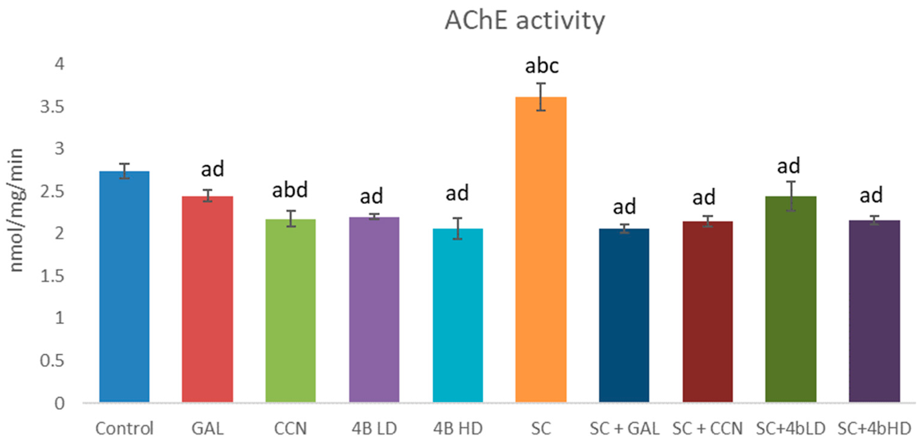
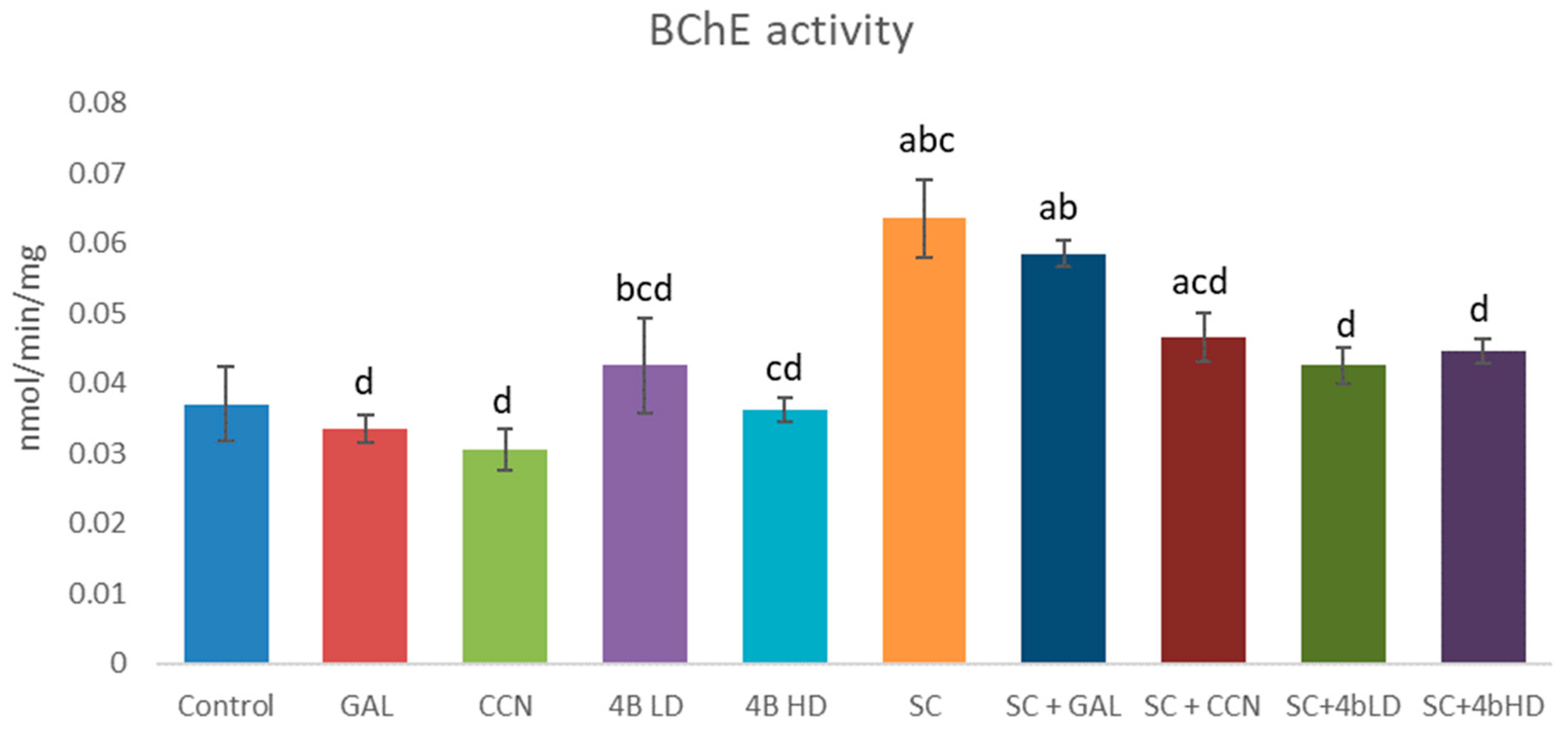
2.2. Assessment of Cholinesterase Inhibition

In the present study, the effects of GAL, CCN and 4b alone and in combination with SC on AChE (Figure 2) and BChE (Figure 3) activity were also evaluated ex vivo. GAL, CCN and both doses of 4b administered alone decreased AChE activity in a statistically significant manner by 11%, 21%, 20% and 25%, respectively. GAL and CCN slightly decreased the BChE activity while 4b LD slightly increased it. Almost no change was observed on BChE activity treated by 4b HD.
Scopolamine administered alone significantly increased both AChE and BChE activities by 32% and 70%, respectively. GAL, CCN and both doses of 4b administered with SC reduced AChE activity by 43%, 41%, 32% and 40%, respectively, compared with the SC group (Figure 2). Regarding BChE inhibition (Figure 3), GAL did not statistically significantly change this enzyme activity, while CCN and both doses of 4b reduced it by 27%, 33% and 30%, respectively.

3. Discussion

Oxidative stress is an important pathological mechanism affecting the pathogenesis of many modern socially significant diseases, including neurodegenerative ones. The brain, with its high oxygen consumption and lipid-rich content, is highly susceptible to oxidative stress. There is no doubt that the imbalance between oxidative stress and the antioxidant capacity of the human body plays a major role in the pathogenesis of AD. The search for new molecules possessing both antioxidant and cholinesterase inhibitory effects would provide great opportunities to therapeutically influence this debilitating disease.
In the present study, we evaluated the antioxidant and cholinesterase inhibitory potential of a novel GAL-CCN hybrid 4b on SC-induced neurotoxicity, a model of Alzheimer’s dementia. CCN increased GSH levels compared with controls and GAL. These results were expected since CCN is known for its antioxidant and anti-inflammatory effects. It increases the synthesis of GSH, the main endogenous cellular protector and antioxidant [16]. CCN enters the structure of the new hybrid 4b, which is probably the reason why the novel compound in both administered doses increased the GSH level. These results confirm the data from our previous studies where 4b, especially at a higher dose, also increased GSH level [11].
Both doses of 4b showed significantly higher SOD activity compared with GAL and CCN. The antioxidant enzymes in the brain are involved in the neutralization and resistance to the toxic effects of ROS. Neurons are highly sensitive to the increased mitochondrial superoxide anion (O2•−) [17]. The first line of defense is the SOD enzyme family, which converts (O2•−) to stable and easily diffusible hydrogen peroxide (H2O2) that is neutralized to water by CAT or GPx [18]. It is possible that the hybrid 4b can specifically activate this enzyme. This fact should be verified in future experiments.
Many studies have reported that oxidative stress is one of the mechanisms with a key role in SC-induced brain damage. SC decreases the activity of SOD, CAT, glutathione S-transferase (GST) and GPx and increases the level of MDA [6]. Several antioxidant compounds, including CCN, could improve SC-induced memory impairment by attenuating oxidative stress markers [6,8], indicating a crucial role of oxidative stress in SC-induced amnesia.
The simultaneous administration of SC with GAL, CCN and 4b improved the brain oxidative status. GAL is an AChE inhibitor and has some antioxidant properties acting as a ROS acceptor [19]. We found similar effects for GAL in our previous studies [11,20], and some other research teams have also found this [21,22].
ROS are potent tissue-damaging molecules in almost all diseases, including AD [23]. The main antioxidant mechanisms include both increased antioxidant enzyme activity and nonenzymatic mechanisms such as increased GSH levels. CAT, SOD and GPx are the main antioxidant enzymes of the brain’s defense system, by which the free radicals produced during the pathological progression of AD are removed. So, the intake of natural or synthetic antioxidants may be beneficial in slowing the progression of AD. CCN has been shown to be an antioxidant in a variety of neurodegenerative diseases, including AD [24,25]. It reduces the serum MDA concentration and has the potential to increase the total antioxidant potential (TAC). The action of CCN on markers of oxidative stress is related to its activity in scavenging radicals, chelating metals and regulating antioxidant enzymes [26]. The presence of CCN fragments in the structure of 4b determined its significant antioxidant effect at both administered doses.
In the present study, GAL, CCN and 4b administered alone reduced the AChE activity. Their effect on BChE was diverse: GAL and CCN slightly decreased it, 4b LD slightly increased it and 4b HD had no effect. SC increased both AChE and BChE activities as it was observed by others [8,27].
Regarding the BChE inhibition, the results showed that GAL did not significantly change this enzyme activity compared with the SC group, while CCN and both doses of 4b substantially reduced the enzyme activity. In our previous study [11], using PAMPA, we found that 4b had better blood–brain barrier (BBB) permeability compared with CCN and GAL, and thus provided a better brain bioavailability. Cholinesterase inhibition has strong clinical evidence in slowing the progression of AD. Several drugs selectively inhibit AChE, but molecules that can also inhibit BChE may provide additional benefits. As AD progresses, brain ACh concentrations may become increasingly dependent on BChE [5]. In this sense, dual enzyme inhibitors may provide a more significant efficacy than AChE-selective agents.
The increased levels of ACh in the brain are associated with the onset and development of depression [28]. Biochemically, depression is caused by a disbalance between adrenergic and cholinergic functions in favor of cholinergic dominance [29]. There is a hypothesis that the depression observed in patients with AD treated with AChE inhibitors is a side effect of the therapy and not a comorbidity [28]. Although there is still no strong evidence, there are many indications to support this hypothesis [30]. In this sense, the higher anticholinesterase effect implies a higher risk of triggering depression. We have not yet examined the prodepressant effects of 4b, if any. The LD50 of 4b in an acute toxicity test on mice is 49 mg/kg orally [11]. For comparison, the oral LD50s of GAL and CCN are 30 mg/kg [31] and more than 2000 mg/kg [32], respectively. The presence of the CCN fragment in the molecule of 4b obviously decreases its toxicity. In the present study, the doses used in the in vivo experiments were 1/10 of LD50 for GAL, CCN and 4b HD. The dose for 4b LD was 1/20 of LD50. Even more, it was found that CCN might improve depressive and anxiety symptoms in people with depression [33]. Considering the low toxicity of 4b, the presence of the CCN fragment in the molecule and the low administered doses, we do not expect 4b to have a prodepressant effect, but this remains to be proven in further studies.

4. Materials and Methods
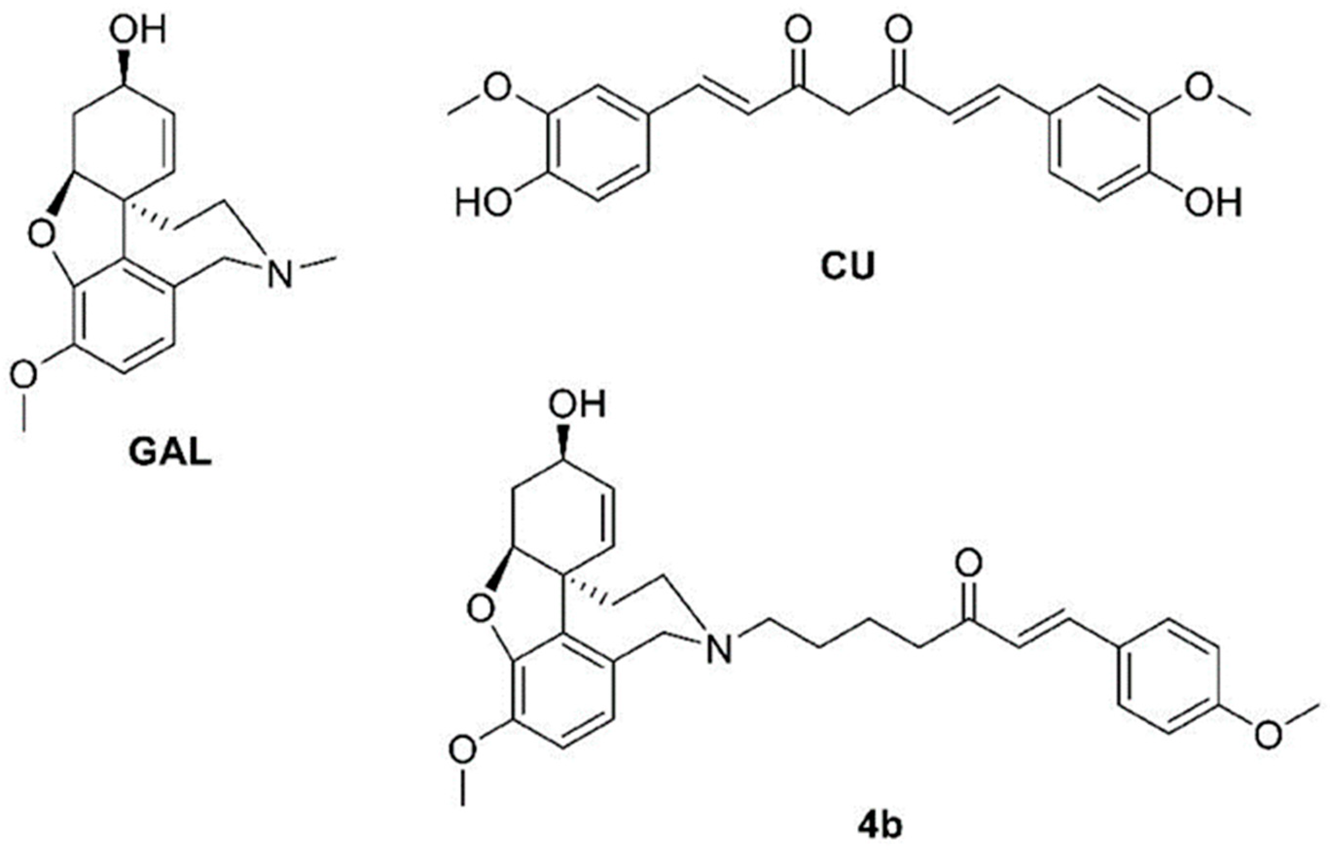
4.1. Materials

Acetylthiocholine iodide (Sigma Aldrich, Taufkirchen, Germany, Mw = 289.18, purity > 98%), beta–Nicotinamide adenine dinucleotide 2′-phosphate reduced tetrasodium salt (NADPH), Bovine serum albumin (Sigma Aldrich, Taufkirchen, Germany, Mw ≈ 66 kD, purity > 96%), Butyrylthiocholine iodide (Thermo Fisher, Kandel, Germany, Mw = 317.23, purity > 98%), Cumene hydroperoxide (Sigma Aldrich, Taufkirchen, Germany, Mw = 152.2, purity > 88%), 1-chloro-2 4-dinitrobenzene (CDNB) (Sigma Aldrich, Taufkirchen, Germany, Mw = 202.56, purity > 97%), Curcumin (BioXtract, Les Isnes, Belgium, Mw = 368.4 g/mol, purity > 98%), Dinitro-5,5 dithiodibenzoic acid (DTNB) (Sigma Aldrich, Taufkirchen, Germany, Mw = 396.35, purity > 98%), Epinephrine (Sigma Aldrich, Taufkirchen, Germany, Mw = 183.20, purity > 99%), Ethylenediaminetetraacetic acid (EDTA) (Sigma Aldrich, Taufkirchen, Germany, Mw = 292.24, purity > 99%), Galantamine HBr (Galen-N Ltd., Sofia, Bulgaria, Mw = 368.3 g/mol, purity > 98%), Glutathione oxidized (GSSG) (Sigma Aldrich, Taufkirchen, Germany, Mw = 612.63, purity > 98%), Glutathione reduced (GSH) (Sigma Aldrich, Taufkirchen, Germany, Mw = 307.32, purity > 98%), Glutathione reductase (GR) from baker’s yeast (S. cerevisiae) (Sigma Aldrich, Taufkirchen, Germany), Hydrogen peroxide sol. 30% (Sigma Aldrich, Taufkirchen, Germany, Mw = 34.01), Scopolamine HBr 3H2O (Sigma Aldrich, Taufkirchen, Germany, Mw = 438.31, purity > 98%), Thiobarbituric acid (Sigma Aldrich, Taufkirchen, Germany, Mw = 144.15 g/mol, purity > 98%), Trichloroacetic acid (Sigma Aldrich, Taufkirchen, Germany, Mw = 163.39 g/mol, purity > 99%) were used. The synthesis of 4b was described elsewhere [8,9].

4.2. Animals

Forty specific pathogen-free male ICR mice (6 weeks old, 25–35 g) were used. The animals were purchased from the National Breeding Center, Sofia, Bulgaria. A minimum of 7 days of acclimatization was allowed before the start of the study. Age-appropriate standard complete commercial pelleted mouse feed and fresh drinking water were available ad libitum during the experimental period of 14 days. The animals were housed in Plexiglas cages (4 per cage) in a 12/12 light/dark cycle under standard laboratory conditions (ambient temperature 20 ± 2 °C and humidity 72 ± 4%).
Mice were housed, maintained and euthanized in accordance with relevant international rules and recommendations as outlined in the European Convention for the Protection of Vertebrate Animals Used for Experimental and Other Scientific Purposes (ETS 123).

4.3. Design of the Experiment

Alzheimer’s type dementia was induced by an ip injection of SC 3 mg/kg to male mice for a period of 14 days. To assess the effect of 4b on SC-induced changes on brain biochemical parameters, 40 male mice were divided into ten groups with four mice in each group (n = 4) [11,20].
  • Group 1—control mice, treated with 0.9 % saline po;
  • Group 2—animals treated with GAL alone as a positive control (3 mg/kg po);
  • Group 3—animals treated with CCN alone as a positive control (25 mg/kg po);
  • Group 4—animals treated with 4b alone (2.5 mg/kg po), (low dose, LD);
  • Group 5—animals treated with 4b alone (5 mg/kg po) (high dose, HD);
  • Group 6—animals treated with SC alone (3 mg/kg ip);
  • Group 7—animals treated with GAL (3 mg/kg po) and SC (3 mg/kg ip 30 min after GAL medication);
  • Group 8—animals treated with CCN (25 mg/kg po) and SC (3 mg/kg ip 30 min after CCN medication);
  • Group 9—animals treated with 4b (2.5 mg/kg po) and SC (3 mg/kg ip 30 min after 4b medication);
  • Group 10—animals treated with 4b (5 mg/kg po) and SC (3 mg/kg ip 30 min after 4b medication).
All treatments were performed over 14 days. During this period, animals were observed daily for behavioral changes and signs of toxicity. On day 14, after the last treatment, the animals were euthanized in strict accordance with the rule of the Animal Ethics Committee and the adopted Directive 2010/63/EU (Directive 2010/63/EU of the European Parliament and of the Council of 22 September 2010 on the protection of animals used for scientific purposes) [34].
Brains were harvested, dried, weighed and homogenized with appropriate buffers. The protein content of brain homogenates was measured using the Lowry method [35]. The brain homogenates were used to assess cholinesterase activity (AChE and BChE) and the oxidative stress biomarkers catalase (CAT), superoxide dismutase (SOD), glutathione peroxidase (GPx), malondialdehyde (MDA) and reduced glutathione (GSH).

4.4. Methods

4.4.1. Measurement of AChE and BChE Inhibition in Brain Homogenate

Brains were homogenized with a 0.1 M phosphate buffer, pH 7.4. Aliquots of the brain homogenates from different groups were used to measure AChE activity for 10 min via the Ellman method [36]. AChE activity was calculated and expressed as nmol/min/mg protein using a molar absorbance coefficient of 13,600 M−1 cm−1. This method is based on the formation of a yellow anion, 5,5′dithio-bis-nitrobenzoic acid, measured by absorbance at 412 nm. The reaction was initiated by adding acetylthiocholine iodide as a substrate. A measurement of BChE inhibition was performed in the same manner with butyrylthiocholine iodide used as a substrate [8]. AChE and BChE activities were calculated and expressed as nmol/min/mg protein using the molar absorption coefficient of 13,600 M−1 cm−1.

4.4.2. Antioxidant Enzyme Activity Measurement

Measured amounts of brains were rinsed in ice-cold saline and minced with scissors. Furthermore, 10% homogenates were prepared in a 0.05 M phosphate buffer (pH = 7.4) and were centrifuged at 7000× g, and the supernatant was used for the antioxidant enzyme assay.
  • Catalase Activity (CAT)
Catalase activity was assessed using the method of Aebi et al. [37]. The CAT activity was determined by monitoring the decomposition of H2O2, which was measured spectrophotometrically by the decrease in absorbance at 240 nm. Enzyme activity was calculated using a molar extinction coefficient of 0.043 mM−1 cm−1 and expressed as nM/minute/mg protein.
  • Superoxide Dismutase Activity (SOD).
Superoxide dismutase activity was measured according to the method of Misra and Fridovich [38] following the spectrophotometric autoxidation of epinephrine at pH = 10.4, 30 °C, using the molar extinction coefficient of 4.02 mM−1 cm−1. The reaction was initiated by the addition of epinephrine. SOD activity was expressed as nmol of epinephrine prevented from autoxidation after the addition of the sample.
  • Glutathione Peroxidase Activity (GPx).
Glutathione Peroxidase activity was measured with NADPH oxidation using a coupled reaction system consisting of glutathione, glutathione reductase and cumene hydroperoxide [39]. The reaction was initiated by adding 50 𝜇L cumene hydroperoxide (1 mg/mL) and the rate of disappearance of NADPH with time was determined by monitoring the absorbance at 340 nm. The results are expressed as nmol/min/mg protein using the molar extinction coefficient of 6.22 mM−1 cm−1.

4.4.3. Measurement of Malondialdehyde (MDA) Levels in Brain Homogenate

The brains were homogenized with a 0.1 M phosphate buffer and EDTA, pH 7.4. Aliquots of the homogenates were heated for 20 min on a water bath (100 °C) with thiobarbituric acid. The amount of thiobarbituric-acid-formed reactive species (TBARS) (expressed as MDA equivalents) was measured spectrophotometrically using the method of Deby and Goutier [40] at a wavelength of 535 nm. The concentration of MDA was calculated using a molar absorption coefficient of 1.56 × 10⁵ M−1 cm−1 and is expressed in nmol/g tissue.

4.4.4. Measurement of Reduced Glutathione (GSH) Levels in Brain Homogenate

Reduced Glutathione was evaluated by measuring nonprotein sulfhydryls after trichloroacetic acid (TCA) protein precipitation using the method described by Bump et al. [41]. The brains were homogenized in 5% TCA (1:10) and centrifuged for 20 min at 4000× g. The reaction mixture contained 0.05 mL of supernatant, 3 mL of 0.05 M phosphate buffer (pH = 8) and 0.02 mL of DTNB reagent. Absorption was determined at a wavelength of 412 nm and the results are expressed as nmol/g tissue.

4.4.5. Statistical Analysis

The MEDCALC statistical program (MedCalc Software bvba, Belgium, v. 12.3) was used to analyze the data. The results are expressed as mean ± SD for four mice in each group. The significance of the data was assessed with a nonparametric Mann–Whitney U test. Values of p < 0.05 were considered statistically significant.

5. Conclusions

The results in the present study expand our knowledge about the multitarget action of the hybrid 4b. It was found that this compound completely protects and eliminates the toxic effects of SC administered in vivo. Moreover, this effect was observed for both AChE and BChE. The protection of 4b against BChE is a novelty, the mechanism of which remains to be elucidated, because apparently it is not due to a direct interaction with the enzyme. The antioxidant activity of the novel compound was recorded here as a reactivation of the antioxidant enzymes CAT, SOD and GPx, injured by SC administration. In this study again, 4b outperformed its parents GAL and CCN. Combining fragments from GAL and CCN provides not just a sum of their activities but adds a new surplus value to the novel hybrid, making it promising for further experimental studies in the field of neurodegenerative processes.

Author Contributions

Conceptualization, R.S.; methodology, R.S.; software, M.A.; investigation, R.S.; resources, G.S. and I.P.; writing—original draft preparation, R.S.; writing—review and editing, I.D., M.A., G.S. and I.P.; visualization, R.S.; supervision, I.D.; project administration, I.D.; funding acquisition, I.D. All authors have read and agreed to the published version of the manuscript.

Funding

This research was funded by the Science and Education for Smart Growth Operational Program and was cofinanced by the European Union through the European Structural and Investment Funds, grant number BG05M2OP001-1.001-0003. The APC was funded by the Bulgarian Ministry of Education and Science, project National Roadmap for Research Infrastructure (2017–2023), Grant No. D01-271/2019.

Institutional Review Board Statement

The animal study protocol was approved by the Animal Care Ethic Committee of the Bulgarian Agency for Food Safety (protocol code No. 208 from 5 October 2018).

Informed Consent Statement

Not applicable.

Data Availability Statement

Not applicable.

Conflicts of Interest

The authors declare no conflict of interest. The funders had no role in the design of the study; in the collection, analyses or interpretation of data; in the writing of the manuscript; or in the decision to publish the results.

References

  1. Dobson, C.M. Alzheimer’s disease: Addressing a twenty-first century plague. Rend. Fis. Acc. Lincei 2015, 26, 251–262. [Google Scholar] [CrossRef] [Green Version]
  2. Alzheimer’s Disease. Mayo Clinic Health Information. 2022. Available online: https://www.mayoclinic.org/diseases-conditions/alzheimers-disease/symptoms-causes/ (accessed on 20 October 2020).
  3. Swomley, A.M.; Butterfield, D.A. Oxidative stress in Alzheimer’s disease and mild cognitive impairment: Evidence from human data provided by redox proteomics. Arch. Toxicol. 2015, 89, 1669–1680. [Google Scholar] [CrossRef] [PubMed]
  4. Poprac, P.; Jomova, K.; Simunkova, M.; Kolar, V.; Rhodes, C.J.; Valko, M. Free radical targeting in human oxidative stress-related diseases. Trends Pharmacol. Sci. 2017, 38, 592–607. [Google Scholar] [CrossRef]
  5. Ballard, C.G. Advances in the treatment of Alzheimer’s disease: Benefits of dual cholinesterase inhibition. Eur. Neurol. 2002, 47, 64–70. [Google Scholar] [CrossRef] [PubMed]
  6. Foyet, H.S.; Keugong, W.E.; Ngatanko, A.H.H.; Assongalem, E.A.; Eyong, G.O.O.D. AChE and antioxidant potential of hydromethanolic leaf extract of Ziziphus mucronata (Rhamnaceae) on scopolamine-induced memory and cognitive dysfunctions in mice. Evid.-Based Suppl. Altern. Med. 2019, 2019, 4568401. [Google Scholar]
  7. Boiangiu, R.S.; Brinza, I.; Hancianu, M.; Erdogan Orhan, I.; Eren, G.; Gunduz, E.; Ertas, H.; Hritcu, L.; Cioanca, O. Cognitive facilitation and antioxidant effects of an essential oil mixture on scopolamine-induced amnesia in rats: Molecular modeling of in vitro and in vivo approaches. Molecules 2020, 25, 1519. [Google Scholar] [CrossRef] [Green Version]
  8. Akinyemi, A.J.; Oboh, G.; Oyeleye, S.I.; Ogunsuyi, O. Antiamnestic effect of curcumin in combination with donepezil, an anticholinester drug: Involvement of the cholinergic system. Neurotox Res. 2017, 31, 560–569. [Google Scholar] [CrossRef]
  9. Rahimzadegan, M.; Soodi, M. Comparison of memory impairment and oxidative stress after single or multiple doses of scopolamine in the rat hippocampus. Basic Clin. Neurosci. 2018, 9, 5–14. [Google Scholar] [CrossRef] [Green Version]
  10. Stavrakov, G.; Philipova, I.; Lukarski, A.; Atanasova, M.; Zheleva, D.; Zhivkova, Z.D.; Ivanov, S.; Atanasova, T.; Konstantinov, S.; Doytchinova, I. Galantamine-curcumin hybrids as dual-site binding acetylcholinesterase inhibitors. Molecules 2020, 25, 3341. [Google Scholar] [CrossRef]
  11. Simeonova, R.; Zheleva, D.; Valkova, I.; Stavrakov, G.; Philipova, I.; Atanasova, M.; Doytchinova, I. A novel galantamine-curcumin hybrid as a potential multi-target agent against neurodegenerative disorders. Molecules 2021, 26, 1865. [Google Scholar] [CrossRef]
  12. Lubrano, V.; Balzan, S. Enzymatic antioxidant system in vascular inflammation and coronary artery disease. World J. Exp. Med. 2015, 5, 218–224. [Google Scholar] [CrossRef]
  13. Ighodaroab, O.M.; Akinloye, O.A. First line defence antioxidants-superoxide dismutase (SOD), catalase (CAT) and glutathione peroxidase (GPX): Their fundamental role in the entire antioxidant defense grid. Alex. J. Med. 2018, 54, 287–293. [Google Scholar]
  14. Ayala, A.; Muñoz, M.F.; Argüelles, S. Lipid peroxidation: Production, metabolism, and signaling mechanisms of malondialdehyde and 4-hydroxy-2-nonenal. Oxid Med. Cell Longev. 2014, 2014, 360438. [Google Scholar] [CrossRef] [PubMed] [Green Version]
  15. Forman, H.J.; Zhang, H.; Rinna, A. Glutathione: Overview of its protective roles, measurement, and biosynthesis. Mol. Asp. Med. 2009, 30, 1–12. [Google Scholar] [CrossRef] [Green Version]
  16. Zheng, S.; Yumei, F.; Chen, A. De novo glutathione synthesis is a prerequisite for curcumin to inhibit hepatic stellate cell (HSC) activation. Free Radic. Biol. Med. 2007, 43, 444–453. [Google Scholar] [CrossRef] [Green Version]
  17. Izuo, N.; Nojiri, H.; Uchiyama, S.; Noda, Y.; Kawakami, S.; Kojima, S.; Sasaki, T.; Shirasawa, T.; Shimizu, T. Brain-specific superoxide dismutase 2 deficiency causes perinatal death with spongiform encephalopathy in mice. Oxid Med. Cell Longev. 2015, 2015, 238914. [Google Scholar] [CrossRef] [PubMed] [Green Version]
  18. Wang, Y.; Branicky, R.; Noë, A.; Hekimi, S. Superoxide dismutases: Dual role in controlling ROS damage and regulating ROS signaling. J. Cell Biol. 2018, 217, 1915–1928. [Google Scholar] [CrossRef] [PubMed] [Green Version]
  19. Traikova, M.; Traikov, T.; Hadzhimitova, V.; Krikoryan, K.; Boyadzhieva, N. Antioxidant properties of galantamine hydrobromide. Z. Naturforsch. C 2003, 58, 361–365. [Google Scholar] [CrossRef]
  20. Simeonova, R.; Vitcheva, V.; Kostadinova, I.; Valkova, I.; Philipova, I.; Stavrakov, G.; Danchev, N.; Doytchinova, I. Biochemical studies on a novel potent acetylcholinesterase inhibitor with dual-site binding for treatment of Alzheimer’s disease. Comptes Rendus Acad. Bulg. Sci. 2021, 74, 219–225. [Google Scholar]
  21. Triana-Vidal, L.E.; Carvajal-Varona, S.M. Protective effect of galantamine against oxidative damage using human lymphocytes: A novel in vitro model. Arch. Med. Res. 2013, 44, 85–92. [Google Scholar] [CrossRef]
  22. Tsvetkova, D.; Obreshkova, D.; Zheleva-Dimitrova, D.; Saso, L. Antioxidant activity of galantamine and some of its derivatives. Curr. Med. Chem. 2013, 20, 4595–4608. [Google Scholar] [CrossRef] [PubMed]
  23. Huang, W.J.; Zhang, X.; Chen, W.W. The role of oxidative stress in Alzheimer’s disease. Biomed. Rep. 2016, 4, 519–522. [Google Scholar] [CrossRef] [PubMed] [Green Version]
  24. Ashok, A.; Andrabi, S.S.; Mansoor, S.; Kuang, Y.; Kwon, B.K.; Labhasetwar, V. Antioxidant therapy in oxidative stress-induced neurodegenerative diseases: Role of nanoparticle-based drug delivery systems in clinical translation. Antioxidants 2022, 11, 408. [Google Scholar] [CrossRef] [PubMed]
  25. Doghri, R.; Ellefi, A.; Degrach, I.; Srairi-Abid, N.; Gati, A. Curcumin attenuated neurotoxicity in sporadic animal model of Alzheimer’s disease. Molecules 2021, 26, 3011. [Google Scholar]
  26. Jakubczyk, K.; Drużga, A.; Katarzyna, J.; Skonieczna-Żydecka, K. Antioxidant potential of curcumin—A meta-analysis of randomized clinical trials. Antioxidants 2020, 9, 1092. [Google Scholar] [CrossRef]
  27. Adedayo, B.C.; Jesubowale, O.S.; Adebayo, A.A.; Oboh, G. Effect of Andrographis paniculata leaves extract on neurobehavioral and biochemical indices in scopolamine-induced amnesic rats. J. Food Biochem. 2021, 45, e13280. [Google Scholar] [CrossRef]
  28. Ownby, R.L.; Crocco, E.; Acevedo, A.; Vanella, A.; Concas, I.; Tinacci, S.; Petralia, A.; Signorelli, M.S.; Aguglia, E. Depression and risk for Alzheimer disease: Systematic review, meta-analysis, and metaregression analysis. Arch. Gen. Psychiatry 2006, 63, 530–538. [Google Scholar] [CrossRef] [Green Version]
  29. Janowsky, D.S.; el-Yousef, M.K.; Davis, J.M. Acetylcholine and depression. Psychosom. Med. 1974, 36, 248–257. [Google Scholar] [CrossRef]
  30. Mitić, M.; Lazarević-Pašti, T. Does the application of acetylcholinesterase inhibitors in the treatment of Alzheimer’s disease lead to depression? Expert Opin. Drug Metab. Toxicol. 2021, 17, 841–856. [Google Scholar] [CrossRef]
  31. Auro-Galantamine. Product Monograph; AuroPharma Inc.: Woodbridge, ON, Canada, 2017. [Google Scholar]
  32. ChemlDpus Datadase, US National Library of Medicine. Available online: https://chem.nlm.nih.gov/chemidplus/ (accessed on 20 October 2020).
  33. Fusar-Poli, L.; Vozza, L.; Gabbiadini, A.; Vanella, A.; Concas, I.; Tinacci, S.; Petralia, A.; Signorelli, M.S.; Aguglia, E. Curcumin for depression: A meta-analysis. Crit. Rev. Food Sci. Nutr. 2020, 60, 2643–2653. [Google Scholar] [CrossRef]
  34. Directive 2010/63/EU of the European Parliament and of the Council on the protection of animals used for scientific purposes. OJEU 2010, L 276, 33–79.
  35. Lowry, O.H.; Rosebrough, N.J.; Farr, A.L.; Randall, R.J. Protein measurement with the Folin phenol reagent. J. Biol. Chem. 1951, 193, 265–275. [Google Scholar] [CrossRef] [PubMed]
  36. Ellman, G.L.; Courtney, K.D.; Andre, J.V.; Featherstone, R.M. A new and rapid colorimetric determination of acetylcholinesterase activity. Biochem. Pharmacol. 1961, 7, 88–95. [Google Scholar] [CrossRef] [PubMed]
  37. Aebi, H. Catalase. In Methods of Enzymatic Analysis; Bergmeyer, H.U., Ed.; Verlag Chemie: Weinheim, Germany, 1974; pp. 673–684. [Google Scholar]
  38. Misra, H.P.; Fridovich, I. The role of superoxide anion in the autoxidation of epinephrine and a simple assay for superoxide dismutase. J. Biol. Chem. 1972, 247, 3170–3175. [Google Scholar] [CrossRef] [PubMed]
  39. Tappel, A.L. Glutathione peroxidase and hydroperoxides. Methods Enzymol. 1978, 52, 506–513. [Google Scholar] [PubMed]
  40. Deby, C.; Goutier, R. New perspectives on the biochemistry of superoxide anion and the efficiency of superoxide dismutases. Biochem. Pharmacol. 1990, 39, 399–405. [Google Scholar] [CrossRef]
  41. Bump, E.A.; Taylor, Y.C.; Brown, J.M. Role of glutathione in the hypoxic cell cytotoxicity of misonidazole. Cancer Res. 1983, 43, 997–1002. [Google Scholar]
Figure 1. Structures of galantamine (GAL), curcumin (CCN) and their hybrid 4b.
Ijms 23 14843 g001
Figure 2. AChE activity measured over 10 min in brain homogenates from mice treated for 14 days with GAL, CCN and 4b (two doses) alone and in combination with SC. a p < 0.05 vs. control; b p < 0.05 vs. GAL; c p < 0.05 vs. CCN; d p < 0.05 vs. SC.
Ijms 23 14843 g002
Figure 3. BChE activity measured for 10 min in brain homogenates from mice treated 14 days with GAL, CCN and 4b (two doses) alone and in combination with SC. a p < 0.05 vs. control; b p < 0.05 vs. GAL; c p < 0.05 vs. CCN; d p < 0.05 vs. SC.
Ijms 23 14843 g003
Table 1. Oxidative stress biomarkers for GAL, CCN and 4b (two doses) alone and in combination with SC. Results are expressed as mean ± SD (n = 4). The significance of the data was assessed using the nonparametric Mann–Whitney U test. Values of p ≤ 0.05 were considered statistically significant.
ParametersControlGALCCN4b LD4b HDSCSC + GALSC + CCNSC + 4b LDSC + 4b HD
CAT nmol/mg/min8.50 ± 0.417.70 ± 0.42 d9.35 ± 0.68 d7.80 ± 0.32 cd7.10 ± 0.70 cd5.35 ± 0.52 abc7.20 ± 0.43 d8.55 ± 0.34 d8.15 ± 0.28 d8.40 ± 0.36 d
SOD nmol/mg/min0.35 ± 0.050.31 ± 0.04 d0.34 ± 0.01 d0.44 ± 0.03 bcd0.41 ± 0.03 bcd0.22 ± 0.03 abc0.31 ± 0.02 d0.34 ± 0.01 d0.33 ± 0.01 d0.34 ± 0.02 d
GPx nmol/mg/min0.43 ± 0.010.39 ± 0.03 d0.40 ± 0.04 d0.44 ± 0.03 d0.46 ± 0.02 d0.21 ± 0.02 abc0.34 ± 0.03 ad0.39 ± 0.05 d0.41 ± 0.03 d0.44 ± 0.02 d
GSH nmol/g tissue1.59 ± 0.031.73 ± 0.07 d2.04 ± 0.12 abd2.06 ± 0.07 abd2.22 ± 0.08 abd1.09 ± 0.13 abc1.66 ± 0.121.83 ± 0.03 cd1.44 ± 0.08 d1.59 ± 0.04 d
MDA nmol/g tissue1.41 ± 0.271.75 ± 0.13 d1.45 ± 0.09 d1.62 ± 0.30 d1.76 ± 0.12 d2.39 ± 0.1 abc1.79 ± 0.08 d1.64 ± 0.05 d1.76 ± 0.03 d1.65 ± 0.16 d
Abbreviations: GAL, galantamine; CCN, curcumin; 4b LD, 4b low dose (2.5 mg/kg); 4b HD, 4b high dose (5 mg/kg); SC, scopolamine; a p < 0.05 vs. control; b.p < 0.05 vs. GAL; c p < 0.05 vs. CCN; d p < 0.05 vs. SC.
Publisher’s Note: MDPI stays neutral with regard to jurisdictional claims in published maps and institutional affiliations.

