Review

Developmental Formation of the GABAergic and Glycinergic Networks in the Mouse Spinal Cord

1
Department of Molecular Anatomy, Graduate School of Medicine, University of the Ryukyus, 207 Uehara, Nishihara 903-0215, Japan
2
Department of Anatomy, Kosin University College of Medicine, Busan 49267, Korea
3
Department of Nutritional Science, Faculty of Health and Welfare, Seinan Jo Gakuin University, Fukuoka 803-0835, Japan
*
Author to whom correspondence should be addressed.
Int. J. Mol. Sci. 2022, 23(2), 834; https://doi.org/10.3390/ijms23020834
Submission received: 28 December 2021 / Revised: 7 January 2022 / Accepted: 11 January 2022 / Published: 13 January 2022
(This article belongs to the Special Issue GABAergic and Glycinergic Neurons)

Abstract

:
Gamma-aminobutyric acid (GABA) and glycine act as inhibitory neurotransmitters. Three types of inhibitory neurons and terminals, GABAergic, GABA/glycine coreleasing, and glycinergic, are orchestrated in the spinal cord neural circuits and play critical roles in regulating pain, locomotive movement, and respiratory rhythms. In this study, we first describe GABAergic and glycinergic transmission and inhibitory networks, consisting of three types of terminals in the mature mouse spinal cord. Second, we describe the developmental formation of GABAergic and glycinergic networks, with a specific focus on the differentiation of neurons, formation of synapses, maturation of removal systems, and changes in their action. GABAergic and glycinergic neurons are derived from the same domains of the ventricular zone. Initially, GABAergic neurons are differentiated, and their axons form synapses. Some of these neurons remain GABAergic in lamina I and II. Many GABAergic neurons convert to a coreleasing state. The coreleasing neurons and terminals remain in the dorsal horn, whereas many ultimately become glycinergic in the ventral horn. During the development of terminals and the transformation from radial glia to astrocytes, GABA and glycine receptor subunit compositions markedly change, removal systems mature, and GABAergic and glycinergic action shifts from excitatory to inhibitory.

1. Introduction

In the mature central nervous system (CNS), which includes the spinal cord, γ-aminobutyric acid (GABA), and glycine, are inhibitory neurotransmitters that negatively regulate neuronal activity [1,2,3,4]. In the spinal cord, there are three types of inhibitory neurons and terminals: GABAergic, GABA/glycine coreleasing, and glycinergic [5,6,7]. These neurons and terminals are arranged in the spinal cord neural circuit and are involved in many vital roles, such as regulating somatic sense, locomotive movement, and respiratory rhythms [8,9,10]. In the first part of this review, we will focus on the three types of neurons and terminals and describe the inhibitory networks in the mature spinal cord from the following viewpoints: (1) distribution of neurons and terminals, (2) receptor composition, (3) removal system, and (4) mechanisms underlying inhibitory transmission. In the latter half of the review, we will focus on morphological development and demonstrate the processes of how mature networks are established through the following neuronal differentiation processes: GABAergic and glycinergic neurons are born in the ventricular zone, migrate in the gray matter, extend their dendrites and axons, and form synapses. During these processes, the neuronal types alter the composition of the receptor subunits changes, the removal system matures, and the action of both neurotransmitters shifts from excitatory to inhibitory.

2. GABAergic and Glycinergic Network in the Mature Spinal Cord

GABAergic and glycinergic synapses are schematically illustrated in Figure 1A,B, respectively. GABA and glycine are synthesized within the neurons and transported from the extracellular space, including the synaptic cleft. The neurotransmitters are loaded into synaptic vesicles and released from the axon terminals. After diffusion in the synaptic cleft, they bind to GABA or glycine receptors at the postsynaptic membrane. In the mature spinal cord, activation of GABA or glycine receptors induces hyperpolarization of the membrane potential and negatively regulates neuronal activity. The action of these neurotransmitters is terminated by their removal from the synaptic cleft into presynaptic terminals and astrocytic sheets that surround the synapses [3,11,12,13].

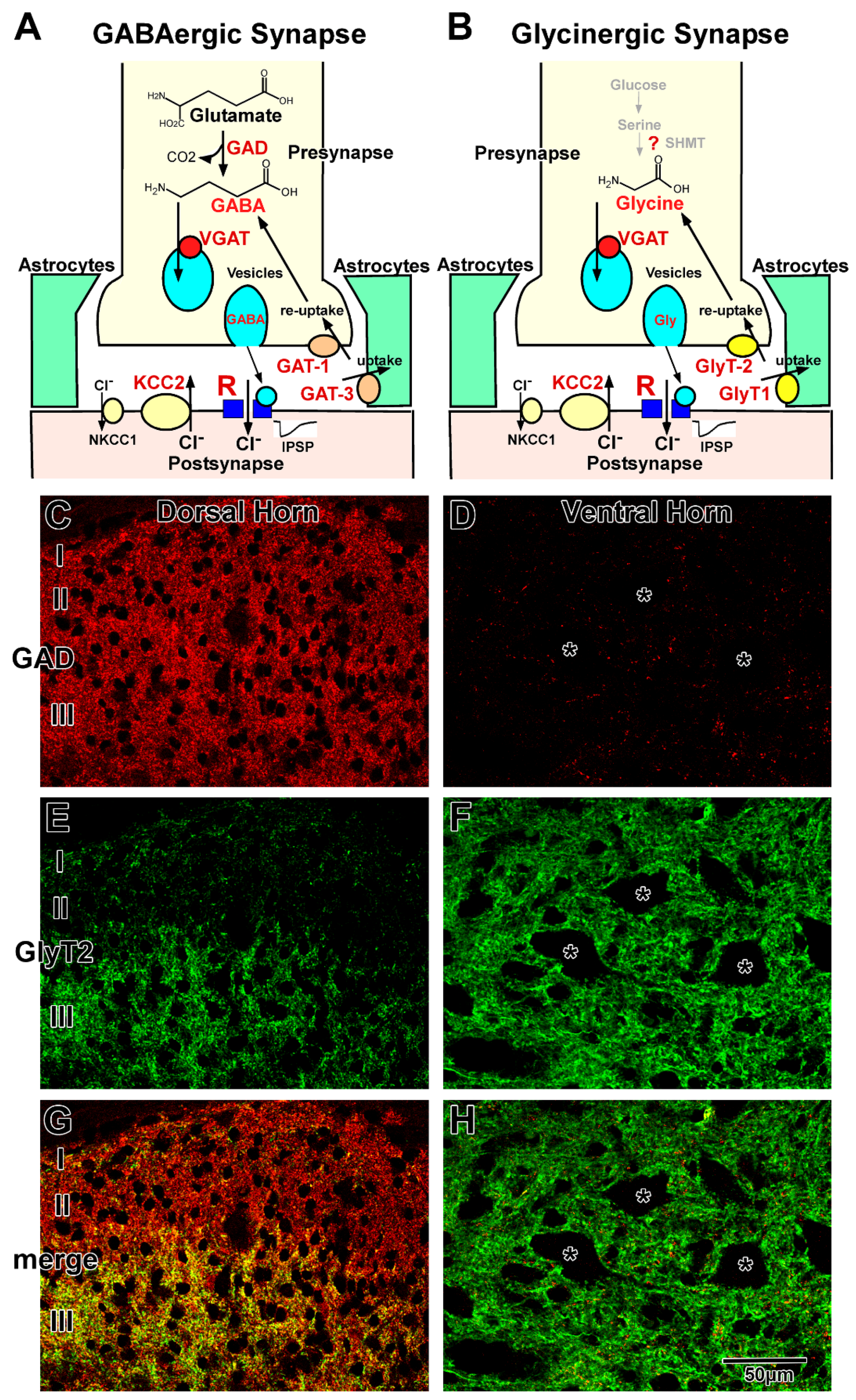
2.1. GABAergic Transmission

GABAergic transmission at the synapse is illustrated in Figure 1A. GABA is synthesized from glutamate by two isoforms of glutamic acid decarboxylase (GAD65 and GAD67) [14]. The primary source of glutamate may be derived from glutamine, which is transported back from astrocytes by glutamine transporter [15]. GABA is loaded into vesicles by vesicular GABA transporter (VGAT), also known as vesicular inhibitory amino acid transporter (VIAAT) [16,17]. GABA is released via the fusion of vesicles with the presynaptic membrane at the nerve terminals and binds to GABA receptors on the postsynaptic membrane.
GABA receptors are classified into three groups based on their pharmacological and biochemical characteristics: GABAA, GABAB, and GABAC. Most of the fast synaptic transmission is mediated by GABAA receptors in the mammalian CNS [1,18]. The GABAA receptor is a member of the ligand-gated ion channel receptor family and is thought to be composed of five heteromeric subunits belonging to seven different subunit families: α1–6, β1–3, γ1–3, δ, ε, π, and θ [1,2,18,19]. Native GABAA receptors contain at least one α, one β, and one γ subunit. The subunit composition varies among the brain regions [20,21,22]. Different subunit compositions exhibit their own characteristic pharmacological and electrophysiological properties [1,2,19,23,24]. The GABAC receptor is also an ion-channel type receptor composed of only single or multiple ρ subunits: ρ1 and ρ2. The GABAC receptor is identified as a bicuculline and baclofen insensitive GABA receptor and is considered a pharmacological variant of GABAA receptors [18,25,26]. The binding of GABA to the GABAA and GABAC receptors induces the influx of chloride ions (Cl) and mediate hyperpolarization of the postsynaptic membrane potential. The GABAB receptor, which consists of two subunits, GABAB1 and GABAB2, is a metabotropic receptor, activates G proteins, negatively regulates the second messenger system, and responds to slow-acting inhibition of channel and receptor functions [27,28,29,30,31,32]. GABAergic transmission is terminated by the reuptake of GABA into the nerve terminals or uptake into the surrounding astrocytes by plasma membrane GABA transporters (GATs) [33].
GATs are high-affinity Na+ and Cl-dependent transporters that cotransport GABA with Na+ and Cl [34,35]. In the CNS, there are three types of GATs: GAT-1, GAT-2, and GAT-3. GAT-2 is localized in leptomeningeal ependymal cells and the choroid plexus [36]. GAT-1 and GAT-3 function at the membranes of axon terminals containing GABAergic vesicles and astrocytic sheets surrounding GABAergic synapses, respectively [37,38,39,40]. In the astrocytes, GABA is degraded into succinate, followed by entering the Krebs cycle [15]. The 2-oxoglutarate in the Krebs cycle is converted to glutamate and glutamine. Astrocytic glutamine is transported back to neurons by a glutamine transporter. This GABA/glutamine cycle may play critical roles in GABA metabolism between neurons and astrocytes [41].

2.2. Glycinergic Transmission

Glycinergic transmission at the synapse is illustrated in Figure 1B. The metabolic pathway of glycine in neurons is still unclear. Although serine hydroxymethyltransferase (SHMT) has been shown to be able to synthesize in the neurons [42,43,44], little is known about the relationship between SHMT and glycine levels [45]. In general, high-affinity uptake systems, mediated by glycine transporter 2 (GlyT2), are considered the principal regulators of intracellular glycine concentrations. The GlyT2 knockout mice die from lack of glycine during the second postnatal week [46,47], which suggests that de novo synthesis by SHMT is not sufficient for glycinergic neurotransmission [4,12,48], and glycine in the neurons may be dominantly transported from extracellular space through blood–brain barrier. Thus, GlyT2 is a reliable marker for glycine-immunoreactive neurons [45]. After being loaded into vesicles via VGAT (VIAAT) [16,17], glycine is released by exocytosis from the nerve terminals and binds to glycine receptors on the postsynaptic membrane. The glycine receptor is a ligand-gated ion channel receptor that consists of five subunits belonging to two subunit families: α1–3 and β in the mammalian CNS [3,49,50]. The α subunit has a strychnine binding site, and the β subunit binds to the scaffolding protein gephyrin. The composition varies among the CNS regions. Different subunit compositions exhibit their own characteristic electrophysiological properties [50,51]. Glycine binding to the receptor induces an influx of Cl (Figure 1A), as observed in GABA binding (Figure 1B). The glycinergic action is terminated by reuptake into the nerve terminals and uptake into the surrounding glia through plasma membrane glycine transporters (GlyTs) [52]. GlyTs are high-affinity Na+ and Cl-dependent transporters that co-transport glycine with Na+ and Cl. There are two types of GlyTs in the CNS: GlyT1 and GlyT2. In the spinal cord, as well as in other brain regions, GlyT1 is localized at the astrocytic sheets that surround glycinergic synapses, and GlyT2 is localized at the membranes of axon terminals that contain glycinergic vesicles [53,54]. In the astrocyte surrounding glycinergic synapses, glycine may be degraded by the glycine cleavage system [55,56].

2.3. GABAergic and Glycinergic Transmission in the Mature Spinal Cord

GABA or GAD immunohistochemistry [57,58,59] and GAD-green fluorescent protein (GFP) labeling [60] demonstrated that the density of GABAergic neurons is high in the dorsal horn and moderate in the middle part of the gray matter central part, whereas GABAergic neurons are scarce or in the ventral horn. The distribution of GABAergic terminals is almost the same as that of GABAergic neurons, with high density observed in the dorsal horn (Figure 1C) and low density observed in the ventral horn (Figure 1D). In contrast, glycine immunohistochemistry [61] and GlyT2 expression analysis [8,10] demonstrated that glycinergic neurons and terminals are homogeneously distributed in the gray matter, except for in the superficial layer of the dorsal horn (Figure 1E,F). In lamina I and II, the density of glycinergic neurons and terminals was lower than that observed in the other laminae (Figure 1E) [61,62]. Furthermore, double staining of GABA/GAD and glycine/GlyT2 demonstrated that colocalizing (functionally coreleasing) neurons and terminals are often detected in the spinal cord (Figure 1G,H) [5,6,7], and electrophysiological studies confirmed that the two neurotransmitters are loaded into the same synaptic vesicles and released simultaneously [63,64]. Coreleasing terminals have been abundantly detected in the deep part of the dorsal horn and middle part of the gray matter. In general, GABAergic neurons and their terminals are dominant in lamina I and II (Figure 1G). Coreleasing neurons and terminals are dominant in lamina III to VI (Figure 1G). Glycinergic neurons and their terminals are dominant in the lamina VII–IX (Figure 1H) [9,53,65,66,67]. Total inhibitory terminals detected as VGAT (VIAAT)-positive dots are homogeneously and ubiquitously distributed in the gray matter [57]. GABA and glycine play distinct roles in motor and sensory functions in the complex network in the dorsal [68,69] and ventral horns [70]. Fast GABAergic transmission is dominantly mediated by GABAA receptors consisting of α3β3γ2 subunits in the dorsal horn and α2(α5)β3γ2 subunits in the motor neurons [21,71]. Glycinergic transmission is mediated by glycine receptors consisting of α1β (two α1 and three β) subunits [51,72]. GABAA and glycine receptors colocalize at the postsynaptic membrane of the coreleasing terminals [73]. GABAB Receptors consisting of B1, 1a and 1b, and B2 mainly play roles in the dorsal horn and motor neuron pool [74,75]. [3H]-baclofen binding assay demonstrated that GABAB receptor activity may be higher in the dorsal horn than in other areas [74]. GABAc receptors, containing only ρ2 subunits, play roles in the dorsal horn, whereas motor neurons express those consisting of both ρ1 and ρ2 subunits [76]. Released GABA is removed into presynaptic terminals by GAT-1 and into astrocytic sheets by GAT-3. In contrast, uptake of released glycine into the astrocytic processes is mediated by GlyT1, and reuptake of glycine into the presynaptic terminals is mediated by GlyT2. Because GAT-1 distribution is identical to that of the GABAergic terminals, GAT-1 is abundantly localized in the dorsal horn and sparsely localized in the ventral horn [77]. In contrast, GlyT2 is homogeneously distributed throughout the gray matter [53,78]. Although the localizations of GABAergic and glycinergic terminals are different, both GAT-3 and GlyT1 are homogeneously distributed throughout the astrocytic sheets surrounding synapses, suggesting that astrocytic uptake may ubiquitously occur regardless of terminal distribution.

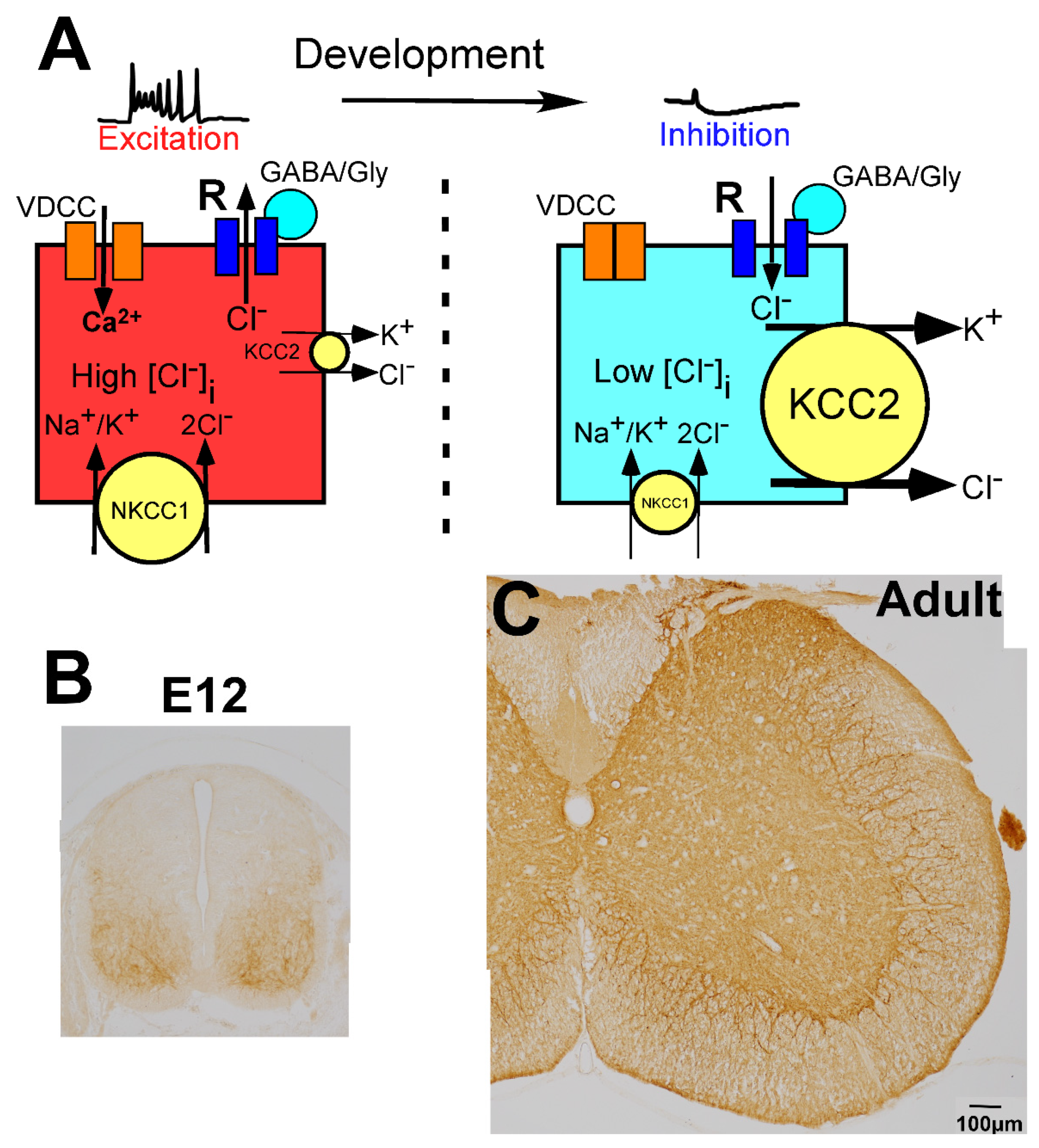
2.4. Regulation of GABAergic and Glycinergic Action by Chloride Transporters

In the CNS, the change in membrane potential exerted by GABA and glycine is determined by the intracellular chloride ion concentration ([Cl]i), which is regulated by the balance of two different chloride cotransporters, Na+-K+-Cl cotransporter 1 (NKCC1) and K+-Cl cotransporter 2 (KCC2) (Figure 2A) [79,80,81]. NKCC1 increases the [Cl]i, whereas KCC2 decreases the [Cl]i. When the action of NKCC1 is relatively high or KCC2 is absent, [Cl]i is high, and GABA and glycine induce depolarization of the membrane potential (Figure 2A, left). In contrast, when KCC2 expression is high compared with the expression of NKCC1, [Cl]i is low, and GABA and glycine act in an inhibitory fashion (Figure 2A, right). In the mature CNS, which includes the spinal cord, KCC2 is highly expressed on the membranes of neuronal cell bodies and dendrites (Figure 2C) [57], and its expression level is very high compared with that of NKCC1 [79,80,81]. Thus, in the mature spinal cord, the [Cl]i is low enough for GABA and glycine to act as inhibitory neurotransmitters [57,82].

3. Development of GABAergic and Glycinergic Neurons and Their Axon Terminals

In general, spinal cord development progresses in the ventral-to-dorsal and rostral-to-caudal directions [83,84]. The formation of synapses, maturation of the removal system, and changes in the action of GABA and glycine also proceed in the same directions. This ventral-to-dorsal development may be regulated by sonic hedgehog signals [85,86,87].

3.1. Early Development of the GABAergic and Glycinergic Neurons

The early development of GABAergic neurons has been precisely described in previous reviews [70,88,89]. Twelve progenitor domains are formed in the spinal cord ventricular zone (neuroepithelial layer), including the vMN and vP0–vP3 domains in the ventral half and the dP1–dP6 domains in the dorsal half (Figure 3A) [83,84]. Each domain produces distinct neuron groups (classes): MN and V0–V3 in the basal plate (ventral half) and dI1–dI6 in the alar plate (dorsal half). The dILA and dILB classes are derived later from the dP4 and dP5 domains. The V0 class is further divided into V0V and V0D subclasses, and the V2 class is subdivided into V2a and V2b subclasses. These classes are characterized by the expression of marker proteins such as Isl1, Evx1/2, and Evx1 [90]. GABAergic neurons are derived from six of the classes (V0D, V1, V2b, dI4, dI6, and dILA). After exiting the cell cycle, neurons move out of the ventricular zone and migrate into the gray matter (Figure 3A). Figure 3 demonstrates the developmental localization of GABAergic neurons in embryonic heterozygous GAD-GFP knock-in mice [60]. In the cervical spinal cord, GABAergic neurons first appear on the surface of the ventricular zone between embryonic day 10 (E10) and E11 (Figure 3B,C). The neurons migrate along distinct routes and finally settle in distinct laminae (Figure 3A). The differentiation processes of GABAergic neurons have been precisely demonstrated using GAD or GABA immunohistochemistry [57,91,92] and GAD-GFP knock-in mice (Figure 3B–G) [60,93]. In contrast, the early developmental processes of glycinergic neurons are still unclear. RNA sequencing studies demonstrated that GlyT2 mRNA-expressing neuron groups were identical to those expressing GAD mRNA [94,95]. GlyT2-GFP histochemistry and glycine immunohistochemistry demonstrated that glycinergic neurons often colocalize with GABA or GAD after E13, suggesting that late embryonic development of glycinergic neurons may be the same as that of GABAergic neurons [62,96]. Taken together, these findings indicate that glycinergic neurons may be derived from the same domains as GABAergic neurons and may appear later than GABAergic neurons.

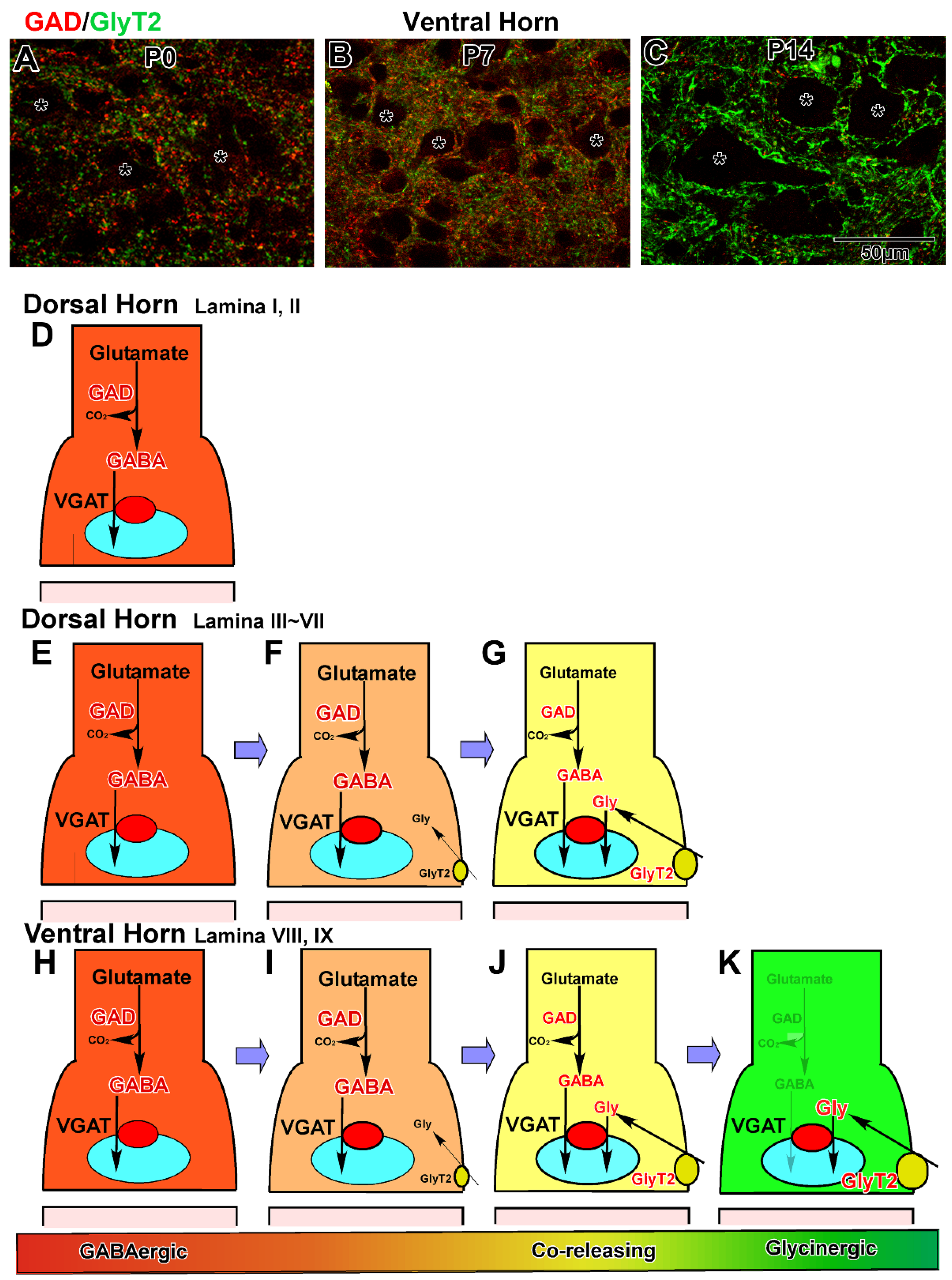
3.2. Development in the Ventral Horn

After motor neurons are differentiated from the MN class in the ventral horn, three classes of cells—V0D, V1, and V2b—develop into GABA and glycinergic neurons under the regulation of various transcriptional factors [70,97] and distinctly participate in the complex network around the motor neurons (Figure 3B–G) [98]. In the cervical spinal cord [57], V0D neurons first appear on the ventral side of the sulcus limitans between E10 and E11. Subsequently, these cells move ventrally and send commissural axons into the contralateral marginal zone (Figure 3C,D). The axons ascend two to four segments and enter the contralateral motor neuron pool [57,70,99,100]. Finally, they give rise to inhibitory neurons in the mature lamina VII. Second, V1 neurons appear before E11, located ventrally to the V0 neurons, and V2b neurons subsequently arise between E12 and E13 (Figure 3C,D). These neurons settle in the ventral horn and extend their axons into the ipsilateral marginal zone. Their axons ascend or descend for several segments [101,102,103]. The V1 neurons develop into the major inhibitory interneurons, including Renshaw cells, in the ventral horn [103]. Inhibitory neurons derived from V2b are low in number among the neurons in lamina VII [104]. Last, the dl6 neurons derived from the dP6 domain migrate in the ventral direction into lamina VII and VIII and take part in the ventral horn network (Figure 3D,E). Glycinergic neurons appear at E13 in the ventral horn [62] when GlyT2 is localized at the axon varicosities in the marginal zone [53]. Double labeling of GABA and glycine demonstrated that glycine-positive neurons often contained GABA immunolabeling during embryonic development [62]. These results suggest that many GABAergic neurons may gradually convert to coreleasing neurons in the ventral horn after E13.
In the ventral horn, GABAergic axon terminals, identified as axon varicosities, first appear in the marginal zone at E11 and are detected within the ventral horn at E13. They markedly increase in number and density after E15 and often surround the cell bodies of large motor neurons at E17 [57]. Until postnatal day seven (P7), they continue to increase in density markedly, and the neuropil region is occupied by numerous GABAergic terminals. In contrast, glycinergic terminals appear in the ventral horn at E15 and continue to increase during embryonic and postnatal development [53]. Double staining of GAD and GlyT2 revealed that GlyT2 immunolabeling was usually localized at GABAergic axon terminals, while GlyT2 single-positive terminals were scarce during embryonic development (Figure 4A). During postnatal development, GABAergic terminals gradually convert to coreleasing terminals in the marginal zone after E14 and in the ventral horn after E16. The coreleasing terminals markedly increase in density during the first postnatal week, and coreleasing terminals become dominant at P7 (Figure 4B). Between P7 and P14, the majority of coreleasing terminals change to glycinergic terminals via the removal of GAD from the terminals (Figure 4C). After P21, glycinergic terminals, detected as GlyT2-positive dots, often surround the large neurons, whereas GABAergic terminals are sparse in the ventral horn [53]. The aforementioned shift in dominant neurotransmitters may underlie the difference in the survival period of GAD67-knockout mice and GlyT2-knockout mice [46,47,52,105,106,107]. Because GABAergic inhibition is dominant at birth, GAD67-knockout mice cannot survive after birth, whereas GlyT2-knockout mice can. In contrast, GlyT2-knockout mice suffer from the neuromotor disorder and die around P10 because the dominant inhibitory input shifts from GABAergic to glycinergic during the second postnatal week.

3.3. Development in the Dorsal Horn

The dI4 class and late-born dILA subclass, derived from the dP4 domain, differentiate into GABAergic neurons in the dorsal horn (Figure 3A) [70,88,89]. GABAergic neurons appear on the surface of the dorsal ventricular zone between E11 and E13, migrate laterally and dorsally, and enter the dorsal horn after E13 (Figure 3C–E) [57]. The number of GABAergic neurons markedly increases until E17, and the neurons distribute throughout lamina I to IV by the day of birth (Figure 3E–G). Glycinergic neurons may be derived from the same dorsal domains under the direction of transcription factors such as Ptfla [89,95]. Double staining with GAD/GABA and GlyT2/glycine suggests that GABAergic neurons in lamina I through IV may remain GABAergic before birth [53,62,96]. The GABAergic neurons convert to coreleasing neurons in lamina IV and lamina III during postnatal development, whereas many GABAergic neurons remain GABAergic in lamina I and II until maturation is complete.
GABAergic axon terminals are first detected in the dorsal horn at E15, markedly increase in density after E17, and are homogeneously distributed in lamina I through V at P0 [53,57]. The density of GABAergic terminals further increases during postnatal development. In contrast, glycinergic terminals are absent in lamina I to IV during embryonic development. During postnatal development, GlyT2 is localized at GABAergic axon terminals, as detected in the ventral horn. Thus, GABAergic terminals gradually shift to coreleasing terminals in lamina IV during the first postnatal week and lamina III during the second postnatal week. For two weeks after birth, coreleasing terminals increase in density in lamina III to V, but many GABAergic terminals remain in lamina I and II until maturation is complete.
The development processes of inhibitory terminal formation and maturation are summarized schematically in Figure 4. First, GABAergic terminals are produced by GAD expression (Figure 4D,E,H). In lamina I and II, many GABAergic neurons and terminals remain (Figure 4D). In lamina III to IX, many GABAergic terminals convert to coreleasing terminals via the expression of GlyT2 (Figure 4F,I). After the initiation of high-affinity glycine uptake, both synthesized GABA and uptaken glycine are loaded into the same synaptic vesicles and coreleased from these terminals (Figure 4G,J). In lamina III to VI, coreleasing neurons and terminals are dominant (Figure 4G). In lamina VII to IX (motor neuron pools) of the ventral horn, many coreleasing terminals become glycinergic via the disappearance of GAD (Figure 4K). The mechanism underlying regional differences in the differentiation of terminals is still unclear. Furthermore, this developmental shift in inhibitory neurotransmitters from GABA to glycine has been reported previously in electrophysiological experiments in the spinal cord [108,109] and other regions [110,111,112,113].

3.4. Developmental Formation of Total Inhibitory Terminals

As VGAT/VIAAT transports not only GABA but also glycine into synaptic vesicles, VGAT immunohistochemistry allows for the visualization of all types of inhibitory terminals, including GABAergic, coreleasing, and glycinergic terminals. Inhibitory terminals appeared in the marginal zone at E11 and ventral horn at E13 [57]. The developmental expression of VGAT exhibits ventral-to-dorsal gradation (Figure 4), which indicates that inhibitory terminals are gradually formed in the ventral–dorsal direction during embryonic and early postnatal development [53,57].

4. Developmental Changes in Ionotropic GABA and Glycine Receptors

4.1. GABAA Receptor

The subunit composition of GABAA receptors changes during spinal cord development [114,115], as in various other brain regions [20,115]. In the rat spinal cord, GABAA receptors first appear in the ventricular zone. The main subunit composition continued to be α4β1γ1. These receptors in the ventricular zone may be independent of synaptic transmission [116] because synapses have not yet been formed within the ventricular zone. In the gray matter, developing neurons start to express the α2, α3, α5, β2, β3, γ2, and γ3 subunits. The α1 subunit appears after birth, but the expression is low to moderate. The α2 subunit continues to be highly expressed in the developing and mature motor neurons in lamina VIII and IX. The expression of the α2 subunit gradually increases in other laminae during embryonic development but decreases after birth. Expression of the α3 and α5 subunits begins homogeneously and increases in intensity during late embryonic and early postnatal development. After birth, α3 expression decreases in the ventral horn but remains high in the dorsal horn. The α5 subunit also decreases in expression throughout the gray matter, but expression remains moderate in the motor neurons. Expression of the β2 and γ3 subunits declines during postnatal development and is low or faint in the mature spinal cord. In contrast, the expression level of the β3 and γ2 subunits continues to increase after birth and remains high [114,115]. The developmental changes in α2, α3, β3, and γ2 subunit expression parallel the change in the formation of GABAergic synapses. In the mature spinal cord, expression of the α2 subunit is high in motor neurons and the surrounding satellite neurons in the ventral horn. The α3 subunit is highly expressed in the dorsal horn and moderately localized in other regions of gray matter. In contrast, expression of the α1 and α5 subunits is weak, whereas the β3 and γ2 subunits are highly expressed throughout the gray matter.

4.2. GABAB Receptor

In the ventricular zone, only the B1 subunit is expressed [117]. In the gray matter, both B1 (1a and 1b) and B2 subunits are highly expressed during the late embryonic and early postnatal period [75]. During postnatal development, both expressions slightly decreased, as shown by [3H]-baclofen binding assay [74].

4.3. GABAC Receptor

After birth, the GABAC receptor ρ1 subunit continued to be expressed throughout the gray matter neurons. In contrast, the ρ2 subunit continued to be expressed in the motor neurons and associated interneurons [76]. Therefore, GABAC receptor subunit composition does not change during postnatal spinal cord development.

4.4. Glycine Receptors

The subunit combination [72,118] and electrophysiological characteristics [51,119,120,121] of glycine receptors also markedly change during spinal cord development. During embryonic development, the α2 subunit is highly and exclusively expressed throughout the gray matter of the rat spinal cord. Around the day of birth, expression of the β and α1 subunits is initiated. After birth, α2 subunit expression gradually decreases, whereas α1 and β subunit expression continue to increase. During postnatal development, the expression of the α3 subunit slightly increases [122]. In the mature spinal cord, α2 and α3 expressions are low. These results suggest that the glycine receptor exists as an α2 homomeric pentamer during embryonic development and may temporally change to an α2β heteromeric receptor. Ultimately, the major composition of this receptor changes to α1β during postnatal development [50]. In addition, those containing the α2 and α3 subunits may play roles at the extra-synaptic region in the dorsal horn, as seen in the hippocampus [123]. These changes in composition agree with the results of in vitro electrophysiological studies [120,121].

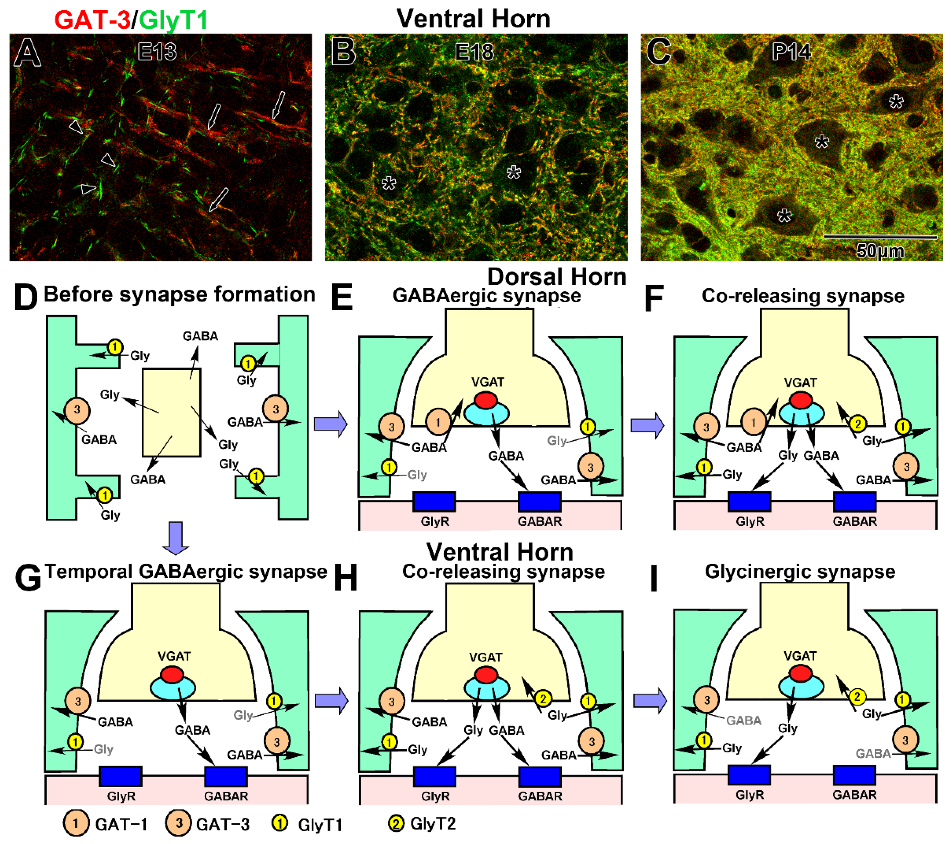
4.5. Developmental Formation of GABA and Glycine Removal System

GABA is removed explicitly by GAT-1 and GAT-3 [35,124,125], and glycine is removed by GlyT1 and GlyT2 during CNS development and in the mature stage (Figure 1A,B) [52]. GAT-1 and GlyT2 are localized at distinct axon terminals, while GAT-3 and GlyT1 are localized on the astrocytic sheets that face the synaptic cleft [53,57].

4.6. Uptake into the Presynaptic Terminals

In the marginal zone and dorsal horn, the GABA removal system at the presynaptic terminals develops simultaneously with the formation of the presynaptic terminals [77]. First, GAT-1 is localized at the GABAergic axon terminals in the marginal zone. In the dorsal horn, GAT-1 localization begins in the axon terminals at E15, and the GAT-1-positive terminals markedly increase in density during embryonic and early postnatal development [77]. During dorsal horn development, localization of GATs spreads in the deep-to-superficial direction, as is observed in GABAergic terminal formation [57]. In the ventral horn, however, although numerous GABAergic terminals and synapses temporally function during embryonic and early postnatal development, GAT-1 immunolabeling is always sparse. These results suggest that the presynaptic GABA removal system does not function at the temporal GABAergic synapses in the ventral horn, and GABA, which is released from transient terminals, may be largely removed into astrocytes near these temporal GABAergic synapses. In summary, the GABA removal system develops simultaneously with the formation of “permanent” presynaptic terminals in the spinal cord [77].
The onset of the GlyT2 expression was almost concomitant to the initial distribution of glycine immunolabeling [53,62,96]. As mentioned in Section 2.2, uptake from the extracellular space through GlyT2 is the main glycine supply pathway, suggesting that GlyT2 localization at the presynaptic terminals may mark the common onset of both glycinergic transmission and glycine removal. Therefore, the development of the glycine removal system proceeds simultaneously with the formation of glycinergic transmission [53].

4.7. Reuptake into the Astrocytes

Both GAT-3 and GlyT1 continue to localize in the astrocytic lineage cells, from radial glial to astrocytes, during spinal cord development [77]. The onset of GAT-3 localization on the radial glia is nearly concomitant with the distribution of GABAergic neurons in the ventral horn at E11 and dorsal horn at E13 before the formation of GABAergic terminals [77]. GlyT1 localization is almost concomitant with the appearance of glycinergic neurons at E13 [62]. When GlyT2-positive glycinergic terminals were detected at E15, GlyT1 had already been localized at the radial glial processes (Figure 5A). These results indicate that GABA and glycine removal systems may function before synaptic transmission and suggest that extrasynaptically released GABA and glycine may be exclusively removed into radial glial processes [126]. During spinal cord development, GAT-3- and GlyT1-expressing radial glia gradually spread to the dorsal area. Initially, GABA and glycine are removed at distinct positions on the radial glial processes. GAT-3 is localized at the shaft of radial processes, whereas GlyT1 is localized at the spine-like profiles of the shafts (Figure 5A). While radial glia differentiates into astroglia and astrocytic processes surrounding the synapses, GABAergic terminals gradually change to coreleasing terminals between E17 to P14. Concomitantly, GAT-3 and GlyT2 gradually colocalize at the astrocytic sheets that face the synaptic clefts (Figure 5B,C). Thus, in the coreleasing synapses, GABA and glycine are released into the same synaptic cleft and removed by adjacent transporters, GlyT1 and GAT-3. Development of the GABA and glycine removal system may be fixed by P21 [77].
Interestingly, although many GABAergic neurons remain GABAergic and do not convert to coreleasing neurons in lamina I and II, GlyT1 is abundantly localized in this region. In addition, after coreleasing terminals give rise to glycinergic terminals by the disappearance of GABA synthesis, GAT-3 continues to be abundantly localized at the astroglia sheets [77]. The developmental formation of the removal system is illustrated in Figure 5. During the middle embryonic stage, extrasynaptically released GABA and glycine are uptaken at the shaft and spines of radial glial processes, respectively (Figure 5D). In the dorsal horn, when GABAergic synapses are formed, GABA is removed through GAT-1 and GAT-3 (Figure 5E). Furthermore, GlyT1 is localized near GAT-3 on the astrocytic sheets and may remove extracellular glycine (Figure 5E). This type of synapse remains in lamina I and II. Next, GlyT2 appears at the presynaptic terminals, and GABAergic synapses convert to coreleasing synapses. In these synapses, GAT-3 and GlyT2 intermingle on the astrocytic sheets (Figure 5F). These synapses remain in lamina III to VII. In contrast, in lamina VIII and IX, when temporal GABAergic synapses are formed, GABA is removed through only GAT-3 (Figure 5G). After GABAergic synapses convert to coreleasing terminals, glycine starts to be removed by GlyT1 into the presynaptic terminals (Figure 5H). Even after coreleasing terminals convert to glycinergic terminals, GAT-1 persists at the astrocytic sheets (Figure 5I).

5. Developmental Changes in GABAergic and Glycinergic Action

In general, GABA and glycine induce depolarization of the membrane potential in the immature CNS, whereas these molecules act as inhibitory neurotransmitters in the mature CNS [79,80,127]. This developmental change in the action of these neurotransmitters occurs as a result of a negative shift in the Cl reversal potential due to the marked increase in KCC2 localization and decrease in NKCC1 expression (Figure 2A) [79,80,81]. Changes in KCC2 activity may play a pivotal role in the fine-tuning of [Cl]i for the following reasons [128,129,130,131]. During development, the expression of NKCC1 does not markedly change [132], whereas changes in the levels of KCC2 correlate with modification of the action of GABA. Transfection of KCC2 into hippocampal neurons converts the action of GABA from excitatory to inhibitory, and GABA is excitatory in KCC2 knockout mice [80,81,128]. Furthermore, after nerve injury, expression of KCC2 is markedly decreased in motor neurons, and the action of GABA and glycine is shifted from inhibitory to excitatory [133,134,135,136]. At E11, weak KCC2 signals appear on the surface of the ventral horn, whereas the dorsal horn is negative for KCC2 expression (Figure 2B). During embryonic development, KCC2 expression gradually increases in intensity, and the KCC2-positive area gradually spreads to the dorsal horn. The gray matter becomes homogeneously labeled by the day of birth [57,137,138]. Concomitantly, NKCC1 expression gradually decreases [138]. These results suggest that the action of GABA and glycine may change from excitation to inhibition in the direction from the ventral-to-dorsal horn [57]. As the expression of KCC2 increases and that of NKCC1 decreases, the reversal potential of Cl and inhibitory postsynaptic potentials (IPSPs) gradually decreases [132,138,139]. Taken together, these observations indicate that GABAergic and glycinergic action in the ventral horn may developmentally change as follows: initially, GABA and glycine mediate depolarization and induce action potentials; next, they mediate depolarization of the membrane potential, but the depolarization is below threshold; and finally, the excitatory inputs are shut around the birthday. This membrane potential is termed “depolarizing IPSP”, and this phenomenon is called a “shunting effect”. Finally, GABA and glycine induce hyperpolarization of the membrane potential [140,141].
The activity of KCC2 is regulated not only by expression level but also through various other mechanisms, such as phosphorylation/dephosphorylation [142,143,144] and membrane trafficking [145,146,147]. For example, phosphorylation of threonine residues 906 and 1007 decreases the activity of KCC2. In the developing CNS, this phosphorylation inhibits KCC2 activity, maintains the excitatory action of GABA and glycine, and may play key roles in morphogenesis [148]. Conversely, continuous phosphorylation affects the postnatal mouse brain functions; phosphomimetic KCC2 knock-in mice cannot survive due to the lack of GABAergic inhibition [149]. The phosphorylation of serine residue 940 increases the influx of K+ and Cl ions. Abnormalities in phosphorylation may cause various neuropsychiatric diseases [143,144]. The phosphorylation of tyrosine residue 1087 is involved in the internalization of KCC2, which results in the downregulation of KCC2 activity [147]. In addition to tyrosine residue phosphorylation, other complex mechanisms may take part in the trafficking and endocytosis of KCC2 and regulate the activity of KCC2 in developing neurons [145,146,147].

6. Discussion

Lastly, we will focus on the processes in which GABAergic excitatory action plays a role. Glycine may play a similar role in the developing CNS. It is thought that GABA may act as a trophic factor in the developing CNS and induce brain morphogenesis. In the developing immature CNS, GABAA receptor-mediated depolarization activates voltage-dependent calcium channels and N-methyl-D-aspartate-type glutamate receptors and elevates cytosolic calcium ion concentration (Figure 2A) [150,151,152,153,154,155,156,157]. The elevation of cytosolic calcium may play roles in various steps in CNS development, such as (1) stop signals for cell proliferation, (2) cell migration, and (3) neuronal maturation, which includes synaptogenesis [11,14,79,80,158,159,160]. GABA acts as an antiproliferation molecule, reduces DNA synthesis in the proliferating precursor cells, and depresses the rate of cellular proliferation via the activation of GABAA receptors and other GABAA receptor-related molecules [116,161]. GABA modulates neuronal migration at the femtomolar (1015 M) to micromolar (μM) level [162,163,164]. Furthermore, exposure of neurons to GABA or GABAA receptor agonists induces the synthesis of neuron-specific molecules such as neuron-specific enolase and neural cell adhesion molecules, enhances the growth rate of neuronal processes, and facilitates synapse formation by inducing the expression and targeting of GABA receptor subunits [159,165,166,167,168,169,170,171,172,173,174,175,176,177,178]. Consequently, it is suggested that lack of GABA synthesis and inhibition of GABA release may cause morphological abnormalities in the CNS, including abnormalities of the spinal cord. To reveal this hypothesis, two types of knockout mice lacking GAD67 [105,106,107] and VGAT [63,179,180,181] were established. Although they have common severe phenotypes, such as omphalocele, cleft palate, hunched posture, loss of movement, and respiratory failure, and cannot survive after birth, no morphological abnormalities were detected in the CNS of these mice. Total GAD knockout mice, which lack both GAD65 and GAD67, also exhibited normal histology in the CNS [107,182]. Furthermore, KCC2-knockout mice, in which GABA and glycine continued to be excitatory, exhibited similar phenotypes [128]. These results suggest that the abnormalities detected in the three types of knockout mice may result from hyperexcitation resulting from the loss of GABAergic and glycinergic inhibition. The inhibitory action by GABA and glycine in the ventral horn may be crucial for the survival of newborn mice. Therefore, the function of GABAergic and glycinergic excitation is still unclear.

7. Conclusions

Initially, six groups of GABAergic neurons are derived from five domains in the ventricular zone. Each group migrates along a distinct route, settles in distinct laminae, and forms synapses. Many GABAergic neurons remain GABAergic, mainly those located in lamina I and II. In other laminae, many of these neurons convert to GABA and glycine coreleasing neurons by initiating glycine reuptake via GlyT2. In the ventral horn, many of these neurons give rise to the glycinergic network after GABA synthesis ceases. During these developmental processes, the subunit compositions and electrophysiological characteristics of GABA and glycine receptors change. During changes in neuronal types, GABA and glycine removal systems mature. Furthermore, the action of GABA and glycine shifts from excitatory to inhibitory.

Author Contributions

Conceptualization, C.S.-O., S.K., J.K., M.S., A.O. and C.T.; development and study of the glycinergic network, C.S.-O., S.K. and M.S.; development of the GABAergic network, Y.K., J.K. and A.O.; data curation, C.T.; writing—original draft preparation, C.T.; writing—review and editing, C.S.-O., S.K., J.K., M.S., A.O. and C.T. All authors have read and agreed to the published version of the manuscript.

Funding

This work was supported by JSPS KAKENHI [Grant-in-Aid for Scientific Research (C) JP18K07823 (CS-O), 21K06394 (CT), and Early-Career Scientists 20K16111(SK) and 19K19871 (YK)].

Institutional Review Board Statement

Not applicable.

Informed Consent Statement

Not applicable.

Data Availability Statement

Not applicable.

Acknowledgments

We are grateful to Makiko Moriyasu-Kurogi and Asako Komesu at the Department of Molecular Anatomy for their assistance in preparing this manuscript. We also thank Nobuhiko Okura for valuable discussions. We thank Emma Longworth-Mills for editing a draft of this manuscript.

Conflicts of Interest

The authors declare no conflict of interest.

Abbreviations

AFanterior funiculus
CNScentral nervous system
[Cl]iintracellular chloride ion concentration
CNScentral nervous system
DHdorsal horn
DRGdorsal root ganglion
dI1–6, dIL, dILA, dILB, MN, V0–3, V0D, V0V, V2a, and V2bneuronal groups, classes, derived from their distinct domains of spinal cord ventricular zone
dP1–dP6, vMN, and vP0–vP3domains of the ventricular zone in embryonic spinal cord
Eembryonic day
FPfloor plate
GABAγ-aminobutyric acid
GADglutamic acid decarboxylase
GAT-1, GAT-2, and GAT-3GABA transporter 1, 2, and 3
Glyglycine
GlyT1 and GlyT2glycine transporter 1 and 2
GFPgreen fluorescence protein
IPSPinhibitory postsynaptic potential
KCC2potassium (K+), chloride (Cl) cotransporter 2
LFlateral funiculus
NKCC1sodium (Na+)-K+-2Cl cotransporter 1
Ppostnatal day
PFposterior funiculus
RGABAA and glycine receptor
RProof plate
SHMTserine hydroxy-methyltransferase
VDCCvoltage-dependent calcium channel
VIAATvesicular inhibitory amino acid transporter
VGATvesicular GABA transporter
VHventral horn
VZventricular zone
I-IXlaminar number of the gray matter
1GAT-1
3GAT-3

References

  1. Macdonald, R.L.; Olsen, R.W. GABAA receptor channels. Annu. Rev. Neurosci. 1994, 17, 569–602. [Google Scholar] [CrossRef]
  2. Olsen, R.W.; Tobin, A.J. Molecular biology of GABAA receptors. FASEB J. 1990, 4, 1469–1480. [Google Scholar] [CrossRef]
  3. Kirsch, J. Glycinergic transmission. Cell Tissue Res. 2006, 326, 535–540. [Google Scholar] [CrossRef]
  4. Legendre, P. The glycinergic inhibitory synapse. Cell. Mol. Life Sci. 2001, 58, 760–793. [Google Scholar] [CrossRef]
  5. Tritsch, N.X.; Granger, A.J.; Sabatini, B.L. Mechanisms and functions of GABA co-release. Nat. Rev. Neurosci. 2016, 17, 139–145. [Google Scholar] [CrossRef] [PubMed]
  6. Vaaga, C.E.; Borisovska, M.; Westbrook, G.L. Dual-transmitter neurons: Functional implications of co-release and co-transmission. Curr. Opin. Neurobiol. 2014, 29, 25–32. [Google Scholar] [CrossRef] [PubMed] [Green Version]
  7. Jonas, P.; Bischofberger, J.; Sandkuhler, J. Corelease of two fast neurotransmitters at a central synapse. Science 1998, 281, 419–424. [Google Scholar] [CrossRef] [Green Version]
  8. Hossaini, M.; French, P.J.; Holstege, J.C. Distribution of glycinergic neuronal somata in the rat spinal cord. Brain Res. 2007, 1142, 61–69. [Google Scholar] [CrossRef]
  9. Todd, A.J.; Sullivan, A.C. Light microscope study of the coexistence of GABA-like and glycine-like immunoreactivities in the spinal cord of the rat. J. Comp. Neurol. 1990, 296, 496–505. [Google Scholar] [CrossRef]
  10. Zeilhofer, H.U.; Studler, B.; Arabadzisz, D.; Schweizer, C.; Ahmadi, S.; Layh, B.; Bosl, M.R.; Fritschy, J.M. Glycinergic neurons expressing enhanced green fluorescent protein in bacterial artificial chromosome transgenic mice. J. Comp. Neurol. 2005, 482, 123–141. [Google Scholar] [CrossRef] [PubMed]
  11. Barker, J.L.; Behar, T.; Li, Y.X.; Liu, Q.Y.; Ma, W.; Maric, D.; Maric, I.; Schaffner, A.E.; Serafini, R.; Smith, S.V.; et al. GABAergic cells and signals in CNS development. Perspect. Dev. Neurobiol. 1998, 5, 305–322. [Google Scholar] [PubMed]
  12. Zafra, F.; Aragon, C.; Gimenez, C. Molecular biology of glycinergic neurotransmission. Mol. Neurobiol. 1997, 14, 117–142. [Google Scholar] [CrossRef]
  13. Martin, D.L.; Rimvall, K. Regulation of gamma-aminobutyric acid synthesis in the brain. J. Neurochem. 1993, 60, 395–407. [Google Scholar] [CrossRef]
  14. Varju, P.; Katarova, Z.; Madarasz, E.; Szabo, G. GABA signalling during development: New data and old questions. Cell Tissue Res. 2001, 305, 239–246. [Google Scholar] [CrossRef] [PubMed]
  15. Bak, L.K.; Schousboe, A.; Waagepetersen, H.S. The glutamate/GABA-glutamine cycle: Aspects of transport, neurotransmitter homeostasis and ammonia transfer. J. Neurochem. 2006, 98, 641–653. [Google Scholar] [CrossRef] [PubMed]
  16. Bedet, C.; Isambert, M.F.; Henry, J.P.; Gasnier, B. Constitutive phosphorylation of the vesicular inhibitory amino acid transporter in rat central nervous system. J. Neurochem. 2000, 75, 1654–1663. [Google Scholar] [CrossRef] [Green Version]
  17. Sagne, C.; El Mestikawy, S.; Isambert, M.F.; Hamon, M.; Henry, J.P.; Giros, B.; Gasnier, B. Cloning of a functional vesicular GABA and glycine transporter by screening of genome databases. FEBS Lett. 1997, 417, 177–183. [Google Scholar] [CrossRef] [Green Version]
  18. Mehta, A.K.; Ticku, M.K. An update on GABAA receptors. Brain Res. Brain Res. Rev. 1999, 29, 196–217. [Google Scholar] [CrossRef]
  19. Sieghart, W. Structure and pharmacology of gamma-aminobutyric acidA receptor subtypes. Pharmacol. Rev. 1995, 47, 181–234. [Google Scholar]
  20. Laurie, D.J.; Seeburg, P.H.; Wisden, W. The distribution of 13 GABAA receptor subunit mRNAs in the rat brain. II. Olfactory bulb and cerebellum. J. Neurosci. 1992, 12, 1063–1076. [Google Scholar] [CrossRef]
  21. Wisden, W.; Gundlach, A.L.; Barnard, E.A.; Seeburg, P.H.; Hunt, S.P. Distribution of GABAA receptor subunit mRNAs in rat lumbar spinal cord. Brain Res. Mol. Brain Res. 1991, 10, 179–183. [Google Scholar] [CrossRef]
  22. Wisden, W.; Laurie, D.J.; Monyer, H.; Seeburg, P.H. The distribution of 13 GABAA receptor subunit mRNAs in the rat brain. I. Telencephalon, diencephalon, mesencephalon. J. Neurosci. 1992, 12, 1040–1062. [Google Scholar] [CrossRef] [Green Version]
  23. Pritchett, D.B.; Sontheimer, H.; Shivers, B.D.; Ymer, S.; Kettenmann, H.; Schofield, P.R.; Seeburg, P.H. Importance of a novel GABAA receptor subunit for benzodiazepine pharmacology. Nature 1989, 338, 582–585. [Google Scholar] [CrossRef] [PubMed]
  24. Vicini, S. New perspectives in the functional role of GABA(A) channel heterogeneity. Mol. Neurobiol. 1999, 19, 97–110. [Google Scholar] [CrossRef] [PubMed]
  25. Bormann, J.; Feigenspan, A. GABAC receptors. Trends Neurosci. 1995, 18, 515–519. [Google Scholar] [CrossRef]
  26. Bormann, J. The ‘ABC’ of GABA receptors. Trends Pharmacol. Sci. 2000, 21, 16–19. [Google Scholar] [CrossRef]
  27. Bormann, J. Electrophysiology of GABAA and GABAB receptor subtypes. Trends Neurosci. 1988, 11, 112–116. [Google Scholar] [CrossRef]
  28. Connors, B.W.; Malenka, R.C.; Silva, L.R. Two inhibitory postsynaptic potentials, and GABAA and GABAB receptor-mediated responses in neocortex of rat and cat. J. Physiol. 1988, 406, 443–468. [Google Scholar] [CrossRef]
  29. LeVine, H., 3rd. Structural features of heterotrimeric G-protein-coupled receptors and their modulatory proteins. Mol. Neurobiol. 1999, 19, 111–149. [Google Scholar] [CrossRef] [PubMed]
  30. Nicoll, R.A. The coupling of neurotransmitter receptors to ion channels in the brain. Science 1988, 241, 545–551. [Google Scholar] [CrossRef] [PubMed]
  31. Jembrek, M.J.; Vlainic, J. GABA Receptors: Pharmacological Potential and Pitfalls. Curr. Pharm. Des. 2015, 21, 4943–4959. [Google Scholar] [CrossRef] [PubMed] [Green Version]
  32. Evenseth, L.S.M.; Gabrielsen, M.; Sylte, I. The GABAB Receptor-Structure, Ligand Binding and Drug Development. Molecules 2020, 25, 3093. [Google Scholar] [CrossRef]
  33. Cherubini, E.; Conti, F. Generating diversity at GABAergic synapses. Trends Neurosci. 2001, 24, 155–162. [Google Scholar] [CrossRef]
  34. Kanner, B.I. Sodium-coupled neurotransmitter transport: Structure, function and regulation. J. Exp. Biol. 1994, 196, 237–249. [Google Scholar] [CrossRef] [PubMed]
  35. Gadea, A.; Lopez-Colome, A.M. Glial transporters for glutamate, glycine, and GABA: II. GABA transporters. J. Neurosci. Res. 2001, 63, 461–468. [Google Scholar] [CrossRef]
  36. Conti, F.; Zuccarello, L.V.; Barbaresi, P.; Minelli, A.; Brecha, N.C.; Melone, M. Neuronal, glial, and epithelial localization of gamma-aminobutyric acid transporter 2, a high-affinity gamma-aminobutyric acid plasma membrane transporter, in the cerebral cortex and neighboring structures. J. Comp. Neurol. 1999, 409, 482–494. [Google Scholar] [CrossRef]
  37. Minelli, A.; Brecha, N.C.; Karschin, C.; DeBiasi, S.; Conti, F. GAT-1, a high-affinity GABA plasma membrane transporter, is localized to neurons and astroglia in the cerebral cortex. J. Neurosci. 1995, 15, 7734–7746. [Google Scholar] [CrossRef]
  38. Minelli, A.; DeBiasi, S.; Brecha, N.C.; Zuccarello, L.V.; Conti, F. GAT-3, a high-affinity GABA plasma membrane transporter, is localized to astrocytic processes, and it is not confined to the vicinity of GABAergic synapses in the cerebral cortex. J. Neurosci. 1996, 16, 6255–6264. [Google Scholar] [CrossRef] [Green Version]
  39. Itouji, A.; Sakai, N.; Tanaka, C.; Saito, N. Neuronal and glial localization of two GABA transporters (GAT1 and GAT3) in the rat cerebellum. Brain Res. Mol. Brain Res. 1996, 37, 309–316. [Google Scholar] [CrossRef]
  40. Takayama, C.; Inoue, Y. Developmental expression of GABA transporter-1 and 3 during formation of the GABAergic synapses in the mouse cerebellar cortex. Brain Res. Dev. Brain Res. 2005, 158, 41–49. [Google Scholar] [CrossRef] [Green Version]
  41. Kolker, S. Metabolism of amino acid neurotransmitters: The synaptic disorder underlying inherited metabolic diseases. J. Inherit. Metab. Dis. 2018, 41, 1055–1063. [Google Scholar] [CrossRef] [PubMed]
  42. Verleysdonk, S.; Martin, H.; Willker, W.; Leibfritz, D.; Hamprecht, B. Rapid uptake and degradation of glycine by astroglial cells in culture: Synthesis and release of serine and lactate. Glia 1999, 27, 239–248. [Google Scholar] [CrossRef]
  43. Beyoglu, D.; Idle, J.R. The glycine deportation system and its pharmacological consequences. Pharmacol. Ther. 2012, 135, 151–167. [Google Scholar] [CrossRef] [PubMed] [Green Version]
  44. Zeilhofer, H.U.; Wildner, H.; Yevenes, G.E. Fast synaptic inhibition in spinal sensory processing and pain control. Physiol. Rev. 2012, 92, 193–235. [Google Scholar] [CrossRef] [PubMed]
  45. Xu, T.L.; Gong, N. Glycine and glycine receptor signaling in hippocampal neurons: Diversity, function and regulation. Prog. Neurobiol. 2010, 91, 349–361. [Google Scholar] [CrossRef] [PubMed]
  46. Gomeza, J.; Ohno, K.; Hulsmann, S.; Armsen, W.; Eulenburg, V.; Richter, D.W.; Laube, B.; Betz, H. Deletion of the mouse glycine transporter 2 results in a hyperekplexia phenotype and postnatal lethality. Neuron 2003, 40, 797–806. [Google Scholar] [CrossRef] [Green Version]
  47. Latal, A.T.; Kremer, T.; Gomeza, J.; Eulenburg, V.; Hulsmann, S. Development of synaptic inhibition in glycine transporter 2 deficient mice. Mol. Cell. Neurosci. 2010, 44, 342–352. [Google Scholar] [CrossRef]
  48. Chalphin, A.V.; Saha, M.S. The specification of glycinergic neurons and the role of glycinergic transmission in development. Front. Mol. Neurosci. 2010, 3, 11. [Google Scholar] [CrossRef] [Green Version]
  49. Laube, B.; Maksay, G.; Schemm, R.; Betz, H. Modulation of glycine receptor function: A novel approach for therapeutic intervention at inhibitory synapses? Trends Pharmacol. Sci. 2002, 23, 519–527. [Google Scholar] [CrossRef] [Green Version]
  50. Lynch, J.W. Molecular structure and function of the glycine receptor chloride channel. Physiol. Rev. 2004, 84, 1051–1095. [Google Scholar] [CrossRef]
  51. Lynch, J.W. Native glycine receptor subtypes and their physiological roles. Neuropharmacology 2009, 56, 303–309. [Google Scholar] [CrossRef] [Green Version]
  52. Eulenburg, V.; Armsen, W.; Betz, H.; Gomeza, J. Glycine transporters: Essential regulators of neurotransmission. Trends Biochem. Sci. 2005, 30, 325–333. [Google Scholar] [CrossRef]
  53. Sunagawa, M.; Shimizu-Okabe, C.; Kim, J.; Kobayashi, S.; Kosaka, Y.; Yanagawa, Y.; Matsushita, M.; Okabe, A.; Takayama, C. Distinct development of the glycinergic terminals in the ventral and dorsal horns of the mouse cervical spinal cord. Neuroscience 2017, 343, 459–471. [Google Scholar] [CrossRef] [Green Version]
  54. Zafra, F.; Aragon, C.; Olivares, L.; Danbolt, N.C.; Gimenez, C.; Storm-Mathisen, J. Glycine transporters are differentially expressed among CNS cells. J. Neurosci. 1995, 15, 3952–3969. [Google Scholar] [CrossRef]
  55. Sato, K.; Yoshida, S.; Fujiwara, K.; Tada, K.; Tohyama, M. Glycine cleavage system in astrocytes. Brain Res. 1991, 567, 64–70. [Google Scholar] [CrossRef]
  56. Kikuchi, G.; Motokawa, Y.; Yoshida, T.; Hiraga, K. Glycine cleavage system: Reaction mechanism, physiological significance, and hyperglycinemia. Proc. Jpn. Acad. Ser. B Phys. Biol. Sci. 2008, 84, 246–263. [Google Scholar] [CrossRef] [Green Version]
  57. Kosaka, Y.; Kin, H.; Tatetsu, M.; Uema, I.; Takayama, C. Distinct development of GABA system in the ventral and dorsal horns in the embryonic mouse spinal cord. Brain Res. 2012, 1486, 39–52. [Google Scholar] [CrossRef] [PubMed]
  58. Todd, A.J.; Maxwell, D.J. GABA in the mammalian spinal cord. In GABA in the Nervous System; Martin, D.L., Olsen, R.W., Eds.; Lippincotto Willams & Wilkins: Philadelphia, PA, USA, 2000; pp. 439–457. [Google Scholar]
  59. Ottersen, O.P.; Storm-Mathisen, J. Glutamate- and GABA-containing neurons in the mouse and rat brain, as demonstrated with a new immunocytochemical technique. J. Comp. Neurol. 1984, 229, 374–392. [Google Scholar] [CrossRef]
  60. Tamamaki, N.; Yanagawa, Y.; Tomioka, R.; Miyazaki, J.; Obata, K.; Kaneko, T. Green fluorescent protein expression and colocalization with calretinin, parvalbumin, and somatostatin in the GAD67-GFP knock-in mouse. J. Comp. Neurol. 2003, 467, 60–79. [Google Scholar] [CrossRef] [PubMed]
  61. Campistron, G.; Buijs, R.M.; Geffard, M. Glycine neurons in the brain and spinal cord. Antibody production and immunocytochemical localization. Brain Res. 1986, 376, 400–405. [Google Scholar] [CrossRef] [Green Version]
  62. Allain, A.E.; Bairi, A.; Meyrand, P.; Branchereau, P. Expression of the glycinergic system during the course of embryonic development in the mouse spinal cord and its co-localization with GABA immunoreactivity. J. Comp. Neurol. 2006, 496, 832–846. [Google Scholar] [CrossRef] [PubMed]
  63. Wojcik, S.M.; Katsurabayashi, S.; Guillemin, I.; Friauf, E.; Rosenmund, C.; Brose, N.; Rhee, J.S. A shared vesicular carrier allows synaptic corelease of GABA and glycine. Neuron 2006, 50, 575–587. [Google Scholar] [CrossRef] [Green Version]
  64. Ishibashi, H.; Yamaguchi, J.; Nakahata, Y.; Nabekura, J. Dynamic regulation of glycine-GABA co-transmission at spinal inhibitory synapses by neuronal glutamate transporter. J. Physiol. 2013, 591, 3821–3832. [Google Scholar] [CrossRef] [PubMed]
  65. Ornung, G.; Shupliakov, O.; Linda, H.; Ottersen, O.P.; Storm-Mathisen, J.; Ulfhake, B.; Cullheim, S. Qualitative and quantitative analysis of glycine- and GABA-immunoreactive nerve terminals on motoneuron cell bodies in the cat spinal cord: A postembedding electron microscopic study. J. Comp. Neurol. 1996, 365, 413–426. [Google Scholar] [CrossRef]
  66. Dougherty, K.J.; Sawchuk, M.A.; Hochman, S. Phenotypic diversity and expression of GABAergic inhibitory interneurons during postnatal development in lumbar spinal cord of glutamic acid decarboxylase 67-green fluorescent protein mice. Neuroscience 2009, 163, 909–919. [Google Scholar] [CrossRef] [Green Version]
  67. Bardoni, R.; Takazawa, T.; Tong, C.K.; Choudhury, P.; Scherrer, G.; Macdermott, A.B. Pre- and postsynaptic inhibitory control in the spinal cord dorsal horn. Ann. N. Y. Acad. Sci. 2013, 1279, 90–96. [Google Scholar] [CrossRef]
  68. Basbaum, A.I.; Bautista, D.M.; Scherrer, G.; Julius, D. Cellular and molecular mechanisms of pain. Cell 2009, 139, 267–284. [Google Scholar] [CrossRef] [PubMed] [Green Version]
  69. Cioffi, C.L. Modulation of Glycine-Mediated Spinal Neurotransmission for the Treatment of Chronic Pain. J. Med. Chem. 2018, 61, 2652–2679. [Google Scholar] [CrossRef]
  70. Goulding, M. Circuits controlling vertebrate locomotion: Moving in a new direction. Nat. Rev. Neurosci. 2009, 10, 507–518. [Google Scholar] [CrossRef]
  71. Paul, J.; Zeilhofer, H.U.; Fritschy, J.M. Selective distribution of GABA(A) receptor subtypes in mouse spinal dorsal horn neurons and primary afferents. J. Comp. Neurol. 2012, 520, 3895–3911. [Google Scholar] [CrossRef]
  72. Malosio, M.L.; Marqueze-Pouey, B.; Kuhse, J.; Betz, H. Widespread expression of glycine receptor subunit mRNAs in the adult and developing rat brain. EMBO J. 1991, 10, 2401–2409. [Google Scholar] [CrossRef] [PubMed]
  73. Todd, A.J.; Watt, C.; Spike, R.C.; Sieghart, W. Colocalization of GABA, glycine, and their receptors at synapses in the rat spinal cord. J. Neurosci. 1996, 16, 974–982. [Google Scholar] [CrossRef] [PubMed] [Green Version]
  74. Dorfman, V.B.; Vega, M.C.; Coirini, H. Age-related changes of the GABA-B receptor in the lumbar spinal cord of male rats and penile erection. Life Sci. 2006, 78, 1529–1534. [Google Scholar] [CrossRef] [PubMed]
  75. Sands, S.A.; Purisai, M.G.; Chronwall, B.M.; Enna, S.J. Ontogeny of GABA(B) receptor subunit expression and function in the rat spinal cord. Brain Res. 2003, 972, 197–206. [Google Scholar] [CrossRef]
  76. Rozzo, A.; Armellin, M.; Franzot, J.; Chiaruttini, C.; Nistri, A.; Tongiorgi, E. Expression and dendritic mRNA localization of GABAC receptor rho1 and rho2 subunits in developing rat brain and spinal cord. Eur. J. Neurosci. 2002, 15, 1747–1758. [Google Scholar] [CrossRef]
  77. Kim, J.; Kosaka, Y.; Shimizu-Okabe, C.; Niizaki, A.; Takayama, C. Characteristic development of the GABA-removal system in the mouse spinal cord. Neuroscience 2014, 262, 129–142. [Google Scholar] [CrossRef] [PubMed]
  78. Jursky, F.; Nelson, N. Localization of glycine neurotransmitter transporter (GLYT2) reveals correlation with the distribution of glycine receptor. J. Neurochem. 1995, 64, 1026–1033. [Google Scholar] [CrossRef] [PubMed] [Green Version]
  79. Owens, D.F.; Kriegstein, A.R. Is there more to GABA than synaptic inhibition? Nat. Rev. Neurosci. 2002, 3, 715–727. [Google Scholar] [CrossRef]
  80. Ben-Ari, Y. Excitatory actions of gaba during development: The nature of the nurture. Nat. Rev. Neurosci. 2002, 3, 728–739. [Google Scholar] [CrossRef] [PubMed]
  81. Payne, J.A.; Rivera, C.; Voipio, J.; Kaila, K. Cation-chloride co-transporters in neuronal communication, development and trauma. Trends Neurosci. 2003, 26, 199–206. [Google Scholar] [CrossRef]
  82. Baccei, M.L.; Fitzgerald, M. Development of GABAergic and glycinergic transmission in the neonatal rat dorsal horn. J. Neurosci. 2004, 24, 4749–4757. [Google Scholar] [CrossRef]
  83. Sagner, A.; Briscoe, J. Establishing neuronal diversity in the spinal cord: A time and a place. Development 2019, 146, dev182154. [Google Scholar] [CrossRef] [Green Version]
  84. Hernandez-Miranda, L.R.; Muller, T.; Birchmeier, C. The dorsal spinal cord and hindbrain: From developmental mechanisms to functional circuits. Dev. Biol. 2017, 432, 34–42. [Google Scholar] [CrossRef] [PubMed] [Green Version]
  85. Briscoe, J.; Ericson, J. Specification of neuronal fates in the ventral neural tube. Curr. Opin. Neurobiol. 2001, 11, 43–49. [Google Scholar] [CrossRef]
  86. Jessell, T.M. Neuronal specification in the spinal cord: Inductive signals and transcriptional codes. Nat. Rev. Genet. 2000, 1, 20–29. [Google Scholar] [CrossRef]
  87. Lee, K.J.; Jessell, T.M. The specification of dorsal cell fates in the vertebrate central nervous system. Annu. Rev. Neurosci. 1999, 22, 261–294. [Google Scholar] [CrossRef] [PubMed] [Green Version]
  88. Helms, A.W.; Johnson, J.E. Specification of dorsal spinal cord interneurons. Curr. Opin. Neurobiol. 2003, 13, 42–49. [Google Scholar] [CrossRef]
  89. Hori, K.; Hoshino, M. GABAergic neuron specification in the spinal cord, the cerebellum, and the cochlear nucleus. Neural Plast. 2012, 2012, 921732. [Google Scholar] [CrossRef] [Green Version]
  90. Goulding, M.; Lamar, E. Neuronal patterning: Making stripes in the spinal cord. Curr. Biol. 2000, 10, R565–R568. [Google Scholar] [CrossRef] [Green Version]
  91. Tran, T.S.; Alijani, A.; Phelps, P.E. Unique developmental patterns of GABAergic neurons in rat spinal cord. J. Comp. Neurol. 2003, 456, 112–126. [Google Scholar] [CrossRef]
  92. Allain, A.E.; Bairi, A.; Meyrand, P.; Branchereau, P. Ontogenic changes of the GABAergic system in the embryonic mouse spinal cord. Brain Res. 2004, 1000, 134–147. [Google Scholar] [CrossRef] [PubMed]
  93. Huang, J.; Chen, J.; Wang, W.; Wei, Y.Y.; Cai, G.H.; Tamamaki, N.; Li, Y.Q.; Wu, S.X. Birthdate study of GABAergic neurons in the lumbar spinal cord of the glutamic acid decarboxylase 67-green fluorescent protein knock-in mouse. Front. Neuroanat. 2013, 7, 42. [Google Scholar] [CrossRef] [PubMed] [Green Version]
  94. Delile, J.; Rayon, T.; Melchionda, M.; Edwards, A.; Briscoe, J.; Sagner, A. Single cell transcriptomics reveals spatial and temporal dynamics of gene expression in the developing mouse spinal cord. Development 2019, 146, dev.173807. [Google Scholar] [CrossRef] [Green Version]
  95. Borromeo, M.D.; Meredith, D.M.; Castro, D.S.; Chang, J.C.; Tung, K.C.; Guillemot, F.; Johnson, J.E. A transcription factor network specifying inhibitory versus excitatory neurons in the dorsal spinal cord. Development 2014, 141, 2803–2812. [Google Scholar] [CrossRef] [Green Version]
  96. Restrepo, C.E.; Lundfald, L.; Szabo, G.; Erdelyi, F.; Zeilhofer, H.U.; Glover, J.C.; Kiehn, O. Transmitter-phenotypes of commissural interneurons in the lumbar spinal cord of newborn mice. J. Comp. Neurol. 2009, 517, 177–192. [Google Scholar] [CrossRef] [PubMed]
  97. Lee, S.K.; Pfaff, S.L. Transcriptional networks regulating neuronal identity in the developing spinal cord. Nat. Neurosci. 2001, 4, 1183–1191. [Google Scholar] [CrossRef]
  98. Stachowski, N.J.; Dougherty, K.J. Spinal Inhibitory Interneurons: Gatekeepers of Sensorimotor Pathways. Int. J. Mol. Sci. 2021, 22, 2667. [Google Scholar] [CrossRef] [PubMed]
  99. Tran, T.S.; Cohen-Cory, S.; Phelps, P.E. Embryonic GABAergic spinal commissural neurons project rostrally to mesencephalic targets. J. Comp. Neurol. 2004, 475, 327–339. [Google Scholar] [CrossRef]
  100. Moran-Rivard, L.; Kagawa, T.; Saueressig, H.; Gross, M.K.; Burrill, J.; Goulding, M. Evx1 is a postmitotic determinant of v0 interneuron identity in the spinal cord. Neuron 2001, 29, 385–399. [Google Scholar] [CrossRef] [Green Version]
  101. Joshi, K.; Lee, S.; Lee, B.; Lee, J.W.; Lee, S.K. LMO4 controls the balance between excitatory and inhibitory spinal V2 interneurons. Neuron 2009, 61, 839–851. [Google Scholar] [CrossRef] [PubMed] [Green Version]
  102. Lundfald, L.; Restrepo, C.E.; Butt, S.J.; Peng, C.Y.; Droho, S.; Endo, T.; Zeilhofer, H.U.; Sharma, K.; Kiehn, O. Phenotype of V2-derived interneurons and their relationship to the axon guidance molecule EphA4 in the developing mouse spinal cord. Eur. J. Neurosci. 2007, 26, 2989–3002. [Google Scholar] [CrossRef] [PubMed]
  103. Alvarez, F.J.; Jonas, P.C.; Sapir, T.; Hartley, R.; Berrocal, M.C.; Geiman, E.J.; Todd, A.J.; Goulding, M. Postnatal phenotype and localization of spinal cord V1 derived interneurons. J. Comp. Neurol. 2005, 493, 177–192. [Google Scholar] [CrossRef] [Green Version]
  104. Zhang, J.; Lanuza, G.M.; Britz, O.; Wang, Z.; Siembab, V.C.; Zhang, Y.; Velasquez, T.; Alvarez, F.J.; Frank, E.; Goulding, M. V1 and v2b interneurons secure the alternating flexor-extensor motor activity mice require for limbed locomotion. Neuron 2014, 82, 138–150. [Google Scholar] [CrossRef] [PubMed] [Green Version]
  105. Condie, B.G.; Bain, G.; Gottlieb, D.I.; Capecchi, M.R. Cleft palate in mice with a targeted mutation in the gamma-aminobutyric acid-producing enzyme glutamic acid decarboxylase 67. Proc. Natl. Acad. Sci. USA 1997, 94, 11451–11455. [Google Scholar] [CrossRef] [Green Version]
  106. Ding, R.; Tsunekawa, N.; Obata, K. Cleft palate by picrotoxin or 3-MP and palatal shelf elevation in GABA-deficient mice. Neurotoxicol. Teratol. 2004, 26, 587–592. [Google Scholar] [CrossRef] [PubMed]
  107. Ji, F.; Kanbara, N.; Obata, K. GABA and histogenesis in fetal and neonatal mouse brain lacking both the isoforms of glutamic acid decarboxylase. Neurosci. Res. 1999, 33, 187–194. [Google Scholar] [CrossRef]
  108. Gao, B.X.; Stricker, C.; Ziskind-Conhaim, L. Transition from GABAergic to glycinergic synaptic transmission in newly formed spinal networks. J. Neurophysiol. 2001, 86, 492–502. [Google Scholar] [CrossRef]
  109. Singer, J.H.; Berger, A.J. Development of inhibitory synaptic transmission to motoneurons. Brain Res. Bull. 2000, 53, 553–560. [Google Scholar] [CrossRef]
  110. Kotak, V.C.; Korada, S.; Schwartz, I.R.; Sanes, D.H. A developmental shift from GABAergic to glycinergic transmission in the central auditory system. J. Neurosci. 1998, 18, 4646–4655. [Google Scholar] [CrossRef] [Green Version]
  111. Nabekura, J.; Katsurabayashi, S.; Kakazu, Y.; Shibata, S.; Matsubara, A.; Jinno, S.; Mizoguchi, Y.; Sasaki, A.; Ishibashi, H. Developmental switch from GABA to glycine release in single central synaptic terminals. Nat. Neurosci. 2004, 7, 17–23. [Google Scholar] [CrossRef] [PubMed]
  112. Nerlich, J.; Rubsamen, R.; Milenkovic, I. Developmental Shift of Inhibitory Transmitter Content at a Central Auditory Synapse. Front. Cell. Neurosci. 2017, 11, 211. [Google Scholar] [CrossRef]
  113. McMenamin, C.A.; Anselmi, L.; Travagli, R.A.; Browning, K.N. Developmental regulation of inhibitory synaptic currents in the dorsal motor nucleus of the vagus in the rat. J. Neurophysiol. 2016, 116, 1705–1714. [Google Scholar] [CrossRef] [Green Version]
  114. Ma, W.; Saunders, P.A.; Somogyi, R.; Poulter, M.O.; Barker, J.L. Ontogeny of GABAA receptor subunit mRNAs in rat spinal cord and dorsal root ganglia. J. Comp. Neurol. 1993, 338, 337–359. [Google Scholar] [CrossRef]
  115. Laurie, D.J.; Wisden, W.; Seeburg, P.H. The distribution of thirteen GABAA receptor subunit mRNAs in the rat brain. III. Embryonic and postnatal development. J. Neurosci. 1992, 12, 4151–4172. [Google Scholar] [CrossRef]
  116. LoTurco, J.J.; Owens, D.F.; Heath, M.J.; Davis, M.B.; Kriegstein, A.R. GABA and glutamate depolarize cortical progenitor cells and inhibit DNA synthesis. Neuron 1995, 15, 1287–1298. [Google Scholar] [CrossRef] [Green Version]
  117. Kim, M.O.; Li, S.; Park, M.S.; Hornung, J.P. Early fetal expression of GABA(B1) and GABA(B2) receptor mRNAs on the development of the rat central nervous system. Brain Res. Dev. Brain Res. 2003, 143, 47–55. [Google Scholar] [CrossRef]
  118. Watanabe, E.; Akagi, H. Distribution patterns of mRNAs encoding glycine receptor channels in the developing rat spinal cord. Neurosci. Res. 1995, 23, 377–382. [Google Scholar] [CrossRef]
  119. Takahashi, T.; Momiyama, A.; Hirai, K.; Hishinuma, F.; Akagi, H. Functional correlation of fetal and adult forms of glycine receptors with developmental changes in inhibitory synaptic receptor channels. Neuron 1992, 9, 1155–1161. [Google Scholar] [CrossRef]
  120. Withers, M.D.; St John, P.A. Embryonic rat spinal cord neurons change expression of glycine receptor subtypes during development in vitro. J. Neurobiol. 1997, 32, 579–592. [Google Scholar] [CrossRef]
  121. Aguayo, L.G.; van Zundert, B.; Tapia, J.C.; Carrasco, M.A.; Alvarez, F.J. Changes on the properties of glycine receptors during neuronal development. Brain Res. Brain Res. Rev. 2004, 47, 33–45. [Google Scholar] [CrossRef] [PubMed]
  122. Jonsson, S.; Morud, J.; Pickering, C.; Adermark, L.; Ericson, M.; Soderpalm, B. Changes in glycine receptor subunit expression in forebrain regions of the Wistar rat over development. Brain Res. 2012, 1446, 12–21. [Google Scholar] [CrossRef]
  123. Aroeira, R.I.; Ribeiro, J.A.; Sebastiao, A.M.; Valente, C.A. Age-related changes of glycine receptor at the rat hippocampus: From the embryo to the adult. J. Neurochem. 2011, 118, 339–353. [Google Scholar] [CrossRef]
  124. Borden, L.A. GABA transporter heterogeneity: Pharmacology and cellular localization. Neurochem. Int. 1996, 29, 335–356. [Google Scholar] [CrossRef]
  125. Jursky, F.; Nelson, N. Developmental expression of GABA transporters GAT1 and GAT4 suggests involvement in brain maturation. J. Neurochem. 1996, 67, 857–867. [Google Scholar] [CrossRef]
  126. Takayama, C.; Inoue, Y. GABAergic signaling in the developing cerebellum. Anat. Sci. Int. 2004, 79, 124–136. [Google Scholar] [CrossRef] [PubMed] [Green Version]
  127. Ben-Ari, Y. The GABA excitatory/inhibitory developmental sequence: A personal journey. Neuroscience 2014, 279, 187–219. [Google Scholar] [CrossRef] [PubMed] [Green Version]
  128. Hubner, C.A.; Stein, V.; Hermans-Borgmeyer, I.; Meyer, T.; Ballanyi, K.; Jentsch, T.J. Disruption of KCC2 reveals an essential role of K-Cl cotransport already in early synaptic inhibition. Neuron 2001, 30, 515–524. [Google Scholar] [CrossRef] [Green Version]
  129. Lee, H.; Chen, C.X.; Liu, Y.J.; Aizenman, E.; Kandler, K. KCC2 expression in immature rat cortical neurons is sufficient to switch the polarity of GABA responses. Eur. J. Neurosci. 2005, 21, 2593–2599. [Google Scholar] [CrossRef] [Green Version]
  130. Rivera, C.; Voipio, J.; Kaila, K. Two developmental switches in GABAergic signalling: The K+-Cl cotransporter KCC2 and carbonic anhydrase CAVII. J. Physiol. 2005, 562, 27–36. [Google Scholar] [CrossRef]
  131. Mahadevan, V.; Woodin, M.A. Regulation of neuronal chloride homeostasis by neuromodulators. J. Physiol. 2016, 594, 2593–2605. [Google Scholar] [CrossRef] [PubMed]
  132. Delpy, A.; Allain, A.E.; Meyrand, P.; Branchereau, P. NKCC1 cotransporter inactivation underlies embryonic development of chloride-mediated inhibition in mouse spinal motoneuron. J. Physiol. 2008, 586, 1059–1075. [Google Scholar] [CrossRef]
  133. Tatetsu, M.; Kim, J.; Kina, S.; Sunakawa, H.; Takayama, C. GABA/glycine signaling during degeneration and regeneration of mouse hypoglossal nerves. Brain Res. 2012, 1446, 22–33. [Google Scholar] [CrossRef]
  134. Kim, J.; Kobayashi, S.; Shimizu-Okabe, C.; Okabe, A.; Moon, C.; Shin, T.; Takayama, C. Changes in the expression and localization of signaling molecules in mouse facial motor neurons during regeneration of facial nerves. J. Chem. Neuroanat. 2018, 88, 13–21. [Google Scholar] [CrossRef]
  135. Nabekura, J.; Ueno, T.; Okabe, A.; Furuta, A.; Iwaki, T.; Shimizu-Okabe, C.; Fukuda, A.; Akaike, N. Reduction of KCC2 expression and GABAA receptor-mediated excitation after in vivo axonal injury. J. Neurosci. 2002, 22, 4412–4417. [Google Scholar] [CrossRef] [Green Version]
  136. Toyoda, H.; Ohno, K.; Yamada, J.; Ikeda, M.; Okabe, A.; Sato, K.; Hashimoto, K.; Fukuda, A. Induction of NMDA and GABAA receptor-mediated Ca2+ oscillations with KCC2 mRNA downregulation in injured facial motoneurons. J. Neurophysiol. 2003, 89, 1353–1362. [Google Scholar] [CrossRef] [PubMed] [Green Version]
  137. Stein, V.; Hermans-Borgmeyer, I.; Jentsch, T.J.; Hubner, C.A. Expression of the KCl cotransporter KCC2 parallels neuronal maturation and the emergence of low intracellular chloride. J. Comp. Neurol. 2004, 468, 57–64. [Google Scholar] [CrossRef]
  138. Stil, A.; Liabeuf, S.; Jean-Xavier, C.; Brocard, C.; Viemari, J.C.; Vinay, L. Developmental up-regulation of the potassium-chloride cotransporter type 2 in the rat lumbar spinal cord. Neuroscience 2009, 164, 809–821. [Google Scholar] [CrossRef] [PubMed]
  139. Vinay, L.; Jean-Xavier, C. Plasticity of spinal cord locomotor networks and contribution of cation-chloride cotransporters. Brain Res. Rev. 2008, 57, 103–110. [Google Scholar] [CrossRef] [PubMed]
  140. Nishimaru, H.; Kakizaki, M. The role of inhibitory neurotransmission in locomotor circuits of the developing mammalian spinal cord. Acta Physiol. 2009, 197, 83–97. [Google Scholar] [CrossRef] [PubMed]
  141. Vinay, L.; Brocard, F.; Pflieger, J.F.; Simeoni-Alias, J.; Clarac, F. Perinatal development of lumbar motoneurons and their inputs in the rat. Brain Res. Bull. 2000, 53, 635–647. [Google Scholar] [CrossRef]
  142. Kahle, K.T.; Deeb, T.Z.; Puskarjov, M.; Silayeva, L.; Liang, B.; Kaila, K.; Moss, S.J. Modulation of neuronal activity by phosphorylation of the K-Cl cotransporter KCC2. Trends Neurosci. 2013, 36, 726–737. [Google Scholar] [CrossRef] [PubMed] [Green Version]
  143. Kahle, K.T.; Delpire, E. Kinase-KCC2 coupling: Cl rheostasis, disease susceptibility, therapeutic target. J. Neurophysiol. 2016, 115, 8–18. [Google Scholar] [CrossRef] [PubMed] [Green Version]
  144. Fukuda, A.; Watanabe, M. Pathogenic potential of human SLC12A5 variants causing KCC2 dysfunction. Brain Res. 2019, 1710, 1–7. [Google Scholar] [CrossRef] [PubMed]
  145. Come, E.; Heubl, M.; Schwartz, E.J.; Poncer, J.C.; Levi, S. Reciprocal Regulation of KCC2 Trafficking and Synaptic Activity. Front. Cell. Neurosci. 2019, 13, 48. [Google Scholar] [CrossRef]
  146. Tang, B.L. K(+)-Cl(−) co-transporter 2 (KCC2)—A membrane trafficking perspective. Mol. Membr. Biol. 2016, 33, 100–110. [Google Scholar] [CrossRef] [Green Version]
  147. Lee, H.H.; Jurd, R.; Moss, S.J. Tyrosine phosphorylation regulates the membrane trafficking of the potassium chloride co-transporter KCC2. Mol. Cell. Neurosci. 2010, 45, 173–179. [Google Scholar] [CrossRef] [Green Version]
  148. Friedel, P.; Kahle, K.T.; Zhang, J.; Hertz, N.; Pisella, L.I.; Buhler, E.; Schaller, F.; Duan, J.; Khanna, A.R.; Bishop, P.N.; et al. WNK1-regulated inhibitory phosphorylation of the KCC2 cotransporter maintains the depolarizing action of GABA in immature neurons. Sci. Signal. 2015, 8, ra65. [Google Scholar] [CrossRef]
  149. Watanabe, M.; Zhang, J.; Mansuri, M.S.; Duan, J.; Karimy, J.K.; Delpire, E.; Alper, S.L.; Lifton, R.P.; Fukuda, A.; Kahle, K.T. Developmentally regulated KCC2 phosphorylation is essential for dynamic GABA-mediated inhibition and survival. Sci. Signal. 2019, 12, eaaw9315. [Google Scholar] [CrossRef]
  150. Connor, J.A.; Tseng, H.Y.; Hockberger, P.E. Depolarization- and transmitter-induced changes in intracellular Ca2+ of rat cerebellar granule cells in explant cultures. J. Neurosci. 1987, 7, 1384–1400. [Google Scholar] [CrossRef] [Green Version]
  151. Yuste, R.; Katz, L.C. Control of postsynaptic Ca2+ influx in developing neocortex by excitatory and inhibitory neurotransmitters. Neuron 1991, 6, 333–344. [Google Scholar] [CrossRef]
  152. Reichling, D.B.; Kyrozis, A.; Wang, J.; MacDermott, A.B. Mechanisms of GABA and glycine depolarization-induced calcium transients in rat dorsal horn neurons. J. Physiol. 1994, 476, 411–421. [Google Scholar] [CrossRef] [PubMed]
  153. Leinekugel, X.; Tseeb, V.; Ben-Ari, Y.; Bregestovski, P. Synaptic GABAA activation induces Ca2+ rise in pyramidal cells and interneurons from rat neonatal hippocampal slices. J. Physiol. 1995, 487 Pt 2, 319–329. [Google Scholar] [CrossRef] [PubMed] [Green Version]
  154. Obrietan, K.; van den Pol, A.N. Growth cone calcium elevation by GABA. J. Comp. Neurol. 1996, 372, 167–175. [Google Scholar] [CrossRef]
  155. Ben-Ari, Y.; Khazipov, R.; Leinekugel, X.; Caillard, O.; Gaiarsa, J.L. GABAA, NMDA and AMPA receptors: A developmentally regulated ‘menage a trois’. Trends Neurosci. 1997, 20, 523–529. [Google Scholar] [CrossRef]
  156. Serafini, R.; Ma, W.; Maric, D.; Maric, I.; Lahjouji, F.; Sieghart, W.; Barker, J.L. Initially expressed early rat embryonic GABA(A) receptor Cl- ion channels exhibit heterogeneous channel properties. Eur. J. Neurosci. 1998, 10, 1771–1783. [Google Scholar] [CrossRef] [PubMed]
  157. Eilers, J.; Plant, T.D.; Marandi, N.; Konnerth, A. GABA-mediated Ca2+ signalling in developing rat cerebellar Purkinje neurones. J. Physiol. 2001, 536, 429–437. [Google Scholar] [CrossRef]
  158. McCarthy, M.M.; Auger, A.P.; Perrot-Sinal, T.S. Getting excited about GABA and sex differences in the brain. Trends Neurosci. 2002, 25, 307–312. [Google Scholar] [CrossRef]
  159. Belhage, B.; Hansen, G.H.; Elster, L.; Schousboe, A. Effects of gamma-aminobutyric acid (GABA) on synaptogenesis and synaptic function. Perspect. Dev. Neurobiol. 1998, 5, 235–246. [Google Scholar]
  160. Kardos, J. Recent advances in GABA research. Neurochem. Int. 1999, 34, 353–358. [Google Scholar] [CrossRef]
  161. Haydar, T.F.; Wang, F.; Schwartz, M.L.; Rakic, P. Differential modulation of proliferation in the neocortical ventricular and subventricular zones. J. Neurosci. 2000, 20, 5764–5774. [Google Scholar] [CrossRef] [Green Version]
  162. Behar, T.N.; Schaffner, A.E.; Colton, C.A.; Somogyi, R.; Olah, Z.; Lehel, C.; Barker, J.L. GABA-induced chemokinesis and NGF-induced chemotaxis of embryonic spinal cord neurons. J. Neurosci. 1994, 14, 29–38. [Google Scholar] [CrossRef]
  163. Behar, T.N.; Li, Y.X.; Tran, H.T.; Ma, W.; Dunlap, V.; Scott, C.; Barker, J.L. GABA stimulates chemotaxis and chemokinesis of embryonic cortical neurons via calcium-dependent mechanisms. J. Neurosci. 1996, 16, 1808–1818. [Google Scholar] [CrossRef] [PubMed] [Green Version]
  164. Behar, T.N.; Schaffner, A.E.; Tran, H.T.; Barker, J.L. GABA-induced motility of spinal neuroblasts develops along a ventrodorsal gradient and can be mimicked by agonists of GABAA and GABAB receptors. J. Neurosci. Res. 1995, 42, 97–108. [Google Scholar] [CrossRef]
  165. Abraham, J.H.; Schousboe, A. Effects of taurine on cell morphology and expression of low-affinity GABA receptors in cultured cerebellar granule cells. Neurochem. Res. 1989, 14, 1031–1038. [Google Scholar] [CrossRef] [PubMed]
  166. Elster, L.; Hansen, G.H.; Belhage, B.; Fritschy, J.M.; Mohler, H.; Schousboe, A. Differential distribution of GABAA receptor subunits in soma and processes of cerebellar granule cells: Effects of maturation and a GABA agonist. Int. J. Dev. Neurosci. 1995, 13, 417–428. [Google Scholar] [CrossRef]
  167. Gao, X.B.; van den Pol, A.N. GABA release from mouse axonal growth cones. J. Physiol. 2000, 523 Pt 3, 629–637. [Google Scholar] [CrossRef]
  168. Carlson, B.X.; Belhage, B.; Hansen, G.H.; Elster, L.; Olsen, R.W.; Schousboe, A. Expression of the GABA(A) receptor alpha6 subunit in cultured cerebellar granule cells is developmentally regulated by activation of GABA(A) receptors. J. Neurosci. Res. 1997, 50, 1053–1062. [Google Scholar] [CrossRef]
  169. Carlson, B.X.; Elster, L.; Schousboe, A. Pharmacological and functional implications of developmentally-regulated changes in GABA(A) receptor subunit expression in the cerebellum. Eur. J. Pharmacol. 1998, 352, 1–14. [Google Scholar] [CrossRef]
  170. Mellor, J.R.; Merlo, D.; Jones, A.; Wisden, W.; Randall, A.D. Mouse cerebellar granule cell differentiation: Electrical activity regulates the GABAA receptor alpha 6 subunit gene. J. Neurosci. 1998, 18, 2822–2833. [Google Scholar] [CrossRef] [Green Version]
  171. Moss, S.J.; Smart, T.G. Constructing inhibitory synapses. Nat. Rev. Neurosci. 2001, 2, 240–250. [Google Scholar] [CrossRef]
  172. Meier, E.; Jorgensen, O.S. Gamma-aminobutyric acid affects the developmental expression of neuron-associated proteins in cerebellar granule cell cultures. J. Neurochem. 1986, 46, 1256–1262. [Google Scholar] [CrossRef] [PubMed]
  173. Meier, E.; Jorgensen, O.S.; Schousboe, A. Effect of repeated treatment with a gamma-aminobutyric acid receptor agonist on postnatal neural development in rats. J. Neurochem. 1987, 49, 1462–1470. [Google Scholar] [CrossRef]
  174. Wolff, J.R.; Joo, F.; Dames, W. Plasticity in dendrites shown by continuous GABA administration in superior cervical ganglion of adult rat. Nature 1978, 274, 72–74. [Google Scholar] [CrossRef]
  175. Spoerri, P.E. Neurotrophic effects of GABA in cultures of embryonic chick brain and retina. Synapse 1988, 2, 11–22. [Google Scholar] [CrossRef]
  176. Barbin, G.; Pollard, H.; Gaiarsa, J.L.; Ben-Ari, Y. Involvement of GABAA receptors in the outgrowth of cultured hippocampal neurons. Neurosci. Lett. 1993, 152, 150–154. [Google Scholar] [CrossRef]
  177. Mitchell, C.K.; Redburn, D.A. GABA and GABA-A receptors are maximally expressed in association with cone synaptogenesis in neonatal rabbit retina. Brain Res. Dev. Brain Res. 1996, 95, 63–71. [Google Scholar] [CrossRef]
  178. Kim, H.Y.; Sapp, D.W.; Olsen, R.W.; Tobin, A.J. GABA alters GABAA receptor mRNAs and increases ligand binding. J. Neurochem. 1993, 61, 2334–2337. [Google Scholar] [CrossRef]
  179. Saito, K.; Kakizaki, T.; Hayashi, R.; Nishimaru, H.; Furukawa, T.; Nakazato, Y.; Takamori, S.; Ebihara, S.; Uematsu, M.; Mishina, M.; et al. The physiological roles of vesicular GABA transporter during embryonic development: A study using knockout mice. Mol. Brain 2010, 3, 40. [Google Scholar] [CrossRef] [PubMed] [Green Version]
  180. Fujii, M.; Arata, A.; Kanbara-Kume, N.; Saito, K.; Yanagawa, Y.; Obata, K. Respiratory activity in brainstem of fetal mice lacking glutamate decarboxylase 65/67 and vesicular GABA transporter. Neuroscience 2007, 146, 1044–1052. [Google Scholar] [CrossRef]
  181. Yamada, M.H.; Nishikawa, K.; Kubo, K.; Yanagawa, Y.; Saito, S. Impaired glycinergic synaptic transmission and enhanced inflammatory pain in mice with reduced expression of vesicular GABA transporter (VGAT). Mol. Pharmacol. 2012, 81, 610–619. [Google Scholar] [CrossRef] [PubMed] [Green Version]
  182. Kakizaki, T.; Oriuchi, N.; Yanagawa, Y. GAD65/GAD67 double knockout mice exhibit intermediate severity in both cleft palate and omphalocele compared with GAD67 knockout and VGAT knockout mice. Neuroscience 2015, 288, 86–93. [Google Scholar] [CrossRef] [PubMed]
Figure 1. GABAergic and glycinergic transmission in the adult spinal cord. (A,B) are Schematic illustrations of GABAergic (A) and glycinergic (B) synapses. Various key molecules are involved in GABAergic and glycinergic transmission. C and D are Immunohistochemistry for GAD (C,D), GlyT2 (E,F), and both (G,H) in the dorsal (C,E,G) and ventral (D,F,H) horns. In lamina I and II, GABAergic terminals are dominant (C), whereas GAD and GlyT2 double-positive colocalizing terminals (yellow) are dominant in lamina III of the dorsal horn (G). In the ventral horn, glycinergic GlyT2-positive terminals (green) are dominant (F), but GABAergic GAD-positive terminals (red) are scarce (F,H).
Ijms 23 00834 g001
Figure 2. Molecular mechanisms underlying developmental changes in the action of GABA and glycine. (A) Schematic illustration of GABA action depending on intracellular Cl concentration ([Cl]i), which is regulated by K+, Cl cotransporter 2 (KCC2), and Na+, K+, Cl cotransporter 1 (NKCC1). In the immature stage, KCC2 expression is low and [Cl]i is high, thus, GABA binding to GABA receptors (R) induces the efflux of chloride ions (Cl) and mediates excitation (left). In contrast, after maturation, KCC2 expression is high and [Cl]i is low, thus, GABA mediates inhibition. When GABA/glycine elevates the membrane potential in the immature spinal cord, Ca2+ enters through the activated voltage-dependent calcium channel (VDCC) (left). (B,C) Developmental localization of KCC2. KCC2 is weakly localized in the ventral horn at E12, but the dorsal part was negative (B). In the adult spinal cord, KCC2 is expressed throughout the gray matter (C).
Ijms 23 00834 g002
Figure 3. Developmental localization of GABAergic neurons in the spinal cord. (A) Schematic illustration of the origin of GABAergic neurons and their migration routes. GABAergic neurons are derived from six classes (V0D, V1, V2b, dI6, dI4, and dIL) that arise from five domains (vP0, vP1, vP2, dP6, and dP4). Each class of GABAergic neurons migrates their own routes and settle in distinct laminae. (BG) Immunohistochemical analysis of GFP in the developing GFP-GAD knock-in mouse spinal cord. GABAergic neurons were absent at E10 (B) and appeared at E11 (B), and expanded their localization during embryonic development in the ventral-to-dorsal direction (DG). In contrast to the mature spinal cord (Figure 1B), many GABAergic neurons were detected homogeneously in the dorsal and ventral horn of the embryonic spinal cord.
Ijms 23 00834 g003
Figure 4. Developmental changes in inhibitory terminals in the spinal cord. (AC) Double labeling of GAD and GlyT2 in the developing ventral horn. Dominant inhibitory terminals are GABAergic (red) at P0 (A) and change to coreleasing terminals at P7 (B) and glycinergic terminals at P14 (C). (DK) Schematic illustrations of the developmental changes in inhibitory terminals. Initially, GABAergic terminals are formed after starting GABA synthesis by GAD in the gray matter (D,E,H). In lamina I and II, GABAergic terminals remain (D). In lamina III to IX, GABAergic terminals convert to coreleasing terminals by the additional glycine reuptake by GlyT2 (F,G,I,J). In lamina VIII and IX, GAD disappears from the terminals, and the coreleasing terminals give rise to glycinergic terminals (K).
Ijms 23 00834 g004
Figure 5. Developmental formation of the GABA and glycine removal system. (AC) Double staining of GAT-3 (red) and GlyT1 (green). GAT-3 is localized at the shaft of the radial glial processes (arrows), whereas GlyT1 is localized at the spines of the processes (arrowheads) at E13. As development proceeds, GAT-3 and GlyT1 immunolabelings gradually merge and surround the motor neurons (asterisks in B,C). (DI) Schematic illustrations of the development of the GABA and glycine removal system. Initially, uptake of extrasynaptically released GABA and glycine occurs at the shafts and spines of radial glial processes, respectively (D). In the ventral horn, GAT-1 is localized at the terminals when GABAergic synapses are formed (E). After the GABAergic synapses convert to coreleasing synapses, GlyT2 is additionally localized at the terminals (F). In contrast, in the ventral horn, GAT-1 is not localized at the terminals even though GABA is synthesized and released in the temporally synapses. After the synapses convert to coreleasing synapses, GlyT2 is also additionally localized at the terminals (G). Even after coreleasing synapses changed to glycinergic synapses, GAT-3 continued to be localized at the astroglial sheets (I).
Ijms 23 00834 g005
Publisher’s Note: MDPI stays neutral with regard to jurisdictional claims in published maps and institutional affiliations.

