Article

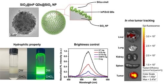
Highly Bright Silica-Coated InP/ZnS Quantum Dot-Embedded Silica Nanoparticles as Biocompatible Nanoprobes

1
Department of Bioscience and Biotechnology, Konkuk University, Seoul 05029, Korea
2
Company of Global Zeus, Hwaseong 18363, Korea
3
Company of BioSquare, Hwaseong 18449, Korea
4
AI-Superconvergence KIURI Translational Research Center, Ajou University School of Medicine, Suwon 16499, Korea
5
School of International Engineering and Science, Jeonbuk National University, Jeonju 54896, Korea
6
Department of Chemical and Biological Engineering, Hanbat University, Daejeon 34158, Korea
7
Department of Urology, Stanford University School of Medicine, Stanford, CA 94305, USA
*
Authors to whom correspondence should be addressed.
These authors contributed equally to this work.
Int. J. Mol. Sci. 2022, 23(18), 10977; https://doi.org/10.3390/ijms231810977
Submission received: 24 August 2022 / Revised: 15 September 2022 / Accepted: 16 September 2022 / Published: 19 September 2022
(This article belongs to the Special Issue Advanced Optical Materials: From Materials to Applications)

Abstract

:
Quantum dots (QDs) have outstanding optical properties such as strong fluorescence, excellent photostability, broad absorption spectra, and narrow emission bands, which make them useful for bioimaging. However, cadmium (Cd)-based QDs, which have been widely studied, have potential toxicity problems. Cd-free QDs have also been studied, but their weak photoluminescence (PL) intensity makes their practical use in bioimaging challenging. In this study, Cd-free QD nanoprobes for bioimaging were fabricated by densely embedding multiple indium phosphide/zinc sulfide (InP/ZnS) QDs onto silica templates and coating them with a silica shell. The fabricated silica-coated InP/ZnS QD-embedded silica nanoparticles (SiO2@InP QDs@SiO2 NPs) exhibited hydrophilic properties because of the surface silica shell. The quantum yield (QY), maximum emission peak wavelength, and full-width half-maximum (FWHM) of the final fabricated SiO2@InP QDs@SiO2 NPs were 6.61%, 527.01 nm, and 44.62 nm, respectively. Moreover, the brightness of the particles could be easily controlled by adjusting the amount of InP/ZnS QDs in the SiO2@InP QDs@SiO2 NPs. When SiO2@InP QDs@SiO2 NPs were administered to tumor syngeneic mice, the fluorescence signal was prominently detected in the tumor because of the preferential distribution of the SiO2@InP QDs@SiO2 NPs, demonstrating their applicability in bioimaging with NPs. Thus, SiO2@InP QDs@SiO2 NPs have the potential to successfully replace Cd-based QDs as highly bright and biocompatible fluorescent nanoprobes.

Graphical Abstract

1. Introduction

Quantum dots (QDs), a type of colloidal semiconductor nanocrystal, have been applied in various bio-fields owing to their good optical properties, such as high fluorescence intensity, low photobleaching, wide absorbance wavelengths, and narrow emission wavelengths, compared to conventional organic fluorescent materials (e.g., organic dyes and fluorescent proteins) [1,2,3,4,5,6]. Among various QDs, cadmium selenide (CdSe)-based QDs have been the most widely studied, owing to their advantages such as high quantum yield (QY), particle stability, and a photoluminescent (PL) emission range across almost the entire visible light region. However, it is well known that cadmium and selenide ions within the CdSe-based QDs could cause serious problems in terms of environmental hazards and toxicity to organisms [7,8,9].
Indium phosphide (InP) QDs, which have been widely studied as Cd-free QDs, were less toxic and harmful to the environment and organisms than CdSe-based QDs [10,11]. In addition, InP QDs have a bulk bandgap of 1.35 eV and an exciton Bohr radius of approximately 10 nm, which enables QDs to have PL emission wavelengths ranging from visible light (blue) to the near infrared [12,13,14]. However, InP QDs typically exhibit a poor QY of <1%, which is attributed to the surface trap states of InP QDs [15,16]. To overcome the low QY of InP QDs, InP-based QDs with an InP core and a shell structure consisting of higher energy bandgap materials such as zinc sulfide (ZnS) were synthesized [17]. The ZnS shell passivated surface defects, prevented the oxidation of the InP core, and significantly increased the QY of InP/ZnS QDs to about 40% [18,19]. Although InP/ZnS QDs possess the potential to be used in biological applications [20,21,22,23,24,25], compared to the well-developed CdSe-based QDs, these still have a lower brightness, which limits the direct application of single InP/ZnS QDs for bioimaging [26,27,28].
To overcome the low brightness of single QDs, approaches for embedding multiple QDs onto the surface of a silica template structure have been leveraged [29,30,31]. As the fabricated silica template-based multi-QDs are brighter than single QDs, they could be used as advanced strategies for applying QDs in biological fields. Among these approaches, silica-coated QD-embedded silica nanoparticles (SiO2@QDs@SiO2 NPs) have many structural advantages that are suited for biological applications [32,33,34,35,36]. The fabrication process of SiO2@QDs@SiO2 NPs yields an efficient assembly of approximately 500 QDs on a single silica template. The fabricated SiO2@QDs@SiO2 NPs exhibited a 200-fold stronger PL emission than those of single QDs. In addition, the silica shell, which is located on the surface of the SiO2@QDs@SiO2 NPs, ensures a good colloidal stability in hydrophilic solvents and facilitates surface modification. A strong fluorescence signal from the SiO2@QDs@SiO2 NP-tagged cells was observed, and the suitability of SiO2@QDs@SiO2 NPs for bioimaging applications was confirmed.
Most studies focus on the fabrication of silica template-based multi-QDs for bioimaging using Cd-based QDs [37,38,39]. However, there has been little progress in the fabrication of silica template-based multi-QDs using InP/ZnS QDs [40,41] and their applications in bioimaging. Miao et al. synthesized Hsp 90α-functionalized mesoporous silica NP-InP/ZnS QD complexes and used them for screening proteins and real-time cell imaging [42]. Perton et al. synthesized polysaccharide-coated stellate mesoporous silica-InP/ZnS QDs and used them for the in vivo fluorescent imaging of zebrafish [43]. However, detailed approaches to materials have not been studied well.
In this study, highly bright and biocompatible bioimaging nanoprobes were fabricated by densely embedding multiple InP/ZnS QDs onto the surfaces of silica templates and coating them with a silica shell. The fabricated silica-coated InP/ZnS QD-embedded SiO2 NPs (SiO2@InP QDs@SiO2 NPs) exhibited hydrophilicity owing to the silica shell on the particle surface. In addition, SiO2@InP QDs@SiO2 NPs showed a much stronger PL intensity than single hydrophilic CdSe/ZnS QDs, and these strong fluorescence signals could be advantageously applied to bioimaging. Furthermore, the brightness control of the SiO2@InP QDs@SiO2 NPs was performed by adjusting the amount of added InP/ZnS QDs during the particle fabrication process. To confirm the biological applicability, cytotoxicity investigation, in vivo biodistribution, and fluorescence imaging were performed, thereby proving that SiO2@InP QDs@SiO2 NPs can be utilized in the field of bioimaging as an alternative to CdSe-based QDs.

2. Results and Discussion

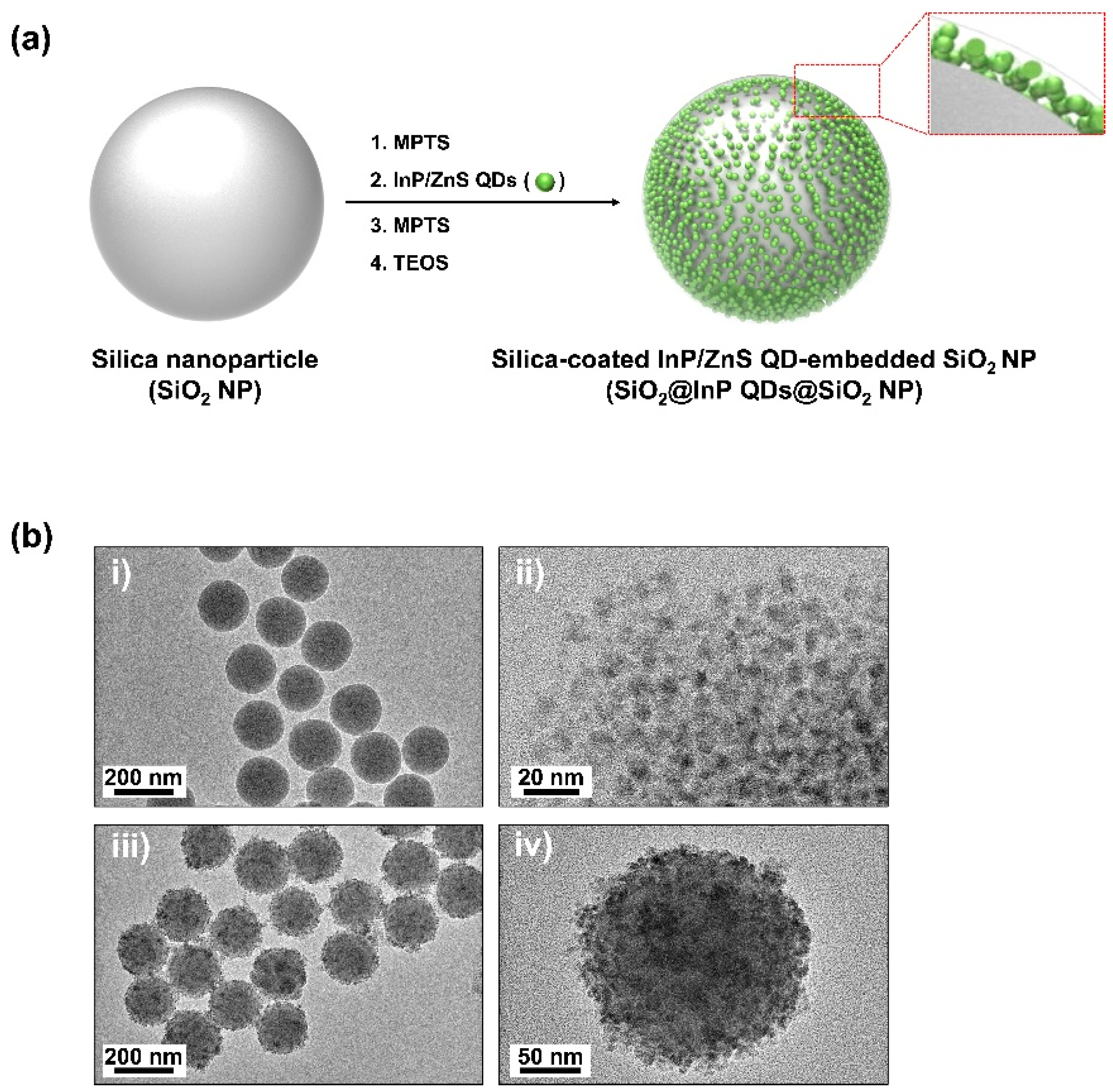
2.1. Fabrication of SiO2@InP QDs@SiO2 NPs

The fabrication flow of the proposed, bright, and biocompatible silica-coated InP/ZnS QD-embedded silica nanoparticles (SiO2@InP QDs@SiO2 NPs) is illustrated in Figure 1a. SiO2 NPs, which were used as templates, were synthesized using a sol–gel process based on the Stöber method [44]. The surface of SiO2 NPs was modified to a thiol (-SH) group, which has high affinity for QDs, by using 3-mercaptopropyltrimethoxysilane (MPTS). Owing to the affinity between the -SH group and InP/ZnS QDs, several InP/ZnS QDs were embedded onto the surface of the thiol-modified SiO2 NPs. The addition of MPTS and NH4OH after embedding increased the number of InP/ZnS QDs embedded on the surface of the thiol-modified SiO2 NPs [28]. To increase biocompatibility and to prevent the leaching of embedded InP/ZnS QDs on SiO2 NPs, these were coated with silica shells by reacting with tetraethyl orthosilicate (TEOS) and NH4OH.
Transmission electron microscopy (TEM) images of SiO2 NPs, InP/ZnS QDs, and SiO2@InP QDs@SiO2 NPs were obtained to confirm the morphology and size of each particle (Figure 1b). SiO2 NPs showed a uniform spherical shape with a size of 172.2 ± 7.2 nm (Figure 1b(i)). The size of the InP/ZnS QDs (Mesolight, Suzhou, China) was estimated to be 5.1 ± 1.0 nm (Figure 1b(ii)). The SiO2@InP QDs@SiO2 NPs were fabricated with similar morphologies and had a final size of 201.4 ± 9.9 nm (Figure 1b(iii)). The fabricated SiO2@InP QDs@SiO2 NPs showed that approximately 1200–1500 InP/ZnS QDs were densely embedded onto the SiO2 NPs, and silica shells were formed on the surfaces of the SiO2@InP QDs NPs (Figure 1b(iv)).

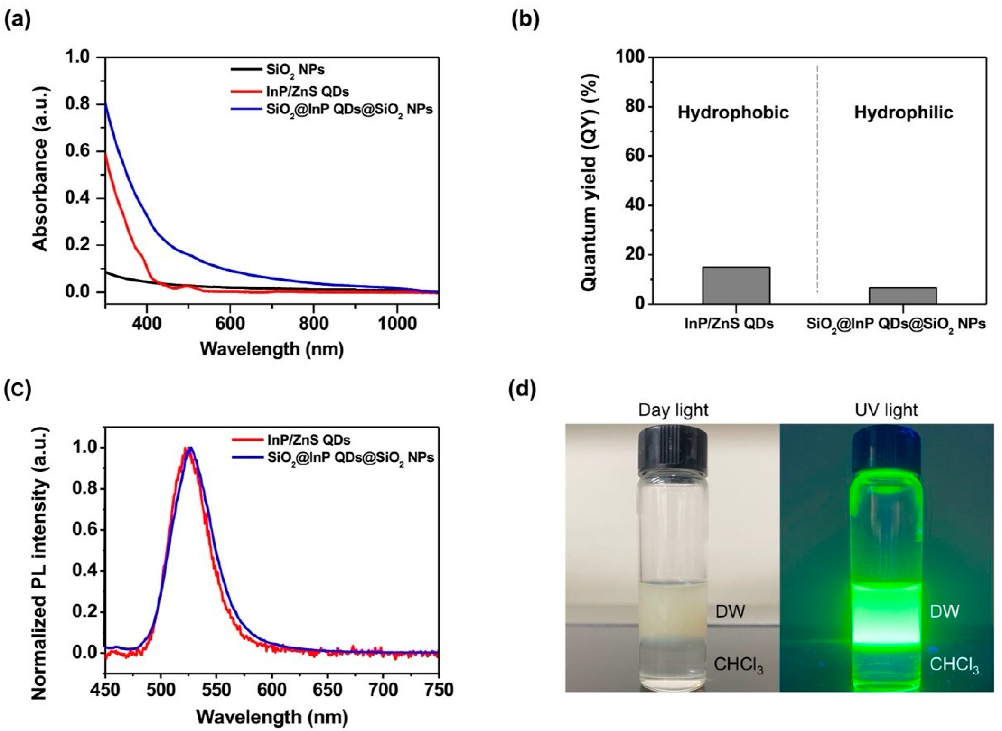
2.2. Characterization of SiO2@InP QDs@SiO2 NPs

To evaluate the photophysical properties of the SiO2@InP QDs@SiO2 NPs, the UV/Vis/NIR absorbance spectra of the SiO2 NPs, InP/ZnS QDs, and SiO2@InP QDs@SiO2 NPs were measured (Figure 2a). The absorbance measurement range was 300–1100 nm. When the absorbance was measured, SiO2 NPs and SiO2@InP QDs@SiO2 NPs were measured at the same concentration (0.1 mg/mL), and InP/ZnS QDs were measured at 0.07 mg/mL, which was based on the amount of added QDs during the fabrication of the SiO2@InP QDs@SiO2 NPs. An absorbance analysis showed that the absorbance of the fabricated SiO2@InP QDs@SiO2 NPs was higher compared to those of the SiO2 NPs and InP/ZnS QDs over the wavelength range of UV/Vis. In addition, both the InP/ZnS QDs and SiO2@InP QDs@SiO2 NPs showed an absorption peak at approximately 500 nm. The absorption spectrum of the SiO2@InP QDs@SiO2 NPs showed that the InP/ZnS QDs were well embedded on the surface of the thiol-modified SiO2 NPs, and that the SiO2@InP QDs@SiO2 NPs maintained the absorption property of the InP/ZnS QDs.
To evaluate the luminous efficiency of the SiO2@InP QDs@SiO2 NPs, the quantum yields (QYs) of the InP/ZnS QDs and SiO2@InP QDs@SiO2 NPs were compared. The QY of the InP/ZnS QDs was 15.02%, and that of SiO2@InP QDs@SiO2 NPs was 6.61% (Figure 2b). Processes such as modifying the surface of QDs and coating silica shells can affect the QYs of QDs [45,46]. These results were also observed in previous studies related to silica-template-based multi-QDs [32,33,36].
To evaluate the emission properties of the SiO2@InP QDs@SiO2 NPs, the PL spectra of InP/ZnS QDs and SiO2@InP QDs@SiO2 NPs were compared (Figure 2c). The maximum emission peak wavelength of InP/ZnS QDs and SiO2@InP QDs@SiO2 NPs were 525.09 nm and 527.01 nm, respectively. The emission peaks of the SiO2@InP QDs@SiO2 NPs shifted little from the emission peak of InP/ZnS QDs. The full-width half-maximum (FWHM) of the InP/ZnS QDs and the SiO2@InP QDs@SiO2 NPs were 41.38 nm and 44.62 nm, respectively. The FWHM values were not significant changed.
The CdSe/ZnS QDs have been most widely used in biological applications because of their excellent QY and photostability [47,48,49]. Therefore, they were set as a comparison group for the SiO2@InP QDs@SiO2 NPs. The selected CdSe/ZnS QDs exhibit a photoluminescence (PL) emission wavelength range similar to that of the InP/ZnS QDs (Figure S1). To compare the QY under aqueous conditions, the hydrophobic ligands of the CdSe/ZnS QDs were replaced with hydrophilic ligands [50]. The QY of the hydrophilic CdSe/ZnS QDs was 87.52% (Figure S2). As the SiO2@InP QDs@SiO2 NPs were fabricated based on InP/ZnS QDs with a low QY, they had a lower QY than the hydrophilic CdSe/ZnS QDs.
To evaluate the luminous intensity, the PL intensities of the SiO2@InP QDs@SiO2 NPs and the hydrophilic CdSe/ZnS QDs were compared. The PL spectra of the SiO2@InP QDs@SiO2 NPs and the hydrophilic CdSe/ZnS QDs with the same particle concentration (2.66 × 10¹² particles/mL) were compared in the visible light region (inset of Figure S3). When each of the particles were irradiated by a light source with an excitation wavelength of 385 nm, the PL intensity of the SiO2@InP QDs@SiO2 NPs in the 500–550 nm emission wavelength range was much stronger than that of the hydrophilic CdSe/ZnS QDs. At an emission wavelength of 527 nm, the SiO2@InP QDs@SiO2 NPs exhibited the maximum fluorescence signal, and the maximum PL intensity of the SiO2@InP QDs@SiO2 NPs was up to 98.4 times higher than that of the hydrophilic CdSe/ZnS QDs (Figure S3). Although the QY of the SiO2@InP QDs@SiO2 NPs was lower than that of the hydrophilic CdSe/ZnS QDs, the SiO2@InP QDs@SiO2 NPs were brighter than the hydrophilic CdSe/ZnS QDs at the same particle concentration because the SiO2@InP QDs@SiO2 NPs contained multiple InP/ZnS QDs. With these optical properties, SiO2@InP QDs@SiO2 NPs are advantageous for applications in fluorescence bioimaging.
To investigate the hydrophilicity of the SiO2@InP QDs@SiO2 NPs, the fabricated SiO2@InP QDs@SiO2 NPs were dispersed in distilled water (DW), and an equal volume of chloroform (CHCl3) was added. The mixture was vortexed for a few minutes and photographed under daylight and UV light after the phase was separated (Figure 2d). As a result, the SiO2@InP QDs@SiO2 NPs with a hydrophilic silica shell on the surface were well dispersed in DW, but not in CHCl3. These features indicate that the SiO2@InP QDs@SiO2 NPs had hydrophilic properties.

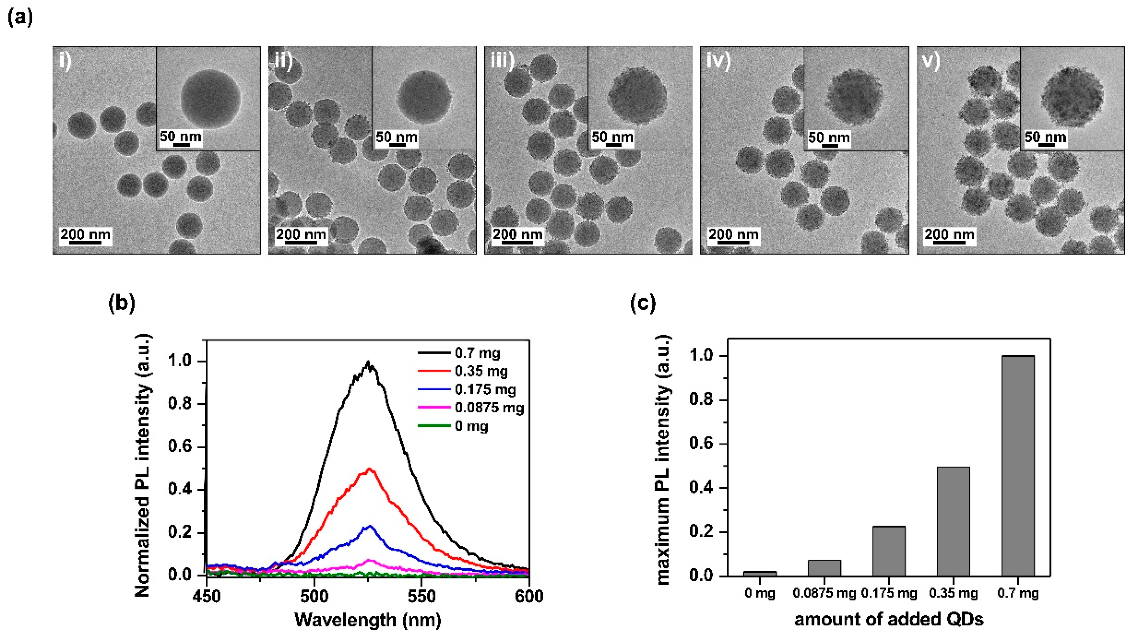
2.3. Brightness Control of SiO2@InP QDs@SiO2 NPs

To evaluate the brightness control of the SiO2@InP QDs@SiO2 NPs, SiO2@InP QDs@SiO2 NPs with different numbers of embedded QDs were fabricated, and their brightnesses were compared. The SiO2@InP QDs@SiO2 NPs, which were fabricated by varying the amount of added QDs (0, 0.0875, 0.175, 0.35, and 0.7 mg of QDs per 1 mg of SiO2 NPs) were analyzed by using TEM (Figure 3a). As the amount of added QDs increased from 0 mg to 0.7 mg per 1 mg of SiO2 NPs, QDs were densely embedded onto the surface of the SiO2 NPs. When the QDs exceeded 0.7 mg, excess QDs aggregated and did not embed onto the surface of the SiO2 NPs (Figure S4).
To evaluate the luminous variation according to the number of QDs in the SiO2@InP QDs@SiO2 NPs, the PL spectra of the SiO2@InP QDs@SiO2 NPs with different numbers of embedded QDs were compared in the visible light region (Figure 3b). As the amount of added QDs increased, the number of embedded QDs on the surface of the SiO2 NPs increased. Consequently, the PL intensity of the SiO2@InP QDs@SiO2 NPs increased. At an emission wavelength of 527 nm, the SiO2@InP QDs@SiO2 NPs exhibited maximum fluorescence, and the maximum PL intensity of the SiO2@InP QDs@SiO2 NPs also increased proportionally with the amount of added QDs (Figure 3c). Based on these results, the brightness of the SiO2@InP QDs@SiO2 NPs could be easily controlled by adjusting the amount of added QDs.

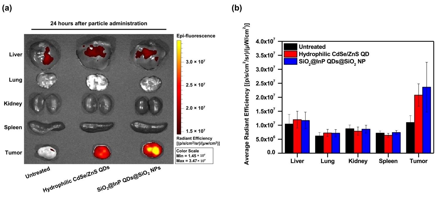
2.4. Cytotoxicity Investigation and In Vivo Biodistribution of SiO2@InP QDs@SiO2 NPs

To evaluate the suitability of the SiO2@InP QDs@SiO2 NPs for bioimaging applications, we first tested the cytotoxicity of these NPs in human cells (Figure S5). SiO2@InP QDs@SiO2 NPs and hydrophilic CdSe/ZnS QDs (3.56 × 10¹¹–1.39 × 10⁹ and 1.96 × 10¹⁴–7.66 × 10¹¹ particles/mL, respectively) were used to treat human lung cancer (A549) cells, and cell viability was determined after 24 h. When A549 cells were treated with the highest concentration of SiO2@InP QDs@SiO2 NPs or hydrophilic CdSe/ZnS QDs, cell viability was not significantly affected compared to the untreated cells (87.9 ± 6.2% and 94.2 ± 5.4% in SiO2@InP QDs@SiO2 NPs and hydrophilic CdSe/ZnS QDs, respectively). Thus, the SiO2@InP QDs@SiO2 NPs showed no significant cytotoxicity against human cells, which is appropriate for bioimaging applications in vivo.
To assess the applicability of SiO2@InP QDs@SiO2 NPs in bioimaging tumors in vivo, tumor syngeneic mice were intravenously administered SiO2@InP QDs@SiO2 NPs and hydrophilic CdSe/ZnS QDs (2.22 × 10¹¹ particles/mL each) via the tail vein. At 24 h after intravenous administration, the biodistributions of the SiO2@InP QDs@SiO2 NPs and hydrophilic CdSe/ZnS QDs in major organs (liver, lung, kidney, and spleen) and in tumors of tumor syngeneic mice were monitored by measuring the fluorescence signal of the QDs using an IVIS imaging system (Figure 4a). Owing to the enhanced permeability and retention (EPR) effect, both SiO2@InP QDs@SiO2 NPs and hydrophilic CdSe/ZnS QDs prominently accumulated at the tumor site compared to those in other major organs [51,52,53]. To investigate the residual fluorescence signal of particles accumulated in the tumor, the average radiant efficacy in major organs and tumors was measured (Figure 4b). The SiO2@InP QDs@SiO2 NPs showed significantly higher fluorescence in the tumor tissue compared to those in other organs and in the untreated control. More importantly, the retention of the SiO2@InP QDs@SiO2 NPs at the tumor site was clearly observed, with a similar or comparable efficiency to that of the hydrophilic CdSe/ZnS QDs. Hence, we suggest that SiO2@InP QDs@SiO2 NPs can be utilized as an alternative to CdSe-based QDs for bioimaging, owing to their outstanding fluorescence signal and excellent biocompatibility.

3. Materials and Methods

3.1. Materials

Indium phosphide/zinc sulfide quantum dots (InP/ZnS QDs, λem. 527 nm) were purchased from Mesolight (Suzhou, China). Cadmium selenide/zinc sulfide quantum dots (CdSe/ZnS QDs, λem. 530 nm) were purchased from ZEUS (Hwaseong, Korea). Tetraethyl orthosilicate (TEOS), 3-mercaptopropyltrimethoxysilane (MPTS), and dichloromethane (DCM) were purchased from Samchun (Pyeongtaek, Korea). Ethyl alcohol (EtOH, 99.9%) and aqueous ammonium hydroxide (NH4OH, 27%) were purchased from Daejung (Siheung, Korea). Chloroform (CHCl3, 99%), tetramethylammonium hydroxide pentahydrate (TMAH, 97%), 3-mercaptopropionic acid (MPA, 99%), paraformaldehyde, and crystal violet were purchased from Sigma-Aldrich (St. Louis, MO, USA). A549 (human non-small cell lung cancer, CCL-185) and 4T1 (mouse breast cancer, CRL-2539) cells were purchased from the American Type Culture Collection (ATCC, Manassas, VA, USA). High-glucose Dulbecco’s modified Eagle medium (DMEM) and fetal bovine serum (FBS) were purchased from Biowest (Nuaille, France). The penicillin–streptomycin solution was purchased from Welgene (Daegu, Korea). Sodium dodecyl sulfate (SDS) was purchased from LPS Solution (Daejeon, Korea). Phosphate-buffered saline (PBS; pH 7.4) was purchased from BYLABS (Hanam, Korea). Eight-week-old male BALB/c mice were purchased from Orient Bio, Inc. (Seongnam, Korea).

3.2. Preparation of Thiol-Modified Silica Nanoparticles

Silica nanoparticles (SiO2 NPs) with an average diameter of approximately 172 nm were prepared using a sol–gel process based on the Stöber method [44]. TEOS (1.6 mL) was mixed with 40 mL of EtOH. NH4OH (3.0 mL) was added to the mixture while stirring, and the mixture was allowed to react at room temperature for 20 h while stirring at 700 rpm. The SiO2 NPs were centrifuged for 15 min at 8500 rpm and washed 5 times with EtOH. After 1 mg of SiO2 NPs were dispersed in a microtube with 980 μL of EtOH, 10 μL of distilled water, 10 μL of MPTS, and 2.5 μL of NH4OH, the mixture was incubated at 50 °C for 1 h. The thiol-group-introduced SiO2 NPs were harvested after centrifugation and washed thrice with EtOH to remove the excess reagents.

3.3. Fabrication of Silica-Coated InP/ZnS QD-Embedded SiO2 NPs (SiO2@InP QDs@ SiO2 NPs)

Thiol-modified SiO2 NPs (1 mg in 100 μL EtOH) and 5 μL distilled water were added to 400 μL DCM and mixed with 0.7 mg of InP/ZnS QDs (25 mg/mL in toluene). The mixture was then incubated at room temperature for 1 h, with sonication for 2 min every 30 min. Next, 5 μL MPTS and 5 μL NH4OH were added to the mixture, and the mixture was incubated at room temperature for 1 h. InP/ZnS QD-embedded SiO2 NPs (SiO2@InP QDs NPs) were centrifuged for 10 min at 8500 rpm and washed thrice with EtOH. The washed SiO2@InP QDs NPs were dispersed in 500 μL EtOH. After dispersion, 5 μL TEOS and 5 μL NH4OH were added to the solution. The mixture was then incubated at room temperature for 20 h. Silica-coated InP/ZnS QD-embedded SiO2 NPs (SiO2@InP QDs@SiO2 NPs) were centrifuged for 10 min at 8500 rpm and washed thrice with EtOH. The SiO2@InP QDs@SiO2 NPs were then dispersed in EtOH to adjust the concentration to 1 mg/mL.

3.4. Surface Modification of CdSe/ZnS QDs

Hydrophilic CdSe/ZnS QDs were prepared by replacing hydrophobic surface ligands (oleic acid) with hydrophilic surface ligands (MPA), as previously described [50]. TMAH (100 mg) and 22.5 μL of MPA were added to 1 mL of CHCl3. The mixture was then incubated at room temperature for 1 h. Subsequently, a clear colorless aqueous layer was formed above the CHCl3 layer. The biphasic solution was mixed via vigorous shaking and allowed to equilibrate for 1 h. The organic phase at the bottom was transferred into a vial for the ligand-exchange reaction with CdSe/ZnS QDs. CdSe/ZnS QDs (0.25 mg, dispersed in 100 μL of CHCl3) were added to the MPA-CHCl3 solution and mixed well. The solution was then allowed to stand at room temperature for 3 h. After the reaction, the MPA-capped QDs separated from the CHCl3 solutions were collected, washed with CHCl3 (thrice), and dispersed in 1 mL of distilled water (final concentration: 0.025 mg/mL).

3.5. Fabrication of SiO2@InP QDs@ SiO2 NPs with Different Number of Embedded QDs

SiO2@InP QDs@SiO2 NPs with different numbers of embedded QDs were fabricated by varying the amount of added QDs with two-fold serial dilutions of 5.6, 2.8, 1.4, 0.7, 0.35, 0.175, 0.0875, and 0 mg per 1 mg of thiol-modified SiO2 NPs. Thiol-modified SiO2 NPs (1 mg in 100 μL EtOH) and 5 μL distilled water were added to 400 μL DCM and mixed with 0 mg to 5.6 mg InP/ZnS QDs (25 mg/mL in toluene). The subsequent fabrication flow for the SiO2@InP QDs@SiO2 NPs with different numbers of embedded QDs was identical to that described above for SiO2@InP QDs@SiO2 NPs.

3.6. Characterization of SiO2@InP QDs@ SiO2 NPs

Transmission electron microscopy (TEM) images of SiO2@InP QDs@SiO2 NPs were obtained using a JEM-2010 system (JEOL, Tokyo, Japan). UV/Vis/NIR absorbance spectra of the SiO2@InP QDs@SiO2 NPs were obtained using a Optizen Pop UV/Vis spectrophotometer (Mecasys, Daejeon, Korea). Photoluminescence (PL) emission spectra of the SiO2@InP QDs@SiO2 NPs were obtained using a Cary Eclipse (Agilent Technologies, Santa Clara, CA, USA). The quantum yield (QY) of the SiO2@InP QDs@SiO2 NPs was measured using a QE-2000 (Otsuka Electronics, Osaka, Japan).

3.7. Cytotoxicity Investigation of SiO2@InP QDs@ SiO2 NPs

A549 cells were maintained in high-glucose DMEM supplemented with 10% FBS, 100 mg/mL streptomycin, and 100 U/mL penicillin, and incubated at 37 °C in 5% CO2. A549 cells were seeded in 96-well plates at a density of 8 × 10³ cells/well in 100 μL of medium and grown at 37 °C for 18 h. The cells were then treated with two-fold serial diluted SiO2@InP QDs@SiO2 NPs and hydrophilic CdSe/ZnS QDs (3.56 × 10¹¹–1.39 × 10⁹ and 1.96 × 10¹⁴–7.66 × 10¹¹ particles/mL, respectively) in 100 μL medium. After incubation at 37 °C for 24 h, cells were washed with PBS and fixed with 4% paraformaldehyde at room temperature for 2 h. Next, the fixed cells were stained with 0.5% crystal violet and washed thrice with distilled water. The crystal violet-stained cells were de-stained with 1% SDS solution. Absorbance was measured at 585 nm using a VICTOR X3 Multilabel Plate Reader (PerkinElmer, Waltham, MA, USA).

3.8. In Vivo Biodistribution of SiO2@InP QDs@ SiO2 NPs

All animal experiments were approved by the Institutional Animal Care and Use Committee (IACUC) of Konkuk University. To establish tumor syngeneic mice, 1 × 10⁶ 4T1 cells were subcutaneously administered to the BALB/c mice. When the tumor had grown to approximately 400 mm3, mice were intravenously administered SiO2@InP QDs@SiO2 NPs and hydrophilic CdSe/ZnS QDs at a dose equivalent to 200 μL (2.22 × 10¹¹ particles/mL) diluted in D5W (dextrose 5% in water) via the tail vein. Mice were euthanized 24 h after intravenous administration, and the major organs (liver, lungs, kidney, spleen, and tumor) were obtained. The biodistributions of SiO2@InP QDs@SiO2 NPs and hydrophilic CdSe/ZnS QDs were monitored, and the fluorescence intensity of each organ was determined using an IVIS imaging system (Xenogen Corp., Hopkinton, MA, USA).

4. Conclusions

In summary, we fabricated highly bright and biocompatible fluorescent nanoprobes, and successfully applied them for bioimaging. SiO2@InP QDs@SiO2 NPs were fabricated by densely embedding approximately 1200–1500 InP/ZnS QDs onto the surface of the SiO2 NPs. The final fabricated SiO2@InP QDs@SiO2 NPs exhibited a QY of 6.61%. The maximum emission peak wavelength and FWMH of the SiO2@InP QDs@SiO2 NPs were 527.01 nm, and 44.62 nm, respectively. Even after the SiO2@InP QDs@SiO2 NPs were fabricated, there were no significant differences between the emission peaks and the FWHM values of the InP/ZnS QDs. In addition, the SiO2@InP QDs@SiO2 NPs showed good colloidal dispersibility in hydrophilic solvents because of the silica shell on their surface, which is advantageous for biological applications. We also confirmed that the brightness of the SiO2@InP QDs@SiO2 NPs could be easily controlled by adjusting the amount of added InP/ZnS QDs mixed with thiol-modified SiO2 NPs. The fabricated SiO2@InP QDs@SiO2 NPs selectively accumulated in the tumors of tumor syngeneic mice, with a fluorescence intensity comparable to that of hydrophilic CdSe/ZnS QDs. Thus, SiO2@InP QDs@SiO2 NPs proved to be useful in biomedical fields with high sensitivity and low toxicity, particularly for bioimaging via in vivo tumor tracking.

Supplementary Materials

The following supporting information can be downloaded at: https://www.mdpi.com/article/10.3390/ijms231810977/s1.

Author Contributions

D.-E.K. and B.-H.J. conceived the idea and designed the experiments; K.-M.H., M.K., S.B. and J.A. performed the experiments; K.-M.H., M.K., S.B., W.K., H.-M.K., J.A., H.S.J., H.S. and J.-W.K. analyzed the data; K.-M.H. and M.K. wrote the manuscript; W.-Y.R., S.H.L., S.-m.P., J.K., D.-E.K. and B.-H.J. supervised the research. All authors have read and agreed to the published version of the manuscript.

Funding

This research was funded by the Ministry of Science and ICT (NRF-2022R1A2C2012883), and supported by the Konkuk University Researcher Fund in 2020 (2020-A019-0280). Further, this research was supported by the Korea Initiative for fostering University of Research and Innovation Program of the National Research Foundation (NRF), funded by the Korean government (MSIT) (No. NRF2021M3H1A104892211).

Institutional Review Board Statement

All animal experiments were approved by the Institutional Animal Care and Use Committee (IACUC) of Konkuk University. The approval number of IACUC of Konkuk University is KU22098.

Informed Consent Statement

Not applicable.

Data Availability Statement

Not applicable.

Acknowledgments

The authors give thanks for the financial support from Konkuk University. Further, the authors give thanks for the financial support from the NRF of Korea.

Conflicts of Interest

The authors declare no conflict of interest.

References

  1. Bruchez, M., Jr.; Moronne, M.; Gin, P.; Weiss, S.; Alivisatos, A.P. Semiconductor nanocrystals as fluorescent biological labels. Science 1998, 281, 2013–2016. [Google Scholar] [CrossRef] [PubMed]
  2. Chan, W.C.; Nie, S. Quantum dot bioconjugates for ultrasensitive nonisotopic detection. Science 1998, 281, 2016–2018. [Google Scholar] [CrossRef] [PubMed]
  3. Chan, W.C.; Maxwell, D.J.; Gao, X.; Bailey, R.E.; Han, M.; Nie, S. Luminescent quantum dots for multiplexed biological detection and imaging. Curr. Opin. Biotechnol. 2002, 13, 40–46. [Google Scholar] [CrossRef]
  4. Dabbousi, B.O.; Rodriguez-Viejo, J.; Mikulec, F.V.; Heine, J.R.; Mattoussi, H.; Ober, R.; Jensen, K.F.; Bawendi, M.G. (CdSe) ZnS core–shell quantum dots: Synthesis and characterization of a size series of highly luminescent nanocrystallites. J. Phys. Chem. B 1997, 101, 9463–9475. [Google Scholar] [CrossRef]
  5. Lim, Y.T.; Kim, S.; Nakayama, A.; Stott, N.E.; Bawendi, M.G.; Frangioni, J.V. Selection of quantum dot wavelengths for biomedical assays and imaging. Mol. Imaging 2003, 2, 15353500200302163. [Google Scholar] [CrossRef]
  6. Park, S.-M.; Aalipour, A.; Vermesh, O.; Yu, J.H.; Gambhir, S.S. Towards clinically translatable in vivo nanodiagnostics. Nat. Rev. Mater. 2017, 2, 17014. [Google Scholar] [CrossRef]
  7. Tarrahi, R.; Movafeghi, A.; Khataee, A.; Rezanejad, F.; Gohari, G. Evaluating the toxic impacts of cadmium selenide nanoparticles on the aquatic plant Lemna minor. Molecules 2019, 24, 410. [Google Scholar] [CrossRef]
  8. Soenen, S.J.; Rivera-Gil, P.; Montenegro, J.-M.; Parak, W.J.; De Smedt, S.C.; Braeckmans, K. Cellular toxicity of inorganic nanoparticles: Common aspects and guidelines for improved nanotoxicity evaluation. Nano Today 2011, 6, 446–465. [Google Scholar] [CrossRef]
  9. Tsoi, K.M.; Dai, Q.; Alman, B.A.; Chan, W.C. Are quantum dots toxic? Exploring the discrepancy between cell culture and animal studies. Acc. Chem. Res. 2013, 46, 662–671. [Google Scholar] [CrossRef]
  10. Brunetti, V.; Chibli, H.; Fiammengo, R.; Galeone, A.; Malvindi, M.A.; Vecchio, G.; Cingolani, R.; Nadeau, J.L.; Pompa, P.P. InP/ZnS as a safer alternative to CdSe/ZnS core/shell quantum dots: In vitro and in vivo toxicity assessment. Nanoscale 2013, 5, 307–317. [Google Scholar] [CrossRef]
  11. Wu, T.; Tang, M. Toxicity of quantum dots on respiratory system. Inhal. Toxicol. 2014, 26, 128–139. [Google Scholar] [CrossRef] [PubMed]
  12. Grim, J.Q.; Manna, L.; Moreels, I. A sustainable future for photonic colloidal nanocrystals. Chem. Soc. Rev. 2015, 44, 5897–5914. [Google Scholar] [CrossRef] [PubMed]
  13. Yoffe, A.D. Semiconductor quantum dots and related systems: Electronic, optical, luminescence and related properties of low dimensional systems. Adv. Phys. 2001, 50, 1–208. [Google Scholar] [CrossRef]
  14. Chen, B.; Li, D.; Wang, F. InP quantum dots: Synthesis and lighting applications. Small 2020, 16, 2002454. [Google Scholar] [CrossRef]
  15. Adam, S.; Talapin, D.; Borchert, H.; Lobo, A.; McGinley, C.; De Castro, A.; Haase, M.; Weller, H.; Möller, T. The effect of nanocrystal surface structure on the luminescence properties: Photoemission study of HF-etched InP nanocrystals. J. Chem. Phys. 2005, 123, 084706. [Google Scholar] [CrossRef] [PubMed]
  16. Reiss, P.; Carriere, M.; Lincheneau, C.; Vaure, L.; Tamang, S. Synthesis of semiconductor nanocrystals, focusing on nontoxic and earth-abundant materials. Chem. Rev. 2016, 116, 10731–10819. [Google Scholar] [CrossRef]
  17. Haubold, S.; Haase, M.; Kornowski, A.; Weller, H. Strongly luminescent InP/ZnS core–shell nanoparticles. ChemPhysChem 2001, 2, 331–334. [Google Scholar] [CrossRef]
  18. Xie, R.; Battaglia, D.; Peng, X. Colloidal InP nanocrystals as efficient emitters covering blue to near-infrared. J. Am. Chem. Soc. 2007, 129, 15432–15433. [Google Scholar] [CrossRef]
  19. Xu, S.; Ziegler, J.; Nann, T. Rapid synthesis of highly luminescent InP and InP/ZnS nanocrystals. J. Mater. Chem. 2008, 18, 2653–2656. [Google Scholar] [CrossRef]
  20. Fan, G.; Wang, C.; Fang, J. Solution-based synthesis of III–V quantum dots and their applications in gas sensing and bio-imaging. Nano Today 2014, 9, 69–84. [Google Scholar] [CrossRef]
  21. Yong, K.-T.; Ding, H.; Roy, I.; Law, W.-C.; Bergey, E.J.; Maitra, A.; Prasad, P.N. Imaging pancreatic cancer using bioconjugated InP quantum dots. ACS Nano 2009, 3, 502–510. [Google Scholar] [CrossRef] [PubMed]
  22. Li, C.; Hosokawa, C.; Suzuki, M.; Taguchi, T.; Murase, N. Preparation and biomedical applications of bright robust silica nanocapsules with multiple incorporated InP/ZnS quantum dots. New J. Chem. 2018, 42, 18951–18960. [Google Scholar] [CrossRef]
  23. Xu, Y.; Lv, Y.; Wu, R.; Li, J.; Shen, H.; Yang, H.; Zhang, H.; Li, L.S. Sensitive Immunoassay based on biocompatible and robust silica-coated Cd-free InP-based quantum dots. Inorg. Chem. 2021, 60, 6503–6513. [Google Scholar] [CrossRef]
  24. Watanabe, T.; Iso, Y.; Isobe, T.; Sasaki, H. Photoluminescence color stability of green-emitting InP/ZnS core/shell quantum dots embedded in silica prepared via hydrophobic routes. RSC Adv. 2018, 8, 25526–25533. [Google Scholar] [CrossRef] [PubMed]
  25. Zhang, L.; Yang, X.-Q.; An, J.; Zhao, S.-D.; Zhao, T.-Y.; Tan, F.; Cao, Y.-C.; Zhao, Y.-D. In vivo tumor active cancer targeting and CT-fluorescence dual-modal imaging with nanoprobe based on gold nanorods and InP/ZnS quantum dots. J. Mater. Chem. B 2018, 6, 2574–2583. [Google Scholar] [CrossRef] [PubMed]
  26. Sharma, P.; Brown, S.; Walter, G.; Santra, S.; Moudgil, B. Nanoparticles for bioimaging. Adv. Colloid Interface Sci. 2006, 123, 471–485. [Google Scholar] [CrossRef]
  27. Brown, R.P.; Gallagher, M.J.; Fairbrother, D.H.; Rosenzweig, Z. Synthesis and degradation of cadmium-free InP and InPZn/ZnS quantum dots in solution. Langmuir 2018, 34, 13924–13934. [Google Scholar] [CrossRef]
  28. Kim, J.; Hwang, D.W.; Jung, H.S.; Kim, K.W.; Pham, X.-H.; Lee, S.-H.; Byun, J.W.; Kim, W.; Kim, H.-M.; Hahm, E. High-quantum yield alloy-typed core/shell CdSeZnS/ZnS quantum dots for bio-applications. J. Nanobiotechnol. 2022, 20, 22. [Google Scholar] [CrossRef]
  29. Pham, X.-H.; Park, S.-M.; Ham, K.-M.; Kyeong, S.; Son, B.S.; Kim, J.; Hahm, E.; Kim, Y.-H.; Bock, S.; Kim, W. Synthesis and application of silica-coated quantum dots in biomedicine. Int. J. Mol. Sci. 2021, 22, 10116. [Google Scholar] [CrossRef]
  30. Huang, L.; Liao, T.; Wang, J.; Ao, L.; Su, W.; Hu, J. Brilliant pitaya-type silica colloids with central–radial and high-density quantum dots incorporation for ultrasensitive fluorescence immunoassays. Adv. Funct. Mater. 2018, 28, 1705380. [Google Scholar] [CrossRef]
  31. Li, D.-Y.; He, X.-W.; Chen, Y.; Li, W.-Y.; Zhang, Y.-K. Novel hybrid structure silica/CdTe/molecularly imprinted polymer: Synthesis, specific recognition, and quantitative fluorescence detection of bovine hemoglobin. ACS Appl. Mater. Interfaces 2013, 5, 12609–12616. [Google Scholar] [CrossRef] [PubMed]
  32. Ha, Y.; Jung, H.S.; Jeong, S.; Kim, H.M.; Kim, T.H.; Cha, M.G.; Kang, E.J.; Pham, X.H.; Jeong, D.H.; Jun, B.H. Fabrication of Remarkably Bright QD Densely-Embedded Silica Nanoparticle. Bull. Korean Chem. Soc. 2019, 40, 9–13. [Google Scholar] [CrossRef]
  33. Jun, B.H.; Hwang, D.W.; Jung, H.S.; Jang, J.; Kim, H.; Kang, H.; Kang, T.; Kyeong, S.; Lee, H.; Jeong, D.H. Ultrasensitive, Biocompatible, Quantum-Dot-Embedded Silica Nanoparticles for Bioimaging. Adv. Funct. Mater. 2012, 22, 1843–1849. [Google Scholar] [CrossRef]
  34. Kim, H.-M.; Oh, C.; An, J.; Baek, S.; Bock, S.; Kim, J.; Jung, H.-S.; Song, H.; Kim, J.-W.; Jo, A. Multi-quantum dots-embedded silica-encapsulated nanoparticle-based lateral flow assay for highly sensitive exosome detection. Nanomaterials 2021, 11, 768. [Google Scholar] [CrossRef] [PubMed]
  35. Bock, S.; Kim, H.-M.; Kim, J.; An, J.; Choi, Y.-S.; Pham, X.-H.; Jo, A.; Ham, K.-M.; Song, H.; Kim, J.-W. Lateral Flow Immunoassay with Quantum-Dot-Embedded Silica Nanoparticles for Prostate-Specific Antigen Detection. Nanomaterials 2021, 12, 33. [Google Scholar] [CrossRef]
  36. Jo, A.; Kim, T.H.; Kim, D.-M.; Kim, H.-M.; Seong, B.; Kim, J.; Pham, X.-H.; Jung, H.S.; Lee, S.H.; Jeong, D.H. Sensitive detection of virus with broad dynamic range based on highly bright quantum dot-embedded nanoprobe and magnetic beads. J. Ind. Eng. Chem. 2020, 90, 319–326. [Google Scholar] [CrossRef]
  37. Chen, L.; Chen, C.; Li, R.; Li, Y.; Liu, S. CdTe quantum dot functionalized silica nanosphere labels for ultrasensitive detection of biomarker. Chem. Commun. 2009, 19, 2670–2672. [Google Scholar] [CrossRef]
  38. Huang, L.; Wu, Q.; Wang, J.; Foda, M.; Liu, J.; Cai, K.; Han, H. A brilliant sandwich type fluorescent nanostructure incorporating a compact quantum dot layer and versatile silica substrates. Chem. Commun. 2014, 50, 2896–2899. [Google Scholar] [CrossRef]
  39. Li, J.; Lv, Y.; Li, N.; Wu, R.; Xing, M.; Shen, H.; Li, L.S.; Chen, X. Robust synthesis of bright multiple quantum dot-embedded nanobeads and its application to quantitative immunoassay. Chem. Eng. J. 2019, 361, 499–507. [Google Scholar] [CrossRef]
  40. Hu, Y.; Fu, A.; Miao, Z.; Zhang, X.; Wang, T.; Kang, A.; Shan, J.; Zhu, D.; Li, W. Fluorescent ligand fishing combination with in-situ imaging and characterizing to screen Hsp 90 inhibitors from Curcuma longa L. based on InP/ZnS quantum dots embedded mesoporous nanoparticles. Talanta 2018, 178, 258–267. [Google Scholar] [CrossRef] [PubMed]
  41. Iso, Y.; Isobe, T. Critical Review—Photostable Fluorescent Cd-Free Quantum Dots Transparently Embedded in Monolithic Silica. ECS J. Solid State Sci. Technol. 2019, 9, 016005. [Google Scholar] [CrossRef]
  42. Miao, Z.; Hu, Y.; Zhang, X.; Yang, X.; Tang, Y.; Kang, A.; Zhu, D. Screening and identification of ligand-protein interactions using functionalized heat shock protein 90-fluorescent mesoporous silica-indium phosphide/zinc sulfide quantum dot nanocomposites. J. Chromatogr. A 2018, 1562, 1–11. [Google Scholar] [CrossRef] [PubMed]
  43. Perton, F.; Harlepp, S.; Follain, G.; Parkhomenko, K.; Goetz, J.G.; Bégin-Colin, S.; Mertz, D. Wrapped stellate silica nanocomposites as biocompatible luminescent nanoplatforms assessed in vivo. J. Colloid Interface Sci. 2019, 542, 469–482. [Google Scholar] [CrossRef]
  44. Stöber, W.; Fink, A.; Bohn, E. Controlled growth of monodisperse silica spheres in the micron size range. J. Colloid Interface Sci. 1968, 26, 62–69. [Google Scholar] [CrossRef]
  45. Kim, S.; Bawendi, M.G. Oligomeric ligands for luminescent and stable nanocrystal quantum dots. J. Am. Chem. Soc. 2003, 125, 14652–14653. [Google Scholar] [CrossRef]
  46. Rogach, A.L.; Nagesha, D.; Ostrander, J.W.; Giersig, M.; Kotov, N.A. “Raisin bun”-type composite spheres of silica and semiconductor nanocrystals. Chem. Mater. 2000, 12, 2676–2685. [Google Scholar] [CrossRef]
  47. Shibu, E.S.; Hamada, M.; Nakanishi, S.; Wakida, S.-I.; Biju, V. Photoluminescence of CdSe and CdSe/ZnS quantum dots: Modifications for making the invisible visible at ensemble and single-molecule levels. Coord. Chem. Rev. 2014, 263, 2–12. [Google Scholar] [CrossRef]
  48. Green, M. Semiconductor quantum dots as biological imaging agents. Angew. Chem. Int. Ed. 2004, 43, 4129–4131. [Google Scholar] [CrossRef]
  49. Wagner, A.M.; Knipe, J.M.; Orive, G.; Peppas, N.A. Quantum dots in biomedical applications. Acta Biomater. 2019, 94, 44–63. [Google Scholar] [CrossRef]
  50. Pong, B.-K.; Trout, B.L.; Lee, J.-Y. Modified ligand-exchange for efficient solubilization of CdSe/ZnS quantum dots in water: A procedure guided by computational studies. Langmuir 2008, 24, 5270–5276. [Google Scholar] [CrossRef]
  51. Kalyane, D.; Raval, N.; Maheshwari, R.; Tambe, V.; Kalia, K.; Tekade, R.K. Employment of enhanced permeability and retention effect (EPR): Nanoparticle-based precision tools for targeting of therapeutic and diagnostic agent in cancer. Mater. Sci. Eng. C 2019, 98, 1252–1276. [Google Scholar] [CrossRef] [PubMed]
  52. Stylianopoulos, T. EPR-effect: Utilizing size-dependent nanoparticle delivery to solid tumors. Ther. Deliv. 2013, 4, 421–423. [Google Scholar] [CrossRef] [PubMed]
  53. Kang, H.; Rho, S.; Stiles, W.R.; Hu, S.; Baek, Y.; Hwang, D.W.; Kashiwagi, S.; Kim, M.S.; Choi, H.S. Size-dependent EPR effect of polymeric nanoparticles on tumor targeting. Adv. Healthc. Mater. 2020, 9, 1901223. [Google Scholar] [CrossRef] [PubMed]
Figure 1. Fabrication of SiO2@InP QDs@SiO2 NPs. (a) Schematic illustration for fabrication of SiO2@InP QDs@SiO2 NPs. (b) Transmission electron microscopy (TEM) images of (i) SiO2 NPs, (ii) InP/ZnS QDs (iii), and (iv) SiO2@InP QDs@SiO2 NPs.
Ijms 23 10977 g001
Figure 2. Characterization of SiO2@InP QDs@SiO2 NPs. (a) UV/Vis/NIR absorbance spectra of SiO2 NPs, InP/ZnS QDs, and SiO2@InP QDs@SiO2 NPs. (b) Comparison of quantum yield (QY) between InP/ZnS QDs and SiO2@InP QDs@SiO2 NPs. (c) Comparison of photoluminescence (PL) spectra between InP/ZnS QDs and SiO2@InP QDs@SiO2 NPs. (d) Digital images of SiO2@InP QDs@SiO2 NPs distributed in distilled water (DW).
Ijms 23 10977 g002
Figure 3. Brightness control of SiO2@InP QDs@SiO2 NPs. (a) TEM image of SiO2@InP QDs@SiO2 NPs via the amount of added QDs. The amount of added QDs were (i) 0 mg, (ii) 0.0875 mg, (iii) 0.175 mg, (iv) 0.35 mg, and (v) 0.7mg per 1 mg of SiO2 NPs. (b) Normalized PL intensity spectra of SiO2@InP QDs@SiO2 NPs via amount of added QDs. (c) Maximum PL intensity of SiO2@InP QDs@SiO2 NPs via amount of added QDs at 527 nm emission wavelength.
Ijms 23 10977 g003
Figure 4. In vivo biodistribution of SiO2@InP QDs@SiO2 NPs. (a) Comparison of biodistribution and fluorescence of particles in major organs and tumors after administration of SiO2@InP QDs@SiO2 NPs and hydrophilic CdSe/ZnS QDs to tumor syngeneic mice. (b) Comparison of average radiant efficiency in major organs and tumors.
Ijms 23 10977 g004
Publisher’s Note: MDPI stays neutral with regard to jurisdictional claims in published maps and institutional affiliations.

