Review

Distinct Responses to Pathogenic and Symbionic Microorganisms: The Role of Plant Immunity

Shandong Provincial Key Laboratory of Animal Resistance Biology, Institute of Biomedical Sciences, College of Life Sciences, Shandong Normal University, Jinan 250014, China
*
Author to whom correspondence should be addressed.
These authors contributed equally to this work.
Int. J. Mol. Sci. 2022, 23(18), 10427; https://doi.org/10.3390/ijms231810427
Submission received: 12 August 2022 / Revised: 6 September 2022 / Accepted: 7 September 2022 / Published: 9 September 2022
(This article belongs to the Section Molecular Microbiology)

Abstract

:
Plants must balance both beneficial (symbiotic) and pathogenic challenges from microorganisms, the former benefitting the plant and agriculture and the latter causing disease and economic harm. Plant innate immunity describes a highly conserved set of defense mechanisms that play pivotal roles in sensing immunogenic signals associated with both symbiotic and pathogenic microbes and subsequent downstream activation of signaling effector networks that protect the plant. An intriguing question is how the innate immune system distinguishes “friends” from “foes”. Here, we summarize recent advances in our understanding of the role and spectrum of innate immunity in recognizing and responding to different microbes. In addition, we also review some of the strategies used by microbes to manipulate plant signaling pathways and thus evade immunity, with emphasis on the use of effector proteins and micro-RNAs (miRNAs). Furthermore, we discuss potential questions that need addressing to advance the field of plant–microbe interactions.

1. Introduction

The constant exposure of plants to microorganisms in the surrounding environment can result in disease and consequent severe economic damage to agriculture [1]. To protect themselves from pathogen attacks, plants have evolved a sophisticated, multilayered immune system involving constitutive and inducible defenses to detect pathogenic microbes [2]. Unlike vertebrates, plants mainly rely on innate immunity to combat pathogens due to their lack of an adaptive immune system.
Plants detect microbes through recognition of specific microbial structures by pattern recognition receptors (PRRs), which always localize at the plasma membrane and work with coreceptors [3]. PRRs consist of receptor-like kinases (RLKs) and receptor-like proteins (RLPs): RLKs possess an intracellular kinase domain, a transmembrane domain, and an extracellular domain, while RLPs lack the intracellular domain but contain the other two domains [4,5,6,7]. RLKs and RLPs, as the first defense barrier, detect microbe-associated molecular patterns (MAMPs) to initiate pattern-triggered immunity (PTI). However, some pathogens can evade PTI by secreting effector proteins into plant cells to interfere with MAMP detection and/or subsequent signal transduction, with consequent disease development. Plants have in turn evolved another robust and rapid immune response, namely effector-triggered immunity (ETI), to detect pathogen effectors, a process that depends on intracellular nucleotide-binding leucine-rich repeat (NLR) proteins [8]. Plant NLRs comprise a C-terminal leucine-rich repeat (LRR) domain, a central nucleotide-binding (NB-ARC) domain, and an N-terminal domain that may be a coiled-coil domain, a resistance to powdery mildew 8 (RPW8) domain, or a toll/interleukin-1 receptor homology (TIR) domain [9,10]. Recently, the structures of some NLRs have been resolved, providing a basis for understanding plant NLRs [9,11,12,13]. Generally, PRRs function as extracellular immune receptors while NLRs mediate intracellular defenses. Together, PRRs and NLRs cooperate to defend against invaders.
However, some microorganisms can establish a symbiotic relationship with the plant that is beneficial to the plant host. For example, arbuscular mycorrhizal (AM) fungi collaborate with most land plants to improve their phosphorus nutrition through the establishment of symbiotic interactions. In addition, another widely studied symbiosis is the relationship between rhizobia and legume plants, in which rhizobia fix atmospheric nitrogen and transfer it to the plant. In these cases, beneficial microbes are subjected to the plant immune system as pathogens, but they could survive. However, the underlying mechanism(s) dictating this outcome remains uncertain. Is symbiosis a result of an absent immune response upon recognition of beneficial microbes or do symbiotic microbes overcome plant defense mechanisms? How plants distinguish “friend” from “foe” is a further intriguing question, particularly as MAMPs are conserved in both symbiotic and pathogenic microbes [14]. Emerging evidence suggests that the establishment of symbiotic interactions is an intricate process that requires contributions from both plants and microorganisms. In this review, we highlight the importance of plant immunity in the recognition of different microbes and discuss how plant immune pathways are tightly manipulated in symbiotic relationships, focusing on the association between plants and AM fungi and rhizobia. Understanding the molecular mechanisms underpinning immune responses and tolerance will give us some guidance to foster beneficial microbial symbiosis while repudiating pathogenic interaction and thus, ultimately, contribute to improving crop production.

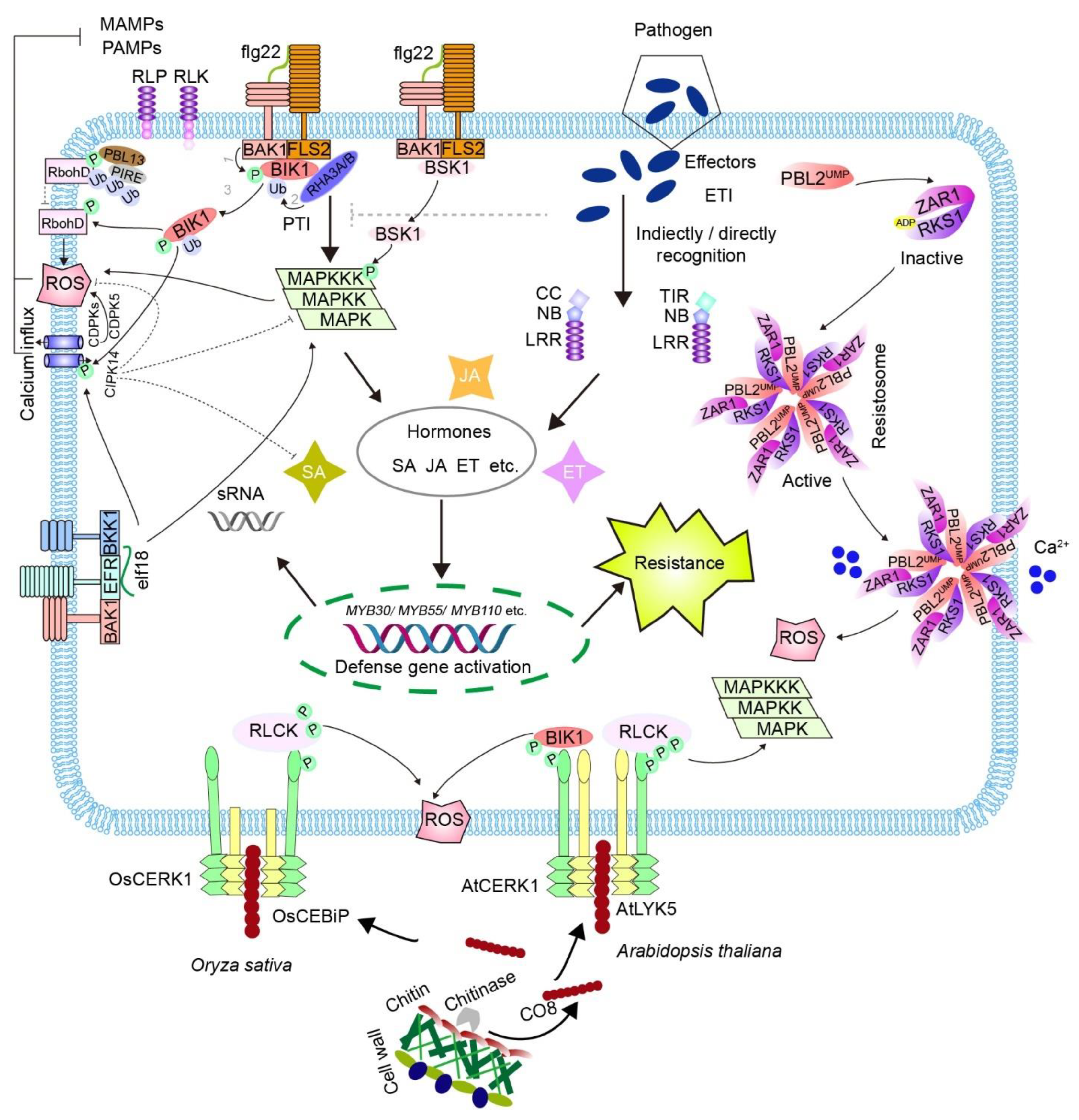
2. The Plant Immune System

The plant immune system includes local immunity with PTI and ETI as well as systemic immunity with systemic acquired resistance (SAR) and induced systemic immunity (ISR). Systemic immunity refers to systemic signals spread from the affected site to unaffected tissues to enhance resistance to a subsequent secondary stress. For example, the differentiated outer cell layers display low protein level of PRRs and deficient PTI in Arabidopsis thaliana. However, these cells could be induced to highly express PRRs and be responsive to pathogens by neighbor cell damage, suggesting that a local immune response could be induced in nonresponsive cells [15]. SAR is triggered by pathogens and is primarily associated with salicylic acid (SA)-dependent signaling [16], whereas ISR is induced by beneficial microbes with jasmonic acid (JA) or ethylene (ET) signaling independent of SA [17]. The SA and JA pathways have a well-documented antagonistic relationship [18]. In addition, a novel immune strategy, termed general non-self response (GNSR), has been identified in Arabidopsis thaliana. GNSR involves a core set of 24 genes that are consistently induced in plant responses to the microbiome, indicating that plants use their immune systems to shape their microbiota [19]. Here, we focus on how the plant innate immunity functions appropriately during its interactions with microbes.
Plants detect microbes through MAMPs or microbial effectors. Plenty of MAMPs have been identified, including β-glucans and the eicosapolyenoic acid (EP) of oomycete, bacterial flagellin peptide flg22, peptidoglycan (PGN), lipopolysaccharide (LPS), EF-Tu peptide elf18, lipopeptides, fungal wall component chitin, and chitosan [20]. The first layer of the immune response begins when PRRs sense MAMPs. This then triggers the activation of downstream plasma membrane-associated receptor-like cytoplasmic kinases (RLCKs), followed by a ROS burst, calcium influx, and mitogen-activated protein kinase (MAPK) cascades encompassing a MAPK kinase kinase (MAPKKK), a MAPK kinase (MAPKK), and a MAPK (MPK) (Figure 1) [21,22]. In addition, MPK cascade activation by chitin elicitor induces the expression of MYB transcription factors MYB30, MYB55, and MYB110, resulting in the accumulation of ferulic acid together with enhanced resistance to pathogens in rice [23]. These studies indicate that robust defense response involves multiple alterations in gene expression, protein modifications, and synthesis of secondary metabolites.
Several RLCKs are thought to link PRRs to key downstream signaling pathways, such as RLCK subfamily II components in Arabidopsis: botrytis-induced kinase 1 (BIK1), PBS1-like 1 (PBL1), and brassinosteroid signaling kinase 1 (BSK1) [22,24,25,26,27]. The extensively studied bacterial PAMPs that elicit immune responses are flg22 and elf18. For instance, once flg22 is detected, FLS2 instantaneously forms a complex with brassinosteroid insensitive 1-associated kinase 1 (BAK1) and concomitantly interacts with BIK1 [24,25]. Subsequently, BIK1 is phosphorylated by BAK1, followed by monoubiquitination by ring-H2 finger A3A (RHA3A) and RHA3B and then dissolution from the FLS2-BAK1 complex to activate immune signaling [28]. As a serine/threonine kinase, activated BIK1 subsequently phosphorylates the N terminus of NADPH oxidase RbohD to promote ROS production [29,30] and cyclic nucleotide-gated channels to increase the concentration of cyclic calcium [31]. Many calcium-dependent kinases (CDPKs), including CDPK5, are also involved in ROS production [32]. However, another Ca2+ sensor, CBL-interacting protein kinase 14 (CIPK14), negatively regulates MAPK-mediated immune responses and SA production [33]. In addition, flg22 activates MAPK cascades to upregulate defense genes [34,35,36]. It has been shown that BSK1, another RLCK member, may transduce upstream cues to activate MAPK cascades. Similar to BIK1, BSK1 associates with FLS2 in the absence of flg22, but disassociates from the complex when flg22 is detected [27]. Activated BSK1 then phosphorylates MAPKKK5 which associates with multiple MAPK kinases to induce downstream signals [36]. Similarly, EFR forms a complex with BAK1 and BAK1-LIKE 1 (BKK1) [37,38] and shares common signaling pathways with FLS2 in response to elf18 [39,40].
Additionally, plants also have powerful mechanisms to sense fungi, which mainly depend on the recognition of chitin. Chitin, a conserved structural component of fungal cell walls, is a polymer of β-1,4-linked N-acetylglucosamine (GlcNAc). To avoid fungal infection, plants secrete chitinases that hydrolyze chitin and release chitooligosaccharides (COs). Chitin hexamers (CO-6) or octamers (CO-8), as breakdown products, are recognized by lysin motif (LysM)-containing RLPs or RLKs to initiate intracellular immune signaling (Figure 1) [41,42]. In Arabidopsis thaliana, chitin is detected by the LysM-receptor kinases AtCERK1 and AtLYK5, in which AtLYK5 has a higher affinity for COs, while AtCERK1 is an indispensable coreceptor with modest affinity [43]. In addition, AtLYK4 was shown to be involved in chitin signaling by associating with AtLYK5 [44], or recognizing COs directly [45]. Notably, AtCERK1, but not AtLYK5 or AtLYK4, has kinase activity, suggesting that AtCERK1 is responsible for initiating immune responses. It has been shown that chitin rapidly induces autophosphorylation of AtCERK1 at multiple residues in the kinase and juxtamembrane domains, which is pivotal for chitin-induced signaling [46]. Activated AtCERK1 thus activates downstream members of RLCK subfamily VII which contains 46 members in Arabidopsis thaliana, including PBL1 to PBL43, PBS1, CDG1, and BIK1 [24,26,47,48,49,50]. In brief, chitin initiates the removal of BIK1 from AtCERK1, and BIK1 in turn phosphorylates and activates NADPH oxidase and cyclic nucleotide-gated channel proteins, such as CNGC2 and CNGC4, leading to ROS production [29] and increased cytosolic calcium [31]. In addition, RLCK VII-4 subfamily members, including PBL19, could phosphorylate MAPKKK5 to activate MAPKKK5-MKK4/5-MPK3/6 cascades and MEKK1 to promote MPK4 activation and defense gene expression [34]. Moreover, activated MPK3/6 and MPK4 in turn phosphorylate MAPKKK5 and MEKK1, serving as positive feedback mechanisms [34].
In Oryza sativa, chitin detection involves OsCEBiP and OsCERK1. Although OsCEBiP binds COs with higher affinity, it does not have a cytoplasmic domain [51]. Considering that OsCERK1 does not associate with chitin but possesses a kinase domain, it is thought that OsCEBiP interacts with chitin and subjects it to its partner OsCERK1 to induce immune signaling [51,52]. Briefly, a single chitin fragment crosslinks two OsCEBiP molecules to promote OsCEBiP dimerization, after which OsCEBiP associates with OsCERK1 to form an active hetero-oligomeric receptor complex, which has been suggested to act as a “sandwich-like” homodimer [52,53]. In this model, OsCERK1 is activated via autophosphorylation and in turn interacts with and phosphorylates OsRLCK185, a member of the RLCK VII family. Subsequently, OsRLCK185 phosphorylates distinct substrates to initiate an immune response [54,55,56,57,58]. In addition, other RLCK VII members, like OsRLCK118 and OsRLCK176, participate in ROS generation by phosphorylating the NADPH oxidase OsRbohB [59].
Although successful pathogens can escape the first layer of plant innate immunity by transferring effector proteins to host plants, plant cells employ NLRs to recognize these effectors both directly and indirectly, and initiate ETI to drive the infected cell to programmed cell death. For example, AvrPphB secreted by Pseudomonas syringae cleaves RLCK PBS1 to suppress PTI [24,60]. However, this process could be monitored by NLR protein resistance to pseudomonas syringae 5 (RPS5) and elicits RPS5-specified ETI [60]. Likewise, the effector protein AvrRpt2 activates NLR protein resistance to pseudomonas syringae 2 (RPS2) through eliminating the RPS2-bound RIN4 protein in Arabidopsis [61,62]. Additionally, the tomato NLR protein Prf associates with Pto kinase to recognize the effector protein AvrPto [63]. Recently, a pan-genomic analysis of numerous Pseudomonas syringae strains identified many unique effector protein sequences that are surprisingly recognized by only a small number of NLRs, suggesting a pervasive role for ETI [64]. Notably, the molecular mechanisms underlying plant NLR activation have recently begun to be revealed by a series of elegant studies that reconstituted and solved the structure of Arabidopsis NLR receptor HOPZ-ACTIVATED RESISTANCE 1 (ZAR1). In two recent seminal articles [9,12], the authors first investigated the 3D structure of the ZAR1-RKS1 complex and showed that the ZAR1-RKS1-PBL2UMP complex induced the replacement of an ADP molecule by an ATP molecule with ZAR1, resulting in the full activation of this complex and formation of the pentameric ZAR1 resistosome [9,65]. More importantly, the ZAR1 resistosome has been shown to form pentameric complexes in the plant cell plasma membrane, where it acts as a calcium-permeable channel triggering plant immune signaling [11]. The discovery of the ZAR1 resistosome is a breakthrough in our understanding of NLR-mediated immunity.
In summary, it is widely accepted that the RLP/RLK-RLCK signaling module plays important roles in MAMP-triggered immune responses. However, the exact contributions of most RLCK members to immune signaling still require further clarification. In addition, the immune signaling pathways are also controlled by negative regulators. For example, the complex formed from RLCK PBL13 and an E3 ligase PIRE phosphorylates RbohD and enhances its ubiquitination to negatively regulate ROS production during immunity [66]. Moreover, great efforts are also needed to uncover how NLR proteins recognize various effectors and initiate immune responses.

3. Interplay between MAMP- and Effector-Triggered Immunity

Remarkably, several recent studies have suggested that PRR- and NLR-mediated signaling extensively cross-talk to significantly influence immune response outcomes [10,67,68,69,70,71,72]. For example, the NLR protein recognition of Peronospora parasitica 2 (RPP2)- and RPP4-mediated ETI-associated pathogen restriction in Arabidopsis against Hyaloperonospora arabidopsidis (Hpa) races Emoy2 and Cala2 requires the RLK/PLP co-receptors BAK1 and BKK1 [37]. Furthermore, avrRpt2 activates RPS2-dependent ETI in wild-type plants [61,62] but fails to elicit effective ETI in fls2/efr/cerk1 and bak1/bkk1/cerk1 triple mutants in Arabidopsis [68,73]. Moreover, BIK1 has recently been identified to be required for ROS burst, immune gene expression, and resistance during ETI [68]. Consistently, pre-activation of ETI by AvrPps4 increases the flg22-induced production of ROS, suggesting that ETI enhances PTI responses [74]. In addition, it has been recently shown that MPK3/6 control the SA component of defense through the expression of some NLR genes, thereby bridging PTI and ETI [69]. Similarly, activation of TIR signaling fosters PTI, as demonstrated by a recent study showing that PTI is attenuated in plants with reduced NLR protein levels [70]. Taken together, there is increasing evidence that PTI and ETI cooperate to guarantee robust immunity, although the precise molecular mechanisms still need further exploration.

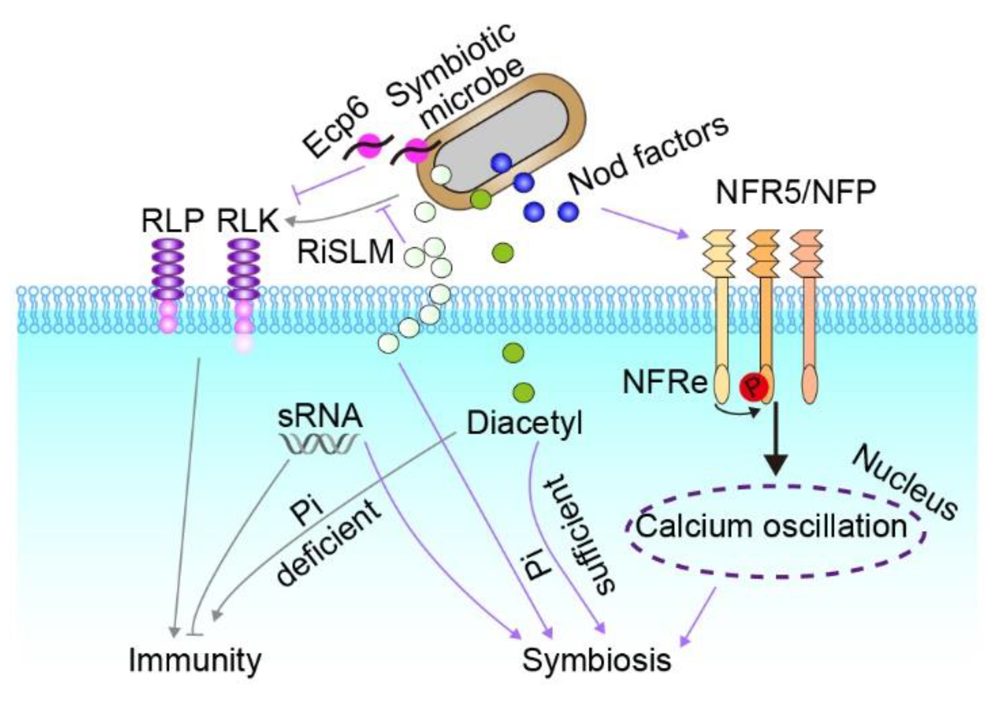
4. Establishment of Symbiosis

The symbiotic interaction between rhizobia and leguminous plants involves multiple processes that begin with pre-infection events in the rhizosphere [75]. Under nitrogen-limiting conditions, flavonoids are produced by legume roots and are secreted into the rhizosphere to recruit symbiotic partners [76]. When flavonoids are detected by rhizobia, lipochitooligosaccharides (LCOs), known as Nod factors, are synthesized [75,77,78]. In symbiosis, Nod factors are also perceived by LysM-RLKs. In Lotus japonicus, Nod factors are recognized by NFR1 kinase and NFR5 pseudokinase, whereas the kinase and pseudokinase are LYK3 and NFP, respectively, in Medicago truncatula [79,80,81,82]. The perception of Nod-LCOs elicits downstream signal-transduction [83], which induces the formation of infection threads and nodule organogenesis [84]. Another important signaling pathway involved in legume-rhizobium symbiosis is the type III secretion system (T3SS). The T3SS is widely found in pathogenic bacteria [85,86], and it delivers virulence effectors to host plants to manipulate host biological processes [87]. Interestingly, some rhizobia possess T3SSs, which can be induced by flavonoids to secrete nodulation outer proteins [88], thereby suppressing plant defense reactions. Nonetheless, some nodulation outer proteins activate plant immune responses [88,89,90]. For example, nodulation outer protein L (NopL) secreted by Rhizobium sp. NGR234 decreases the protein level of chitinase and therefore suppresses the transcription of resistance genes in both Nicotiana tabacum and Lotus japonicus [88]. In addition, nodulation outer protein M (NopM) is also secreted by Rhizobium sp. NGR234 and it has E3 ubiquitin ligase activity in vitro. Transgenic Nicotiana benthamiana plants that express nopM exhibit suppressed flg22-induced PTI. However, NopM induces the expression of resistance genes in the transgenic plants [89]. All these data suggest that signaling pathways involved in immune responses are tightly regulated in the establishment of symbiosis.
Similarly, the establishment of AM symbiosis requires presymbiotic communication which depends on mutual signal exchange between fungi and host plants. The recipient plant root extrudes strigolactones (SLs), a class of phytohormone, which act as stimulants of hyphal branching and fungal metabolism [91]. Simultaneously, AMFs secrete Myc factors including sulfated and nonsulfated mycorrhizal-lipochitooligosaccharides (Myc-LCOs) and short-chain COs, such as CO4 and CO5, to elicit presymbiosis responses in the host [92,93]. Both Myc-LCOs and COs are chemically closely related to the MAMPs CO7 and CO8, which elicit plant PTI [43,94,95,96]. It has been shown that OsCERK1 is involved in AM symbiosis [97,98] and recognition of COs, as shown in a study in which CO-dependent nuclear Ca2+ spiking was impaired when OsCERK was mutated in rice [99]. Furthermore, OsCERK1 homologs in Medicago truncatula and Pisum sativum, MtLYK9 and PsLYK9, also function as bifunctional receptors that participate in AM symbiosis and defense responses [100,101].
Importantly, given that OsCERK1 is unable to bind to CO4 and CO5, it is therefore likely that there are additional proteins other than CEBiP and NFR5 that associate with OsCERK1 for AMF ligand perception [102]. Recently, Myc factor receptor 1 (OsMYR1) was identified as an OsCERK1 binding partner for detecting Myc-CO4, as demonstrated by the finding that the Osmyr1 mutant had low colonization with the AM fungus and decreased symbiotic responses [103]. The same group also determined the molecular mechanism by which the OsMYR1/OsCERK1 complex elicits symbiosis rather than an immune response [104]. Briefly, the symbiotic receptor OsMYR1 was shown to disrupt the formation of the OsCERK1-OsCEBiP complex and stop OsCERK1 from phosphorylating the downstream substrate OsGEF1, thereby decreasing the sensitivity of rice to MAMPs [104]. The perception of Myc factors by RLKs is thought to contribute to the establishment of AM symbiosis through activating symbiotic responses including robust nuclear Ca2+ oscillations in the rhizodermis and transcriptional regulation of AM symbiosis-associated genes [105,106].
The structural similarity of LysM-RLKs or the dual functions of the same RLK have prompted the question of how RLKs initiate specific signaling for symbiosis or immunity. Recently, several studies have begun to answer this question. An elegant study performed in Lotus japonicus and Medicago truncatula revealed that the ectodomain of chitin-binding receptor LjLYS6/MtLYK9 enables the kinase to bind long COs such as CO6, CO7, and CO8 instead of CO4, indicating the importance of the ectodomain in discriminating immunogenic and symbiotic factors [107]. Subsequently, another elegant study identified that two diverging motifs in the LysM1 domain of NFR1 and CERK6 are responsible for discriminating between immunity and symbiotic functions in Lotus japonicus [41]. The molecular mechanisms governing specific recognition of distinct signals by LysM receptors across different plant species still need further investigation. In addition, a lysin motif effector, RiSLM, has been shown to protect chitin from chitinases, and in doing so it efficiently interferes with chitin-triggered immunity to facilitate arbuscular mycorrhizal symbiosis [108]. In summary, the strategies used by plants to distinguish pathogenic from symbiotic microbes are complicated and still need investigation. The comparative analysis of different model plants using proteomics, transcriptomics, and structural analysis would help deepen our understanding of immunity and symbiosis.

5. Tight Regulation of Plant Immunity during Symbiosis

Several studies have now shown that the establishment of symbiotic associations is always accompanied by a transient immune response [109,110,111]. For example, a transcriptome analysis of Bradyrhizobium japonicum-inoculated soybean showed strong induction of plant defense-related genes in the 12 h after inoculation but with expression returning to the baseline within 24 h [109]. Similarly, AM fungus Glomus intraradices colonization increased the transcriptional level of defense genes in rice leaves [111]. Therefore, the symbiotic associations appear to be established on the basis of tightly regulated immunity. Here, we focus on how plant immunity is tightly controlled during symbiosis.
Symbionts can evade recognition by the plant through MAMP divergence or effectors that actively suppress defense responses (Figure 2). Bacterial polysaccharides, as polyanionic molecules, can chelate calcium ions and thus suppresses innate immunity to enhance symbiosis [112,113]. In line with this, roots inoculated with an exopolysaccharide-deficient mutant of Sinorhizobium meliloti express upregulated plant defense genes compared to roots inoculated with the wild type [113]. Furthermore, NFs have been shown to partially inhibit PTI in a nodulation-independent manner, and this inhibition occurrs in both legumes and non-legumes [114]. In addition, some LysM receptors, such as the newly identified epidermal LysM receptor (NFRe), guarantee robust symbiotic signaling through phosphorylating NFR5, which in turn regulates NFRe downstream signaling. Consistently, mutants of Nfre show an increased calcium spiking interval and fewer nodules in the presentence of rhizobia [84]. Moreover, diacetyl, a bacterial volatile compound, can transit immunity to symbiosis depending on phosphate availability. Diacetyl partially inhibits the ROS burst and fosters symbiont colonization while phosphate is abundant. However, diacetyl could enhance hormone-mediated immunity under phosphate-deficient conditions [115]. Another important strategy by which microbes invade the host plant immune system is by interfering with plant PRR function via the delivery of effector proteins or small RNAs to the apoplast or the host cell [116,117,118]. One example is Ecp6, a protein containing the LysM domain, which competes with plant chitin receptors for ligand binding to prevent host immune activation through PRR ligand deprivation [119,120,121]. In addition, a recent study revealed that the symbiosis receptor-like kinase (SymRK) associates with LjBAK1 to directly inhibit LjBAK1 kinase activity and thus suppress innate immunity [122]. Co-infection of the legume Medicago truncatula with rhizobium and pathogenic bacteria shows that nodules display a weak defense response upon pathogen infection and provide a diffusion barrier to prevent pathogens from spreading to the rest of the plant [123], indicating spatiotemporal suppression of PTI during symbiosis. It is of great significance to investigate how plants maintain immune responses in a spatiotemporally specific manner while employing distinct strategies for plant–microbe symbioses.
Plants also seem to directly suppress immunity signaling with endogenous small RNAs (sRNAs) to promote symbiotic associations [124]. For example, upon rhizobia infection, miR169 binds to polysomes and is decreased in polysomal complexes to accelerate nodulation [125]. In another example, miR171b, expressed in arbuscule-containing cells, promotes symbiosis as well [126]. Briefly, LOST MERISTEMS 1 (LOM1) is identified to be a positive regulator of AM symbiosis and is a substrate of miR171 family members. However, miR171b has a mismatched cleavage site and lacks the ability to downregulate the transcription level of LOM1. Moreover, miR171b expression protects the LOM1 gene from silencing by other miR171 family members. Moreover, high-throughput sRNA sequencing of maize roots identified many miRNAs that regulate symbiosis via lipid metabolism, phosphate starvation, or fatty acid metabolism [127]. For example, decreased levels of miR399 are able to inhibit the expression of Pi starvation-inducible genes in the direct Pi-uptake pathway, leading to reorganization of Pi uptake. Additionally, the decreased expression of several miRNAs, such as zma-miR399b-5p, results in an increase of fatty acid biosynthetic process genes’ expression, which promotes fatty acid metabolism and lipid delivery from plants to AM fungi. A recent study reported that transfer-RNA-derived sRNA fragments from rhizobial bacteria modulate host nodulation-associated genes by employing the host’s RNAi machinery [128]. This transfer of sRNAs and subsequent gene silencing in the target organism is known as cross-kingdom RNAi. Although many miRNAs have been reported to function in symbiosis, whether symbiotic microbes transfer sRNAs to the host plant to repress plant immunity is largely unknown.

6. Conclusion and Perspectives

Recent studies have provided a detailed understanding of plant immunity. Microbial MAMPs and/or effectors sensed by host plants trigger PTI and/or ETI. Studies of the PRR and NLR networks have revealed that the perception of pathogenic invasion is highly complex and probably requires the cooperation of multiple immunogenic signals [12]. Similarly, accumulating evidence indicates that a successful response requires complex cooperation of multiple pathways
The establishment of mutualistic symbiosis requires beneficial microbes to either avoid producing MAMPs, repress plant immunity, or both. Although some effector proteins or small RNAs have been found to negate plant immunity, there are also more challenging questions that relate to the interplay between immunity and symbiosis signaling. For example, how do cross-kingdom sRNAs work synergistically? How does the plant cell respond to many diverse signals received simultaneously? Are there any other strategies employed by microbes to restrain host plant immunity? To solve these questions, transcriptomic and proteomic analyses might be useful to screen for differentially expressed genes and proteins at the global level during different stages of infection. Moreover, single-cell sequencing technology is likely to be useful for identifying novel genes and proteins involved in immunity and symbiosis at the single-cell level. Furthermore, a novel technology called proteolysis-targeting chimeras (PROTACs), which degrade targeted proteins, could help us investigate the functions of newly identified proteins [129,130]. In addition, we cannot ignore the roles of protein post-translational modifications, such as ubiquitination, phosphorylation, and acetylation, in plant–microbe interactions.
There has historically been a focus on the early stages of symbiosis and plant immunity, but it is also interesting and necessary to understand the maintenance and termination of these processes. Plants respond to invading microbes through complicated networks, but clearly and comprehensively understanding these plant–microbe interactions will provide the foundation to engineer disease resistance genes into important crops and help increase crop yield.

Author Contributions

L.J., X.Y. and F.Q. wrote the manuscript. X.Y. provided help in drawing the figures. F.Q. drew the figures and revised the manuscript. All authors have read and agreed to the published version of the manuscript.

Funding

This research was funded by the National Natural Science Foundation of China, grant number 32000481, the China Postdoctoral Science Foundation, grant number 2020M672115, and the Natural Science Foundation of Shandong Province, grant number ZR2020QC072.

Institutional Review Board Statement

Not applicable.

Informed Consent Statement

Not applicable.

Conflicts of Interest

The authors declare no conflict of interests.

References

  1. Savary, S.; Willocquet, L.; Pethybridge, S.J.; Esker, P.; McRoberts, N.; Nelson, A. The global burden of pathogens and pests on major food crops. Nat. Ecol. Evol. 2019, 3, 430–439. [Google Scholar] [CrossRef] [PubMed]
  2. Spoel, S.H.; Dong, X. How do plants achieve immunity? Defence without specialized immune cells. Nat. Rev. Immunol. 2012, 12, 89–100. [Google Scholar] [CrossRef]
  3. Dangl, J.L.; Horvath, D.M.; Staskawicz, B.J. Pivoting the plant immune system from dissection to deployment. Science 2013, 341, 746–751. [Google Scholar] [CrossRef] [PubMed]
  4. Shiu, S.H.; Bleecker, A.B. Plant receptor-like kinase gene family: Diversity, function, and signaling. Sci. STKE 2001, 2001, re22. [Google Scholar] [CrossRef] [PubMed]
  5. Shiu, S.H.; Karlowski, W.M.; Pan, R.; Tzeng, Y.H.; Mayer, K.F.; Li, W.H. Comparative analysis of the receptor-like kinase family in Arabidopsis and rice. Plant Cell 2004, 16, 1220–1234. [Google Scholar] [CrossRef]
  6. Wang, G.; Ellendorff, U.; Kemp, B.; Mansfield, J.W.; Forsyth, A.; Mitchell, K.; Bastas, K.; Liu, C.M.; Woods-Tör, A.; Zipfel, C.; et al. A genome-wide functional investigation into the roles of receptor-like proteins in Arabidopsis. Plant Physiol. 2008, 147, 503–517. [Google Scholar] [CrossRef]
  7. Fritz-Laylin, L.K.; Krishnamurthy, N.; Tör, M.; Sjölander, K.V.; Jones, J.D. Phylogenomic analysis of the receptor-like proteins of rice and Arabidopsis. Plant Physiol. 2005, 138, 611–623. [Google Scholar] [CrossRef]
  8. Cui, H.; Tsuda, K.; Parker, J.E. Effector-triggered immunity: From pathogen perception to robust defense. Annu. Rev. Plant Biol. 2015, 66, 487–511. [Google Scholar] [CrossRef]
  9. Wang, J.; Wang, J.; Hu, M.; Wu, S.; Qi, J.; Wang, G.; Han, Z.; Qi, Y.; Gao, N.; Wang, H.W.; et al. Ligand-triggered allosteric ADP release primes a plant NLR complex. Science 2019, 364, eaav5868. [Google Scholar] [CrossRef]
  10. Lu, Y.; Tsuda, K. Intimate association of PRR- and NLR-mediated signaling in plant immunity. Mol. Plant Microbe Interact. 2021, 34, 3–14. [Google Scholar] [CrossRef]
  11. Bi, G.; Su, M.; Li, N.; Liang, Y.; Dang, S.; Xu, J.; Hu, M.; Wang, J.; Zou, M.; Deng, Y.; et al. The ZAR1 resistosome is a calcium-permeable channel triggering plant immune signaling. Cell 2021, 184, 3528–3541.e3512. [Google Scholar] [CrossRef]
  12. Wang, J.; Hu, M.; Wang, J.; Qi, J.; Han, Z.; Wang, G.; Qi, Y.; Wang, H.W.; Zhou, J.M.; Chai, J. Reconstitution and structure of a plant NLR resistosome conferring immunity. Science 2019, 364, eaav5870. [Google Scholar] [CrossRef]
  13. Ma, S.; Lapin, D.; Liu, L.; Sun, Y.; Song, W.; Zhang, X.; Logemann, E.; Yu, D.; Wang, J.; Jirschitzka, J.; et al. Direct pathogen-induced assembly of an NLR immune receptor complex to form a holoenzyme. Science 2020, 370, eabe3069. [Google Scholar] [CrossRef]
  14. Zamioudis, C.; Pieterse, C.M. Modulation of host immunity by beneficial microbes. Mol. Plant Microbe Interact. 2012, 25, 139–150. [Google Scholar] [CrossRef]
  15. Zhou, F.; Emonet, A.; Denervaud Tendon, V.; Marhavy, P.; Wu, D.; Lahaye, T.; Geldner, N. Co-incidence of Damage and Microbial Patterns Controls Localized Immune Responses in Roots. Cell 2020, 180, 440–453.e418. [Google Scholar] [CrossRef]
  16. Vlot, A.C.; Dempsey, D.A.; Klessig, D.F. Salicylic Acid, a multifaceted hormone to combat disease. Annu. Rev. Phytopathol. 2009, 47, 177–206. [Google Scholar] [CrossRef]
  17. Pieterse, C.M.; Zamioudis, C.; Berendsen, R.L.; Weller, D.M.; Van Wees, S.C.; Bakker, P.A. Induced systemic resistance by beneficial microbes. Annu. Rev. Phytopathol. 2014, 52, 347–375. [Google Scholar] [CrossRef] [PubMed]
  18. Glazebrook, J. Contrasting mechanisms of defense against biotrophic and necrotrophic pathogens. Annu. Rev. Phytopathol. 2005, 43, 205–227. [Google Scholar] [CrossRef]
  19. Maier, B.A.; Kiefer, P.; Field, C.M.; Hemmerle, L.; Bortfeld-Miller, M.; Emmenegger, B.; Schäfer, M.; Pfeilmeier, S.; Sunagawa, S.; Vogel, C.M.; et al. A general non-self response as part of plant immunity. Nat. Plants 2021, 7, 696–705. [Google Scholar] [CrossRef]
  20. Versluys, M.; Tarkowski, L.P.; Van den Ende, W. Fructans as DAMPs or MAMPs: Evolutionary prospects, cross-tolerance, and multistress resistance potential. Front. Plant Sci. 2016, 7, 2061. [Google Scholar] [CrossRef] [Green Version]
  21. Boller, T.; Felix, G. A renaissance of elicitors: Perception of microbe-associated molecular patterns and danger signals by pattern-recognition receptors. Annu. Rev. Plant Biol. 2009, 60, 379–406. [Google Scholar] [CrossRef]
  22. Tang, D.; Wang, G.; Zhou, J.M. Receptor Kinases in Plant-Pathogen Interactions: More Than Pattern Recognition. Plant Cell 2017, 29, 618–637. [Google Scholar] [CrossRef] [PubMed]
  23. Kishi-Kaboshi, M.; Seo, S.; Takahashi, A.; Hirochika, H. The MAMP-responsive MYB transcription factors MYB30, MYB55 and MYB110 activate the HCAA synthesis pathway and enhance immunity in rice. Plant Cell Physiol. 2018, 59, 903–915. [Google Scholar] [CrossRef]
  24. Zhang, J.; Li, W.; Xiang, T.; Liu, Z.; Laluk, K.; Ding, X.; Zou, Y.; Gao, M.; Zhang, X.; Chen, S.; et al. Receptor-like cytoplasmic kinases integrate signaling from multiple plant immune receptors and are targeted by a Pseudomonas syringae effector. Cell Host Microbe 2010, 7, 290–301. [Google Scholar] [CrossRef]
  25. Lu, D.; Wu, S.; Gao, X.; Zhang, Y.; Shan, L.; He, P. A receptor-like cytoplasmic kinase, BIK1, associates with a flagellin receptor complex to initiate plant innate immunity. Proc. Natl. Acad. Sci. USA 2010, 107, 496–501. [Google Scholar] [CrossRef]
  26. Veronese, P.; Nakagami, H.; Bluhm, B.; Abuqamar, S.; Chen, X.; Salmeron, J.; Dietrich, R.A.; Hirt, H.; Mengiste, T. The membrane-anchored BOTRYTIS-INDUCED KINASE1 plays distinct roles in Arabidopsis resistance to necrotrophic and biotrophic pathogens. Plant Cell 2006, 18, 257–273. [Google Scholar] [CrossRef]
  27. Shi, H.; Shen, Q.; Qi, Y.; Yan, H.; Nie, H.; Chen, Y.; Zhao, T.; Katagiri, F.; Tang, D. BR-SIGNALING KINASE1 physically associates with FLAGELLIN SENSING2 and regulates plant innate immunity in Arabidopsis. Plant Cell 2013, 25, 1143–1157. [Google Scholar] [CrossRef]
  28. Ma, X.; Claus, L.A.N.; Leslie, M.E.; Tao, K.; Wu, Z.; Liu, J.; Yu, X.; Li, B.; Zhou, J.; Savatin, D.V.; et al. Ligand-induced monoubiquitination of BIK1 regulates plant immunity. Nature 2020, 581, 199–203. [Google Scholar] [CrossRef]
  29. Kadota, Y.; Sklenar, J.; Derbyshire, P.; Stransfeld, L.; Asai, S.; Ntoukakis, V.; Jones, J.D.; Shirasu, K.; Menke, F.; Jones, A.; et al. Direct regulation of the NADPH oxidase RBOHD by the PRR-associated kinase BIK1 during plant immunity. Mol. Cell 2014, 54, 43–55. [Google Scholar] [CrossRef]
  30. Li, L.; Li, M.; Yu, L.; Zhou, Z.; Liang, X.; Liu, Z.; Cai, G.; Gao, L.; Zhang, X.; Wang, Y.; et al. The FLS2-associated kinase BIK1 directly phosphorylates the NADPH oxidase RbohD to control plant immunity. Cell Host Microbe 2014, 15, 329–338. [Google Scholar] [CrossRef] [Green Version]
  31. Tian, W.; Hou, C.; Ren, Z.; Wang, C.; Zhao, F.; Dahlbeck, D.; Hu, S.; Zhang, L.; Niu, Q.; Li, L.; et al. A calmodulin-gated calcium channel links pathogen patterns to plant immunity. Nature 2019, 572, 131–135. [Google Scholar] [CrossRef]
  32. Dubiella, U.; Seybold, H.; Durian, G.; Komander, E.; Lassig, R.; Witte, C.P.; Schulze, W.X.; Romeis, T. Calcium-dependent protein kinase/NADPH oxidase activation circuit is required for rapid defense signal propagation. Proc. Natl. Acad. Sci. USA 2013, 110, 8744–8749. [Google Scholar] [CrossRef]
  33. Ma, Y.; Chen, Q.; He, J.; Cao, J.; Liu, Z.; Wang, J.; Yang, Y. The kinase CIPK14 functions as a negative regulator of plant immune responses to Pseudomonas syringae in Arabidopsis. Plant Sci. 2021, 312, 111017. [Google Scholar] [CrossRef]
  34. Bi, G.; Zhou, Z.; Wang, W.; Li, L.; Rao, S.; Wu, Y.; Zhang, X.; Menke, F.L.H.; Chen, S.; Zhou, J.M. Receptor-like cytoplasmic kinases directly link diverse pattern recognition receptors to the activation of mitogen-activated protein kinase cascades in Arabidopsis. Plant Cell 2018, 30, 1543–1561. [Google Scholar] [CrossRef]
  35. Yamada, K.; Yamaguchi, K.; Shirakawa, T.; Nakagami, H.; Mine, A.; Ishikawa, K.; Fujiwara, M.; Narusaka, M.; Narusaka, Y.; Ichimura, K.; et al. The Arabidopsis CERK1-associated kinase PBL27 connects chitin perception to MAPK activation. EMBO J. 2016, 35, 2468–2483. [Google Scholar] [CrossRef]
  36. Yan, H.; Zhao, Y.; Shi, H.; Li, J.; Wang, Y.; Tang, D. BRASSINOSTEROID-SIGNALING KINASE1 Phosphorylates MAPKKK5 to Regulate Immunity in Arabidopsis. Plant Physiol. 2018, 176, 2991–3002. [Google Scholar] [CrossRef]
  37. Roux, M.; Schwessinger, B.; Albrecht, C.; Chinchilla, D.; Jones, A.; Holton, N.; Malinovsky, F.G.; Tör, M.; de Vries, S.; Zipfel, C. The Arabidopsis leucine-rich repeat receptor-like kinases BAK1/SERK3 and BKK1/SERK4 are required for innate immunity to hemibiotrophic and biotrophic pathogens. Plant Cell 2011, 23, 2440–2455. [Google Scholar] [CrossRef]
  38. Chinchilla, D.; Zipfel, C.; Robatzek, S.; Kemmerling, B.; Nurnberger, T.; Jones, J.D.; Felix, G.; Boller, T. A flagellin-induced complex of the receptor FLS2 and BAK1 initiates plant defence. Nature 2007, 448, 497–500. [Google Scholar] [CrossRef]
  39. Zipfel, C.; Kunze, G.; Chinchilla, D.; Caniard, A.; Jones, J.D.; Boller, T.; Felix, G. Perception of the bacterial PAMP EF-Tu by the receptor EFR restricts Agrobacterium-mediated transformation. Cell 2006, 125, 749–760. [Google Scholar] [CrossRef]
  40. Jeworutzki, E.; Roelfsema, M.R.; Anschutz, U.; Krol, E.; Elzenga, J.T.; Felix, G.; Boller, T.; Hedrich, R.; Becker, D. Early signaling through the Arabidopsis pattern recognition receptors FLS2 and EFR involves Ca-associated opening of plasma membrane anion channels. Plant J. 2010, 62, 367–378. [Google Scholar] [CrossRef]
  41. Bozsoki, Z.; Gysel, K.; Hansen, S.B.; Lironi, D.; Kronauer, C.; Feng, F.; de Jong, N.; Vinther, M.; Kamble, M.; Thygesen, M.B.; et al. Ligand-recognizing motifs in plant LysM receptors are major determinants of specificity. Science 2020, 369, 663–670. [Google Scholar] [CrossRef]
  42. Narusaka, Y.; Shinya, T.; Narusaka, M.; Motoyama, N.; Shimada, H.; Murakami, K.; Shibuya, N. Presence of LYM2 dependent but CERK1 independent disease resistance in Arabidopsis. Plant Signal. Behav. 2013, 8, e25345. [Google Scholar] [CrossRef]
  43. Cao, Y.; Liang, Y.; Tanaka, K.; Nguyen, C.T.; Jedrzejczak, R.P.; Joachimiak, A.; Stacey, G. The kinase LYK5 is a major chitin receptor in Arabidopsis and forms a chitin-induced complex with related kinase CERK1. eLife 2014, 3, e03766. [Google Scholar] [CrossRef]
  44. Xue, D.X.; Li, C.L.; Xie, Z.P.; Staehelin, C. LYK4 is a component of a tripartite chitin receptor complex in Arabidopsis thaliana. J. Exp. Bot. 2019, 70, 5507–5516. [Google Scholar] [CrossRef]
  45. Wan, J.; Tanaka, K.; Zhang, X.C.; Son, G.H.; Brechenmacher, L.; Nguyen, T.H.; Stacey, G. LYK4, a lysin motif receptor-like kinase, is important for chitin signaling and plant innate immunity in Arabidopsis. Plant Physiol. 2012, 160, 396–406. [Google Scholar] [CrossRef]
  46. Petutschnig, E.K.; Jones, A.M.; Serazetdinova, L.; Lipka, U.; Lipka, V. The lysin motif receptor-like kinase (LysM-RLK) CERK1 is a major chitin-binding protein in Arabidopsis thaliana and subject to chitin-induced phosphorylation. J. Biol. Chem. 2010, 285, 28902–28911. [Google Scholar] [CrossRef]
  47. Shiu, S.H.; Bleecker, A.B. Receptor-like kinases from Arabidopsis form a monophyletic gene family related to animal receptor kinases. Proc. Natl. Acad. Sci. USA 2001, 98, 10763–10768. [Google Scholar] [CrossRef]
  48. Swiderski, M.R.; Innes, R.W. The Arabidopsis PBS1 resistance gene encodes a member of a novel protein kinase subfamily. Plant J. 2001, 26, 101–112. [Google Scholar] [CrossRef]
  49. Muto, H.; Yabe, N.; Asami, T.; Hasunuma, K.; Yamamoto, K.T. Overexpression of constitutive differential growth 1 gene, which encodes a RLCKVII-subfamily protein kinase, causes abnormal differential and elongation growth after organ differentiation in Arabidopsis. Plant Physiol. 2004, 136, 3124–3133. [Google Scholar] [CrossRef]
  50. Kim, T.W.; Guan, S.; Burlingame, A.L.; Wang, Z.Y. The CDG1 kinase mediates brassinosteroid signal transduction from BRI1 receptor kinase to BSU1 phosphatase and GSK3-like kinase BIN2. Mol. Cell 2011, 43, 561–571. [Google Scholar] [CrossRef] [Green Version]
  51. Kaku, H.; Nishizawa, Y.; Ishii-Minami, N.; Akimoto-Tomiyama, C.; Dohmae, N.; Takio, K.; Minami, E.; Shibuya, N. Plant cells recognize chitin fragments for defense signaling through a plasma membrane receptor. Proc. Natl. Acad. Sci. USA 2006, 103, 11086–11091. [Google Scholar] [CrossRef] [PubMed]
  52. Shimizu, T.; Nakano, T.; Takamizawa, D.; Desaki, Y.; Ishii-Minami, N.; Nishizawa, Y.; Minami, E.; Okada, K.; Yamane, H.; Kaku, H.; et al. Two LysM receptor molecules, CEBiP and OsCERK1, cooperatively regulate chitin elicitor signaling in rice. Plant J. 2010, 64, 204–214. [Google Scholar] [CrossRef]
  53. Squeglia, F.; Berisio, R.; Shibuya, N.; Kaku, H. Defense against pathogens: Structural insights into the mechanism of chitin induced activation of innate immunity. Curr. Med. Chem. 2017, 24, 3980–3986. [Google Scholar] [CrossRef] [PubMed]
  54. Yamaguchi, K.; Yamada, K.; Ishikawa, K.; Yoshimura, S.; Hayashi, N.; Uchihashi, K.; Ishihama, N.; Kishi-Kaboshi, M.; Takahashi, A.; Tsuge, S.; et al. A receptor-like cytoplasmic kinase targeted by a plant pathogen effector is directly phosphorylated by the chitin receptor and mediates rice immunity. Cell Host Microbe 2013, 13, 347–357. [Google Scholar] [CrossRef] [PubMed]
  55. Wang, C.; Wang, G.; Zhang, C.; Zhu, P.; Dai, H.; Yu, N.; He, Z.; Xu, L.; Wang, E. OsCERK1-mediated chitin perception and immune signaling requires receptor-like cytoplasmic kinase 185 to activate an MAPK cascade in rice. Mol. Plant 2017, 10, 619–633. [Google Scholar] [CrossRef]
  56. Yamada, K.; Yamaguchi, K.; Yoshimura, S.; Terauchi, A.; Kawasaki, T. Conservation of chitin-induced MAPK signaling pathways in rice and arabidopsis. Plant Cell Physiol. 2017, 58, 993–1002. [Google Scholar] [CrossRef]
  57. Wang, J.; Liu, X.; Zhang, A.; Ren, Y.; Wu, F.; Wang, G.; Xu, Y.; Lei, C.; Zhu, S.; Pan, T.; et al. A cyclic nucleotide-gated channel mediates cytoplasmic calcium elevation and disease resistance in rice. Cell Res. 2019, 29, 820–831. [Google Scholar] [CrossRef] [PubMed]
  58. Yamaguchi, K.; Yoshimura, Y.; Nakagawa, S.; Mezaki, H.; Yoshimura, S.; Kawasaki, T. OsDRE2 contributes to chitin-triggered response through its interaction with OsRLCK185. Biosci. Biotechnol. Biochem. 2019, 83, 281–290. [Google Scholar] [CrossRef]
  59. Fan, J.; Bai, P.; Ning, Y.; Wang, J.; Shi, X.; Xiong, Y.; Zhang, K.; He, F.; Zhang, C.; Wang, R.; et al. The monocot-specific receptor-like kinase SDS2 controls cell death and immunity in rice. Cell Host Microbe 2018, 23, 498–510.e495. [Google Scholar] [CrossRef]
  60. Ade, J.; DeYoung, B.J.; Golstein, C.; Innes, R.W. Indirect activation of a plant nucleotide binding site-leucine-rich repeat protein by a bacterial protease. Proc. Natl. Acad. Sci. USA 2007, 104, 2531–2536. [Google Scholar] [CrossRef] [Green Version]
  61. Axtell, M.J.; Staskawicz, B.J. Initiation of RPS2-specified disease resistance in Arabidopsis is coupled to the AvrRpt2-directed elimination of RIN4. Cell 2003, 112, 369–377. [Google Scholar] [CrossRef]
  62. Mackey, D.; Belkhadir, Y.; Alonso, J.M.; Ecker, J.R.; Dangl, J.L. Arabidopsis RIN4 is a target of the type III virulence effector AvrRpt2 and modulates RPS2-mediated resistance. Cell 2003, 112, 379–389. [Google Scholar] [CrossRef]
  63. Mucyn, T.S.; Clemente, A.; Andriotis, V.M.; Balmuth, A.L.; Oldroyd, G.E.; Staskawicz, B.J.; Rathjen, J.P. The tomato NBARC-LRR protein Prf interacts with Pto kinase in vivo to regulate specific plant immunity. Plant Cell 2006, 18, 2792–2806. [Google Scholar] [CrossRef] [PubMed]
  64. Laflamme, B.; Dillon, M.M.; Martel, A.; Almeida, R.N.D.; Desveaux, D.; Guttman, D.S. The pan-genome effector-triggered immunity landscape of a host-pathogen interaction. Science 2020, 367, 763–768. [Google Scholar] [CrossRef]
  65. Wang, J.; Chern, M.; Chen, X. Structural dynamics of a plant NLR resistosome: Transition from autoinhibition to activation. Sci. China Life Sci. 2020, 63, 617–619. [Google Scholar] [CrossRef]
  66. Lee, D.; Lal, N.K.; Lin, Z.D.; Ma, S.; Liu, J.; Castro, B.; Toruno, T.; Dinesh-Kumar, S.P.; Coaker, G. Regulation of reactive oxygen species during plant immunity through phosphorylation and ubiquitination of RBOHD. Nat. Commun. 2020, 11, 1838. [Google Scholar] [CrossRef]
  67. Yuan, M.; Ngou, B.P.M.; Ding, P.; Xin, X.F. PTI-ETI crosstalk: An integrative view of plant immunity. Curr. Opin. Plant Biol. 2021, 62, 102030. [Google Scholar] [CrossRef] [PubMed]
  68. Yuan, M.; Jiang, Z.; Bi, G.; Nomura, K.; Liu, M.; Wang, Y.; Cai, B.; Zhou, J.M.; He, S.Y.; Xin, X.F. Pattern-recognition receptors are required for NLR-mediated plant immunity. Nature 2021, 592, 105–109. [Google Scholar] [CrossRef]
  69. Lang, J.; Genot, B.; Bigeard, J.; Colcombet, J. MAP kinases 3 and 6 control salicylic acid signaling by upregulating NLR receptors during pattern- and effector-triggered immunity. J. Exp. Bot. 2022, 73, 2190–2205. [Google Scholar] [CrossRef]
  70. Tian, H.; Wu, Z.; Chen, S.; Ao, K.; Huang, W.; Yaghmaiean, H.; Sun, T.; Xu, F.; Zhang, Y.; Wang, S.; et al. Activation of TIR signalling boosts pattern-triggered immunity. Nature 2021, 598, 500–503. [Google Scholar] [CrossRef]
  71. Pruitt, R.N.; Locci, F.; Wanke, F.; Zhang, L.; Saile, S.C.; Joe, A.; Karelina, D.; Hua, C.; Frohlich, K.; Wan, W.L.; et al. The EDS1-PAD4-ADR1 node mediates Arabidopsis pattern-triggered immunity. Nature 2021, 598, 495–499. [Google Scholar] [CrossRef]
  72. Zhai, K.; Liang, D.; Li, H.; Jiao, F.; Yan, B.; Liu, J.; Lei, Z.; Huang, L.; Gong, X.; Wang, X.; et al. NLRs guard metabolism to coordinate pattern- and effector-triggered immunity. Nature 2022, 601, 245–251. [Google Scholar] [CrossRef] [PubMed]
  73. Xin, X.F.; Nomura, K.; Aung, K.; Velásquez, A.C.; Yao, J.; Boutrot, F.; Chang, J.H.; Zipfel, C.; He, S.Y. Bacteria establish an aqueous living space in plants crucial for virulence. Nature 2016, 539, 524–529. [Google Scholar] [CrossRef] [PubMed]
  74. Ngou, B.P.M.; Ahn, H.K.; Ding, P.; Jones, J.D.G. Mutual potentiation of plant immunity by cell-surface and intracellular receptors. Nature 2021, 592, 110–115. [Google Scholar] [CrossRef] [PubMed]
  75. Spaink, H.P. The molecular basis of infection and nodulation by rhizobia: The ins and outs of sympathogenesis. Annu. Rev. Phytopathol. 1995, 33, 345–368. [Google Scholar] [CrossRef]
  76. Peters, N.K.; Frost, J.W.; Long, S.R. A plant flavone, luteolin, induces expression of Rhizobium meliloti nodulation genes. Science 1986, 233, 977–980. [Google Scholar] [CrossRef]
  77. Dénarié, J.; Cullimore, J. Lipo-oligosaccharide nodulation factors: A minireview new class of signaling molecules mediating recognition and morphogenesis. Cell 1993, 74, 951–954. [Google Scholar] [CrossRef]
  78. Long, S.R. Rhizobium symbiosis: Nod factors in perspective. Plant Cell 1996, 8, 1885–1898. [Google Scholar] [CrossRef]
  79. Radutoiu, S.; Madsen, L.H.; Madsen, E.B.; Felle, H.H.; Umehara, Y.; Grønlund, M.; Sato, S.; Nakamura, Y.; Tabata, S.; Sandal, N.; et al. Plant recognition of symbiotic bacteria requires two LysM receptor-like kinases. Nature 2003, 425, 585–592. [Google Scholar] [CrossRef]
  80. Smit, P.; Limpens, E.; Geurts, R.; Fedorova, E.; Dolgikh, E.; Gough, C.; Bisseling, T. Medicago LYK3, an entry receptor in rhizobial nodulation factor signaling. Plant Physiol. 2007, 145, 183–191. [Google Scholar] [CrossRef] [Green Version]
  81. Igolkina, A.A.; Bazykin, G.A.; Chizhevskaya, E.P.; Provorov, N.A.; Andronov, E.E. Matching population diversity of rhizobial nodA and legume NFR5 genes in plant-microbe symbiosis. Ecol. Evol. 2019, 9, 10377–10386. [Google Scholar] [CrossRef] [PubMed]
  82. Broghammer, A.; Krusell, L.; Blaise, M.; Sauer, J.; Sullivan, J.T.; Maolanon, N.; Vinther, M.; Lorentzen, A.; Madsen, E.B.; Jensen, K.J.; et al. Legume receptors perceive the rhizobial lipochitin oligosaccharide signal molecules by direct binding. Proc. Natl. Acad. Sci. USA 2012, 109, 13859–13864. [Google Scholar] [CrossRef] [PubMed]
  83. Charpentier, M.; Oldroyd, G.E. Nuclear calcium signaling in plants. Plant Physiol 2013, 163, 496–503. [Google Scholar] [CrossRef] [PubMed]
  84. Murakami, E.; Cheng, J.; Gysel, K.; Bozsoki, Z.; Kawaharada, Y.; Hjuler, C.T.; Sorensen, K.K.; Tao, K.; Kelly, S.; Venice, F.; et al. Epidermal LysM receptor ensures robust symbiotic signalling in Lotus japonicus. eLife 2018, 7, e33506. [Google Scholar] [CrossRef]
  85. Tampakaki, A.P. Commonalities and differences of T3SSs in rhizobia and plant pathogenic bacteria. Front. Plant Sci. 2014, 5, 114. [Google Scholar] [CrossRef]
  86. Portaliou, A.G.; Tsolis, K.C.; Loos, M.S.; Zorzini, V.; Economou, A. Type III Secretion: Building and Operating a Remarkable Nanomachine. Trends Biochem. Sci. 2016, 41, 175–189. [Google Scholar] [CrossRef] [PubMed]
  87. Lara-Tejero, M.; Kato, J.; Wagner, S.; Liu, X.; Galán, J.E. A sorting platform determines the order of protein secretion in bacterial type III systems. Science 2011, 331, 1188–1191. [Google Scholar] [CrossRef] [PubMed]
  88. Bartsev, A.V.; Deakin, W.J.; Boukli, N.M.; McAlvin, C.B.; Stacey, G.; Malnoë, P.; Broughton, W.J.; Staehelin, C. NopL, an effector protein of Rhizobium sp. NGR234, thwarts activation of plant defense reactions. Plant Physiol. 2004, 134, 871–879. [Google Scholar] [CrossRef]
  89. Xin, D.W.; Liao, S.; Xie, Z.P.; Hann, D.R.; Steinle, L.; Boller, T.; Staehelin, C. Functional analysis of NopM, a novel E3 ubiquitin ligase (NEL) domain effector of Rhizobium sp. strain NGR234. PLoS Pathog. 2012, 8, e1002707. [Google Scholar] [CrossRef] [PubMed]
  90. Miwa, H.; Okazaki, S. How effectors promote beneficial interactions. Curr. Opin. Plant Biol. 2017, 38, 148–154. [Google Scholar] [CrossRef]
  91. Akiyama, K.; Matsuzaki, K.; Hayashi, H. Plant sesquiterpenes induce hyphal branching in arbuscular mycorrhizal fungi. Nature 2005, 435, 824–827. [Google Scholar] [CrossRef]
  92. Genre, A.; Chabaud, M.; Balzergue, C.; Puech-Pages, V.; Novero, M.; Rey, T.; Fournier, J.; Rochange, S.; Becard, G.; Bonfante, P.; et al. Short-chain chitin oligomers from arbuscular mycorrhizal fungi trigger nuclear Ca2+ spiking in Medicago truncatula roots and their production is enhanced by strigolactone. New Phytol. 2013, 198, 190–202. [Google Scholar] [CrossRef]
  93. Maillet, F.; Poinsot, V.; Andre, O.; Puech-Pages, V.; Haouy, A.; Gueunier, M.; Cromer, L.; Giraudet, D.; Formey, D.; Niebel, A.; et al. Fungal lipochitooligosaccharide symbiotic signals in arbuscular mycorrhiza. Nature 2011, 469, 58–63. [Google Scholar] [CrossRef]
  94. Miya, A.; Albert, P.; Shinya, T.; Desaki, Y.; Ichimura, K.; Shirasu, K.; Narusaka, Y.; Kawakami, N.; Kaku, H.; Shibuya, N. CERK1, a LysM receptor kinase, is essential for chitin elicitor signaling in Arabidopsis. Proc. Natl. Acad. Sci. USA 2007, 104, 19613–19618. [Google Scholar] [CrossRef]
  95. Liu, T.; Liu, Z.; Song, C.; Hu, Y.; Han, Z.; She, J.; Fan, F.; Wang, J.; Jin, C.; Chang, J.; et al. Chitin-induced dimerization activates a plant immune receptor. Science 2012, 336, 1160–1164. [Google Scholar] [CrossRef]
  96. Jones, J.D.; Dangl, J.L. The plant immune system. Nature 2006, 444, 323–329. [Google Scholar] [CrossRef]
  97. Miyata, K.; Hayafune, M.; Kobae, Y.; Kaku, H.; Nishizawa, Y.; Masuda, Y.; Shibuya, N.; Nakagawa, T. Evaluation of the role of the LysM receptor-like kinase, OsNFR5/OsRLK2 for AM symbiosis in rice. Plant Cell Physiol. 2016, 57, 2283–2290. [Google Scholar] [CrossRef]
  98. Zhang, X.; Dong, W.; Sun, J.; Feng, F.; Deng, Y.; He, Z.; Oldroyd, G.E.; Wang, E. The receptor kinase CERK1 has dual functions in symbiosis and immunity signalling. Plant J. 2015, 81, 258–267. [Google Scholar] [CrossRef]
  99. Carotenuto, G.; Chabaud, M.; Miyata, K.; Capozzi, M.; Takeda, N.; Kaku, H.; Shibuya, N.; Nakagawa, T.; Barker, D.G.; Genre, A. The rice LysM receptor-like kinase OsCERK1 is required for the perception of short-chain chitin oligomers in arbuscular mycorrhizal signaling. New Phytol. 2017, 214, 1440–1446. [Google Scholar] [CrossRef] [PubMed]
  100. Leppyanen, I.V.; Shakhnazarova, V.Y.; Shtark, O.Y.; Vishnevskaya, N.A.; Tikhonovich, I.A.; Dolgikh, E.A. Receptor-like kinase LYK9 in pisum sativum L. is the CERK1-like receptor that controls both plant immunity and AM symbiosis development. Int. J. Mol. Sci. 2017, 19, 8. [Google Scholar] [CrossRef] [PubMed] [Green Version]
  101. Gibelin-Viala, C.; Amblard, E.; Puech-Pages, V.; Bonhomme, M.; Garcia, M.; Bascaules-Bedin, A.; Fliegmann, J.; Wen, J.; Mysore, K.S.; le Signor, C.; et al. The Medicago truncatula LysM receptor-like kinase LYK9 plays a dual role in immunity and the arbuscular mycorrhizal symbiosis. New Phytol. 2019, 223, 1516–1529. [Google Scholar] [CrossRef] [PubMed]
  102. Miyata, K.; Kozaki, T.; Kouzai, Y.; Ozawa, K.; Ishii, K.; Asamizu, E.; Okabe, Y.; Umehara, Y.; Miyamoto, A.; Kobae, Y.; et al. The bifunctional plant receptor, OsCERK1, regulates both chitin-triggered immunity and arbuscular mycorrhizal symbiosis in rice. Plant Cell Physiol. 2014, 55, 1864–1872. [Google Scholar] [CrossRef] [PubMed]
  103. He, J.; Zhang, C.; Dai, H.; Liu, H.; Zhang, X.; Yang, J.; Chen, X.; Zhu, Y.; Wang, D.; Qi, X.; et al. A LysM receptor heteromer mediates perception of arbuscular mycorrhizal symbiotic signal in rice. Mol. Plant 2019, 12, 1561–1576. [Google Scholar] [CrossRef]
  104. Zhang, C.; He, J.; Dai, H.; Wang, G.; Zhang, X.; Wang, C.; Shi, J.; Chen, X.; Wang, D.; Wang, E. Discriminating symbiosis and immunity signals by receptor competition in rice. Proc. Natl. Acad. Sci. USA 2021, 118, e2023738118. [Google Scholar] [CrossRef]
  105. Zipfel, C.; Oldroyd, G.E. Plant signalling in symbiosis and immunity. Nature 2017, 543, 328–336. [Google Scholar] [CrossRef]
  106. Choi, J.; Summers, W.; Paszkowski, U. Mechanisms underlying establishment of arbuscular mycorrhizal symbioses. Annu. Rev. Phytopathol. 2018, 56, 135–160. [Google Scholar] [CrossRef]
  107. Bozsoki, Z.; Cheng, J.; Feng, F.; Gysel, K.; Vinther, M.; Andersen, K.R.; Oldroyd, G.; Blaise, M.; Radutoiu, S.; Stougaard, J. Receptor-mediated chitin perception in legume roots is functionally separable from Nod factor perception. Proc. Natl. Acad. Sci. USA 2017, 114, e8118–e8127. [Google Scholar] [CrossRef]
  108. Zeng, T.; Rodriguez-Moreno, L.; Mansurkhodzaev, A.; Wang, P.; van den Berg, W.; Gasciolli, V.; Cottaz, S.; Fort, S.; Thomma, B.; Bono, J.J.; et al. A lysin motif effector subverts chitin-triggered immunity to facilitate arbuscular mycorrhizal symbiosis. New Phytol. 2020, 225, 448–460. [Google Scholar] [CrossRef]
  109. Libault, M.; Farmer, A.; Brechenmacher, L.; Drnevich, J.; Langley, R.J.; Bilgin, D.D.; Radwan, O.; Neece, D.J.; Clough, S.J.; May, G.D.; et al. Complete transcriptome of the soybean root hair cell, a single-cell model, and its alteration in response to Bradyrhizobium japonicum infection. Plant Physiol. 2010, 152, 541–552. [Google Scholar] [CrossRef]
  110. Arthikala, M.K.; Sánchez-López, R.; Nava, N.; Santana, O.; Cárdenas, L.; Quinto, C. RbohB, a Phaseolus vulgaris NADPH oxidase gene, enhances symbiosome number, bacteroid size, and nitrogen fixation in nodules and impairs mycorrhizal colonization. New Phytol. 2014, 202, 886–900. [Google Scholar] [CrossRef]
  111. Campos-Soriano, L.; García-Martínez, J.; San Segundo, B. The arbuscular mycorrhizal symbiosis promotes the systemic induction of regulatory defence-related genes in rice leaves and confers resistance to pathogen infection. Mol. Plant Pathol. 2012, 13, 579–592. [Google Scholar] [CrossRef]
  112. Jones, K.M.; Sharopova, N.; Lohar, D.P.; Zhang, J.Q.; VandenBosch, K.A.; Walker, G.C. Differential response of the plant Medicago truncatula to its symbiont Sinorhizobium meliloti or an exopolysaccharide-deficient mutant. Proc. Natl. Acad. Sci. USA 2008, 105, 704–709. [Google Scholar] [CrossRef]
  113. Aslam, S.N.; Newman, M.A.; Erbs, G.; Morrissey, K.L.; Chinchilla, D.; Boller, T.; Jensen, T.T.; De Castro, C.; Ierano, T.; Molinaro, A.; et al. Bacterial polysaccharides suppress induced innate immunity by calcium chelation. Curr. Biol. 2008, 18, 1078–1083. [Google Scholar] [CrossRef]
  114. Liang, Y.; Cao, Y.; Tanaka, K.; Thibivilliers, S.; Wan, J.; Choi, J.; Kang, C.; Qiu, J.; Stacey, G. Nonlegumes respond to rhizobial Nod factors by suppressing the innate immune response. Science 2013, 341, 1384–1387. [Google Scholar] [CrossRef]
  115. Morcillo, R.J.; Singh, S.K.; He, D.; An, G.; Vilchez, J.I.; Tang, K.; Yuan, F.; Sun, Y.; Shao, C.; Zhang, S.; et al. Rhizobacterium-derived diacetyl modulates plant immunity in a phosphate-dependent manner. EMBO J. 2020, 39, e102602. [Google Scholar] [CrossRef]
  116. Block, A.; Alfano, J.R. Plant targets for Pseudomonas syringae type III effectors: Virulence targets or guarded decoys? Curr. Opin. Microbiol. 2011, 14, 39–46. [Google Scholar] [CrossRef]
  117. Tyler, B.M. Entering and breaking: Virulence effector proteins of oomycete plant pathogens. Cell Microbiol. 2009, 11, 13–20. [Google Scholar] [CrossRef]
  118. Chisholm, S.T.; Coaker, G.; Day, B.; Staskawicz, B.J. Host-microbe interactions: Shaping the evolution of the plant immune response. Cell 2006, 124, 803–814. [Google Scholar] [CrossRef]
  119. Bolton, M.D.; van Esse, H.P.; Vossen, J.H.; de Jonge, R.; Stergiopoulos, I.; Stulemeijer, I.J.; van den Berg, G.C.; Borras-Hidalgo, O.; Dekker, H.L.; de Koster, C.G.; et al. The novel Cladosporium fulvum lysin motif effector Ecp6 is a virulence factor with orthologues in other fungal species. Mol. Microbiol. 2008, 69, 119–136. [Google Scholar] [CrossRef]
  120. de Jonge, R.; Thomma, B.P. Fungal LysM effectors: Extinguishers of host immunity? Trends Microbiol. 2009, 17, 151–157. [Google Scholar] [CrossRef]
  121. de Jonge, R.; van Esse, H.P.; Kombrink, A.; Shinya, T.; Desaki, Y.; Bours, R.; van der Krol, S.; Shibuya, N.; Joosten, M.H.; Thomma, B.P. Conserved fungal LysM effector Ecp6 prevents chitin-triggered immunity in plants. Science 2010, 329, 953–955. [Google Scholar] [CrossRef]
  122. Feng, Y.; Wu, P.; Liu, C.; Peng, L.; Wang, T.; Wang, C.; Tan, Q.; Li, B.; Ou, Y.; Zhu, H.; et al. Suppression of LjBAK1-mediated immunity by SymRK promotes rhizobial infection in Lotus japonicus. Mol. Plant 2021, 14, 1935–1950. [Google Scholar] [CrossRef] [PubMed]
  123. Benezech, C.; Berrabah, F.; Jardinaud, M.F.; Le Scornet, A.; Milhes, M.; Jiang, G.; George, J.; Ratet, P.; Vailleau, F.; Gourion, B. Medicago-sinorhizobium-ralstonia co-infection reveals legume nodules as pathogen confined infection sites developing weak defenses. Curr. Biol. 2020, 30, 351–358.e354. [Google Scholar] [CrossRef]
  124. Song, X.; Li, Y.; Cao, X.; Qi, Y. MicroRNAs and their regulatory roles in plant-environment interactions. Annu. Rev. Plant Biol. 2019, 70, 489–525. [Google Scholar] [CrossRef]
  125. Reynoso, M.A.; Blanco, F.A.; Bailey-Serres, J.; Crespi, M.; Zanetti, M.E. Selective recruitment of mRNAs and miRNAs to polyribosomes in response to rhizobia infection in Medicago truncatula. Plant J. 2013, 73, 289–301. [Google Scholar] [CrossRef]
  126. Couzigou, J.M.; Lauressergues, D.; Andre, O.; Gutjahr, C.; Guillotin, B.; Becard, G.; Combier, J.P. Positive gene regulation by a natural protective miRNA enables arbuscular mycorrhizal symbiosis. Cell Host Microbe 2017, 21, 106–112. [Google Scholar] [CrossRef]
  127. Xu, Y.; Zhu, S.; Liu, F.; Wang, W.; Wang, X.; Han, G.; Cheng, B. Identification of arbuscular mycorrhiza fungi responsive microRNAs and their regulatory network in maize. Int. J. Mol. Sci. 2018, 19, 3201. [Google Scholar] [CrossRef]
  128. Ren, B.; Wang, X.; Duan, J.; Ma, J. Rhizobial tRNA-derived small RNAs are signal molecules regulating plant nodulation. Science 2019, 365, 919–922. [Google Scholar] [CrossRef]
  129. Gadd, M.S.; Testa, A.; Lucas, X.; Chan, K.H.; Chen, W.; Lamont, D.J.; Zengerle, M.; Ciulli, A. Structural basis of PROTAC cooperative recognition for selective protein degradation. Nat. Chem. Biol. 2017, 13, 514–521. [Google Scholar] [CrossRef]
  130. Paiva, S.L.; Crews, C.M. Targeted protein degradation: Elements of PROTAC design. Curr. Opin. Chem. Biol. 2019, 50, 111–119. [Google Scholar] [CrossRef]
Figure 1. Schematic summary of plant innate immune signaling pathways. MAMPs, sensed by membrane-localized PRRs which work with RLCKs, trigger immune responses, which typically involve MAPK cascades, a ROS burst, a Ca2+ influx, and upregulation of transcription factors such as MYB30, MYB55, and MYB110 to induce defense gene expression. For example, upon flg22 detection, BIK1 is phosphorylated by FLS2-BAK1, followed by monoubiquitination by RHA3A/B and then dissolution from the FLS2-BAK1 complex to phosphorylates RbohD to promote ROS production and cyclic nucleotide-gated channels to increase the concentration of cyclic calcium. ROS production is negatively regulated by the PBL13-PIRE complex which phosphorylates and ubiquitinates RbohD. Additionally, BSK1 disassociates from the FLS2-BAK1 complex when flg22 is detected and subsequently phosphorylates MAPKKK to induce downstream signals. In addition, Ca2+-dependent kinases (CDPKs), such as CDPK5, have been shown to be involved in PTI by activating NADPH oxidases. However, another Ca2+ sensor, CBL-interacting protein kinase 14 (CIPK14), negatively regulates MAPK-mediated immune responses and SA production. Furthermore, the EFR/BAK1/BKK1 complex shares common signaling pathways with FLS2 in response to elf18. In addition, fungal chitin is hydrolyzed by chitinase and releases COs to trigger an immune response. For example, CO8 is recognized by AtCERK/AtLYK5 and OsCERK/OsCEBiP in Arabidopsis thaliana and Oryza sativa, respectively. AtCERK/AtLYK5 and OsCERK/OsCEBiP associates with their RLCKs to initiate downstream immune responses, including a ROS burst and activation of an MPK cascade. Some pathogens secrete effector proteins into the host cell to interfere with MAMP detection and/or subsequent signal transduction. In response, plants have evolved ETI to detect pathogen effectors, which is mediated by intracellular NLR receptor proteins such as CC-NLR and TIR-NLR. For example, uridylylated PBL2 (PBL2UMP) is recruited by the ZAR1-RKS1 complex, which forms the ZAR1-RKS1-PBL2UMP resistosome. The resistosome localizes to the membrane and is controlled by Ca2+ to initiate ETI by triggering a ROS burst. Endogenous phytohormones, such as SA, JA, and ET, as well as small RNAs (sRNAs) are also induced and contribute to plant immunity.
Ijms 23 10427 g001
Figure 2. Signaling pathways employed by microbes to evade plant immunity and establish symbiosis. Beneficial microbes establish symbiotic interactions by secreting effectors that interfere with plant immune systems. Nod factors, detected by NFR5/NFP, trigger calcium oscillations in the nucleus and promote symbiosis. Recently, a new LysM receptor, NFRe, has been identified and guarantees robust symbiotic signaling through phosphorylating NFR5. Furthermore, RiSLM, a lysin motif effector, suppresses recognition of microbes by plants by protecting the cell walls from chitinases. Similarly, the Ecp6 protein, which contains the LysM domain, competes with plant chitin receptors for ligand binding to prevent host immune activation. Moreover, sRNAs from microbes have been found to impact the host immune system and help establish symbiosis. In addition, microbial diacetyl modulates the transition from immunity to symbiosis in a phosphate-dependent manner.
Ijms 23 10427 g002
Publisher’s Note: MDPI stays neutral with regard to jurisdictional claims in published maps and institutional affiliations.

