Review

The Arrival of Gene Therapy for Patients with Hemophilia A

by
Giancarlo Castaman
1,†,
Giovanni Di Minno
2,†,
Raimondo De Cristofaro
3 and
Flora Peyvandi
4,5,*
1
Center for Bleeding Disorders, Department of Oncology, Careggi University Hospital, Largo Brambilla 3, 50134 Firenze, Italy
2
Regional Reference Centre for Hemo-Coagulation Diseases, Federico II University, Via S. Pansini 5, 80131 Naples, Italy
3
Servizio Malattie Emorragiche e Trombotiche, Dipartimento di Medicina e Chirurgia Traslazionale, Fondazione Policlinico Universitraio “A. Gemelli” IRCCS, Università Cattolica S. Cuore Roma, Largo Francesco Vito, 1, 00168 Rome, Italy
4
Angelo Bianchi Bonomi Hemophilia and Thrombosis Center, Fondazione Luigi Villa, Fondazione IRCCS Ca’ Granda Ospedale Maggiore Policlinico, 20122 Milan, Italy
5
Department of Pathophysiology and Transplantation, Università degli Studi di Milano, Via Pace 9, 20122 Milan, Italy
*
Author to whom correspondence should be addressed.
These authors contributed equally to this work.
Int. J. Mol. Sci. 2022, 23(18), 10228; https://doi.org/10.3390/ijms231810228
Submission received: 25 July 2022 / Revised: 1 September 2022 / Accepted: 3 September 2022 / Published: 6 September 2022

Abstract

:
Historically, the standard of care for hemophilia A has been intravenous administration of exogenous factor VIII (FVIII), either as prophylaxis or episodically. The development of emicizumab, a humanized bispecific monoclonal antibody mimicking activated FVIII, was a subsequent advance in treatment. However, both exogenous FVIII and emicizumab require repeated and lifelong administration, negatively impacting patient quality of life. A recent breakthrough has been the development of gene therapy. This allows a single intravenous treatment that could result in long-term expression of FVIII, maintenance of steady-state plasma concentrations, and minimization (or possibly elimination) of bleeding episodes for the recipient’s lifetime. Several gene therapies have been assessed in clinical trials, with positive outcomes. Valoctocogene roxaparvovec (an adeno-associated viral 5-based therapy encoding human B domain-deleted FVIII) is expected to be the first approved gene therapy in European countries, including Italy, in 2022. Some novel challenges exist including refining patient selection criteria, managing patient expectations, further elucidation of the durability and variability of transgene expression and long-term safety, and the development of standardized ‘hub and spoke’ centers to optimize and monitor this innovative treatment. Gene therapy represents a paradigm shift, and may become a new reference standard for treating patients with hemophilia A.

1. Introduction

Hemophilia is a hemorrhagic disease that includes two distinct genetic disorders caused by missing or defective clotting factor VIII (FVIII; hemophilia A) or clotting factor IX (hemophilia B) [1,2,3,4,5,6].
FVIII is a large protein that is endogenously produced, primarily in the liver by sinusoidal endothelial cells (rather than hepatocytes) [4,7,8,9]. Disease severity in hemophilia A is classified according to the plasma level of FVIII activity [2,10]. The severe form of hemophilia A is defined as a FVIII activity level <1% of normal, the moderate form as a FVIII level of 1–5% of normal, and the mild form as a FVIII level >5% to <40% of normal [10]. Endogenous FVIII activity levels of <1 IU/dL (1%) present an increased risk of spontaneous and trauma-related bleeding events, including bleeding into the joints that leads to hemophilic arthropathy. In contrast, an endogenous FVIII activity level >1 IU/dL is associated with a low risk of breakthrough bleeding and progressive joint destruction [11,12].
Currently available therapeutic options for hemophilia A (primarily, intravenous administration of exogenous FVIII) are associated with several limitations and there is an unmet need for improved care in these patients. Gene therapy represents an emerging effective long-term treatment modality for hemophilia that potentially bypasses the complications of other therapies, reducing the number of breakthrough bleeding events and the need for frequent infusions, thereby improving patient health-related quality of life (QoL) [4]. Despite its potential, several issues remain to be fully clarified for hemophilia A gene therapy, including patient selection criteria, the durability and variability of transgene expression, and long-term safety. In addition, the complexity and potential complications of prescribing, administering, and monitoring patients undergoing gene therapy necessitate specific considerations for the centers that will offer this therapy, i.e., expert ‘hubs’, in which a multidisciplinary team will be required to ensure maximum benefit from therapy and optimal levels of treatment and care.
This paper provides an overview of the results of clinical trials of gene therapy for hemophilia A, outlines challenges and opportunities for patients, and provides an expert opinion on the arrival in Italy of gene therapy for patients with hemophilia A.

2. Currently Available Therapies and Their Limitations

2.1. Exogenous Coagulation Factors

Factor VIII

Currently, intravenous administration of exogenous FVIII, either as prophylaxis or episodically to treat bleeding events, trauma, or surgical procedures, constitutes the gold standard in hemophilia A treatment [2,13,14,15]. However, exogenous coagulation factors represent a significant burden to patients, impacting their health-related QoL.
The short biological half-life of the standard FVIII protein requires frequent infusions (2–3 times per week) to maintain trough levels above 1%. To overcome this burden, long-acting FVIII products have been developed that require less frequent intravenous infusions, although the half-life of these products is only 1.4–1.5-times longer than the previously available short-acting products [16]. The requirement for frequent dosing may create venous access problems, poses an obstacle to patient adherence and the proper use and adoption of prophylaxis, and is associated with significant financial costs, which have resulted in substantial global variation in access to this standard of care.
Further, prophylaxis with exogenous coagulation factors can be associated with a “sawtooth” pattern of plasma FVIII levels, in which FVIII increases to high levels immediately after infusion and then falls rapidly to near baseline, which can lead to breakthrough bleeding. In addition, prophylaxis requires significant planning in everyday life. This is particularly important for individuals participating in high-risk activities, such as contact sports.
An immune response against therapeutic FVIII can seriously complicate replacement factor treatment [2,5], due to the development of neutralizing alloantibodies (inhibitors) to FVIII that render the exogenous factor ineffective [5,17,18]. The incidence of neutralizing antibody development is the highest in patients with severe hemophilia A (up to 30%) [18,19].
Finally, prophylactic FVIII therapy is not effective for all patients, with arthropathy still developing in some patients, further reducing their QoL [20,21].

2.2. Emicizumab

In addition to long-acting FVIII products, another step towards improving QoL for patients with hemophilia A has been the development of the FVIII-mimicking product emicizumab.
Emicizumab is a humanized bispecific monoclonal antibody that binds, and bridges activated factor IX and factor X, mimicking the function of activated FVIII [14,22,23,24,25]. Emicizumab is administered subcutaneously and regardless of the presence or absence of inhibitors [26]. However, emicizumab must be administered as maintenance therapy (every 7, 14, or 28 days) [14,22,27], and breakthrough bleeds and surgery still require the use of FVIII or by-passing agents in patients with inhibitors.

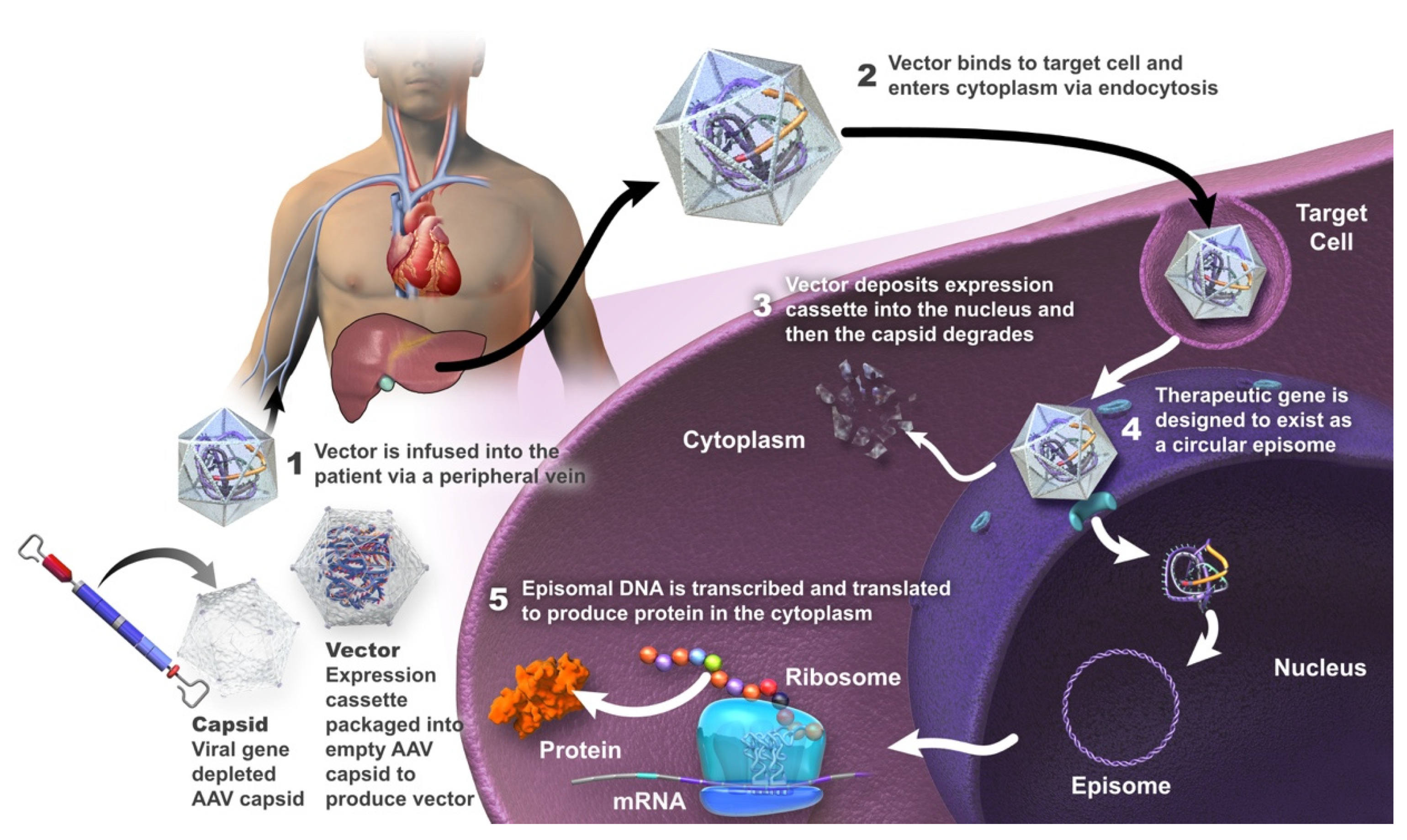
3. Gene Therapy in Hemophilia A

Gene therapy is a treatment strategy used to repair or provide a functional copy of a gene that is either absent or expressed as a non-functional protein [4,28,29,30]. Treating a genetic disease with gene therapy requires that the transgene (or its protein product) be delivered to the physiologically relevant target tissue or tissues, be stably expressed, and not interfere with the functional integrity of the cells in these tissues [31,32,33]. The ultimate goal of gene therapy for patients with hemophilia A is the production of a treatment that is given as a one-time infusion and that allows adequate long-term expression of the deficient FVIII, with the maintenance of steady-state plasma FVIII concentrations. This would minimize (or ideally eliminate) bleeding episodes and thereby decrease the patient and societal burden of the disease [34,35,36,37].
After more than three decades of investigation, gene therapy in hemophilia A has moved beyond proof of concept, toward a realistic expectation that the first product may soon be available. Indeed, such a gene therapy is likely to be approved in Europe in 2022.
Hemophilia A is an ideal candidate for gene therapy [32,38,39,40,41,42]. Its well-characterized monogenic nature of inheritance allows for the correction of a single gene to provide symptomatic relief [32,43]. In hemophilia A, gene transfer strategies employing adeno-associated viral (AAV) vectors to target liver hepatocytes are the closest to regulatory approval. Lentiviral vectors (LVs) targeting hematopoietic stem and progenitor cells ex vivo have shown promise in animal models [44] and have advanced into clinical testing [6]; however, lentiviral vectors integrate into DNA and may potentially induce oncogenesis [45,46,47].

Adeno-Associated Viral Vectors

To date, AAV vectors have demonstrated the greatest clinical success for in vivo gene delivery [29,48,49], and will be the focus of this article (Figure 1).
AAV vectors are engineered from a non-pathogenic, non-enveloped linear, single-stranded DNA parvovirus; wild-type AAV is naturally replication-defective and requires a helper virus for replication [4,50,51,52]. These vectors are minimally integrative and associated with a low risk of insertional mutagenesis [48,53,54,55]. The host transcription machinery transcribes the transgene into mRNA, which is then translated into the protein of interest [6]. It is considered that the genome of the recombinant AAV vector does not undergo site-specific integration in the host DNA or alter the genetic process, but is largely episomal in the nucleus of transduced cells [48].

4. Hemophilia A Clinical Trials Involving Gene Therapy

Several AAV-based gene therapies for hemophilia A are currently under evaluation in clinical studies (Table 1) [56,57,58,59,60,61,62,63,64,65,66,67,68,69,70,71]. In these clinical trials, FVIII replacement therapy was administered in the event of a breakthrough bleeding episode [58,62,65], as gene therapy does not preclude “rescue” therapy, or the return to standard therapy should gene therapy fail.

4.1. Valoctocogene Roxaparvovec

Valoctocogene roxaparvovec is a recombinant codon-optimized AAV5 vector that expresses the SQ variant of B-domain-deleted human FVIII with a hybrid liver-specific transcription promoter. It is likely to be the first gene therapy to be approved as, in June 2022, EMA recommended granting conditional marketing authorization in the EU. The expression cassette is inserted between two AAV serotype 2 inverted terminal repeats [69]. The non-enveloped icosahedral AAV5 capsid delivers the transgene predominantly to the liver [69]. The vector itself is manufactured using a baculovirus–Spodoptera frugiperda (Sf9) insect–cell production system [69].
In an ongoing phase ½ study (NCT02576795), an infusion of valoctocogene roxaparvovec was administered to 15 adults with severe hemophilia A (FVIII ≤ 1 IU/dL) at doses of 6 × 10¹² viral genome (vg)/kg (n = 1), 2 × 10¹³ vg/kg (n = 1), 6 × 10¹³ vg/kg (n = 7) or 4 × 10¹³ vg/kg (n = 6) [64,65,69]. Four of seven and three of six participants in the 6 × 10¹³ vg/kg and 4 × 10¹³ vg/kg cohorts, respectively, maintained median FVIII levels > 5 IU/dL at 4 and 5 years after administration [64]. Individuals in the 6 × 10¹³ vg/kg cohort had a mean ABR of 0.7 treated bleeds/year during year 5, resulting in a cumulative mean ABR of 0.8 treated bleeds/year from week 5 onward (95% reduction from baseline) [64]. Individuals in the 4 × 10¹³ vg/kg cohort had a mean ABR of 1.7 treated bleeds/year during year 4, resulting in a cumulative mean ABR of 1.0 treated bleeds/year from week 5 onward, a 92% reduction from baseline [64]. QoL was maintained in patients receiving the higher dose [64]. The most common adverse events were transient, asymptomatic, and mild-to-moderate ALT elevations [64].
In the phase 3 study (NCT03370913), a single 6 × 10¹³ vg/kg valoctocogene roxaparvovec infusion was administered to 134 adult males with severe hemophilia A (FVIII ≤ 1 IU/dL) on FVIII prophylaxis (negative for FVIII inhibitors) [63]. CSA-assessed FVIII activity increased by a mean of 41.9 IU/dL at weeks 49–52 (baseline FVIII activity was assumed to be 1 IU/dL, as there was no washout of FVIII therapy before valoctocogene roxaparvovec infusion). An 83.8% reduction in mean ABR and 98.6% reduction in FVIII infusion rate were reported. Data for two years’ post-infusion are available for 17 study participants [63]. In these individuals, the mean FVIII activity level was 24.4 IU/dL at week 104, compared with 42.2 IU/dL at weeks 49–52 after infusion. At week 104, 18% (n = 3) of study participants had a median FVIII activity level of ≥40 IU/dL, 59% (n = 10) had an activity level of >5 to <40 IU/dL, and 24% (n = 4) had an activity level of <5 IU/dL. All study participants experienced at least one adverse event, with an increase in ALT (85.8% of participants) and aspartate aminotransferase (AST; 35.1%) levels, headache (38.1%) and nausea (37.3%) being the most common. Most events were of grade 1 or 2, but 8.2% of patients had a grade 3 increase in ALT, and five patients reported a serious adverse event that was considered to be treatment-related. All serious adverse events resolved, as did 96.2% of ALT elevations (with treatment), and no participants died, withdrew from the study because of adverse events, or developed FVIII inhibitors. FVIII activity levels >150 IU/dL were found in 7 (5.2%) patients at weeks 49–52, but none developed thromboembolism [63].

4.2. Dirloctocogene Samoparvovec

Dirloctocogene samoparvovec (SPK-8011) is a recombinant-AAV vector consisting of SPK200 (a bioengineered capsid derived from AAV3 (specifically, subtype LK03)) with a liver-specific, truncated transthyretin enhancer and promoter, a synthetic intron sequence, and codon-optimized FVIII cDNA encoding FVIII-SQ and manufactured with transient triple transfection of human embryonic kidney cells (HEK293 cells) [58].
In a phase ½ trial (NCT03003533/NCT03432520), 18 men with hemophilia A were enrolled in four dose cohorts; the lowest-dose cohort received 5 × 10¹¹ vg/kg and the highest-dose cohort received 2 × 10¹² vg/kg, with glucocorticoids administered in cases of suspected immune response [58]. Two participants lost all FVIII expression because of an anti-AAV capsid cellular immune response that was not sensitive to immune suppression. In the remaining 16 men, FVIII expression was maintained, with 12 men followed for >2 years. These men had a 91.5% reduction in ABR after dirloctocogene samoparvovec administration. Elevated ALT levels were seen in seven study participants, the majority of which were mild, with the exception of a grade 2 elevation in one participant that required hospitalization for treatment. Adverse events related to glucocorticoids were reported in four participants. No cases of FVIII inhibitory antibody development were reported.

4.3. SPK-8016

A phase ½, open-label, non-randomized, dose-finding study is investigating the efficacy of SPK-8016 in adult men with clinically severe hemophilia A who have not developed FVIII inhibitors and had no AAV neutralizing antibodies (NCT03734588) [70]. In three patients, there was evidence for unstable FVIII:C and cellular immunity against AAV. Patients were put on daily oral corticosteroids at weeks 3–7 for 43–38 weeks. Preliminary results demonstrated sustained FVIII levels (6.2–21.8%) after 52 weeks in four men who received SPK-8016 at a dose of 5 × 10¹¹ vg/kg. An optimization of the immunomodulatory regimen is being developed with the potential for obtaining clinically meaningful FVIII activity using low vector doses [70].

4.4. Giroctocogene Fitelparvovec

Giroctocogene fitelparvovec comprises an AAV serotype 6 vector (AAV2/6) encoding the cDNA for B domain-deleted human FVIII. This gene therapy has been investigated in a phase ½ trial (NCT03061201) in which 11 patients were each given four ascending doses: 9 × 10¹¹, 2 × 10¹², 1 × 10¹³, and 3 × 10¹³ vg/kg (n = 2, 2, 2 and 5 patients, respectively) [71]. Four patients in the 3 × 10¹³ vg/kg cohort have available data, which demonstrated mean FVIII activity maintained in the mild to normal range (30.9% measured by CCA; 46.4% measured by one-stage assay) to week 104. The ABR was zero for the first-year post-infusion and 0.9 throughout the follow-up, and two patients experienced a total of three bleeding events (two traumatic, one unknown; one in a target joint) that necessitated treatment with exogenous FVIII. No patients have resumed prophylaxis. Common treatment-related adverse events were elevated liver enzyme levels (ALT n = 5/11 patients, and AST n = 3/11), pyrexia (n = 3/11), and tachycardia (n = 2/11). Four patients in the 3 × 10¹³ vg/kg cohort had an ALT elevation that required >7 days of corticosteroid treatment, but these were managed with a tapering course of corticosteroids, and FVIII activity was maintained. No patients developed inhibitors to FVIII.
Giroctocogene fitelparvovec was being evaluated in a phase 3 study in men with moderately severe to severe hemophilia A (AFFINE; NCT04370054) [57], but the US Food and Drug Administration has placed this study on hold until proposed protocol amendments have been implemented, due to FVIII levels being >150% in some participants [57].

4.5. TAK-754

A phase ½ trial (NCT03370172) is currently evaluating TAK-754, a modified AAV serotype 8 gene therapy, in four men with severe hemophilia A [56]. Participants were treated in two ascending dose cohorts (2 × 10¹² capsid particles [cp]/kg and 6 × 10¹² cp/kg) [56]. FVIII expression declined significantly during the tapering of corticosteroids, and three of the four patients have resumed FVIII replacement therapy after ≥10 months’ follow-up. The utilization of corticosteroids did not prevent the loss of transgene expression. The reason behind transcript expression loss is being investigated [56].

4.6. BAY 2599023

BAY 2599023 (AAVhu37.hFVIIIco) comprises an AAV vector with a capsid serotype hu37 (AAVhu37), and a genome that directs expression of a codon-optimized B-domain-deleted human FVIII under the control of a liver-specific promoter/enhancer combination [67].
A phase ½ study (NCT03588299) is being conducted in adult males who have severe hemophilia A with no history of FVIII inhibitors, no detectable immunity to the AAVhu37 capsid, and ≥150 exposure days to FVIII products [67,68]. Nine patients have received a single infusion of BAY 2599023 at doses of 0.5, 1.0, and 2.0 × 10¹³ gene copies/kg (n = 2, 2, and 5, respectively) [67,68]. Successful proof-of-concept was achieved with measurable, stable expression of endogenous FVIII for up to >23 months [67,68]. Bleeding protection has been observed, with patients in the two higher dose groups not requiring prophylaxis with FVIII products from approximately 6–12 weeks after gene transfer, and no spontaneous bleeds requiring treatment being reported once FVIII levels reached >11 IU/dL. Mild-to-moderate ALT elevations have been observed in five patients but these were successfully managed with corticosteroids or famotidine [68].

4.7. AAV8-HLP-hFVIII-V3

AAV-HLP-hFVIII-V3 encodes a 17-amino-acid peptide with six N-linked glycosylation motifs from the human FVIII B domain, pseudotyped with AAV serotype 8 capsid, and has been shown to mediate three-fold higher FVIII expression compared with valoctocogene roxaparvovec in an animal model [61]. In a phase ½ study (NCT03001830), three men with severe hemophilia A received a single infusion of AAV8-HLP-hFVIII-V3 [61], with one man receiving a dose of 6 × 10¹¹ vg/kg and the subsequent two patients each receiving a dose of 2 × 10¹² vg/kg. The patients were followed up for 13–47 weeks after vector administration. FVIII activity levels were >5% in all three men, with normalization of FVIII levels in one patient. Serum ALT levels were elevated in two of the patients between 4 and 6 weeks after gene transfer but this responded to corticosteroids, with no loss of transgene expression. No study participants have developed FVIII inhibitors.

5. Opportunities Associated with Gene Therapy in Hemophilia A

Gene therapy is a disease-transforming therapy that has the potential to become the new standard for the treatment of patients with hemophilia A [72]. It may enable the patient to achieve a sustained physiological level of endogenously produced FVIII protein that could provide effective prophylaxis without the need for exogenous factor replacement therapy [40,42]. Such continuous endogenous expression of physiological levels of FVIII can be expected to eliminate breakthrough bleeding and micro-hemorrhages [72]. Gene therapy may, thus, provide protection against further bleeding-induced joint damage [73]. While the clinical studies reported to date indicate a cessation of joint bleeds [63,65], further data are needed to further investigate whether this leads to a reduction in joint damage, although this can be anticipated.
Given that even minimal increases in FVIII levels can lead to significantly improved bleeding outcomes, it is expected that gene therapy will provide a resultant improvement in the QoL of the individual with hemophilia A [38,72].

6. Challenges of Gene Therapy in Hemophilia A

Successful gene therapy was first reported, in 2011, with intravenously administered AAV-based liver-directed gene therapy in patients with severe hemophilia B [74]. In contrast, gene therapy directed at hemophilia A has lagged behind that directed at hemophilia B, largely due to the limitations inherent in the size of the FVIII gene and the structure of FVIII, problems achieving therapeutic levels of the transgenic protein, and cellular immune responses to the capsid of the AAV vector [75] (also identified in trials of therapy for hemophilia B, although to a lesser extent), as well as issues related to the duration and variability of this form of therapy [1,4,6].

6.1. FVIII Structure

The gene for FVIII is located at the tip of the long arm of the X chromosome. At over 180 kb, it is one of the largest known genes. It comprises 26 exons, which encode a polypeptide chain of 2351 amino acids (19 signal peptides and a 2332-amino-acid mature polypeptide chain). Factor VIII is composed of six domains, A1-A2-B-A3-C1-C2, with three acidic regions (α1-3) bordering the A domains. The A domains are each approximately 330 residues in length, the B domain approximately 900 residues, and the C domains each approximately 150 residues [76].
FVIII, and the FVIII complementary DNA (cDNA) for FVIII (~7 kb), exceed the packaging capacity of AAV vectors (~5 kb) [40]. This restriction has been overcome by using FVIII transgenes optimized for packaging through the removal of the B domain, which comprises a large proportion of the gene but is not required for clotting function [48,77,78,79]. All current FVIII transgene constructs utilize a B domain-deleted (or truncated) cDNA [1,80].

6.2. FVIII Expression

The target cell for FVIII gene therapy is the hepatocyte, which is not the native cell for the endogenous FVIII synthesis [7,8]. Nevertheless, expression of FVIII in hepatocytes generates a functional and physiological protein that has restored hemostasis in animal models and clinical studies [81,82]. In contrast, other currently available therapies involve the administration of exogenous FVIII by infusion or the subcutaneous administration of an FVIII-mimicking compound [2,14].
The expression of FVIII and the duration of this expression between patients is variable, not only between the different studies, but also within dose cohorts and within the patients themselves over time [83]. Low levels of FVIII expression may be overcome by introducing liver-specific promoters and enhancers [84].

6.3. Infusion-Related Adverse Reactions

Infusion-related adverse reactions can occur with gene therapy for hemophilia A, but are generally infrequent, resolve with treatment and/or slowing or pausing the infusion, and do not usually prevent completion of the infusion. In a phase ½ study (NCT02576795) of valoctocogene roxaparvovec (an AAV5-based gene therapy encoding human B domain-deleted FVIII), one patient was hospitalized due to grade 2 pyrexia with myalgia and headache that occurred within 24 h after the infusion; the symptoms resolved within 48 h after treatment with acetaminophen (paracetamol) [65]. In a phase 3 study of valoctocogene roxaparvovec (NCT03370913), seven study participants (5.2%) had systemic hypersensitivity and three reported serious infusion-related reactions (maculopapular rash and presyncope; anaphylactic reaction; and hypersensitivity reaction) during or shortly after the infusion [63]. However, the infusion reactions were managed by slowing or pausing the infusion and administering supportive medications (e.g., antihistamines, antipyretics, or glucocorticoids), such that all participants completed the infusion. During a phase ½ trial of dirloctocogene samoparvovec (NCT03003533; NCT03432520), one participant had an acute infusion-related reaction, characterized by vomiting, myalgia, back pain, and pyrexia 12 h after administration, which resolved within 72 h after antipyretic treatment [58]. One patient experienced hypotension and fever approximately 6 h after infusion of the highest dose of giroctocogene fitelparvovec in a phase ½ study (NCT03061201); the events fully resolved with treatment and did not delay post-infusion discharge [60].

6.4. Anti-AAV Neutralizing Antibodies

Exposure to wild-type AAV results in priming of the body’s immune system against the virus, with the development of both humoral and T-cell immunity that can neutralize the vector and reduce treatment efficacy [85,86]. At present, patient selection in gene therapy trials in hemophilia A includes screening for pre-existing immunity to identify and enroll patients who are negative for anti-AAV capsid neutralizing antibodies [6,87].
The prevalence of anti-AAV immunoglobulin G and neutralizing antibodies differs for each AAV type within the general population. Although data are somewhat inconsistent, studies suggest a higher prevalence of antibodies against AAV1 and AAV2 than against AAV5, AAV6, AAV8, and AAV9 [88,89,90,91]. This suggests that the use of vectors based on the latter AAV types may avoid pre-existing immunity and, therefore, have advantages for use in gene therapy.
Individuals without pre-existing immunity who are treated with AAV-based gene therapy are likely to develop high-titer antibodies to the vector. For example, in a phase ½ study of valoctocogene roxaparvovec in which patients with pre-existing immunity to AAV5 were excluded from enrolment, anti-AAV5 antibodies were detected in all patients (n = 15) after treatment (follow-up was at least 104 weeks, with a maximum of 183 weeks) [92]. All patients had seroconverted at week 8 post-treatment (the first time point assessed). The titer peaked 40 weeks after administration and remained stable through to week 104, with no apparent dose–response relationship. Further, there was no correlation between the immune response and safety (alanine transaminase (ALT) levels) or efficacy measures. Cross-reactivity to other AVV serotypes was also observed [92].
Further research is needed to determine the implications of such immune responses for the repeated administration of gene therapy, whether repeated administration of the same gene therapy or that of different therapies that use AAV-type vectors for which cross-reactivity has been observed.

6.5. Elevation of Liver Enzymes

AAV-based gene therapy may induce a degree of hepatotoxicity, which is usually transient and asymptomatic. Increased levels of serum ALT are commonly seen. The mechanism of this toxicity has not been clearly established but could be related to the development of an anti-AAV capsid peptide cytotoxic T cell response, endoplasmic reticulum stress, and associated hepatocyte apoptosis due to high expression of FVIII, or a direct effect of vector particle load [80].
Clinical studies in patients with hemophilia A have reported increases in ALT level in a high proportion of patients [56,58,60,61,63,65,67,69,70]. In a phase ½ study of valoctocogene roxaparvovec in patients with hemophilia A, an increase in ALT was reported in 11 of the 15 study participants (73.3%; 14 events). The increases were not generally associated with loss of FVIII activity or a T-cell immune response to viral capsid peptides [65]. All elevations in ALT were mild (13 events grade 1 and 1 event grade 2), non-serious and transient, and there were no symptoms or sequelae suggestive of clinically significant hepatocyte injury or liver dysfunction [65]. There were no consistent cellular immune responses to FVIII or AAV5 capsid detected in any participant [65]. Similarly, transient elevations of ALT were seen in patients participating in the phase 3 study of valoctocogene roxaparvovec [63]. The elevations occurred in 115 participants (85.8%) and lasted for a median duration of 15 days. While most events were grade 1 or 2 in severity, 11 participants (8.2%) experienced 12 events of grade 3 ALT elevations (>5 to 20 times the upper limit of normal), with two events deemed to be serious, although none met the criteria for significant liver injury. All events were managed with glucocorticoids and resolved [63]. In a phase ½ study of dirloctocogene samoparvovec, elevated ALT levels were seen in seven participants (39%; 13 events) [58]. All the elevations were considered mild, except for that in one patient who experienced a grade 2 elevation that constituted a serious AE.
During clinical studies, increases in ALT levels were managed with a tapering course of glucocorticoids [58,65,69]. In the phase 3 study of valoctocogene roxaparvovec, 79.1% of patients received glucocorticoids to manage ALT level elevations, with a median duration of treatment of 230 days (range 22–551 days) [63].
Participants receiving prednisolone also showed a concomitant increase in FVIII activity after valoctocogene roxaparvovec [69], raising the question of whether prednisolone may directly regulate transgene expression; however, in animal models of hemophilia treated with valoctocogene roxaparvovec, prednisolone did not regulate FVIII expression [93].
While steroids may provide immunosuppression, they can be associated with adverse effects (e.g., weight gain, edema, irritability, adrenal insufficiency, negative effects on bone health, diabetes, and hypertension), as well as an increased risk of infection [35,58]. Indeed, almost 72% of the patients treated with glucocorticoids in the phase 3 study of valoctocogene roxaparvovec experienced such adverse events, with the most common being acne, insomnia, Cushing’s syndrome, and weight gain [63]. Serious adverse events attributed to glucocorticoids occurred in 2.7% of study participants [63]. In a few cases, hepatotoxicity may be unresponsive to oral steroids, requiring the administration of intravenous methylprednisolone or alternative immune-regulatory agents (e.g., tacrolimus or azathioprine) [80].

6.6. Oncogenesis

The risk of genomic insertional mutagenesis after AAV-mediated gene transfer is considered to be low due to the episomic nature of the transinfected cDNA [6]. Durable FVIII expression has been observed in an animal model of severe hemophilia A, with no late-toxicity or oncogenesis [94]. A study in hemophilia dogs treated with AAV8 or AAV9 vectors expressing canine FVIII and followed for 10 years did not find any evidence of hepatic tumors or altered liver function, although there were some AAV integration events in genomic DNA and clonal expansion of cells that harbored vector integration in genes potentially associated with growth control [54].
A patient enrolled in a phase 3 clinical trial of a gene therapy for hemophilia B (etranacogene dezaparvovec) developed a hepatocellular carcinoma, although an independent investigation concluded that it was ‘highly unlikely’ that this was caused by the therapy, given the low level of vector integration in the patient’s tissue and the finding that the patient had a number of risk factors for hepatocellular carcinoma [95]. There is currently no evidence that gene therapies for hemophilia A are associated with a risk of oncogenesis. A patient who received valoctocogene roxaparvovec in a phase ½ trial developed salivary gland cancer, but it was deemed unrelated to the therapy [96]. There was no evidence of hepatocellular carcinoma in a phase ½ trial of dirloctocogene samoparvovec; this was specifically reported, having been investigated using ultrasonographic assessment of the liver [58]. In a phase ½ trial of giroctocogene fitelparvovec, no ‘hepatic masses’ were detected [71].
Ongoing investigation in animal models, long-term follow-up of clinical studies, and surveillance using long-term registries of patients treated with gene therapy are required to confirm that the risk of oncogenesis is minimal.

6.7. Durability and Variability of Transgene Expression

The clinical trials completed so far have not answered fully all issues related to gene therapy in hemophilia A. Open questions remain as to why expression levels of FVIII decrease over time and why there is intra- and inter-patient variability [83]. It is yet to be determined whether the vector serotype, manufacturing process, expression of FVIII in hepatocytes, FVIII protein unfolding, episomal structural changes, or other factors contribute to this decline/variability in FVIII levels [1]. The impact of liver growth and hepatocyte turnover on AAV vector genome persistence, and the efficacy and durability of factor expression in adults, adolescents, and pediatric groups is also still unclear [97]. Variability may or may not be related to durability [98]. Identifying the causes of inter- and intra-individual variability needs to be a research priority, as finding the root causes of the variability might permit potential mitigants to be identified [98].
In light of these questions, the durability/variability of hepatocyte-expressed FVIII in humans continues to be investigated in clinical trials [1,6]. In a phase ½ study in 15 patients with hemophilia A who received a single dose of valoctocogene roxaparvovec, follow-up data to 5 years indicated that 10 out of 13 participants continued to have FVIII levels above the severe hemophilia A range (>1 IU/dL) [64]. Data from patients up to 2 years post-infusion who are taking part in the phase 3 study of valoctocogene roxaparvovec (n = 17) are also encouraging, with 76% having a median FVIII activity level of >5 IU/dL at this time point, and 18% having a level of ≥40 IU/dL [63]. In a phase ½ study of dirloctocogene samoparvovec, FVIII expression was maintained in 16/18 individuals, with the mean FVIII activity being 6.9% of the normal value in an FVIII chromogenic assay (CSA) at >52 weeks after vector administration [99]. However, two of 18 individuals with hemophilia A lost all FVIII expression because of an anti-AAV capsid cellular immune response that was not sensitive to immune suppression [99].
Unfortunately, the positive sustained responses in the majority of patients in clinical trials poses concerns that these patients may be lost to follow-up [100]. Long-term surveillance of these patients is essential with regards to their clinical bleeding patterns, levels of transgenic clotting factor, the possible development of clotting factor immune response, and the need for regular hepatic imaging assessment to ensure that no neoplastic process is developing [100]. There is also a need for long-term follow-up and post-marketing surveillance through registries to establish if the efficacy and tolerability of gene therapy observed in clinical trials are maintained long term in trial participants and in the real-world population [97].

6.8. Patient Expectations

Gene therapy has the potential to change the quality of life of patients with hemophilia A by reducing the frequency of bleeding events, thereby allowing patients to lead lives that are less impacted by the disease [83,101,102]. However, it is important that patient expectations surrounding their eligibility, access to treatment, and treatment outcomes are managed appropriately, as this therapy may not be suitable for all patients [83,102,103]. This is because there is variability in FVIII expression between patients, FVIII expression may not be durable in all patients, and some patients may be positive for anti-AAV capsid neutralizing antibodies and, therefore, not suitable candidates for gene therapy.
People living with hemophilia A need to be fully informed regarding the potential benefits and risks of gene therapy [102]. It will be essential that physicians pay close attention to what information they share with their patients, choose the optimum way to do so, and determine how to ensure sufficient patient understanding of the information provided [102].
At present, there are no data on the effects of gene therapy in children with hemophilia, and no ongoing trials. As such, the expectations of the parents of these children regarding the appropriateness of gene therapy in younger patients with hemophilia will need to be managed accordingly.

7. Assessment of Efficacy of FVIII Expression

The measurement of FVIII activity is a direct and accessible way to determine transgene expression, but there are differences in standard clinical laboratory measurements of clotting activity, with the one-stage assay providing higher levels of FVIII activity than the CSA [104,105]. One-stage assays and/or chromogenic assays have been employed to measure circulating FVIII or FIX levels and duration of transgene expression in liver-directed gene therapy studies. A 1.6-fold difference between the values obtained by the two methods was encountered in comparative studies for the B-domain-deleted FVIII-SQ in valoctocogene roxaparvovec AAV5-FVIII-SQ [105]. Likewise, differences between chromogenic and one-stage FIX assays with one-stage values ≅ 2-fold higher than chromogenic assay values were found for the transgene expression of Factor IX–R338L [106]. In our opinion, clinical outcomes are likely to define which assay best measures FVIII/FIX levels, thus uncovering the real efficacy/safety profile of the transgenic factors. In the only phase 3 trial of gene therapy for hemophilia completed and published to date (of valoctocogene roxaparvovec), the primary endpoint was the change from baseline in median FVIII activity during weeks 49–52, assessed using the CSA [62,63]. We would like to point out that, as successfully experimented with the BDD-FVIII recombinant product, the use of a standard reference factor provided by the company, would assure accuracy and precision in measuring the achieved levels of a transgenic factor in treated patients in a simple one-stage assay [107].
In clinical trials, a holistic assessment of patient outcomes is needed [80,83]. Measures of QoL and hemophilia-specific patient-reported outcomes need to be used [83,101]. Annualized bleeding rate (ABR) is a patient-reported outcome in which the occurrence of bleeding events, their location, severity and whether there was a precipitating event is recorded by the patient. The collection of QoL outcomes will enable a better understanding of the cost–benefit of gene therapies in patients with hemophilia A [1]. An early cost-effectiveness analysis compared valoctocogene roxaparvovec with standard half-life FVIII or emicizumab in patients with severe hemophilia A without FVIII antibodies using a Dutch societal perspective with a 10-year time horizon [108]. This suggested that valoctocogene roxaparvovec may result in improved health and lower cost compared with prophylactic FVIII and emicizumab.
One issue that remains to be elucidated with any gene therapy for hemophilia A is whether this treatment will longer-term benefit joint health and structure, as determined by imaging analysis [80].

8. Implementing Gene Therapy in a Real-World Setting

Gene therapy is a complex treatment process and when approved for reimbursement it will likely first be offered at comprehensive care centers with significant expertise in the management of patients with hemophilia. A recent joint publication from the European Association for Haemophilia and Allied Disorders and the European Haemophilia Consortium suggested that a modified ‘hub-and-spoke’ model, incorporating a long-term safety and efficacy surveillance system, be introduced to ensure appropriate prescription, administration, and monitoring of gene therapy in patients with severe hemophilia A [109,110].
Given the imminent approval of gene therapy for hemophilia A in Italy, there is a need to determine how this treatment service will be organized on national and local levels and how patients who are candidates for gene therapy in Italy will be assessed, treated, and managed [111]. In our opinion, the hub-and-spoke model should be followed for the best cost-effectiveness and resource utilization. Hubs should manage all aspects of therapy delivery and data collection, while spokes should take care of pre-screening and patient selection. A set of detailed selection criteria will need to be elaborated by multidisciplinary teams present in the centers involved; however, there will have to be flexibility in the system as patients’ individual situations can be very different and should not lead to an a priori exclusion. We believe that contacts with patients should be kept by a single designated member of the multidisciplinary team, who will act as a point of contact throughout the procedure and the follow-up. Such a person will have the preparation to provide information to the patient including the possibility of transgene expression loss and the necessity of returning to other treatments. An active engagement with patients is very important and can be obtained through an adequate explanation and support given to the patient. Patient association groups can play an important role in this aspect. Adequate laboratory surveillance needs to be available at all stages pre- and post-procedure. In particular, a reliable standardized determination of anti-AAV antibodies and monitoring of FVIII levels must be ensured together with safety monitoring (e.g., liver function). A structured follow-up must be provided to all patients, especially, in the first year after gene therapy.

9. Conclusions

The current treatment of hemophilia A involves frequent intravenous injections and the risk of FVIII inhibitor development, which can negatively impact patient QoL. Gene therapy for patients with hemophilia A represents the potential for a single treatment that could allow for long-term expression of the deficient FVIII, with the maintenance of steady-state plasma FVIII concentrations, thereby minimizing bleeding episodes for the whole lifetime of the recipient and decreasing the burden of their disease. FVIII expressed through gene therapy is a physiological protein rather than a mimicking factor or an infused exogenous FVIII concentrate, and steady-state physiological plasma FVIII levels may avoid the deterioration of joint status.
Gene therapy for the management of hemophilia A has now moved beyond proof of concept, with the realistic expectation that it may become available to patients in European countries, including Italy, in 2022 [49,80]. It is likely that the first licensed product will be valoctocogene roxaparvovec, for which the longest duration efficacy and safety data (5 years) for any gene therapy currently under investigation are available. These data indicate sustained secretion of FVIII in patients who have received valoctocogene roxaparvovec, such that ABR and FVIII replacement therapy were minimized [64].
We believe that improved patient selection and management of patient expectations will enable the benefits of gene therapy to be maximized. Consequently, gene therapy could represent a paradigm shift, becoming a new reference standard in the treatment of patients with hemophilia A, enabling long-lasting treatment and improved clinical and patient-centered outcomes, including enhanced QoL, for many patients with this disease.

Author Contributions

Conceptualization: G.C., G.D.M., R.D.C. and F.P.; writing—review and editing: G.C., G.D.M., R.D.C. and F.P. All authors have read and agreed to the published version of the manuscript.

Funding

Medical writing assistance for this manuscript and the APC was funded by BioMarin.

Institutional Review Board Statement

Not applicable.

Informed Consent Statement

Not applicable.

Data Availability Statement

No new data were created or analyzed in this study. Data sharing is not applicable to this article.

Acknowledgments

We would like to thank Monique Curran, who wrote the outline of this manuscript on behalf of Springer Healthcare Communications, and Kate Palmer from Springer Healthcare Communications who wrote the subsequent drafts. This medical writing assistance was funded by BioMarin.

Conflicts of Interest

Giancarlo Castaman has received speaker’s fees from Bayer, Grifols, LFB, Roche, SOBI, Novo Nordisk, Werfen, and Kedrion; research funding directly from their institution from CSL Behring, Pfizer, and SOBI; and, during the last 2 years, participated in advisory boards for Bayer, BioMarin, Takeda, CSL Behring, Novo Nordisk, Pfizer, Roche, Sanofi, SOBI, and Uniqure. Raimondo De Cristofaro has received honoraria for advisory board participation from Bayer, SOBI, and Takeda. Flora Peyvandi has received honoraria for participating as a speaker at educational meetings from Grifols and Roche, and on advisory boards organized by Sanofi, SOBI, Takeda, Roche, and BioMarin. Giovanni Di Minno has received speaker’s fees from Sanofi, BioMarin, Novo Nordisk, Pfizer, Takeda, and CSL Behring. BioMarin provided funding for medical writing support for this manuscript. BioMarin had no input into the content of the manuscript. The authors take full responsibility for the drafting of this manuscript and in the final decision to publish.

References

  1. Arruda, V.R.; Weber, J.; Samelson-Jones, B.J. Gene Therapy for Inherited Bleeding Disorders. Semin. Thromb. Hemost. 2021, 47, 161–173. [Google Scholar] [CrossRef]
  2. Berntorp, E.; Fischer, K.; Hart, D.P.; Mancuso, M.E.; Stephensen, D.; Shapiro, A.D.; Blanchette, V. Haemophilia. Nat. Rev. Dis. Prim. 2021, 7, 45. [Google Scholar] [CrossRef]
  3. Castaman, G.; Matino, D. Hemophilia A and B: Molecular and clinical similarities and differences. Haematologica 2019, 104, 1702–1709. [Google Scholar] [CrossRef]
  4. Perrin, G.Q.; Herzog, R.W.; Markusic, D.M. Update on clinical gene therapy for hemophilia. Blood 2019, 133, 407–414. [Google Scholar] [CrossRef]
  5. Peyvandi, F.; Garagiola, I.; Young, G. The past and future of haemophilia: Diagnosis, treatments, and its complications. Lancet 2016, 388, 187–197. [Google Scholar] [CrossRef]
  6. Pipe, S.W.; Gonen-Yaacovi, G.; Segurado, O.G. Hemophilia A gene therapy: Current and next-generation approaches. Expert Opin. Biol. Ther. 2022, 1–17. [Google Scholar] [CrossRef]
  7. Everett, L.A.; Cleuren, A.; Khoriaty, R.N.; Ginsburg, D. Murine coagulation factor VIII is synthesized in endothelial cells. Blood 2014, 123, 3697–3705. [Google Scholar] [CrossRef]
  8. Fahs, S.A.; Hille, M.T.; Shi, Q.; Weiler, H.; Montgomery, R.R. A conditional knockout mouse model reveals endothelial cells as the principal and possibly exclusive source of plasma factor VIII. Blood 2014, 123, 3706–3713. [Google Scholar] [CrossRef]
  9. Shahani, T.; Covens, K.; Lavend’Homme, R.; Jazouli, N.; Sokal, E.; Peerlinck, K.; Jacquemin, M. Human liver sinusoidal endothelial cells but not hepatocytes contain factor VIII. J. Thromb. Haemost. 2014, 12, 36–42. [Google Scholar] [CrossRef]
  10. Blanchette, V.S.; Key, N.S.; Ljung, L.R.; Manco-Johnson, M.J.; van den Berg, H.M.; Srivastava, A. Definitions in hemophilia: Communication from the SSC of the ISTH. J. Thromb. Haemost. 2014, 12, 1935–1939. [Google Scholar] [CrossRef]
  11. Collins, P.W.; Blanchette, V.S.; Fischer, K.; Björkman, S.; Oh, M.; Fritsch, S.; Schroth, P.; Spotts, G.; Astermark, J.; Ewenstein, B.; et al. Break-through bleeding in relation to predicted factor VIII levels in patients receiving prophylactic treatment for severe hemophilia A. J. Thromb. Haemost. 2009, 7, 413–420. [Google Scholar] [CrossRef]
  12. Uijl, I.E.M.D.; Fischer, K.; Van Der Bom, J.G.; Grobbee, D.E.; Rosendaal, F.R.; Plug, I. Analysis of low frequency bleeding data: The association of joint bleeds according to baseline FVIII activity levels. Haemophilia 2011, 17, 41–44. [Google Scholar] [CrossRef]
  13. Castaman, G.; Linari, S. Prophylactic versus on-demand treatments for hemophilia: Advantages and drawbacks. Expert Rev. Hematol. 2018, 11, 567–576. [Google Scholar] [CrossRef]
  14. Mahlangu, J.; Oldenburg, J.; Paz-Priel, I.; Negrier, C.; Niggli, M.; Mancuso, M.E.; Schmitt, C.; Jiménez-Yuste, V.; Kempton, C.; Dhalluin, C.; et al. Emicizumab Prophylaxis in Patients Who Have Hemophilia A without Inhibitors. N. Engl. J. Med. 2018, 379, 811–822. [Google Scholar] [CrossRef]
  15. Mannucci, P.M.; Cortesi, P.A.; Di Minno, M.N.D.; Sanò, M.; Mantovani, L.G.; Di Minno, G. Comparative analysis of the pivotal studies of extended half-life recombinant FVIII products for treatment of haemophilia A. Haemophilia 2021, 27, e422–e433. [Google Scholar] [CrossRef]
  16. Ar, M.C.; Balkan, C.; Kavaklı, K. Extended Half-Life Coagulation Factors: A New Era in the Management of Haemophilia Patients. Turk. J. Hematol. 2019, 36, 141–154. [Google Scholar] [CrossRef]
  17. Darby, S.C.; Keeling, D.M.; Spooner, R.J.D.; Kan, S.W.; Giangrande, P.L.F.; Collins, P.W.; Hill, F.G.H.; Hay, C.R.M.; Organisation, U.H.C.D. The incidence of factor VIII and factor IX inhibitors in the hemophilia population of the UK and their effect on subsequent mortality, 1977–1999. J. Thromb. Haemost. 2004, 2, 1047–1054. [Google Scholar] [CrossRef]
  18. Witmer, C.; Young, G. Factor VIII inhibitors in hemophilia A: Rationale and latest evidence. Ther. Adv. Hematol. 2013, 4, 59–72. [Google Scholar] [CrossRef]
  19. Rosendaal, F.R.; Palla, R.; Garagiola, I.; Mannucci, P.M.; Peyvandi, F. Genetic risk stratification to reduce inhibitor development in the early treatment of hemophilia A: A SIPPET analysis. Blood 2017, 130, 1757–1759. [Google Scholar] [CrossRef]
  20. Oldenburg, J. Optimal treatment strategies for hemophilia: Achievements and limitations of current prophylactic regimens. Blood 2015, 125, 2038–2044. [Google Scholar] [CrossRef]
  21. Olivieri, M.; Kurnik, K.; Pfluger, T.; Bidlingmaier, C. Identification and long-term observation of early joint damage by magnetic resonance imaging in clinically asymptomatic joints in patients with haemophilia A or B despite prophylaxis. Haemophilia 2012, 18, 369–374. [Google Scholar] [CrossRef]
  22. Blair, H.A. Emicizumab: A Review in Haemophilia, A. Drugs 2019, 79, 1697–1707. [Google Scholar] [CrossRef]
  23. Franchini, M.; Marano, G.; Pati, I.; Candura, F.; Profili, S.; Veropalumbo, E.; Masiello, F.; Catalano, L.; Piccinini, V.; Vaglio, S.; et al. Emicizumab for the treatment of haemophilia A: A narrative review. Blood Transfus. 2019, 17, 223–228. [Google Scholar] [CrossRef]
  24. Kitazawa, T.; Igawa, T.; Sampei, Z.; Muto, A.; Kojima, T.; Soeda, T.; Yoshihashi, K.; Okuyama-Nishida, Y.; Saito, H.; Tsunoda, H.; et al. A bispecific antibody to factors IXa and X restores factor VIII hemostatic activity in a hemophilia A model. Nat. Med. 2012, 18, 1570–1574. [Google Scholar] [CrossRef]
  25. Sampei, Z.; Igawa, T.; Soeda, T.; Okuyama-Nishida, Y.; Moriyama, C.; Wakabayashi, T.; Tanaka, E.; Muto, A.; Kojima, T.; Kitazawa, T.; et al. Identification and Multidimensional Optimization of an Asymmetric Bispecific IgG Antibody Mimicking the Function of Factor VIII Cofactor Activity. PLoS ONE 2013, 8, e57479. [Google Scholar] [CrossRef]
  26. Oldenburg, J.; Mahlangu, J.N.; Kim, B.; Schmitt, C.; Callaghan, M.U.; Young, G.; Santagostino, E.; Kruse-Jarres, R.; Negrier, C.; Kessler, C.; et al. Emicizumab Prophylaxis in Hemophilia A with Inhibitors. N. Engl. J. Med. 2017, 377, 809–818. [Google Scholar] [CrossRef]
  27. Pipe, S.W.; Shima, M.; Lehle, M.; Shapiro, A.; Chebon, S.; Fukutake, K.; Key, N.S.; Portron, A.; Schmitt, C.; Podolak-Dawidziak, M.; et al. Efficacy, safety, and pharmacokinetics of emicizumab prophylaxis given every 4 weeks in people with haemophilia A (HAVEN 4): A multicentre, open-label, non-randomised phase 3 study. Lancet Haematol. 2019, 6, e295–e305. [Google Scholar] [CrossRef]
  28. Anguela, X.M.; High, K.A. Entering the Modern Era of Gene Therapy. Annu. Rev. Med. 2019, 70, 273–288. [Google Scholar] [CrossRef]
  29. Kumar, S.R.; Markusic, D.M.; Biswas, M.; High, K.A.; Herzog, R.W. Clinical development of gene therapy: Results and lessons from recent successes. Mol. Ther. Methods Clin. Dev. 2016, 3, 16034. [Google Scholar] [CrossRef]
  30. Maestro, S.; Weber, N.D.; Zabaleta, N.; Aldabe, R.; Gonzalez-Aseguinolaza, G. Novel vectors and approaches for gene therapy in liver diseases. JHEP Rep. 2021, 3, 100300. [Google Scholar] [CrossRef]
  31. High, K.A.; Roncarolo, M.G. Gene Therapy. N. Engl. J. Med. 2019, 381, 455–464. [Google Scholar] [CrossRef] [PubMed]
  32. Lisowski, L.; Staber, J.M.; Wright, J.F.; Valentino, L.A. The intersection of vector biology, gene therapy, and hemophilia. Res. Pract. Thromb. Haemost. 2021, 5, e12586. [Google Scholar] [CrossRef] [PubMed]
  33. Marchesini, E.; Morfini, M.; Valentino, L. Recent Advances in the Treatment of Hemophilia: A Review. Biol. Targets Ther. 2021, 15, 221–235. [Google Scholar] [CrossRef] [PubMed]
  34. Miesbach, W.; Pasi, K.J.; Pipe, S.W.; Hermans, C.; O’Mahony, B.; Guelcher, C.; Steiner, B.; Skinner, M.W. Evolution of haemophilia integrated care in the era of gene therapy: Treatment centre’s readiness in United States and EU. Haemophilia 2021, 27, 511–514. [Google Scholar] [CrossRef]
  35. Rodríguez-Merchán, E.; De Pablo-Moreno, J.; Liras, A. Gene Therapy in Hemophilia: Recent Advances. Int. J. Mol. Sci. 2021, 22, 7647. [Google Scholar] [CrossRef]
  36. Rodriguez-Santana, I.; DasMahapatra, P.; Burke, T.; Hakimi, Z.; Bartelt-Hofer, J.; Nazir, J.; O’Hara, J. Differential humanistic and economic burden of mild, moderate and severe haemophilia in european adults: A regression analysis of the CHESS II study. Orphanet J. Rare Dis. 2022, 17, 148. [Google Scholar] [CrossRef]
  37. Zhou, Z.-Y.; Koerper, M.A.; Johnson, K.A.; Riske, B.; Baker, J.R.; Ullman, M.; Curtis, R.G.; Poon, J.-L.; Lou, M.; Nichol, M. Burden of illness: Direct and indirect costs among persons with hemophilia A in the United States. J. Med. Econ. 2015, 18, 457–465. [Google Scholar] [CrossRef]
  38. Uijl, I.E.M.D.; Bunschoten, E.P.M.; Roosendaal, G.; Schutgens, R.E.G.; Biesma, D.H.; Grobbee, D.E.; Fischer, K. Clinical severity of haemophilia A: Does the classification of the 1950s still stand? Haemophilia 2011, 17, 849–853. [Google Scholar] [CrossRef]
  39. Doshi, B.S.; Arruda, V.R. Gene therapy for hemophilia: What does the future hold? Ther. Adv. Hematol. 2018, 9, 273–293. [Google Scholar] [CrossRef]
  40. Mancuso, M.E.; Mahlangu, J.N.; Pipe, S.W. The changing treatment landscape in haemophilia: From standard half-life clotting factor concentrates to gene editing. Lancet 2021, 397, 630–640. [Google Scholar] [CrossRef]
  41. Mannucci, P.M.; Tuddenham, E.G. The Hemophilias—From Royal Genes to Gene Therapy. N. Engl. J. Med. 2001, 344, 1773–1779. [Google Scholar] [CrossRef] [PubMed]
  42. Peyvandi, F.; Garagiola, I. Clinical advances in gene therapy updates on clinical trials of gene therapy in haemophilia. Haemophilia 2019, 25, 738–746. [Google Scholar] [CrossRef] [PubMed]
  43. Nathwani, A.C.; Tuddenham, E.G.D. Haemophilia, the journey in search of a cure. 1960–2020. Br. J. Haematol. 2020, 191, 573–578. [Google Scholar]
  44. Olgasi, C.; Borsotti, C.; Merlin, S.; Bergmann, T.; Bittorf, P.; Adewoye, A.B.; Wragg, N.; Patterson, K.; Calabria, A.; Benedicenti, F.; et al. Efficient and safe correction of hemophilia A by lentiviral vector-transduced BOECs in an implantable device. Mol. Ther. Methods Clin. Dev. 2021, 23, 551–566. [Google Scholar] [CrossRef] [PubMed]
  45. Hacein-Bey-Abina, S.; Garrigue, A.; Wang, G.P.; Soulier, J.; Lim, A.; Morillon, E.; Clappier, E.; Caccavelli, L.; Delabesse, E.; Beldjord, K.; et al. Insertional oncogenesis in 4 patients after retrovirus-mediated gene therapy of SCID-X1. J. Clin. Investig. 2008, 118, 3132–3142. [Google Scholar] [CrossRef] [PubMed]
  46. Hernández, R.J.; Calabria, A.; Sanvito, F.; De Mattia, F.; Farinelli, G.; Scala, S.; Visigalli, I.; Carriglio, N.; De Simone, M.; Vezzoli, M.; et al. Hematopoietic Tumors in a Mouse Model of X-linked Chronic Granulomatous Disease after Lentiviral Vector-Mediated Gene Therapy. Mol. Ther. 2021, 29, 86–102. [Google Scholar] [CrossRef]
  47. Schlimgen, R.; Howard, J.; Wooley, D.; Thompson, M.; Baden, L.R.; Yang, O.O.; Christiani, D.C.; Mostoslavsky, G.; Diamond, D.V.; Duane, E.G.; et al. Risks Associated with Lentiviral Vector Exposures and Prevention Strategies. J. Occup. Environ. Med. 2016, 58, 1159–1166. [Google Scholar] [CrossRef]
  48. Colella, P.; Ronzitti, G.; Mingozzi, F. Emerging Issues in AAV-Mediated In Vivo Gene Therapy. Mol. Ther. Methods Clin. Dev. 2018, 8, 87–104. [Google Scholar] [CrossRef]
  49. Mendell, J.R.; Al-Zaidy, S.A.; Rodino-Klapac, L.R.; Goodspeed, K.; Gray, S.J.; Kay, C.N.; Boye, S.L.; Boye, S.E.; George, L.A.; Salabarria, S.; et al. Current Clinical Applications of In Vivo Gene Therapy with AAVs. Mol. Ther. 2021, 29, 464–488. [Google Scholar] [CrossRef]
  50. Dunbar, C.E.; High, K.A.; Joung, J.K.; Kohn, D.B.; Ozawa, K.; Sadelain, M. Gene therapy comes of age. Science 2018, 359, eaan4672. [Google Scholar] [CrossRef]
  51. Goverdhana, S.; Puntel, M.; Xiong, W.; Zirger, J.; Barcia, C.; Curtin, J.; Soffer, E.; Mondkar, S.; King, G.; Hu, J.; et al. Regulatable gene expression systems for gene therapy applications: Progress and future challenges. Mol. Ther. 2005, 12, 189–211. [Google Scholar] [CrossRef] [PubMed]
  52. Li, C.; Samulski, R.J. Engineering adeno-associated virus vectors for gene therapy. Nat. Rev. Genet. 2020, 21, 255–272. [Google Scholar] [CrossRef]
  53. Li, H.; Malani, N.; Hamilton, S.R.; Schlachterman, A.; Bussadori, G.; Edmonson, S.E.; Shah, R.; Arruda, V.R.; Mingozzi, F.; Wright, J.F.; et al. Assessing the potential for AAV vector genotoxicity in a murine model. Blood 2011, 117, 3311–3319. [Google Scholar] [CrossRef] [PubMed]
  54. Nguyen, G.N.; Everett, J.K.; Kafle, S.; Roche, A.M.; Raymond, H.E.; Leiby, J.; Wood, C.; Assenmacher, C.-A.; Merricks, E.P.; Long, C.T.; et al. A long-term study of AAV gene therapy in dogs with hemophilia A identifies clonal expansions of transduced liver cells. Nat. Biotechnol. 2021, 39, 47–55. [Google Scholar] [CrossRef]
  55. Rosas, L.E.; Grieves, J.L.; Zaraspe, K.; La Perle, K.; Fu, H.; McCarty, D.M. Patterns of scAAV Vector Insertion Associated with Oncogenic Events in a Mouse Model for Genotoxicity. Mol. Ther. 2012, 20, 2098–2110. [Google Scholar] [CrossRef] [PubMed]
  56. Chapin, J.; Allen, G.; Alvarez-Roman, M.; Ayash-Rashkovsky, M.; Jaime, F.; Maggiore, C.; Mingot-Castellano, M.; Rajavel, K.; Rauch, A.; Susen, S. Results from a phase 1/2 safety and dose escalation study of TAK-754, an AAV8 vector with a codon-optimized B-domain-deleted factor VIII transgene in severe hemophilia A. Haemophilia 2021, 27, 122. [Google Scholar]
  57. European Haemophilia Consortium; National Hemophilia Foundation; World Federation of Hemophilia. FDA Places the Pfizer/Sangamo Therapeutics Phase 3 AFFINE Haemophilia a Gene Therapy Study on Clinical Hold. Available online: https://www.hemophilia.org/sites/default/files/document/files/Pfizer-GT-pause-clean.pdf (accessed on 22 November 2021).
  58. George, L.A.; Monahan, P.E.; Eyster, M.E.; Sullivan, S.K.; Ragni, M.V.; Croteau, S.E.; Rasko, J.E.; Recht, M.; Samelson-Jones, B.J.; MacDougall, A.; et al. Multiyear Factor VIII Expression after AAV Gene Transfer for Hemophilia A. N. Engl. J. Med. 2021, 385, 1961–1973. [Google Scholar] [CrossRef]
  59. High, K.A.; George, L.A.; Eyster, M.E.; Sullivan, S.K.; Ragni, M.V.; Croteau, S.E.; Samelson-Jones, B.J.; Evans, M.; Joseney-Antoine, M.; Macdougall, A. A phase 1/2 trial of investigational SPK-8011 in hemophilia a demonstrates durable expression and prevention of bleeds. Blood 2018, 132, 487. [Google Scholar] [CrossRef]
  60. Leavitt, A.D.; Konkle, B.A.; Stine, K.; Visweshwar, N.; Harrington, T.J.; Giermasz, A.; Arkin, S.; Fang, A.; Plonski, F.; Smith, L. Updated follow-up of the Alta Study, a phase 1/2 study of giroctocogene fitelparvovec (SB-525) gene therapy in adults with severe hemophilia A. Blood 2020, 136, 12. [Google Scholar] [CrossRef]
  61. Nathwani, A.C.; Tuddenham, E.; Chowdary, P.; McIntosh, J.; Lee, D.; Rosales, C.; Phillips, M.; Pie, J.; Junfang, Z.; Meagher, M.M. GO-8: Preliminary results of a Phase I/II dose escalation trial of gene therapy for haemophilia A using a novel human factor VIII variant. Blood 2018, 132, 489. [Google Scholar] [CrossRef]
  62. Ozelo, M.; Mahlangu, J.; Pasi, K.; Giermasz, A.; Leavitt, A.D.; Laffan, M.; Symington, E.; Quon, D.V.; Wang, J.D.; Peerlinck, K.; et al. Efficacy and safety of valoctocogene roxaparvovec adeno-associated virus gene transfer for severe hemophilia A: Results from the phase 3 GENEr8-1 trial. Res. Pract. Thromb. Haemost. 2021, 5 (Suppl. 2), 89. [Google Scholar]
  63. Ozelo, M.C.; Mahlangu, J.; Pasi, K.J.; Giermasz, A.; Leavitt, A.D.; Laffan, M.; Symington, E.; Quon, D.V.; Wang, J.-D.; Peerlinck, K.; et al. Valoctocogene Roxaparvovec Gene Therapy for Hemophilia A. N. Engl. J. Med. 2022, 386, 1013–1025. [Google Scholar] [CrossRef]
  64. Pasi, K.J.; Laffan, M.; Rangarajan, S.; Robinson, T.M.; Mitchell, N.; Lester, W.; Symington, E.; Madan, B.; Yang, X.; Kim, B.; et al. Persistence of haemostatic response following gene therapy with valoctocogene roxaparvovec in severe haemophilia A. Haemophilia 2021, 27, 947–956. [Google Scholar] [CrossRef]
  65. Pasi, K.J.; Rangarajan, S.; Mitchell, N.; Lester, W.; Symington, E.; Madan, B.; Laffan, M.; Russell, C.B.; Li, M.; Pierce, G.F.; et al. Multiyear Follow-up of AAV5-hFVIII-SQ Gene Therapy for Hemophilia A. N. Engl. J. Med. 2020, 382, 29–40. [Google Scholar] [CrossRef]
  66. Pipe, S.; Becka, M.; Detering, E.; Vanevski, K.; Lissitchkov, T. First-in-human Gene Therapy Study of AAVhu37 Capsid Vector Technology in Severe Hemophilia A. Blood 2019, 134, 4630. [Google Scholar] [CrossRef]
  67. Pipe, S.; Hay, C.; Sheehan, J.; Lissitchkov, T.; Leebeek, F.; Coppens, M.; Detering, E.; Ribeiro, S.; Vanevski, K. First-in-human gene therapy study of AAVhu37 capsid vector technology in severe hemophilia A: Safety and FVIII activity results. Res. Pract. Thromb. Haemost. 2020, 4, 27–28. [Google Scholar]
  68. Pipe, S.W.; Sheehan, J.P.; Coppens, M.; Eichler, H.; Linardi, C.; Wiegmann, S.; Hay, C.R.; Lissitchkov, T. First-in-Human Dose-Finding Study of AAVhu37 Vector-Based Gene Therapy: BAY 2599023 Has Stable and Sustained Expression of FVIII over 2 Years. Blood 2021, 138, 3971. [Google Scholar] [CrossRef]
  69. Rangarajan, S.; Walsh, L.; Lester, W.; Perry, D.; Madan, B.; Laffan, M.; Yu, H.; Vettermann, C.; Pierce, G.F.; Wong, W.Y.; et al. AAV5–Factor VIII Gene Transfer in Severe Hemophilia A. N. Engl. J. Med. 2017, 377, 2519–2530. [Google Scholar] [CrossRef]
  70. Sullivan, S.; Barrett, J.; Drelich, D.; Tarantino, M.; MacDougall, A.; Joseney-Antoine, M.; Wachtel, K.; Jaworski, K.; Curran, M.; Kuranda, K. SPK-8016: Preliminary results from a phase 1/2 clinical trial of gene therapy for hemophilia A. Haemophilia 2021, 136, 129–130. [Google Scholar]
  71. Visweshwar, N.; Harrington, T.J.; Leavitt, A.D.; Konkle, B.A.; Giermasz, A.; Stine, K.; Rupon, J.; Di Russo, G.; Tseng, L.-J.; Resa, M.D.L.A.; et al. Updated Results of the Alta Study, a Phase 1/2 Study of Giroctocogene Fitelparvovec (PF-07055480/SB-525) Gene Therapy in Adults with Severe Hemophilia a. Blood 2021, 138, 564. [Google Scholar] [CrossRef]
  72. Nathwani, A.C. Gene therapy for hemophilia. Hematol. Am. Soc. Hematol. Educ. Program 2019, 2019, 1–8. [Google Scholar] [CrossRef]
  73. Zhang, F.; Yan, X.; Li, M.M.; Hua, B.; Xiao, X.; Monahan, P.E.; Sun, J. Exploring the Potential Feasibility of Intra-Articular Adeno-Associated Virus-Mediated Gene Therapy for Hemophilia Arthropathy. Hum. Gene Ther. 2020, 31, 448–458. [Google Scholar] [CrossRef]
  74. Nathwani, A.C.; Tuddenham, E.G.D.; Rangarajan, S.; Rosales, C.; McIntosh, J.; Linch, D.C.; Chowdary, P.; Riddell, A.; Pie, A.J.; Harrington, C.; et al. Adenovirus-Associated Virus Vector–Mediated Gene Transfer in Hemophilia B. N. Engl. J. Med. 2011, 365, 2357–2365. [Google Scholar] [CrossRef] [PubMed]
  75. Mingozzi, F.; High, K.A. Immune responses to AAV vectors: Overcoming barriers to successful gene therapy. Blood 2013, 122, 23–36. [Google Scholar] [CrossRef]
  76. Lenting, P.J.; van Mourik, J.A.; Mertens, K. The life cycle of coagulation factor VIII in view of its structure and function. Blood 1998, 92, 3983–3996. [Google Scholar] [CrossRef] [PubMed]
  77. Lind, P.; Larsson, K.; Spira, J.; Sydow-Backman, M.; Almstedt, A.; Gray, E.; Sandberg, H. Novel Forms of B-Domain-Deleted Recombinant Factor VIII Molecules. Construction and Biochemical Characterization. Eur. J. Biochem. 1995, 232, 19–27. [Google Scholar] [CrossRef] [PubMed]
  78. McIntosh, J.; Lenting, P.J.; Rosales, C.; Lee, D.; Rabbanian, S.; Raj, D.; Patel, N.; Tuddenham, E.G.D.; Christophe, O.D.; McVey, J.H.; et al. Therapeutic levels of FVIII following a single peripheral vein administration of rAAV vector encoding a novel human factor VIII variant. Blood 2013, 121, 3335–3344. [Google Scholar] [CrossRef]
  79. Pittman, D.D.; Alderman, E.M.; Tomkinson, K.N.; Wang, J.H.; Giles, A.R.; Kaufman, R.J. Biochemical, immunological, and in vivo functional characterization of B-domain-deleted factor VIII. Blood 1993, 81, 2925–2935. [Google Scholar]
  80. Batty, P.; Lillicrap, D. Hemophilia Gene Therapy: Approaching the First Licensed Product. HemaSphere 2021, 5, e540. [Google Scholar] [CrossRef]
  81. Lisowski, L.; Dane, A.P.; Chu, K.; Zhang, Y.; Cunningham, S.C.; Wilson, E.M.; Nygaard, S.; Grompe, M.; Alexander, I.E.; Kay, M.A. Selection and evaluation of clinically relevant AAV variants in a xenograft liver model. Nature 2014, 506, 382–386. [Google Scholar] [CrossRef]
  82. Nathwani, A.C.; Gray, J.T.; Ng, C.Y.C.; Zhou, J.; Spence, Y.; Waddington, S.; Tuddenham, E.G.D.; Kemball-Cook, G.; McIntosh, J.; Boon-Spijker, M.; et al. Self-complementary adeno-associated virus vectors containing a novel liver-specific human factor IX expression cassette enable highly efficient transduction of murine and nonhuman primate liver. Blood 2006, 107, 2653–2661. [Google Scholar] [CrossRef] [PubMed]
  83. Leebeek, F.W.G.; Miesbach, W. Gene therapy for hemophilia: A review on clinical benefit, limitations, and remaining issues. Blood 2021, 138, 923–931. [Google Scholar] [CrossRef] [PubMed]
  84. Chuah, M.K.; Petrus, I.; De Bleser, P.; Le Guiner, C.; Gernoux, G.; Adjali, O.; Nair, N.; Willems, J.; Evens, H.; Rincon, M.Y.; et al. Liver-Specific Transcriptional Modules Identified by Genome-Wide In Silico Analysis Enable Efficient Gene Therapy in Mice and Non-Human Primates. Mol. Ther. 2014, 22, 1605–1613. [Google Scholar] [CrossRef]
  85. Verdera, H.C.; Kuranda, K.; Mingozzi, F. AAV Vector Immunogenicity in Humans: A Long Journey to Successful Gene Transfer. Mol. Ther. 2020, 28, 723–746. [Google Scholar] [CrossRef]
  86. Weber, T. Anti-AAV Antibodies in AAV Gene Therapy: Current Challenges and Possible Solutions. Front. Immunol. 2021, 12, 658399. [Google Scholar] [CrossRef]
  87. Stanford, S.; Pink, R.; Creagh, D.; Clark, A.; Lowe, G.; Curry, N.; Pasi, J.; Perry, D.; Fong, S.; Hayes, G.; et al. Adenovirus-associated antibodies in UK cohort of hemophilia patients: A seroprevalence study of the presence of adenovirus-associated virus vector-serotypes AAV5 and AAV8 neutralizing activity and antibodies in patients with hemophilia A. Res. Pract. Thromb. Haemost. 2019, 3, 261–267. [Google Scholar] [CrossRef] [Green Version]
  88. Boutin, S.; Monteilhet, V.; Veron, P.; Leborgne, C.; Benveniste, O.; Montus, M.F.; Masurier, C. Prevalence of Serum IgG and Neutralizing Factors Against Adeno-Associated Virus (AAV) Types 1, 2, 5, 6, 8, and 9 in the Healthy Population: Implications for Gene Therapy Using AAV Vectors. Hum. Gene Ther. 2010, 21, 704–712. [Google Scholar] [CrossRef]
  89. Halbert, C.L.; Miller, A.D.; McNamara, S.; Emerson, J.; Gibson, R.L.; Ramsey, B.; Aitken, M.L. Prevalence of Neutralizing Antibodies Against Adeno-Associated Virus (AAV) Types 2, 5, and 6 in Cystic Fibrosis and Normal Populations: Implications for Gene Therapy Using AAV Vectors. Hum. Gene Ther. 2006, 17, 440–447. [Google Scholar] [CrossRef]
  90. Kruzik, A.; Fetahagic, D.; Hartlieb, B.; Dorn, S.; Koppensteiner, H.; Horling, F.M.; Scheiflinger, F.; Reipert, B.M.; de la Rosa, M. Prevalence of Anti-Adeno-Associated Virus Immune Responses in International Cohorts of Healthy Donors. Mol. Ther. Methods Clin. Dev. 2019, 14, 126–133. [Google Scholar] [CrossRef]
  91. Jeune, V.L.; Joergensen, J.A.; Hajjar, R.J.; Weber, T. Pre-existing Anti–Adeno-Associated Virus Antibodies as a Challenge in AAV Gene Therapy. Hum. Gene Ther. Methods 2013, 24, 59–67. [Google Scholar] [CrossRef]
  92. Long, B.R.; Veron, P.; Kuranda, K.; Hardet, R.; Mitchell, N.; Hayes, G.M.; Wong, W.Y.; Lau, K.; Li, M.; Hock, M.B.; et al. Early Phase Clinical Immunogenicity of Valoctocogene Roxaparvovec, an AAV5-Mediated Gene Therapy for Hemophilia A. Mol. Ther. 2021, 29, 597–610. [Google Scholar] [CrossRef] [PubMed]
  93. Zhang, L.; Handyside, B.; Murphy, R.; Sihn, C.-R.; Xie, L.; Vitelli, C.; Harmon, D.; Sisó, S.; Liu, S.; Bullens, S.; et al. Prednisolone Does Not Regulate Factor VIII Expression in Mice Receiving AAV5-hFVIII-SQ: Valoctocogene Roxaparvovec. Mol. Ther. Methods Clin. Dev. 2020, 17, 13–20. [Google Scholar] [CrossRef] [PubMed]
  94. Sabatino, D.E.; Lange, A.M.; Altynova, E.S.; Sarkar, R.; Zhou, S.; Merricks, E.P.; Franck, H.G.; Nichols, T.C.; Arruda, V.R.; Kazazian, H.H., Jr. Efficacy and Safety of Long-term Prophylaxis in Severe Hemophilia A Dogs Following Liver Gene Therapy Using AAV Vectors. Mol. Ther. 2011, 19, 442–449. [Google Scholar] [CrossRef]
  95. GlobalNewswire. Uniqure Announces Findings from Reported Case of Hepatocellular Carcinoma (HCC) in Hemophilia B Gene Therapy Program. Available online: https://www.globenewswire.com/news-release/2021/03/29/2200653/0/en/uniQure-Announces-Findings-from-Reported-Case-of-Hepatocellular-Carcinoma-HCC-in-Hemophilia-B-Gene-Therapy-Program.html (accessed on 23 May 2022).
  96. BioMarin. A Statement for the Haemophilia Community from BioMarin Regarding a Serious Adverse event, Deemed Unrelated, by the Independent Data Safety Monitoring Committee, to the BioMarin Haemophilia a Gene Therapy Phase 1/2 Trial (Clinical Studies Are Ongoing). Available online: https://www.ehc.eu/wp-content/uploads/BioMarin-Haemophilia-EMEA-Program-Update-for-Patient-Associations-04FEB22-MMRC-BMN27-00217.pdf (accessed on 20 May 2022).
  97. Konkle, B.A.; Recht, M.; Hilger, A.; Marks, P. The critical need for postmarketing surveillance in gene therapy for haemophilia. Haemophilia 2021, 27, 126–131. [Google Scholar] [CrossRef] [PubMed]
  98. Pierce, G.F.; Kaczmarek, R.; Noone, D.; O’Mahony, B.; Page, D.; Skinner, M.W. Gene therapy to cure haemophilia: Is robust scientific inquiry the missing factor? Haemophilia 2020, 26, 931–933. [Google Scholar] [CrossRef]
  99. Spark Therapeutics. Spark Therapeutics’ SPK-8011 Suggests Stable and Durable Factor Viii Expression in Largest Phase 1/2 Gene Therapy Study in Hemophilia A to Date. Available online: https://www.globenewswire.com/news-release/2021/07/21/2266633/0/en/Spark-Therapeutics-SPK-8011-Suggests-Stable-and-Durable-Factor-VIII-Expression-in-Largest-Phase-1-2-Gene-Therapy-Study-in-Hemophilia-A-to-Date.html (accessed on 5 November 2021).
  100. Batty, P.; Lillicrap, D. Gene therapy for hemophilia: Current status and laboratory consequences. Int. J. Lab. Hematol. 2021, 43, 117–123. [Google Scholar] [CrossRef]
  101. Miesbach, W.; Klamroth, R. The Patient Experience of Gene Therapy for Hemophilia: Qualitative Interviews with Trial Patients. Patient Prefer. Adherence 2020, 14, 767–770. [Google Scholar] [CrossRef]
  102. Woollard, L.; Gorman, R.; Rosenfelt, D.J. Improving patient informed consent for haemophilia gene therapy: The case for change. Ther. Adv. Rare Dis. 2021, 2, 26330040211047244. [Google Scholar] [CrossRef]
  103. Sidonio, R.F.; Pipe, S.W.; Callaghan, M.U.; Valentino, L.A.; Monahan, P.E.; Croteau, S.E. Discussing investigational AAV gene therapy with hemophilia patients: A guide. Blood Rev. 2021, 47, 100759. [Google Scholar] [CrossRef]
  104. Bowyer, A.E.; Lowe, A.E.; Tiefenbacher, S. Laboratory issues in gene therapy and emicizumab. Haemophilia 2021, 27, 142–147. [Google Scholar] [CrossRef]
  105. Rosen, S.; Tiefenbacher, S.; Robinson, M.; Huang, M.; Srimani, J.; MacKenzie, D.; Christianson, T.; Pasi, K.J.; Rangarajan, S.; Symington, E.; et al. Activity of transgene-produced B-domain–deleted factor VIII in human plasma following AAV5 gene therapy. Blood 2020, 136, 2524–2534. [Google Scholar] [CrossRef]
  106. George, L.A.; Sullivan, S.K.; Giermasz, A.; Rasko, J.E.; Samelson-Jones, B.J.; Ducore, J.; Cuker, A.; Sullivan, L.M.; Majumdar, S.; Teitel, J.; et al. Hemophilia B Gene Therapy with a High-Specific-Activity Factor IX Variant. N. Engl. J. Med. 2017, 377, 2215–2227. [Google Scholar] [CrossRef]
  107. Jacquemin, M.; Vodolazkaia, A.; Toelen, J.; Schoeters, J.; Van Horenbeeck, I.; Vanlinthout, I.; Debasse, M.; Peerlinck, K.; Vodolozkaia, A. Measurement of B-domain-deleted ReFacto AF activity with a product-specific standard is affected by choice of reagent and patient-specific factors. Haemophilia 2018, 24, 675–682. [Google Scholar] [CrossRef]
  108. Ham, R.M.T.; Walker, S.M.; Soares, M.O.; Frederix, G.W.; Leebeek, F.W.; Fischer, K.; Coppens, M.; Palmer, S.J. Modeling Benefits, Costs, and Affordability of a Novel Gene Therapy in Hemophilia A. HemaSphere 2022, 6, e679. [Google Scholar] [CrossRef]
  109. European Association for Haemophilia and Allied Disorders. EAHAD-EHC Joint Statement on: Promoting Hub-and-Spoke Model for the Treatment of Haemophilia and Rare Bleeding Disorders Using Gene Therapies. Available online: http://eahad.org/wp-content/uploads/2020/05/Hub-and-Spoke.pdf (accessed on 20 May 2022).
  110. Miesbach, W.; Chowdary, P.; Coppens, M.; Hart, D.P.; Jimenez-Yuste, V.; Klamroth, R.; Makris, M.; Noone, D.; Peyvandi, F. Delivery of AAV-based gene therapy through haemophilia centres—A need for re-evaluation of infrastructure and comprehensive care: A Joint publication of EAHAD and EHC. Haemophilia 2021, 27, 967–973. [Google Scholar] [CrossRef]
  111. Di Minno, G.; Castaman, G.; De Cristofaro, R.; Brunetti-Pierri, N.; Pastore, L.; Castaldo, G.; Trama, U.; Di Minno, M. Progress, and prospects in the therapeutic armamentarium of persons with congenital hemophilia. Defining the place for liver-directed gene therapy. Blood Rev. 2022, 101011. [Google Scholar] [CrossRef]
Figure 1. Summary of mechanism of action of gene therapy using an adeno-associated viral (AAV) vector.
Ijms 23 10228 g001
Table 1. Gene therapy trials involving adeno-associated viral (AAV) vectors in patients with hemophilia A.
Gene TherapyAdenovirus SerotypeClinical Stage (Trial Name;
ClinicalTrials.gov Identifier)
Starting Year/Status on ClinicalTrials.govCompany
Valoctocogene roxaparvovec (AAV5-hFVIII-SQ; BMN 270)AAV5Phase 3
(GENEr8-1; NCT03370913; NCT03392974) [63]
2017, active, not recruiting; 2018, active, not recruitingBioMarin
Phase ½ (NCT02576795) [64,65,69]2015, active, not recruiting
Dirloctocogene samoparvovec (SPK 8011)AAV3Phase ½
(NCT03003533; NCT03432520) [58]
2016, recruiting; 2018, enrolling by invitationSpark Therapeutics; Roche
SPK-8016AAVPhase ½
(NCT03734588) [70]
2018, recruitingSpark Therapeutics; Roche
Giroctocogene fitelparvovec
(SB-525; PF-07055480)
rAAV6Phase 3
(AFFINE; NCT04370054) [57] [on hold]
2020, active, not recruitingPfizer (Sangamo)
Phase ½
(NCT03061201) [60,71]
2017, active, not recruiting
TAK-754
(previously SHP654 and BAX 888)
AAV8Phase ½
(NCT03370172) [56]
2017, active, not recruitingTakeda-Shire
BAY 2599023AAVhu37Phase ½
(NCT03588299) [67]
2018, recruitingBayer-Ultragenix Pharmaceutics
AAV8-HLP-hFVIII-V3AAV8Phase ½
(GO-8; NCT03001830) [61]
2016, recruitingUniversity College, London
Publisher’s Note: MDPI stays neutral with regard to jurisdictional claims in published maps and institutional affiliations.

