Review

PAX8 in the Junction between Development and Tumorigenesis

1
Bruce and Ruth Rappaport Faculty of Medicine, Technion–Israel Institute of Technology, Haifa 3109601, Israel
2
Clinical Research Institute at Rambam, Rambam Health Care Campus, Haifa 3109601, Israel
3
Division of Oncology, Rambam Health Care Campus, Haifa 3109601, Israel
*
Author to whom correspondence should be addressed.
Int. J. Mol. Sci. 2022, 23(13), 7410; https://doi.org/10.3390/ijms23137410
Submission received: 27 May 2022 / Revised: 25 June 2022 / Accepted: 28 June 2022 / Published: 3 July 2022
(This article belongs to the Special Issue PAX Genes in Health and Diseases)

Abstract

:
Normal processes of embryonic development and abnormal transformation to cancer have many parallels, and in fact many aberrant cancer cell capabilities are embryonic traits restored in a distorted, unorganized way. Some of these capabilities are cell autonomous, such as proliferation and resisting apoptosis, while others involve a complex interplay with other cells that drives significant changes in neighboring cells. The correlation between embryonic development and cancer is driven by shared proteins. Some embryonic proteins disappear after embryogenesis in adult differentiated cells and are restored in cancer, while others are retained in adult cells, acquiring new functions upon transformation to cancer. Many embryonic factors embraced by cancer cells are transcription factors; some are master regulators that play a major role in determining cell fate. The paired box (PAX) domain family of developmental transcription factors includes nine members involved in differentiation of various organs. All paired box domain proteins are involved in different cancer types carrying pro-tumorigenic or anti-tumorigenic roles. This review focuses on PAX8, a master regulator of transcription in embryonic development of the thyroid, kidney, and male and female genital tracts. We detail the role of PAX8 in each of these organ systems, describe its role during development and in the adult if known, and highlight its pro-tumorigenic role in cancers that emerge from PAX8 expressing organs.

1. Introduction

The understanding that high grade serous ovarian cancer (HGSOC), the most common subtype of ovarian cancer, originates from the fallopian tube epithelium, rather than from the ovary, has brought on a paradigm shift in ovarian cancer research, mainly in the search for prevention and early detection tools [1]. However, another important consequence of this discovery is the understanding that fallopian tube lineage markers might play a role in HGSOC. Soon afterward the fallopian tube emerged as the organ-of-origin of HGSOC, several manuscripts were published highlighting the critical role of PAX8 in HGSOC [2,3,4]. We were especially intrigued by the reactivation of a critical embryonic developmental factor in cancer. In this manuscript, we aim to explore the reactivation of this specific fallopian tube embryonic marker in the context of other developmental processes that are reactivated in cancer, and in the context of other PAX8 roles.
The many similarities between embryonic development and cancer progression are increasingly recognized, as these are both processes that require interaction and coordination among many cell types [5,6]. Cell proliferation, migration, and epithelial to mesenchymal transitions (EMT) are some of the shared processes [5]. Embryo implantation in the uterus is similar to cancer progression by utilizing processes such as invasion and angiogenesis [7]. Another common aspect is the immortality of cells; human preimplantation embryonic cells, similarly to cancer cells, are immortal and therefore can be indefinitely cultured in vitro [8,9]. An interesting concept when comparing development and cancer progression is that of organizing cells [6]. During development it is well established that in very early embryos there are “organizer” cells that are higher in the cell hierarchy and can influence other cells to change their differentiation [6,10]. In tumors, cancer cells command tissue microenvironment cells to assume tumor-supporting roles. The affected microenvironment cells include fibroblasts, nerve cells, adipocytes [11], immune cells [12], and endothelial cells [13]. Therefore, targeting of key cancer cell drivers affects not only the proliferation of cancer cells, but also curbs the supporting tumor microenvironment [12]. Examples of key intracellular drivers that also affect the microenvironment are KRAS [14], the ERK pathway, and intracellular transcription factors such as MYC [15], HIF-1 [16], and PAX8 [17,18]. Nevertheless, it is important to note that while these factors influence embryos to differentiate in an organized fashion, cancer progression is disorganized and includes dedifferentiation [6].
Many transcription factors, and transcription factor families, are involved in the regulation of embryonic differentiation and cancer transformation and progression. Some of these are factors that are expressed and play a key role in embryonic stages disappearing in adult cells and are reactivated in cancer. One example of such a gene is the well-known octamer-binding-protein, OCT4 (also known as OCT3), a product of the POU homeobox gene which is expressed in pre-implantation embryos and human tumors but not in normal somatic tissues [19]. OCT4 is specific to oocytes, the pluripotent cells of the embryo, and the germ line in humans and mice [20]. Similarly, OCT4 plays a major role in cancer stem cells, a subpopulation of cancer cells that is responsible for chemotherapy resistance [21,22].
Another group of transcription factors that are essential in embryonic development and tumorigenesis are the homeobox (HOX) genes [23,24]. The HOX genes play a central role in embryonic patterning and are responsible for the organization of the organs on the head–tail axis of the embryo. They are organized genomically in clusters residing in different chromosomes [25] and an altered expression of defined HOX clusters is found in different cancers, including alteration of HOXA genes in breast and cervical cancers, HOXB in colon cancer, HOXC in prostate and lung cancers, and HOXD in colon and breast cancer [26]. The involvement of HOX genes in cancer is therefore complex, with deregulation mechanisms of specific HOX genes differing among cancer types [27]. There is also a second class of homeobox genes that are not organized in these clusters and are called divergent genes [28]. The divergent genes also encode for transcription factors that play central roles in embryonic development and tumorigenesis [29]. Among them are the Distal-less homeobox family of genes [30], the NKX family of homeobox proteins, and the paired box domain family of proteins that will be detailed in the next section [31,32].

2. Paired Box Domain Family of Developmental Proteins

The paired box (PAX) domain proteins are homeobox divergent genes, which are vital regulators of embryogenesis, involved in tissue development and cell lineage specification and promote cell proliferation, migration, and survival [33]. Mutations and genetic aberrations in PAX family members are associated with extensive developmental defects and malignancies in humans and mice [33,34,35]. In mammals, there are nine PAX gene family members each encoding a nuclear transcription factor, and all are identified by a highly evolutionary conserved 128 amino acid paired box DNA-binding domain (Prd) at the N-terminal region of the protein [36].
The PAX family members are sub-classified into four paralog groups based on the presence or absence of a homeodomain (HD) region in the N-terminal DNA binding region and the presence of an octapeptide (OP) [37]. See Figure 1 for an illustration of PAX8 protein domains. Most PAX family members exist in several isoforms because of alternative splicing. The ability to produce different RNA transcripts, and thus several different protein structures from each gene, increases the functional diversity of PAX proteins. The expression of PAX proteins in adult tissues was recently summarized [38]. All PAX proteins play pro- or anti-tumorigenic roles in cancer. For a clear and succinct summary see [39].

3. PAX8 Is a Member of the Paired Box Domain Gene Family

The transcription factor PAX8 is a member of the paired box domain gene family, and, as with all paired box domain proteins, PAX8 expression is known to be essential in early embryonic development [42].
PAX8 carries an N-terminal DNA binding domain and a C-terminal transactivation domain. The DNA binding domain is composed of a paired box region, an octapeptide, and a truncated homeodomain. The paired box domain of PAX8 consists of two distinct subdomains known as PAI (N-terminal amino acids 1–72) and RED (C-terminal amino acids 77–128), each composed of three helices in a helix-turn-helix motif. Both subdomains are essential for DNA binding and a flexible linker connects these domains [43,44] (Figure 1). The gene encoding for PAX8 is located on chromosome 2q14.1 and has six splice variants, generated by alternative splicing (variants a–f). These isoforms exhibit different transcription properties but do not alter the DNA binding domain. PAX8A is the longest isoform, encoded by 12 exons, leading to a 450 amino acid protein. PAX8E and PAX8F were observed only in the placenta, where isoforms C and D are missing. Interestingly, the expression of isoforms E and F is abundant at the early embryonic stages and then gradually decreases. PAX8A becomes the predominant transcript in the late embryonic life of mice and in the post-embryonic life of mice and humans [45,46].

4. PAX8 in Thyroid Organogenesis and Cancer

4.1. The Role of PAX8 in Thyroid Organogenesis

The mammalian thyroid gland, located in the neck region, consists of two cell types: thyroid follicular cells (TFC) and C cells. The TFCs and C cells arise from different embryonic origins; TFCs originate from embryonic endodermal cells, and the source of C cells is neuro-ectodermal, originating from the neural crest at early embryogenesis [47]. TFCs are responsible for the formation of thyroid follicles that serve as storage and regulation structures of thyroid hormones (T3 and T4), while C cells are distributed in the inter-follicular space and produce calcitonin [48]. Mammalian thyroid differentiation is mainly governed by thyroid transcription factors (TTFs), namely NKX2.1, FOXE1, HHEX, and PAX8. These factors are detected in different embryonic tissues, although all four of them are synergistically expressed exclusively in thyroid follicular cells, suggesting that their co-expression is vital at early stages of thyroid morphogenesis [49].
The development of the murine thyroid begins on embryonic day 8 (E8), while PAX8 expression is first seen at E8.5 and maintained through the complete differentiation and organogenesis of the thyroid at E16.5. Pax8−/− mice develop a normal primordial thyroid and budding (E9.5), but by E11.5 the primitive thyroid is smaller than its wild-type counterpart and a complete loss of thyroid follicular structures is seen by E12.5. Accordingly, Pax8−/− mice show a severe hypothyroidism phenotype: thyroid hypoplasia, low birth weight, developmental delays, and overall high mortality rates during the first few weeks after birth [42]. The early presence of PAX8 preserves the survival of thyroid precursor cells by activation of the anti-apoptotic protein BCL2 [50]. However the development of the thyroid is not dependent on the expression of the anti-apoptotic protein BCL2 and likely also has other mechanisms since BCL2 knockout mice were shown to have a normal thyroid gland [51]. Another PAX8 transcriptional target that plays an important role in thyroid development is Foxe1. Pax8−/− mice show a dramatic downregulation of FOXE1 expression on E9 as compared to WT mice, suggesting that Foxe1 is a target gene of PAX8 in the early primordial thyroid [52]. However, while in Foxe1 knockout mice the primordial thyroid cells do not migrate to the underlying mesenchyme (an essential process for thyroid formation), suggesting that FOXE1 is essential for cell migration, in Pax8−/− mice the migration of thyroid primordial cells on E10 is similar to WT mice. This suggests that thyroid primordial cells can migrate to the underlying mesenchyme in a mechanism that is PAX8- and FOXE1-independent [52,53].
In humans, the development of the thyroid begins 20 days post fertilization (E20) when the thyroid anlage appears [54,55]. This is the first step in the process of specification that ultimately leads to the development of the thyroid. The thyroid anlage develops by thickening of the endodermal epithelium of the foregut, equivalent to mouse thyroid development on E8.5 [56]. In humans, on E24 the thyroid bud forms and starts to migrate towards the ultimate location of the thyroid near the trachea until the migration process completes on E45–E50 [57]. On E60, the forming thyroid fuses with the ultimobranchial bodies, which are the embryonic origin of the thyroid C-cells [57,58]. PAX8 expression is seen throughout the process of embryonic human thyroid formation, from specification to migration to the terminally differentiated thyroid phenotype by E70 [58]. PAX8 expression persists in adult thyroid follicular cells where it regulates the expression of many genes involved in the thyroid hormone-synthesizing machinery, such as thyroglobulin (TG), thyroid peroxidase (TPO), and sodium iodide symporter (NIS) [59,60]. Some patients with thyroid dysgenesis were found to have PAX8 heterozygous mutations. Most of the mutations were localized to the paired box domain, preventing binding to target sites and thus leading to a decreased expression of target genes [61,62,63]. Only few mutations were described in the promoter region of the PAX8 gene, explaining reduced protein expression [64,65]. In some of the PAX8 mutations DNA binding alternation is not the main defect, but rather the impaired synergism of the protein with other thyroid transcription factors, such as NKX2-1, or poor recruitment of transcriptional coactivators such as p300 [64,66,67].
The regulation of PAX8 in thyroid development is unclear. In adult cells, a complex network for the regulation of PAX8 expression and activity is seen. For example, the Hippo pathway mediator TAZ was shown to positively and negatively regulate the transcriptional activity of PAX8 on different promoters [68,69]. Interestingly, TAZ also regulates the activity of another thyroid embryonic factor, TTF1 [69].

4.2. The Role of PAX8 in Thyroid Carcinoma

There are several subtypes of epithelial thyroid cancers that arise from the follicular cells of the thyroid. These are divided into differentiated thyroid cancers, which are the majority, and undifferentiated cancers, that are much more aggressive [70]. Differentiated thyroid cancers are further classified into papillary thyroid cancers (PTC), which are about 70–80% of differentiated thyroid cancers, follicular thyroid carcinomas (FTC), comprising about 10–15% of differentiated thyroid tumors, and a small minority of other rare subtypes [71,72,73]. Undifferentiated thyroid cancers are mostly comprised of anaplastic thyroid cancers, which are less than 2% of the thyroid cancers that arise from the follicular cells of the thyroid [74].
PAX8 expression can be found in 100% of papillary thyroid carcinomas, in 91–100% of thyroid follicular carcinomas, and in variable levels of up to 80% in anaplastic thyroid carcinomas [75,76,77]. Furthermore, 30% of follicular thyroid carcinomas contain a balanced translocation, t(2;3) (q13;p25), that results in fusion of the PAX8 and peroxisome proliferator-activated receptor γ (PPARG) genes with concomitant expression of a PAX8–PPARγ fusion protein (PPFP). PPFP can also be found in the follicular variant of papillary thyroid carcinoma, albeit at a much lower rate [78].
PPARγ encodes for a nuclear hormone receptor transcription factor whose activity is related to adipocyte differentiation, lipid and carbohydrate metabolism, and cellular proliferation and differentiation [71,79]. PPARγ is expressed at very low levels in normal thyroids and has no known function in that organ. PPFP demonstrates a well-documented oncogenic capacity through a mechanism in which it acts as a dominant-negative inhibitor of wild-type PPARγ. Likely the PAX8 part of PPFP does not affect the function of PPFP, but rather the high expression of this fusion protein is a result of the PAX8 promoter [80].
The role of PAX8 itself in thyroid carcinomas that do not harbor the PPFP fusion protein is unclear. PAX8 target genes were shown to be dysregulated in PTC, but the biological implications of these changes were not tested [81]. BCL2 was shown to be a PAX8 target gene during embryonic development, but there is no evidence suggesting that PAX8 regulates BCL2 in thyroid cancer, although both genes are co-regulated by the same microRNA [82]. FOXE1 is another suggested PAX8 target gene in thyroid development that to the best of our knowledge was not shown to be a PAX8 target gene in thyroid cancers. The overall differences and similarities between PAX8 target genes in thyroid development, adult thyroids, and thyroid cancers remain to be shown.
TTF1 and PAX8 are both markers of embryonic thyroid development that are often co-expressed in thyroid cancer cells, and TTF1 was shown to regulate the expression of PAX8 in thyroid cancer cells. Of interest, the effect of PAX8 expression of cancer cell proliferation and migration is relatively minor, and TTF1 has a more pronounced effect on the tumorigenic properties of thyroid cancer cells [83].

5. PAX8 in Kidney Development and Cancer

5.1. The Role of PAX8 in Kidney Development
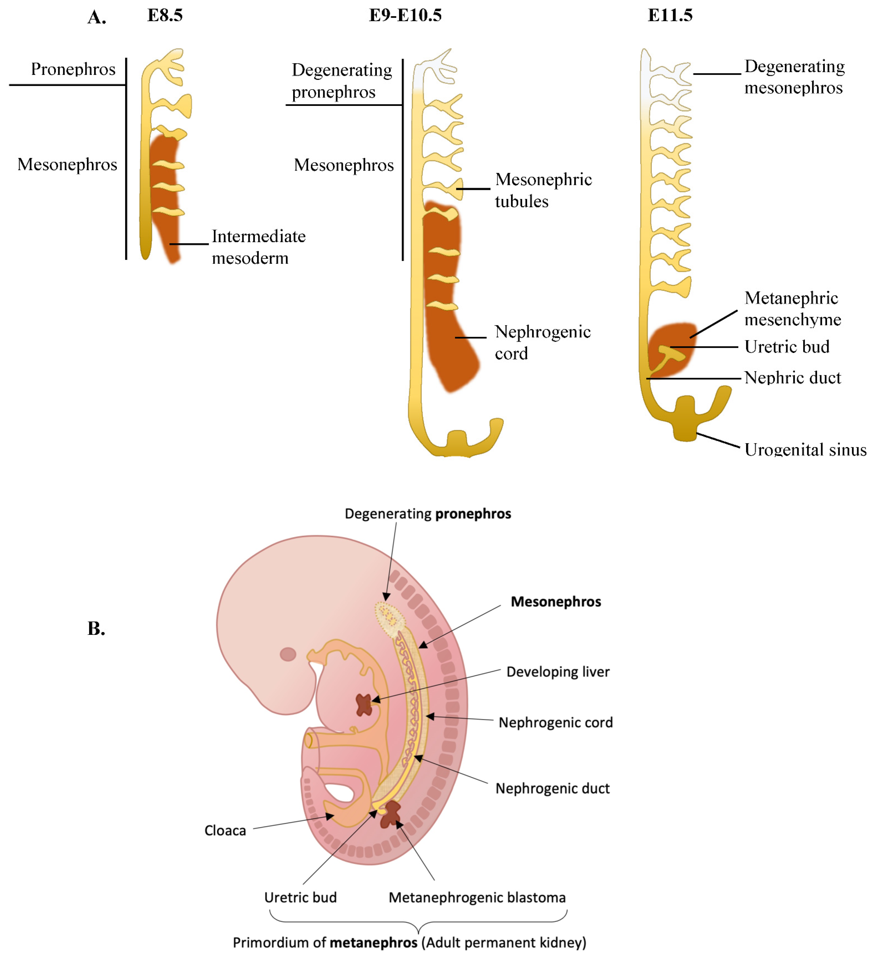
The renal system is composed of the kidney and the urogenital tract. Renal organogenesis is a step-wise process that starts from the development of the pronephros arising from the intermediate mesoderm on E8.5 of mice, turning into the mesonephros and then the metanephros, the latter of which initiates development on E11.5 in mice [84,85] (Figure 2). Kidney organogenesis begins on E8.5 when the intermediate mesoderm undergoes mesenchymal-to-epithelial transition (MET) that gives rise to the pronephros, also termed nephric (Wolffian) duct [84]. The nephric duct then elongates caudally during the transformation of the pronephros to the mesonephros, and as it elongates it induces the formation of mesonephric tubules from the adjacent intermediate mesoderm (also called nephrogenic cord or nephrogenic mesenchyme) [86]. Caudally, the nephric ducts fuse with urogenital sinus (the ventral part of the cloaca), the latter giving rise to the adult bladder [87]. Around E10.5, the caudal part of the pronephros (Wolffian duct) gives rise to a branching outgrowth called the ureteric bud [86].
The ureteric bud grows into a special part of the nephric cord called the metanephric mesenchyme, and the ureteric bud induces the metanephric mesenchyme to turn into metanephric nephrons that will eventually give rise to the kidney [88]. The ureteric bud itself develops into the urogenital tract. The mesonephros and the mesonephric tubules start degenerating on E14.5 and almost entirely disappear in 24 h [89,90]. In females, the mesonephros entirely disappears, while in males some of the cranial mesonephric tubules turn into the epididymal ducts of the testes [85].
PAX8 expression is detected in renal development even before the pronephros is formed, around E7–8 in mice (6–7 somite stage), and is retained during embryogenesis all the way through the developmental process until the adult kidney is formed [91]. However, Pax8 knockout mice show no kidney abnormalities, suggesting that PAX8 expression is redundant in the kidney [42]. Another PAX protein that plays a role in kidney development is the PAX8 close family member PAX2. PAX2 is expressed in the intermediate mesoderm during the nephric anlage stage (before pronephros formation) in the 8–9 somite stage, slightly later then PAX8 expression [91]. However, in PAX2 knockout mice, the pronephros and mesonephros are formed but the metanephros fails to form, resulting in a lack of kidney and genital tract formation [92,93,94]. Combined PAX2 and PAX8 knockout mice show a more severe kidney phenotype, where even the pronephros and mesonephros are not formed. This suggests that PAX8 can compensate for the lack of PAX2 during pronephros and mesonephros development [91]. Of note, while the upper collecting system is PAX8-positive and stems from the metanephros, the bladder stems from differentiation of the urogenital system and is accordingly PAX8-negative [95].
There is very little information regarding the role of PAX8 in the adult renal kidney, and likely most of its role is shared with PAX2. However, it was shown that in inner medullary collecting duct epithelia of adult mice that PAX8, but not PAX2, binds the promoter of Slc14a2. Slc14a2 is the gene encoding for urea transporters and regulating the expression of urea transporters in response to high salt conditions to maintain salt homeostasis [96].
Figure 2. Illustration of embryonic kidney development in mice. (A) An illustration of the kidney development timeline in mice. (B) Mouse kidney development on E11 shown in the context of the whole mouse embryo [97].
Ijms 23 07410 g002

5.2. The Role of PAX8 in Renal Cancers

PAX8 is expressed in several tumors of the urogenital system, such as in the majority of pediatric Wilms tumors [98,99,100], ~80% of renal cell carcinomas (RCC), and nephrogenic adenomas. Tumors of the collecting system diverge in their PAX8 expression. The upper part of the collecting system stems from the PAX8-positive metanephros, and accordingly 23% of tumors of the upper part, specifically of the renal pelvis, are PAX8-positive. The lower part of the collecting system arises from the PAX8-negative urogenital sinus, and tumors that arise from the lower part, i.e., tumors of the ureters and bladder, are all PAX8-negative [77,95,101].
There is a paucity of information regarding the role of PAX8 in RCC. PAX8 plays a positive proliferative role in RCC, and PAX8 knockdown leads to growth arrest and senescence [102,103]. This induction of growth arrest occurs via two different mechanisms. First, PAX8 knockdown was shown to transcriptionally reduce E2F1 expression and reduce the expression of several E2F1 target genes leading to E2F1-dependent growth arrest. Interestingly, and perhaps paradoxically, PAX8 was also shown to stabilize protein levels of RB, the negative regulator of E2F1 [103]. This suggests that PAX8 can perhaps regulate E2F1 levels both positively in a direct manner and negatively in an indirect manner. Another mechanism that was recently suggested for the positive role of PAX8 on cell proliferation is activation of various metabolic pathways in RCC, among them ferroxidase ceruloplasmin, thus altering the cell’s iron metabolism [102].

6. PAX8 Expression in Female Reproductive Tract Development and Cancer

6.1. The Role of PAX8 in Female Reproductive Tract Organogenesis

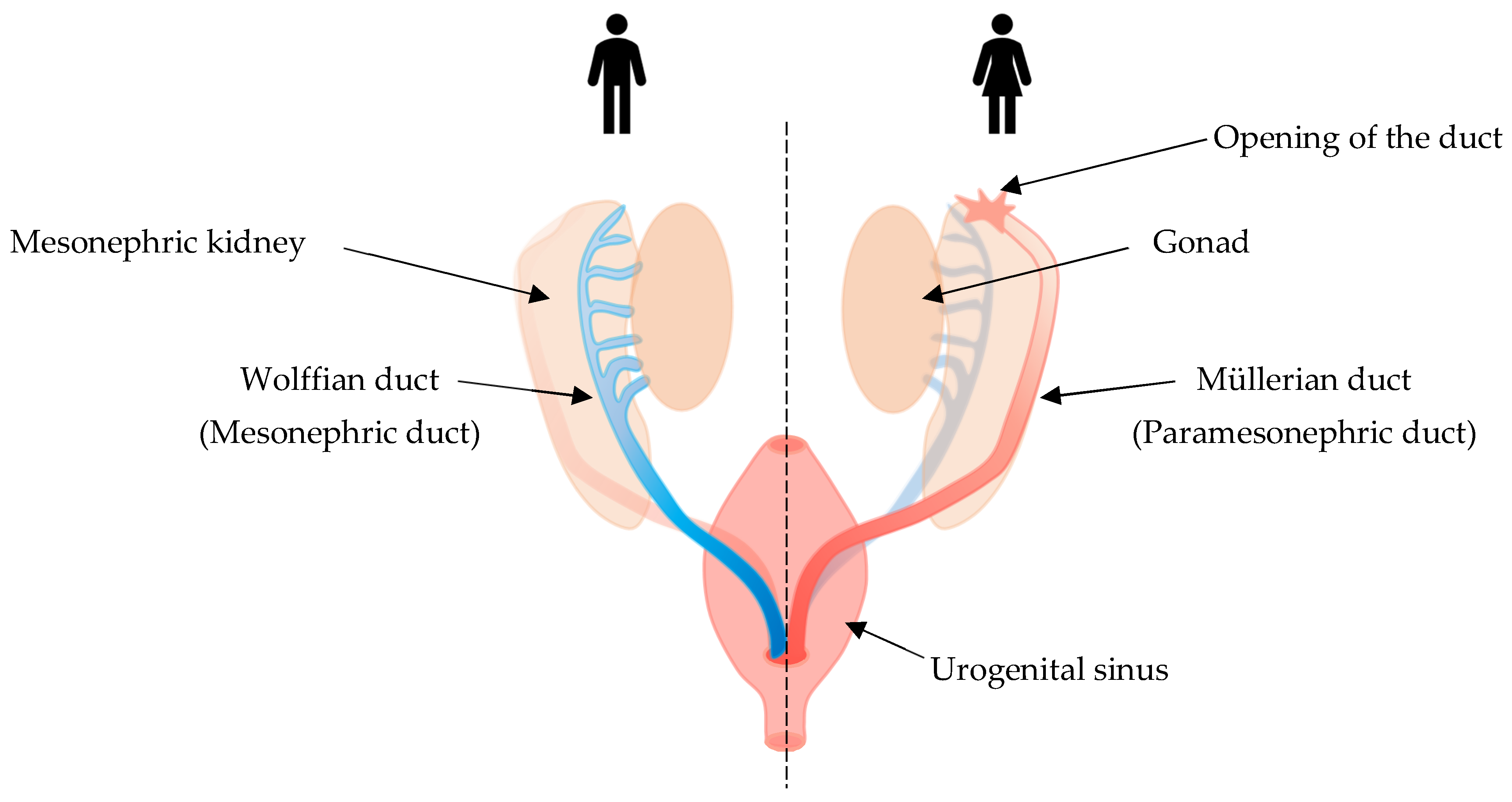
In early embryonic stages, the urogenital system is derived from the intermediate mesoderm of the gastrula [104]. The Müllerian ducts develop in close proximity to the Wolffian ducts, which eventually form the urinary tract [105]. In humans, the two Müllerian ducts begin to migrate medially and then fuse and descend into the pelvis at around 6 weeks of gestational age [106]. Fusion is generally complete by 14 weeks of gestational age, at which time the structures canalize [106,107]. During this time, the urogenital sinus invaginates and differentiates into the sinovaginal bulb [108]. At approximately 12 weeks of gestation, the sinovaginal bulb grows cephalad and fuses with the most caudal portion of the Müllerian ducts [109,110] (Figure 3). In mice, a similar process occurs where the Müllerian duct starts to form by invagination of the surface epithelium of the anterior mesonephros (embryonic kidney) around E11.5 [111]. The Müllerian duct then elongates until it is completed by E13.5, when the duct has reached and conjoins the urogenital sinus [111,112].
In the murine Müllerian duct, PAX8 is detected as early as E11.5–E12.5 [113,114]. Although it has been shown to be expressed in the entire Müllerian duct, PAX8 knockout mice lack endometrial development; however, there is normal development of other structures that arise from the Müllerian duct, such as the upper part of the vagina, cervix, and oviduct [115,116]. The latter suggests that other transcription factors compensate for the absence of PAX8 in these structures [115]. In the adult human uterus, PAX8 is expressed in all epithelial cells of the endometrium. In the fallopian tube, PAX8 is expressed in the secretory epithelial cells, but not in ciliary epithelial cells [77]. In the cervix, PAX8 is expressed in the glandular cells of the uterine cervix (endocervix) whilst not detected in the squamous epithelial cells (ectocervix) [77]. The role of PAX8 in the normal adult female genital tract is unknown.

6.2. The Role of PAX8 in Tumors of the Female Genital System

Tumors arising from the Müllerian system are divided into uterine and extra uterine cancers [117]. The most common female genital tract extra-uterine cancer is epithelial ovarian cancer (EOC). The most common subtype of EOC is high-grade serous carcinoma (HGSC), which in the majority of cases arises from the fallopian tube epithelium [1].
PAX8 was suggested in recent years to be a highly specific marker for identification of Müllerian tumors, both uterine and extra-uterine [116,118]. Different studies have shown that PAX8 is expressed in 89–92% of EOC [77,119,120] and 98–100% of endometrial tumors [77,118]. Of note, all non-mucinous EOC subtypes are PAX8-positive in the vast majority of tumors, and only mucinous ovarian carcinomas are mostly PAX8-negative, suggesting a different embryonic origin [77,121]. In uterine mixed Müllerian tumors (MMMT), which encompass both epithelial and sarcomatous components, PAX8 is expressed in the epithelial component of 97% of all uterine MMMTs but is much less common in the sarcomatous component [122]. For cervical cancer, there is consensus that squamous cell carcinomas of the cervix do not express PAX8, but there is a range in the rate of adenocarcinomas of the cervix that are positive, from 83% to none, likely due to small sample sizes [77,123]. Therefore, PAX8 is clinically used as a good IHC marker for diagnosing Müllerian tumors. One study calculated the sensitivity of PAX8 staining for diagnoses as ranging from moderate to high sensitivity, with high specificity for diagnosing carcinomas of Müllerian origin [124].
PAX8 plays many oncogenic roles in different subtypes of EOC. We and others have shown that PAX8 inhibits HGSC cell proliferation via inhibition of apoptosis [2,3,125]. We have shown that PAX8 activates mutated oncogenic p53 and pro-proliferative cytoplasmic p21, together driving the pro-proliferative effect of PAX8 [3]. Regulation of apoptosis by PAX8 was also shown in a FOXM1-dependent mechanism [125]. A recent study suggests that PAX8 has a pro-angiogenic role in HGSCs via its interaction with another developmental factor, SOX17. PAX8 and SOX17 are co-expressed in ovarian cancer cells [126] and together inhibit the expression of SERPINE1, an anti-angiogenic factor, thereby promoting angiogenesis [18]. The PAX8 and SOX17 complex was also shown to regulate other genes involved in the cell cycle and morphogenesis. Interestingly, PAX8 and SOX17 were shown to be co-regulated by CDK12/13, and biochemical inhibition of CDK12/13 downregulates the expression of both proteins [126]. Another work has shown that PAX8 cooperates with PRDM3 (the protein product of the MECOM genomic locus) to regulate genes that are involved in adhesion and the extracellular matrix [127]. The role of PAX8 in HGSC migration was also shown to be mediated by PKCα [17]. Another role for PAX8 is binding of YAP1/TEAD proteins and transcriptional activation of the Hippo/YAP signaling pathway [128], known to regulate pro-survival genes [129]. Taken together, the information regarding the role of PAX8 in HGSCs fits its definition as a master regulator transcription factor of HGSCs, regulating about 30,000 genes and governing many hallmarks of cancer such as evading apoptosis, angiogenesis, and migration [2,3,17,18,127].
In non-HGSC EOC subtypes, PAX8 was shown to promote anchorage-independent growth, likely by increasing cellular adhesion, thereby contributing to EOC tumorigenicity [130]. Furthermore, PAX8 was shown to have a role in the migration and invasion of EOC [4]. Other studies have shown that in non-HGSC EOC, PAX8 exerts its pro-proliferative role via inhibition of growth arrest and subsequent transcriptional induction of E2F1 expression [103]. Overall, the role of PAX8 in HGSCs and non-HGSC EOC is similar, and different from the minor role of PAX8 in benign cells of fallopian tube lineage [128].
Similar to the non-essential role of PAX8 in fallopian tube development [115], it seems to be also non-essential in adult fallopian tube secretory epithelial cells (FTSEC). However, when FTSEC transform, the role of PAX8 becomes essential to these cells and the number of target genes significantly rises. It is unclear what the exact mechanism is that leads to the major change in the role of PAX8 during transformation. Some suggested mechanisms are epigenomic changes [128], or changes in the role of its target genes, such as a mutation in p53 and an aberrant role of p21 [3].
Another interesting question is the source of PAX8 expression in non-HGSC EOC. PAX8 is a lineage marker of FTSEC, and therefore it is not surprising that it can be found in HGSCs, which arise from transformation of FTSEC. However, non-HGSC EOC is not considered to arise from FTSEC, which raises a question regarding the origin of PAX8 expression in these cells. Interestingly, endometrioid and clear cell carcinomas also express PAX8 in the majority of tumors [77], although these subtypes do not originate from PAX8-positive FTSEC. It was proposed that these tumor types arise from endometriosis, PAX8-positive endometrial cells ectopically expressed outside the uterus [131,132]. Alternatively, these tumors may arise from ovarian surface epithelial cells, and there have been reports of PAX8 expression in ovarian surface epithelium [133]. However, ovarian surface epithelial cells are more commonly considered PAX8-negative, and occasional PAX8-positive cells in the ovarian surface epithelium are thought to be FTSEC or endometrial cells that migrate to the ovarian surface [116,134], leaving PAX8-positive endometriosis cells as the main cell of origin for clear cell and endometrioid ovarian cancers.
In endometrial cancer, PAX8 is expressed in over 90% of tumors of endometrioid and serous histologies [77]. We have published on the role of PAX8 in an especially aggressive subtype of endometrial cancer, uterine serous papillary carcinoma. We have shown that in uterine serous papillary carcinoma, PAX8 plays a pro-proliferative and anti-apoptotic role that is mediated via transcriptional activation of cytoplasmic p21, which has an anti-apoptotic role in this disease [135].

7. PAX8 in the Male Reproductive System—Development and Cancer

7.1. The Role of PAX8 in Male Reproductive System Organogenesis

The main purpose of the male reproductive system is generation, storage, and ejaculation of sperm as well as the production of androgens. Spermatogenesis occurs in the seminiferous tubules of the testes. Sperm maturation mostly occurs in the periphery of the seminiferous tubules. As the sperm matures it moves into the lumen of the tubules and then, via a net of small tubes called the rete testes, through small ducts, called efferent ductules, and into the epididymis. The sperm matures and becomes motile in the epididymis and eventually, during sexual arousal, moves into the vas-deferens where it is combined with fluid coming from the seminal vesicles in the ejaculatory duct, and from there both sperm and fluid are ejaculated via the urethra [136]. The testes, epididymis, vas deferens, ejaculatory ducts, and seminal vesicles are all derived from the Wolffian (mesonephric) duct, which degenerates in the female embryo (Figure 3) [137]. The testes also contain Leydig cells which produce testosterone, the major driver of male genital tract differentiation [136].
In mice, around E13.5, the mesonephric tubules develop into the efferent ductules that connect the rete testis with the upper part of the epididymis [138]. PAX8 expression is detectable at E9.5 in the developing mesonephric duct and also later at E15.5 in the epididymis of male mice [139]. PAX8 expression is also seen in progenitor cells of the rete testis as early as E10.5, showing that the rete testes are also partly derived from the mesonephric duct [140].
Male Pax8 knockout mice are infertile due to malformation and obstruction of the genital tracts. Specifically, Pax8 knockout mice have either agenesis or significant degeneration of the efferent ductules and epididymis. The lack of drainage of the testes results in secondary disorganization of the seminiferous epithelium and atrophy of the testes that progresses with age, likely due to sustained pressure from the drainage blockade. This suggests a role for PAX8 in embryonic development of the efferent ducts and epididymis. In Pax8 knockout mice, there was also some degeneration in the development of the vas deferens, another mesonephros derived organ. However, since overall organs that derive from Wolffian structures develop, it is likely that, in early stages of male genital tract development, the loss of PAX8 is compensated by other factors, such as PAX2. In later stages however, when proper morphogenesis of the efferent ductules and the epididymis occurs, the role of PAX8 is critical. This is also evident from the finding that the seminal vesicles, which are also PAX8-positive and derived from the mesonephric duct, are intact in PAX8 knockout mice. Of note, the development of the seminal vesicles is androgen-dependent and is intact, suggesting proper hormonal function in these mice [141].
In adulthood, in mice and humans, PAX8 is expressed in the epithelium of the seminal vesicles, epididymis, testicular efferent ductules, and rete testes, but not in the urothelium, germ cells, Leydig cells, Sertoli cells, or the prostatic epithelium [142]. These distinctive patterns are probably related to the different embryonic origins of these organs that do not originate in the mesonephric ducts [143,144].

7.2. The Role of PAX8 in Male Genital Tract Neoplasms

A limited number of male genital tract epithelial tumors show PAX8 immuno-positivity, all of which are rare tumors. PAX8-positive staining can be found in primary epithelial tumors that result from transformation of Wolffian derived organs. Specifically, PAX8 expression is seen in serous cystadenoma of the epididymis, carcinoma of the rete testis, Wolffian adnexal tumors, and endometrioid carcinoma of the seminal vesicle [142,145].
PAX8 staining can aid in differentiating these tumors from other male genitourinary tumors that are PAX8-negative, such as urinary tract, testicular, and prostate cancer [142].

8. PAX8 in Other Organs

Over the years, several studies looked for PAX8 expression in other organs that was not the well-characterized expression found in the thyroid, kidney and genital tracts. However, most of these studies used a polyclonal antibody for PAX8 (Proteintech, cat #10336) which was later found to be cross-reactive with other PAX family members such as PAX2, PAX5, and PAX6 [146,147]. The high homology of the N-terminal paired domain of all PAX family members renders N-terminal antibodies, or polyclonal antibodies that detect a large fragment of the protein, to be specifically prone to such cross-reactivity [148]. Therefore, in studies looking for PAX8 expression in organs previously unknown to express the protein, either a C-terminal antibody should be used [149] or mRNA expression should be tested. Western blot analysis using siRNA is another useful tool to determine antibody specificity.
In light of these caveats, the literature pertaining to PAX8 expression in other organs should be critically addressed. For example, the involvement of PAX8 in normal pancreatic islet cells was suggested using the aforementioned PAX8 polyclonal antibody [142]. However, other publications have shown that a more specific PAX8 antibody does not detect PAX8 in developing or adult islet cells, that PAX8 mRNA is not expressed in adult islet cells, and that the antibody that was used to detect PAX8 in islet cells is cross-reactive with PAX6 [150]. Of note, although PAX8 is not expressed in normal pancreatic islet cells when tested using appropriate methods, its expression is activated during pregnancy [151]. Interestingly, a father and daughter carrying a germline PAX8 mutation were described. The father had suffered from type 2 diabetes, while the daughter developed gestational diabetes mellitus, further implicating PAX8 in pancreatic islet cell function during pregnancy [152]. Another paper described a single nucleotide polymorphism (SNP) in the PAX8 locus in Afro-Americans that correlated with elevated risk of type II diabetes [153]. Therefore, while PAX8 is not reliably detected in normal islet cells, it can still be implicated in islet function under certain conditions.
Accordingly, the expression of PAX8 was described in pancreatic neuroendocrine tumors (PNET) [142,154,155] but disputed by others based on antibody cross-reactivity [149,150,156]. Staining of PAX8 in PNET was clearly shown to be a result of antibody cross-reactivity with another PAX family member, PAX6 [150,156].
PAX8 is expressed in the developing brain, specifically in the forebrain and hindbrain, but disappears in the adult brain [157,158,159]. In terms of the function of PAX8 in the developing brain, PAX8 knockout mice did not show brain abnormalities [42,160]. PAX8 does plays a role in mouse inner ear morphogenesis and development, but this role is non-essential since PAX8 knockout mice have no defects in inner ear development [161].
In brain cancers, PAX8 was shown to be expressed in medulloblastoma but only in the subtypes associated with sonic hedgehog and wingless signaling, and not in groups 3 or 4 [159]. Interestingly, PAX8 expression in medulloblastoma was an independent marker of good prognosis, and PAX8 knockdown reduced proliferation, a reverse role compared to its known role in Müllerian tumors. The expression of PAX8 was also studied in glioblastoma, but with contradictory results. Khanlou et al. showed that in glioblastoma sections PAX8 expression was seen focally in only one of nine examined cores [158], while Hung et al. showed PAX8 expression in the vast majority of glioblastoma samples. Of importance, Hung et al. detected the expression of PAX8 protein using two different antibodies, including a C-terminal protein that likely is not cross-reactive with other PAX proteins, and using reverse transcription polymerase chain reaction (RT-PCR), therefore excluding antibody cross-reactivity as the basis for the results. PAX8 expression was also seen rarely in low-grade astrocytomas and grade 1 meningioma tumors. Malignant meningioma (grade 3) exhibited PAX8 expression in 4/4 examined tumors [162].
Two comprehensive immunohistochemical studies reported expression of PAX8 in 100% of a sub population of lymphocytes (presumably B cells) in normal lymphoid tissues. In addition, positive PAX8 diffuse staining has been observed in 100% of the tested lymphomas. However, this study used a polyclonal PAX8 antibody (Proteintech cat #10336) which was subsequently shown to be cross-reactive with PAX5 that is known to be expressed in B-cells. Accordingly, PAX8 was not detected in normal B-cells nor in B-cell lymphomas using a PAX8 C-terminal antibody that does not cross-react with PAX5. Furthermore, PAX8 mRNA levels were not detected in any of the B-cell lymphoma cell lines studied [147].
This example highlights the importance of using specific antibodies when reporting PAX8 expression in different tissues.

9. Summary

PAX8 is a lineage marker with important developmental roles in the thyroid, Wolffian, and Müllerian systems in mice and humans. The expression of PAX8 persists in adult tissues but the role of PAX8 in adult non-thyroid tissues is unclear. When PAX8 expressing cells are transformed, PAX8 gains a new oncogenic role. There is debate regarding the expression of PAX8 in other tissues such as the brain, where expression subsides in the adult brain and turns back upon transformation. The comparison of PAX8 expression and its role in development and cancerous tissues highlights the connections between embryonic development and cancer formation.

Author Contributions

Writing—Original Draft Preparation, R.R.K.; Writing—Review and Editing, R.R.K., Z.M. and R.P.; Supervision, R.P.; Funding Acquisition, R.P. All authors have read and agreed to the published version of the manuscript.

Funding

R.P. is supported by grants from the Israel Science Foundation (1990/18); the Beverley Librach Abshez Initiative for Ovarian and Female Reproductive System Cancers Grant (grant no. 21-303-AI) from the Israel Cancer Research Fund; the Len and Susan Mark Phase II initiative for Ovarian and Uterine/MMMT grant from the Israel Cancer Research Fund; Israel Cancer Association’s USA Board of Directors grant; the EU’s Marie Curie Actions (FP7 People) program; and the Women’s Health Fund at the Rambam Health Care Campus.

Institutional Review Board Statement

Ethical review and approval were waived for this study, as it consists solely of a literature review and does not involve any human or animal studies.

Informed Consent Statement

Not applicable.

Data Availability Statement

Not applicable.

Acknowledgments

We would like to thank Debby Mir for editing of this manuscript and the members of the Perets lab for helpful discussions on the role of PAX8 in gynecological malignancies.

Conflicts of Interest

The authors declare no conflict of interest.

References

  1. Perets, R.; Drapkin, R. It’s Totally Tubular…Riding The New Wave of Ovarian Cancer Research. Cancer Res. 2016, 76, 10–17. [Google Scholar] [CrossRef] [PubMed] [Green Version]
  2. Cheung, H.W.; Cowley, G.S.; Weir, B.A.; Boehm, J.S.; Rusin, S.; Scott, J.A.; East, A.; Ali, L.D.; Lizotte, P.H.; Wong, T.C.; et al. Systematic investigation of genetic vulnerabilities across cancer cell lines reveals lineage-specific dependencies in ovarian cancer. Proc. Natl. Acad. Sci. USA 2011, 108, 12372–12377. [Google Scholar] [CrossRef] [PubMed] [Green Version]
  3. Ghannam-Shahbari, D.; Jacob, E.; Kakun, R.R.; Wasserman, T.; Korsensky, L.; Sternfeld, O.; Kagan, J.; Bublik, D.R.; Aviel-Ronen, S.; Levanon, K.; et al. PAX8 activates a p53-p21-dependent pro-proliferative effect in high grade serous ovarian carcinoma. Oncogene 2018, 37, 2213–2224. [Google Scholar] [CrossRef] [PubMed]
  4. Di Palma, T.; Lucci, V.; de Cristofaro, T.; Filippone, M.G.; Zannini, M. A role for PAX8 in the tumorigenic phenotype of ovarian cancer cells. BMC Cancer 2014, 14, 292. [Google Scholar] [CrossRef] [PubMed] [Green Version]
  5. Aiello, N.; Stanger, B.Z. Echoes of the embryo: Using the developmental biology toolkit to study cancer. Dis. Model. Mech. 2016, 9, 105–114. [Google Scholar] [CrossRef] [PubMed] [Green Version]
  6. Li, J.; Stanger, B.Z. The tumor as organizer model. Science 2019, 363, 1038–1039. [Google Scholar] [CrossRef] [PubMed]
  7. Murray, M.J.; Lessey, B.A. Embryo Implantation and Tumor Metastasis: Common Pathways of Invasion and Angiogenesis. Semin. Reprod. Med. 1999, 17, 275–290. [Google Scholar] [CrossRef]
  8. Evans, M.J.; Kaufman, M.H. Establishment in culture of pluripotential cells from mouse embryos. Nature 1981, 292, 154–156. [Google Scholar] [CrossRef]
  9. Matsui, Y.; Zsebo, K.; Hogan, B.L. Derivation of pluripotential embryonic stem cells from murine primordial germ cells in culture. Cell 1992, 70, 841–847. [Google Scholar] [CrossRef]
  10. Cousin, H. Spemann–Mangold Grafts. Cold Spring Harb. Protoc. 2019, 2019, pdb-rot097345. [Google Scholar] [CrossRef]
  11. Oudin, M.; Weaver, V.M. Physical and Chemical Gradients in the Tumor Microenvironment Regulate Tumor Cell Invasion, Migration, and Metastasis. Cold Spring Harb. Symp. Quant. Biol. 2016, 81, 189–205. [Google Scholar] [CrossRef]
  12. Wellenstein, M.D.; de Visser, K.E. Cancer-Cell-Intrinsic Mechanisms Shaping the Tumor Immune Landscape. Immunity 2018, 48, 399–416. [Google Scholar] [CrossRef] [Green Version]
  13. Folkman, J. Tumor angiogenesis: Therapeutic implications. N. Engl. J. Med. 1971, 285, 1182–1186. [Google Scholar] [CrossRef]
  14. Buscail, L.; Bournet, B.; Cordelier, P. Role of oncogenic KRAS in the diagnosis, prognosis and treatment of pancreatic cancer. Nat. Rev. Gastroenterol. Hepatol. 2020, 17, 153–168. [Google Scholar] [CrossRef]
  15. Meškytė, E.M.; Keskas, S.; Ciribilli, Y. MYC as a Multifaceted Regulator of Tumor Microenvironment Leading to Metastasis. Int. J. Mol. Sci. 2020, 21, 7710. [Google Scholar] [CrossRef]
  16. Semenza, G.L.; Wang, G.L. A nuclear factor induced by hypoxia via de novo protein synthesis binds to the human erythropoietin gene enhancer at a site required for transcriptional activation. Mol. Cell Biol. 1992, 12, 5447–5454. [Google Scholar]
  17. Hardy, L.R.; Pergande, M.; Esparza, K.; Heath, K.N.; Önyüksel, H.; Cologna, S.M.; Burdette, J.E. Proteomic analysis reveals a role for PAX8 in peritoneal colonization of high grade serous ovarian cancer that can be targeted with micelle encapsulated thiostrepton. Oncogene 2019, 38, 6003–6016. [Google Scholar] [CrossRef]
  18. Chaves-Moreira, D.; Mitchell, M.A.; Arruza, C.; Rawat, P.; Sidoli, S.; Nameki, R.; Reddy, J.; Corona, R.I.; Afeyan, L.K.; Klein, I.A.; et al. The transcription factor PAX8 promotes angiogenesis in ovarian cancer through interaction with SOX17. Sci. Signal. 2022, 15. [Google Scholar] [CrossRef]
  19. Monk, M.; Holding, C. Human embryonic genes re-expressed in cancer cells. Oncogene 2001, 20, 8085–8091. [Google Scholar] [CrossRef] [Green Version]
  20. Takahashi, K.; Tanabe, K.; Ohnuki, M.; Narita, M.; Ichisaka, T.; Tomoda, K.; Yamanaka, S. Induction of Pluripotent Stem Cells from Adult Human Fibroblasts by Defined Factors. Cell 2007, 131, 861–872. [Google Scholar] [CrossRef] [Green Version]
  21. Zhang, X.; Zhao, W.; Li, Y. Stemness-related markers in cancer. Cancer Transl. Med. 2017, 3, 87–95. [Google Scholar] [CrossRef] [Green Version]
  22. Gwak, J.M.; Kim, M.; Kim, H.J.; Jang, M.H.; Park, S.Y. Expression of embryonal stem cell transcription factors in breast cancer: Oct4 as an indicator for poor clinical outcome and tamoxifen resistance. Oncotarget 2017, 8, 36305–36318. [Google Scholar] [CrossRef] [Green Version]
  23. Bhatlekar, S.; Fields, J.Z.; Boman, B.M. HOX genes and their role in the development of human cancers. Klin. Wochenschr. 2014, 92, 811–823. [Google Scholar] [CrossRef]
  24. Montavon, T.; Soshnikova, N. Hox gene regulation and timing in embryogenesis. Semin. Cell Dev. Biol. 2014, 34, 76–84. [Google Scholar] [CrossRef]
  25. Domsch, K.; Papagiannouli, F.; Lohmann, I. Chapter Five—he HOX–Apoptosis Regulatory Interplay in Development and Disease. In Current Topics in Developmental Biology; Steller, H., Ed.; Academic Press: Cambridge, MA, USA, 2015; Volume 114, pp. 121–158. [Google Scholar]
  26. Paço, A.; de Bessa Garcia, S.A.; Castro, J.L.; Costa-Pinto, A.; Freitas, R. Roles of the HOX Proteins in Cancer Invasion and Metastasis. Cancers 2020, 13, 10. [Google Scholar] [CrossRef]
  27. Smith, J.; Zyoud, A.; Allegrucci, C. A Case of Identity: HOX Genes in Normal and Cancer Stem Cells. Cancers 2019, 11, 512. [Google Scholar] [CrossRef] [Green Version]
  28. Mark, M.; Rijli, F.M.; Chambon, P. Homeobox Genes in Embryogenesis and Pathogenesis. Pediatr. Res. 1997, 42, 421–429. [Google Scholar] [CrossRef]
  29. Lou, Y.; Fallah, Y.; Yamane, K.; E Berg, P. BP1, a potential biomarker for breast cancer prognosis. Biomarkers Med. 2018, 12, 535–545. [Google Scholar] [CrossRef] [Green Version]
  30. Tan, Y.; Testa, J. DLX Genes: Roles in Development and Cancer. Cancers 2021, 13, 3005. [Google Scholar] [CrossRef]
  31. Holland, P.W.H. Evolution of homeobox genes. Wiley Interdiscip. Rev. Dev. Biol. 2012, 2, 31–45. [Google Scholar] [CrossRef]
  32. Holland, P.W.H.; Booth, H.A.F.; Bruford, E. Classification and nomenclature of all human homeobox genes. BMC Biol. 2007, 5, 1–28. [Google Scholar] [CrossRef] [PubMed] [Green Version]
  33. Lang, D.; Powell, S.K.; Plummer, R.S.; Young, K.P.; Ruggeri, B.A. PAX genes: Roles in development, pathophysiology, and cancer. Biochem. Pharmacol. 2007, 73, 1–14. [Google Scholar] [CrossRef] [PubMed]
  34. Chi, N.; Epstein, J.A. Getting your Pax straight: Pax proteins in development and disease. Trends Genet. 2002, 18, 41–47. [Google Scholar] [CrossRef]
  35. Robson, E.J.D.; He, S.-J.; Eccles, M.R. A PANorama of PAX genes in cancer and development. Nat. Cancer 2006, 6, 52–62. [Google Scholar] [CrossRef]
  36. Noll, M. Evolution and role of Pax genes. Curr. Opin. Genet. Dev. 1993, 3, 595–605. [Google Scholar] [CrossRef]
  37. Balczarek, K.A.; Lai, Z.C.; Kumar, S. Evolution of functional diversification of the paired box (Pax) DNA-binding domains. Mol. Biol. Evol. 1997, 14, 829–842. [Google Scholar] [CrossRef] [Green Version]
  38. Thompson, B.; Davidson, E.A.; Liu, W.; Nebert, D.W.; Bruford, E.A.; Zhao, H.; Dermitzakis, E.T.; Thompson, D.C.; Vasiliou, V. PAX Genes in Cancer; Friends specific variant expression and involvement in human disease. Hum. Genet. 2021, 140, 381–400. [Google Scholar] [CrossRef]
  39. Li, C.G.; Eccles, M.R. PAX Genes in Cancer; Friends or Foes? Front. Genet. 2012, 3, 6. [Google Scholar] [CrossRef] [Green Version]
  40. Jumper, J.; Evans, R.; Pritzel, A.; Green, T.; Figurnov, M.; Ronneberger, O.; Tunyasuvunakool, K.; Bates, R.; Žídek, A.; Potapenko, A.; et al. Highly accurate protein structure prediction with AlphaFold. Nature 2021, 596, 583–589. [Google Scholar] [CrossRef]
  41. Varadi, M.; Anyango, S.; Deshpande, M.; Nair, S.; Natassia, C.; Yordanova, G.; Yuan, D.; Stroe, O.; Wood, G.; Laydon, A.; et al. AlphaFold Protein Structure Database: Massively expanding the structural coverage of protein-sequence space with high-accuracy models. Nucleic Acids Res. 2022, 50, D439–D444. [Google Scholar] [CrossRef]
  42. Mansouri, A.; Chowdhury, K.; Gruss, P. Follicular cells of the thyroid gland require Pax8 gene function. Nat. Genet. 1998, 19, 87–90. [Google Scholar] [CrossRef]
  43. Campagnolo, M.; Pesaresi, A.; Zelezetsky, I.; Geremia, S.; Randaccio, L.; Bisca, A.; Tell, G. Structural Studies on Pax-8 Prd Domain/DNA Complex. J. Biomol. Struct. Dyn. 2007, 24, 429–441. [Google Scholar] [CrossRef]
  44. Codutti, L.; van Ingen, H.; Vascotto, C.; Fogolari, F.; Corazza, A.; Tell, G.; Quadrifoglio, F.; Viglino, P.; Boelens, R.; Esposito, G. The Solution Structure of DNA-free Pax-8 Paired Box Domain Accounts for Redox Regulation of Transcriptional Activity in the Pax Protein Family. J. Biol. Chem. 2008, 283, 33321–33328. [Google Scholar] [CrossRef] [Green Version]
  45. Kozmik, Z.; Kurzbauer, R.; Dörfler, P.; Busslinger, M. Alternative splicing of Pax-8 gene transcripts is developmentally regulated and generates isoforms with different transactivation properties. Mol. Cell. Biol. 1993, 13, 6024–6035. [Google Scholar]
  46. Szczepanek-Parulska, E.; Szaflarski, W.; Piątek, K.; Budny, B.; Jaszczyńska-Nowinka, K.; Biczysko, M.; Wierzbicki, T.; Skrobisz, J.; Zabel, M.; Ruchała, M. Alternative 3’ acceptor site in the exon 2 of human PAX8 gene resulting in the expression of unknown mRNA variant found in thyroid hemiagenesis and some types of cancers. Acta Biochim. Pol. 2013, 60, 573–578. [Google Scholar] [CrossRef] [Green Version]
  47. Johansson, E.; Andersson, L.; Örnros, J.; Carlsson, T.; Ingeson-Carlsson, C.; Liang, S.; Dahlberg, J.; Jansson, S.; Parrillo, L.; Zoppoli, P.; et al. Revising the embryonic origin of thyroid C cells in mice and humans. Development 2015, 142, 3519–3528. [Google Scholar]
  48. Mauchamp, J.; Mirrione, A.; Alquier, C.; André, F. Follicle-like structure and polarized monolayer: Role of the extracellular matrix on thyroid cell organization in primary culture. Biol. Cell 1998, 90, 369–380. [Google Scholar] [CrossRef]
  49. Fernández, L.P.; López-Márquez, A.; Santisteban, P. Thyroid transcription factors in development, differentiation and disease. Nat. Rev. Endocrinol. 2014, 11, 29–42. [Google Scholar] [CrossRef] [Green Version]
  50. Fagman, H.; Amendola, E.; Parrillo, L.; Zoppoli, P.; Marotta, P.; Scarfò, M.; De Luca, P.; de Carvalho, D.P.; Ceccarelli, M.; De Felice, M.; et al. Gene expression profiling at early organogenesis reveals both common and diverse mechanisms in foregut patterning. Dev. Biol. 2011, 359, 163–175. [Google Scholar] [CrossRef]
  51. Veis, D.J.; Sorenson, C.M.; Shutter, J.R.; Korsmeyer, S.J. Bcl-2-deficient mice demonstrate fulminant lymphoid apoptosis, polycystic kidneys, and hypopigmented hair. Cell 1993, 75, 229–240. [Google Scholar] [CrossRef]
  52. Parlato, R.; Rosica, A.; Rodriguez-Mallon, A.; Affuso, A.; Postiglione, M.P.; Arra, C.; Mansouri, A.; Kimura, S.; Di Lauro, R.; De Felice, M. An integrated regulatory network controlling survival and migration in thyroid organogenesis. Dev. Biol. 2004, 276, 464–475. [Google Scholar] [CrossRef] [Green Version]
  53. De Felice, M.; Ovitt, C.; Biffali, E.; Rodriguez-Mallon, A.; Arra, C.; Anastassiadis, K.; Macchia, P.E.; Mattei, M.-G.; Mariano, A.; Schöler, H.; et al. A mouse model for hereditary thyroid dysgenesis and cleft palate. Nat. Genet. 1998, 19, 395–398. [Google Scholar] [CrossRef]
  54. Fisher, D.A.; Dussault, J.H.; Sack, J.; Chopra, I.J. Ontogenesis of hypothalamic pituitary thyroid function and metabolism in man, sheep, and rat. Recent Prog. Horm. Res. 1977, 33, 59–116. [Google Scholar]
  55. Sgalitzer, K.E. Contribution to the study of the morphogenesis of the thyroid gland. J. Anat. 1941, 75, 389–405. [Google Scholar]
  56. Kaufman, M.H.; Bard, J.B.L. The Anatomical Basis of Mouse Development; Gulf Professional Publishing: Houston, TX, USA, 1999. [Google Scholar]
  57. Polak, M.; Sura-Trueba, S.; Chauty, A.; Szinnai, G.; Carré, A.; Castanet, M. Molecular Mechanisms of Thyroid Dysgenesis. Horm. Res. Paediatr. 2004, 62, 14–21. [Google Scholar] [CrossRef]
  58. De Felice, M.; Di Lauro, R. Thyroid Development and Its Disorders: Genetics and Molecular Mechanisms. Endocr. Rev. 2004, 25, 722–746. [Google Scholar] [CrossRef] [Green Version]
  59. di Magliano, M.P.; Di Lauro, R.; Zannini, M. Pax8 has a key role in thyroid cell differentiation. Proc. Natl. Acad. Sci. USA 2000, 97, 13144–13149. [Google Scholar] [CrossRef] [Green Version]
  60. D’Andrea, B.; Iacone, R.; Di Palma, T.; Nitsch, R.; Baratta, M.G.; Nitsch, L.; Di Lauro, R.; Zannini, M. Functional Inactivation of the Transcription Factor Pax8 through Oligomerization Chain Reaction. Mol. Endocrinol. 2006, 20, 1810–1824. [Google Scholar] [CrossRef] [PubMed] [Green Version]
  61. Macchia, P.E.; Lapi, P.; Krude, H.; Pirro, M.T.; Missero, C.; Chiovato, L.; Souabni, A.; Baserga, M.; Tassi, V.; Pinchera, A.; et al. PAX8 mutations associated with congenital hypothyroidism caused by thyroid dysgenesis. Nat. Genet. 1998, 19, 83–86. [Google Scholar] [CrossRef] [PubMed]
  62. Hermanns, P.; Grasberger, H.; Cohen, R.; Freiberg, C.; Dörr, H.G.; Refetoff, S.; Pohlenz, J. Two cases of thyroid dysgenesis caused by different novel PAX8 mutations in the DNA-binding region: In vitro studies reveal different pathogenic mechanisms. Thyroid 2013, 23, 791–796. [Google Scholar] [CrossRef] [PubMed]
  63. Nettore, I.C.; Cacace, V.; De Fusco, C.; Colao, A.; Macchia, P.E. The molecular causes of thyroid dysgenesis: A systematic review. J. Endocrinol. Investig. 2013, 36, 654–664. [Google Scholar]
  64. Hermanns, P.; Grasberger, H.; Refetoff, S.; Pohlenz, J. Mutations in the NKX2.5 gene and the PAX8 promoter in a girl with thyroid dysgenesis. J. Clin. Endocrinol. Metab. 2011, 96, E977–E981. [Google Scholar] [CrossRef] [Green Version]
  65. Hermanns, P.; Shepherd, S.; Mansor, M.; Schulga, J.; Jones, J.; Donaldson, M.; Pohlenz, J. A new mutation in the promoter region of the PAX8 gene causes true congenital hypothyroidism with thyroid hypoplasia in a girl with Down’s syndrome. Thyroid 2014, 24, 939–944. [Google Scholar] [CrossRef] [Green Version]
  66. Grasberger, H.; Ringkananont, U.; LeFrancois, P.; Abramowicz, M.; Vassart, G.; Refetoff, S. Thyroid transcription factor 1 rescues PAX8/p300 synergism impaired by a natural PAX8 paired domain mutation with dominant negative activity. Mol. Endocrinol. 2005, 19, 1779–1791. [Google Scholar] [CrossRef] [Green Version]
  67. Di Palma, T.; Zampella, E.; Filippone, M.G.; Macchia, P.E.; Ris-Stalpers, C.; De Vroede, M.; Zannini, M. Characterization of a novel loss-of-function mutation of PAX8 associated with congenital hypothyroidism. Clin. Endocrinol. 2010, 73, 808–814. [Google Scholar] [CrossRef]
  68. Fernández-Méndez, C.; Santisteban, P. A Critical Balance Between PAX8 and the Hippo Mediator TAZ Determines Sodium/Iodide Symporter Expression and Function. Thyroid 2022, 32, 315–325. [Google Scholar] [CrossRef]
  69. Di Palma, T.; D’Andrea, B.; Liguori, G.L.; Liguoro, A.; de Cristofaro, T.; Del Prete, D.; Pappalardo, A.; Mascia, A.; Zannini, M. TAZ is a coactivator for Pax8 and TTF-1, two transcription factors involved in thyroid differentiation. Exp. Cell Res. 2009, 315, 162–175. [Google Scholar] [CrossRef]
  70. Dralle, H.; Machens, A.; Basa, J.; Fatourechi, V.; Franceschi, S.; Hay, I.D.; Nikiforov, Y.E.; Pacini, F.; Pasieka, J.L.; Sherman, S.I. Follicular cell-derived thyroid cancer. Nat. Rev. Dis. Primers 2015, 1, 15077. [Google Scholar] [CrossRef]
  71. Parameswaran, R.; Brooks, S.; Sadler, G.P. Molecular pathogenesis of follicular cell derived thyroid cancers. Int. J. Surg. 2010, 8, 186–193. [Google Scholar] [CrossRef] [Green Version]
  72. LiVolsi, V.A. Papillary thyroid carcinoma: An update. Mod. Pathol. 2011, 24, S1–S9. [Google Scholar] [CrossRef]
  73. Enewold, L.; Zhu, K.; Ron, E.; Marrogi, A.J.; Stojadinovic, A.; Peoples, G.E.; Devesa, S.S. Rising Thyroid Cancer Incidence in the United States by Demographic and Tumor Characteristics, 1980–2005. Cancer Epidemiol. Biomark. Prev. 2009, 18, 784–791. [Google Scholar] [CrossRef] [Green Version]
  74. Pstrąg, N.; Ziemnicka, K.; Bluyssen, H.; Wesoły, J. Thyroid cancers of follicular origin in a genomic light: In-depth overview of common and unique molecular marker candidates. Mol. Cancer 2018, 17, 1–17. [Google Scholar] [CrossRef] [Green Version]
  75. Nonaka, D.; Tang, Y.; Chiriboga, L.; Rivera, M.; Ghossein, R.A. Diagnostic utility of thyroid transcription factors Pax8 and TTF-2 (FoxE1) in thyroid epithelial neoplasms. Mod. Pathol. 2007, 21, 192–200. [Google Scholar] [CrossRef]
  76. Bishop, J.A.; Sharma, R.; Westra, W.H. PAX8 immunostaining of anaplastic thyroid carcinoma: A reliable means of discerning thyroid origin for undifferentiated tumors of the head and neck. Hum. Pathol. 2011, 42, 1873–1877. [Google Scholar] [CrossRef] [PubMed]
  77. Laury, A.R.; Perets, R.; Piao, H.; Krane, J.F.; Barletta, J.A.; French, C.; Chirieac, L.R.; Lis, R.; Loda, M.; Hornick, J.L.; et al. A Comprehensive Analysis of PAX8 Expression in Human Epithelial Tumors. Am. J. Surg. Pathol. 2011, 35, 816–826. [Google Scholar] [CrossRef] [PubMed] [Green Version]
  78. French, C.A.; Alexander, E.K.; Cibas, E.S.; Nose, V.; Laguette, J.; Faquin, W.; Garber, J.; Moore, F.; Fletcher, J.A.; Larsen, P.R.; et al. Genetic and Biological Subgroups of Low-Stage Follicular Thyroid Cancer. Am. J. Pathol. 2003, 162, 1053–1060. [Google Scholar] [CrossRef] [Green Version]
  79. Tai, T.-A.C.; Jennermann, C.; Brown, K.K.; Oliver, B.B.; MacGinnitie, M.A.; Wilkison, W.O.; Brown, H.R.; Lehmann, J.M.; Kliewer, S.A.; Morris, D.C.; et al. Activation of the Nuclear Receptor Peroxisome Proliferator-activated Receptor γ Promotes Brown Adipocyte Differentiation. J. Biol. Chem. 1996, 271, 29909–29914. [Google Scholar] [CrossRef] [Green Version]
  80. Raman, P.; Koenig, R.J. PAX8-PPARγ fusion protein in thyroid carcinoma. Nat. Rev. Endocrinol. 2014, 10, 616–623. [Google Scholar] [CrossRef]
  81. Rosignolo, F.; Sponziello, M.; Durante, C.; Puppin, C.; Mio, C.; Baldan, F.; Di Loreto, C.; Russo, D.; Filetti, S.; Damante, G. Expression of PAX8 Target Genes in Papillary Thyroid Carcinoma. PLoS ONE 2016, 11, e0156658. [Google Scholar] [CrossRef]
  82. Credendino, S.C.; Bellone, M.L.; Lewin, N.; Amendola, E.; Sanges, R.; Basu, S.; Sepe, R.; Decaussin-Petrucci, M.; Tinto, N.; Fusco, A.; et al. A ceRNA Circuitry Involving the Long Noncoding RNA Klhl14-AS, Pax8, and Bcl2 Drives Thyroid Carcinogenesis. Cancer Res. 2019, 79, 5746–5757. [Google Scholar] [CrossRef] [Green Version]
  83. Dupain, C.; Ali, H.M.; Mouhoub, T.A.; Urbinati, G.; Massaad-Massade, L. Induction of TTF-1 or PAX-8 expression on proliferation and tumorigenicity in thyroid carcinomas. Int. J. Oncol. 2016, 49, 1248–1258. [Google Scholar] [CrossRef] [Green Version]
  84. Bouchard, M.; Pfeffer, P.; Busslinger, M. Functional equivalence of the transcription factors Pax2 and Pax5 in mouse development. Development 2000, 127, 3703–3713. [Google Scholar] [CrossRef]
  85. Vetter, M.R.; Gibley, C.W. Morphogenesis and histochemistry of the developing mouse kidney. J. Morphol. 1966, 120, 135–155. [Google Scholar] [CrossRef]
  86. Davidson, A.J. Mouse kidney development. In StemBook; Harvard Stem Cell Institut: Cambridge, MA, USA, 2008. [Google Scholar]
  87. Tan, W.-H.; Gilmore, E.C.; Baris, H.N. Human Developmental Genetics. In Emery and Rimoin’s Principles and Practice of Medical Genetics, 6th ed.; Academic Press: Cambridge, MA, USA, 2013; pp. 1–63. [Google Scholar] [CrossRef]
  88. Birchmeier, W. Epithelial Morphogenesis in Development and Disease; CRC Press: Boca Raton, FL, USA, 2003. [Google Scholar]
  89. Sainio, K.; Hellstedt, P.; Kreidberg, J.; Saxen, L.; Sariola, H. Differential regulation of two sets of mesonephric tubules by WT-1. Development 1997, 124, 1293–1299. [Google Scholar] [CrossRef]
  90. Smith, C.; Mackay, S. Morphological development and fate of the mouse mesonephros. J. Anat. 1991, 174, 171–184. [Google Scholar]
  91. Bouchard, M.; Souabni, A.; Mandler, M.; Neubüser, A.; Busslinger, M. Nephric lineage specification by Pax2 and Pax8. Genes Dev. 2002, 16, 2958–2970. [Google Scholar] [CrossRef] [Green Version]
  92. Dressler, G.R.; Wilkinson, J.E.; Rothenpieler, U.W.; Patterson, L.T.; Williams-Simons, L.; Westphal, H. Deregulation of Pax-2 expression in transgenic mice generates severe kidney abnormalities. Nature 1993, 362, 65–67. [Google Scholar] [CrossRef]
  93. Dressler, G.; Deutsch, U.; Chowdhury, K.; Nornes, H.; Gruss, P. Pax2, a new murine paired-box-containing gene and its expression in the developing excretory system. Development 1990, 109, 787–795. [Google Scholar] [CrossRef]
  94. Sanyanusin, P.; Schimmenti, L.; McNoe, L.A.; Ward, T.A.; Pierpont, M.E.M.; Sullivan, M.J.; Dobyns, W.; Eccles, M.R. Mutation of the PAX2 gene in a family with optic nerve colobomas, renal anomalies and vesicoureteral reflux. Nat. Genet. 1995, 9, 358–364. [Google Scholar] [CrossRef]
  95. Tong, G.-X.; Yu, W.M.; Beaubier, N.T.; Weeden, E.M.; Hamele-Bena, D.; Mansukhani, M.M.; O’Toole, K.M. Expression of PAX8 in normal and neoplastic renal tissues: An immunohistochemical study. Mod. Pathol. 2009, 22, 1218–1227. [Google Scholar] [CrossRef] [Green Version]
  96. Laszczyk, A.M.; Higashi, A.Y.; Patel, S.R.; Johnson, C.; Soofi, A.; Abraham, S.; Dressler, G.R. Pax2 and Pax8 Proteins Regulate Urea Transporters and Aquaporins to Control Urine Concentration in the Adult Kidney. J. Am. Soc. Nephrol. 2020, 31, 1212–1225. [Google Scholar] [CrossRef] [PubMed]
  97. Kousta, E.; Papathanasiou, A.; Skordis, N. Sex determination and disorders of sex development according to the revised nomenclature and classification in 46, XX individuals. Hormones 2010, 9, 218–231. [Google Scholar] [CrossRef]
  98. Tagge, E.P.; Hanson, P.; Re, G.G.; Othersen, H.; Smith, C.D.; Garvin, A. Paired box gene expression in Wilms’ tumor. J. Pediatr. Surg. 1994, 29, 134–141. [Google Scholar] [CrossRef]
  99. Sefidbakht, S.; Khorsand-Rahimzadeh, A.; Omidi, S.; Mohsenpourian, S.; Mirzaian, E. Expression of PAX2 and PAX8 in Wilms Tumor: A Tissue Microarray-based Immunohistochemical Study. Iran. J. Pathol. 2021, 16, 310–315. [Google Scholar] [CrossRef]
  100. Poleev, A.; Fickenscher, H.; Mundlos, S.; Winterpacht, A.; Zabel, B.; Fidler, A.; Gruss, P.; Plachov, D. PAX8, a human paired box gene: Isolation and expression in developing thyroid, kidney and Wilms’ tumors. Development 1992, 116, 611–623. [Google Scholar] [CrossRef]
  101. Ozcan, A.; de la Roza, G.; Ro, J.Y.; Shen, S.S.; Truong, L.D. PAX2 and PAX8 Expression in Primary and Metastatic Renal Tumors: A Comprehensive Comparison. Arch. Pathol. Lab. Med. 2012, 136, 1541–1551. [Google Scholar] [CrossRef] [Green Version]
  102. Bleu, M.; Gaulis, S.; Lopes, R.; Sprouffske, K.; Apfel, V.; Holwerda, S.; Pregnolato, M.; Yildiz, U.; Cordo’, V.; Dost, A.F.M.; et al. PAX8 activates metabolic genes via enhancer elements in Renal Cell Carcinoma. Nat. Commun. 2019, 10, 1–10. [Google Scholar] [CrossRef] [Green Version]
  103. Li, C.G.; Nyman, J.E.; Braithwaite, A.W.; Eccles, M.R. PAX8 promotes tumor cell growth by transcriptionally regulating E2F1 and stabilizing RB protein. Oncogene 2011, 30, 4824–4834. [Google Scholar] [CrossRef] [Green Version]
  104. Coalson, R.E.; Tomasek, J.J. Urogenital System. In Embryology; Springer: Berlin/Heidelberg, Germany, 1992; pp. 95–102. [Google Scholar] [CrossRef]
  105. Kobayashi, A.; Behringer, R.R. Developmental genetics of the female reproductive tract in mammals. Nat. Rev. Genet. 2003, 4, 969–980. [Google Scholar] [CrossRef]
  106. Wilson, D.; Bordoni, B. Embryology, Mullerian Ducts (Paramesonephric Ducts). In StatPearls; StatPearls Publishing: Tampa, FL, USA, 2022. [Google Scholar]
  107. Aimen, F.M.; Atef, Y.; Majed, G.; Manel, M.; Monia, M.; Radhouane, A.; Hedi, R.; Khaled, N. Spontaneous pregnancy after vaginoplasty in a patient presenting a congenital vaginal aplasia. Asian Pac. J. Reprod. 2016, 5, 351–353. [Google Scholar] [CrossRef]
  108. Robboy, S.J.; Kurita, T.; Baskin, L.; Cunha, G.R. New insights into human female reproductive tract development. Differentiation 2017, 97, 9–22. [Google Scholar] [CrossRef]
  109. Joki-Erkkilä, M.M.; Heinonen, P.K. Presenting and long-term clinical implications and fecundity in females with obstructing vaginal malformations. J. Pediatr. Adolesc. Gynecol. 2003, 16, 307–312. [Google Scholar] [CrossRef]
  110. Sanfilippo, J.S.; Lara-Torre, E.; Gomez-Lobo, V. Sanfilippo’s Textbook of Pediatric and Adolescent Gynecology, 2nd ed.; CRC Press: Boca Raton, FL, USA, 2019. [Google Scholar]
  111. Kobayashi, A.; Shawlot, W.; Kania, A.; Behringer, R.R. Requirement of Lim1 for female reproductive tract development. Development 2004, 131, 539–549. [Google Scholar] [CrossRef] [Green Version]
  112. Orvis, G.D.; Behringer, R.R. Cellular mechanisms of Müllerian duct formation in the mouse. Dev. Biol. 2007, 306, 493–504. [Google Scholar] [CrossRef] [Green Version]
  113. Vainio, S.; Heikkilä, M.; Kispert, A.; Chin, N.; McMahon, A.P. Female development in mammals is regulated by Wnt-4 signalling. Nature 1999, 397, 405–409. [Google Scholar] [CrossRef]
  114. Carroll, T.J.; Park, J.-S.; Hayashi, S.; Majumdar, A.; McMahon, A.P. Wnt9b Plays a Central Role in the Regulation of Mesenchymal to Epithelial Transitions Underlying Organogenesis of the Mammalian Urogenital System. Dev. Cell 2005, 9, 283–292. [Google Scholar] [CrossRef] [Green Version]
  115. Mittag, J.; Winterhager, E.; Bauer, K.; Grümmer, R. Congenital Hypothyroid Female Pax8-Deficient Mice Are Infertile Despite Thyroid Hormone Replacement Therapy. Endocrinology 2007, 148, 719–725. [Google Scholar] [CrossRef] [Green Version]
  116. Bowen, N.J.; Logani, S.; Dickerson, E.B.; Kapa, L.B.; Akhtar, M.; Benigno, B.B.; McDonald, J.F. Emerging roles for PAX8 in ovarian cancer and endosalpingeal development. Gynecol. Oncol. 2007, 104, 331–337. [Google Scholar] [CrossRef]
  117. Dubeau, L. Pathogenesis of serous, extra-uterine Müllerian epithelial cancer and therapeutic implications. Transl. Cancer Res. 2015, 4, 3–13. [Google Scholar] [PubMed]
  118. Nonaka, D.; Chiriboga, L.; Soslow, R.A. Expression of Pax8 as a Useful Marker in Distinguishing Ovarian Carcinomas From Mammary Carcinomas. Am. J. Surg. Pathol. 2008, 32, 1566–1571. [Google Scholar] [CrossRef] [PubMed]
  119. Chai, H.; Ren, Q.; Fan, Q.; Ye, L.; Du, G.; Du, H.; Xu, W.; Li, Y.; Zhang, L.; Cheng, Z. PAX8 is a potential marker for the diagnosis of primary epithelial ovarian cancer. Oncol. Lett. 2017, 14, 5871–5875. [Google Scholar] [CrossRef]
  120. Laury, A.R.; Hornick, J.L.; Perets, R.; Krane, J.F.; Corson, J.; Drapkin, R.; Hirsch, M.S. PAX8 Reliably Distinguishes Ovarian Serous Tumors From Malignant Mesothelioma. Am. J. Surg. Pathol. 2010, 34, 627–635. [Google Scholar] [CrossRef] [Green Version]
  121. Tong, G.-X.; Devaraj, K.; Hamele-Bena, D.; Yu, W.M.; Turk, A.; Chen, X.; Wright, J.D.; Greenebaum, E. Pax8: A marker for carcinoma of Müllerian origin in serous effusions. Diagn. Cytopathol. 2010, 39, 562–566. [Google Scholar] [CrossRef]
  122. Holmes, B.J.; Gown, A.M.; Vang, R.; Ronnett, B.M.; Yemelyanova, A. PAX8 Expression in Uterine Malignant Mesodermal Mixed Tumor (Carcinosarcoma). Int. J. Gynecol. Pathol. 2014, 33, 425–431. [Google Scholar] [CrossRef]
  123. Tacha, D.E.; Zhou, D.; Cheng, L. Expression of PAX8 in normal and neoplastic tissues: A comprehensive immunohistochemical study. Appl. Immunohistochem. Mol. Morphol. 2011, 19, 293–299. [Google Scholar] [CrossRef] [PubMed]
  124. Tavanafar, Z.; Heidarpour, M. Diagnostic utility of PAX8 in differentiation of mullerian from non-mullerian tumors. Adv. Biomed. Res. 2014, 3, 96. [Google Scholar] [CrossRef]
  125. Rodgers, L.H.; Hainmhire, E.; Young, A.N.; Burdette, J.E. Loss of PAX8 in high-grade serous ovarian cancer reduces cell survival despite unique modes of action in the fallopian tube and ovarian surface epithelium. Oncotarget 2016, 7, 32785–32795. [Google Scholar] [CrossRef] [Green Version]
  126. Lin, L.; Shi, K.; Zhou, S.; Cai, M.-C.; Zhang, C.; Sun, Y.; Zang, J.; Cheng, L.; Ye, K.; Ma, P.; et al. SOX17 and PAX8 constitute an actionable lineage-survival transcriptional complex in ovarian cancer. Oncogene 2022, 41, 1767–1779. [Google Scholar] [CrossRef]
  127. Bleu, M.; Mermet-Meillon, F.; Apfel, V.; Barys, L.; Holzer, L.; Salvy, M.B.; Lopes, R.; Barbosa, I.A.M.; Delmas, C.; Hinniger, A.; et al. PAX8 and MECOM are interaction partners driving ovarian cancer. Nat. Commun. 2021, 12, 1–12. [Google Scholar] [CrossRef]
  128. Elias, K.M.; Emori, M.M.; Westerling, T.; Long, H.; Budina-Kolomets, A.; Li, F.; Macduffie, E.; Davis, M.; Holman, A.; Lawney, B.; et al. Epigenetic remodeling regulates transcriptional changes between ovarian cancer and benign precursors. JCI Insight 2016, 1. [Google Scholar] [CrossRef] [PubMed] [Green Version]
  129. Hua, G.; Lv, X.; He, C.; Remmenga, S.W.; Rodabough, K.J.; Dong, J.; Yang, L.; Lele, S.M.; Yang, P.; Zhou, J.; et al. YAP induces high-grade serous carcinoma in fallopian tube secretory epithelial cells. Oncogene 2015, 35, 2247–2265. [Google Scholar] [CrossRef] [PubMed] [Green Version]
  130. Adler, E.K.; Corona, R.I.; Lee, J.M.; Rodriguez-Malave, N.; Mhawech-Fauceglia, P.; Sowter, H.; Hazelett, D.J.; Lawrenson, K.; Gayther, S.A. The PAX8 cistrome in epithelial ovarian cancer. Oncotarget 2017, 8, 108316–108332. [Google Scholar] [CrossRef] [Green Version]
  131. Crozier, M.A.; Copeland, L.J.; Silva, E.G.; Gershenson, D.M.; Stringer, C. Clear cell carcinoma of the ovary: A study of 59 cases. Gynecol. Oncol. 1989, 35, 199–203. [Google Scholar] [CrossRef]
  132. Kurman, R.J.; Shih, I.-M. The Origin and Pathogenesis of Epithelial Ovarian Cancer: A Proposed Unifying Theory. Am. J. Surg. Pathol. 2010, 34, 433–443. [Google Scholar] [CrossRef] [Green Version]
  133. Adler, E.; Mhawech-Fauceglia, P.; Gayther, S.A.; Lawrenson, K. PAX8 expression in ovarian surface epithelial cells. Hum. Pathol. 2015, 46, 948–956. [Google Scholar] [CrossRef] [Green Version]
  134. Ozcan, A.; Liles, N.; Coffey, D.; Shen, S.S.; Truong, L.D. PAX2 and PAX8 expression in primary and metastatic müllerian epithelial tumors: A comprehensive comparison. Am. J. Surg. Pathol. 2011, 35, 1837–1847. [Google Scholar] [CrossRef]
  135. Fares, B.; Berger, L.; Bangiev-Girsh, E.; Kakun, R.R.; Ghannam-Shahbari, D.; Tabach, Y.; Zohar, Y.; Gottlieb, E.; Perets, R. PAX8 plays an essential antiapoptotic role in uterine serous papillary cancer. Oncogene 2021, 40, 5275–5285. [Google Scholar] [CrossRef]
  136. Gurung, P.; Yetiskul, E.; Jialal, I. Physiology, Male Reproductive System. In StatPearls; StatPearls Publishing: Tampa, FL, USA, 2022. [Google Scholar]
  137. Drews, U. Local mechanisms in sex specific morphogenesis. Cytogenet. Genome Res. 2000, 91, 72–80. [Google Scholar] [CrossRef]
  138. Brune, R.; Bard, J.; Dubreuil, C.; Guest, E.; Hill, W.; Kaufman, M.; Stark, M.; Davidson, D.; Baldock, R. A Three-Dimensional Model of the Mouse at Embryonic Day 9. Dev. Biol. 1999, 216, 457–468. [Google Scholar] [CrossRef] [PubMed] [Green Version]
  139. Bouchard, M.; Souabni, A.; Busslinger, M. Tissue-specific expression of cre recombinase from thePax8 locus. Genesis 2004, 38, 105–109. [Google Scholar] [CrossRef] [PubMed]
  140. Major, A.T.; Estermann, M.A.; Smith, C.A. Anatomy, Endocrine Regulation, and Embryonic Development of the Rete Testis. Endocrinology 2021, 162, bqab046. [Google Scholar] [CrossRef]
  141. Wistuba, J.; Mittag, J.; Luetjens, C.M.; Cooper, T.G.; Yeung, C.-H.; Nieschlag, E.; Bauer, K. Male congenital hypothyroid Pax8−/− mice are infertile despite adequate treatment with thyroid hormone. J. Endocrinol. 2007, 192, 99–109. [Google Scholar] [CrossRef] [Green Version]
  142. Ozcan, A.; Shen, S.S.; Hamilton, C.; Anjana, K.; Coffey, D.; Krishnan, B.; Truong, L.D. PAX 8 expression in non-neoplastic tissues, primary tumors, and metastatic tumors: A comprehensive immunohistochemical study. Mod. Pathol. 2011, 24, 751–764. [Google Scholar] [CrossRef] [Green Version]
  143. Grote, D.; Souabni, A.; Busslinger, M.; Bouchard, M. Pax 2/8-regulated Gata 3 expression is necessary for morphogenesis and guidance of the nephric duct in the developing kidney. Development 2006, 133, 53–61. [Google Scholar] [CrossRef] [Green Version]
  144. Narlis, M.; Grote, D.; Gaitan, Y.; Boualia, S.K.; Bouchard, M. Pax2 and Pax8 Regulate Branching Morphogenesis and Nephron Differentiation in the Developing Kidney. J. Am. Soc. Nephrol. 2007, 18, 1121–1129. [Google Scholar] [CrossRef] [Green Version]
  145. Tong, G.-X.; Memeo, L.; Colarossi, C.; Hamele-Bena, D.; Magi-Galluzzi, C.; Zhou, M.; Lagana, S.M.; Harik, L.; Oliver-Krasinski, J.M.; Mansukhani, M.; et al. PAX8 and PAX2 Immunostaining Facilitates the Diagnosis of Primary Epithelial Neoplasms of the Male Genital Tract. Am. J. Surg. Pathol. 2011, 35, 1473–1483. [Google Scholar] [CrossRef]
  146. Zong, Y.; Xiong, Y.; Dresser, K.; Yang, M.; Bledsoe, J.R. Polyclonal PAX8 expression in carcinomas of the biliary tract — Frequent non-specific staining represents a potential diagnostic pitfall. Ann. Diagn. Pathol. 2021, 53, 151762. [Google Scholar] [CrossRef]
  147. Moretti, L.; Medeiros, L.J.; Kunkalla, K.; Williams, M.D.; Singh, R.R.; Vega, F. N-terminal PAX8 polyclonal antibody shows cross-reactivity with N-terminal region of PAX5 and is responsible for reports of PAX8 positivity in malignant lymphomas. Mod. Pathol. 2011, 25, 231–236. [Google Scholar] [CrossRef] [Green Version]
  148. Ordóñez, N.G. Value of PAX 8 Immunostaining in Tumor Diagnosis: A Review and Update. Adv. Anat. Pathol. 2012, 19, 140–151. [Google Scholar] [CrossRef]
  149. Tacha, D.; Qi, W.; Zhou, D.; Bremer, R.; Cheng, L. PAX8 Mouse Monoclonal Antibody [BC12] Recognizes a Restricted Epitope and Is Highly Sensitive in Renal Cell and Ovarian Cancers But Does Not Cross-react With B Cells and Tumors of Pancreatic Origin. Appl. Immunohistochem. Mol. Morphol. 2013, 21, 59–63. [Google Scholar] [CrossRef] [PubMed]
  150. Lorenzo, P.I.; Moreno, C.M.J.; Delgado, I.; Cobo-Vuilleumier, N.; Meier, R.; Gomez-Izquierdo, L.; Berney, T.; Garcia-Carbonero, R.; Rojas, A.; Gauthier, B.R. Immunohistochemical assessment of Pax8 expression during pancreatic islet development and in human neuroendocrine tumors. Histochem. Cell Biol. 2011, 136, 595–607. [Google Scholar] [CrossRef] [PubMed] [Green Version]
  151. Rieck, S.; White, P.; Schug, J.; Fox, A.J.; Smirnova, O.; Gao, N.; Gupta, R.K.; Wang, Z.V.; Scherer, P.E.; Keller, M.P.; et al. The Transcriptional Response of the Islet to Pregnancy in Mice. Mol. Endocrinol. 2009, 23, 1702–1712. [Google Scholar] [CrossRef] [PubMed] [Green Version]
  152. Martin-Montalvo, A.; López-Noriega, L.; Jimenez-Moreno, C.; Herranz, A.; Lorenzo, P.I.; Cobo-Vuilleumier, N.; Tamayo, A.; Gonzalez-Guerrero, C.; Hofsteede, J.S.; Lebreton, F.; et al. Transient PAX8 Expression in Islets During Pregnancy Correlates With β-Cell Survival, Revealing a Novel Candidate Gene in Gestational Diabetes Mellitus. Diabetes 2019, 68, 109–118. [Google Scholar] [CrossRef] [Green Version]
  153. Elbein, S.C.; Das, S.K.; Hallman, D.M.; Hanis, C.L.; Hasstedt, S.J. Genome-Wide Linkage and Admixture Mapping of Type 2 Diabetes in African American Families From the American Diabetes Association GENNID (Genetics of NIDDM) Study Cohort. Diabetes 2009, 58, 268–274. [Google Scholar] [CrossRef] [Green Version]
  154. Sangoi, A.R.; Ohgami, R.S.; Pai, R.K.; Beck, A.H.; McKenney, J.K.; Pai, R.K. PAX8 expression reliably distinguishes pancreatic well-differentiated neuroendocrine tumors from ileal and pulmonary well-differentiated neuroendocrine tumors and pancreatic acinar cell carcinoma. Mod. Pathol. 2010, 24, 412–424. [Google Scholar] [CrossRef] [Green Version]
  155. Long, K.B.; Srivastava, A.; Hirsch, M.S.; Hornick, J.L. PAX8 Expression in Well-differentiated Pancreatic Endocrine Tumors: Correlation With Clinicopathologic Features and Comparison With Gastrointestinal and Pulmonary Carcinoid Tumors. Am. J. Surg. Pathol. 2010, 34, 723–729. [Google Scholar] [CrossRef]
  156. Moreno, C.M.J.; Lorenzo, P.I.; Delgado, I.; Cobo-Vuilleumier, N.; Gomez-Izquierdo, L.; Garcia-Carbonero, R.; Rojas, A.; Gauthier, B.R. Pax8 Detection in Well-Differentiated Pancreatic Endocrine Tumors: How Reliable is it? Am. J. Surg. Pathol. 2011, 35, 1906–1908. [Google Scholar] [CrossRef]
  157. Plachov, D.; Chowdhury, K.; Walther, C.; Simon, D.; Guenet, J.; Gruss, P. Pax8, a murine paired box gene expressed in the developing excretory system and thyroid gland. Development 1990, 110, 643–651. [Google Scholar] [CrossRef]
  158. Khanlou, N.; Shintaku, P.; Yi, J.; Moatamed, N. Evaluation of PAX8 Expression in Brain Tissue and Related Neoplasms. Appl. Immunohistochem. Mol. Morphol. 2016, 24, 207–209. [Google Scholar] [CrossRef] [PubMed]
  159. Harter, P.N.; Baumgarten, P.; Zinke, J.; Schilling, K.; Baader, S.; Hartmetz, A.K.; Schittenhelm, J.; Beschorner, R.; Liebner, S.; Schulte, D.; et al. Paired box gene 8 (PAX8) expression is associated with sonic hedgehog (SHH)/wingless int (WNT) subtypes, desmoplastic histology and patient survival in human medulloblastomas. Neuropathol. Appl. Neurobiol. 2015, 41, 165–179. [Google Scholar] [CrossRef] [PubMed]
  160. Asano, M.; Gruss, P. Pax-5 is expressed at the midbrain-hindbrain boundary during mouse development. Mech. Dev. 1992, 39, 29–39. [Google Scholar] [CrossRef]
  161. Bouchard, M.; de Caprona, D.; Busslinger, M.; Xu, P.; Fritzsch, B. Pax2 and Pax8 cooperate in mouse inner ear morphogenesis and innervation. BMC Dev. Biol. 2010, 10, 89. [Google Scholar] [CrossRef] [PubMed] [Green Version]
  162. Hung, N.; Chen, Y.-J.; Taha, A.; Olivecrona, M.; Boet, R.; Wiles, A.; Warr, T.; Shaw, A.; Eiholzer, R.; Baguley, B.C.; et al. Increased paired box transcription factor 8 has a survival function in Glioma. BMC Cancer 2014, 14, 159. [Google Scholar] [CrossRef] [Green Version]
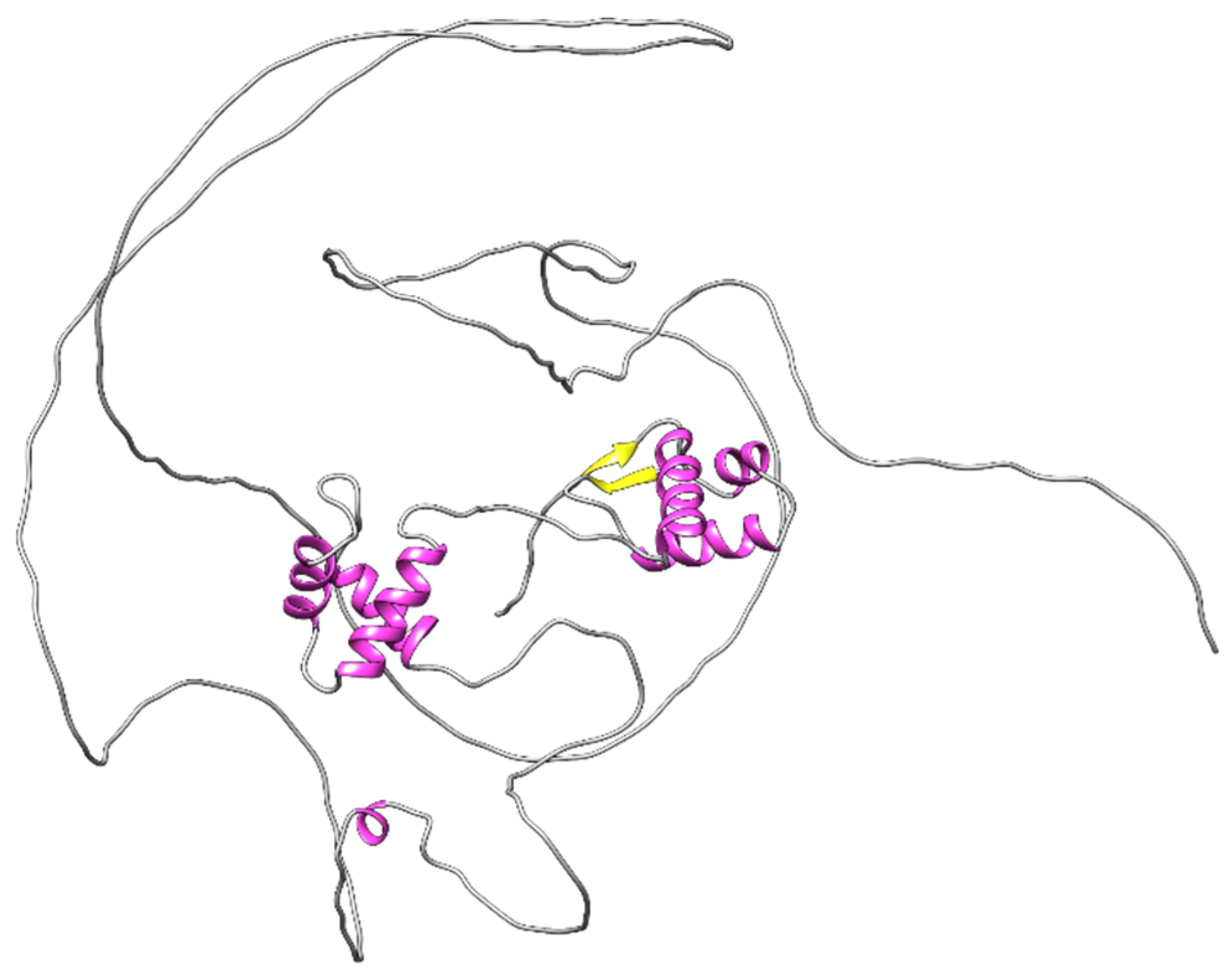
Figure 1. Alpha fold prediction of the structural organization of the PAX8 protein. The PAI and RED subdomains that are composed of helix-turn-helix motifs are seen in pink. A β hairpin is seen in yellow at the N-terminus of the PAI subdomain. Both PAI and RED subdomains are predicted to bind DNA. The octapeptide, homeodomain, and transactivation domains are predicted to be disorganized structures [40,41].
Ijms 23 07410 g001
Figure 3. Illustration of the development of the male and female genital tract. Degenerating ducts are shaded.
Ijms 23 07410 g003
Publisher’s Note: MDPI stays neutral with regard to jurisdictional claims in published maps and institutional affiliations.