Article

N-Methyltransferase CaASHH3 Acts as a Positive Regulator of Immunity against Bacterial Pathogens in Pepper

1
Key Laboratory of Applied Genetics of Universities in Fujian Province, Fujian Agriculture and Forestry University, Fuzhou 350002, China
2
Department of Plant Breeding and Genetics, Ghazi University, Dera Ghazi Khan 32200, Pakistan
3
Department of Botany, Government College University, Faisalabad 38000, Pakistan
4
State Key Laboratory of Subtropical Silviculture, Zhejiang A&F University, 666 Wusu Street, Lin’An, Hangzhou 311300, China
5
Department of Biology, Faculty of Applied Science, Umm Al-Qura University, Makkah 21955, Saudi Arabia
*
Authors to whom correspondence should be addressed.
These authors contributed equally to this work.
Int. J. Mol. Sci. 2022, 23(12), 6492; https://doi.org/10.3390/ijms23126492
Submission received: 30 April 2022 / Revised: 24 May 2022 / Accepted: 27 May 2022 / Published: 10 June 2022
(This article belongs to the Special Issue Plant Disease Resistance 2.0)

Abstract

:
Proteins with conserved SET domain play a critical role in plant immunity. However, the means of organization and functions of these proteins are unclear, particularly in non-model plants such as pepper (Capsicum annum L.). Herein, we functionally characterized CaASHH3, a member of class II (the ASH1 homologs H3K36) proteins in pepper immunity against Ralstonia solanacearum and Pseudomonas syringae pv tomato DC3000 (Pst DC3000). The CaASHH3 was localized in the nucleus, and its transcript levels were significantly enhanced by R. solanacearum inoculation (RSI) and exogenous application of salicylic acid (SA), methyl jasmonate (MeJA), ethephon (ETH), and abscisic acid (ABA). Knockdown of CaASHH3 by virus-induced gene silencing (VIGS) compromised peppers’ resistance to RSI. Furthermore, silencing of CaASHH3 impaired hypersensitive-response (HR)-like cell death response due to RSI and downregulated defense-associated marker genes, including CaPR1, CaNPR1, and CaABR1. The CaASHH3 protein was revealed to affect the promoters of CaNPR1, CaPR1, and CaHSP24. Transiently over-expression of CaASHH3 in pepper leaves elicited HR-like cell death and upregulated immunity-related marker genes. To further study the role of CaASHH3 in plant defense in vivo, CaASHH3 transgenic plants were generated in Arabidopsis. Overexpression of CaASHH3 in transgenic Arabidopsis thaliana enhanced innate immunity against Pst DC3000. Furthermore, CaASHH3 over-expressing transgenic A. thaliana plants exhibited upregulated transcriptional levels of immunity-associated marker genes, such as AtNPR1, AtPR1, and AtPR2. These results collectively confirm the role of CaASHH3 as a positive regulator of plant cell death and pepper immunity against bacterial pathogens, which is regulated by signaling synergistically mediated by SA, JA, ET, and ABA.

1. Introduction

Plants have evolved various distinctive mechanisms to cope with rapidly varying environmental conditions [1,2]. It has been observed that tolerance is enhanced after repeated exposure to similar environmental stress. This phenomenon of augmented tolerance against repeated exposure to same environmental stress is known as acclimation [3,4]. For instance, Arabidopsis plants after exposure to high temperature (38 °C) showed higher tolerance to subsequent high temperature stress (45 °C) as compared untreated plants with high temperature treatment [5,6].
Moreover, plants are exposed to biotic stresses, e.g., microbial pathogen attack in addition to abiotic stresses. Defense responses are induced locally by recognition of microbe-associated molecular patterns (MAMPs), via membrane proteins of plants named as pattern recognition receptors [7,8,9]. The epitope peptide flg22 is a model bacterial MAMP that is derivative of flagellin [10]. This basic defense system is known as pattern-triggered immunity (PTI) [11]. The PTI is induced by the expression of defense-associated genes and deposition of callose [12]. Additionally, systemic resistance is generally associated with a sensitization of stimulus reaction called priming [13,14]. Priming allows cells to counter very low levels of stress in more speedy and vigorous manner as compared to non-primed cells [15]. Usually, primed plants show quicker and stronger activation of several defense responses that are induced after microbial pathogen infection [16].
Histones are assembled with DNA to build nucleosomes that are a fundamental component of chromatin. The histone tails can be methylated post-transnationally at manifold lysine sites and diverse levels of methylation (mono, di, and tri) [17,18]. Histone H3 lysine 36 (H3K36) methylation is an evolutionarily conserved epigenetic mark normally supposed to be involved with active chromatin [19]. Thus far, enzymes that consign methyl groups on Lys36 residues have been expressed in various yeasts and animals, with different preferences for various states of methylation. The H3K36 methylation has also been involved in different processes such as transcription commencement, suppression of transcription, dose compensation, alternative splicing, DNA replication, and DNA repair [19]. At least six lysine residues on histone H3 (K4, K9, K27, K36, and K79) and on histone H4 (K20) are targeted by histone lysine methyltransferases (HKMTs) [20,21]. Except for H3K79, SET-domain-possessing HKMTs have the capability to transmit one or several methyl groups to the ε-nitrogen of specific lysine residues on histones [22,23].
Distinct biological processes are involved in plant development that are differentially affected by environmental conditions [24]. According to annotations in the Pfam and ChromeDB databases (http://www.chromdb.org/, accessed on 11 February 2022) and prior phylogenetic analyses, plants may have developed diverse epigenetic mechanisms against various environmental stresses during growth and development [25,26]. Epigenetic mechanisms such as covalent histone modifications play a critical role in activation or repression of transcription by producing more “open” or “close” chromatin configurations, respectively [27]. Generally, openness of chromatin enhances the genome accessibility to transcription machinery such as RNA polymerase II (RNAPII), thus triggering the transcription process. Therefore, primed genes maybe in an open chromatin configuration. Innate immunity priming is currently associated with the modifications of chromatin in promoter regions of WRKY TFs [28] and also SA- and PTI-associated genes [29,30].
Proteins with a conserved SET domain are found in different organisms including viruses, bacteria, archaea, and eukayota. On the basis of the annotation from Pfam and Chrom, DB 47, 37, and 35 SET domain proteins are found in Arabidopsis, rice, and maize, respectively [31]. Plant SET proteins are classified into seven classes on the basis of their phylogentic relationship, domain organization, and sequence homology [25]. The role of SET domain proteins is still needed to elucidate in non-model plants including pepper.
Pepper (Capsicum annuum L.) is an important agricultural crop worldwide. Pepper is an important member of the Solanaceae family. It is generally sown on uplands in temperate weather conditions and mostly challenged by various soil-borne pathogens and diseases during its life cycle. Diseases, i.e., phytophthora blight and bacterial wilts [32,33,34] are major reasons for poor production of pepper and great threat to economic losses, especially under high temperature and high humidity (HTHH) [35,36]. Ralstonia solanacearum is a destructive soil-borne bacterium and causes wilt disease, which threatens the production of >200 economically vital plants species, including pepper and tobacco [37]. The research on genetics of plants’ disease resistance against R. solanacearum will be extremely beneficial for producing new superior crop varieties.
In this study, we reported a member of class II (the ASH1 homologs H3K36) plant SET domain protein family in pepper, named CaASHH3. We characterized expression and functions of CaASHH3 and found that it was induced by infection of R. solanacearum and application of various exogenous phytohormones, including SA, MeJA, ETH, and ABA. Transient over-expression of CaASHH3 in pepper plants caused HR-like cell death and silencing of CaASHH3 in pepper plants, which compromised the resistance against R. solanacearum infection. We also reported that CaASHH3 over-expression in transgenic Arabidopsis thaliana confers higher innate immunity against the bacterial pathogen Pseudomonas syringae pv tomato DC3000 (Pst DC3000). These results infer that CaASHH3 is a positive regulator of plant cell death and pepper immunity against bacterial pathogen R. solanacearum and Pst DC3000.

2. Results

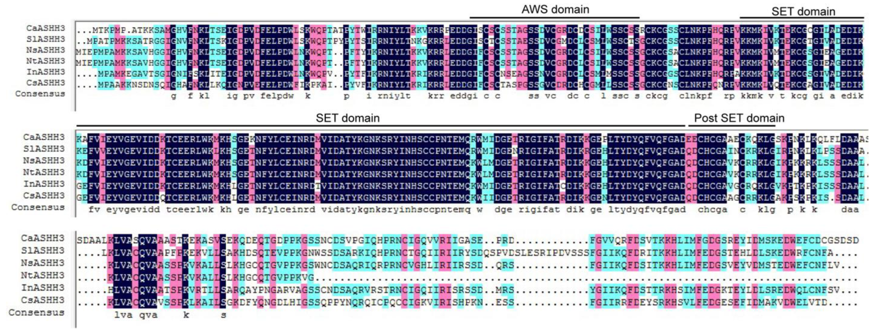
2.1. Cloning and Sequence Analysis of the Full-Length cDNA of CaASHH3

The presence of SET-domain in CaASHH3 protein implies its potential role in pepper immunity. The cDNA fragment of CaASHH3 (Ca methyl transferase) that contained 1146 bp open-reading frame (ORF) was cloned (LOC107875209) by using gene-specific primers (Table S1). Its deduced amino acid sequence containing AWS-domain, SET-domain, and post-SET-domain was 381 amino acid residues in length and was classified into class II (the ASH1 homologs H3K36) [25] (Figure 1). The size of predicted protein was 42.64 kDa. The CaASHH3 shares 83% of amino acid identities with SlASHH3, NsASHH3, and NtASHH3; 82% of amino acid identities with InASHH3; and 79% with CsASHH3 (Figure S1).

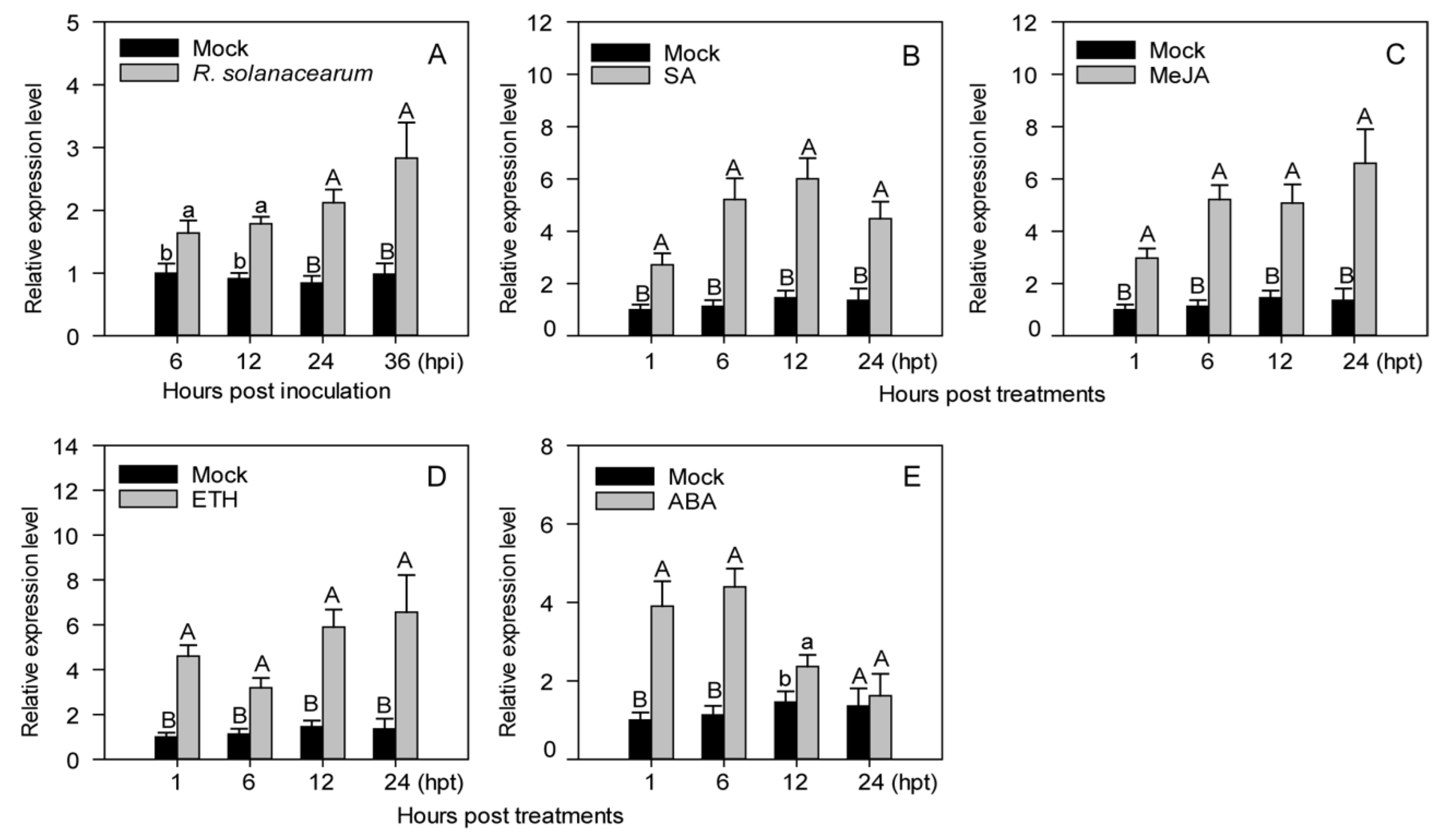
2.2. Transcriptional Expression Levels of CaASHH3 Were Upregulated by R. solanacearum Inoculation and Exogenously Supplied Phytohormones including SA, MeJA, ETH, and ABA

The transcript levels of CaASHH3 were significantly enhanced in leaves inoculated with R. solanacearum but not in the leaves without treatment (MgCl2) (Figure 2A). The increased CaASHH3 transcript levels were maintained between 6 and 36 h post-inoculation (hpi) with the highest levels noticed at 36 hpi (Figure 2A). The results of transcriptional upregulation suggested that CaASHH3 might play a critical role in pepper plants’ immunity against R. solanacearum infection.
We determined the role of these phytohormones in regulation of CaASHH3 expression in pepper plants. Real-time qRT-PCR was used to assess the relative abundance of CaASHH3 transcriptional expression of pepper plants supplied with exogenous SA, MeJA, ABA, and ETH. After application of 1 mM SA, CaASHH3 transcript levels were significantly enhanced from 1 to 24 hpt (hours post treatment) as compared with the control (mock) plants, and the highest transcript level was recorded at 12 hpt (Figure 2B). The exogenous application of 100 µM MeJA significantly increased the transcriptional expression levels of CaASHH3 from 1 to 24 hpt as compared to the mock plants and topped at 24 hpt (Figure 2C). The CaASHH3 transcript levels were significantly increased after treatment with 100 µM ETH and maintained 1 to 24 hpt as compared to mock. The highest transcript level was observed at 24 hpt (Figure 2D). After application of 100 µM ABA, CaASHH3 transcript levels were increased from 1to 24 hpt as compared to mock plants, and it was highest at 6 hpt (Figure 2E).

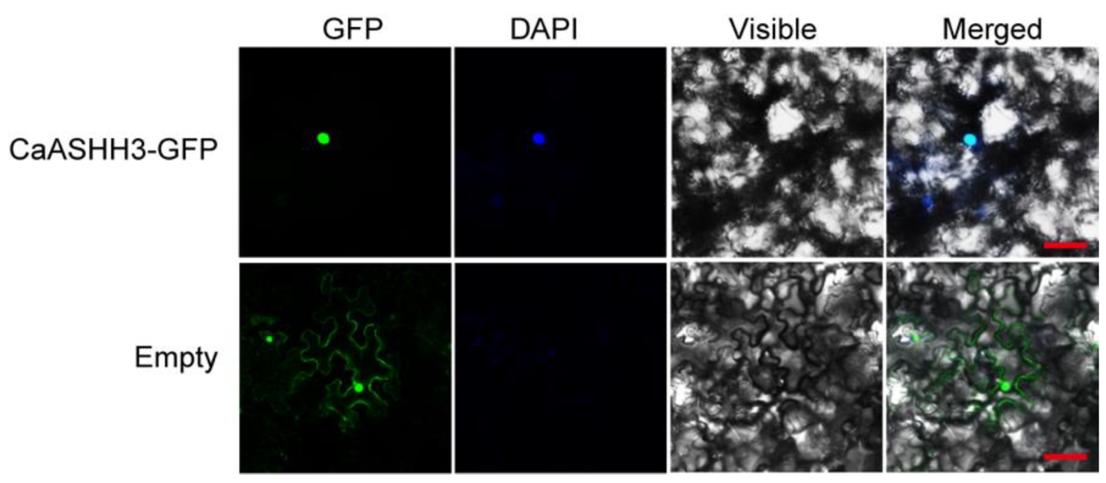
2.3. CaASHH3 Was Localized to the Nucleus

Sequence analysis using WoLFPSORT (http://www.genscript.com/psort/wolf_psort.html accessed on 17 February 2022) showed that the predicted CaASHH3 protein possesses a putative nuclear localization signal. A CaASHH3–GFP fusion construct driven by the constitutive CMV 35S promoter was generated. This construct was transiently expressed in N. benthamiana leaves to confirm its localization. The construct p35S:GFP was used as a control. The results displayed the exclusive localization of CaASHH3-GFP to the nucleus. On the other hand, the p35S:GFP control was located in the multiple sites in subcellular portions, including the cytoplasm and nucleus (Figure 3). These results inferred that CaASHH3 is localized to the nucleus.
The CaASHH3 was exclusively found to be localized in nucleus of N. benthamiana leaves, transiently over-expressed in 35S:CaASHH3-GFP. The GFP signal for the control N. benthamiana leaves was found throughout the cell, including the nucleus, cytoplasm, and membrane. Green color indicates GFP, and blue color indicates DAPI staining of the nucleus. Cyan color indicates merger of GFP and the DAPI-stained nucleus. Images were taken by confocal microscopy at 48 hpi of Agrobacteria with 35S:CaASHH3-GFP construct and 35S-GFP construct (control).

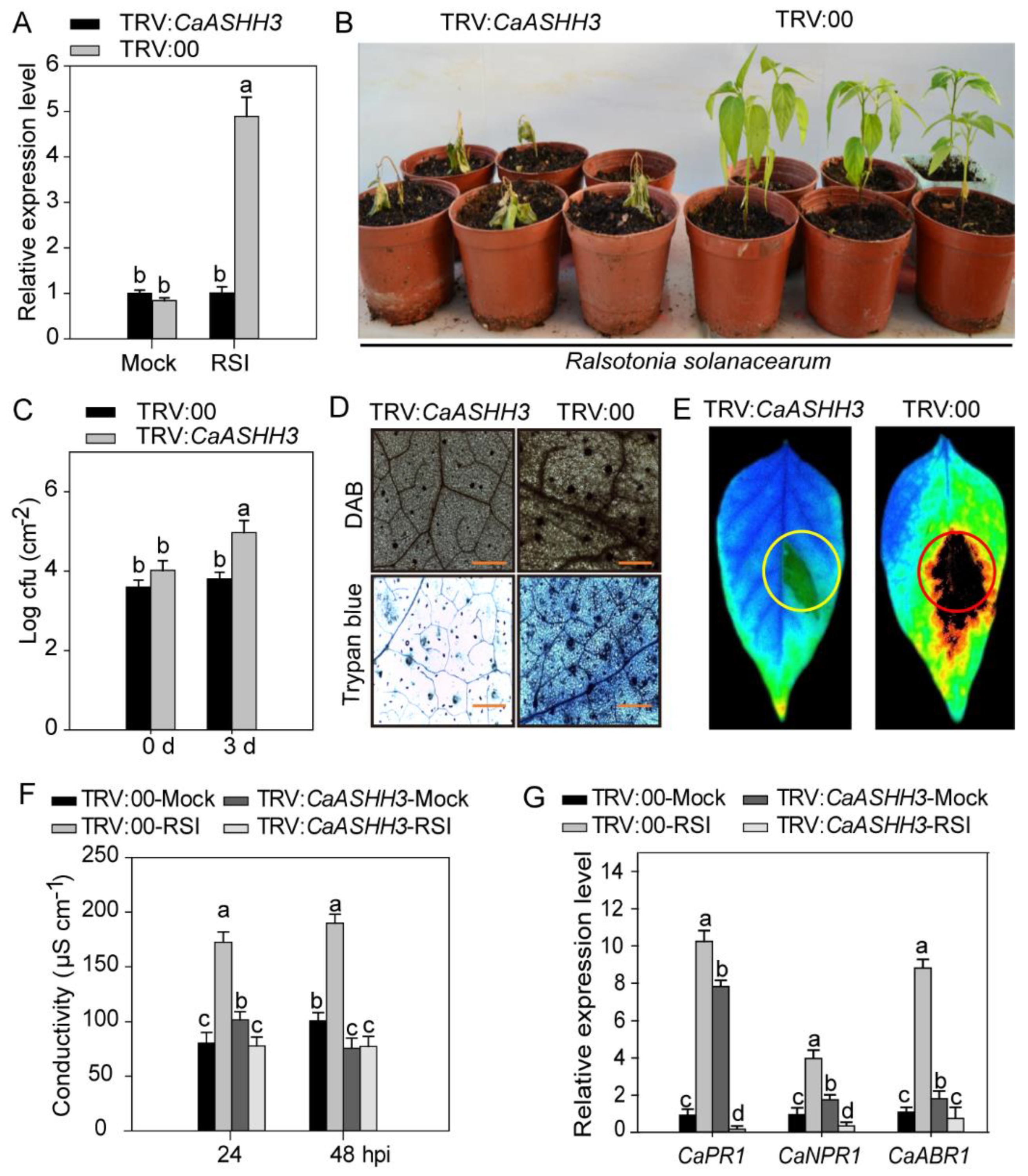
2.4. CaASHH3-Silencing Compromised the Resistance of Pepper against R. solanacearum Infection

The loss-of-function experiments in pepper seedlings were performed to investigate the role of CaASHH3 in immunity against pathogens by VIGS assay. We constructed TRV:CaPDS, which knocked down the phytoene desaturase gene (PDS), which induced a photo bleaching phenotype as an additional control to check the successful silencing of the desired gene. A total of 50 plants of TRV:00 and 50 plants of TRV:CaASHH3 were acquired. Six plants were selected randomly for verifying the gene silencing efficiency by treating R. solanacearum strain ‘FJC100301’ by root inoculation. The results showed that transcript levels of CaASHH3 were reduced to ≈30% in FJC100301-inoculated TRV:CaASHH3 pepper plants as compared to TRV:00 plants, showing the successful silencing of CaASHH3 (Figure 4A).
Ten plants of TRV:CaASHH3 and TRV:00 were randomly selected and inoculated with R. solanacearum ‘FJC100301’ in the roots. At 7 dpi, strong wilting symptoms appeared on TRV:CaASHH3-silenced plants, but TRV:00-unsilenced plants exhibited only weak disease symptoms (Figure 4B). In the meanwhile, we observed significant magnification in the growth of R. solanacearum in CaASHH3-silenced plants, manifested by higher values of cfu at 3 dpi as compared to unsilenced plants (Figure 4C). Histochemical staining, i.e., trypan blue and DAB staining, were performed to evaluate the cell death and H2O2 accumulation of these R. solanacearum-infected, CaASHH3-silenced, and unsilenced plants. At 48 hpi, a clear deeper DAB staining (dark brown) and trypan blue color showing HR response and cell death in unsilenced plants leaves were detected. Nevertheless, DAB and trypan blue staining were distinctly reduced in CaASHH3-silenced leaves (Figure 4D).
The fluorescent modulation meter was used to take the photos of cell death in TRV:CaASHH3 and TRV:00-unsilenced plants after infection with R. solanacearum strain ‘FJC100301’. We recorded obvious and strong cell death in the leaves of unsilenced plants and very weak or slight cell death in CaASHH3-silenced plants’ leaves (Figure 4E). We estimated electrical conductivity to analyze the cell necrosis severity caused by plasma membrane damage after RSI. The leaves of unsilenced plant exhibited stronger ion leakage as compared to CaASHH3-silenced plants at 24 and 48 hpi (Figure 4F).
The real-time qRT-PCR was used to detect the transcript abundance levels of defense-related genes involved in the response against pathogen inoculation. The transcriptional abundance of the defense-related pepper genes, i.e., CaPR1, CaNPR1, and CaABR1 were reduced in CaASHH3-silenced plants’ leaves as compared to unsilenced plants at 24 hpi (Figure 4G).

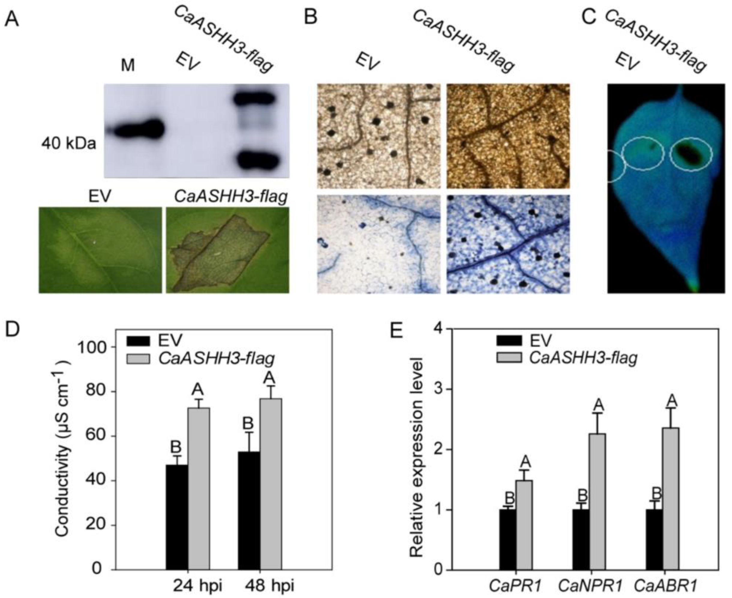
2.5. Transient Over-Expression of CaASHH3 Caused HR-like Cell Death and Accumulation of H2O2 in the Leaves of Pepper Plants

The successful expression of CaASHH3 was confirmed by Western blotting experiment (Figure 5A). The DAB staining was performed to assess H2O2 production in pepper leaves transiently over-expressing CaASHH3. The DAB staining color was very weak or absent in the leaves transiently over-expressing the 35S:00-empty vector as compared to transiently over-expressing 35S:CaASHH3 (Figure 5B). Trypan blue staining was used to identify the cell death. We observed no HR-mediated cell death in the leaves transiently expressing empty vector, while the leaves expressing 35S:CaASHH3 construct induced a very strong and clear necrotic response (Figure 5B). The HR mechanism is used by plants for preventing the disease spread from infected to the healthy cells. This defense response includes the ROS production and cell death in infected tissues. We assessed involvement of CaASHH3 in the regulation of HR mechanism. The CaASHH3 was transiently over-expressed in leaves by infiltrating A. tumefaciens GV3101 harboring 35S:00 and 35S:CaASHH3. There was very strong HR-like cell death caused by over-expression of 35S:CaASHH3, while no or feeble HR-like cell death was caused by 35S:00-empty vector detected under modulation meter (Figure 5C).
Changes in electrical conductivity were recorded to assess the cell necrosis caused by plasma membrane damage in cells expressing 35S:CaASHH3. The pepper leaves transiently over-expressing 35S:CaASHH3 displayed significantly high ion leakage at 24 and 48 hpi as compared to leaves expressing the 35S:00-empty vector (Figure 5D). We used the real-time qRT-PCR analysis to determine the transcript levels of known defense-related genes inclusive of ET biosynthesis-associated CaPR1, CaNPR1, and CaABR1. These results indicated steady increase in relative transcript levels of CaPR1, CaNPR1, and CaABR1 during transient over-expression of CaASHH3 as compared to 35S:00-empty vector at 24 hpi (Figure 5E). Keeping these results in view, it is confirmed that transient over-expression of CaASHH3 can induce HR-like cell death and a play critical role in pepper plant immunity to pathogens.

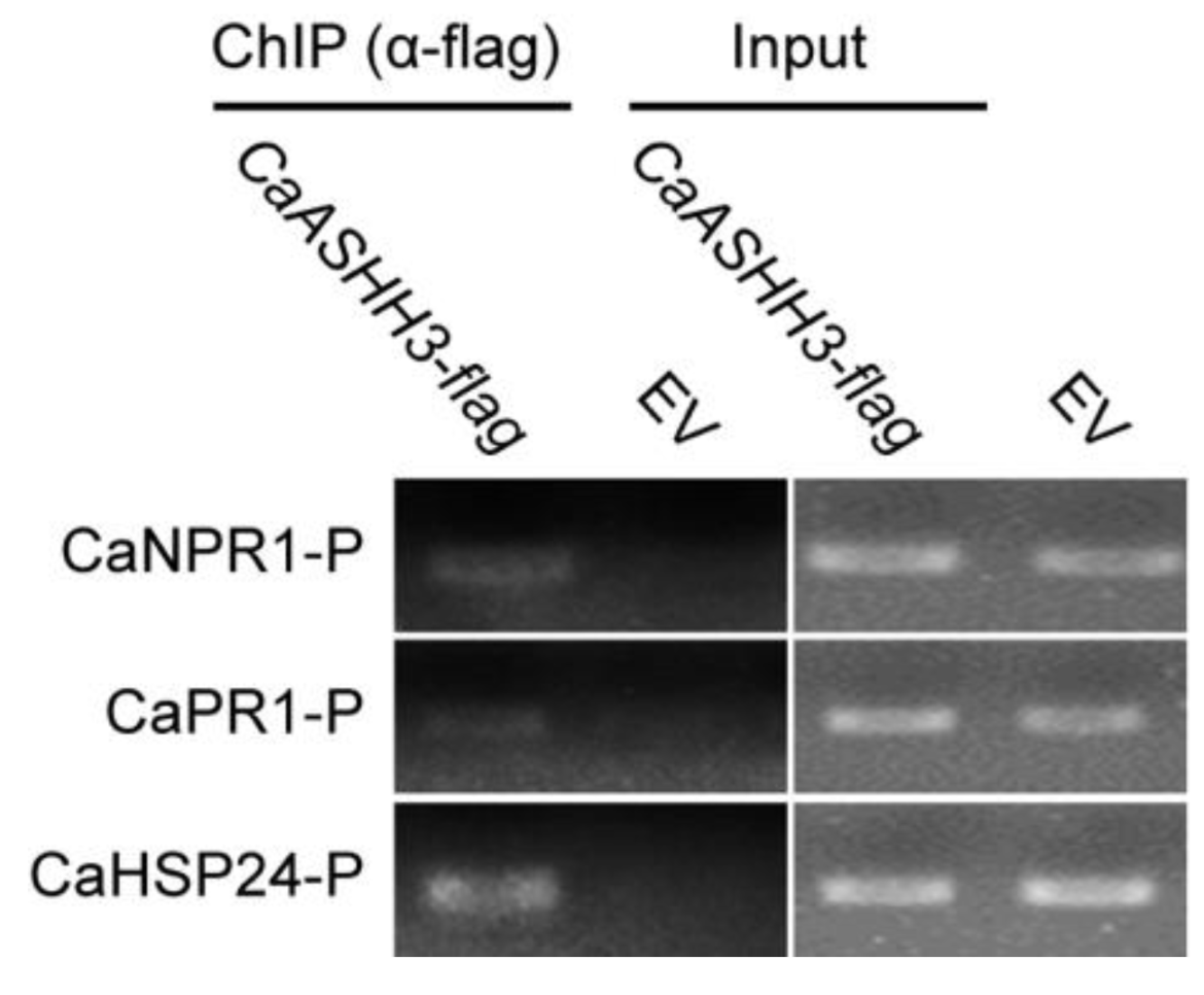
2.6. ChIP-PCR Analysis to Test Chromatin Modification in Pepper

To evaluate possible histone modification in pepper plants after exposure to recurring stresses, chromatin immune precipitation (ChIP) was carried out by using histone-modification-specific antibodies. The chromatin was sheared into fragments of 300 to 500 bps in length, and then the protein–DNA complex was immunoprecipitated with HA antibody. The DNA was released and immunoprecipitated for further ChIP-PCR. The specific primers pair of known defense-associated genes including CaNPR1, CaPR1, and CaHSP24 was used for ChIP-PCR, and immunoprecipitated fragments of DNA were used as PCR template. The results indicated that CaASHH3 directly bind to histones (Figure 6)
The CaASHH3 binds to histones of different defense-associated marker genes detected by ChIP-PCR. The CaASHH3-Flag was transiently over-expressed in pepper leaves and the chromatin was isolated; moreover, the DNA–protein complex was immuno-precipitated using anti-HA antibodies and adjusted to the same concentration, and PCR was performed using primer pairs based on CaNPR1, CaNPR1, and CaHSP24.

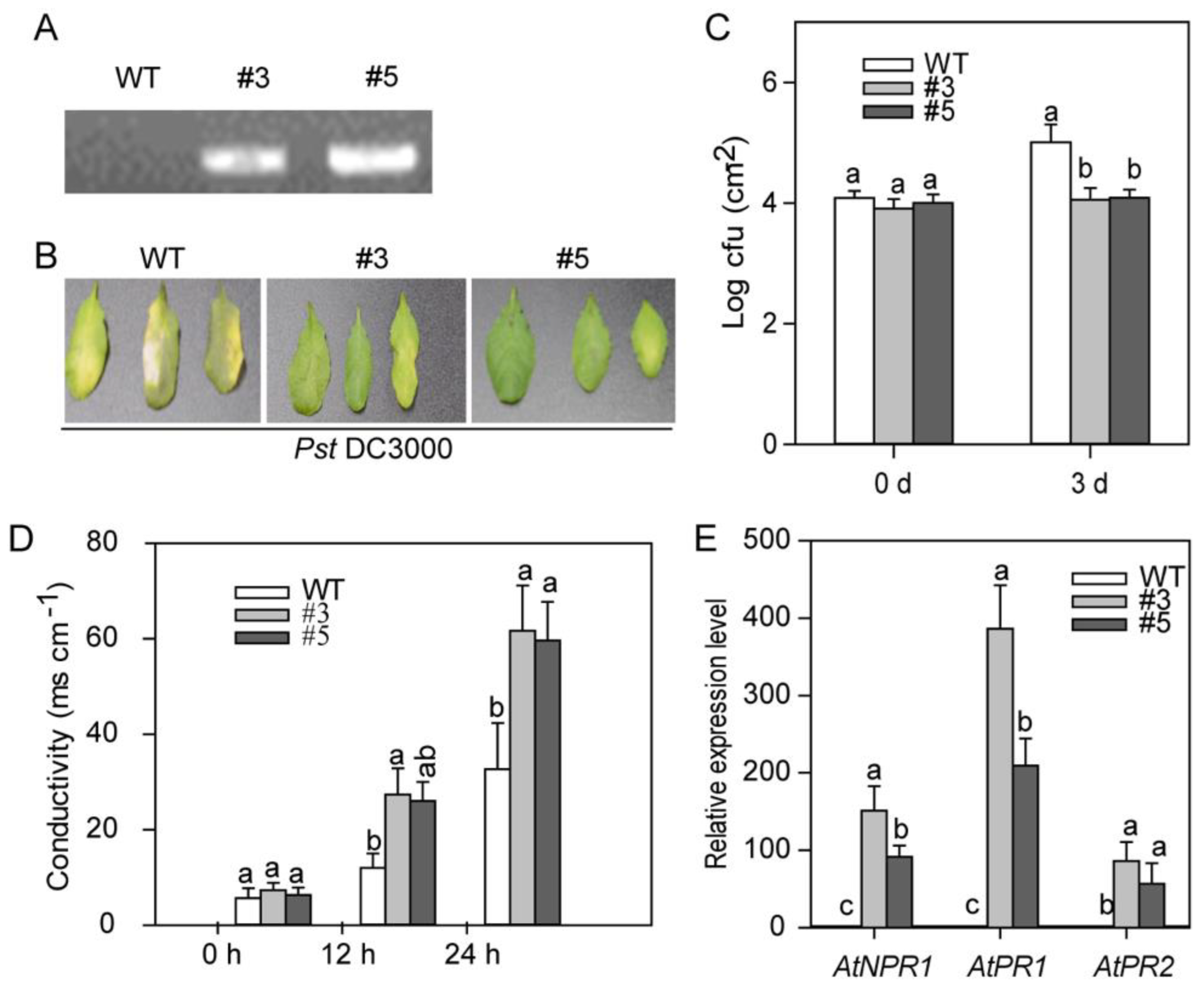
2.7. CaASHH3-OX (Over-Expression) in Transgenic Arabidopsis Plants Enhanced the Resistance to Bacterial Pathogen

As the stable transformation is very hard to attain in pepper plants to further study the role of CaASHH3 in plant defense in vivo. CaASHH3 transgenic plants were generated in Arabidopsis that over-expressed the complete precursor under the control of constitutive 35S promoter (Figure 7). To confirm the CaASHH3-OX transgenic expression in independent homozygous T3 Arabidopsis lines, PCR analysis was performed by using genomic DNA extracted from CaASHH3-OX transgenic Arabidopsis plants and from wild-type plants (Col-0). The CaASHH3 gene was amplified from two CaASHH3-OX plants, while PCR product was not detected for wild-type Arabidopsis plants (Figure 7A). The phenotype of CaASHH3-OX Arabidopsis plants was indistinguishable from wild-type plants under normal growth conditions. To check the disease resistance against bacterial pathogen, both wild-type and CaASHH3-OX Arabidopsis plants were inoculated with P. syringe pv tomato DC3000 by using needless syringe. Wild-type plants displayed very clear typical chlorotic symptoms on treated leaves at 3 dpi, whereas there were no obvious symptoms on CaASHH3-OX lines (Figure 7B). To verify that these disease symptoms reveal bacterial pathogen propagation in plants, the growth of bacterial pathogen was observed in plant leaves at 0 and 3 dpi. The results showed that there was increase in growth of pathogen in wild-type plants as compared with CaASHH3-OX plants, manifested by higher cfu values at 0- and 3-day intervals (Figure 7C). These results showed that CaASHH3 over-expression confers enhanced resistance to plants against pathogen. Cell death related to HR response was measured by using electrolyte leakage assay. The CaASHH3-OX leaves showed high levels of electrolyte leakage as compared to wild-type plants at 0, 12, and 24 hpi with P. syringe pv tomato Pst DC3000 (Figure 7D).
To examine whether this enhanced immunity against the pathogen in CaASHH3-OX transgenic Arabidopsis plants is linked to the expression of SA-dependent PR genes, we evaluated the transcriptional expression of AtNPR1, AtPR1, and AtPR2 in wild-type and CaASHH3-OX plants. The results indicated that the transcript levels of these defense-related marker genes were more abundant in CaASHH3-OX plants as compared to wild-type plants at 24 hpi with P. syringe pv tomato Pst DC3000 (Figure 7E). These results infer that SA-dependent immunity mechanism may play a critical role in CaASHH3-OX-enhanced disease resistance against bacterial pathogen attack.

3. Discussion

It is well known that plants are regularly exposed to pathogen attack throughout their entire life cycle. A great extent of the knowledge of how plants cope with pathogen attack has been yielded by the previous studies on model plants such as Arabidopsis, rice, or tobacco [2,24,38]. The SA or JA/ET signaling pathways are first to be turned on after plants encounter microorganisms [33,39,40]. SA-related signaling pathways are involved in resistance to bio-trophic pathogens, while JA/ET-dependent signaling pathways are involved in resistance against necro-trophic pathogens [41,42,43].
The DNA methylation and histone modifications play a crucial role in transcriptional regulations in plants [44,45,46]. Previous studies revealed that many chromatin and histone modifiers, as well as remodelers, have been involved in plant defense against biotic and abiotic stresses [46,47]. Immune responses are controlled by several epigenetic phenomena and histone modification post-transcriptionally, both synergistically and antagonistically over plant defense and transcription [48,49,50]. This is how different methylases, demethylases, and chromatin re-modelers play their role in various aspects of immunity. Some of them are positive and others are negative regulators of either particular aspects or of total immunity [49]. Many histone methyltransferases and de-methylases act as immunity regulators. Both types of enzymes have crucial role of regulation of histone methylation in plant defense. Some reported methyltransferases that are involved in plant immunity are ATX1, SDG8, and SDG25. Mutants of these methyltransferases are defective in basal defense, necro-trophic resistance, and SAR, respectively [26,51,52,53,54].
We cloned a Capsicum annum gene member of class II (ASH1 homologs H3K36) plant set domain protein family, named CaASHH3, that possesses methyl-transferase activity. The over-expression of CaASHH3 induced the HR-like cell death response in pepper leaves, but loss of function experiments of CaASHH3 compromised the plant resistance against pathogen and enhanced the cfu value in CaASHH3-silenced plants. Moreover, transgenic Arabidopsis ASHH3-OX showed more resistance as compared to wild-type plants. These data back the notion that CaASHH3 acts as a positive regulator and plays a critical role in plant defense against pathogen. The involvement of CaASSH3 in regulation of pepper immunity was further supported by the data that CaASHH3 transcriptional expression levels were significantly upregulated by RSI.
Since genes upregulated upon exposure to certain stress are repeatedly found to have a role in response to that stress [55], we inferred that CaASSH3 might act as a positive regulator in pepper’s resistance to RSI. The declined immunity of pepper plants was accompanied with amplified growth of inoculated Ralstonia solanacearum and downregulated [56,57], SA-associated CaPR1 [57,58,59]; immunity-associated CaNPR1 [60]; and CaABR1 [61] under treatment to RSI. These studies support our hypothesis and are further supported by the results from loss-of-function experiments of CaASHH3 by VIGS, gain-of-function experiments by transient over-expression, and CaASHH3-OX over-expression in transgenic Arabidopsis, respectively. The silencing of CaASHH3 by VIGS significantly enhanced the vulnerability of pepper plants to RSI.
In comparison, transient over-expression of CaASHH3 distinctively triggered HR-like cell death and accumulation of H2O2, consistent with upregulation of immunity-associated CaPR1, CaNPR1, and CaABR1. These findings of loss-of-function and gain-of-function experiments strongly recommend the role of CaASHH3 as a positive regulator of cell death and immunity in pepper against bacterial pathogen. The similar plant HR has been observed in the case of CaZNF830 and CabZIP53 [32,39]. The distinguished plant responses in pepper and Arabidopsis confirmed the contribution of set of genes as well as transcription factors in plant innate immunity. The upregulation of defense marker genes is directly coupled with CaASHH3 and other mentioned genes that ultimately support plant survival against Ralstonia and P. syringae pv DC 3000.
To further examine whether over-expression of CaASHH3 bestows defense responses to heterologous plants against pathogen attack, we produced CaASHH3-OX transgenic lines in A. thaliana that were resistant to virulent P. syringae pv DC 3000 attack. Resistance responses in transgenic Arabidopsis plants included accumulation of ROS; damage of plasma membrane; necrotic disease symptoms; and upregulation of immunity-associated marker genes, including AtNPR1, AtPR1, and AtPR2. These results recommend that over-expression of CaASHH3 gene in Arabidopsis increases basal defense or R-gene-mediated resistance against bacterial pathogen.
Phyto-hormones including SA, JA, and ET are involved in plant immunity signaling pathways. The SA triggers resistance against biotrophic pathogens, whereas JA and ET have a role in plant immunity against necrotrophic pathogens [2,62,63]. Production of hormones, i.e., SA, JA, and ET, is often coupled with ETI or PTI. These phytohormones can work synergistically or antagonistically depending upon their concentrations during defense singling [39,64]. These signaling components have a synergistic relationship during PTI. On the other hand, these signaling components have a compensatory relationship during ETI [40,65,66]. In our study, CaASHH3 was constantly noticed as being induced by exogenous foliar application of phytohormones, including SA, MEJA, ETH, and ABA. The checked SA-, JA-, ET-, and ABA-based immunity-associated marker genes; SA-associated CaPR1 and CaNPR1 [67,68,69,70,71,72]; and ABA-associated CaABR1 [73] were downregulated by silencing of CaASHH3 by VIGS under RSI, whereas these HR-associated, SA-dependent, and ABA-dependent immunity-associated PR genes (CaPR1, CaNPR1, and CaABR1) were upregulated by transient over-expression of CaASHH3 in pepper. Transcriptional expression levels of immunity-associated marker genes AtNPR1, AtPR1, and AtPR2 were also significantly upregulated in ASHH3-OX-over-expressing transgenic Arabidopsis plants. These data indicate that CaASHH3 takes part in defense signaling, which is mediated synergistically by SA, JA, and ABA, hence leading to PTI.
In chromatin immuno-precipitation assay, CaASHH3 bound to promoter of immunity-associated marker genes CaNPR1, CaPR1, and CaHSP24, with these results suggesting that these immunity-associated marker genes are the target of CaASHH3 and are directly regulated by CaASHH3.

4. Materials and Methods

4.1. Plant Materials and Growth Conditions

The seeds of pepper (Capsicum annuum L.) cultivar ‘GZ03’, Arabidopsis thaliana (ecotype ‘Col-0’), and Nicotiana benthamiana were obtained from pepper breeding group at Fujian Agriculture and Forestry University (www.fafu.edu.cn, accessed on 10 February 2022), Fuzhou, China. The seeds of pepper cv. ‘GZ03’, A. thaliana, and N. benthamiana were sown in a soil mixture comprising peat, moss, and perlite, [2/1(v/v)] in plastic pots (2.2 L), and placed in the growth chamber under controlled conditions of 25 °C, 60–70 µmol photons m−2 s−1, a relative humidity of 70%, and 16 h light/8 h dark photoperiod. The seeds of Arabidopsis were sterilized with 70% ethanol for 1 min prior to in vitro culture followed by treatment with 2% sodium hydroxide for 10 min. Ultimately, the seeds were washed with sterilized distilled water for 5 times and were sown in Petri dishes on solid Murashige and Skoog (MS) media for two weeks in growth chamber at a 16 h light and 8 h dark photoperiod.

4.2. Vectors Construction

The gateway technology was employed to construct the vectors. The full length ORF of CaASHH3 (with or without terminal codon) was cloned into the entry vector pDONR207 by using BP reaction to construct satellite vectors. Then, LR reaction was employed to transfer the full length ORF of CaASHH3 into destination vectors, including pMDC83, CD3688 (Flag-tag), pK7WG2, and CD3687 (HA-tag) to generate vectors for subcellular localization, over-expression, and ChIP assay, respectively. To construct vectors for virus-induced gene silencing (VIGS), a specific 310 bps fragment in 3′-untranslated region (UTR) of CaASHH3 was selected, and the specificity was confirmed by BLAST against genome sequence in the database of CM334 (http://peppergenome.snu.ac.kr/) and Zunla-1 (http://peppersequence.genomics.cn/page/species/blast.jsp, accessed on 13 February 2022). The specific fragment was cloned into the entry vector pDONR207 by BP reaction and then transferred into the destination PYL279 vector by LR reaction. The primers used in vector construction are given in Table S1.

4.3. Pathogens and Inoculation Procedures

A virulent pathogen strain R. solanacearum ‘FJC100301’ was isolated from disease-infected pepper plants from Fujian province (China). Exudates of the stem and the stem vascular tissue portion from these plants were purified by using previously described tetrazolium chloride method [33,74]. Virulent R. solanacearum strain ‘FJC100301’ was cultured overnight in SPA medium (200 g potato, 20 g sucrose, 3 g beef extract, 5 g tryptone, and 1 L of ddH2O) at 200 rpm and 28 °C in thermo-control shaker. The cultivated R. solanacearum was then centrifuged at 6500 rpm and 28 °C for 10 min. The pellet was dissolved in distilled sterilized 10 mM MgCl2. Bacterial cell density was diluted to 108 cfu mL−1 (OD600 = 0.8). To examine the effect of R. solanacearum inoculation (RSI) on transcript levels of CaASHH3 and the resistance of pepper plants to RSI, pepper plants were infected with 10 µL R. solanacearum into the top third leaf with the help of a needleless syringe. The corresponding leaf samples were collected at the specified time intervals for RNA extraction and histochemical staining, including DAB and trypan blue staining. To examine the CaASHH3-silenced phenotype under R. solanacearum infection, pepper roots were wounded by giving minor cuts with a glass rod and then infected with R. solanacearum. After RSI, the plants were grown in a growth chamber under controlled conditions at 28 ± 2 °C, 60–70 µmol photons m−2 s−1, relative humidity of 70% and under a 16 h light/8 h dark photoperiod.
The virulent bacterial pathogen P. syringae pv. Tomato DC3000 (Pst DC3000) was also used in this study. To prepare bacterial pathogen suspension for the infection of Arabidopsis plants, bacterial strain was cultivated overnight at 200 rpm and 28 °C in LB media containing desired antibiotic. Afterwards, this suspension was centrifuged at 6500 rpm and 28 °C for 10 min. The liquid supernatant was discarded, and solid pellet was dissolved in distilled sterilized 10 mM MgCl2 and diluted to a 105 cfu mL−1. Leaves of 5–6-week-old Arabidopsis plants were inoculated with Pst DC3000 pathogen suspension by using a needleless syringe. The infected plants were placed in a moist chamber for 18 h and then shifted to a growth room. The samples were harvested for examining phenotype, measuring electrolyte conductivity, colony forming units (cfu), and RNA extraction at the desired time points.

4.4. Treatment of Pepper Plants with Exogenously Applied Phytohormones

To study the response of phytohormones, healthy pepper plants at four-leaf stage were treated with foliar application of 1 mM SA, 100 µM MeJA, 100 µM ETH, and 100 µM ABA. Mock plants were treated with foliar application of sterilized ddH2O. The phyto-hormones and mock-treated samples were harvested for RNA extraction at specific time intervals.

4.5. Subcellular Localization Experiment of CaASHH3

The Agrobacterium tumefaciens strain GV3101 possessing 35S:CaASHH3-GFP constructs and 35S:GFP (used as control) were cultured overnight in LB media containing recommended antibiotics. The Agrobacterium cells were centrifuged, and the pellet was collected and dissolved in induction medium (10 mM MES, 10 mM MgCl2, 200 µM acetosyringone, (pH 5.6)) and diluted to OD600 = 0.8. Agrobacterium cells possessing the 35S:CaASHH3-GFP and 35S:GFP were injected into N. benthamiana leaves by using a needless syringe. Previously described 4,6-diamidino-2-phenylindole staining (DAPI) was carried out [75] to exhibit the nucleus. The GFP and DAPI fluorescence signals were perceived, and images were captured by utilizing a Leica fluorescence light microscope (Leica, Wetzlar, Germany) with an excitation wavelength of 488 nm, and an excitation wavelength of 405 nm with a 435–480 nm band-pass emission filter.

4.6. Virus-Induced Gene Silencing (VIGS) in Pepper Plants

For CaASHH3 silencing experiments, the Tobacco Rattle Virus (TRV)-based VIGS system was used in pepper plants according to our previous studies [2,42,76]. Agrobacterium tumefaciens strain ‘GV3101’ containing pYL192 and pYL279-CaASHH3 or pYL279 were cultured overnight in the shaker at 200 rpm and 28 °C. This Agrobacterium culture was centrifuged at 6500 rpm and 28 °C for 10 min. The pellet was collected by centrifugation and dissolved in induction medium (10 mM MES, 10 mM MgCl2, 200 µM acetosyringone, (pH 5.6)) and diluted to OD600 = 0.6. Agrobacterium strains with pYL192 vector and pYL279 vector, pYL279-CaASHH3, and pYL192-PDS (OD600 = 0.6) were mixed in a 1:1 ratio. This mixture was infiltrated into cotyledons of 2-week-old pepper plants using a needleless syringe. The Agrobacterium-injected plants were placed in a growth chamber at 16 °C in the dark for 56 h with 45% relative humidity and transferred into a growth room at 25 ± 2 °C, 60–70 µmol photons m−2 s−1 and a relative humidity of 70%, under a 16 h light/8 h dark cycle.

4.7. Agrobacterium-Mediated Transient CaASHH3 Over-Expression Assay

Agrobacterium tumefaciens strain ‘GV3101’ possessing 35S:CaASHH3-flag and 35S:00 vector (empty vector used as control) was cultured overnight to OD600 = 1 in LB media containing the corresponding antibiotics in a shaker at 200 rpm and 28 °C. Then, the cultured Agrobacterium was centrifuged, the supernatant was discarded, and the pellet was dissolved in induction medium (10 mM MES, 10 mM MgCl2, 200 µM acetosyringone, (pH 5.6)) and diluted to OD600 = 0.8. This suspension was injected into pepper leaves with the help of a syringe without a needle. The plants were kept in a growth room, and then samples were collected for further analysis at the desired time point.

4.8. Histochemical Staining

Trypan blue and 3,3′-diaminobenzidine (DAB) staining was performed for CaASHH3 transiently over-expressed pepper leaves and VIGS-treated, CaASHH3-silenced and un-silenced pepper leaves inoculated with R. solanacearum according to a previously published method [8,57]. For trypan blue staining, treated pepper leaves were boiled in trypan blue staining solution (10 mL lactic acid, 10 mL glycerol, 10 mL phenol, 40 mL ethanol, 10 mL ddH2O, and 1 mL trypan blue) for 30 min, stored at room temperature for 8 h, followed by transfer into choral hydrate solution (2.5 g of chloral hydrate dissolved in 1 mL of distilled water), and these leaves were again boiled thrice to destain for 20 min each time. Finally, samples were kept in 70% glycerol. For DAB staining, the treated pepper leaves were dipped in 1 mg/mL of DAB solution and kept at room temperature overnight. Lactic acid/glycerol/absolute ethanol (1:1:3 (v/v/v)) solution was used to destain the DAB-stained pepper leaves and was placed in 95% absolute ethanol [77]. The images of DAB and trypan-blue-stained leaves were captured by camera and a light microscope (Leica, Wetzlar, Germany).

4.9. RNA Extraction and Real-Time qRT-PCR

Total RNA was extracted from pepper leaves by using TRIzol reagent (Invitrogen, Carlsbad, CA, USA) and reverse-transcribed by using a Prime Script RT-PCR kit (TaKaRa, Dalian, China). To check the relative transcript levels of selected marker genes, qRT-PCR was carried out with specific primers (Table S2) according to the manufacturer’s instructions for the Bio-Rad Real-Time PCR system (Bio-Rad, Foster City, CA, USA) and SYBR premix Ex Taq II system (TaKaRa Perfect Real Time). Real-time qRT-PCR and corresponding data processing were performed as described previously [24,78].

4.10. Measurement of Ion Conductivity

Electrolyte ion conductivity (ion leakage) was measured according to a previously described method with slight amendments [9,79]. The six leaf discs (4 mm in diameter) were cut with a hole-puncher, then washed with sterilized ddH2O thrice and directly incubated in 10 mL of double distilled water. These discs were placed in a shaker with gently shaking (60 rpm) for 1 h at room temperature. The ion conductivity was assessed with a conductivity meter (Mettler Toledo 326 Mettler, Zurich, Switzerland).

4.11. Immunoblotting

Total protein from pepper samples was extracted by using protein extraction buffer as described earlier [39]. The extracted protein was incubated overnight with anti-HA agarose at 4 °C (Thermo Fisher Scientific, Waltham, Massachusetts, USA). Magnetic crack was used to collect the beads, and they were washed thrice by tris-buffered saline (TBS) and tween 20 (0.05%). Eluted proteins were observed using immunoblotting and by anti-HA-peroxidase (Abcam, Cambridge, UK).

4.12. ChIP Assays

The ChIP assays were accomplished as described in earlier studies [80,81]. Briefly, Agrobacterium cells possessing the construct 35S:CaASHH3-flag or 35S:00-flag were injected into the leaves at the eight-leaf stage. The treated leaf samples were collected at the required time interval, and extraction of chromatin and precipitation was conducted from these 5- to 6-week-old leaves (1.5 to 2 g of leaf tissue) treated with 1% formaldehyde. The chromatin was sheared to an average length of 300 to 500 bp by sonication and immuno-precipitated with antibodies against hemagglutinin (HA; Santa Cruz Biotechnology). A total of 10 mg of antibodies were used for ChIP analysis, washed with TBST buffer, and reverse cross-linked, and finally the DNA was eluted with sterilized ddH2O. The immuno-precipitated DNA was analyzed for enrichment of CaASHH3 at the promoter region of target genes by using common ChIP-PCR. Every sample was quantified thrice. The primers used for ChIP-PCR analysis in ChIP assays are listed in Table S3.

4.13. Generation of CaASHH3-OX-Overexpressing Transgenic Arabidopsis Plants

The full-length CaASHH3 cDNA sequence was cloned and integrated into the entry vector pDONR207 by BP reaction. Then, LR reaction was carried out to ligate this fragment into destination pK7WG2 vector. The vector pK7WG2 harboring the cauliflower mosaic virus 35S promoter–CaASHH3 construct was transformed into Agrobacterium tumefaciens strain GV3101. Agrobacterium-mediated transformation was conducted to generate CaASHH3-OX-overexpressing transgenic Arabidopsis plants by using the floral dip method [82]. Seeds were harvested from these transgenic Arabidopsis plants and sown in Petri plates on MS agar media containing 50 µg mL−1 kanamycin to obtain independent transgenic lines. The PCR was carried out on confirming the insertion of CaASHH3 cDNA in the genome of transgenic Arabidopsis plants.

Supplementary Materials

The following supporting information can be downloaded at https://www.mdpi.com/article/10.3390/ijms23126492/s1.

Author Contributions

Conceptualization, A.H., L.K., M.A., Z.L., and S.H.; data curation, A.H., L.K., and M.I.K.; formal analysis, M.F.A. and M.I.K.; funding acquisition, M.A., Z.L., and S.H.; investigation, A.N. and Z.L.; methodology, A.N.; project administration, Z.L. and S.H.; software, A.H. and L.K.; supervision, S.H.; visualization, M.F.A. and M.I.K.; writing—original draft, A.H. and L.K.; writing—review and editing, A.N., M.F.A., M.A., M.I.K., Z.L., and S.H. All authors have read and agreed to the published version of the manuscript.

Funding

This research was funded by the National Natural Science Foundation of China (31572136, 32172553, and 31372061), the Scientific Research Foundation of the Graduate School of Fujian Agriculture and Forestry University (324-1122yb045) and the Development Fund Project of Fujian Agriculture and Forestry University (CXZX2016158, CXZX2017548, and CXZX2018114).

Institutional Review Board Statement

Not applicable.

Informed Consent Statement

Not applicable.

Data Availability Statement

All the data are within the manuscript and its supporting information files.

Conflicts of Interest

The authors declare no conflict of interest.

References

  1. Pastori, G.M.; Foyer, C.H. Common components, networks, and pathways of cross-tolerance to stress. The central role of “redox” and abscisic acid-mediated controls. Plant Physiol. 2002, 129, 460–468. [Google Scholar] [CrossRef] [PubMed] [Green Version]
  2. Hussain, A.; Li, X.; Weng, Y.; Liu, Z.; Ashraf, M.F.; Noman, A.; Yang, S.; Ifnan, M.; Qiu, S.; Yang, Y. CaWRKY22 Acts as a Positive Regulator in Pepper Response to Ralstonia solanacearum by Constituting Networks with CaWRKY6, CaWRKY27, CaWRKY40, and CaWRKY58. Int. J. Mol. Sci. 2018, 19, 1426. [Google Scholar] [CrossRef] [PubMed] [Green Version]
  3. Sung, D.-Y.; Kaplan, F.; Lee, K.-J.; Guy, C.L. Acquired tolerance to temperature extremes. Trends Plant Sci. 2003, 8, 179–187. [Google Scholar] [CrossRef]
  4. Zhu, J.; Dong, C.-H.; Zhu, J.-K. Interplay between cold-responsive gene regulation, metabolism and RNA processing during plant cold acclimation. Curr. Opin. Plant Biol. 2007, 10, 290–295. [Google Scholar] [CrossRef]
  5. Schramm, F.; Larkindale, J.; Kiehlmann, E.; Ganguli, A.; Englich, G.; Vierling, E.; Koskull-Döring, V. A cascade of transcription factor DREB2A and heat stress transcription factor HsfA3 regulates the heat stress response of Arabidopsis. Plant J. 2008, 53, 264–274. [Google Scholar] [CrossRef] [Green Version]
  6. Larkindale, J.; Vierling, E. Core genome responses involved in acclimation to high temperature. Plant Physiol. 2008, 146, 748–761. [Google Scholar] [CrossRef] [Green Version]
  7. Boller, T.; Felix, G. A renaissance of elicitors: Perception of microbe-associated molecular patterns and danger signals by pattern-recognition receptors. Annu. Rev. Plant Biol. 2009, 60, 379–406. [Google Scholar] [CrossRef]
  8. Ifnan Khan, M.; Zhang, Y.; Liu, Z.; Hu, J.; Liu, C.; Yang, S.; Hussain, A.; Furqan Ashraf, M.; Noman, A.; Shen, L. CaWRKY40b in Pepper Acts as a Negative Regulator in Response to Ralstonia solanacearum by Directly Modulating Defense Genes Including CaWRKY40. Int. J. Mol. Sci. 2018, 19, 1403. [Google Scholar] [CrossRef] [Green Version]
  9. Cai, H.; Yang, S.; Yan, Y.; Xiao, Z.; Cheng, J.; Wu, J.; Qiu, A.; Lai, Y.; Mou, S.; Guan, D. CaWRKY6 transcriptionally activates CaWRKY40, regulates Ralstonia solanacearum resistance, and confers high-temperature and high-humidity tolerance in pepper. J. Exp. Bot. 2015, 66, 3163–3174. [Google Scholar] [CrossRef] [Green Version]
  10. Gómez-Gómez, L.; Felix, G.; Boller, T. A single locus determines sensitivity to bacterial flagellin in Arabidopsis thaliana. Plant J. 1999, 18, 277–284. [Google Scholar] [CrossRef]
  11. Tsuda, K.; Katagiri, F. Comparing signaling mechanisms engaged in pattern-triggered and effector-triggered immunity. Curr. Opin. Plant Biol. 2010, 13, 459–465. [Google Scholar] [CrossRef] [PubMed]
  12. Boudsocq, M.; Willmann, M.R.; McCormack, M.; Lee, H.; Shan, L.; He, P.; Bush, J.; Cheng, S.-H.; Sheen, J. Differential innate immune signalling via Ga 2+ sensor protein kinases. Nature 2010, 464, 418. [Google Scholar] [CrossRef] [PubMed] [Green Version]
  13. Zimmerli, L.; Métraux, J.-P.; Mauch-Mani, B. β-Aminobutyric acid-induced protection of Arabidopsis against the necrotrophic fungus Botrytis cinerea. Plant Physiol. 2001, 126, 517–523. [Google Scholar] [CrossRef] [PubMed] [Green Version]
  14. Gruner, K.; Griebel, T.; Návarová, H.; Attaran, E.; Zeier, J. Reprogramming of plants during systemic acquired resistance. Front. Plant Sci. 2013, 4, 252. [Google Scholar] [CrossRef] [Green Version]
  15. Návarová, H.; Bernsdorff, F.; Döring, A.-C.; Zeier, J. Pipecolic acid, an endogenous mediator of defense amplification and priming, is a critical regulator of inducible plant immunity. Plant Cell 2012, 24, 5123–5141. [Google Scholar] [CrossRef] [Green Version]
  16. van Hulten, M.; Pelser, M.; Van Loon, L.; Pieterse, C.M.; Ton, J. Costs and benefits of priming for defense in Arabidopsis. Proc. Natl. Acad. Sci. 2006, 103, 5602–5607. [Google Scholar] [CrossRef] [Green Version]
  17. Liu, C.; Lu, F.; Cui, X.; Cao, X. Histone methylation in higher plants. Annu. Rev. Plant Biol. 2010, 61, 395–420. [Google Scholar] [CrossRef]
  18. Xu, C.; Bian, C.; Yang, W.; Galka, M.; Ouyang, H.; Chen, C.; Qiu, W.; Liu, H.; Jones, A.E.; MacKenzie, F. Binding of different histone marks differentially regulates the activity and specificity of polycomb repressive complex 2 (PRC2). Proc. Natl. Acad. Sci. 2010, 107, 19266–19271. [Google Scholar] [CrossRef] [Green Version]
  19. Wagner, E.J.; Carpenter, P.B. Understanding the language of Lys36 methylation at histone H3. Nat. Rev. Mol. Cell Biol. 2012, 13, 115–126. [Google Scholar] [CrossRef] [Green Version]
  20. Turner, B.M. Histone acetylation and an epigenetic code. Bioessays 2000, 22, 836–845. [Google Scholar] [CrossRef]
  21. Martin, C.; Zhang, Y. The diverse functions of histone lysine methylation. Nat. Rev. Mol. Cell Biol. 2005, 6, 838. [Google Scholar] [CrossRef] [PubMed]
  22. Zhang, Y.; Reinberg, D. Transcription regulation by histone methylation: Interplay between different covalent modifications of the core histone tails. Genes Dev. 2001, 15, 2343–2360. [Google Scholar] [CrossRef] [PubMed] [Green Version]
  23. Lachner, M.; Jenuwein, T. The many faces of histone lysine methylation. Curr. Opin. Cell Biol. 2002, 14, 286–298. [Google Scholar] [CrossRef]
  24. Dang, F.F.; Wang, Y.N.; Yu, L.; Eulgem, T.; Lai, Y.; Liu, Z.Q.; Wang, X.; Qiu, A.L.; Zhang, T.X.; Lin, J. CaWRKY40, a WRKY protein of pepper, plays an important role in the regulation of tolerance to heat stress and resistance to Ralstonia solanacearum infection. Plant Cell Environ. 2013, 36, 757–774. [Google Scholar] [CrossRef]
  25. Ng, D.W.; Wang, T.; Chandrasekharan, M.B.; Aramayo, R.; Kertbundit, S.; Hall, T.C. Plant SET domain-containing proteins: Structure, function and regulation. Biochim. Biophys. Acta (BBA)-Gene Struct. Expr. 2007, 1769, 316–329. [Google Scholar] [CrossRef] [Green Version]
  26. Berr, A.; McCallum, E.J.; Alioua, A.; Heintz, D.; Heitz, T.; Shen, W.-H. Arabidopsis histone methyltransferase SET DOMAIN GROUP8 mediates induction of the jasmonate/ethylene pathway genes in plant defense response to necrotrophic fungi. Plant Physiol. 2010, 154, 1403–1414. [Google Scholar] [CrossRef] [Green Version]
  27. Roudier, F.; Teixeira, F.K.; Colot, V. Chromatin indexing in Arabidopsis: An epigenomic tale of tails and more. Trends Genet. 2009, 11, 511–517. [Google Scholar] [CrossRef]
  28. Jaskiewicz, M.; Conrath, U.; Peterhänsel, C. Chromatin modification acts as a memory for systemic acquired resistance in the plant stress response. EMBO Rep. 2011, 12, 50–55. [Google Scholar] [CrossRef] [Green Version]
  29. Luna, E.; Bruce, T.J.; Roberts, M.R.; Flors, V.; Ton, J. Next-generation systemic acquired resistance. Plant Physiol. 2012, 158, 844–853. [Google Scholar] [CrossRef] [Green Version]
  30. Po-Wen, C.; Singh, P.; Zimmerli, L. Priming of the Arabidopsis pattern-triggered immunity response upon infection by necrotrophic Pectobacterium carotovorum bacteria. Mol. Plant Pathol. 2013, 14, 58–70. [Google Scholar] [CrossRef]
  31. Bateman, A.; Coin, L.; Durbin, R.; Finn, R.D.; Hollich, V.; Griffiths-Jones, S.; Khanna, A.; Marshall, M.; Moxon, S.; Sonnhammer, E.L. The Pfam protein families database. Nucleic Acids Res. 2004, 32, D138–D141. [Google Scholar] [CrossRef] [PubMed]
  32. Noman, A.; Liu, Z.; Yang, S.; Shen, L.; Hussain, A.; Ashraf, M.F.; Khan, M.I.; He, S. Expression and functional evaluation of CaZNF830 during pepper response to Ralstonia solanacearum or high temperature and humidity. Microb. Pathog. 2018, 118, 336–346. [Google Scholar] [CrossRef] [PubMed]
  33. Shen, L.; Liu, Z.; Yang, S.; Yang, T.; Liang, J.; Wen, J.; Liu, Y.; Li, J.; Shi, L.; Tang, Q. Pepper CabZIP63 acts as a positive regulator during Ralstonia solanacearum or high temperature–high humidity challenge in a positive feedback loop with CaWRKY40. J. Exp. Bot. 2016, 67, 2439–2451. [Google Scholar] [CrossRef] [PubMed] [Green Version]
  34. Liu, Z.; Shi, L.; Yang, S.; Lin, Y.; Weng, Y.; Li, X.; Hussain, A.; Noman, A.; He, S. Functional and Promoter Analysis of ChiIV3, a Chitinase of Pepper Plant, in Response to Phytophthora capsici Infection. Int. J. Mol. Sci. 2017, 18, 1661. [Google Scholar] [CrossRef] [PubMed] [Green Version]
  35. Rizhsky, L.; Liang, H.; Mittler, R. The combined effect of drought stress and heat shock on gene expression in tobacco. Plant Physiol. 2002, 130, 1143–1151. [Google Scholar] [CrossRef] [Green Version]
  36. Dang, F.; Wang, Y.; She, J.; Lei, Y.; Liu, Z.; Eulgem, T.; Lai, Y.; Lin, J.; Yu, L.; Lei, D. Overexpression of CaWRKY27, a subgroup IIe WRKY transcription factor of Capsicum annuum, positively regulates tobacco resistance to Ralstonia solanacearum infection. Physiol. Plant. 2014, 150, 397–411. [Google Scholar] [CrossRef]
  37. Poueymiro, M.; Cunnac, S.; Barberis, P.; Deslandes, L.; Peeters, N.; Cazale-Noel, A.-C.; Boucher, C.; Genin, S. Two type III secretion system effectors from Ralstonia solanacearum GMI1000 determine host-range specificity on tobacco. Mol. Plant-Microbe Interact. 2009, 22, 538–550. [Google Scholar] [CrossRef] [Green Version]
  38. De-La-Peña, C.; Rangel-Cano, A.; Alvarez-Venegas, R. Regulation of disease-responsive genes mediated by epigenetic factors: Interaction of Arabidopsis–Pseudomonas. Mol. Plant Pathol. 2012, 13, 388–398. [Google Scholar] [CrossRef]
  39. Noman, A.; Hussain, A.; Ashraf, M.F.; Khan, M.I.; Liu, Z.; He, S. CabZIP53 is targeted by CaWRKY40 and act as positive regulator in pepper defense against Ralstonia solanacearum and thermotolerance. Environ. Exp. Bot. 2019, 159, 138–148. [Google Scholar] [CrossRef]
  40. Noman, A.; Hussain, A.; Adnan, M.; Khan, M.I.; Ashraf, M.F.; Zainab, M.; Khan, K.A.; Ghramh, H.A.; He, S. A novel MYB transcription factor CaPHL8 provide clues about evolution of pepper immunity againstsoil borne pathogen. Microb. Pathog. 2019, 137, 103758. [Google Scholar] [CrossRef]
  41. Liu, Z.; Shi, L.; Weng, Y.; Zou, H.; Li, X.; Yang, S.; Qiu, S.; Huang, X.; Huang, J.; Hussain, A. ChiIV3 acts as a novel target of WRKY40 to mediate pepper immunity against Ralstonia solanacearum infection. Mol. Plant-Microbe Interact. 2019, 32, 1121–1133. [Google Scholar] [CrossRef] [PubMed]
  42. Liu, Z.-Q.; Liu, Y.-Y.; Shi, L.-P.; Yang, S.; Shen, L.; Yu, H.-X.; Wang, R.-Z.; Wen, J.-Y.; Tang, Q.; Hussain, A. SGT1 is required in PcINF1/SRC2-1 induced pepper defense response by interacting with SRC2-1. Sci. Rep. 2016, 6, 21651. [Google Scholar] [CrossRef] [PubMed]
  43. Adnan, M.; Islam, W.; Noman, A.; Hussain, A.; Anwar, M.; Khan, M.U.; Akram, W.; Ashraf, M.F.; Raza, M.F. Q-SNARE protein FgSyn8 plays important role in growth, DON production and pathogenicity of Fusarium graminearum. Microb. Pathog. 2020, 140, 103948. [Google Scholar] [CrossRef] [PubMed]
  44. Smale, S.T.; Tarakhovsky, A.; Natoli, G. Chromatin contributions to the regulation of innate immunity. Annu. Rev. Immunol. 2014, 32, 489–511. [Google Scholar] [CrossRef] [PubMed]
  45. Obata, Y.; Furusawa, Y.; Hase, K. Epigenetic modifications of the immune system in health and disease. Immunol. Cell Biol. 2015, 93, 226–232. [Google Scholar] [CrossRef] [Green Version]
  46. Ramirez-Prado, J.S.; Abulfaraj, A.A.; Rayapuram, N.; Benhamed, M.; Hirt, H. Plant immunity: From signaling to epigenetic control of defense. Trends Plant Sci. 2018, 23, 833–844. [Google Scholar] [CrossRef]
  47. Ding, B.; Wang, G.-L. Chromatin versus pathogens: The function of epigenetics in plant immunity. Front. Plant Sci. 2015, 6, 675. [Google Scholar] [CrossRef] [Green Version]
  48. Antunez-Sanchez, J.; Naish, M.; Ramirez-Prado, J.S.; Ohno, S.; Huang, Y.; Dawson, A.; Manza-Mianza, D.; Ariel, F.; Raynaud, C.; Wibowo, A. A new role for histone demethylases in the maintenance of plant genome integrity. Elife 2020, 9, e58533. [Google Scholar] [CrossRef]
  49. Ramirez-Prado, J.S.; Piquerez, S.J.; Bendahmane, A.; Hirt, H.; Raynaud, C.; Benhamed, M. Modify the histone to win the battle: Chromatin dynamics in plant–pathogen interactions. Front. Plant Sci. 2018, 9, 355. [Google Scholar] [CrossRef]
  50. Berr, A.; Ménard, R.; Heitz, T.; Shen, W.H. Chromatin modification and remodelling: A regulatory landscape for the control of Arabidopsis defence responses upon pathogen attack. Cell. Microbiol. 2012, 14, 829–839. [Google Scholar] [CrossRef]
  51. Lu, F.; Li, G.; Cui, X.; Liu, C.; Wang, X.J.; Cao, X. Comparative analysis of JmjC domain-containing proteins reveals the potential histone demethylases in Arabidopsis and rice. J. Integr. Plant Biol. 2008, 50, 886–896. [Google Scholar] [CrossRef] [PubMed]
  52. Dutta, A.; Choudhary, P.; Caruana, J.; Raina, R. JMJ 27, an Arabidopsis H3K9 histone demethylase, modulates defense against Pseudomonas syringae and flowering time. Plant J. 2017, 91, 1015–1028. [Google Scholar] [CrossRef] [PubMed] [Green Version]
  53. Alvarez-Venegas, R.; Avramova, Z. Methylation patterns of histone H3 Lys 4, Lys 9 and Lys 27 in transcriptionally active and inactive Arabidopsis genes and in atx1 mutants. Nucleic Acids Res. 2005, 33, 5199–5207. [Google Scholar] [CrossRef] [PubMed] [Green Version]
  54. Lee, S.; Fu, F.; Xu, S.; Lee, S.Y.; Yun, D.-J.; Mengiste, T. Global regulation of plant immunity by histone lysine methyl transferases. Plant Cell 2016, 28, 1640–1661. [Google Scholar] [CrossRef] [PubMed] [Green Version]
  55. Hsu, F.-C.; Chou, M.-Y.; Chou, S.-J.; Li, Y.-R.; Peng, H.-P.; Shih, M.-C. Submergence confers immunity mediated by the WRKY22 transcription factor in Arabidopsis. Plant Cell 2013, 25, 2699–2713. [Google Scholar] [CrossRef] [PubMed] [Green Version]
  56. Choi, J.; Huh, S.U.; Kojima, M.; Sakakibara, H.; Paek, K.-H.; Hwang, I. The cytokinin-activated transcription factor ARR2 promotes plant immunity via TGA3/NPR1-dependent salicylic acid signaling in Arabidopsis. Dev. Cell 2010, 19, 284–295. [Google Scholar] [CrossRef] [Green Version]
  57. Choi, D.S.; Hwang, I.S.; Hwang, B.K. Requirement of the cytosolic interaction between PATHOGENESIS-RELATED PROTEIN10 and LEUCINE-RICH REPEAT PROTEIN1 for cell death and defense signaling in pepper. Plant Cell 2012, 24, 1675–1690. [Google Scholar] [CrossRef] [Green Version]
  58. Kim, D.S.; Hwang, B.K. The pepper MLO gene, CaMLO2, is involved in the susceptibility cell-death response and bacterial and oomycete proliferation. Plant J. 2012, 72, 843–855. [Google Scholar] [CrossRef]
  59. Hwang, I.S.; Kim, N.H.; Choi, D.S.; Hwang, B.K. Overexpression of Xanthomonas campestris pv. vesicatoria effector AvrBsT in Arabidopsis triggers plant cell death, disease and defense responses. Planta 2012, 236, 1191–1204. [Google Scholar] [CrossRef]
  60. Üstün, S.; Bartetzko, V.; Börnke, F. The Xanthomonas campestris type III effector XopJ targets the host cell proteasome to suppress salicylic-acid mediated plant defence. PLoS Pathog. 2013, 9, e1003427. [Google Scholar] [CrossRef]
  61. Choi, D.S.; Hwang, B.K. Proteomics and functional analyses of pepper abscisic acid–responsive 1 (ABR1), which is involved in cell death and defense signaling. Plant Cell 2011, 23, 823–842. [Google Scholar] [CrossRef] [PubMed] [Green Version]
  62. Kunkel, B.N.; Brooks, D.M. Cross talk between signaling pathways in pathogen defense. Curr. Opin. Plant Biol. 2002, 5, 325–331. [Google Scholar] [CrossRef]
  63. Jacobo-Velázquez, D.A.; González-Agüero, M.; Cisneros-Zevallos, L. Cross-talk between signaling pathways: The link between plant secondary metabolite production and wounding stress response. Sci. Rep. 2015, 5, 8608. [Google Scholar] [CrossRef] [PubMed] [Green Version]
  64. Tsuda, K.; Sato, M.; Stoddard, T.; Glazebrook, J.; Katagiri, F. Network properties of robust immunity in plants. PLoS Genet. 2009, 5, e1000772. [Google Scholar] [CrossRef] [Green Version]
  65. Bagnaresi, P.; Biselli, C.; Orrù, L.; Urso, S.; Crispino, L.; Abbruscato, P.; Piffanelli, P.; Lupotto, E.; Cattivelli, L.; Valè, G. Comparative transcriptome profiling of the early response to Magnaporthe oryzae in durable resistant vs susceptible rice (Oryza sativa L.) genotypes. PLoS ONE 2012, 7, e51609. [Google Scholar] [CrossRef] [Green Version]
  66. Matić, S.; Bagnaresi, P.; Biselli, C.; Carneiro, G.A.; Siciliano, I.; Valé, G.; Gullino, M.L.; Spadaro, D. Comparative transcriptome profiling of resistant and susceptible rice genotypes in response to the seedborne pathogen Fusarium fujikuroi. BMC Genom. 2016, 17, 608. [Google Scholar] [CrossRef] [Green Version]
  67. Li, X.; Xia, B.; Jiang, Y.; Wu, Q.; Wang, C.; He, L.; Peng, F.; Wang, R. A new pathogenesis-related protein, LrPR4, from Lycoris radiata, and its antifungal activity against Magnaporthe grisea. Mol. Biol. Rep. 2010, 37, 995–1001. [Google Scholar] [CrossRef]
  68. Zhang, H.; Huang, Z.; Xie, B.; Chen, Q.; Tian, X.; Zhang, X.; Zhang, H.; Lu, X.; Huang, D.; Huang, R. The ethylene-, jasmonate-, abscisic acid-and NaCl-responsive tomato transcription factor JERF1 modulates expression of GCC box-containing genes and salt tolerance in tobacco. Planta 2004, 220, 262–270. [Google Scholar] [CrossRef]
  69. Gu, Y.-Q.; Wildermuth, M.C.; Chakravarthy, S.; Loh, Y.-T.; Yang, C.; He, X.; Han, Y.; Martin, G.B. Tomato transcription factors Pti4, Pti5, and Pti6 activate defense responses when expressed in Arabidopsis. Plant Cell 2002, 14, 817–831. [Google Scholar] [CrossRef] [Green Version]
  70. Bhattarai, K.K.; Xie, Q.-G.; Mantelin, S.; Bishnoi, U.; Girke, T.; Navarre, D.A.; Kaloshian, I. Tomato susceptibility to root-knot nematodes requires an intact jasmonic acid signaling pathway. Mol. Plant-Microbe Interact. 2008, 21, 1205–1214. [Google Scholar] [CrossRef] [Green Version]
  71. Vicedo, B.; Flors, V.; de la O Leyva, M.; Finiti, I.; Kravchuk, Z.; Real, M.D.; García-Agustín, P.; González-Bosch, C. Hexanoic acid-induced resistance against Botrytis cinerea in tomato plants. Mol. Plant-Microbe Interact. 2009, 22, 1455–1465. [Google Scholar] [CrossRef] [PubMed] [Green Version]
  72. O'Donnell, P.J.; Schmelz, E.; Block, A.; Miersch, O.; Wasternack, C.; Jones, J.B.; Klee, H.J. Multiple hormones act sequentially to mediate a susceptible tomato pathogen defense response. Plant Physiol. 2003, 133, 1181–1189. [Google Scholar] [CrossRef] [PubMed] [Green Version]
  73. Yang, S.-H.; Sharrocks, A.D.; Whitmarsh, A.J. Transcriptional regulation by the MAP kinase signaling cascades. Gene 2003, 320, 3–21. [Google Scholar] [CrossRef]
  74. Kelman, A. The relationship of pathogenicity of Pseudomonas solanacearum to colony appearance in a tetrazolium medium. Phytopathology 1954, 44, 693–695. [Google Scholar]
  75. Wang, Y.; Dang, F.; Liu, Z.; Wang, X.; Eulgem, T.; Lai, Y.; Yu, L.; She, J.; Shi, Y.; Lin, J. C a WRKY 58, encoding a group I WRKY transcription factor of C apsicum annuum, negatively regulates resistance to R alstonia solanacearum infection. Mol. Plant Pathol. 2013, 14, 131–144. [Google Scholar] [CrossRef]
  76. Shen, L.; Yang, S.; Yang, T.; Liang, J.; Cheng, W.; Wen, J.; Liu, Y.; Li, J.; Shi, L.; Tang, Q. CaCDPK15 positively regulates pepper responses to Ralstonia solanacearum inoculation and forms a positive-feedback loop with CaWRKY40 to amplify defense signaling. Sci. Rep. 2016, 6, 22439. [Google Scholar] [CrossRef] [Green Version]
  77. Korasick, D.A.; McMichael, C.; Walker, K.A.; Anderson, J.C.; Bednarek, S.Y.; Heese, A. Novel functions of Stomatal Cytokinesis-Defective 1 (SCD1) in innate immune responses against bacteria. J. Biol. Chem. 2010, 285, 23342–23350. [Google Scholar] [CrossRef] [Green Version]
  78. He, S. Genome-wide identification and transcriptional expression analysis of mitogen-activated protein kinase and mitogen-activated protein kinase kinase genes in Capsicum annuum. Front. Plant Sci. 2015, 6, 780. [Google Scholar]
  79. Hwang, I.S.; Hwang, B.K. The pepper mannose-binding lectin gene CaMBL1 is required to regulate cell death and defense responses to microbial pathogens. Plant Physiol. 2011, 155, 447–463. [Google Scholar] [CrossRef] [Green Version]
  80. Singh, P.; Yekondi, S.; Chen, P.-W.; Tsai, C.-H.; Yu, C.-W.; Wu, K.; Zimmerli, L. Environmental history modulates Arabidopsis pattern-triggered immunity in a HISTONE ACETYLTRANSFERASE1–dependent manner. Plant Cell 2014, 26, 2676–2688. [Google Scholar] [CrossRef] [Green Version]
  81. Liu, B.; Wei, G.; Shi, J.; Jin, J.; Shen, T.; Ni, T.; Shen, W.H.; Yu, Y.; Dong, A. SET DOMAIN GROUP 708, a histone H3 lysine 36-specific methyltransferase, controls flowering time in rice (Oryza sativa). New Phytol. 2016, 210, 577–588. [Google Scholar] [CrossRef] [PubMed] [Green Version]
  82. Clough, S.J.; Bent, A.F. Floral dip: A simplified method forAgrobacterium-mediated transformation ofArabidopsis thaliana. Plant J. 1998, 16, 735–743. [Google Scholar] [CrossRef] [PubMed] [Green Version]
Figure 1. Multiple sequence alignment of sequences of proteins related to CaASHH3. Comparison of deduced amino acid sequence of CaASHH3 with related proteins of Solanum lycopersicum SlASHH3 (XP004228631.1), Nicotiana sylvestris NsASHH3 (XP009773063.1), Nicotiana tabacum NtASHH3 (XP016443214.1), Ipomoea nil InASHH3 (XP019152077.1), and Citrus sinensis CsASHH3 (XP006478510.1). Green shading indicates 50–75% similarity, red shading denotes 75–100% similarity, and black shading represents 100% similarity. Alignment was carried out by DNAMAN5.
Ijms 23 06492 g001
Figure 2. Real-time qRT-PCR analysis for relative transcript levels of CaASHH3 in pepper leaves subjected to Ralstonia solanacearum and exogenous application of phytohormones. (A) qRT-PCR analysis of relative transcriptional expression levels of CaASHH3 in pepper leaves at various time intervals after treatment with virulent R. solanacearum strain ‘FJC100301’. (BE) qRT-PCR analysis of relative transcriptional expression levels of CaASHH3 at various time intervals after treatment with 1 mM SA (B), 100 µM MeJA (C), 100 µM ETH (D), and 100 µM ABA (E). The relative transcriptional expression levels in R. solanacearum-treated and phytohormone-treated leaves were compared with mock plants (which normalized to a relative transcriptional expression level of 1). Error bars indicate the standard error. Different letters above the bars indicate significant differences between means from four biological replicates based on Fisher’s protected LSD test: uppercase letters represent p < 0.01; lower case letters represent p < 0.05.
Ijms 23 06492 g002
Figure 3. The localization of CaASHH3 in the nucleus. Bars = 50 µm.
Ijms 23 06492 g003
Figure 4. The CaASHH3 silencing impaired the pepper plants’ resistance to Ralstonia solanacearum inoculation. (A) Real-time qRT-PCR analysis of CaASHH3 transcript accumulation in CaASHH3-silenced (TRV:CaASHH3) and unsilenced plants (TRV:00) inoculated with R. solanacearum. (B) Phenotypic effect of R. solanacearum treatment on CaASHH3-silenced and unsilenced plants at 7 dpi. (C) Comparison of growth of R. solanacearum in CaASHH3-silenced and unsilenced plants inoculated with R. solanacearum at 0 and 3 dpi. (D) DAB and trypan blue staining in R. solanacearum-infected CaASHH3-silenced and unsilenced leaves at 2 dpi. Scale bar = 50 µm. (E) Assessment of cell death in R. solanacearum-infected, CaASHH3-silenced and unsilenced leaves under fluorescent modulation meter. (F) Measurement of electrolyte leakage as ion conductivity to check the cell death responses in leaf discs of CaASHH3-silenced and unsilenced plants at 24 and 48 hpi and without inoculation of R. solanacearum. (G) qRT-PCR expression of transcript levels of defense-associated marker genes in CaASHH3-silenced and unsilenced plants after inoculation and without inoculation of R. solanacearum at 24 hpi, respectively. The relative transcriptional expression levels of CaASHH3 in mock-treated unsilenced plants were normalized to 1. Error bars indicate the standard error. Data represent the means ± SE from four biological replicates. Different letters indicate significant differences between means, as determined by Fisher’s protected LSD test: lowercase letters represent p < 0.05.
Ijms 23 06492 g004
Figure 5. The HR-like cell death and transcriptional expression of immunity-associated marker genes induced by transient over-expression of 35S:CaASHH3. (A) The successful over-expression of CaASHH3-flag in pepper leaves as detected by Western blotting. (B) HR-like cell death triggered by transient over-expression of CaASHH3-flag detected by phenotype, DAB, and trypan blue staining at 2 dpi. Scale bar = 50 µm. (C) HR-like cell death caused by transient over-expression of CaASHH3-flag under fluorescent modulation meter. (D) Measurement of ion conductivity (electrolyte leakage) to evaluate the cell death response in leaf discs of peppers after over-expression of CaASHH3-flag at 24 and 48 h, respectively. (E) qRT-PCR analysis of the transcriptional expression levels of immunity-associated marker genes in CaASHH3-flag-expressed pepper leaves at 24 hpi. The relative transcriptional expression levels of empty-vector-treated control plants were normalized to 1. Error bars indicate the standard error of means. Data represent the means ± SE from four biological replicates. The different letters above the bars shows significant differences between means, as determined by Fisher’s protected LSD test: uppercase letters represent p < 0.01.
Ijms 23 06492 g005
Figure 6. ChIP-PCR analysis indicating the interaction of CaASHH3 and promoters of defense-related marker genes.
Ijms 23 06492 g006
Figure 7. Enhanced resistance of CaASSH3-OX over-expressing transgenic Arabidopsis plants to Pseudomonas Syringae pv tomato (Pst) DC3000 infection. (A) Extraction of genomic DNA and PCR analysis of CaASHH3 insertion in wild-type and CaASHH3-OX transgenic lines. (B) Disease symptoms of wild-type or CaASHH3-OX over-expressing transgenic plants inoculated with virulent Pseudomonas syringae pv tomato (Pst) DC3000 strains. Leaves of 4-week-old CaASHH3-OX transgenic and wild-type Arabidopsis plants were infiltrated by needless syringe with a suspension (105 cfu mL−1) of virulent Pst DC3000. Disease symptoms were photographed at 3 dpi. (C) Bacterial growth in leaves of wild-type and CaASHH3-OX transgenic lines was investigated at 0 and 3 days after inoculation with virulent P. syringae pv tomato DC3000 (105 cfu mL−1). (D) Quantification of electrolyte leakage from leaf tissues of CaASSH3-OX transgenic and wild-type plants inoculated with virulent bacterial pathogen Pst DC3000. Samples were harvested at 0, 12, and 24 h post-inoculation. (E) Real-time quantitative PCR of defense-related genes in leaves of wild-type and CaASSH3-OX transgenic plants after inoculation with virulent P. syringae pv tomato DC 3000 (105 cfu mL−1) at 24 h (hours) time interval. The relative transcriptional expressions levesl of mock-treated plants were normalized to 1. Error bars indicate the standard error. Data represent the means ± SE from four biological replicates. Different alphabetical letters indicate significant differences between means, as determined by Fisher’s protected LSD test: lower case letters represent p < 0.05.
Ijms 23 06492 g007
Publisher’s Note: MDPI stays neutral with regard to jurisdictional claims in published maps and institutional affiliations.

