Article

Role of Etanercept and Infliximab on Nociceptive Changes Induced by the Experimental Model of Fibromyalgia

1
Department of Biomedical, Dental and Morphological and Functional Imaging University of Messina, Via Consolare Valeria, 98125 Messina, Italy
2
Department of Chemical, Biological, Pharmaceutical and Environmental Sciences, University of Messina, Viale Ferdinando Stagno D’Alcontres 31, 98166 Messina, Italy
3
Department of Veterinary Sciences, University of Messina, 98168 Messina, Italy
4
Department of Clinical and Experimental Medicine, University of Messina, Via Consolare Valeria, 98125 Messina, Italy
*
Authors to whom correspondence should be addressed.
These authors contributed equally to this work.
These authors shared senior authorship.
Int. J. Mol. Sci. 2022, 23(11), 6139; https://doi.org/10.3390/ijms23116139
Submission received: 29 April 2022 / Revised: 26 May 2022 / Accepted: 27 May 2022 / Published: 30 May 2022
(This article belongs to the Special Issue State-of-the-Art Biochemistry)

Abstract

:
Background: Fibromyalgia is a clinical condition that affects 1% to 5% of the population. No proper therapy has been currently found. It has been described that inflammation plays a central role in the nerve sensitizations that characterize the pathology. Methods: This paper aimed to evaluate the efficacy of etanercept and infliximab in the management of pain sensitization. Fibromyalgia was induced by three injections once a day of reserpine at the dose of 1 mg/kg. Etanercept (3 mg/kg) and infliximab (10 mg/kg) were administered the day after the last reserpine injection and then 5 days after that. Behavioral analyses were conducted once a week, and molecular investigations were performed at the end of the experiment. Results: Our data confirmed the major effect of infliximab administration as compared to etanercept: infliximab administration strongly reduced pain sensitization in thermal hyperalgesia and mechanical allodynia. From the molecular point of view, infliximab reduced the activation of microglia and astrocytes and the expression of the purinergic P2X7 receptor ubiquitously expressed on glia and neurons. Downstream of the P2X7 receptor, infliximab also reduced p38-MAPK overexpression induced by the reserpine administration. Conclusion: Etanercept and infliximab treatment caused a significant reduction in pain. In particular, rats that received infliximab showed less pain sensitization. Moreover, infliximab reduced the activation of microglia and astrocytes, reducing the expression of the purinergic receptor P2X7 and p38-MAPK pathway.

1. Introduction

Fibromyalgia is a clinical syndrome characterized by chronic, widespread pain, depression, and fatigue [1,2]. It affects approximately the 2–5.8% of people worldwide [3,4,5]. It is characterized by an inadequate transduction of nociceptive signaling and is a consequence of hypersensitivity to non-noxious stimuli [6,7]. Even though it is considered a non-inflammatory disorder [8,9,10], changes in inflammatory mediators [11,12,13,14,15] have been detected. Clinical and experimental evidence shows that inflammation has a key role in the development and perpetuation of chronic pain [16,17,18,19]. In particular, increased pro-inflammatory cytokine levels have been documented in the spinal cord of suffering animals, and their expression correlates with pain-like behaviors [20,21].
These discoveries support the theory that the pro-inflammatory microenvironment [22] and the release of mediators [23] are critical for the development of neuropathic pain. These mediators are produced by cells of inflammatory/immune origin and also include spinal glial cells [24,25,26,27,28] and Schwann cells [29,30,31]. Following activation, glia cells release pro-inflammatory cytokines/chemokines such as tumor necrosis factor-α (TNF), interleukin-1 beta (IL-1β), interleukin-6 (IL-6), interleukin-10 (IL-10), and chemokine (C–C motif) ligand 2 (CCL-2), also known as monocyte chemoattractant protein 1 (MCP-1), as well as nerve growth factor (NGF), glutamate, and substance P (SP) [32,33,34], substances with the potential for pain amplification. Animals and human studies underline the importance of a pro-inflammatory agent that modulates pain [35,36]. Increased inflammation in the nervous system has been associated with enhanced pain states in animal and human studies. The expression of Toll-like receptor 4 (TLR4) is upregulated in microglia, leading to the production and potential release of a number of potent, locally acting chemicals, including excitatory amino acids, nitric oxide, prostaglandins, leukotriene, nerve growth factor, and superoxides [34]. Activated microglia and astrocytes may also release pro-inflammatory cytokines [37]. Thus, the environment may contribute considerable locally acting spinal cord pro-inflammatory chemicals that result in neuro-inflammation within the central nervous system [34]. Anti-inflammatory compounds may have different roles in the regulation of inflammatory and neuropathic pain. Recent papers describe the role of infliximab and etanercept in the modulation of chronic pain. In rheumatic disorders and chronic pain models, infliximab, etanercept, and adalimumab have shown analgesic effects. Mechanical allodynia in a rat model of central neuropathic pain due to T13 spinal cord hemisection was attenuated by the immediate, but not delayed, intrathecal administration of etanercept at 1–4 weeks post-spinal cord injury [38]. An initial pilot study using subcutaneous etanercept to treat patients admitted to a hospital with acute severe sciatica showed improved pain scores [39]. Similarly, an open-label study with infliximab revealed promising results [40]. Subsequent randomized, controlled trials failed to support the benefits of systemic infliximab treatment [40,41,42,43], but a recent report did show the positive benefits of epidurally administered etanercept in the treatment of sciatica [44]. To date, we are unaware of any randomized, controlled clinical trials of infliximab or etanercept in treating other types of neuropathic pain. In this study, we evaluated the effects of etanercept and infliximab administrations in an animal model of fibromyalgia, focusing on the molecular pathways related and the modulation of the pain-like behaviors.

2. Results

2.1. Experimental Timeline

In order to investigate the effects of TNF-α inhibitors, etanercept and infliximab, on fibromyalgia, rats were subcutaneously injected with reserpine 1 mg/kg for 3 consecutive days and were intraperitoneally injected with etanercept (3 mg/kg) or infliximab (10 mg/kg) after the last reserpine injection and 5 days after that (Figure 1).

2.2. Effect of TNF-α Inhibitors on Pain Hypersensitivity Induced by Fibromyalgia

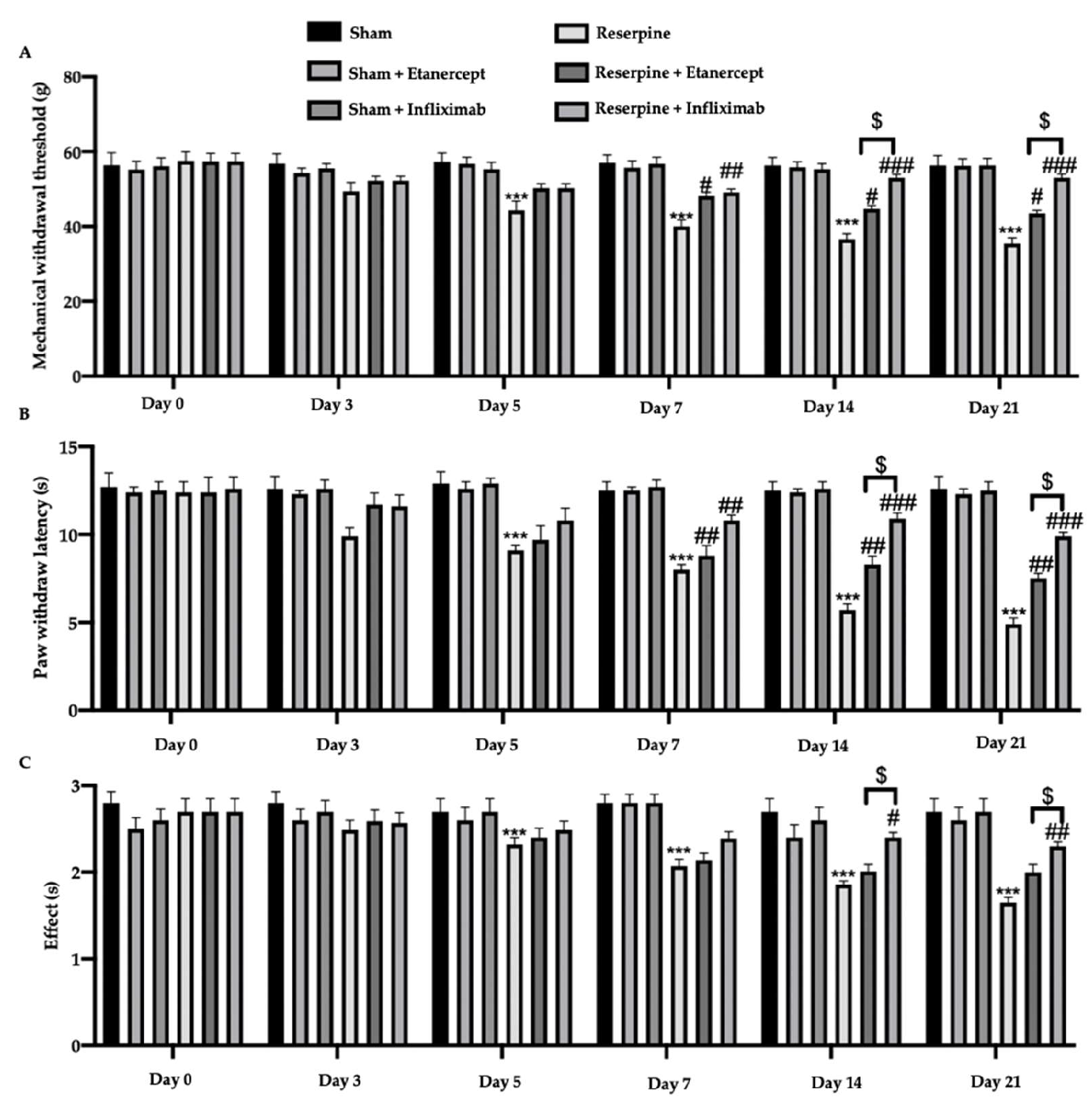
Three days after the first reserpine injection, the animals showed hypersensitivity to mechanical (Figure 2A) and thermal (Figure 2B,C) stimuli as compared to the sham group. The reserpine group showed an increased hypersensitivity, also at different time points, in particular at 5, 7, 14, and 21 days after the first reserpine injection. Etanercept and infliximab treatments importantly attenuated this overreaction (Figure 2A–C). In particular, this effect was prominent in infliximab-treated rats that, from 7 days from the reserpine injection, showed an increased withdrawal threshold and latency. No differences among the sham groups were detected; thus, no other results are shown about the sham + etanercept and sham + infliximab groups.

2.3. Effect of TNF-α Inhibitors on Pain-Related Mediators Induced by Fibromyalgia

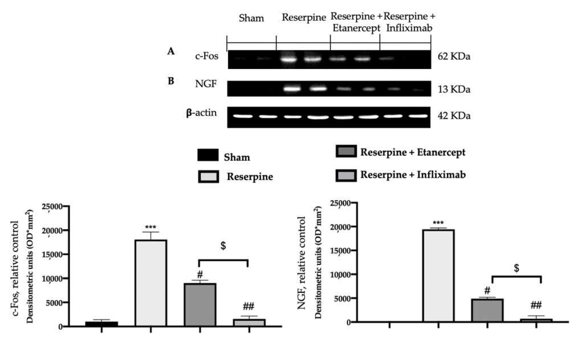
Western blot analyses were conduced to investigate the activation of the expression of the pain-related inflammatory mediators. Lumbar spinal cord tissues collected from the reserpine group showed an increased expression of c-FOS (Figure 3A) and nerve growth factor (NGF) (Figure 3B) as compared to the samples collected from the sham animals. Differently, the samples collected from the etanercept and infliximab groups showed strongly reduced expressions of both mediators. In particular, infliximab showed a stronger effect of inhibition on c-FOS and NGF as compared to etanercept (Figure 3A,B).

2.4. Effect of TNF-α Inhibitors on Pro-Inflammatory Cytokines’ Overexpression Induced by Fibromyalgia

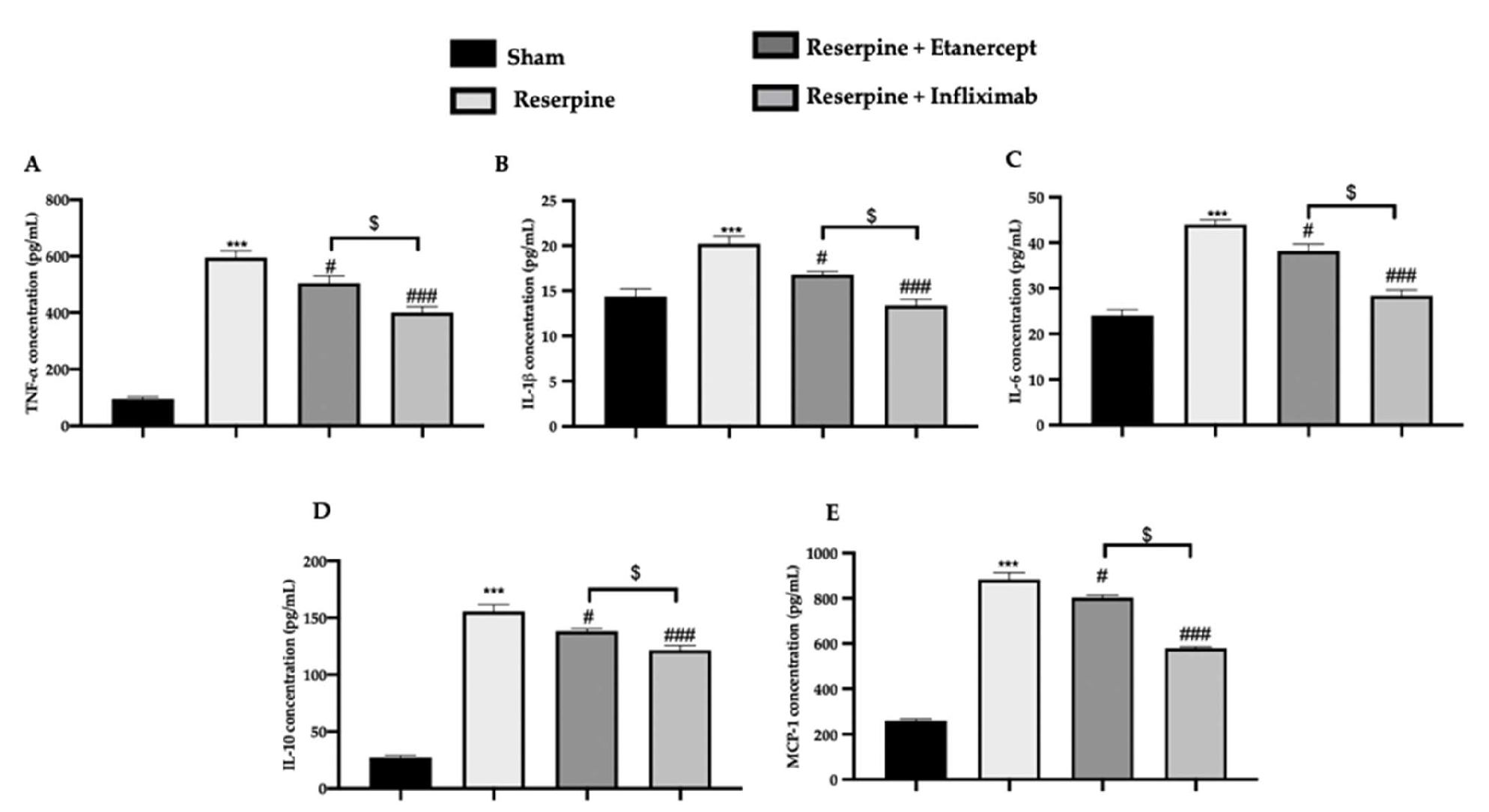
Lumbar spinal cord tissues collected from the reserpine group showed an increased expression of pro-inflammatory cytokines as compared to the sham group (Figure 4A–E). Differently, samples collected from the etanercept and infliximab groups showed strongly reduced expressions of TNF-α (Figure 4A), IL-1β (Figure 4B), IL-6 (Figure 4C), IL10 (Figure 4D), and MCP-1 (Figure 4E). In particular, infliximab showed a stronger effect of the inhibition of pro-inflammatory cytokines’ expression as compared to etanercept.

2.5. Effect of TNF-α Inhibitors on Glia Activation Induced by Fibromyalgia

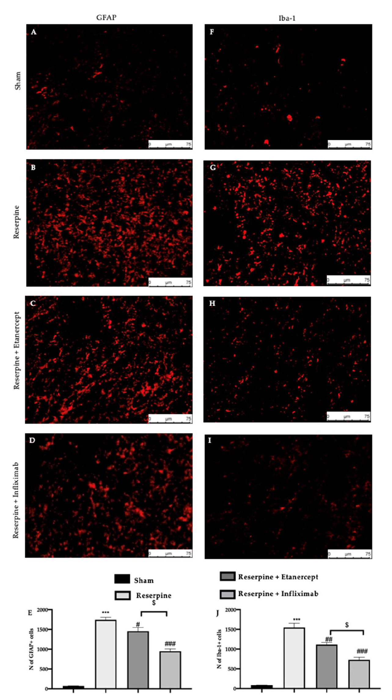
Immunofluorescence analysis showed increased GFAP (Figure 5B) and Iba-1 (Figure 5G) expressions in the lumbar spinal cord tissue of the reserpine group as compared to the sham group (Figure 5A,F, respectively). Infliximab and etanercept administrations reduced both GFAP (Figure 5C,D) and Iba-1 (Figure 5H,I) expressions. In particular, this effect was prominent in the infliximab-treated rats for both the GFAP (Figure 5E) and Iba-1 (Figure 5J) levels.

2.6. Effect of TNF-α Inhibitors on Purinergic Receptor and p38-MAPK Expression Induced by Fibromyalgia

Western blot analysis showed an important increase in the P2X7 expression in lumbar spinal cord harvested from the reserpine group as compared to the sham group. Etanercept and infliximab administrations importantly reduced P2X7 levels (Figure 6A). Additionally, Western blot analysis was employed to evaluate MAPK activation. In particular, p-38 was found phosphorylated in the reserpine group as compared to the sham group (Figure 6B). Etanercept and infliximab treatments significantly reduced its phosphorylation (Figure 6B), but infliximab strongly reduced both proteins’ expressions.

3. Discussion

The data from this study, acquired using an experimental model of fibromyalgia, indicate that the administration of infliximab and etanercept displayed an analgesic effect.
We employed an experimental animal model of fibromyalgia, which was developed through the administration of reserpine, a biogenic amine depletor; long-lasting widespread nociceptive hypersensitivities were exhibited in rats [45,46,47,48,49]. It is noteworthy that the time course of pain-related behaviors was in parallel with the decrease in monoamine neurotransmitters after the reserpine treatment. Thus, monoamine depletion appears to cause nociceptive hypersensitivity in a rat reserpine-induced pain model, and the model could be useful to study pathological mechanisms of chronic, widespread pain such as with fibromyalgia. In our model of fibromyalgia, the animals showed hyperalgesia and allodynia and increased TNFR expression as compared to a sham group. This hypersensitivity to thermal and mechanical stimuli was reduced by etanercept and infliximab administrations. Both treatments showed important analgesic effects. Notably, the highly significant antinociceptive effects were observed in the infliximab-treated rats. Infliximab showed a strong potential to reduce fibromyalgia-evoked pain. Well in line with the literature, our data confirmed the major effect of infliximab administration as compared to etanercept: infliximab administration strongly reduced pain sensitization in thermal hyperalgesia and mechanical allodynia. These data are well in line with a reduced expression of the pain-related pro-inflammatory mediators and cytokines in the spinal cord. In particular, both infliximab and etanercept did not cross the blood–brain barrier; thus, the central effects seen here are consequences of the peripheral effects [50].
It has been described that astrocytes alone are not able to self-maintain this pro-inflammatory state but, in turn, activate microglia. Infliximab administration strongly reduced the activation of microglia and astrocytes, as compared to etanercept. Under this pathological condition, the activated microglia and astrocytes release a large amount of ATP, which activates specific purinoceptors, receptors ubiquitously expressed on glia and neurons [51,52]. ATP is a fundamental regulator of microglia activity [53,54], including cytokines’ expression. It has been described that ATP is involved in the novo synthesis of pro-inflammatory cytokines from animal microglia through the activation of the P2X7 receptor. In response to the increased ATP, the ionotropic purinergic receptor induces Ca2+ in microglia [51], which activates the pro-inflammatory cytokines’ secretion. Downstream of the P2X7 receptor involves the MAP kinase activation [55,56]. Microglial p38-MAPK has been described as being involved in the development of neuropathic pain in studies on spinal cord injury [57,58] and peripheral nerve [59,60,61] injury. Infliximab administration significantly reduced P2X7 and p38 expression as compared to etanercept.
Overall, there is published evidence that infliximab does not bind rat TNF-α [62], while there is no evidence about etanercept [63]. This paper shows the indirect effect in rat models by anti-human-TNF-α antibody preparation such as etanercept and/or infliximab. The observed effects would be a “placebo” effect resulting from just giving an antibody in general, not specifically targeting TNF-α.
Our data showed the key role of the infliximab and etanercept in the development of fibromyalgia, its role in the activation of astrocytes and microglia, and its modulation of the signaling pathway involved in the modulation of pain.

4. Materials and Methods

4.1. Animals

Male Sprague–Dawley rats (200–220 g, 6–8 weeks old, Envigo, Milan, Italy) were employed for this paper. Food and water were administered ad libitum. The University of Messina Review Board for animal care approved the study. All in vivo experiments followed the new regulations of the USA (Animal Welfare Assurance No. A5594-01), Europe (EU Directive 2010/63), Italy (D.Lgs 2014/26), and the ARRIVE guidelines.

4.2. Experimental Model

Reserpine administration was performed by subcutaneous injection of 1 mg/kg for 3 consecutive days [50]. Reserpine (Sigma-Aldrich, Saint Louis, MO, USA) was dissolved in distilled water with 0.5% acetic acid (vehicle). Sham animals received the same volume of vehicle but no reserpine administrations.

4.3. Treatment

Etanercept (3 mg/kg) was intraperitoneally injected the day after the last reserpine injection and on post-injection day 5. Infliximab (10 mg/kg) was intraperitoneally injected the day after the last reserpine injection and on post-injection day 5. The doses of etanercept and infliximab were based on previous studies [64].

4.4. Experimental Froups

Then, rats were randomly divided into several groups (n = 15):
Sham: Rats were subcutaneously injected with vehicle instead of reserpine and treated with saline the day after the last reserpine injection and 5 days after.
Sham + etanercept: Rats were subcutaneously injected with saline instead of reserpine and intraperitoneally injected with etanercept.
Sham + infliximab: Rats were subcutaneously injected with saline instead of reserpine and intraperitoneally injected with infliximab.
Reserpine: Rats were administered with reserpine as previously described.
Reserpine + etanercept: Rats were administered with reserpine as previously described and intraperitoneally injected with etanercept.
Reserpine + infliximab: Rats were administered with reserpine as previously described and intraperitoneally injected with infliximab.
Twenty-one days after reserpine injection, behavioural analysis were conducted, animals were sacrificed by isoflurane overdose (concentration to 5%) and L4–L6 area of spinal cord were collected for molecular analysis.

4.5. Behavioral Analysis

4.5.1. Von Frey Hair Test

Mechanical allodynia was evaluated on day 0 and 3, 5, 7, 14, and 21 days post-reserpine injection using a dynamic plantar von Frey hair esthesiometer, as already described [65]. The device is comprised of a force transducer equipped with a plastic tip. When pressure is applied to the tip, the force applied is calculated. The tip was applied to the plantar area, and a rising, upward force was exerted [66]. The withdrawal threshold was defined as the force, expressed in grams, at which the mouse removed its paw.

4.5.2. Hot Plate Test

On day 0 and 3, 5, 7, 14, and 21 days post-reserpine injection the hot plate test was performed. The hot-plate latency was calculated using a metal surface maintained at 53.6 °C. The rat was monitored, and the licking of a hind paw was set as the end point. Maximal latency accepted was 45 s [67].

4.5.3. Tail Flick Warm Water Test

On day 0 and 3, 5, 7, 14, and 21 days post reserpine injection, the tail-flick, warm water test was performed to evaluate pain threshold. The rat’s tail was immersed in warm water (50 ± 0.5 °C), and the time between tail input and retraction was recordered. A maximum tail-flick latency of 10 s was employed to minimize tissue damage [67].

4.6. Western Blot Analysis

Western blot analysis was performed on lumbar spinal cord tissues. For this analysis, n = 5 animals for each group were employed. Cytosolic and nuclear extracts were prepared, with slight modifications of published procedures [68,69]. Tissues from each rat were suspended in extraction Buffer A, containing 0.2 mM PMSF, 0.15 mM pepstatin A, 20 mM leupeptin, and 1 mM sodium orthovanadate, homogenized at the highest setting for 2 min, and centrifuged at 12,000× g rpm for 4 min at 4 °C [70,71,72]. Supernatants represented the cytosolic fraction. The pellets, containing enriched nuclei, were resuspended in Buffer B, containing 1% Triton X-100, 150 mM NaCl, 10 mM Tris-HCl pH 7.4, 1 mM EGTA, 1 mM EDTA, 0.2 mM PMSF, 20 mm leupeptin, and 0.2 mM sodium orthovanadate [73]. After centrifugation for 10 min at 12,000 rpm at 4 °C, the supernatants contained the nuclear protein [74]. Protein concentrations were estimated by the Bio-Rad protein assay using bovine serum albumin as a standard [71]. Briefly, samples were heated to 100 °C for 5 min, and equal amounts of protein were separated on SDS-PAGE gel and transferred to nitrocellulose membrane [69,70]. The membranes were probed with specific antibodies, anti-c-FOS (sc-166940; Santa Cruz Biotechnology, Dallas, TX, USA), anti-NGF (sc-32300; Santa Cruz Biotechnology), anti-P2X7 ((Cell Signalling Technology, Danvers, MA, USA), or anti-p-p38 (sc-7983; Santa Cruz Biotechnology), in 1x PBS (phosphate-buffered saline), 5% w/v non-fat dried milk, and 0.1% Tween-20 at 4 °C, overnight [61,66]. To control the equal amounts of proteins, blots also were probed with an antibody against the b-actin protein (Santa Cruz Biotechnology). Signals were examined with an enhanced chemiluminescence (ECL) detection system reagent (Thermo Fisher, Waltham, MA, USA). The relative expression of the protein bands was quantified by densitometry with BIORAD ChemiDocTM XRS+ software and standardized to b-actin. The blot was stripped with glycine 2% and re-incubated several times to optimize the detection of the proteins and to visualize other proteins, minimizing the number of gels and transfers. The experiments were performed in triplicate and repeated three times with similar results.

4.7. Immunofluorescence Analysis

Immunofluorescence analysis was performed on lumbar spinal cord tissues. For this analysis n = 5 animals for each group were employed. N = 5 different fields from n = 5 different animals were evaluated. The areas between the lumbar vertebrae (L5–6) were harvested, fixed and decalcified [75]. In particular, the tissues were fixed for 24 h in a formaldehyde solution (10% in PBS) at room temperature and decalcified in Osteosoft solution (Merck Millipore, Milan, Italy). Next, the samples were dehydrated through a graded series of ethanol and xylene and embedded in BioPlast Plus (Bio Optica, Milan, Italy) [76,77]. Sections (5 μm in thickness) were prepared from tissues. After deparaffinization and rehydration, sections were boiled in 0.1 M citrate buffer for 1 min [78,79]. Non-specific adsorption was diminished by incubating the sections in 2% (v/v) normal goat serum in PBS for 20 min [80,81]. The sections were incubated with primary antibodies, anti-GFAP (Santa Cruz Biotechnology) or anti Iba-1 (Santa Cruz Biotechnology), overnight in a humidified chamber at 37 °C [74]. The sections were washed with PBS and were incubated with FITC-conjugated anti-rabbit Alexa Fluor-594 antibody (1:2000 v/v Molecular Probes, UK) for 1 h at 37 °C. The sections were analyzed using a Leica DM6 microscope (Leica Microsystems SpA, Milan, Italy) associated with Leica LAS X Navigator software (Leica Microsystems SpA, Milan, Italy). For immunofluorescence, the photographs were the outcomes of at least three independent experiments.

4.8. ELISA Analysis

The concentrations of TNF-α, IL-1β, IL-6, IL10, and MCP-1 were measured. Briefly, spinal cord tissues were homogenated in 1 mL PBS with 10 μL protease inhibitor at low speed for ~20 seconds [72]. The samples were centrifuged at 14,000× g at 4 °C for 15 minutes; supernatants were employed, using respective ELISA kits according to the manufacturer’s protocol, and analyzed using a microplate reader [61]. The values are expressed as pg/mL.

4.9. Statistical Evaluation

All values in the figures and text are expressed as mean ± standard error of the mean (SEM) of N number of animals. The results were analyzed by one-way ANOVA followed by a Bonferroni post hoc test for multiple comparisons. A p-value < 0.05 was considered significant; * p < 0.05 vs. sham; # p < 0.05 vs. vehicle; ** p < 0.01 vs. sham; ## p < 0.01 vs. vehicle; *** p < 0.001 vs. sham; ###p < 0.001 vs. vehicle; $ p < 0.05 infliximab vs. etanercept.

Author Contributions

Conceptualization, M.C.; data curation, R.S.; formal analysis, R.D.; investigation, T.G., G.F. and Y.M.; project administration, R.F. and D.I.; resources, D.D.P.; supervision, S.C.; writing—review and editing, R.D.P. All authors have read and agreed to the published version of the manuscript.

Funding

This research received no external funding.

Institutional Review Board Statement

The study was conducted according to the guidelines of the Declaration of Helsinki and approved by the Institutional Review Board for animal care (OPBA) of the University of Messina.

Informed Consent Statement

Not applicable.

Data Availability Statement

The data presented in this study are available on request from the corresponding author.

Conflicts of Interest

The authors declare no conflict of interest.

References

  1. Maffei, M.E. Fibromyalgia: Recent Advances in Diagnosis, Classification, Pharmacotherapy and Alternative Remedies. Int. J. Mol. Sci. 2020, 21, 7877. [Google Scholar] [CrossRef] [PubMed]
  2. Qureshi, A.G.; Jha, S.K.; Iskander, J.; Avanthika, C.; Jhaveri, S.; Patel, V.H.; Potini, B.R.; Azam, A.T. Diagnostic Challenges and Management of Fibromyalgia. Cureus 2021, 13, e18692. [Google Scholar] [CrossRef] [PubMed]
  3. Rodríguez, D.F.G.; Abud-Mendoza, C. Physiopathology of fibromyalgia. Reumatol. Clin. (Engl. Ed.) 2020, 16, 191–194. [Google Scholar] [CrossRef] [PubMed]
  4. D’Agnelli, S.; Arendt-Nielsen, L.; Gerra, M.C.; Zatorri, K.; Boggiani, L.; Baciarello, M.; Bignami, E. Fibromyalgia: Genetics and epigenetics insights may provide the basis for the development of diagnostic biomarkers. Mol. Pain 2019, 15, 1744806918819944. [Google Scholar] [CrossRef] [PubMed]
  5. Sarzi-Puttini, P.; Giorgi, V.; Marotto, D.; Atzeni, F. Fibromyalgia: An update on clinical characteristics, aetiopathogenesis and treatment. Nat. Rev. Rheumatol. 2020, 16, 645–660. [Google Scholar] [CrossRef] [PubMed]
  6. Latremoliere, A.; Woolf, C.J. Central sensitization: A generator of pain hypersensitivity by central neural plasticity. J. Pain 2009, 10, 895–926. [Google Scholar] [CrossRef] [Green Version]
  7. Bair, M.J.; Krebs, E.E. Fibromyalgia. Ann. Intern. Med. 2020, 172, ITC33–ITC48. [Google Scholar] [CrossRef]
  8. Menzies, V.; Lyon, D.E.; Elswick, R.K., Jr.; Montpetit, A.J.; McCain, N.L. Psychoneuroimmunological Relationships in Women with Fibromyalgia. Biol. Res. Nurs. 2014, 15, 219–225. [Google Scholar] [CrossRef]
  9. Bradley, L.A. Pathophysiology of Fibromyalgia. Am. J. Med. 2009, 122, S22–S30. [Google Scholar] [CrossRef] [Green Version]
  10. Peritore, A.F.; Crupi, R.; Scuto, M.; Gugliandolo, E.; Siracusa, R.; Impellizzeri, D.; Cordaro, M.; D’Amico, R.; Fusco, R.; Di Paola, R.; et al. The Role of Annexin A1 and Formyl Peptide Receptor 2/3 Signaling in Chronic Corticosterone-Induced Depression-Like behaviors and Impairment in Hippocampal-Dependent Memory. CNS Neurol. Disord.—Drug Targets 2020, 19, 27–43. [Google Scholar] [CrossRef]
  11. García, J.J.; Cidoncha, A.; Bote, M.E.; Hinchado, M.D.; Ortega, E. Altered profile of chemokines in fibromyalgia patients. Ann. Clin. Biochem. 2013, 51, 576–581. [Google Scholar] [CrossRef] [PubMed]
  12. Behm, F.G.; Gavin, I.M.; Karpenko, O.; Lindgren, V.; Gaitonde, S.; Gashkoff, P.A.; Gillis, B.S. Unique immunologic patterns in fibromyalgia. BMC Clin. Pathol. 2012, 12, 25. [Google Scholar] [CrossRef] [PubMed] [Green Version]
  13. Rodríguez-Pintó, I.; Agmon-Levin, N.; Howard, A.; Shoenfeld, Y. Fibromyalgia and cytokines. Immunol. Lett. 2014, 161, 200–203. [Google Scholar] [CrossRef] [PubMed]
  14. Benlidayi, I.C. Role of inflammation in the pathogenesis and treatment of fibromyalgia. Rheumatol. Int. 2019, 39, 781–791. [Google Scholar] [CrossRef] [PubMed]
  15. Theoharides, T.C.; Tsilioni, I.; Bawazeer, M. Mast Cells, Neuroinflammation and Pain in Fibromyalgia Syndrome. Front. Cell. Neurosci. 2019, 13, 353. [Google Scholar] [CrossRef]
  16. Üçeyler, N.; Rogausch, J.P.; Toyka, K.V.; Sommer, C. Differential expression of cytokines in painful and painless neuropathies. Neurology 2007, 69, 42–49. [Google Scholar] [CrossRef]
  17. Genevay, S.; Finckh, A.; Zufferey, P.; Viatte, S.; Balagué, F.; Gabay, C. Adalimumab in acute sciatica reduces the long-term need for surgery: A 3-year follow-up of a randomised double-blind placebo-controlled trial. Ann. Rheum. Dis. 2012, 71, 560–562. [Google Scholar] [CrossRef] [Green Version]
  18. Alexander, G.M.; Peterlin, B.L.; Perreault, M.J.; Grothusen, J.R.; Schwartzman, R.J. Changes in plasma cytokines and their soluble receptors in complex regional pain syndrome. J. Pain 2012, 13, 10–20. [Google Scholar] [CrossRef]
  19. Conti, P.; Gallenga, C.E.; Caraffa, A.; Ronconi, G.; Kritas, S.K. Impact of mast cells in fibromyalgia and low-grade chronic inflammation: Can IL-37 play a role? Dermatol. Ther. 2020, 33, e13191. [Google Scholar] [CrossRef]
  20. DeLeo, J.A.; Rutkowski, M.D.; Stalder, A.K.; Campbell, I.L. Transgenic expression of TNF by astrocytes increases mechanical allodynia in a mouse neuropathy model. NeuroReport 2000, 11, 599–602. [Google Scholar] [CrossRef]
  21. Hatashita, S.; Sekiguchi, M.; Kobayashi, H.; Konno, S.-I.; Kikuchi, S.-I. Contralateral neuropathic pain and neuropathology in dorsal root ganglion and spinal cord following hemilateral nerve injury in rats. Spine (Phila Pa 1976) 2008, 33, 1344–1351. [Google Scholar] [CrossRef] [PubMed]
  22. Sommer, C.; Galbraith, J.A.; Heckman, H.M.; Myers, R.R. Pathology of experimental compression neuropathy producing hyperesthesia. J. Neuropathol. Exp. Neurol. 1993, 52, 223–233. [Google Scholar] [CrossRef] [PubMed]
  23. Frisén, J.; Risling, M.; Fried, K. Distribution and axonal relations of macrophages in a neuroma. Neuroscience 1993, 55, 1003–1013. [Google Scholar] [CrossRef]
  24. Inoue, K.; Tsuda, M.; Tozaki-Saitoh, H. Modification of neuropathic pain sensation through microglial ATP receptors. Purinergic Signal. 2007, 3, 311–316. [Google Scholar] [CrossRef] [PubMed] [Green Version]
  25. Adler, J.E.; Nico, L.; VandeVord, P.; Skoff, A.M. Modulation of neuropathic pain by a glial-derived factor. Pain Med. 2009, 10, 1229–1236. [Google Scholar] [CrossRef] [Green Version]
  26. Hansson, E. Could chronic pain and spread of pain sensation be induced and maintained by glial activation? Acta Physiol. 2006, 187, 321–327. [Google Scholar] [CrossRef]
  27. Moss, A.; Beggs, S.; Vega-Avelaira, D.; Costigan, M.; Hathway, G.J.; Salter, M.W.; Fitzgerald, M. Spinal microglia and neuropathic pain in young rats. Pain 2007, 128, 215–224. [Google Scholar] [CrossRef]
  28. Thacker, M.A.; Clark, A.K.; Bishop, T.; Grist, J.; Yip, P.K.; Moon, L.D.; Thompson, S.W.N.; Marchand, F.; McMahon, S.B. CCL2 is a key mediator of microglia activation in neuropathic pain states. Eur. J. Pain 2009, 13, 263–272. [Google Scholar] [CrossRef]
  29. Bergsteinsdottir, K.; Kingston, A.; Jessen, K.R. Rat Schwann cells can be induced to express major histocompatibility complex class II moleculesin vivo. J. Neurocytol. 1992, 21, 382–390. [Google Scholar] [CrossRef]
  30. Constable, A.L.; Armati, P.J.; Toyka, K.V.; Hartung, H.-P. Production of prostanoids by Lewis rat Schwann cells in vitro. Brain Res. 1994, 635, 75–80. [Google Scholar] [CrossRef]
  31. Bolin, L.M.; Verity, A.N.; Silver, J.E.; Shooter, E.M.; Abrams, J.S. Interleukin-6 production by schwann cells and induction in sciatic nerve injury. J. Neurochem. 1995, 64, 850–858. [Google Scholar] [CrossRef] [PubMed]
  32. Sofroniew, M.V.; Howe, C.L.; Mobley, W.C. Nerve growth factor signaling, neuroprotection, and neural repair. Annu. Rev. Neurosci. 2001, 24, 1217–1281. [Google Scholar] [CrossRef] [PubMed]
  33. Watkins, L.R.; Maier, S.F.; Goehler, L.E. Cytokine-to-brain communication: A review & analysis of alternative mechanisms. Life Sci. 1995, 57, 1011–1026. [Google Scholar] [CrossRef] [PubMed]
  34. Milligan, E.D.; Watkins, L.R. Pathological and protective roles of glia in chronic pain. Nat. Rev. Neurosci. 2009, 10, 23–36. [Google Scholar] [CrossRef]
  35. Empl, M.; Renaud, S.; Erne, B.; Fuhr, P.; Straube, A.; Schaeren-Wiemers, N.; Steck, A.J. TNF-alpha expression in painful and nonpainful neuropathies. Neurology 2001, 56, 1371–1377. [Google Scholar] [CrossRef]
  36. Covey, W.C.; Ignatowski, T.A.; Renauld, A.E.; Knight, P.R.; Nader, N.D.; Spengler, R.N. Expression of neuron-associated tumor necrosis factor alpha in the brain is increased during persistent pain. Reg. Anesth. Pain Med. 2002, 27, 357–366. [Google Scholar] [CrossRef]
  37. Watkins, L.R.; Maier, S.F. Immune regulation of central nervous system functions: From sickness responses to pathological pain. J. Intern. Med. 2005, 257, 139–155. [Google Scholar] [CrossRef]
  38. Marchand, F.; Tsantoulas, C.; Singh, D.; Grist, J.; Clark, A.K.; Bradbury, E.J.; McMahon, S.B. Effects of Etanercept and Minocycline in a rat model of spinal cord injury. Eur. J. Pain 2009, 13, 673–681. [Google Scholar] [CrossRef]
  39. Genevay, S.; Stingelin, S.; Gabay, C. Efficacy of etanercept in the treatment of acute, severe sciatica: A pilot study. Ann. Rheum. Dis. 2004, 63, 1120–1123. [Google Scholar] [CrossRef] [Green Version]
  40. Karppinen, J.; Korhonen, T.; Malmivaara, A.; Paimela, L.; Kyllonen, E.; Lindgren, K.A.; Rantanen, P.; Tervonen, O.; Niinimaki, J.; Seitsalo, S.; et al. Tumor necrosis factor-alpha monoclonal antibody, infliximab, used to manage severe sciatica. Spine (Phila Pa 1976) 2003, 28, 750–753, Discussion in 753–754. [Google Scholar] [CrossRef]
  41. Korhonen, T.; Karppinen, J.; Malmivaara, A.; Autio, R.; Niinimäki, J.; Paimela, L.; Kyllönen, E.; Lindgren, K.-A.; Tervonen, O.; Seitsalo, S.; et al. Efficacy of infliximab for disc herniation-induced sciatica: One-year follow-up. Spine (Phila Pa 1976) 2004, 29, 2115–2119. [Google Scholar] [CrossRef] [PubMed]
  42. Korhonen, T.; Karppinen, J.; Paimela, L.; Malmivaara, A.; Lindgren, K.-A.; Bowman, C.; Hammond, A.; Kirkham, B.; Järvinen, S.; Niinimäki, J.; et al. The Treatment of disc herniation-induced sciatica with infliximab: One-year follow-up results of FIRST II, a randomized controlled trial. Spine (Phila Pa 1976) 2006, 31, 2759–2766. [Google Scholar] [CrossRef] [PubMed]
  43. Cohen, S.P.; Wenzell, D.; Hurley, R.W.; Kurihara, C.; Buckenmaier, C.C., 3rd; Griffith, S.; Larkin, T.M.; Dahl, E.; Morlando, B.J. A double-blind, placebo-controlled, dose–response pilot study evaluating intradiscal etanercept in patients with chronic discogenic low back pain or lumbosacral radiculopathy. Anesthesiology 2007, 107, 99–105. [Google Scholar] [CrossRef] [PubMed]
  44. Cohen, S.P.; Bogduk, N.; Dragovich, A.; Buckenmaier, C.C., 3rd; Griffith, S.; Kurihara, C.; Raymond, J.; Richter, P.J.; Williams, N.; Yaksh, T.L. Randomized, Double-blind, placebo-controlled, dose-response, and preclinical safety study of transforaminal epidural etanercept for the treatment of sciatica. Anesthesiology 2009, 110, 1116–1126. [Google Scholar] [CrossRef] [PubMed] [Green Version]
  45. Nagakura, Y.; Oe, T.; Aoki, T.; Matsuoka, N. Biogenic amine depletion causes chronic muscular pain and tactile allodynia accompanied by depression: A putative animal model of fibromyalgia. Pain 2009, 146, 26–33. [Google Scholar] [CrossRef] [PubMed]
  46. Taguchi, T.; Katanosaka, K.; Yasui, M.; Hayashi, K.; Yamashita, M.; Wakatsuki, K.; Kiyama, H.; Yamanaka, A.; Mizumura, K. Peripheral and spinal mechanisms of nociception in a rat reserpine-induced pain model. Pain 2015, 156, 415–427. [Google Scholar] [CrossRef]
  47. Tamano, R.; Ishida, M.; Asaki, T.; Hasegawa, M.; Shinohara, S. Effect of spinal monoaminergic neuronal system dysfunction on pain threshold in rats, and the analgesic effect of serotonin and norepinephrine reuptake inhibitors. Neurosci. Lett. 2016, 615, 78–82. [Google Scholar] [CrossRef]
  48. Wells, J.A.; Shibata, S.; Fujikawa, A.; Takahashi, M.; Saga, T.; Aoki, I. Functional MRI of the Reserpine-Induced Putative Rat Model of Fibromyalgia Reveals Discriminatory Patterns of Functional Augmentation to Acute Nociceptive Stimuli. Sci. Rep. 2017, 7, 38325. [Google Scholar] [CrossRef] [Green Version]
  49. Uchida, M.; Kobayashi, O.; Yoshida, M.; Miwa, M.; Miura, R.; Saito, H.; Nagakura, Y. Coexistence of Alterations of Gastrointestinal Function and Mechanical Allodynia in the Reserpine-Induced Animal Model of Fibromyalgia. Dig. Dis. Sci. 2019, 64, 2538–2547. [Google Scholar] [CrossRef]
  50. Fusco, R.; Siracusa, R.; D’Amico, R.; Peritore, A.F.; Cordaro, M.; Gugliandolo, E.; Crupi, R.; Impellizzeri, D.; Cuzzocrea, S.; Di Paola, R. Melatonin Plus Folic Acid Treatment Ameliorates Reserpine-Induced Fibromyalgia: An Evaluation of Pain, Oxidative Stress, and Inflammation. Antioxidants 2019, 8, 628. [Google Scholar] [CrossRef] [Green Version]
  51. Inoue, K.; Nakajima, K.; Morimoto, T.; Kikuchi, Y.; Koizumi, S.; Illes, P.; Kohsaka, S.; Inoue, K.; Nakajima, K.; Morimoto, T.; et al. ATP stimulation of Ca2+-dependent plasminogen release from cultured microglia. Br. J. Pharmacol. 1998, 123, 1304–1310. [Google Scholar] [CrossRef] [PubMed] [Green Version]
  52. Abbracchio, M.P.; Burnstock, G.; Verkhratsky, A.; Zimmermann, H. Purinergic signalling in the nervous system: An overview. Trends Neurosci. 2009, 32, 19–29. [Google Scholar] [CrossRef] [PubMed]
  53. Bianco, F.; Pravettoni, E.; Colombo, A.; Schenk, U.; Möller, T.; Matteoli, M.; Verderio, C. Astrocyte-Derived ATP Induces Vesicle Shedding and IL-1β Release from Microglia. J. Immunol. 2005, 174, 7268–7277. [Google Scholar] [CrossRef] [PubMed] [Green Version]
  54. Koizumi, S.; Ohsawa, K.; Inoue, K.; Kohsaka, S. Purinergic receptors in microglia: Functional modal shifts of microglia mediated by P2 and P1 receptors. Glia 2013, 61, 47–54. [Google Scholar] [CrossRef]
  55. Suzuki, T.; Hide, I.; Ido, K.; Kohsaka, S.; Inoue, K.; Nakata, Y. Production and Release of Neuroprotective Tumor Necrosis Factor by P2X7 Receptor-Activated Microglia. J. Neurosci. 2004, 24, 1–7. [Google Scholar] [CrossRef] [Green Version]
  56. Lister, M.F.; Sharkey, J.; Sawatzky, D.A.; Hodgkiss, J.P.; Davidson, D.J.; Rossi, A.G.; Finlayson, K. The role of the purinergic P2X7 receptor in inflammation. J. Inflamm. 2007, 4, 5. [Google Scholar] [CrossRef] [Green Version]
  57. Crown, E.D.; Ye, Z.; Johnson, K.M.; Xu, G.-Y.; McAdoo, D.J.; Hulsebosch, C.E. Increases in the activated forms of ERK 1/2, p38 MAPK, and CREB are correlated with the expression of at-level mechanical allodynia following spinal cord injury. Exp. Neurol. 2006, 199, 397–407. [Google Scholar] [CrossRef]
  58. Crown, E.D.; Gwak, Y.S.; Ye, Z.; Johnson, K.M.; Hulsebosch, C.E. Activation of p38 MAP kinase is involved in central neuropathic pain following spinal cord injury. Exp. Neurol. 2008, 213, 257–267. [Google Scholar] [CrossRef] [Green Version]
  59. Terayama, R.; Omura, S.; Fujisawa, N.; Yamaai, T.; Ichikawa, H.; Sugimoto, T. Activation of microglia and p38 mitogen-activated protein kinase in the dorsal column nucleus contributes to tactile allodynia following peripheral nerve injury. Neuroscience 2008, 153, 1245–1255. [Google Scholar] [CrossRef]
  60. Wen, Y.-R.; Suter, M.R.; Ji, R.-R.; Yeh, G.-C.; Wu, Y.-S.; Wang, K.-C.; Kohno, T.; Sun, W.-Z.; Wang, C.-C. Activation of p38 Mitogen-activated Protein Kinase in Spinal Microglia Contributes to Incision-induced Mechanical Allodynia. Anesthesiology 2009, 110, 155–165. [Google Scholar] [CrossRef] [Green Version]
  61. Crupi, R.; Palma, E.; Siracusa, R.; Fusco, R.; Gugliandolo, E.; Cordaro, M.; Impellizzeri, D.; De Caro, C.; Calzetta, L.; Cuzzocrea, S.; et al. Protective Effect of Hydroxytyrosol Against Oxidative Stress Induced by the Ochratoxin in Kidney Cells: In vitro and in vivo Study. Front Vet Sci 2020, 7, 136. [Google Scholar] [CrossRef] [PubMed] [Green Version]
  62. Zanella, J.M.; Burright, E.N.; Hildebrand, K.; Hobot, C.; Cox, M.; Christoferson, L.; McKay, W.F. Effect of etanercept, a tumor necrosis factor-alpha inhibitor, on neuropathic pain in the rat chronic constriction injury model. Spine (Phila Pa 1976) 2008, 33, 227–234. [Google Scholar] [CrossRef] [PubMed]
  63. Chen, X.; DuBois, D.C.; Almon, R.R.; Jusko, W.J. Interrelationships between Infliximab and Recombinant Tumor Necrosis Factor-alpha in Plasma Using Minimal Physiologically Based Pharmacokinetic Models. Drug Metab Dispos 2017, 45, 790–797. [Google Scholar] [CrossRef] [PubMed] [Green Version]
  64. Andrade, P.; Hoogland, G.; Del Rosario, J.S.; Steinbusch, H.W.; Visser-Vandewalle, V.; Daemen, M.A. Tumor necrosis factor-α inhibitors alleviation of experimentally induced neuropathic pain is associated with modulation of TNF receptor expression. J. Neurosci. Res. 2014, 92, 1490–1498. [Google Scholar] [CrossRef]
  65. Ferrier, J.; Marchand, F.; Balayssac, D. Assessment of mechanical allodynia in rats using the electronic von frey test. Bio-protocol 2016, 6, e1933. [Google Scholar] [CrossRef]
  66. Cordaro, M.; Siracusa, R.; Fusco, R.; D’Amico, R.; Peritore, A.F.; Gugliandolo, E.; Genovese, T.; Scuto, M.; Crupi, R.; Mandalari, G.; et al. Cashew (Anacardium occidentale L.) Nuts Counteract Oxidative Stress and Inflammation in an Acute Experimental Model of Carrageenan-Induced Paw Edema. Antioxidants (Basel) 2020, 9, 660. [Google Scholar] [CrossRef]
  67. Di Paola, R.; Fusco, R.; Gugliandolo, E.; Crupi, R.; Evangelista, M.; Granese, R.; Cuzzocrea, S. Co-micronized Palmitoylethanolamide/Polydatin Treatment Causes Endometriotic Lesion Regression in a Rodent Model of Surgically Induced Endometriosis. Front. Pharmacol. 2016, 7, 382. [Google Scholar] [CrossRef] [Green Version]
  68. Fusco, R.; Cordaro, M.; Siracusa, R.; D’Amico, R.; Genovese, T.; Gugliandolo, E.; Peritore, A.F.; Crupi, R.; Impellizzeri, D.; Cuzzocrea, S.; et al. Biochemical Evaluation of the Antioxidant Effects of Hydroxytyrosol on Pancreatitis-Associated Gut Injury. Antioxidants 2020, 9, 781. [Google Scholar] [CrossRef]
  69. Fusco, R.; Gugliandolo, E.; Siracusa, R.; Scuto, M.; Cordaro, M.; D’Amico, R.; Evangelista, M.; Peli, A.; Peritore, A.F.; Impellizzeri, D.; et al. Formyl Peptide Receptor 1 Signaling in Acute Inflammation and Neural Differentiation Induced by Traumatic Brain Injury. Biology 2020, 9, 238. [Google Scholar] [CrossRef]
  70. Fusco, R.; Cordaro, M.; Genovese, T.; Impellizzeri, D.; Siracusa, R.; Gugliandolo, E.; Peritore, A.F.; D’Amico, R.; Crupi, R.; Cuzzocrea, S.; et al. Adelmidrol: A New Promising Antioxidant and Anti-Inflammatory Therapeutic Tool in Pulmonary Fibrosis. Antioxidants 2020, 9, 601. [Google Scholar] [CrossRef]
  71. Fusco, R.; Cordaro, M.; Siracusa, R.; Peritore, A.F.; Gugliandolo, E.; Genovese, T.; D’Amico, R.; Crupi, R.; Smeriglio, A.; Mandalari, G.; et al. Consumption of Anacardium Occidentale L. (Cashew Nuts) Inhibits Oxidative Stress through Modulation of the Nrf2/HO-1 and NF-kB Pathways. Molecules 2020, 25, 4426. [Google Scholar] [CrossRef] [PubMed]
  72. Cordaro, M.; Fusco, R.; D’Amico, R.; Siracusa, R.; Peritore, A.F.; Gugliandolo, E.; Genovese, T.; Crupi, R.; Mandalari, G.; Cuzzocrea, S.; et al. Cashew (Anacardium occidentale L.) Nuts Modulate the Nrf2 and NLRP3 Pathways in Pancreas and Lung after Induction of Acute Pancreatitis by Cerulein. Antioxidants 2020, 9, 992. [Google Scholar] [CrossRef] [PubMed]
  73. D’Amico, R.; Fusco, R.; Cordaro, M.; Siracusa, R.; Peritore, A.F.; Gugliandolo, E.; Crupi, R.; Scuto, M.; Cuzzocrea, S.; Di Paola, R.; et al. Modulation of NLRP3 Inflammasome through Formyl Peptide Receptor 1 (Fpr-1) Pathway as a New Therapeutic Target in Bronchiolitis Obliterans Syndrome. Int. J. Mol. Sci. 2020, 21, 2144. [Google Scholar] [CrossRef] [PubMed] [Green Version]
  74. Peritore, A.F.; Siracusa, R.; Fusco, R.; Gugliandolo, E.; D’Amico, R.; Cordaro, M.; Crupi, R.; Genovese, T.; Impellizzeri, D.; Cuzzocrea, S.; et al. Ultramicronized Palmitoylethanolamide and Paracetamol, a New Association to Relieve Hyperalgesia and Pain in a Sciatic Nerve Injury Model in Rat. Int. J. Mol. Sci. 2020, 21, 3509. [Google Scholar] [CrossRef] [PubMed]
  75. Fusco, R.; Gugliandolo, E.; Campolo, M.; Evangelista, M.; Di Paola, R.; Cuzzocrea, S. Effect of a new formulation of micronized and ultramicronized N-palmitoylethanolamine in a tibia fracture mouse model of complex regional pain syndrome. PLoS ONE 2017, 12, e0178553. [Google Scholar] [CrossRef] [PubMed] [Green Version]
  76. Gugliandolo, E.; D’Amico, R.; Cordaro, M.; Fusco, R.; Siracusa, R.; Crupi, R.; Impellizzeri, D.; Cuzzocrea, S.; di Paola, R. Neuroprotective Effect of Artesunate in Experimental Model of Traumatic Brain Injury. Front. Neurol. 2018, 9, 590. [Google Scholar] [CrossRef] [Green Version]
  77. Impellizzeri, D.; Peritore, A.F.; Cordaro, M.; Gugliandolo, E.; Siracusa, R.; Crupi, R.; D’Amico, R.; Fusco, R.; Evangelista, M.; Cuzzocrea, S.; et al. The neuroprotective effects of micronized PEA (PEA-m) formulation on diabetic peripheral neuropathy in mice. FASEB J. 2019, 33, 11364–11380. [Google Scholar] [CrossRef]
  78. Gugliandolo, E.; D’Amico, R.; Cordaro, M.; Fusco, R.; Siracusa, R.; Crupi, R.; Impellizzeri, D.; Cuzzocrea, S.; Di Paola, R. Effect of PEA-OXA on neuropathic pain and functional recovery after sciatic nerve crush. J. Neuroinflammation 2018, 15, 1–13. [Google Scholar] [CrossRef] [Green Version]
  79. Impellizzeri, D.; Siracusa, R.; Cordaro, M.; Crupi, R.; Peritore, A.F.; Gugliandolo, E.; D’Amico, R.; Petrosino, S.; Evangelista, M.; Di Paola, R.; et al. N-Palmitoylethanolamine-oxazoline (PEA-OXA): A new therapeutic strategy to reduce neuroinflammation, oxidative stress associated to vascular dementia in an experimental model of repeated bilateral common carotid arteries occlusion. Neurobiol. Dis. 2019, 125, 77–91. [Google Scholar] [CrossRef]
  80. Di Paola, R.; Fusco, R.; Impellizzeri, D.; Cordaro, M.; Britti, D.; Morittu, V.M.; Evangelista, M.; Cuzzocrea, S. Adelmidrol, in combination with hyaluronic acid, displays increased anti-inflammatory and analgesic effects against monosodium iodoacetate-induced osteoarthritis in rats. Arthritis Res. Ther. 2016, 18, 291. [Google Scholar] [CrossRef] [Green Version]
  81. Fusco, R.; D’Amico, R.; Cordaro, M.; Gugliandolo, E.; Siracusa, R.; Peritore, A.F.; Crupi, R.; Impellizzeri, D.; Cuzzocrea, S.; Di Paola, R. Absence of formyl peptide receptor 1 causes endometriotic lesion regression in a mouse model of surgically-induced endometriosis. Oncotarget 2018, 9, 31355–31366. [Google Scholar] [CrossRef] [PubMed] [Green Version]
Figure 1. Schematic of study design.
Ijms 23 06139 g001
Figure 2. Efficacy of etanercept and infliximab administration on behavioural changes reserpine induced. Behavioural test: (A) Von Frey test, (B) hot plate test, (C) tail-flick test. For these analyses n = 5 animals for each group were employed. Repeated ANOVA analysis followed by a Bonferroni post-hoc test for multiple comparisons were employed to analyse the effect of treatments in reserpine model along time. A p-value <0.05 was considered significant. # p < 0.05 vs. vehicle, ## p < 0.01 vs. vehicle, *** p < 0.001 vs. sham, ### p < 0.001 vs. vehicle, $ p < 0.05 infliximab vs. etanercept.
Ijms 23 06139 g002
Figure 3. Efficacy of etanercept and infliximab administration on c-FOS and NGR overexpression reserpine induced. Western Blot analysis of: (A) c-FOS and (B) NGF expressions. For these analyses n = 5 animals for each group were employed. Shown is a representative blot of lysates from five animals per group, together with a densitometric analysis normalized to housekeeping proteins. Results were analysed by one-way ANOVA followed by a Bonferroni post-hoc test for multiple comparisons. A p-value <0.05 was considered significant. # p < 0.05 vs. vehicle, ## p < 0.01 vs. vehicle, *** p < 0.001 vs. sham, $ p < 0.05 infliximab vs. etanercept.
Ijms 23 06139 g003
Figure 4. Efficacy of etanercept and infliximab administration on cytokines overexpression reserpine induced. ELISA analysis of: (A) TNF-α, (B) IL-1β, (C) IL-6, (D) IL10 and (E) MCP-1 expressions. For these analyses n = 5 animals fror each group were employed. Results were analysed one-way ANOVA followed by a Bonferroni post-hoc test for multiple comparisons. A p-value <0.05 was considered significant. *** p < 0.001 vs. sham. # p < 0.05 vs. vehicle, ### p < 0.001 vs. vehicle, $ p < 0.05 infliximab vs. etanercept.
Ijms 23 06139 g004
Figure 5. Efficacy of etanercept and infliximab administration on microglia and astrocytes reserpine induced. Immunofluorescence analysis of GFAP: (A) Sham, (B) Reserpine, (C) Reserpine+Etanercept, (D) Reserpine+Infliximab, (E) Number of GFAP positive cells. Immunofluorescence analysis of Iba-1: (F) Sham, (G) Reserpine, (H) Reserpine+Etanercept, (I) Reserpine+Infliximab, (J) Number of Iba-1 positive cells. Scale bar: 75μm. Magnification 40X. For these analyses n = 5 animals for each group were employed. Results were analysed by one-way ANOVA followed by a Bonferroni post-hoc test for multiple comparisons. A p-value <0.05 was considered significant. # p < 0.05 vs. vehicle, ## p < 0.01 vs. vehicle, *** p < 0.001 vs. sham, ### p < 0.001 vs. vehicle, $ p < 0.05 infliximab vs. etanercept.
Ijms 23 06139 g005
Figure 6. Efficacy of etanercept and infliximab administration on P2X7 and phospo-p38 overexpression reserpine induced. Western Blot analysis of: (A) P2X7 and (B) phospo-p38 expressions. For these analyses n = 5 animals for each group were employed. Shown is a representative blot of lysates from five animals per group, together with a densitometric analysis normalized to housekeeping proteins. Results were analysed by one-way ANOVA followed by a Bonferroni post-hoc test for multiple comparisons. A p-value <0.05 was considered significant. # p < 0.05 vs. vehicle, ## p < 0.01 vs. vehicle, *** p < 0.001 vs. sham, $ p < 0.05 infliximab vs. etanercept.
Ijms 23 06139 g006
Publisher’s Note: MDPI stays neutral with regard to jurisdictional claims in published maps and institutional affiliations.