Article

Genome-Wide Identification, Comparison, and Expression Analysis of Transcription Factors in Ascidian Styela clava

1
Sars-Fang Centre, MoE Key Laboratory of Marine Genetics and Breeding, College of Marine Life Sciences, Ocean University of China, Qingdao 266003, China
2
Laboratory for Marine Biology and Biotechnology, Qingdao National Laboratory for Marine Science and Technology, Qingdao 266237, China
3
Institute of Evolution and Marine Biodiversity, Ocean University of China, Qingdao 266003, China
*
Authors to whom correspondence should be addressed.
These authors contributed equally to this work.
Int. J. Mol. Sci. 2021, 22(9), 4317; https://doi.org/10.3390/ijms22094317
Submission received: 15 January 2021 / Revised: 9 March 2021 / Accepted: 6 April 2021 / Published: 21 April 2021
(This article belongs to the Section Molecular Genetics and Genomics)

Abstract

:
Tunicates include diverse species, as they are model animals for evolutionary developmental biology study. The embryonic development of tunicates is known to be extensively regulated by transcription factors (TFs). Styela clava, the globally distributed invasive tunicate, exhibits a strong capacity for environmental adaptation. However, the TFs were not systematically identified and analyzed. In this study, we reported 553 TFs categorized into 60 families from S. clava, based on the whole genome data. Comparison of TFs analysis among the tunicate species revealed that the gene number in the zinc finger superfamily displayed the most significant discrepancy, indicating this family was under the highly evolutionary selection and might be related to species differentiation and environmental adaptation. The greatest number of TFs was discovered in the Cys2His2-type zinc finger protein (zf-C2H2) family in S. clava. From the point of temporal view, more than half the TFs were expressed at the early embryonic stage. The expression correlation analysis revealed the existence of a transition for TFs expression from early embryogenesis to the later larval development in S. clava. Eight Hox genes were identified to be located on one chromosome, exhibiting different arrangement and expression patterns, compared to Ciona robusta (C. intestinalis type A). In addition, a total of 23 forkhead box (fox) genes were identified in S. clava, and their expression profiles referred to their potential roles in neurodevelopment and sensory organ development. Our data, thus, provides crucial clues to the potential functions of TFs in development and environmental adaptation in the leathery sea squirt.

1. Introduction

Transcription factors (TFs) specifically recognize the DNA sequence through DNA-binding domains (DBDs) on itself, controlling transcription, and performing the first step in decoding the DNA sequence [1]. All TFs have at least one DBD, through which they attach to a specific sequence of DNA fragment adjacent to the genes, and then the transcriptions of these genes are either activated or repressed [2]. DBD is a crucial standard for the identification and classification of TFs. The DBD, regulatory regions, and biological functions of TFs are largely conserved across metazoans, although the number and diversity of TFs in different organism are variable [2,3,4]. TFs play extensively and essentially regulatory functions in diversely biological processes, such as cell differentiation [5], organ development [6], inflammatory response [7], and body-axis building [8]. In metazoans, TFs were identified and classified into 78 TF families in the REGULATOR database [9]. Genome-wide TFs databases are also available, such as the Animal Transcription Factor DataBase (Animal TFDB) [10]. In these databases, TFs of the representative species, such as Homo sapiens and Drosophila melanogaster are identified and classified.
Tunicates occupy a crucially phylogenetic status between invertebrate and vertebrate, which are closely related to the vertebrates in evolution. The characteristic development process makes them a great study model for developmental biology. In Ciona robusta (C. intestinalis type A), TFs were identified at the genome level [11,12,13,14,15,16]. Most Ciona TFs are expressed maternally and were demonstrated to regulate early embryonic development. For example, brachyury, belonging to the T-box TF family, regulates the notochord cell specification and morphogenesis, through the downstream genes mediated by cis-regulatory modules [17,18,19,20], such as the nuclear factor of activated T cells 5 (nfat5), T-box2/3 (Tbx2/3), and signal transducer and activator of transcription 5 (stat5) [21,22]. Whereas, FoxD activates the expression of the zinc finger of the cerebellum-like (ZicL) part, which binds and promotes the expression of brachyury [23,24,25].
The various TFs in different animals were evolutionarily related to their regulatory ways. For example, hox genes were conserved throughout vertebrates [26,27], but in tunicates, the number and arrangement of hox genes were quite different, indicating the different roles of hox genes in tunicates [26,27].
Styela clava is a globally distributed ascidian species that shows a strong environmental adaptation [28,29,30]. In this study, we performed genome-wide identification and analysis for TFs in S. clava. The expanded and contracted TF families were also identified through comparison with other species. Furthermore, the expression profiles of TFs in S. clava were also analyzed. These results provided insights into the understanding of the regulatory mechanisms of TFs in embryogenesis and environmental adaptation in tunicates, and the evolution of the TF families.

2. Results

2.1. Identification of TFs in the S. clava Genome

A total of 17,428 protein-coding genes were identified, 15,734 of which were annotated in the S. clava genome [28]. Based on our previous study, we further identified 553 TF genes, which were distributed in 60 TF families in the S. clava genome, according to the domain annotation of proteins and the types of DBD. In these 60 TF families, the Cys2His2-type zinc finger protein (zf-C2H2) family had the largest number of TFs, with 154 genes (27.85% of total TFs) (Table 1). The Homeodomain family and the basic helix–loop–helix (bHLH) family were the second and third largest TF family, with 73 (13.20%) and 41 (7.41%) TFs, respectively (Table 1). There were 16 orphan TFs, belonging to the TF family with only one member including the ALL1-fused gene from chromosome 4 (AF-4), the CCAAT-binding transcription factor subunit B_nuclear transcription factor Y subunit alpha (CBFB_NFYA), and interferon regulatory factors-3 (IRF-3), etc. (Table 1).

2.2. Comparison Analysis of S. clava TFs

To compare the TFs in S. clava with other species, the same approach was utilized to identify TFs in the species genomes. Three species, including human (H. sapiens), cephalochordate (the lancelet, Branchiostoma floridae), nematode (Caenorhabditis elegans), and five tunicates, including C. robusta, Molgula oculata, Oikopleura dioica, Botrylloides leachii, and Botryllus schlosseri were selected for comparison analysis. The results showed that there were 51 families shared by S. clava, H. sapiens, B. floridae, and C. elegans (Figure 1A, Table S1). There were also 51 families shared among the five ascidian species (Figure 1B, Table S1). There were 44 gene families shared between these two groups. Seven gene families including Transcriptional Coactivator p15 (PC4), Cysteine/serine-rich nuclear protein N-terminus (CSRNP_N), DM, CBFB_NFYA, Helix-turn-helix (HTH_psq), Heat shock factor type DNA binding (HSF_DNA-binding), and the MIZ-type zinc finger (zf-MIZ) were only shared among the species in Figure 1A, while the other seven gene families including RHD_DNA_binding, BEAF and DREF-type zinc finger protein (zf-BED), AF-4, Homeo-prospero domain (HPD), IRF-3, IRF, and NLS-bindingm and DNA-binding, and the dimerization domains of Nrf1 (Nrf1_DNA-binding) were only shared among the species in Figure 1B (Table S1).
To explore the expansion and contraction of TFs families, we compared the TFs families in tunicates with other species. Nine human TF families could not be found in tunicates and C. elegans (Table 1). Among these nine families, two were found in B. floridae (Table 1). The thanatos-associated proteins (THAP) family was increased in Stolidobranchia ascidians, including S. clava, M. oculata, B. leachii, and B. schlosseri, compared to H. sapiens (Table 1). Compared to B. floridae and C. elegans, general transcription factor IIi (GTF2I), Signal Transducer, and the Activator of Transcription binding (STAT_binding) families were only found in tunicates (Figure 1A, Table 1 and Table S1).
In the S. clava genome, six TF families had the largest number of TFs among the six tunicates, including the bHLH, the Cold shock domain (CSD), the CTF/NF-I family transcription modulation region (CTF_NFI), NHR1 homology to TAF (TAFH), zf-BED, and Sp100, AIRE-1, NucP41/75, and DEAF-1 (SAND) families (Table 1). Two TF families in the S. clava genome had the least number of TFs among the six tunicates, including the forkhead box (fox) and Leucine-Rich Repeat in the Flightless-Interaction Protein (LRRFIP) families (Table 1). The number of TFs in S. clava were more than that in C. robusta, O. dioica, and B. leachii (Table 1). The expansion mainly concentrated on the zf-C2H2 family, in which 59, 82, and 44 more TFs were identified in S. clava than that in C. robusta, O. dioica, and B. leachii, respectively (Table 1). Overall, comparison analysis revealed that the gene number of the zf-C2H2 family showed the most significant change among tunicates, indicating that the zinc finger family genes were under adaptative changes.

2.3. Expression of S. clava TFs at Different Developmental Stages

To explore the expression profiles of TFs during the development of S. clava, we acquired the fragments Per Kilobase of gene per million mapped reads (FPKM) value of each TF in 2-cell–8-cell embryos (2–8 cells), gastrula embryos (gast), neurula embryos (neu), tailbud-stage embryos (tb), hatched swimming larvae (hsl), tail-regressed larvae (trl), and metamorphic juveniles (mj) by RNA-sequencing [28]. These data were further validated through quantitative real-time PCR. The results showed that the relative expression levels of the randomly selected genes were coincident with the results of FPKM values (Figure S1). To analyze the expression profiles of TFs in S. clava during early development, we clustered 547 TFs by the weighted correlation network analysis (WGCNA) method [31] and visualized the expression level by a heat map (Figure 2). The TF genes were clustered into nine modules, labeled with different colors, based on the results of the WGCNA analysis. The turquoise module contained the most TFs, nearly one third of TFs, which were highly expressed at the 2–8 cells stage and the gast stage (Table 2). A total of 290 TF genes (52.44% of total TF genes) were expressed (with FPKM value >10) at the 2–8 cell stage in S. clava, about 161 of them were classified in the turquoise module. The expression level of these genes in this module decreased from the embryo to the larvae stage. The brown module contained nearly one fifth of TFs that were mainly expressed at the tb stage (Table 2, Figure 2).
Based on the expression profiles of each module, we categorized nine modules into three groups. Group I contained TFs that were highly expressed during 2 cell stage to neu stage, including turquoise, blue, magenta, and pink modules. Group II contained TFs that were highly expressed during the tb stage, including the brown module. Group III contained TFs that were highly expressed during the hsl stage to mj stage, including the yellow, red, green, and black modules. More than half of the TFs (58.94%) were clustered into Group I, almost three times than that in Group II (18.25%) and Group III (22.81%) (Table 2). In Group I, zf-C2H2, homeodomain, and the bHLH family were the largest TFs (Table 2). In Group II, homeodomain and the bHLH families were the largest TFs (Table 2). In Group III, homeodomain, zf-C2H2, and fox families were the largest TFs (Table 2). Overall, the homeodomain TF family was dispersed into each group, while the Fox family was highly expressed during the larval development in Group III.
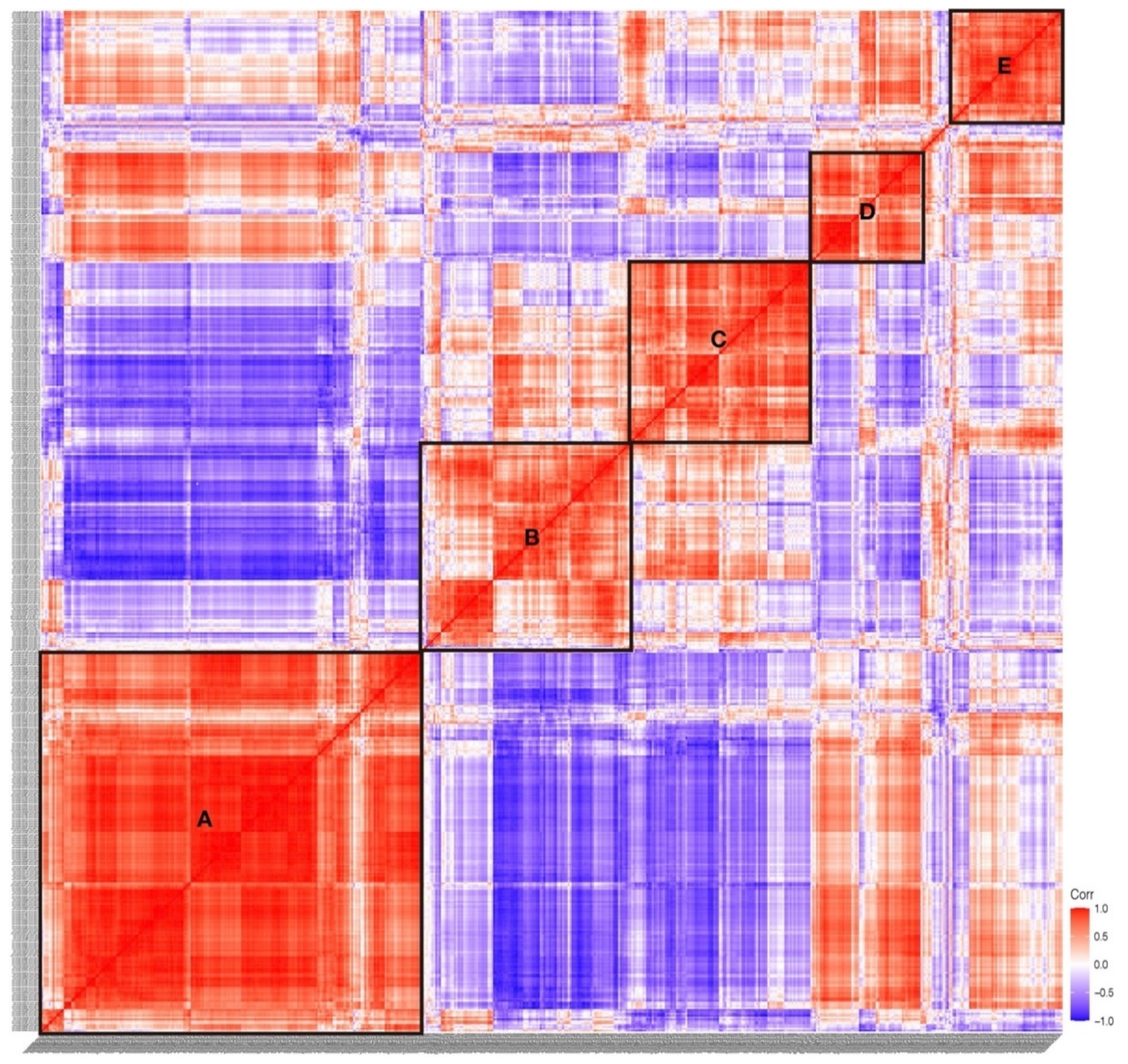
According to the expression correlation heat map of TFs, we found that five boxes of TFs showed significant correlation, which are indicated in box A to E (Figure 3). We found that the TFs in Group I were mainly distributed in box A, box D, and box E, TFs in Group II and Group III were mainly distributed in box C and box B, respectively (Table S2). The expression correlation results showed that the TFs expressed in the embryogenesis and larvae development had a low correlation, indicating the discrepancy of TFs in regulating the process of embryogenesis and larvae development.

2.4. Hox Genes

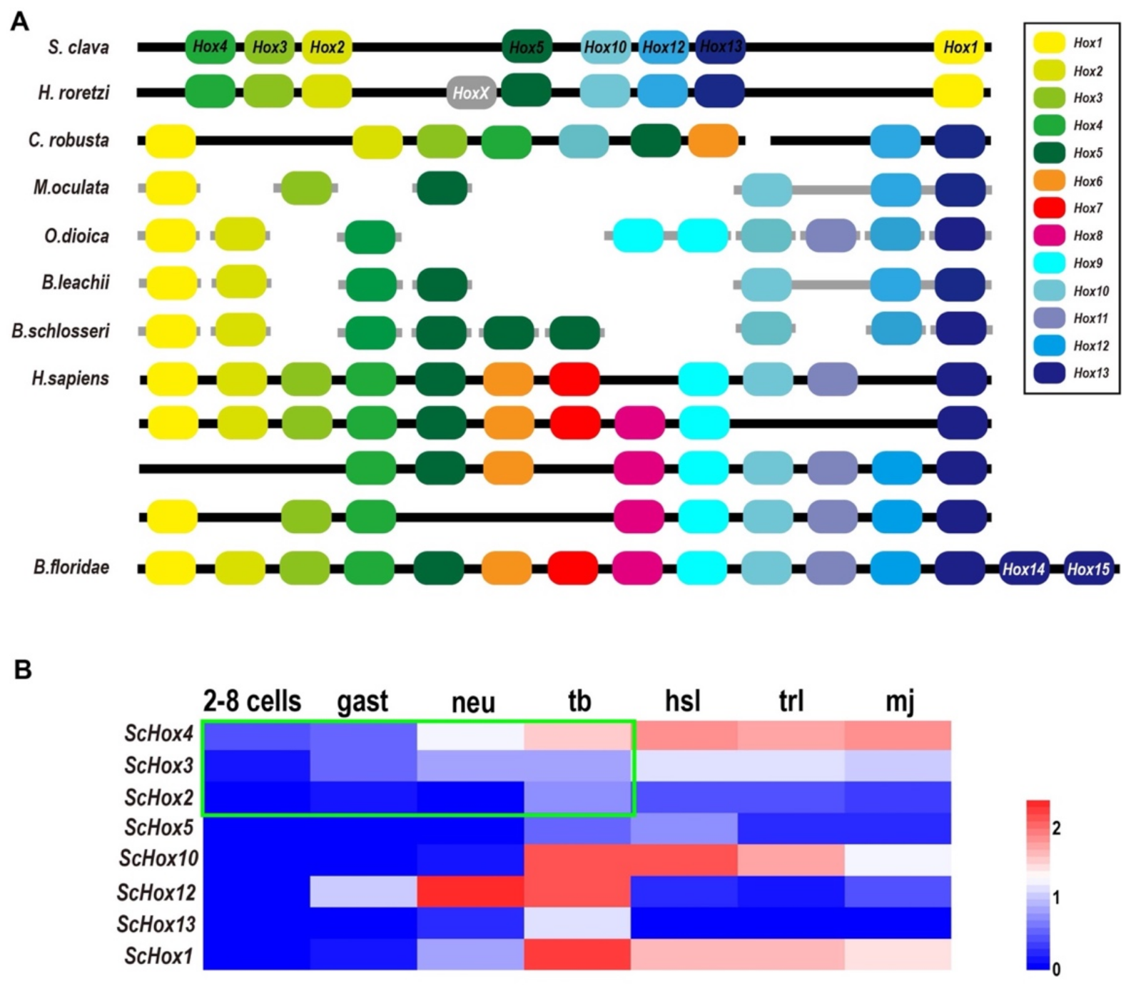
Hox genes are important members of the homeobox TF family and share common development mechanisms in regulating the anteroposterior body axis [8]. In vertebrates, there are 13 paralog groups (PGs) for Hox genes. Through phylogenetic analysis and blast annotation, we identified eight Hox genes in S. clava, including ScHox1, ScHox2, ScHox3, ScHox4, ScHox5, ScHox10, ScHox12, and ScHox13, which were classified into eight PGs (Figure S2). The molecular phylogenetic tree provided a convincing evidence that ScHox1, ScHox2, ScHox3, ScHox10, ScHox12, and ScHox13 belonged to the PG1, PG2, PG3, PG10, PG12, and PG13, respectively (Figure S2). ScHox4 and ScHox5 were identified by sequence alignment. The results showed that they belonged to PG4 (e-value = 2 × 10−57) and PG5 (e-value = 5 × 10−52), respectively. All Hox genes identified in the S. clava genome were distributed on one single chromosome according to the Hi-C result [28]. Hox genes were also distributed on one single chromosome in Halocynthia roretzi with similar arrangement, and on two chromosomes in the C. robusta genome (Figure 4A). In other tunicates mentioned in this study, the Hox genes were distributed on several scaffolds (Figure 4A). In H. sapiens, four Hox clusters existed in the genome and were distributed on four chromosomes (Figure 4A). B. floridae contained all 13 kinds of Hox genes and were distributed on one single chromosome (Figure 4A). In tunicates, there are nine Hox genes in C. robusta, O. dioica, H. roretzi, and B. schlosseri genome [32,33,34,35]. There are six, seven, and eight Hox genes in the M. oculata, B. leachii, and S. clava genome [27,35], respectively (Figure 4A).
The expression profiles of Hox genes in S. clava showed that ScHox4 and ScHox12 were initially expressed at the neu stage, ScHox1, ScHox10, and ScHox13 were initially expressed at the tb stage, ScHox3 was initially expressed at the hsl stage, and expression values of ScHox2 and ScHox5 were low during early development (Figure 4B). Expression of ScHox13 were restricted at the tb stage (Figure 4B). While ScHox12 were not expressed at the hsl stage and the trl stage, after initial expression at the neu stage (Figure 4B). Among the sub-cluster of ScHox2, ScHox3, and ScHox4, ScHox4 was initially expressed at 2–8 cells stage, earlier than the expression of ScHox3 and ScHox2 that were expressed at the gast stage and tb stage (Figure 4B).

2.5. Zinc Finger Family

Zinc finger superfamily contains the greatest number of TFs in the S. clava genome (Table 1). The quantity variation of zinc finger TFs was the main contributor to the different number of TFs between species that we mentioned. There were 11 families in the zinc finger superfamily, including THAP, zf-BED, zf-C2H2, etc. (Table 1). Among these families, the THAP family was expanded in the tunicate genome and the zf-BED family was expanded in the S. clava genome, among the nine species we analyzed (Table 1).
The Zf-C2H2 family was the largest family in the S. clava genome, and also in the other eight species we analyzed (Table 1). According to the domain analysis, there are two kinds of zf-C2H2 proteins in the S. clava genome, including ZBTB and zf-C2H2. The ZBTB proteins were characterized by containing two domains, the BTB domain and zf-C2H2 domain. According to the feature of ZBTB, we identified 12 ZBTB genes in the S. clava genome, more than the other six tunicates analyzed (Figure 5A).
The zf-C2H2 genes were the majority of the zf-C2H2 family. There are 132 zf-C2H2 genes mapped on 16 chromosomes of the S. clava genome (Figure 5B). The Chr 5 processed the greatest number of zf-C2H2 genes (Figure 5B). Those zf-C2H2 genes, which were highly expressed after the neu stage (red rectangles), were located on nine of 16 chromosomes, among which Chr 3 was the most common. The wide distribution of the zf-C2H2 genes indicates the crucial regulatory roles of the zf-C2H2 domain on activating the downstream gene expression in various biological processes.

2.6. Forkhead Box Family

A total of 23 Fox genes were identified in the S. clava genome and they could be grouped into 15 Fox subclasses (Figure 6 and Figure S3). The largest Fox subclass was FoxI, which contained four ScFox genes (Figure 6). The expression profiles of the ScFox genes were varied during early development (Figure 6, Table 2). Five of them were highly expressed before the tb stage, eight were highly expressed during the tb stage, and ten were highly expressed during larvae development (Figure 6). In the S. clava genome, we could not find the homologous genes of FoxB, FoxK, FoxL, and FoxS. The expression profiles showed that the ScFox genes were highly expressed in several stages during early development. This expression pattern was also presented in the Yesso scallop (Patinopecten yessoensis) [36] and sea urchin [37]. Similar patterns presented in early development in these species indicated that ScFox genes are widely involved in regulating the processes of early development of embryogenesis.

3. Discussion

Tunicates, as the closest relatives of vertebrate, show a special rates and patterns of molecular evolution [38]. Identification and analysis of important gene families were also performed, based on the sequenced genomes in several tunicate species [35,39]. In this study, we screened and identified 553 TFs in the S. clava genome and revealed their potential roles in environmental adaptation and early embryonic development, through their expression profiles.
S. clava showed a broad tolerance of environmental conditions [28]. Compared to B. floridae and C. elegans, there are more CSD TFs and immune response-related TFs, (such as IRF genes) in S. clava. CSD is a crucial characteristic of cold shock proteins (CSP), which were upregulated under the low temperature in S. clava [28]. IRF genes play an important role in immune response in C. savignyi [40,41]. The expansion of these TFs might help to improve the fitness to low temperature conditions and improve their ability of immune response to adapt to new environment.
TFs highly expressed during 2–8 cells stage is an important group that we identified in this study. Through gene ontology (GO) enrichment analysis, the biology processes of maternal TFs mainly concentrate in the response/cellular response, signaling, and regulation of biological process (Figure S4). More than half of TFs involved in these processes are annotated as nuclear receptors, which regulate the activation of genes and control many biological processes, such as cell proliferation, cell cycle, and metabolism [42]. For example, thyroid hormone receptor α1 (THRα1) controls the cell proliferation through the Wnt /β-catenin pathway [43]. In C. robusta, the combination of GATA and Ets induced FGF9/16/20 in the neural tissue [44,45,46,47]. TFs highly expressed during 2–8 cells stage in S. clava might act as “responders” and “signal transmitters” to control the expression of the downstream genes and regulate early embryogenesis.
Proportion of the expressed TFs had increased from 2–8 cells stage to the tb stage (Figure S5) and was stable from the hsl stage to the mj stage. In the previous study, the different species showed very similar overall TF expression patterns, with TF expression increasing during the initial stages of development among D. rerio, C. robusta, D. melanogaster, and C. elegans [48]. We compared the TF expression between S. clava and C. robusta and found similar patterns, with TF expression increasing from 2–8 cells stage to tb stage (Figure S4). These results indicate that the most TFs might play a conserved role in early development.
S. clava larvae undergo metamorphosis after hatching. TFs, whose expression are significantly up-regulated after hatching might be involved into the regulation of this process. We classified those genes into two types. One is immune response TFs, which include cyclic AMP-dependent transcription factor (ATF-3), IRF1, IRF4, and B-cell lymphoma 6 (BCL6), which have strong activities in immune response regulation and maturation of the immune system [49,50,51,52]. High expression of these TFs indicates that the immune system and inflammatory reaction are involved in the metamorphosis of S. clava, and these immune responses also appeared in ascidian Boltenia villosa [53]. The others are the thyroid hormone, retinoic acid signaling TFs, and nuclear receptors, including thyroid hormone receptor alpha (THRα), vitamin D 25-hydroxylase (CYP2R1), and retinoic acid receptor alpha (RARα). Thyroid hormone and retinoic acid signaling pathways were demonstrated to be essential in the metamorphosis of S. clava [28,54]. Nuclear receptors are a superfamily of TFs that function in the regulation of various metabolic processes [42,55]. The metamorphosis process is regulated through interaction of hormone and nuclear receptors, which also existed in fishes [56]. Nuclear receptors, involved in the TH and RA signaling pathways, had potential in regulating metamorphosis in S. clava, according to the expression profiles.
Eight ScHox genes were identified in the S. clava genome and they were located in one chromosome. This was consistent with the location of Hox genes in H. roretzi, while Hox genes in genome of C. robusta were located in two chromosomes [27,33,35]. The similar location of Hox genes on the chromosome between S. clava and H. roretzi might be related to their close phylogenetic relationship [28]. Whole-cluster temporal collinearity (WTC) is a typical expression characteristic for Hox genes in vertebrate [57,58]. However, it is not applicable in tunicates. Expression profile of Hox genes in S. clava showed a subcluster temporal collinearity (STC), which was reflected in the ScHox2–4 cluster. STC was also presented in other invertebrate species, such as scallop P. yessoensis [59] and ascidian C. robusta [32]. In C. robusta, the Hox genes were spatially expressed in the central nervous system of larvae and the gut of the juvenile [32]. CrHox10 and CrHox12 play important roles in the neuronal and tail development in C. robusta [60]. These investigations suggest the potential roles of Hox genes in the development of the tail and the nervous system in S. clava.
The expression profiles of Hox1, Hox3, Hox4, Hox10, and Hox12 genes in S. clava were similar to the expression profiles of these genes in C. robusta [32]. The expression of Hox2 and Hox5 were very low in S. clava. The expression profiles of Hox13 were different between S. clava and C. robusta. ScHox13 was expressed in the tb stage, but CrHox13 was detectable only in the juvenile [32]. However, the number and distribution of Hox genes seemed not to be related to their body plan at the larval stage.
Fox genes are characterized by the highly conserved forkhead motif, which is known to be a “winged-helix” DNA-binding domain [61,62]. Fox genes play important roles in embryogenesis and metabolism [62]. A total of 22 Fox genes were identified in the genome of sea urchin [37]. Number of Fox genes in S. clava genome was nearly same as that of the sea urchin. Compared to the sea urchin, FoxE, FoxH, and FoxR subclasses in S. clava were found, and FoxI subclass was expanded, but FoxB, FoxK, and FoxL subclasses were not found. In S. clava, genes in the FoxE and FoxI subclasses were highly expressed after hatching. In vertebrates, FoxE4, FoxI1, and FoxI2 play important roles in len, otic placode, and retina formation and development, respectively [63,64,65]. ScFoxE and ScFoxI might have similar functions in the regulation of neuro and sensory organ formation.
In conclusion, we identified 553 TFs belonging to 60 TF families in the S. clava genome. The expression profiles provided a possible clue for the functions of different TF genes in embryogenesis, environmental adaptation, and metamorphosis in S. clava (Figure 7). Our study provides gene resources and a new perspective to understand the evolution and function of TFs in the leathery sea squirt.

4. Materials and Methods

4.1. Animals and Embryos

The adults of S. clava were collected from Weihai City, China, and cultured in seawater at 18 °C in the laboratory. The eggs and sperm were collected separately from different individuals for fertilization at room temperature. The embryos and larvae were collected for RNA extraction at different stages. The study was approved by the Ocean University of China Institutional Animal Care and Use Committee (OUC-IACUC) prior to the initiation of the study (Approval number: “2021-0032-0012”, 15 April 2019). All experiments and relevant methods were carried out in accordance with the approved guidelines and regulations of OUC-IACUC.

4.2. Transcriptome Sequencing and WGCNA Analysis

The transcriptome sequencing and data were described in a previous study [28]. The co-expression gene network for 21 transcriptomic datasets was constructed using the R package WGCNA, with the parameters of softPower = 12, minimum module size = 300, cutting height = 0.99, and deepSplit = F [31]. The genes that were expressed in at least one developmental stage were used for network construction.

4.3. Identification and Classification of TFs

The TFs were identified and classified, based on the conserved DBDs. After obtaining all protein sequences at the genome level, we removed random sequences through CD-HIT (v4.6.8) [66] and analyzed the protein domain according to the Hidden Markov Model (HMM) profiles from the Pfam database (version 31.0) [67], applying the hmmscan program in the HMMER package(v3.1b2) [68]. 85 Pfam ID of DBDs from REGULATOR database (http://www.bioinformatics.org/regulator/page.php?act=family, accessed on 8 December 2020) and the Animal TFDB database (http://bioinfo.life.hust.edu.cn/AnimalTFDB/#!/species, accessed on 8 December 2020) were used for analysis [9,10]. The Pfam IDs were showed in Table S3. Most genes containing DBDs were screened with the e-value threshold of 10−4 and were regarded as TFs. Some of thresholds of DBD were chosen according to the threshold in Animal TFDB 3.0 [10], including the threshold of the bHLH domain with 10−2, thresholds of the HMG, Homeodomian, zf-BED, zf-C2H2 domains with 10−3, and the thresholds of the zf-CCCH domains with 10−20. The TFs were classified into different families, according to the DBDs.

4.4. Heat Map, Phylogenetic Analysis, Domain Analysis, and Expression Correlation Analysis

We normalized the FPKM values through log10(FPKM + 1) and imported the normalized values into the Heml software (v1.0) [69]. We performed molecular phylogenetic tree analysis using the MEGA software (v7.0) [70] through the Maximum-Likelihood (ML) method and beautified the figure through iTOL (https://itol.embl.de/, accessed on 13 December 2020) online [71]. The multiple sequence alignments were conducted by Clustal W [72]. The gene expression correlation Heat map were constructed by the corrplot package and the ggcorrplot package on R studio. The correlation coefficient indicated the correlation between two genes. The correlation coefficient was greater than 0 for positive correlation (shown in red), and less than 0 for negative correlation (shown in blue). (https://github.com/taiyun/corrplot, accessed on 21 December 2020).

4.5. Quantitative Real-Time PCR

Total RNA was extracted through the TRIZOL method. Reverse transcription experiments were performed according to HiScript II Q RT SuperMix for QPCR (+gDNA wiper) (Vazyme R223-01, Nanjing, China). 18S rRNA was chosen as an internal standard. The primers used in experiments are shown in Table S4. Quantitative real-time PCR was carried out by the ChamQ SYBR Color QPCR Master Mix (Vazyme Q411-01, Nanjing, China). The relative gene expression levels were calculated using the comparative Ct method with the formula 2−∆∆Ct [73].

4.6. Gene Ontology (GO) Enrichment Analysis

The GO enrichment analysis was performed by the OmicShare tools online (https://www.omicshare.com, accessed on 23 September 2020). The target genes were maternal expressed TF genes, the background genes were all TF genes. We obtained the top 20 of GO enrichment results for further analysis and made the bubble chart.

4.7. Data Availability

The genome sequences of S. clava were deposited in NCBI, under the BioProject number PRJNA523448. The transcriptome data of S. clava used in this study were also deposited in the NCBI SRA database, with the accession numbers SRR8599814 to SRR8599834.
The genome resources of C. robusta, H. sapiens, and C. elegans were downloaded from the Ensembl (https://asia.ensembl.org/index.html, version101, accessed on 1 July 2020) [74]. The genome resources of O. dioica and B. floridae were acquired from NCBI (https://www.ncbi.nlm.nih.gov/, accessed on 1 July 2020), and the genome resources of M. oculata, B. leachii, and B. schlosseri were acquired from ANISEED (https://www.aniseed.cnrs.fr/, accessed on 1 July 2020) [75].

Supplementary Materials

The following are available online at https://www.mdpi.com/article/10.3390/ijms22094317/s1. Figure S1: Expressions of partial TFs detected by RNA-Seq and quantitative real-time PCR (RT-qPCR). Figure S2: Phylogenetic tree of hox genes. Figure S3: Phylogenetic tree of fox genes. Figure S4: Function annotation of maternal TFs. Figure S5. TF expression through early development of S. clava. Table S1: The families represented by the number in Figure 1. Table S2: Expression profile information of TFs in the S. clava genome. Table S3: Pfam IDs of each TF domains. Table S4: Primer sequence used in RT-qPCR.

Author Contributions

B.D. and J.W. conceived the project. J.Z., J.W. and H.Y. analyzed the data and performed the experiments. J.Z. and J.W. prepared the figures and wrote the manuscript. B.D. and H.Y. revised the manuscript. All authors have read and agreed to the published version of the manuscript.

Funding

This research was funded by the National Key Research and Development Program of China (2019YFE0190900; 2018YFD0900705), the National Natural Science Foundation of China (31771649).

Institutional Review Board Statement

The study was approved by the Ocean University of China Institutional Animal Care and Use Committee (OUC-IACUC) prior to the initiation of the study (Approval number: “2021-0032-0012”, 15 April 2019). All experiments and relevant methods were carried out in accordance with the approved guidelines and regulations of OUC-IACUC.

Informed Consent Statement

Not applicable.

Data Availability Statement

The genome sequences of S. clava were deposited in NCBI under the BioProject number PRJNA523448. The transcriptome data of S. clava used for biological analysis were also deposited in the NCBI SRA database, with the accession numbers SRR8599814 to SRR8599834. The genome resources of C. robusta, H. sapiens, and C. elegans were downloaded from Ensembl (https://asia.ensembl.org/index.html), the genome resources of O. dioica and B. floridae were acquired from NCBI (https://www.ncbi.nlm.nih.gov/, accessed on 1 July 2020), and the genome resources of M. oculata, B. leachii, and B. schlosseri were acquired from ANISEED (https://www.aniseed.cnrs.fr/, accessed on 1 July 2020).

Acknowledgments

We thank the members of the Dong laboratory, including Xiuxia Yang and Xiaoming Zhang for their critical reading and invaluable comments on the manuscript. We appreciate the computing resources provided on IEMB-1, a high-performance computing cluster operated by the Institute of Evolution and Marine Biodiversity.

Conflicts of Interest

The authors declare no conflict of interest.

References

  1. Lambert, S.A.; Jolma, A.; Campitelli, L.F.; Das, P.K.; Yin, Y.; Albu, M.; Chen, X.; Taipale, J.; Hughes, T.R.; Weirauch, M.T. The Human Transcription Factors. Cell 2018, 172, 650–665. [Google Scholar] [CrossRef] [PubMed]
  2. Mitchell, P.J.; Tjian, R. Transcriptional regulation in mammalian cells by sequence-specific DNA binding proteins. Science 1989, 245, 371–378. [Google Scholar] [CrossRef] [PubMed]
  3. Bejerano, G.; Pheasant, M.; Makunin, I.; Stephen, S.; Kent, W.J.; Mattick, J.S.; Haussler, D. Ultraconserved elements in the human genome. Science 2004, 304, 1321–1325. [Google Scholar] [CrossRef] [PubMed]
  4. Carroll, S.B. Evo-devo and an expanding evolutionary synthesis: A genetic theory of morphological evolution. Cell 2008, 134, 25–36. [Google Scholar] [CrossRef]
  5. Lee, T.I.; Young, R.A. Transcriptional regulation and its misregulation in disease. Cell 2013, 152, 1237–1251. [Google Scholar] [CrossRef]
  6. Arnold, H.-H.; Braun, T. The role of Myf-5 in somitogenesis and the development of skeletal muscles in vertebrates. J. Cell Sci. 1993, 104, 957–960. [Google Scholar]
  7. Peter, J. Nuclear Factor-kB—A pivotal transcription factor in chronic inflammatory diseases. Mech. Dis. 1997, 336, 1066–1071. [Google Scholar]
  8. Mallo, M. Reassessing the Role of Hox Genes during Vertebrate Development and Evolution. Trends Genet. 2018, 34, 209–217. [Google Scholar] [CrossRef]
  9. Wang, K.; Nishida, H. REGULATOR: A database of metazoan transcription factors and maternal factors for developmental studies. BMC Bioinform. 2015, 16, 114. [Google Scholar] [CrossRef]
  10. Hu, H.; Miao, Y.R.; Jia, L.H.; Yu, Q.Y.; Zhang, Q.; Guo, A.Y. AnimalTFDB 3.0: A comprehensive resource for annotation and prediction of animal transcription factors. Nucleic Acids Res. 2019, 47, D33–D38. [Google Scholar] [CrossRef]
  11. Chiba, S.; Awazu, S.; Itoh, M.; Chin-Bow, S.T.; Satoh, N.; Satou, Y.; Hastings, K.E. A genomewide survey of developmentally relevant genes in Ciona intestinalis. IX. Genes for muscle structural proteins. Dev. Genes Evol. 2003, 213, 291–302. [Google Scholar] [CrossRef]
  12. Wada, S.; Tokuoka, M.; Shoguchi, E.; Kobayashi, K.; Di Gregorio, A.; Spagnuolo, A.; Branno, M.; Kohara, Y.; Rokhsar, D.; Levine, M.; et al. A genomewide survey of developmentally relevant genes in Ciona intestinalis. Dev. Genes Evol. 2003, 213, 222–234. [Google Scholar] [CrossRef]
  13. Yagi, K.; Satou, Y.; Mazet, F.; Shimeld, S.M.; Degnan, B.; Rokhsar, D.; Levine, M.; Kohara, Y.; Satoh, N. A genomewide survey of developmentally relevant genes in Ciona intestinalis. III. Genes for Fox, ETS, nuclear receptors and NFkappaB. Dev. Genes Evol. 2003, 213, 235–244. [Google Scholar] [CrossRef]
  14. Yamada, L.; Kobayashi, K.; Degnan, B.; Satoh, N.; Satou, Y. A genomewide survey of developmentally relevant genes in Ciona intestinalis. IV. Genes for HMG transcriptional regulators, bZip and GATA/Gli/Zic/Snail. Dev. Genes Evol. 2003, 213, 245–253. [Google Scholar] [CrossRef]
  15. Imai, K.S.; Hino, K.; Yagi, K.; Satoh, N.; Satou, Y. Gene expression profiles of transcription factors and signaling molecules in the ascidian embryo: Towards a comprehensive understanding of gene networks. Development 2004, 131, 4047–4058. [Google Scholar] [CrossRef]
  16. Miwata, K.; Chiba, T.; Horii, R.; Yamada, L.; Kubo, A.; Miyamura, D.; Satoh, N.; Satou, Y. Systematic analysis of embryonic expression profiles of zinc finger genes in Ciona intestinalis. Dev. Biol. 2006, 292, 546–554. [Google Scholar] [CrossRef]
  17. Katikala, L.; Aihara, H.; Passamaneck, Y.J.; Gazdoiu, S.; Jose-Edwards, D.S.; Kugler, J.E.; Oda-Ishii, I.; Imai, J.H.; Nibu, Y.; Di Gregorio, A. Functional Brachyury binding sites establish a temporal read-out of gene expression in the Ciona notochord. PLoS Biol. 2013, 11, e1001697. [Google Scholar] [CrossRef]
  18. Di Gregorio, A. The notochord gene regulatory network in chordate evolution: Conservation and divergence from Ciona to vertebrates. Curr. Top. Dev. Biol. 2020, 139, 325–374. [Google Scholar] [CrossRef]
  19. Yasuo, H.; Satoh, N. An ascidian homolog of the mouse Brachyury (T) gene is expressed exclusively in notochord cells at the fate restricted stage. Dev. Growth Differ. 1994, 36, 9–18. [Google Scholar] [CrossRef]
  20. Corbo, J.C.; Levine, M.; Zeller, R.W. Characterization of a notochord-specific enhancer from the Brachyury promoter region of the ascidian, Ciona intestinalis. Development 1997, 124, 589–602. [Google Scholar]
  21. Jose-Edwards, D.S.; Kerner, P.; Kugler, J.E.; Deng, W.; Jiang, D.; Di Gregorio, A. The identification of transcription factors expressed in the notochord of Ciona intestinalis adds new potential players to the brachyury gene regulatory network. Dev. Dyn. 2011, 240, 1793–1805. [Google Scholar] [CrossRef] [PubMed]
  22. Jose-Edwards, D.S.; Oda-Ishii, I.; Nibu, Y.; Di Gregorio, A. Tbx2/3 is an essential mediator within the Brachyury gene network during Ciona notochord development. Development 2013, 140, 2422–2433. [Google Scholar] [CrossRef] [PubMed]
  23. Imai, K.S.; Satoh, N.; Satou, Y. An essential role of a FoxD gene in notochord induction in Ciona embryos. Development 2002, 129, 3441–3453. [Google Scholar] [PubMed]
  24. Yagi, K.; Satou, Y.; Satoh, N. A zinc finger transcription factor, ZicL, is a direct activator of Brachyury in the notochord specification of Ciona intestinalis. Development 2004, 131, 1279–1288. [Google Scholar] [CrossRef]
  25. Imai, K.S.; Satou, Y.; Satoh, N. Multiple functions of a Zic-like gene in the differentiation of notochord, central nervous system and muscle in Ciona savignyi embryos. Development 2002, 129, 2723–2732. [Google Scholar]
  26. Lemons, D.; McGinnis, W. Genomic Evolution of Hox Gene Clusters. Science 2006, 313, 1918–1922. [Google Scholar] [CrossRef]
  27. DeBiasse, M.B.; Colgan, W.N.; Harris, L.; Davidson, B.; Ryan, J.F. Inferring Tunicate Relationships and the Evolution of the Tunicate Hox Cluster with the Genome of Corella inflata. Genome Biol. Evol. 2020, 12, 948–964. [Google Scholar] [CrossRef]
  28. Wei, J.; Zhang, J.; Lu, Q.; Ren, P.; Guo, X.; Wang, J.; Li, X.; Chang, Y.; Duan, S.; Wang, S.; et al. Genomic basis of environmental adaptation in the leathery sea squirt (Styela clava). Mol. Ecol. Resour. 2020. [Google Scholar] [CrossRef]
  29. Goldstien, S.J.; Schiel, D.R.; Gemmell, N.J. Regional connectivity and coastal expansion: Differentiating pre-border and post-border vectors for the invasive tunicate Styela clava. Mol. Ecol. 2010, 19, 874–885. [Google Scholar] [CrossRef]
  30. Goldstien, S.J.; Dupont, L.; Viard, F.; Hallas, P.J.; Nishikawa, T.; Schiel, D.R.; Gemmell, N.J.; Bishop, J.D. Global phylogeography of the widely introduced North West Pacific ascidian Styela clava. PLoS ONE 2011, 6, e16755. [Google Scholar] [CrossRef]
  31. Langfelder, P.; Horvath, S. WGCNA: An R package for weighted correlation network analysis. BMC Bioinform. 2008, 9, 559. [Google Scholar] [CrossRef]
  32. Ikuta, T.; Yoshida, N.; Satoh, N.; Saiga, H. Ciona intestinalis Hox gene cluster: Its dispersed structure and residual colinear expression in development. Proc. Natl. Acad. Sci. USA 2004, 101, 15118–15123. [Google Scholar] [CrossRef] [PubMed]
  33. Seo, H.-C.; Edvardsen, R.B.; Maeland, A.D. Hox cluster disintegration with persistent anteroposterior order of expression in Oikopleura dioica. Nature 2004, 431, 67–71. [Google Scholar] [CrossRef]
  34. Sekigami, Y.; Kobayashi, T.; Omi, A.; Nishitsuji, K.; Ikuta, T.; Fujiyama, A.; Satoh, N.; Saiga, H. Hox gene cluster of the ascidian, Halocynthia roretzi, reveals multiple ancient steps of cluster disintegration during ascidian evolution. Zoological. Lett. 2017, 3, 17. [Google Scholar] [CrossRef]
  35. Blanchoud, S.; Rutherford, K.; Zondag, L.; Gemmell, N.J.; Wilson, M.J. De novo draft assembly of the Botrylloides leachii genome provides further insight into tunicate evolution. Sci. Rep. 2018, 8, 5518. [Google Scholar] [CrossRef]
  36. Wu, S.; Zhang, Y.; Li, Y.; Wei, H.; Guo, Z.; Wang, S.; Zhang, L.; Bao, Z. Identification and expression profiles of Fox transcription factors in the Yesso scallop (Patinopecten yessoensis). Gene 2020, 733, 144387. [Google Scholar] [CrossRef]
  37. Tu, Q.; Brown, C.T.; Davidson, E.H.; Oliveri, P. Sea urchin Forkhead gene family: Phylogeny and embryonic expression. Dev. Biol. 2006, 300, 49–62. [Google Scholar] [CrossRef]
  38. Berna, L.; Alvarez-Valin, F. Evolutionary Genomics of Fast Evolving Tunicates. Genome Biol. Evol. 2014, 6, 1724–1738. [Google Scholar] [CrossRef]
  39. Dehal, P.; Satou, Y.; Campbell, R.K.; Chapman, J.; Degnan, B.; De Tomaso, A.; Davidson, B.; Di Gregorio, A.; Gelpke, M.; Goodstein, D.M.; et al. The draft genome of Ciona intestinalis: Insights into chordate and vertebrate origins. Science 2002, 298, 2157–2167. [Google Scholar] [CrossRef]
  40. Ikushima, H.; Negishi, H.; Taniguchi, T. The IRF family transcription factors at the interface of innate and adaptive immune responses. Cold Spring Harb. Symp. Quant. Biol. 2013, 78, 105–116. [Google Scholar] [CrossRef] [PubMed]
  41. Zhang, Z.; Wei, J.; Ren, R.; Zhang, X. Anti-virus effects of interferon regulatory factors (IRFs) identified in ascidian Ciona savignyi. Fish Shellfish. Immunol. 2020, 106, 273–282. [Google Scholar] [CrossRef] [PubMed]
  42. Sever, R.; Glass, C.K. Signaling by Nuclear Receptors. Cold Spring Harb. Perspect. Biol. 2013, 5, a016709. [Google Scholar] [CrossRef] [PubMed]
  43. Plateroti, M.; Kress, E.; Mori, J.I.; Samarut, J. Thyroid Hormone Receptor α1 Directly Controls Transcription of the β-Catenin Gene in Intestinal Epithelial Cells. Mol. Cell. Biol. 2006, 26, 3204–3214. [Google Scholar] [CrossRef]
  44. Vincent Bertrand, C.H.; Caillol, D.; Popovici, C.; Lemaire, P. Neural Tissue in Ascidian Embryos Is Induced by FGF9/16/20, Acting via a Combination of Maternal GATA and Ets Transcription Factors. Cell 2003, 115, 615–627. [Google Scholar] [CrossRef]
  45. Miya, T.; Nishida, H. An Ets transcription factor, HrEts, is target of FGF signaling and involved in induction of notochord, mesenchyme, and brain in ascidian embryos. Dev. Biol. 2003, 261, 25–38. [Google Scholar] [CrossRef]
  46. Hotta, K. Brachyury-downstream gene sets in a chordate, Ciona intestinalis: Integrating notochord specification, morphogenesis and chordate evolution. Evol. Dev. 2008, 10, 37–51. [Google Scholar] [CrossRef]
  47. Gainous, T.B.; Wagner, E.; Levine, M. Diverse ETS transcription factors mediate FGF signaling in the Ciona anterior neural plate. Dev. Biol. 2015, 399, 218–225. [Google Scholar] [CrossRef]
  48. Schep, A.N.; Adryan, B. A comparative analysis of transcription factor expression during metazoan embryonic development. PLoS ONE 2013, 8, e66826. [Google Scholar] [CrossRef]
  49. Nurieva, R.I.; Chung, Y.; Martinez, G.J.; Yang, X.O.; Tanaka, S.; Matskevitch, T.D.; Wang, Y.H.; Dong, C. Bcl6 mediates the development of T follicular helper cells. Science 2009, 325, 1001–1005. [Google Scholar] [CrossRef]
  50. Wang, H.; Mo, P.; Ren, S.; Yan, C. Activating transcription factor 3 activates p53 by preventing E6-associated protein from binding to E6. J. Biol. Chem. 2010, 285, 13201–13210. [Google Scholar] [CrossRef]
  51. Dou, L.; Liang, H.F.; Geller, D.A.; Chen, Y.F.; Chen, X.P. The regulation role of interferon regulatory factor-1 gene and clinical relevance. Hum. Immunol. 2014, 75, 1110–1114. [Google Scholar] [CrossRef]
  52. Nam, S.; Lim, J.S. Essential role of interferon regulatory factor 4 (IRF4) in immune cell development. Arch. Pharm. Res. 2016, 39, 1548–1555. [Google Scholar] [CrossRef]
  53. Davidson, B.; Swalla, B.J. A molecular analysis of ascidian metamorphosis reveals activation of an innate immune response. Development 2002, 129, 4739–4751. [Google Scholar]
  54. Ross, A.C.; Zolfaghari, R. Cytochrome P450s in the regulation of cellular retinoic acid metabolism. Annu. Rev. Nutr. 2011, 31, 65–87. [Google Scholar] [CrossRef]
  55. Nikolenko, J.V.; Krasnov, A.N. Nuclear receptors: Structure and mechanisms of action. Russ. J. Genet. 2007, 43, 234–240. [Google Scholar] [CrossRef]
  56. Shao, C.; Bao, B.; Xie, Z.; Chen, X.; Li, B.; Jia, X.; Yao, Q.; Ortí, G.; Li, W.; Li, X.; et al. The genome and transcriptome of Japanese flounder provide insights into flatfish asymmetry. Nat. Genet. 2016, 49, 119–124. [Google Scholar] [CrossRef]
  57. Izpisu’a-Belmonte, J.-C.; Falkenstein, H.; Dollé, P.; Duboule, A.R.D. Murine genes related to the Drosophila AbdB homeotic genes are sequentially expressed during development of the posterior part of the body. EMBO J. 1991, 10, 2279–2289. [Google Scholar] [CrossRef]
  58. Pascual-Anaya, J.; Sato, I.; Sugahara, F.; Higuchi, S.; Paps, J.; Ren, Y.; Takagi, W.; Ruiz-Villalba, A.; Ota, K.G.; Wang, W.; et al. Hagfish and lamprey Hox genes reveal conservation of temporal colinearity in vertebrates. Nat. Ecol. Evol. 2018, 2, 859–866. [Google Scholar] [CrossRef]
  59. Wang, S.; Zhang, J.; Jiao, W.; Li, J.; Xun, X.; Sun, Y.; Guo, X.; Huan, P.; Dong, B.; Zhang, L.; et al. Scallop genome provides insights into evolution of bilaterian karyotype and development. Nat. Ecol. Evol. 2017, 1, 120. [Google Scholar] [CrossRef]
  60. Ikuta, T.; Satoh, N.; Saiga, H. Limited functions of Hox genes in the larval development of the ascidian Ciona intestinalis. Development 2010, 137, 1505–1513. [Google Scholar] [CrossRef]
  61. Weigel, D.; Jürgens, G.; Küttner, F.; Seifert, E.; Jäckle, H. The homeotic gene fork head encodes a nuclear protein and is expressed in the terminal regions of the Drosophila embryo. Cell 1989, 57, 645–658. [Google Scholar] [CrossRef]
  62. Carlsson, P.; Mahlapuu, M. Forkhead transcription factors: Key players in development and metabolism. Dev. Biol. 2002, 250, 1–23. [Google Scholar] [CrossRef] [PubMed]
  63. Solomon, K.S.; Kudoh, T.; Dawid, I.B.; Fritz, A. Zebrafish foxi1 mediates otic placode formation and jaw development. Development 2003, 130, 929–940. [Google Scholar] [CrossRef] [PubMed]
  64. Solomon, K.S.; Logsdon, J.M., Jr.; Fritz, A. Expression and phylogenetic analyses of three zebrafish FoxI class genes. Dev. Dyn. 2003, 228, 301–307. [Google Scholar] [CrossRef]
  65. Zilinski, C.; Brownell, I.; Hashimoto, R.; Medina-Martinez, O.; Swindell, E.C.; Jamrich, M. Expression of FoxE4 and Rx visualizes the timing and dynamics of critical processes taking place during initial stages of vertebrate eye development. Dev. Neurosci. 2004, 26, 294–307. [Google Scholar] [CrossRef]
  66. Huang, Y.; Niu, B.; Gao, Y.; Fu, L.; Li, W. CD-HIT Suite: A web server for clustering and comparing biological sequences. Bioinformatics 2010, 26, 680–682. [Google Scholar] [CrossRef]
  67. Finn, R.D.; Coggill, P.; Eberhardt, R.Y.; Eddy, S.R.; Mistry, J.; Mitchell, A.L.; Potter, S.C.; Punta, M.; Qureshi, M.; Sangrador-Vegas, A.; et al. The Pfam protein families database: Towards a more sustainable future. Nucleic Acids. Res. 2016, 44, D279–D285. [Google Scholar] [CrossRef]
  68. Potter, S.C.; Luciani, A.; Eddy, S.R.; Park, Y.; Lopez, R.; Finn, R.D. HMMER web server: 2018 update. Nucleic Acids. Res. 2018, 46, W200–W204. [Google Scholar] [CrossRef]
  69. Deng, W.; Wang, Y.; Liu, Z.; Cheng, H.; Xue, Y. HemI: A toolkit for illustrating heatmaps. PLoS ONE 2014, 9, e111988. [Google Scholar] [CrossRef]
  70. Kumar, S.; Stecher, G.; Tamura, K. MEGA7: Molecular Evolutionary Genetics Analysis Version 7.0 for Bigger Datasets. Mol. Biol. Evol. 2016, 33, 1870–1874. [Google Scholar] [CrossRef]
  71. Letunic, I.; Bork, P. Interactive Tree Of Life (iTOL) v4: Recent updates and new developments. Nucleic Acids Res. 2019, 47, W256–W259. [Google Scholar] [CrossRef]
  72. Thompson, J.D.; Higgins, D.G.; Gibson, T.J. CLUSTAL W: Improving the sensitivity of progressive multiple sequence alignment through sequence weighting, position-specific gap penalties and weight matrix choice. Nucleic Acids Res. 1994, 22, 4673–4680. [Google Scholar] [CrossRef]
  73. Livak, K.J.; Schmittgen, T.D. Analysis of relative gene expression data using real-time quantitative PCR and the 2(-Delta Delta C(T)) Method. Methods 2001, 25, 402–408. [Google Scholar] [CrossRef]
  74. Howe, K.L.; Achuthan, P.; Allen, J.; Allen, J.; Alvarez-Jarreta, J.; Amode, M.R.; Armean, I.M.; Azov, A.G.; Bennett, R.; Bhai, J.; et al. Ensembl 2021. Nucleic Acids Res. 2021, 49, D884–D891. [Google Scholar] [CrossRef]
  75. Dardaillon, J.; Dauga, D.; Simion, P.; Faure, E.; Onuma, T.A.; DeBiasse, M.B.; Louis, A.; Nitta, K.R.; Naville, M.; Besnardeau, L.; et al. ANISEED 2019: 4D exploration of genetic data for an extended range of tunicates. Nucleic Acids Res. 2020, 48, D668–D675. [Google Scholar] [CrossRef]
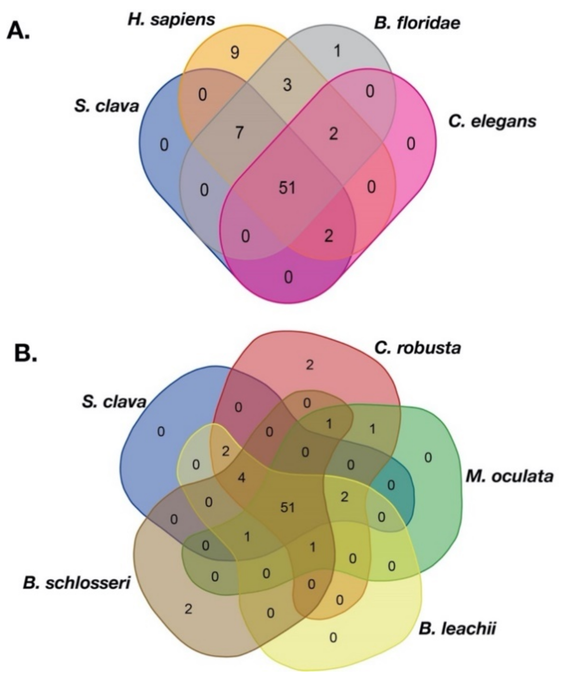
Figure 1. Veen of the TF families among different species. (A) TF families among S. clava (blue), H. sapiens (dark yellow), B. floridae (gary), and C. elegans (pink). (B) TF families among S. clava (blue), C. robusta (red), M. oculata (green), B. leachii (yellow), and B. schlosseri (brown). The transcription factor families contained in each part are listed in Table S1.
Ijms 22 04317 g001
Figure 2. Expression patterns of TFs in S. clava genome—547 TFs in S. clava genome are shown in the heat map. They are classified into nine modules, including turquoise, blue, magenta, pink, brown, yellow, red, green, and black module through the WGCNA analysis. The turquoise, blue, magenta, and pink modules are classified into Group I, in which the TFs were highly expressed during 2–8 cells stage to the neural stage; the brown module is classified into Group II, in which TFs were highly expressed during the tailbud stage; the yellow, red, green, and black modules are classified into Group III, in which TFs were highly expressed from the hatched swimming larvae stage to the metamorphosis juvenile stage. The scale bar indicates the centered FPKM values. The abbreviation of different developmental stages are as follows—2-cell–8-cell embryos (2–8 cells), gastrula embryos (gast), neurula embryos (neu), tailbud-stage embryos (tb), hatched swimming larvae (hsl), tail-regressed larvae (trl), and metamorphic juveniles (mj).
Ijms 22 04317 g002
Figure 3. Expression correlation of TFs in S. clava. The correlation score showed between −1.0 (deep blue) to 1.0 (deep red). The red color indicated that these two TFs showed positive expression correlation, and the blue color showed negative expression correlation. Five significant correlation groups are indicated by box A to box E.
Ijms 22 04317 g003
Figure 4. Comparison of Hox genes in different species and the expression patterns of ScHox genes. (A) Schema depicting Hox gene linkages retained in the genomes of seven tunicates, H. sapiens and B. floridae. Orthologous Hox genes are indicated by the same color of rounded rectangles and the legend is showed on the right. HoxX gene was an unclassified Hox gene in the H. roretzi genome. The spatial distribution of Hox genes in different species on the chromosome or scaffold are indicated by thick black line or thick gray line, respectively. (B) Heat map of the ScHox genes. Gene names are shown on the left. The scale bar indicates the FPKM values, which are dealt with log10(FPKM + 1) but not centered. The green box shows a subcluster-level temporal co-linearity (STC) expression pattern among the ScHox2, ScHox3, and ScHox4 cluster. Shorthand of different developmental stage, which is showed above the heat map, is mentioned above.
Ijms 22 04317 g004
Figure 5. Classification of zf-C2H2 genes. (A) Statistics of zf-C2H2 genes and ZBTB genes in zf-C2H2 family in S. clava, C. robusta, M. oculata, O. dioica, B. leachii, and B. schlosseri. The blue volume indicates the number of zf-C2H2 genes, and the orange volume indicates the number of ZBTB genes. (B) The distribution of zf-C2H2 genes on chromosome. The rectangles indicated the zf-C2H2 genes and the colors indicate the expression levels of each zf-C2H2 genes, according to the WGCNA analysis (mentioned in Figure 2, the black and red rectangles indicate the high expression of the zf-C2H2 genes during 2 cells to neu stages, and during the tbl to mj stages, respectively). The chromosomes are labeled on the left. The black horizontal line indicates scaffold.
Ijms 22 04317 g005
Figure 6. Identification and expression of ScFox genes in S. clava. The Gene IDs, subclasses, Gene names of 23 scfox genes are listed (left side). The expression profiles for each gene through different stages (2–8 cells, gast, neu, tb, hsl, trl, and mj stage) are shown on the right side. The scale bar indicates the centered FPKM values.
Ijms 22 04317 g006
Figure 7. Summary of the TF expression and their potential roles in the embryogenesis and larval development of S. clava. Expression profiles indicates that TFs play essential roles during embryogenesis and larval development. We showed different stages of embryos at the bottom of the figure, including 2–8 cells stage, gast stage, neu stage, mid-tailbud stage, late-tailbud stage, hsl stage, trl stage, and mj stage. We summarized the predicted function of TFs genes into three parts and showed it in different colors, including embryogenesis (green), environmental adaptation (yellow), and metamorphosis (red). The word cloud pictures show the family of the expressed TF genes at different stages. The font size indicates the number of expressed TF genes in the TF families.
Ijms 22 04317 g007
Table 1. Number of TFs and TF families in S. clava, C. robusta, M. oculata, O. dioica, B. leachii, B. schlosseri, H. sapiens, B. floridae, and C. elegans.
TF SuperfamilyTF FamilyS. clavaC. robustaM. oculataO. dioicaB. leachiiB. schlosseriH. sapiensB. floridaeC. elegans
AF-4111111810
ARID4574421545
BTD222223621
bZIPbZIP_113161221157371411
bZIP_267786212817
CBF_beta111111111
NF-YCBFB_NFYA110110112
CBFD_NFYB_HMF898138445169
CG-1011000211
CP2232223821
CSD9547441725
CSRNP_N110212311
MH1CTF_NFI210010410
MH145711451147
E2F_TDP2443322045
ETSEts121512111412291210
ETS_PEA3_N000000410
Forkhead box232524262727552816
GCFC222122312
GCM000000210
GTF2I0100001500
bHLHbHLH4139362237201027338
Myc_N011001410
SIM_C000000200
HMG_box2020141918211022916
HomeoboxHomeodomain73767470744526410885
CUT213332837
PBC1111151112
Pou2335321763
HPD221111301
HSF_DNA-binding110412951
HTH_psq623090251
IRFIRF556285340
IRF-3132049940
LAG1-DNAbinding000001000
LRRFIP013111511
MBD324032732
Myb_DNA-binding1213117131125167
NCU-G1101012110
NDT80_PhoG113012212
Nrf1_DNA-binding111011130
P53321135321
PAX765852959
PC4110111321
RFX_DNA_binding2331321051
RHD_DNA_binding3232451320
Runt111112311
SAND321212744
SRF-TF322321532
STATSTAT_alpha000102200
STAT_binding222222512
STAT_int010000020
TAFH211111631
T-box10888813191019
TEA111211411
TF_AP-2121112514
TF_Otx000000100
TIG117612162918154
TSC22211021415
Tub111211522
Vert_HS_TF000000300
zinc finger THAP2182704049973
GATA54455217712
DM5202737811
Nuclear ReceptorHormone_receptor6667592810151
Androgen_receptor000000100
Oest_receptor000000100
Prog_receptor000000100
GCR000000100
zf-C41512143311213221124
zf-BED1726089309
zf-C2H2154951937211031074874258
zf-C2HC532245821
zf-LITAF-like41253241114
zf-MIZ223210622
zf-NF-X1112011111
Total TFs55345655742552269318671240703
Total TF families606457516160746455
Table 2. Number of TFs in the WGCNA modules.
TF FamilyTurquoiseBlueMagentaPinkBrownYellowRedGreenBlackTotal
AF-41000000001
ARID1110100004
BTD1000010002
bZIP_130002300513
bZIP_23200000016
CBF_beta0000000101
CBFB_NFYA1000000001
CBFD_NFYB_HMF2112020008
CP21000000012
CSD2110113009
CSRNP_N0001000001
CTF_NFI2000000002
CUT0100100002
DM1120100005
E2F_TDP1001000002
Ets30032301012
Forkhead box41035431223
GATA4000010005
GCFC2000000002
HLH1070313230341
HMG_box92122002220
Homeodomain542637834372
Hormone_recepter2000021016
HPD0000100012
HSF_DNA-binding1000000001
HTH_psq0210100206
IRF2000000035
IRF-30000010001
MBD1000110003
MH13001000004
Myb_DNA-binding63101000011
NCU-G11000000001
NDT80_PhoG0000010001
Nrf1_DNA-binding1000000001
P532000010003
PAX2001301007
PBC1000000000
PC40010000001
Pou1000100002
RFX_DNA_binding1001000002
RHD_DNA_binding0000010023
Runt0000000011
SAND1100100003
SRF-TF2100000003
STAT_alpha0000000000
STAT_binding0000010012
TAFH1000000012
T-box11313100010
TEA0000000101
TF_AP-20100000001
THAP61046002221
TIG60102001111
TSC222000000002
Tub0000100001
zf-BED32072201017
zf-C2H26724201275735150
zf-C2HC2200100005
zf-C401132321215
zf-LITAF-like1101100004
zf-MIZ1000100002
zf-NF-X11000000001
Total17461365210044232037
Publisher’s Note: MDPI stays neutral with regard to jurisdictional claims in published maps and institutional affiliations.