Article

Exosomes from Plasma of Neuroblastoma Patients Contain Doublestranded DNA Reflecting the Mutational Status of Parental Tumor Cells

1
Dipartimento di Biotecnologie, Università degli Studi di Verona, 37134 Verona, Italy
2
Laboratorio di Biologia Molecolare, IRCCS Giannina Gaslini, 16147 Genova, Italy
3
U.O.C. Genetica Medica, IRCCS Giannina Gaslini, 16147 Genova, Italy
4
Epidemiologia e Biostatistica, IRCCS Giannina Gaslini, 16147 Genova, Italy
5
Divisione di Oncologia, IRCCS Giannina Gaslini, 16147 Genova, Italy
6
IRCCS Giannina Gaslini, 16147 Genova, Italy
*
Author to whom correspondence should be addressed.
These authors contributed equally to this work.
Int. J. Mol. Sci. 2021, 22(7), 3667; https://doi.org/10.3390/ijms22073667
Submission received: 13 March 2021 / Revised: 28 March 2021 / Accepted: 30 March 2021 / Published: 1 April 2021

Abstract

:
Neuroblastoma (NB) is an aggressive infancy tumor, leading cause of death among preschool age diseases. Here we focused on characterization of exosomal DNA (exo-DNA) isolated from plasma cell-derived exosomes of neuroblastoma patients, and its potential use for detection of somatic mutations present in the parental tumor cells. Exosomes are small extracellular membrane vesicles secreted by most cells, playing an important role in intercellular communications. Using an enzymatic method, we provided evidence for the presence of double-stranded DNA in the NB exosomes. Moreover, by whole exome sequencing, we demonstrated that NB exo-DNA represents the entire exome and that it carries tumor-specific genetic mutations, including those occurring on known oncogenes and tumor suppressor genes in neuroblastoma (ALK, CHD5, SHANK2, PHOX2B, TERT, FGFR1, and BRAF). NB exo-DNA can be useful to identify variants responsible for acquired resistance, such as mutations of ALK, TP53, and RAS/MAPK genes that appear in relapsed patients. The possibility to isolate and to enrich NB derived exosomes from plasma using surface markers, and the quick and easy extraction of exo-DNA, gives this methodology a translational potential in the clinic. Exo-DNA can be an attractive non-invasive biomarker for NB molecular diagnostic, especially when tissue biopsy cannot be easily available.

1. Introduction

Neuroblastoma (NB), the most common extracranial solid tumor occurring in childhood, arises from neural crest cells of the sympathetic nervous system [1,2]. NB presents a high clinical heterogeneity that ranges from spontaneous regression to aggressive metastasis [3,4]. NB patients diagnosed with metastatic disease, or older than 18 months, or when the tumor carries genomic amplification of MYCN oncogene and/or segmental chromosome abnormalities, are considered to be at high risk of death [5]. The other NB patients are classified as intermediate or low risk. Besides to MYCN amplification, the rearrangements of TERT gene, encoding the catalytic subunit of telomerase, or inactivating mutations in ATRX and ARID1A loci, encoding chromatin-remodeling proteins, have been detected mainly in high-risk NB [6,7,8,9,10]. Moreover, NB shows recurrent mutations in ALK gene that encodes a receptor tyrosine kinase [11,12]. Mutations in genes of the RAS and p53 pathways have been described in relapsed NB, and they occur in both high- and non-high-risk tumors although at lower frequencies in the latter group [13,14]. Other somatic mutations have been detected in primary NB albeit rarely (<5%), including nucleotide alterations in PTPRD gene encoding a protein tyrosine phosphatase, in ODZ3 gene encoding a transmembrane protein involved in neuronal development, and in PTPN11 gene encoding a receptor tyrosine kinase [8,9].
Recently, liquid biopsies have been mostly employed in the detection of tumoral biomarkers allowing a non-invasive diagnosis especially in case the tissue biopsy was difficult to collect [15]. Two of the most important biomarkers classes in the field of liquid biopsy are the cell-free DNA (cfDNA) and exosomes [16]. Exosomes are small extracellular membrane vesicles of endocytic origin, secreted by most cells and playing a central role in intercellular communications [17,18]. Exosomes are released by NB cells too [19,20], and it has been demonstrated that exosomal DNA (exo-DNA) derived from tumor cells showed 20-fold more DNA than the one isolated from fibroblasts [21]. As reported by Khalert et al., exosomes contain double stranded (ds) DNA that could be >10 Kb long [22]. On the contrary, cfDNA is generally more fragmented (between 120–220 bp), maybe due to its nucleosomal association [23]. Furthermore, the tumor-derived fraction of cfDNA, known as circulating tumor DNA (ctDNA), is usually even more fragmented than healthy cfDNA, with a size between 90 and 150 bp [24].
In this study, given the recent findings on the biological content of exosomes, including chromosomal DNA [21,25], we focused on characterization of the NB exo-DNA and its potential use as a non-invasive biomarker for screening of somatic mutations present in the parental cells.

2. Results

2.1. Patients Characteristics

Nineteen NB patients were enrolled as described in Section 4. Primary tumor DNA from fifteen patients for which there was enough available material, was firstly analyzed by array-CGH, and disclosed a genomic profile with multiple segmental chromosome gains or losses in twelve specimens, or with only numerical chromosome aberrations in three samples (Table 1).

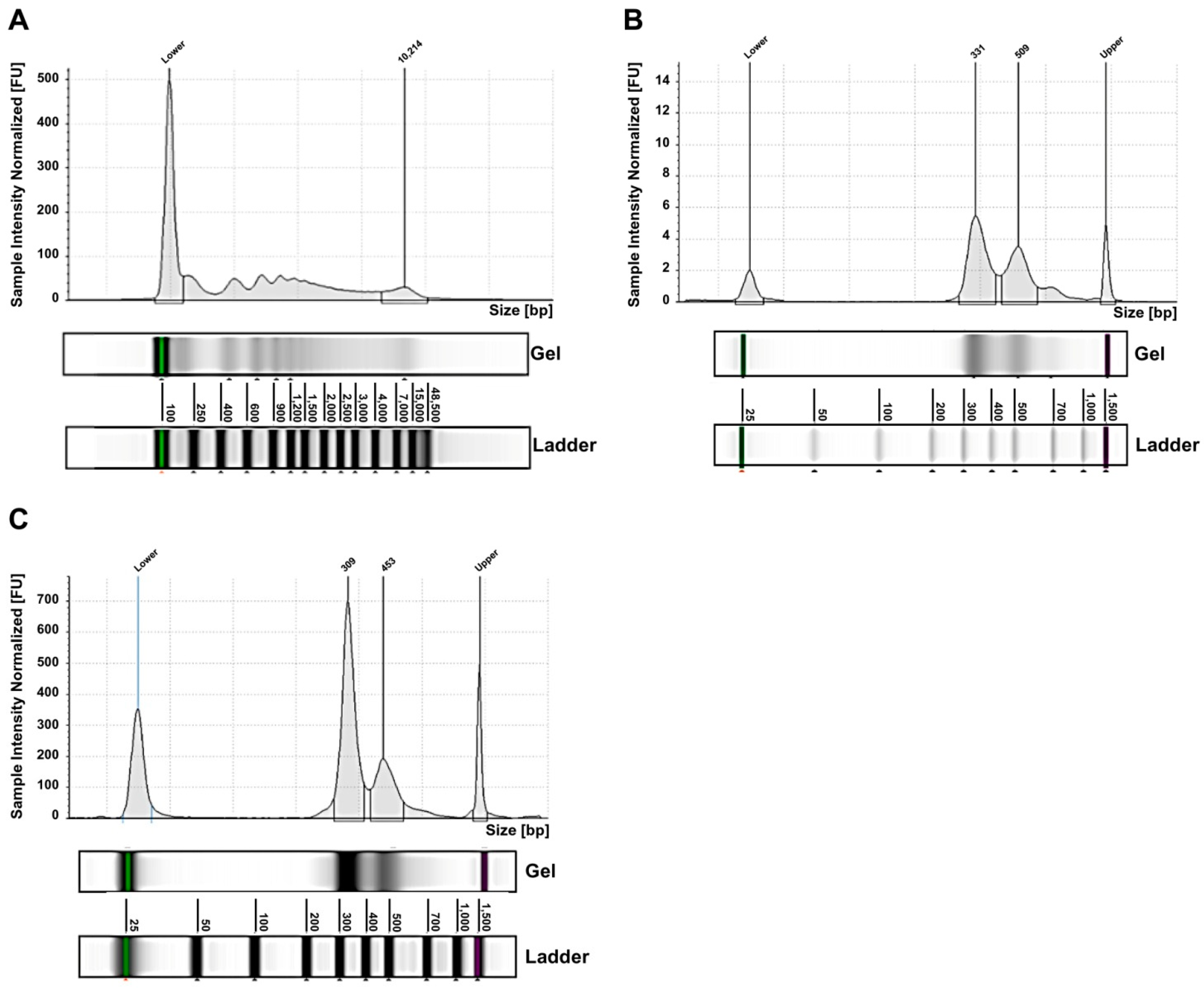
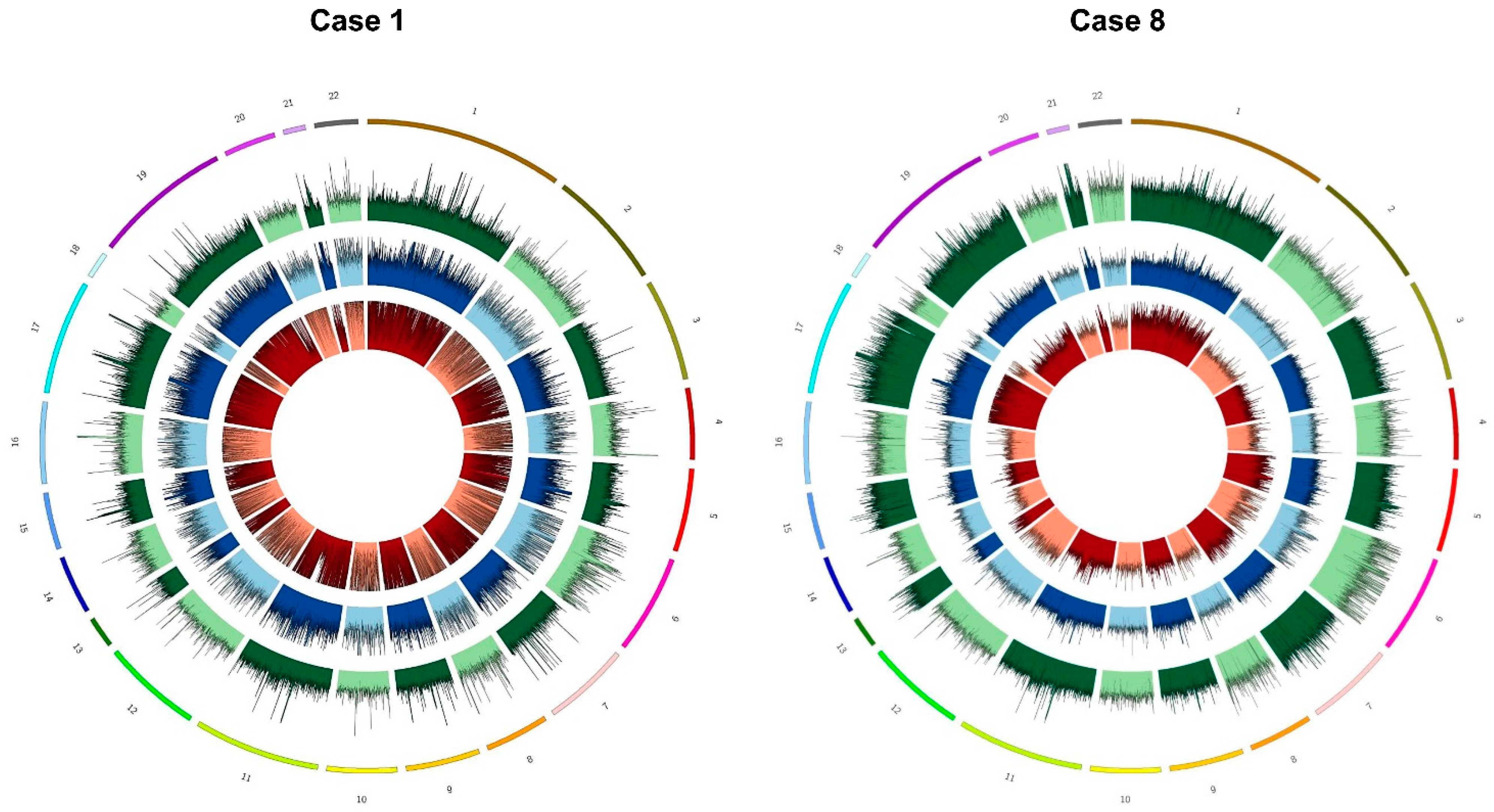
2.2. Exosomes from NB Patients Contain Genomic DsDNA Covering All Chromosomes

Exosomes were purified from peripheral blood samples of the nineteen NB patients at onset of the disease and, in four cases, at the time of relapse too. The NB cell-derived exosomes are nanoparticles already well characterized in terms of size (30–150 nm) [26], and positivity for the GD2 marker, specifically expressed on NB cells surface, as we described in our previous manuscript [19]. In that work, we analyzed NB plasma-derived exosomes by flow cytometry using the anti-tetraspanin CD9 mAb, recognizing the typical exosomal surface marker, and the anti-GD2 mAb to confirm their purity. This analysis showed that about 85–90% of NB-derived exosomes expressed CD9, and that the mean percentage of GD2+ exosomes was over 50%. Therefore, we demonstrated that plasma samples from NB patients contain NB cell-derived exosomes [19]. Exosomes were treated with DNase I, before extracting the exo-DNA, to eliminate any external DNA contamination.
For the NB cases analyzed by whole-exome sequencing (WES), in addition to exo-DNA, the other available DNA specimens were the tumor DNA extracted from needle aspiration or micro-biopsy tissue (for ten patients only) and the genomic blood DNA (gDNA). Fluorimetric sizing analysis of exo-DNA showed a size distribution ranging from 186 bp to 48 kb, with distinct peaks (Figure 1A). Exo-DNA was mechanically fragmented in order to obtain the desired insert size (280 bp) however, due to its initial profile, the final library profiles exhibited two characteristic peaks, the first one of 310–340 bp and the second one of 450–500 bp (Figure 1B). After exome enrichment capture, this size distribution was maintained (Figure 1C).
Exome enriched libraries performed for all nineteen NB cases were sequenced on an Illumina Novaseq 6000, using the 150 bp paired-end mode. Considering the fragments obtained from sequencing and their alignment on the reference genome GRCh38/hg38, the median sequenced fragment size of exo-DNA specimens reached 191 bp, confirming a lower fragmentation pattern typically observed for cfDNA. Tumor biopsy and genomic DNA specimens resulted in a fragment length of 306 and 308 bp, respectively, in line with the expected sequenced size of WES libraries. Observed coverage for all exo-DNA specimens was greater than 50×, with a median value of 110×; median coverage observed for gDNA and tumor DNA was of 147× and 131×, respectively. More specifically, the percentage of the target covered by at least 5 (%5×: 97.99%) and 10 reads (%10×: 97.79%) in exo-DNA reflected similar performance of WES on gDNA and tumor samples (Supplementary Table S1). Indeed, the coverage was uniform along all chromosomes of exo-DNA specimens (Figure 2).
Most importantly, it was possible to genotype the whole exome through the analysis of exo-DNA (the median genotypability value (%PASS) obtained on the entire target region (design) was 97.8) (Supplementary Table S1). In particular, the integration of unique molecular identifiers (UMIs) in the sample preparation, as described in Section 4, allowed a more accurate identification of PCR duplicated fragments. Moreover, the removal of fragments deriving from repetitive regions of the genome using the UMI’s bioinformatics pipeline increased the genotypability along all the target regions. These results showed higher genotypability of the exome (more than 1%) respect to previously reported WES analyses on genomic DNA, with a sequenced fragment length of 350 bp [27]. To be noticed, the exosomes genotype was higher (more than 2%) than in exomes, with even shorter fragment sizes (200 bp) and read length (75 bp) [27]. Given the large size of the entire target region, the genotypability for each chromosome in the different DNA specimens was investigated, looking for genotypability differences. No substantial differences emerged among chromosomes within the same DNA specimen, confirming a high genotypability distributed along the entire exome. The median of the samples for each tumor DNA, exo-DNA at onset, exo-DNA at relapse is reported in Table 2.
By comparing the genotype data between exo-DNA and gDNA, we noted that the genotypability of exo-DNA (in particular at onset) was equal if not higher than control. The coding regions of autosomal chromosomes were all well represented in the exo-DNA at onset (%PASS > 73% with a mean of 90.86%), as was for gDNA (%PASS > 71% with a mean of 87.24%). The exo-DNA at relapse, which in some cases had lower DNA amount, showed a %PASS above 70% for all chromosomes with an average of 86.4%. Tumor DNA had a good %PASS (>72%) too, with an average of 89.46%.

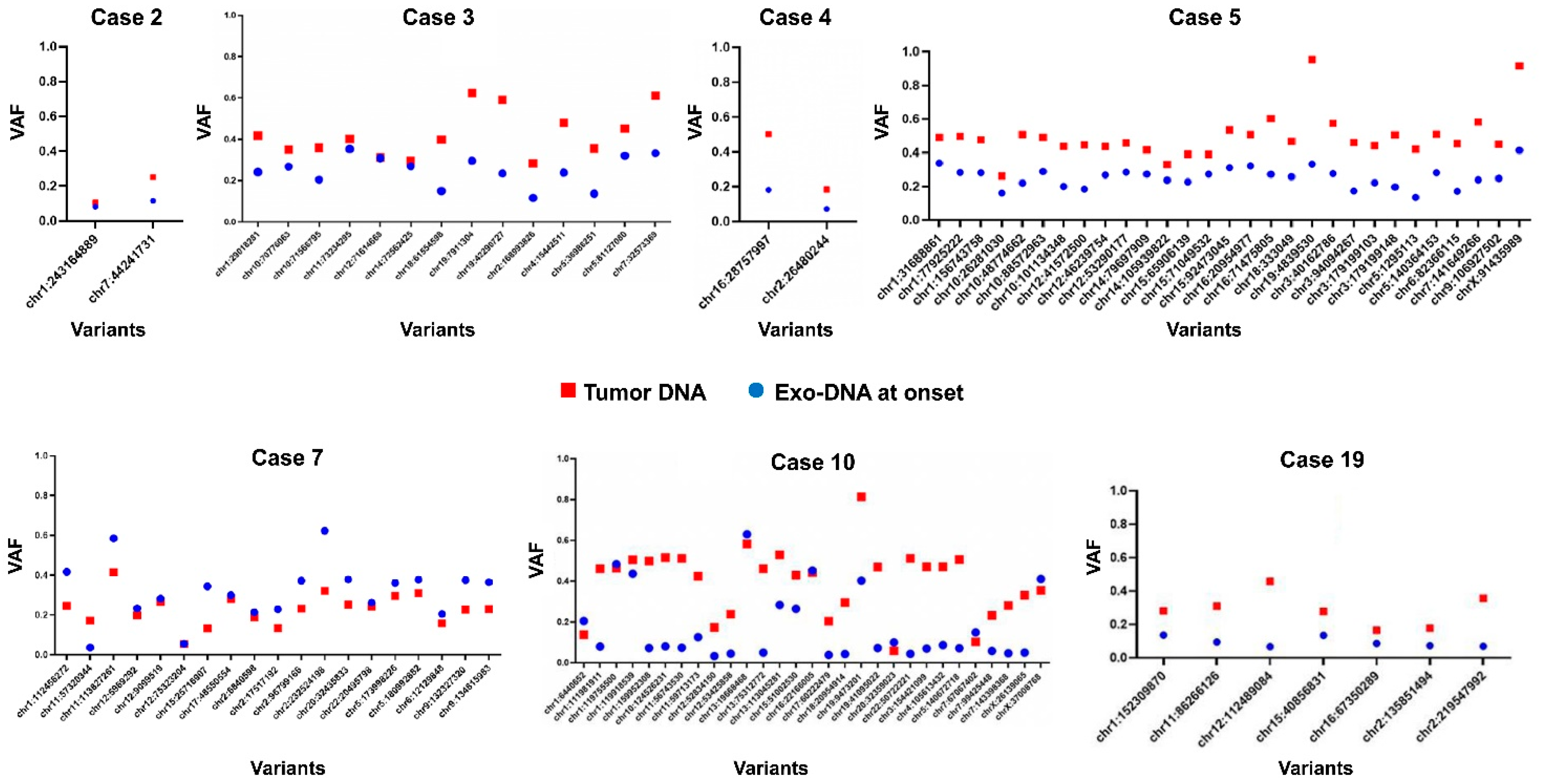
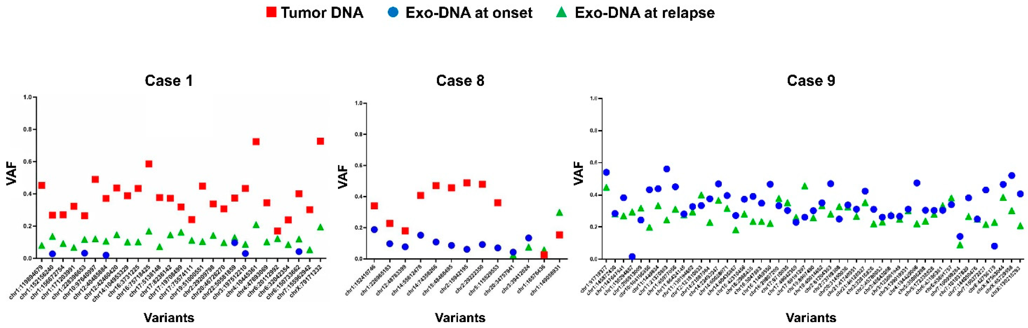
2.3. Tumor DNA Content in Exo-DNA Samples

The somatic single nucleotide variants (SNVs) identified in the tumor DNA, exo-DNA at onset and at relapse were then evaluated. For this purpose, the number of SNVs in common between the different DNA specimens available for each case was calculated. In ten NB cases with available exo-DNA at onset and the corresponding tumor DNA, a higher overall number of SNVs was identified in the exo-DNA compared to tumor DNA (Figure 3A,B). This could mean that SNVs detected in exo-DNA arose from tumor sites different from the tumor DNA site of extraction, highlighting a spatial genetic heterogeneity in NB. In the NB cases with all DNA specimens available (exo-DNA at onset, exo-DNA at relapse, tumor DNA) many SNVs were found to be exclusive of a single specimen, with low concordance among them (Figure 3B). The number of SNVs in common between exo-DNA at onset and exo-DNA at relapse remained low, with a greater number of variants identified for the relapse (Figure 3C).
Subsequently, the frequency of the SNVs identified in the different DNA specimens was investigated. By observing the frequency graphs of the variants identified in the NB cases with available exo-DNA at onset and the corresponding tumor DNA, we identified the presence of clones with lower frequency SNVs in the exo-DNA compared to the tumor DNA (Figure 4). The exo-DNA at relapse had SNVs with higher frequency than the exo-DNA at onset, probably as indicator of clonal evolution (Figure 5). NB cases with only exo-DNA at onset available, showed the same trend seen for other cases, with most of the identified SNVs having low frequency (Supplementary Figure S1).
To investigate if the dsDNA extracted from NB cell-derived exosomes reflected the mutational status of their parental tumor cells, we performed further analysis on the somatic SNVs found in common between different DNA specimens of the same case, evaluating their allelic frequency. Considering the NB cases with available tumor DNA and exo-DNA at onset, we observed that SNVs frequency in the exo-DNA at onset was generally lower than in tumor DNA, probably indicating less tumor content in exo-DNA (Figure 6).
In the NB cases with all DNA specimens available, tumor SNVs appeared to have a higher frequency than both exo-DNA at onset and exo-DNA at relapse, as expected (Figure 7). The last two SNVs in case 8 displayed an abnormal behavior, with a frequency in the tumor DNA lower than in exo-DNA at relapse. The exo-DNA at relapse often showed higher frequency SNVs than the exo-DNA at onset, probably indicating some sort of clonal evolution inducing the relapse itself and the consequent poor prognosis of the patient. The WES results therefore demonstrated that parental tumor SNVs were detectable in the exo-DNA even if with lower frequency.

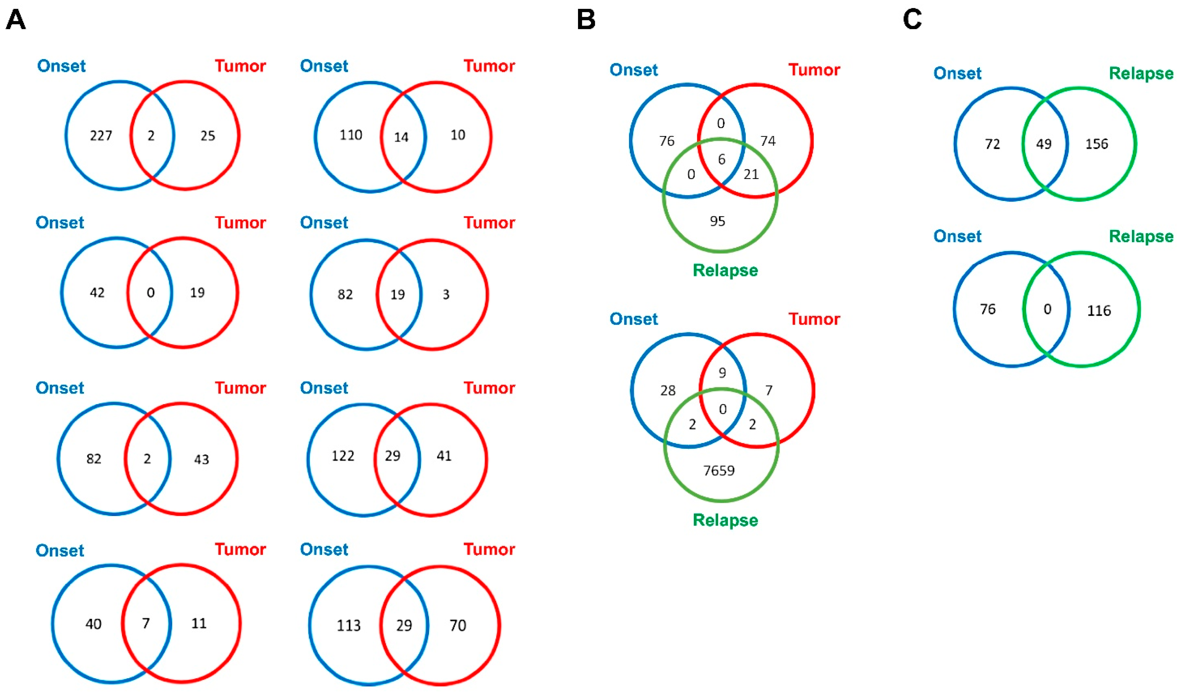
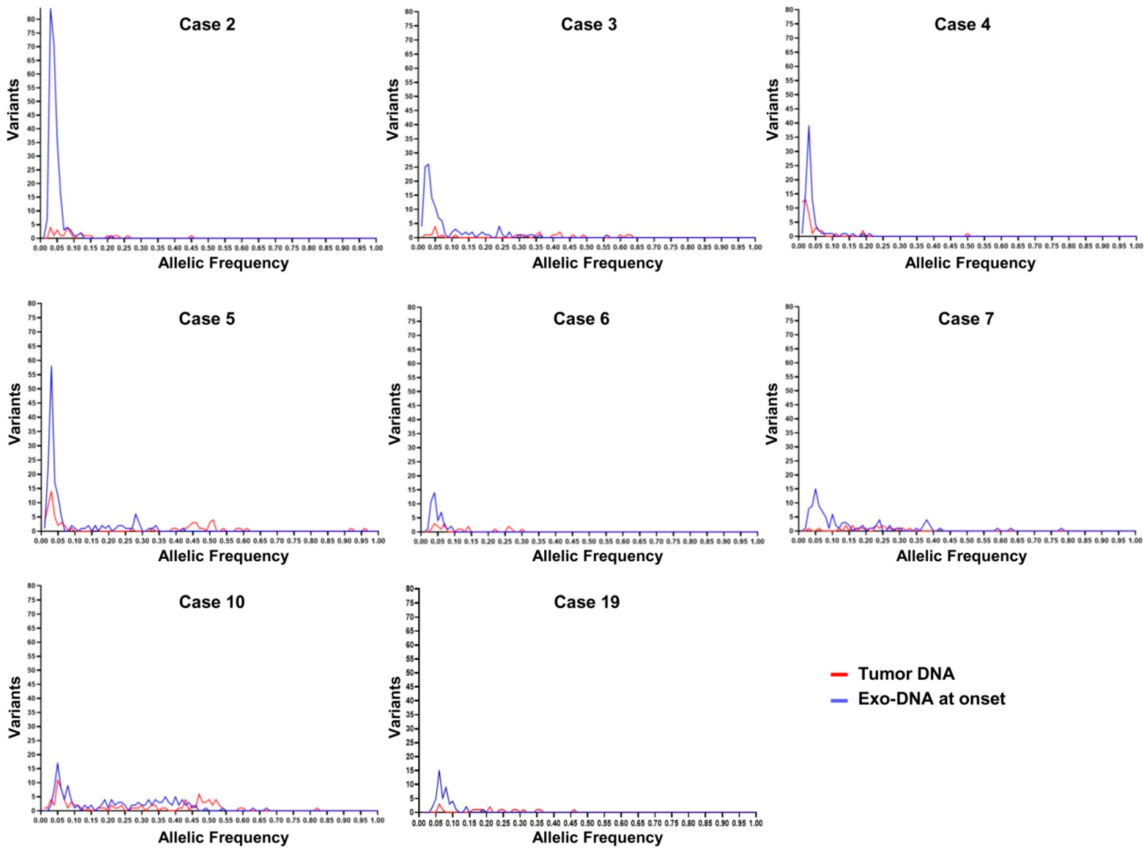
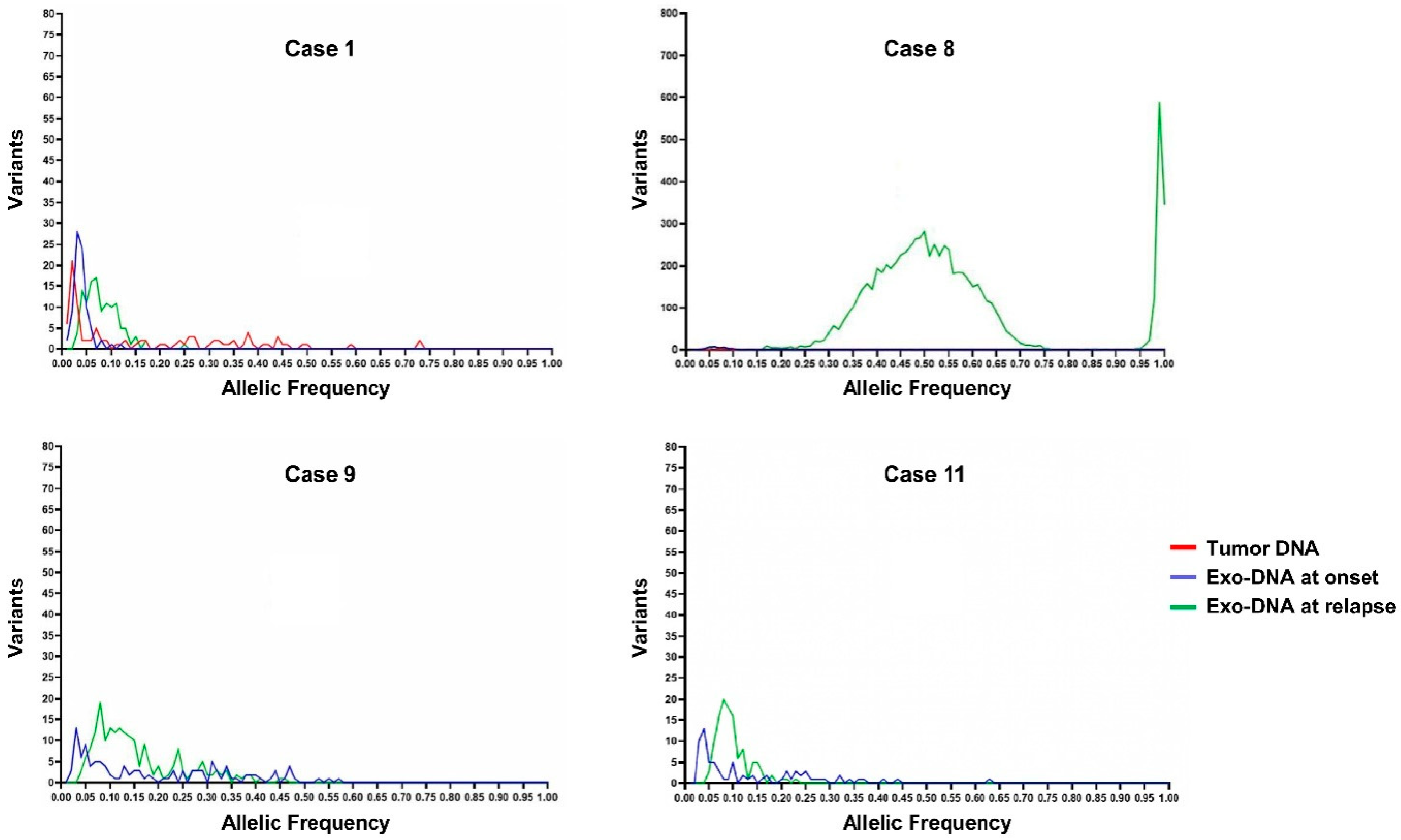
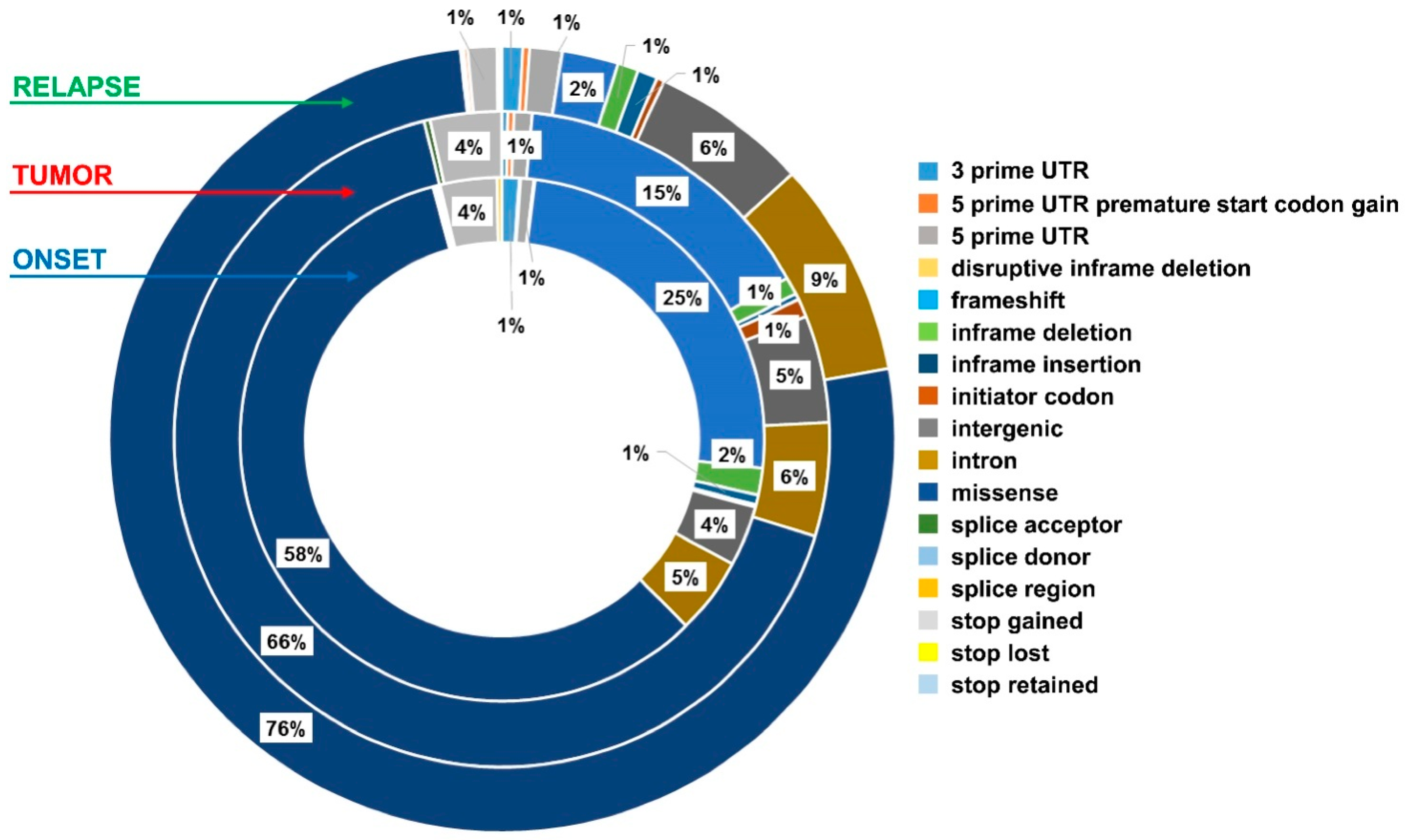
2.4. Exo-DNA-Derived Tumor Mutation Load

The tumor mutation load (TML) resulting from the non-synonymous variants number per megabase (Mb) identified in the target region [28], was considered for the exo-DNA and the tumor DNA. TML value was calculated by WES data for each available specimen, then deriving the median for each type of specimen. Median TML was 1.5 non-synonymous variants per Mb in exo-DNA at onset, 3.55 non-synonymous variants per Mb in exo-DNA at relapse and 0.45 non-synonymous variants per Mb in tumor DNA. These results indicated that tumor DNA TML was in line with the known value already reported for NB, that is a median frequency of 0.48 non-synonymous variants per Mb [9]. Interestingly, TML of exo-DNA both at onset and at relapse was higher than in tumor DNA. The higher TML value observed in the exosomes at onset is probably due to the exo-DNA somatic SNVs not detectable in tumor DNA. Our cohort of NB patients with high TML value in exo-DNA had a worse outcome in comparison with patients with lower values.

2.5. Exo-DNA Contains SNVs of Known Neuroblastoma Driver Genes

The annotation of the SNVs with tumor databases had allowed the classification of many of them from a clinical point of view [29,30]. Main analysis identified somatic SNVs associated with NB in many of the exo-DNAs in the COSMIC somatic mutations database. Known driver and tumor suppressor genes in NB were frequently mutated in the exo-DNA (ALK, CHD5, SHANK2, PHOX2B, TERT, FGFR1, and BRAF). In Table 3, we reported the somatic SNVs identified in the different DNA specimens, for each analyzed NB case. To notice, some clinical variants of ALK gene were present in several cases.
The majority of the identified somatic SNVs are missense variants, confirming what has already been reported in the literature (Figure 8).

2.6. Most Frequently Mutated Genes in Exo-DNA and in Tumor DNA

Next, we evaluated whether in this NB patient cohort there were frequently mutated genes different between exo-DNA and tumor DNA samples. Table 4 reports, for each DNA specimen, the list of the 20 most significantly enriched genes. The results showed that most mutated genes did not always match in the different DNA specimens. In particular, the exo-DNA at onset displayed a more significant enrichment than the exo-DNA at relapse and the tumor DNA. In particular, TYW1 gene was the most mutated gene in exo-DNA at onset, while AHNAK2 and ALK genes were the most mutated genes in the exo-DNA at relapse and in primary tumor DNA, respectively. ALK is one of the most significantly mutated gene in the exo-DNA at onset and in tumor DNA. Particularly mutated regions (hotspots) have also been identified in exo-DNA at onset within the listed genes (Table 5), and chromosome 7 appeared to be the most mutated one. Among these genes, we found new ones, not already associated with NB, that were frequently mutated in this cohort of patients, such as TYW1 and DPP6.

2.7. Identified Copy Number Variations Involved in NB Oncogenesis

All copy number variations (CNVs) found within the known driver genes related to NB or to other tumors, were investigated [4,8,9]. We found that some genes of interest displayed CNVs in different DNA specimens (Table 6; Supplementary Table S2). In particular, CNVs affected genes involved in the RAS pathway (PTPN11, NF1, KRAS, and BRAF) and other genes already detected in SNVs analysis (ALK, and SHANK2). For the cases with multiple available DNA specimens, it was not always possible to identify tumor CNVs in the exo-DNA, anyway a few CNVs have been detected in multiple DNA specimens. For example, MYCN amplification has been observed in tumor DNA and in exo-DNA both at onset and at relapse. For patients with only exo-DNA at onset available, many known NB driver genes were duplicated or deleted. Notably, MYCN, TERT, and SHANK2 genes showed in some cases a very high copy number (greater than 10).

3. Discussion

Exosomes are extracellular membrane vesicles with a size ranging from 30 to 150 nm. These vesicles are released by most cells, including tumor cells, and therefore can be used as a source of DNA, messenger RNA, micro-RNA and proteins [7,18,21,25]. Circulating exosomes represent new components of the tumor microenvironment, and constitute a reliable source of biomarkers that allows real-time monitoring of the disease [19,20,26]. Exosomes purified from peripheral blood sample only require a simple blood draw, which can be repeated multiple times with minimal impact for patients. The use of exosomes as source of non-invasive biomarkers makes possible to test tumor sites more frequently. The advancement of personalized medicine in oncology is based on the development of non-invasive approaches for stratification, diagnosis, monitoring of therapeutic response, and detection of patients’ minimal residual disease. Exosomes represent this type of experimental approach, and are able to provide reliable and solid information on disease, metabolism and on the genomic profile of parental tumor cells.
Therapeutic stratification of NB patients is based on clinical and molecular risk factors that correlate with prognosis, and it is essential for treatment [1,2,3,4,5]. However, patients with similar clinical-pathological parameters who receive the same treatment may have different clinical courses. For this reason, it is important to identify new and better prognostic markers capable of directing therapy towards personalized medicine. Since approximately 50% of high-risk NB patients die despite treatment, new strategies are needed to improve therapy. The clinical, radiological and biological markers currently used in clinical routine are not able to predict in an early and accurate way a possible therapeutic failure that would justify a change in treatment. Exosomes represent an interesting method to obviate the limited amount of tumor tissue obtainable by needle aspiration or micro-biopsy, as well as an alternative for patients who cannot undergo tissue biopsy.
Our study provides evidence that exosomes isolated from plasma samples of NB patients contain double stranded tumor DNA spanning all chromosomes. We suggest a new source of circulating DNA in addition to plasma derived cell-free DNA (cfDNA) resulting from cell lysis or apoptosis [15,31,32]. Indeed, the substantial difference between exo-DNA and cfDNA is that the former derives from living cells, whereas the latter comes, in large part, from dead ones. Therefore, the exo-DNA can better reflect the malignant and aggressive tumor cells populations, and better highlights on tumor dynamics. Potentially, the exo-DNA is much more clinically useful than the cfDNA, which is diluted in plasma or in other body fluids. The exo-DNA is stable and protected inside vesicles, that also carry tumor related miRNA [19], and proteins [33], which have been used successfully as tumor biomarkers. Moreover, the exosomes can be isolated and enriched by exploiting their specific surface markers, thus enriching in exo-DNA too.
We demonstrated by WES that exo-DNA covers the whole exome, obtaining results very similar to those previously reported by WES on genomic DNA [27]. More specifically, the fragmentation pattern obtained for the exosomes could allow sequencing longer reads (150 bp), compared to cfDNA, that appears more fragmented. This, in combination with the use of UMIs during sample preparation, leads to an accurate analysis of the entire exome starting from exo-DNA sequencing. Noteworthy, the NB exo-DNA displays mutations identical to the parental tumor cells DNA, despite the limited number of analyzed samples. Some low frequency variants (e.g., 2/3 reads to support the variant), identified in one specimen only, were present in another specimen of the same case at a lower frequency (e.g., 1 read to support the variant). While in the first specimen the variant was called thanks to the higher number of supporting reads, in the other specimens the variant was not identified. This could have led to the discovery of fewer variants in common between different DNA specimens, under-estimating the tumoral variants present in the exo-DNA. Differences existing between tumoral and exosome specimens’ coverage may have influenced the variant calling. The exo-DNA contains information regarding somatic SNVs and CNVs of ALK, MYCN, PTPN11, ATRX, SMARCA4, TP53, NF1, CHD5, TERT, BRAF, SHANK2, KRAS, and CDKN1B genes present in their parental cells too. The frequency of the somatic SNVs in the exo-DNA at onset is generally lower than in tumor DNA, probably indicating clonal mutational events in the exo-DNA. We identified SNVs in the exo-DNA that were not present in tumor DNA at onset of disease, in particular mutations of ALK, ATRX, NF1, and TERT genes, probably coming from other tumor sites. The exosomes derive from cells of distinct tumor and/or metastatic sites, so they can represent the whole tumor, whereas a micro-biopsy tissue cannot, and this is particularly important for the great intra-tumoral heterogeneity of NB [15]. This aspect underlines the relevance of analyzing exo-DNA for an eventual personalized medicine. In exo-DNA samples at relapse, we observed the majority of mutations detected at diagnosis, plus an increased number of somatic SNVs such as mutations of ALK, TP53, and RAS/MAPK genes. Perhaps the specific SNVs observed at the time of relapse were present as a minor subclone at diagnosis and were not detected by standard coverage WES. The exo-DNA at the time of relapse has somatic SNVs with a higher frequency than the exo-DNA at onset of disease, and probably this is an indicator of clonal evolution. Some genes involved in the RAS/MAPK pathway have somatic SNVs in exo-DNA at the time of relapse, allowing the identification of mechanisms involved in NB progression and acquired resistance [13]. The study of SNVs detected in exo-DNA at the time of relapse, and the chance to identify these mechanisms as soon as possible, could be particularly important for the therapeutic treatment of NB patients. Interestingly, the TML values of exo-DNA both at onset and at relapse are higher than values of tumor DNA. TML has been shown to be a predictor for therapy response [28]. Koeppel and co-authors explained how the advantage of WES over targeted resequencing is not primarily the identification of new variants, but the possibility to obtain a global view of mutations at the target level too. In addition, it seems that our NB patients with high TML values in exo-DNA had a worse outcome than those with lower values. Furthermore, the ability to obtain biomarkers such as TML using non-invasive samples increases the interest of exo-DNA analysis. We observed in exo-DNA at onset hotspot mutations of genes not typically associated with NB such as TYW1 and DPP6. The TYW1 gene encodes for an iron-sulfur protein with protective functions against the adverse iron effects on neuronal functions [34,35]. The DPP6 gene encodes for the dipeptidyl aminopeptidase-like protein 6, promoting cell surface expression of the potassium channel, and generating a current gradient critical for the regulation of dendritic excitability in hippocampal neurons [36]. We observed mutations also in THSD7A gene, coding for a new potential tumor antigen that might represent a putative therapeutic target for cancer therapy [37]. The OR8G5 gene encodes for the olfactory receptor 8G5, a G-protein-coupled receptor mainly expressed in olfactory sensory neurons [38]. Moreover, olfactory receptors may be expressed also in non-olfactory tissues, likely representing new tumor markers [39]. Although these mutations are not currently associated with NB, they could be evaluated further.
Our study has several limitations, such as the relatively small number of exo-DNA samples and the not availability of the corresponding primary tumor DNA for all patients. Nevertheless, here we provide new insights into liquid biopsy subject, to try to achieve a finest personalized medicine for children affected by NB. Future works based on a larger NB samples size are required to confirm present results.

4. Materials and Methods

4.1. Patients and Samples

Nineteen patients with a diagnosis of NB were collected at BIT-NB Biobank of IRCCS Gaslini, Genova, Italy. Of these, fourteen were high-risk, and five were intermediate or low risk as defined by the International Neuroblastoma Risk Group (INRG) classification system [5]. Inclusion condition for the patients analyzed in this study was the availability of plasma obtained at onset of the disease. For four patient’s, plasma samples collected at the time of relapse were also available. The clinical data were derived from the Italian Neuroblastoma Registry, where the clinical characteristics of NB patients were saved in a pseudo-anonymized manner. Written informed consent was obtained from children’s parents to report each case. This study was undertaken in accordance with the ethical principles of the Declaration of Helsinki, and it was approved by the Italian Institutional Ethics Committee (Measure n° 270/17 related to the clinical study protocol IGG-NCA-AP-2016).

4.2. Sample Collection and Processing

NB samples content was confirmed by review of hematoxylin and eosin-stained tumor sections by the local pathologists. Tumor DNAs were extracted from fresh NB tissue using MasterPure DNA Purification Kit (Lucigen, Middleton, WI, USA), according to manufacturer’s instructions. Genomic germline DNA was extracted for each NB patient from peripheral blood leukocytes using the QIAamp DNA Blood Midi Kit (QIAGEN, Hilden, Germany). Blood samples were collected in EDTA tubes and plasma was separated from blood by centrifugation at 1600× g for 10 min at 4 °C twice, followed by careful aliquoting and freezing at −80 °C within 1 to 24 h [40].

4.3. Exo-DNA Purification and Quantification

We isolated exosomes from nineteen plasma NB samples collected at onset of disease and, for four patients, at the time of relapse too, as previously described [19]. Exosomes were isolated with the specific Exo-RNeasy serum/plasma Midi Kit (QIAGEN). The exosomes were incubated in 200 µL PBS with 1 µL DNase I (1 unit/µL) (Promega, Madison, WI, USA) at 37 °C for 30 min. Then, 5 µL of DNase stop solution (Promega) was added, and the exosomes were heated at 65 °C for 5 min. Following, the exosomes were washed and resuspended in 200 µL PBS [41]. Exosomes were lysed with Triton X-100 (2%), SDS (0.1%) and Buffer AL (QIAGEN) as described by Vagner et al. [42]. Finally, the exo-DNA was extracted using the QIAamp MinElute ccfDNA Mini Kit (QIAGEN). Exo-DNA quantity and quality were assessed with 1× dsDNA HS assay on Qubit 4.0 fluorometer (ThermoFisher, Waltham, MA, USA) and with high sensitivity DNA assay on Agilent Bioanalyzer (Agilent Technologies, Santa Clara, CA, USA), respectively.

4.4. Genomic Profiling of NB Primary Tumors

DNA from fifteen NB primary tumors was tested by high-resolution oligonucleotide a-CGH using the 4 × 180 K Kit (Agilent Technologies) with a mean resolution of approximately 25 kb [43]. The altered chromosomal regions were detected using the algorithm ADM-1 (threshold 10) with 0.5 Mb window size to reduce false positives. Chromosome positions were determined using GRCh38/hg19 (UCSC Genome Browser, http://genome.ucsc.edu, release 7 July 2000), accessed on 5 January 2021.

4.5. Library Construction and Exome Capture of Exo-DNA

Quality control of gDNA was performed using the DNA Broad Range Qubit Assay (ThermoFisher) and the 4150 TapeStation System (Agilent Technologies). Samples were sheared to 300 bp using the Covaris S220 instrument (Covaris, Woburn, MA, USA) according to the manufacturer’s protocol. Library construction was performed using the KAPA Hyper Prep (Roche, Basel, Switzerland) with 50 ng or all the available amount of sheared DNA as input for each sample. Both exo-DNA at onset and exo-DNA at relapse were adapter ligated with xGen Dual Index UMI Adapters (IDT, Coralville, IA, USA). Library quality control was performed using the DNA Broad Range Qubit Assay (ThermoFisher) and the 4150 TapeStation System (Agilent Technologies). Exomes were captured using the Twist Human Core Exome + RefSeq (Twist Bioscience, San Francisco, CA, USA) following the manufacture’s recommendation. Enriched libraries were quantified by real-time PCR using the KAPA Library Quantification Kit for Illumina platforms on the QuantStudio3 Real-Time PCR Systems (ThermoFisher) and polled at equimolar concentrations. Sequencing was done on the Novaseq 6000 platform (Illumina, San Diego, CA, USA) using the 150 bp paired-end mode.

4.6. Bioinformatics Pipeline

FastqQ files were quality controlled using FastQC (http://www.bioinformatics.babraham.ac.uk/projects/fastqc/, accessed on 30 October 2020). Low quality nucleotides and adaptors have been trimmed using sickle vl.33 (https://github.com/najoshi/sickle, accessed on 30 October 2020) and scythe v0.991 (https://github.com/vsbuffalo/scythe, accessed on 3 November 2020), respectively. Reads were consequently aligned to the reference human genome sequence version GRCh38/hg38 using BWA-MEM v0.7.15 (https://arxiv.org/abs/1303.3997, accessed on 3 November 2020). For the exosome and tumor samples processed with UMI, fgbio v1.1.0 was used to first integrate the UMI information into the BAM files and to group the reads by UMI using the “Annotate Bam with Umis” and “Group Reads by Umi” functions, respectively. Consensus reads based on UMI were then generated through the “Call Molecular Consensus Reads” function (using parameters: —error-rate-post-umi = 30 and —mim-reads = 1) The consensus reads were then re-mapped to the human references genome. All generated BAM files were cleaned by local realignment around insertion–deletion sites, duplicate marking (only for the samples processed without UMI), and recalibration using Genome Analysis Toolkit v3.8.1.6. Overlapping regions of the BAM file were clipped using BamUtil v1.4.14 to avoid counting multiple reads representing the same fragment. CallableLoci in GATK v3.8 was used to identify callable regions of the target (genotypability), with minimum read depths of 3 and 10. CollectHsMetrics by Picard v2.17.10 was used to calculate fold enrichment and FOLD 80 penalty values to determine enrichment quality. Somatic variant calling was then performed with Mutect2 (GATK v4.1.9.0) in the “tumor with matched normal” mode, considering as “tumor” the exo-DNA and tumor samples, and as “normal” the germline genomic DNA (gDNA). As a preliminary step, a “panel of normal” VCF file was created using all the germinal gDNA samples to further refine the somatic analysis. Variants were then filtered by quality (filter PASS) and target design region. Annotation and prioritization of variants was carried out using VarSeq software (GoldenHelix, Bozeman, MT, USA). The annotation process included databases of somatic mutations (COSMIC v90, TCGA Variants 3 March 2019, GHI, ICGC Simple Somatic Mutations 28, GHI, CIViC—Variant Clinical Evidence Summaries 1 July 2020, WUSTL, CIViC—Genes 1 July 2020, WUSTL), the RefSeq Genes 109 Interim v2.1 NCBI and the dbSNP 153 NCBI databases. A flag was applied for variants present in more than one tissue in the same individual.

4.6.1. Copy Number Variations (CNVs)

CNVs in exo-DNA and in tumor samples were identified using the tool EXCAVATOR2 with default setting, pairing each tumor DNA and exo-DNA with the corresponding gDNA sample. Then, results were filtered using a selected list of genes known to be recurrently mutated in NB (ALK, TERT, ATRX, LIN28B, TP53, SMARCA4, CDKN1B, PHOX2B, CHD5, MYCN, SHANK2, PTPRD, FGFR1, PTPN11, NF1, NRAS, KRAS, and BRAF) [9].

4.6.2. Tumor Mutation Load (TML)

Tumor mutation load (TML) of exo-DNA and of tumor DNA samples was calculated extracting the number of all the non-synonymous SNVs from these samples and dividing it by the target design length (Mb).

4.6.3. Enrichment Analysis

An enrichment analysis was performed in order to identify the most mutated genes in the exo-DNA and in the DNA from tumor samples among all patients. The annotated VCF files were processed by the MutEnricher v1.3.1 software (https://rdcu.be/b51ka, accessed on 19 November 2020) using the “coding” parameter to generate p-values of enrichment for both hotspot sites and genic regions.

4.6.4. Concordance Rate

Common CNVs shared between the exo-DNA and tumor DNA were retrieved using the SnpSift v4.3 tool. A concordance rate (percentage of concordance) was calculated considering the mean number of common CNVs identified for each pair exo-DNA and tumor DNA among all individuals.

5. Conclusions

Here we give evidence, for the first time, that the double stranded DNA contained in exosomes derived from plasma samples of NB patients represents the whole tumor DNA. Exo-DNA can be exploited to identify SNVs and CNVs present in parental NB cells. The possibility to isolate and enrich NB-derived exosomes from plasma using surface markers, and to extract exo-DNA quickly and easily, give this method a significant translational potential in the clinic. Exo-DNA can be an attractive circulating biomarker for NB molecular diagnostic and for clinical outcome prediction. Indeed, exo-DNA can be useful to identify early mechanisms of acquired resistance, such as mutations of ALK, TP53, and RAS/MAPK genes, occurring in relapsed patients. Future clinical studies to define exo-DNA utility in personalized NB therapy are warranted.

Supplementary Materials

The following are available online at https://www.mdpi.com/article/10.3390/ijms22073667/s1.

Author Contributions

Conceptualization, A.P. and M.D.; Methodology, C.D.E., B.I., S.M., C.B., D.L., M.D. and A.P.; Statistical analysis, C.D.E., B.I., S.M., C.B. and D.L.; Validation, C.D.E., B.I., S.M., C.B., D.L. and P.D.M.; Formal analysis C.D.E., B.I., S.M., C.B., D.L. and M.D.; Investigation, C.D.E., B.I., S.M., C.B., D.L., M.O. and A.P.; Resources, A.P., M.M., A.G., G.E. and F.Z.; Data curation, C.D.E., B.I., M.O. and A.P.; Writing—original draft preparation, A.P.; Writing—review and editing, C.D.E., B.I., P.D.M., M.O. and A.P.; Visualization, M.O., C.D.E. and B.I.; Supervision, A.P.; Project administration, A.P.; Funding acquisition, A.P. All authors have read and agreed to the published version of the manuscript.

Funding

This research was funded by the Cinque per mille dell’IRPEF Finanziamento della ricerca sanitaria; Finanziamento Ricerca Corrente 2019, Ministero della Salute; Compagnia San Paolo.

Institutional Review Board Statement

The study was conducted according to the guidelines of the Declaration of Helsinki, and approved by the Italian Institutional Ethics Committee (Measure n° 270/17 related to the clinical study protocol IGG-NCA-AP-2016; approved 15 December 2016).

Informed Consent Statement

Written informed consent was obtained from children’s parents to report each case in this paper.

Data Availability Statement

All the data are reported in the present paper and in the supplementary materials.

Acknowledgments

We thank BIT-NB Biobank of IRCCS Gaslini for providing NB specimens.

Conflicts of Interest

The authors declare no conflict of interest. The funders had no role in the design of the study; in the collection, analyses, or interpretation of data; in the writing of the manuscript, or in the decision to publish the results.

Abbreviations

NBNeuroblastoma
MYCNv-myc myelocytomatosis viral related oncogene, neuroblastoma derived
ALKAnaplastic Limphoma Kinase
RASRat Sarcoma
INRGInternational Neuroblastoma Risk Group
a-CGHArray-Comparative Genome Hybridization
chrChromosome
mAbMonoclonal antibody
dsDNADouble stranded DNA
gDNAGenomic DNA
cfDNACell-free DNA
ctDNACirculating tumor DNA
exo-DNAExosomal DNA
WESWhole Exome Sequencing
UMIUnique Molecular Identifier
SNVSingle Nucleotide Variant
CNVCopy Number Variation
VAFVariant Allele Frequency
TMLTumor Mutation Load
VCFVariant Call Format
COSMICCatalogue of Somatic Mutations in Cancer

References

  1. Cheung, N.K.; Dyer, M.A. Neuroblastoma: Developmental biology, cancer genomics and immunotherapy. Nat. Rev. Cancer 2013, 13, 397–411. [Google Scholar] [CrossRef]
  2. Matthay, K.K.; Maris, J.M.; Schleiermacher, G.; Nakagawara, A.; Mackall, C.L.; Diller, L.; Weiss, W.A. Neuroblastoma. Nat. Rev. Dis. Primers 2016, 2, 16078. [Google Scholar] [CrossRef] [PubMed]
  3. Speleman, F.; Park, J.R.; Henderson, T.O. Neuroblastoma: A Tough Nut to Crack. Am. Soc. Clin. Oncol. Educ. Book 2016, 35, e548–e557. [Google Scholar] [CrossRef]
  4. Bosse, K.R.; Maris, J.M. Advances in the translational genomics of neuroblastoma: From improving risk stratification and revealing novel biology to identifying actionable genomic alterations. Cancer 2016, 122, 20–33. [Google Scholar] [CrossRef] [PubMed]
  5. Cohn, S.L.; Pearson, A.D.; London, W.B.; Monclair, T.; Ambros, P.F.; Brodeur, G.M.; Faldum, A.; Hero, B.; Iehara, T.; Machin, D.; et al. INRG Task Force. The International Neuroblastoma Risk Group (INRG) classification system: An INRG Task Force report. J. Clin. Oncol. 2009, 27, 289–297. [Google Scholar] [CrossRef] [PubMed]
  6. Peifer, M.; Hertwig, F.; Roels, F.; Dreidax, D.; Gartlgruber, M.; Menon, R.; Krämer, A.; Roncaioli, J.L.; Sand, F.; Heuckmann, J.M.; et al. Telomerase activation by genomic rearrangements in high-risk neuroblastoma. Nature 2015, 526, 700–704. [Google Scholar] [CrossRef] [PubMed]
  7. Valentijn, L.J.; Koster, J.; Zwijnenburg, D.A.; Hasselt, N.E.; van Sluis, P.; Volckmann, R.; van Noesel, M.M.; George, R.E.; Tytgat, G.A.M.; Molenaar, J.J.; et al. TERT rearrangements are frequent in neuroblastoma and identify aggressive tumors. Nat. Genet. 2015, 47, 1411–1414. [Google Scholar] [CrossRef]
  8. Molenaar, J.J.; Koster, J.; Zwijnenburg, D.A.; van Sluis, P.; Valentijn, L.J.; van der Ploeg, I.; Hamdi, M.; van Nes, J.; Westerman, B.A.; van Arkel, J.; et al. Sequencing of neuroblastoma identifies chromothripsis and defects in neuritogenesis genes. Nature 2012, 483, 589–593. [Google Scholar] [CrossRef]
  9. Pugh, T.J.; Morozova, O.; Attiyeh, E.F.; Asgharzadeh, S.; Wei, J.S.; Auclair, D.; Carter, S.L.; Cibulskis, K.; Hanna, M.; Kiezun, A.; et al. The genetic landscape of high-risk neuroblastoma. Nat. Genet. 2013, 45, 279–284. [Google Scholar] [CrossRef]
  10. Ackermann, S.; Cartolano, M.; Hero, B.; Welte, A.; Kahlert, Y.; Roderwieser, A.; Bartenhagen, C.; Walter, E.; Gecht, J.; Kerschke, L.; et al. A mechanistic classification of clinical phenotypes in neuroblastoma. Science 2018, 362, 1165–1170. [Google Scholar] [CrossRef]
  11. Bresler, S.C.; Weiser, D.A.; Huwe, P.J.; Park, J.H.; Krytska, K.; Ryles, H.; Laudenslager, M.; Rappaport, E.F.; Wood, A.C.; McGrady, P.W.; et al. ALK mutations confer differential oncogenic activation and sensitivity to ALK inhibition therapy in neuroblastoma. Cancer Cell 2014, 26, 682–694. [Google Scholar] [CrossRef]
  12. Mossé, Y.P.; Laudenslager, M.; Longo, L.; Cole, K.A.; Wood, A.; Attiyeh, E.F.; Laquaglia, M.J.; Sennett, R.; Lynch, J.E.; Perri, P.; et al. Identification of ALK as a major familial neuroblastoma predisposition gene. Nature 2008, 455, 930–935. [Google Scholar] [CrossRef]
  13. Eleveld, T.F.; Oldridge, D.A.; Bernard, V.; Koster, J.; Daage, L.C.; Diskin, S.J.; Schild, L.; Bentahar, N.B.; Bellini, A.; Chicard, M.; et al. Relapsed neuroblastomas show frequent RAS-MAPK pathway mutations. Nat. Genet. 2015, 47, 864–871. [Google Scholar] [CrossRef] [PubMed]
  14. Schramm, A.; Köster, J.; Assenov, Y.; Althoff, K.; Peifer, M.; Mahlow, E.; Odersky, A.; Beisser, D.; Ernst, C.; Henssen, A.G.; et al. Mutational dynamics between primary and relapse neuroblastomas. Nat. Genet. 2015, 47, 872–877. [Google Scholar] [CrossRef] [PubMed]
  15. Chicard, M.; Colmet-Daage, L.; Clement, N.; Danzon, A.; Bohec, M.; Bernard, V.; Sylvain Baulande, S.; Bellini, A.; Deveau, P.; Gaëlle Pierron, G.; et al. Whole-Exome Sequencing of Cell-Free DNA Reveals Temporo-spatial Heterogeneity and Identifies Treatment-Resistant Clones in Neuroblastoma. Clin. Cancer Res. 2018, 24, 939–949. [Google Scholar] [CrossRef] [PubMed]
  16. Kahlert, C. Liquid Biopsy: Is There an Advantage to Analyzing Circulating Exosomal DNA Compared to cfDNA or Are They the Same? Cancer Res. 2019, 79, 2462–2465. [Google Scholar] [CrossRef]
  17. Gangoda, L.; Boukouris, S.; Liem, M.; Kalra, H.; Mathivanan, S. Extracellular vesicles including exosomes are mediators of signal transduction: Are they protective or pathogenic? Proteomics 2015, 15, 260–271. [Google Scholar] [CrossRef] [PubMed]
  18. Thery, C.; Zitvogel, L.; Amigorena, S. Exosomes: Composition, biogenesis and function. Nat. Rev. Immunol. 2002, 2, 569–579. [Google Scholar] [CrossRef]
  19. Morini, M.; Cangelosi, D.; Segalerba, D.; Marimpietri, D.; Raggi, F.; Castellano, A.; Fruci, D.; de Mora, J.F.; Cañete, A.; Yáñez, Y.; et al. Exosomal microRNAs from Longitudinal Liquid Biopsies for the Prediction of Response to Induction Chemotherapy in High-Risk Neuroblastoma Patients: A Proof-of-Concept SIOPEN Study. Cancers 2019, 11, 1476. [Google Scholar] [CrossRef]
  20. Fonseka, P.; Liem, M.; Ozcitti, C.; Adda, C.G.; Ang, C.S.; Mathivanan, S. Exosomes from N-Myc amplified neuroblastoma cells induce migration and confer chemoresistance to non-N-Myc amplified cells: Implications of intra-tumour heterogeneity. J. Extracell. Vesicles 2019, 8, 1597614. [Google Scholar] [CrossRef]
  21. Thakur, B.; Zhang, H.; Becker, A.; Matei, I.; Huang, Y.; Costa Silva, B.; Zheng, Y.; Hoshino, A.; Brazier, H.; Xiang, J.; et al. Double-stranded DNA in exosomes: A novel biomarker in cancer detection. Cell Res. 2014, 24, 766–769. [Google Scholar] [CrossRef] [PubMed]
  22. Kahlert, C.; Melo, S.A.; Protopopov, A.; Tang, J.; Seth, S.; Koch, O.; Zhang, J.; Weitz, J.; Chin, L.; Futreal, A.; et al. Identification of double stranded genomic DNA spanning all chromosomes with mutated KRAS and P53 DNA in the serum exosomes of patients with pancreatic cancer. J. Biol. Chem. 2014, 289, 3869–3875. [Google Scholar] [CrossRef] [PubMed]
  23. Alcaide, M.; Cheung, M.; Hillman, J.; Rassekh, S.R.; Deyell, R.J.; Batist, G.; Karsan, A.; Wyatt, A.W.; Johnson, N.; Scott, D.W.; et al. Evaluating the quantity, quality and size distribution of cell-free DNA by multiplex droplet digital PCR. Sci. Rep. 2020, 10, 12564. [Google Scholar] [CrossRef] [PubMed]
  24. Mouliere, F.; Chandrananda, D.; Piskorz, A.M.; Moore, E.K.; Morris, J.; Ahlborn, L.B.; Mair, R.; Goranova, T.; Marass, F.; Heider, K.; et al. Enhanced detection of circulating tumor DNA by fragment size analysis. Sci. Transl. Med. 2018, 10, eaat4921. [Google Scholar] [CrossRef]
  25. Takahashi, A.; Okada, R.; Nagao, K.; Kawamata, Y.; Hanyu, A.; Yoshimoto, S.; Takasugi, M.; Watanabe, S.; Kanemaki, M.T.; Obuse, C.; et al. Exosomes maintain cellular homeostasis by excreting harmful DNA from cells. Nat. Commun. 2018, 9, 4109. [Google Scholar] [CrossRef]
  26. Zhang, Y.; Liu, Y.; Liu, H.; Tang, W.H. Exosomes: Biogenesis, biologic function and clinical potential. Cell Biol. Sci. 2019, 9, 19. [Google Scholar] [CrossRef]
  27. Iadarola, B.; Xumerle, L.; Lavezzari, D.; Paterno, M.; Marcolungo, L.; Beltrami, C.; Fortunati, E.; Mei, D.; Vetro, A.; Guerrini, R.; et al. Shedding light on dark genes: Enhanced targeted resequencing by optimizing the combination of enrichment technology and DNA fragment length. Sci. Rep. 2020, 10, 9424. [Google Scholar] [CrossRef]
  28. Koeppel, F.; Blanchard, S.; Jovelet, C.; Genin, B.; Marcaillou, C.; Martin, E.; Rouleau, E.; Solary, E.; Soria, J.C.; André, F.; et al. Whole exome sequencing for determination of tumor mutation load in liquid biopsy from advanced cancer patients. PLoS ONE 2017, 12, e0188174. [Google Scholar] [CrossRef]
  29. Möhrmann, L.; Huang, H.J.; Hong, D.S.; Tsimberidou, A.M.; Fu, S.; Piha-Paul, S.A.; Subbiah, V.; Karp, D.D.; Naing, A.; Krug, A.; et al. Liquid Biopsies Using Plasma Exosomal Nucleic Acids and Plasma Cell-Free DNA Compared with Clinical Outcomes of Patients with Advanced Cancers. Clin. Cancer Res. 2018, 24, 181–188. [Google Scholar] [CrossRef]
  30. Brady, S.W.; Liu, Y.; Xiaotu Ma, X.; Gout, A.M.; Hagiwara, K.; Zhou, X.; Wang, J.; Macias, M.; Chen, X.; Easton, J.; et al. Pan-neuroblastoma analysis reveals age- and signature-associated driver alterations. Nat. Commun. 2020, 11, 5183. [Google Scholar] [CrossRef]
  31. Van Roy, N.; Van Der Linden, M.; Menten, B.; Dheedene, A.; Vandeputte, C.; Van Dorpe, J.; Laureys, G.; Renard, M.; Sante, T.; Lammens, T.; et al. Shallow Whole Genome Sequencing on Circulating Cell-Free DNA Allows Reliable Noninvasive Copy-Number Profiling in Neuroblastoma Patients. Clin. Cancer Res. 2017, 23, 6305–6314. [Google Scholar] [CrossRef]
  32. Lodrini, M.; Sprüssel, A.; Astrahantseff, K.; Tiburtius, D.; Konschak, R.; Lode, H.N.; Fischer, M.; Keilholz, U.; Eggert, A.; Deubzer, H.E. Using droplet digital PCR to analyze MYCN and ALK copy number in plasma from patients with neuroblastoma. Oncotarget 2017, 8, 85234–85251. [Google Scholar] [CrossRef] [PubMed]
  33. Colletti, M.; Petretto, A.; Galardi, A.; Di Paolo, V.; Tomao, L.; Lavarello, C.; Inglese, E.; Bruschi, M.; Lopez, A.A.; Pascucci, L.; et al. Proteomic Analysis of Neuroblastoma-Derived Exosomes: New Insights into a Metastatic Signature. Proteomics 2017, 17, 23–24. [Google Scholar] [CrossRef] [PubMed]
  34. Noma, A.; Suzuki, T. Ribonucleome analysis identified enzyme genes responsible for wybutosine synthesis. Nucleic Acids Symp. Ser. 2006, 50, 65–66. [Google Scholar] [CrossRef]
  35. Salvador, G.A.; Oteiza, P.I. Iron overload triggers redox-sensitive signals in human IMR-32 neuroblastoma cells. Neurotoxicology 2011, 32, 75–82. [Google Scholar] [CrossRef] [PubMed]
  36. Sun, W.; Maffie, J.K.; Lin, L.; Petralia, R.S.; Rudy, B.; Hoffman, D.A. DPP6 establishes the A-type K (+) current gradient critical for the regulation of dendritic excitability in CA1 hippocampal neurons. Neuron 2011, 71, 1102–1115. [Google Scholar] [CrossRef] [PubMed]
  37. Stahl, P.R.; Hoxha, E.; Wiech, T.; Schröder, C.; Simon, R.; Stahl, R.A. THSD7A expression in human cancer. Genes Chromosomes Cancer 2017, 56, 314–327. [Google Scholar] [CrossRef] [PubMed]
  38. Flegel, C.; Manteniotis, S.; Osthold, S.; Hatt, H.; Gisselmann, G. Expression profile of ectopic olfactory receptors determined by deep sequencing. PLoS ONE 2013, 8, e55368. [Google Scholar] [CrossRef]
  39. Sanz, G.; Leray, I.; Dewaele, A.; Sobilo, J.; Lerondel, S.; Bouet, S.; Grébert, D.; Monnerie, R.; Pajot-Augy, E.; Mir, L.M. Promotion of cancer cell invasiveness and metastasis emergence caused by olfactory receptor stimulation. PLoS ONE 2014, 9, e85110. [Google Scholar] [CrossRef] [PubMed]
  40. Gerber, T.; Taschner-Mandl, S.; Saloberger-Sindhöringer, L.; Popitsch, N.; Heitzer, E.; Witt, V.; Geyeregger, R.; Hutter, C.; Schwentner, R.; Ambros, I.M.; et al. Assessment of Pre-Analytical Sample Handling Conditions for Comprehensive Liquid Biopsy Analysis. J. Mol. Diagn. 2020, 22, 1070–1086. [Google Scholar] [CrossRef] [PubMed]
  41. Balaj, L.; Lessard, R.; Dai, L.; Cho, Y.J.; Pomeroy, S.L.; Breakefield, X.O.; Skog, J. Tumour microvesicles contain retrotransposon elements and amplified oncogene sequences. Nat. Commun. 2011, 2, 180. [Google Scholar] [CrossRef] [PubMed]
  42. Vagner, T.; Spinelli, C.; Minciacchi, V.R.; Balaj, L.; Zandian, M.; Conley, A.; Zijlstra, A.; Freeman, M.R.; Demichelis, F.; De, S.; et al. Large extracellular vesicles carry most of the tumour DNA circulating in prostate cancer patient plasma. J. Extracell Vesicles. 2018, 7, 1505403. [Google Scholar] [CrossRef] [PubMed]
  43. Ognibene, M.; Morini, M.; Garaventa, A.; Podestà, M.; Pezzolo, A. Identification of a minimal region of loss on chromosome 6q27 associated with poor survival of high-risk neuroblastoma patients. Cancer Biol. Ther. 2020, 21, 391–399. [Google Scholar] [CrossRef] [PubMed]
Figure 1. Exo-DNA size profiling (Tape Station 4150). (A) Representative electropherogram of exo-DNA. (B) Exo-DNA library. (C) Target enrichment capture of exo-DNA (FU = fluorescence unit; bp = base pair).
Ijms 22 03667 g001
Figure 2. Circular view of chromosomes coverage for two representative cases. Each color represents a different DNA specimen: Tumor DNA (red), exo-DNA at onset (blue), exo-DNA at relapse (green).
Ijms 22 03667 g002
Figure 3. Venn diagrams of somatic single nucleotide variants (SNVs) shared by exo-DNA and tumor DNA. (A) SNVs in common between exo-DNA at onset and the corresponding tumor DNA. (B) SNVs in common among exo-DNA at onset, exo-DNA at relapse, and the corresponding tumor DNA. (C) SNVs in common between exo-DNA at onset and exo-DNA at relapse.
Ijms 22 03667 g003
Figure 4. Somatic SNVs frequency in neuroblastoma (NB) cases with exo-DNA at onset and tumor DNA.
Ijms 22 03667 g004
Figure 5. Somatic SNVs frequency in NB cases with exo-DNA at relapse.
Ijms 22 03667 g005
Figure 6. Frequency of the somatic SNVs in common between tumor DNA and exo-DNA at onset (VAF = variant allele frequency).
Ijms 22 03667 g006
Figure 7. Frequency of the somatic SNVs in common between exo-DNA at relapse and tumor DNA/exo-DNA at onset. (VAF = variant allele frequency).
Ijms 22 03667 g007
Figure 8. Mutation types in exo-DNA and in tumor DNA.
Ijms 22 03667 g008
Table 1. Patients’ characteristics and primary tumor DNA genomic profile analysis.
CaseRisk GroupAge at Diagnosis (months)RelapseMYCN StatusGenomic Profile by a-CGHChromosome AlterationsLast Follow-Up
1M61YesGainSegmental2p+, +4, +6, +7, +8, +13, +18, 17q+, 11q−, 1q+, 11q+, 12q+, 14q+, +20, 21q−, Xp+, −Y Alive with evidence of disease
2L234NoSingle copyNumerical−3, +6, −9, −11, +13, −14, −16, +17, +18, −19, −22, −X Alive with evidence of disease
3L2165NoSingle copySegmental7p−, 19p−, 19q−, 21q−Alive
4L21NoSingle copyNumerical−3, −4, +7, −9, −10, −11, +14, −16, +17, +18, −X, −YAlive
5M118NoSingle copySegmental4q+, 10p+, 17p−, 17q+, 19p+, 22q−Alive with evidence of disease
6L228NoSingle copyNumerical+6, + 7, +13, +15, +17Alive
7M45NoAmplifiedSegmental1p−, 2p+, 4p−, 12q+, 17q+, 19p−Dead
8M17YesSingle copySegmental4p−, +6, +7, +13, −9, −10, −11, −12, −14, +15, 16p−, 16q−, +17, −X, +Y Dead
9M88YesAmplifiedSegmental2q−, 7p+, 7q+, 11q−, 11q+, 12q+, 17q+, 19q−, Xp+Dead
10M64NoAmplifiedSegmental+1, 2p+, +3, +7, −5, −8, 17q+, 11q−Dead
11M45YesSingle copyNot performed-Dead
12M70NoAmplifiedNot performed-Dead
13M58NoGainSegmental1p+, 2p+, 3p−, 5q+, 7q+, 13q+,14q−, 17q+, 11q−, 18p−, 19q−, −YAlive with evidence of disease
14M134NoSingle copySegmental1q+, +5, +7, +12, +13, +17, 17q+, +18Alive with evidence of disease
15M24NoAmplifiedNot performed-Dead
16M212NoSingle copySegmental−1, 2p+, −3, 9p−, +10, +11, +12, 14q− Alive with evidence of disease
17M82NoSingle copyNot performed-Alive
18M46NoGainSegmental2p+, 2q+, 3p−, 4p−, +7, 7q+, −8, 8p−, 9p+, +12, 17q+, 11q−, +18, 19p−, 22q+, −X, Xq−Alive with evidence of disease
19Ms10YesSingle copySegmental1p−, −14, 18p−, 19q−, −YAlive with evidence of disease (progression and relapse)
Table 2. Genotypability values of whole-exome sequencing (WES) managed on genomic blood DNA (gDNA), tumor DNA, and exosomal DNA (exo-DNA).
ChromosomeGenomic DNATumor DNAExo-DNA at OnsetExo-DNAat Relapse
%PASS%PASS%PASS%PASS
Chr183.3087.4289.2182.26
Chr284.2188.5390.2682.35
Chr389.5490.6590.5187.66
Chr491.1592.4493.5790.54
Chr579.0981.0780.7978.28
Chr693.5894.8695.6193.53
Chr785.8788.7589.7984.72
Chr878.8779.8681.177.98
Chr983.7585.7886.6982.88
Chr1089.9393.2395.1189.18
Chr1191.7393.9896.0491.00
Chr1292.2194.2796.491.40
Chr1390.7592.7697.7590.81
Chr1494.8797.1497.6193.77
Chr1578.6982.8383.7577.87
Chr1677.9481.0482.6376.47
Chr1791.5793.5294.1190.38
Chr1888.0391.4996.5288.92
Chr1997.0297.2197.296.36
Chr2098.4898.4298.2697.91
Chr2171.4872.9673.4170.49
Chr2287.2289.9392.6787.08
ChrX83.3088.8389.2788.11
Table 3. Somatic SNVs identified in tumor DNA and in exo-DNA samples.
N° Cases12345678910111213141516171819
Genes of interest
ALK c.3824 G > AX X X
ALK c.3522 C > A X X X
ALK c.4587 C > G
ALK c.4338 G > T
ALK c.3509 T > A X
ALK c.1260 T > C
ALK c.1853 G > A
ALK c.1827 G > A
CHD5 c.4789 G > A
CHD5 c.1857 C > T
CHD5 c.635 C > T
CDKN1B c.326 T > G
PHOX2B c.752 C > T
PHOX2B c.722_738 del
DENND3 c.2053 G > T X
TP53 c.811 G > A
ATRX c.3028 G > T
TERT c.510 G > T
TERT c.2297_2299 del
BRAF c.1803 A > T
BRAF c.1741 A > T
MYCN c.131 C > T X
SHANK2 c.2900 A > G
SHANK2 c.1175-5810
SHANK2 c.5404 G > A
SHANK2 c.122 C > A
PTPRD c.1335 T > C
FGFR1 c.1625 del A
FGFR1 c.1638 C > G X
PTPN11 c.1508G > T
KIT c.2394 C > T X
PLAG1 c.1372 C > A X
KCNJ12 c.167 A > C X
Legend: Ijms 22 03667 i001 Mutation detected only in tumor DNA; Ijms 22 03667 i002 Mutation detected only in exo-DNA at onset; Ijms 22 03667 i003 Mutation detected only in exo-DNA at relapse; Ijms 22 03667 i004 Concordant mutation in tumor DNA and in exo-DNA at onset; Ijms 22 03667 i005 Concordant mutation in tumor DNA and in exo-DNA at relapse; X: SNVs frequently associated with NB.
Table 4. The 20 most frequently mutated genes in exo-DNA and in tumor DNA.
Exo-DNA at Onset Exo-DNA at RelapseTumor DNA
GeneFDR_BH
p-Value
GeneFDR_BH
p-Value
GeneFDR_BH
p-Value
TYW19.79 × 1029AHNAK20.00075ALK4.18 × 10−8
ALK3.02 × 109OR52R10.00075PRDM24.7 × 10−5
MUC167.44 × 109OR10H30.00075NRXN34.7 × 10−5
DPP69.93 × 109C17orf780.00075SI4.7 × 10−5
OR8G53.26 × 108DEGS10.00075OTOF0.000119
ALPK15.92 × 106C5orf380.00075MXI10.00134
MYH46.75 × 106THEGL0.00114C5orf470.00134
LOC1053709807.87 × 106CPO0.00114TMEM1980.00134
NCKAP5L9.28 × 106CYP3A430.00114ASCL20.00134
FLG1.99 × 105RETSAT0.00114C1QTNF70.00134
TRIOBP1.99 × 105C16orf890.00114AP1M10.00134
NPIPB21.99 × 105GTPBP40.00114ENO30.00134
DTD21.99 × 105TYR0.00114CLEC9A0.00134
PSG92.35 × 105SLC2A100.00114EED0.00134
FRAS13.18 × 105C14orf390.00165SRL0.00134
HYDIN3.55 × 105TAP10.00185NPTXR0.00134
MUC43.55 × 105NUAK10.00185P2RY20.00134
TMEM14B4.46 × 105DNAI10.00197LCE3D0.00134
ASB114.55 × 105DTNA0.00205LCE2D0.00134
TAF114.55 × 105NPAS40.00222MYCN0.00134
Table 5. Hotspot regions detected in exo-DNA at onset.
Exo-DNA at Onset FDR_BH p-Value
chr6:41198279-41198279 (TREML2 gene)3.35 × 10⁻¹⁷
chr7:154053011-154053011 (DPP6 gene)3.33 × 10¹⁸
chr7:154052929-154052929 (DPP6 gene)3.87 × 10⁻¹⁸
chr7:11831836-11831843 (THSD7A gene) 1.94 × 10⁻¹⁰
chr7:67067402-67067402 (TYW1 gene)9.61 × 1059
chr11:124265668-124265676 (OR8G5 gene)2.85 × 10⁻¹⁶
chr12:123937252-123937252 (CCDC92 gene) 1.2 × 10⁻¹⁷
chr14:31457378-31457378 (DTD2 gene) 1.2 × 10⁻¹⁷
chr19:42740350-42740352 (PSG3 gene) 1.14 × 10⁻¹⁰
Table 6. Genes displaying copy number variations (CNVs) in exo-DNA and in tumor DNA.
Gene.Exo-DNA at Onset Exo-DNA at RelapseTumor DNA
NF1732
BRAF513
MYCN612
ALK512
SHANK2602
PTPN11403
TP53304
ATRX114
CDKN1B402
KRAS302
TERT311
CHD5210
PHOX2B102
PTPRD101
SMARCA4101
FGFR1101
Publisher’s Note: MDPI stays neutral with regard to jurisdictional claims in published maps and institutional affiliations.

