Review

Short Peptides Make a Big Difference: The Role of Botany-Derived AMPs in Disease Control and Protection of Human Health

1
Institute of Urban Agriculture, Chinese Academy of Agricultural Sciences, Chengdu Agricultural Science and Technology Center, Chengdu 610000, China
2
Key Laboratory of Plant Hormones and Development Regulation of Chongqing, School of Life Sciences, Chongqing University, Chongqing 401331, China
3
Zhengzhou Research Base, State Key Laboratory of Cotton Biology, School of Agricultural Science of Zhengzhou University, Zhengzhou 450000, China
4
School of Plant Sciences and Food Security, Tel-Aviv University, Tel-Aviv 69978, Israel
5
Hainan Yazhou Bay Seed Laboratory, Sanya 572025, China
*
Author to whom correspondence should be addressed.
These authors have contributed equally to this work and share first authorship.
Int. J. Mol. Sci. 2021, 22(21), 11363; https://doi.org/10.3390/ijms222111363
Submission received: 30 September 2021 / Revised: 16 October 2021 / Accepted: 19 October 2021 / Published: 21 October 2021
(This article belongs to the Special Issue Biological Systems at the Protein Level 2.0)

Abstract

:
Botany-derived antimicrobial peptides (BAMPs), a class of small, cysteine-rich peptides produced in plants, are an important component of the plant immune system. Both in vivo and in vitro experiments have demonstrated their powerful antimicrobial activity. Besides in plants, BAMPs have cross-kingdom applications in human health, with toxic and/or inhibitory effects against a variety of tumor cells and viruses. With their diverse molecular structures, broad-spectrum antimicrobial activity, multiple mechanisms of action, and low cytotoxicity, BAMPs provide ideal backbones for drug design, and are potential candidates for plant protection and disease treatment. Lots of original research has elucidated the properties and antimicrobial mechanisms of BAMPs, and characterized their surface receptors and in vivo targets in pathogens. In this paper, we review and introduce five kinds of representative BAMPs belonging to the pathogenesis-related protein family, dissect their antifungal, antiviral, and anticancer mechanisms, and forecast their prospects in agriculture and global human health. Through the deeper understanding of BAMPs, we provide novel insights for their applications in broad-spectrum and durable plant disease prevention and control, and an outlook on the use of BAMPs in anticancer and antiviral drug design.

1. Introduction

Pathogenic microorganisms pose a great threat to plants, animals, and humans. Global crop losses caused by bacteria, fungi, and viruses amount to USD 220 billion annually. Humans have also fought against pathogenic microorganisms throughout history. Catastrophic events such as smallpox, plague, and influenza change the course of human development time and time again. At present, we are still suffering from the devastating influence of SARS-CoV-2. During the “arms race” against pathogens, all organisms have evolved strategies to survive pathogenic infections. Among them, plants have developed unique, multi-level defense mechanisms against pathogens, including the use of physical barriers in the cell wall, induction of hypersensitive defense responses, expression of resistance proteins, and synthesis of antimicrobial peptides (AMPs) [1]. Botany-derived AMPs (BAMPs), a class of ubiquitous cationic polypeptides with less than 10 kDa of molecular weight, are the first line of defense in the non-specific innate immune system of plants [2]. Composed of 20–60 amino acid residues, BAMPs are characterized by strong basicity, thermal stability, and broad-spectrum antimicrobial activity. Unlike the well-known PTI (pattern-associated triggered immunity) and ETI (effector-associated triggered immunity) immune responses in plants, the broad-spectrum antimicrobial activity of BAMPs is largely due to targeting lipid structures of microbial cell membranes, thus disrupting membrane structure [3,4] and leading to content leakage and cell death through a combination of membrane lysis and cytotoxicity [5,6]. However, more complex mechanisms also exist, including interaction with specific lipids, cell cycle arrest, reactive oxygen species (ROS) production, programmed cell death, autophagy, cell signaling, and immune response [7,8]. BAMPs are thus responsible for durable resistance in plants.
Since the first BAMP was isolated from wheat, 352 BAMPs have been identified in different plant species [9], which has greatly enriched our knowledge of BAMPs. Additionally, the structures of more than 70 BAMPs have been elucidated. These BAMPs not only exhibit antimicrobial activity in plants, but some also exert anticancer and antiviral effects in humans. They are mainly classified as those with typical structures rich in cysteine residues (such as cyclotides, defensins, thionins, lipid transfer proteins (LTPs), snakins, hevein-like, and knottin-like), and those with atypical structures with few/no cysteine residues (such as 2S albumin, hairpinins, puroindolines, myrosinase binding protein, glycine-rich peptides, αβ-trumpet, and short non-disulfide peptides) [10]. The amino acid composition and structure of BAMPs vary greatly among different classes, usually forming multiple pairs of disulfide bonds that confer structural and thermodynamic stability. Their common features include: (a) ribosome-derived secretory proteins, and the precursor consists of a signal peptide at the N-terminus, a mature AMP structural domain, and an acidic domain at the C-terminus, (b) small molecular weight, (c) cysteine-rich, forming 2–6 intramolecular S-S bonds, and (d) compact structure, conferring thermal, chemical, and enzymatic stability [11]. BAMPs play an important role in regulating plant growth and development, as well as responses to abiotic stress (drought, cold, salt, injury). Moreover, they are highly induced under biotic stress, especially upon pathogen infection [12]. In the field of plant protection, great progress has been made in improving disease resistance in crops by inducing homologous or heterologous expression of BAMPs. For example, heterologous expression of radish defensin RsAFP2 in tobacco, tomato, and wheat significantly improves crop resistance to fungi [13,14], while expression of petunia defensins PhDef1 and PhDef2 increases protection against Fusarium oxysporum in banana crops [15]. In the medical field, BAMPs have been found to significantly inhibit the proliferation of cancer cells and viruses, such as the nonspecific lipid transfer protein (NTP) isolated from Narcissus tazetta [16]. At present, the direct and prevalent mechanism underlying the microbicidal effects of BAMPs is strengthening the membrane permeability, thus leading to metabolite leakage and ultimately cell death [1,8,17]. Additionally, BAMPs play an indirect microbicidal role by targeting organelles, nucleic acids, proteins, and cascade signaling pathways in pathogens after internalization through cell surface receptors or active permeation [1,8,17].
The classification of BAMPs, their structure–function relationships, and research progress in plant development and host defense have been thoroughly described previously (see reviews in [1,8,17]). This review will focus on the properties and functions of BAMPs belonging to the pathogenesis-related (PR) protein family, antimicrobial mechanisms of BAMPs, potential links between membrane components (lipids and membrane proteins), and mechanisms of action, intracellular targets, and potential applications of BAMPs in plant disease control and human disease treatment. This review deepens our understanding of BAMPs in plant protection and human health, and provides theoretical input for future research on BAMPs, thus enabling the development of new disease control strategies and therapeutics.

2. BAMP Diversity and Mechanisms of Action in Plants

In this section, we will describe the antimicrobial activity and antimicrobial mechanisms of BAMPs belonging to the PR family (Table 1). Since they can be cleaved to form mature active peptides that inhibit pathogens, the PR1 and protease inhibitor PR6 with low molecular weight and cysteine-rich are also categorized as BAMPs and reviewed in this section.

2.1. Plant Defensins: Interacting with Fungal Membrane Components and Targeting Intracellular Pathways

Plant defensins, which belong to the PR12 family, are basic BAMPs consisting of 45–54 amino acid residues and four disulfide bonds with less than 5 kDa of molecular weight (Figure 1A) [18]. Their antimicrobial activity is mainly directed against fungi and oomycetes, with relatively few effects on bacteria [12]. Since initial isolation from barley and wheat endosperm in 1990, defensins have been subsequently isolated and identified from different tissues of various monocotyledonous and dicotyledonous plants [19]. Moreover, defensins are widely present in insects and animals for protection against invasion by bacteria, fungi, or viruses, and are an important component of the immune response system. Plant defensins can be broadly classified into classes I and II based on the structure of their precursor proteins. Class I defensins contain endoplasmic reticulum (ER) signaling sequences and mature defensin domains, while class II defensins contain a C-terminal prepropeptide (CTPP) of 27–33 amino acid residues in addition to the characteristics of class I defensins. They are usually produced in solanaceous species and constitutively expressed in flowers and fruits [12]. Class I defensins enter the secretory pathway directly upon synthesis. They lack the signal sequences for post-translational modification or subcellular targeting, and accumulate in the cell wall and extracellular space [19]. With a CTPP that targets vesicles, class II defensins mostly undergo proteolysis in the vesicle to release mature short peptides [12,20]. Mature defensins consist of five segments of non-conserved loops, linking α-helices and β-strands to form high-level structures. Differences in the loop sequences confer different functions, including inhibition of protein synthesis, antimicrobial activity, heavy metal tolerance, plant development, and blocking of ion channels [21].
The amphiphilic characteristics of defensins allow them to bind specifically to the sphingolipid or phospholipid structure of the pathogenic fungal plasma membrane, and preferentially to lipid II [22,23], sphingolipid mannosyldiinositolphosphorylceramide (M(IP)2C) [24], glycosylceramide (GlcCer) [25], phosphatidic acid (PA) [26], and phosphatidylinositol-4,5-bisphosphate (PI(4,5)P2) [27]. Their binding specificity is usually mediated by the loop 5 region [21,22]. For example, defensins RsAFP2 in radish and DmAMP1 in dahlia specifically bind to GlcCer and M(IP)2C on the fungal membrane, respectively. The binding of DmAMP1 to M(IP)2C triggers rapid fungal responses that include increased Ca2+ uptake and K+ efflux, irreversible changes in membrane permeability, and activation of the fungal cell wall integrity (CWI) pathway [25,28,29]. The interaction of RsAFP2 and MsDef1 with GlcCer in the cell wall and plasma membrane induces ROS production, followed by damaging proteins, lipids, and DNA, and activating apoptosis or programmed cell death [30]. Further, RsAFP2 and MsDef1 act as signaling molecules, activating the mitogen-activated protein kinase (MAPK) signaling cascade response [31,32,33].
However, some defensins, such as MtDef4, NaD1, Psd1, and HsAFP1, require internalization into cells to function. These peptides enter the cytoplasm in different ways, including energy-dependent endocytosis, polyamine transport systems, and passive transport. MtDef4 from Tribulus terrestris is internalized into fungal cells via binding to PA, exerting an antimicrobial effect in vivo by disrupting Ca2+ homeostasis and interacting with unknown intracellular targets [34]. NaD1, a class II defensin produced in the flowers of ornamental tobacco, protects reproductive tissues against damage by fungal pathogens. NaD1 requires the presence of a cell wall to initiate its specific lethal effect on fungal cells, where it binds tightly to PI(4,5)P2 to form a dimer before being translocated to the cytoplasm and interacting with intracellular targets to trigger ROS and nitric oxide (NO) production, permeabilization of the plasma membrane, granulation of the cytoplasm, and cell death [33,35]. HsAFP1, an antimicrobial peptide isolated from the seeds of Hemerocallis pigmenti, binds to the fungal cell wall and plasma membrane via loop 5, penetrates the cytoplasm after internalization into pathogen cells, targets mitochondria, inhibits the respiratory chain, produces ROS, and induces apoptosis, thus leading to pathogenic cell death [36]. Interestingly, defensins such as Psd1 from Pisum sativum can also enter the nucleus via nuclear migration to target and inhibit cyclin F, leading to cell cycle arrest and cell death [37].
The various interaction mechanisms between plant defensins and pathogens explain why these short peptides have contributed to innate plant immunity as potent antimicrobial molecules for thousands of years, and why they are attractive antimicrobial drug candidates for agricultural and clinical use. Although the mechanism by which plant defensins inhibit pathogen growth at the plasma membrane level through specific binding to sphingolipids (phospholipids) on the plasma membrane has been demonstrated and generally accepted, little is known about the internalization mechanism and the mode of action within pathogens.

2.2. CAPE Peptides (PR1): Binding to Sterols on Pathogen Membrane and Inhibiting Programmed Cell Death

PR1 is a small, secreted or vesicle-targeting antimicrobial protein specifically induced by salicylic acid (SA) [38]. The mature protein contains approximately six conserved cysteine residues, forming a tertiary α-β-α structure through three pairs of disulfide bonds (Figure 1B). As a marker protein for PTI and systemic acquired resistance (SAR), PR1 provides broad-spectrum resistance to a wide range of pathogens [39,40]. PR1 is also found in yeast, insects, and vertebrates, including humans, which form the cysteine-rich secretory protein (CAP) superfamily, together with cysteine-rich secretory proteins (CRISPs) and antigen 5. Although PR1 is one of the most abundant proteins in the apoplast during pathogenic infection, little is known about its mechanism of action [18]. The role of PR1 in plant–pathogen interactions remains unclear.
PR1a from tobacco was the first PR1 antimicrobial protein to be identified. Overexpression of PR1a significantly increases the tolerance of Nicotiana tabacum to tobacco downy mildew (Peronospora tabacina) and black shank (Phytophthora parasitica var. nicotianae) [41]. The synergistic overexpression of PR1a with P14c from tomato significantly inhibits the germination of Phytophthora infestans spores and suppresses its colonization in the host [42]. P. infestans is a sterol-auxotrophic pathogen that is highly sensitive to PR1 because PR1 sequesters sterols from the pathogen membrane, thus inhibiting pathogen growth [43]. Additionally, sterol-prototrophic pathogenic fungi exhibit high sensitivity to PR1 when their sterol biosynthesis is blocked, suggesting a positive correlation between the sterol-binding capacity of PR1 and its antifungal properties, and that the antimicrobial action of PR1 depends on the sterol synthesis ability of different microorganisms [43]. Further study demonstrated that PR1 bind sterols through its conserved CAP structural domain, which is essential for its antimicrobial activity and is conserved in the CAP superfamily and pathogen-related yeast (PRY) proteins [44].
In addition to sequestering sterols, PR1 can also inhibit programmed cell death upon pathogen infection [45] and induce the expression of host defense-related genes by releasing CAPE1 (CAP-derived peptide 1), a defense signal peptide. CAPE1, with the conserved motif PxGNxxxxxPY, originally derived from the last 11 amino acid residues of the C-terminus of tomato P14c, and is later cleaved at the C-terminus of PR1 and PR5 in a variety of plants [46,47]. CAPE1 generates resistance to Pseudomonas syringae DC3000 and the larvae of Spodoptera litura by inducing the expression of defense-related genes, and induces immune responses against herbivores, pathogens, and abiotic stress [46,47]. These data suggest that CAPE1 plays an important role in inducing host immune responses. Significant progress has been made in our understanding of PR1 function since CAPE1 peptides were identified and their roles in biotic stress responses were established.
Moreover, an increasing number of studies have shown that PR1 promotes cell death by interacting with a variety of pathogen effectors [48]. For example, the ToxA effector of wheat pathogens Parastagonospora nodorum and Pyrenophora tritici-repentis interacts with the dimeric TaPR1-5 protein, leading to increased host cell necrosis [48]. Additionally, the P. nodorum effector SnTox3 interacts with multiple wheat PR1 family members [49] and prevents TaPR1 from releasing the TaCAPE1 peptide [40]. Furthermore, the important virulence factor SsCP1 in Sclerotinia sclerotiorum targets PR1 in Arabidopsis thaliana to inhibit its function, but SsCP1 can also be recognized by the plant, which triggers defense responses that lead to compatible interaction [50]. Interactions between PR1 and pathogen effectors suggest that when pathogens invade susceptible host plants, they secrete effectors to target PR1, thereby inhibiting its antimicrobial activity and achieving pathogen colonization and infection in the host. Conversely, resistant host plants express and secrete PR1 to target pathogen effectors or key virulence proteins associated with pathogenicity when invaded by pathogens, thereby achieving antimicrobial activity to control disease.

2.3. Thionins (PR13): Binding to Phospholipids and Disrupting Membrane Permeability

Thionins are a class of small molecular weight (5 kDa), cysteine-rich, cytotoxic, basic BAMPs that belong to the PR13 family [51]. They contain 6 or 8 cysteines, and 3 or 4 disulfide bonds (Figure 1C). Originally isolated from cereals, approximately 100 thionins have since been identified in 15 monocotyledonous and dicotyledonous plant species [52]. Thionins are divided into two families with different structures, origins, and functions: α/β-thionins and γ-thionins. α/β-thionins are true PR13 family members, whereas γ-thionins are in common with defensins and belong to the PR12 family [53]. α/β-thionins have high sequence similarity with 45–48 amino acid residues and 3–4 disulfide bonds, and can be classified into types I–V [51]. Except for type IV thionins, which are neutral peptides, the other types of thionins are basic, with type V thionins consisting of a class of truncated peptides without cytotoxicity. The N-terminus of the thionin precursor protein contains a leading peptide of about 20 amino acid residues, while the C-terminus contains an acidic peptide of about 60 amino acid residues to neutralize the cationic bioactive peptide prior to its final maturation stage. Cleavage of the leading peptide is required for its toxic activity, and the mature domain is less conserved than the two flanking domains [54].
Thionins play an important role in the defense against pathogen invasion, with overexpression of thionin-encoding genes increasing disease resistance. For example, overexpression of endogenous Thi2.1 enhances the resistance of A. thaliana to F. oxysporum [55], while heterologous overexpression of Thi2.1 from A. thaliana in tomato results in enhanced resistance to bacterial wilt and blight [56]. Further, high-level expression of the hordothionin gene from barley in tobacco confers resistance to Pseudomonas syringae [57], while rice plants expressing the oat thionin gene display enhanced resistance to bacterial diseases [58]. Various studies have confirmed that thionins can act directly on cell membranes and exert antimicrobial activity by altering membrane permeability or forming ion channels [52]. Positively charged thionins have a strong electrostatic binding capacity to negatively charged phospholipids on the microbial membrane, which are naturally present as a protein–lipid complex [59]. In the resistance to pathogen invasion, thionins disrupt pathogenic membrane permeability in a dose-dependent manner, with a critical dose (approximately 1 μM) directly associated with antimicrobial activity and membrane lysis, and an approximately 1 h duration of activity [60]. In addition, thionins interact with lipid transfer proteins (LTPs) to exert synergistic antimicrobial activity, suggesting that these proteins may cooperate in membrane binding and/or permeation [52,61].
Besides cytotoxicity, thionins are involved in other cellular processes. Experiments performed on artificial cell membranes and different cell lines demonstrated that thionin treatment depolarizes membranes, causes cell lysis, and increases Ca2+ channel permeability [62]. After binding to calmodulin, thionins activate endogenous phospholipase A2 (PLA2) and adenylate cyclase, inhibit protein kinase C, and suppress DNA and protein synthesis in cell-free systems [52]. Furthermore, thionins possess thioredoxin activity and are involved in redox regulation of enzymes as a secondary messenger [62]. These events following cell membrane disruption amplify the initial toxic effects of thionins and disrupt many key cellular processes, ultimately leading to cell death [63]. Overall, thionins directly target negatively charged phospholipids in the cell membrane rather than specific protein receptors on the cell surface [52]. Any membrane containing neutral or moderately cationic phospholipids can also be disrupted by thionins [64].

2.4. Lipid Transfer Proteins (LTPs, PR14): Nonspecifically Transporting Lipids

LTPs are a class of multigene-encoded, abundant, soluble, and structurally compact cationic small peptides [65]. As the name implies, the primary function of LTPs is to facilitate the transfer of various types of lipids, including phosphatidylinositol, phosphatidylcholine, and galactolipids [66]. With their low specificity for lipid substrates, plant LTPs are also known as nonspecific lipid transfer proteins (nLTPs). Their protein structure contains four conserved disulfide bonds, and 4–5 α-helices fold to form a tight, heat- and denaturant-insensitive three-dimensional structure (Figure 1D). Hydrophobic cavities are formed to facilitate lipid binding and transport [67]. Based on the spacing between cysteine residues, sequence differences, and post-translational modifications, LTPs are divided into five major classes (LTP1, LTP2, LTPc, LTPd, LTPg) and five minor classes (LTPe, LTPf, LTPh, LTPj, LTPK) [68]. Among the major classes, LTPd and LTPg are present in all terrestrial plants, suggesting that they may be the earliest evolved LTPs, whereas LTP1 and LTP2, the most well-studied LTPs in flowering plants, likely evolved later and are not found in algae, mosses, or other non-seed plants [68]. The N-terminus of the LTP precursor contains a signal peptide for the cellular secretion pathway that locates LTPs in the intercellular space outside the plasma membrane [69]. LTPs have a variety of biological activities, such as promoting cell expansion and plant growth [70], participating in lipid metabolism [71], and being responsible for wax and lipid barrier polymer deposition [72,73,74]. In addition, LTPs are an important part of plant defense, with their encoding genes being abundantly expressed in response to pathogen infection. Furthermore, transgenic overexpression of LTP genes enhances host tolerance to pathogen infection; thus, LTPs are classified as members of the PR14 protein family. Homologous overexpression of LTP genes and heterologous overexpression of barley LTP genes in A. thaliana enhance plant tolerance to P. syringae and Botrytis cinerea [75], while the expression of barley LTP genes in tobacco also enhances its resistance to P. syringae [76]. CaLTP1 isolated from Capsicum annuum seeds exerts antimicrobial activity against Saccharomyces cerevisiae, Pseudomonas tropicalis, and Colletotrichum lindemuthianum, causing morphological damage by penetrating the plasma, and leading to pseudo-hyphae formation [77]. All four nsLTP homologous peptides (CW18–21) isolated from barley and maize can inhibit Clavibacter michiganensis, P. solanacearum, and F. solani [61]. Other LTPs, such as Ha-AP10 [78], Ace-AMP1 [79], and NTP [61,80], can inhibit the growth, development, and pathogenicity of pathogenic fungi and bacteria to varying degrees.
LTPs in plants exhibit broad-spectrum antimicrobial activity by inhibiting the growth of pathogens, and exhibit low toxicity to plant and mammalian cells [81]. However, little has been reported on their antimicrobial mechanism. It has been hypothesized that LTPs may interact with lipids in microbial membranes, causing the lipids to translocate to the extracellular compartment, thus leading to membrane permeation or apoptosis [82]. The detailed mechanism of action can be found in the model of LTPs associated with carcinogenesis (see Section 3.3).

2.5. Proteinase-Inhibitor (PIs, PR6): Inhibiting Proteinase Activity That Is Essential for Pathogen Growth and Pathogenicity

PIs, which belong to the PR6 family, are a subclass of tomato/potato inhibitor I-related serine PIs, with a molecular weight of 8 kDa and 4 disulfide bonds (Figure 1E) [83,84]. Since all types of PIs can interact with pathogenic proteases to exert host defense functions, limiting PIs to serine protease inhibitors is controversial [85,86]. PIs have multiple biological functions, including regulation of endogenous proteases during seed dormancy, mobilization of protein reserves [86], and host defense [87]. During disease defense, PIs can reduce pathogen aggressiveness by inhibiting the lyase activity required for fungal pathogenicity [88], blocking the replication cycle of viruses [89], and inhibiting the digestive enzyme activity of nematodes and insects, thereby limiting the release of amino acids [90]. Both fungal and bacterial infections can induce substantial expression of PI genes. For example, P. infestans [91] and P. syringae [92] induce the expression of PI genes in tomato. In vitro experiments have shown that barley trypsin PI could inhibit Alternaria brassicola, Ascochyta pisi, F. culmorum, and Verticillium dahliae [93], and the activity is synergistically enhanced when combined with thionin (PR13). In addition, HyPep, a serine PI isolated from C. annuum seeds, is able to completely inhibit the growth of S. cerevisiae and C. tropicalis at 25 mg/mL, leading to cell aggregation and pseudo-hyphae formation [94]. Buckwheat trypsin PI is able to inhibit the protease activity necessary for the pathogenicity of B. cinerea in vitro [88,95]. Lorito et al. [96] concluded that PIs can inhibit fungal growth by inhibiting endogenous trypsin that is essential for chitin synthase, thus blocking chitin synthesis in fungal cell walls [97]. Since the role of specific microbial proteases in pathogenicity is unclear, the effects of plant PIs on the activity of these enzymes need further study.
Table 1. Main families of botany-derived antimicrobial peptides (BAMPs) and their modes of action.
FamilyRepresentative PeptideSourcesMode of ActionReferences
Defensin
(PR12)
DmAMP1Dahlia merkiiDmAMP1 binds to M(IP)2C in the membrane, resulting in potassium efflux, calcium uptake, membrane permeability change, and CWI pathway activation.[25,28,29]
RsAFP2Raphanus sativaRsAFP2 binds to GlcCer in the cell wall and plasma membrane, resulting in ROS production, apoptosis, ion fluxes, and CWI pathway activation.[30,31]
MsDef1Medicago sativaMsDef1 interacts with GlcCer in the cell wall and membrane, resulting in the activation of MAPK cascade in the CWI pathway, and disruption of Ca2+ signaling and homeostasis, contributing to fungal cell death.[32,33,98]
MtDef4Mendicago trucatulaMtDef4 binds to PA and is internalized into the fungal cell, resulting in the disruption of Ca2+ signaling and homeostasis in a different way to MsDef1. An interaction with unknown intracellular targets has been proposed. [34]
NaD1Nicotiana alataNaD1 binds to PI(4,5)P2 and dimerizes in the membrane. The dimer is internalized into the cytoplasm and interacts with intracellular targets to trigger the ROS and NO production.[33,35]
HsAFP1Heuchera sanguineaHsAFP1 binds to the fungal cell wall and plasma membrane via loop 5. It moves into the cytoplasm and targets mitochondria, produces ROS, and induces programmed cell death.[36]
Psd1Pisum sativumPsd1 binds to GlcCer in the plasma membrane. It moves to the cytoplasm and interacts with cyclin F in the nucleus, which results in cell cycle arrest and fungal cell death.[37]
PR1PR1aNicotiana tabacumPR1 binds to sterol and sequesters it from pathogens. It inhibits programmed cell death at the pathogen infection sites, and induces the expression of host defense genes by releasing CAPE1 peptide.[43,45,46,47]
P14cSolanum lycopersicum
Thionins
(PR13)
α1-purothionin
β-purothionin
Triticum aestivumUpon application of purothionin, there is a depolarization of the membrane and Ca2+ ion permeability increases. β-purothionin interacts with dimyristoyl-phosphatidylglycerol, and inhibits protein kinase C.[59,99]
ThioninPyrularia puberaThionin leads to membrane depolarization, influx of exogenous Ca2+, and activation of PLA2 and adenylate cyclase.[62]
viscotoxins A3
viscotoxins B
Viscum albumViscotoxins directly interact with DNA and RNA, interfering with nucleic acid synthesis.[100]
Lipid transfer proteins
(LTPs, PR14)
Ca-LTP1Capsicum annuumCa-LTP1 penetrates the plasma membrane and causes morphological damage, accompanied by pseudo-mycelia formation.[77]
Ha-AP10Helianthus annuusHa-AP10 interacts with phospholipids and produces a direct cytotoxic effect on fungal cells mediated by membrane permeabilization.[78]
Proteinase inhibitor
(PIs, PR6)
HyPepCapsicum annuumHyPep inhibits α-amylase and serine proteinases, and causes cell aggregation and pseudo-mycelia formation.[94]

3. Antimicrobial Activity of BAMPs against Targeted Organisms and Cancer Cells

In addition to defending against plant disease, BAMPs are also potential candidates for the treatment of human diseases. Here, we mainly focus on the antifungal, antiviral, and anticancer activity of BAMPs in plants and humans.

3.1. Antifungal Activity

Pathogenic fungi are considered a greater threat to plant and animal biodiversity than other taxonomic classes [101]. As part of the intrinsic immune system, BAMPs have great potential to be developed as novel antifungal agents due to their broad-spectrum activity, selective targeting, multiple mechanisms of action, and limited cell cytotoxicity [7,102]. Elucidating the functional mechanism of BAMPs is key to uncovering their application potential and developing new therapeutic approaches.
The mechanisms underlying the antifungal action of BAMPs can be grouped into the following categories (Figure 2A): (1) BAMPs interact with the fungal cell surface. Through electrostatic action, BAMPs adsorb onto the membrane surface of pathogens, and form a central lumen in the membrane through barrel-stave, carpet, or toroidal pore mechanisms, which induces lipid bending and eventually leads to pore formation [7]. (2) BAMPs bind to the components on the fungal cell membrane. The cell membrane is mainly composed of sterols, phospholipids, and sphingolipids. Based on the polar head groups, phospholipids can be further divided into PA, phosphatidylcholine, phosphatidylethanolamine (PE), phosphatidylglycerol, phosphatidylserine (PS), and inosinephosphatidylinositides, while sphingolipids are divided into sphingomyelin and glycosphingolipids. Many BAMPs can interact with cell membrane components, such as phospholipids and sphingolipids. For example, NaD1 kills fungal cells by binding to PA via the 39th arginine [103]. GlcCer, the most common glycosphingolipid in fungi [104,105], also plays an important role in the antifungal activity of BAMPs, including MsDef1, Psd1, Psd2, PvD1, Sd5, and RsAFP2. Interestingly, although most sphingolipids are not essential for cell survival, they are key regulators of the pathogenicity of various fungi and are essential components needed for infection in vivo [106]. (3) BAMPs interact with the fungal cell wall. Fungal cell walls are mainly composed of glucan, chitin, and glycosylated proteins [107]. GlcCer is also an important component of fungal cell walls [108]. A variety of BAMPs are able to bind to major cell wall components, such as RsAFP2 targeting GlcCer, and hevein-like peptides binding chitin, thereby inhibiting cell wall formation and pathogenic fungal growth [109]. Notably, BAMPs and human-derived AMPs such as histatin 5 and neutrophil defensin 1 (HNP-1) have a similar mechanism of inhibiting cell wall biosynthesis [110]. (4) BAMPs act on intracellular targets and participate in cellular signaling pathways. The antifungal mode of BAMPs in vivo mainly includes the induction of endogenous ROS production and programmed cell death, mitochondrial dysfunction, adenosine triphosphate (ATP) efflux, cell cycle disorder, cation homeostasis disruption, autophagy induction, and vesicular dysfunction [7]. BAMPs such as HsAFP1 and NaD1 are capable of internalization within the fungal cytoplasm through endocytosis to exert their antifungal effects [7]. Notably, the internalization process of BAMPs is often species-specific. For example, OefDef1.1 transfers to the cytoplasm at the plant germling and pathogenic conidia stages in F. oxysporum, whereas the internalization only occurs at the germ development stage in B. cinerea [111]. Furthermore, BAMP internalization is not indispensable for the induction of intracellular mechanisms. For example, RsAFP2 does not need to be internalized within C. albicans to induce ROS production and programmed cell death [31].

3.2. Antiviral Activity

BAMPs can control viral infection by disrupting viral envelope structures, blocking the interaction of viruses with host cells, and inhibiting viral replication (Table 2). StPIP1, a pathogen-associated molecular pattern (PAMP)-inducible peptide in potato, triggers plant defense responses against potato Y virus (PVY) through inducing ROS production, callose deposition, and defense-related gene expression during compatible interaction with PVY [112]. Peptides A22 and A64 interfere with viral replication by binding to the origin of replication loop structure (OriRep) of tomato golden mosaic virus (TGMV) [113]. Co-expression of these two peptides in tomato plants infected with tomato yellow leaf curl virus (TYLCV) or tobacco mottle virus (ToMoV) effectively reduces the disease symptoms [113]. Consistent with the action mechanism of A22 and A64, the AmPep1 peptide obtained from the globulin of Amaranthus hypochondriacus seed highly binds to the OriRep of TYLCV and pepper yellow vein virus (PHYVV), inhibiting viral replication and alleviating disease symptoms in Nicotiana benthamiana [114]. This was the first report of direct exogenous expression of a peptide for controlling plant DNA virus. Since then, Rudolph et al. [115] improved tobacco resistance to tomato chlorotic spot virus (TCSV), groundnut ringspot virus (GRSV), chrysanthemum stem necrosis virus (CSNV), impatiens necrotic spot virus (INSV), iris yellow spot virus (IYSV), physalis severe mottle virus (PSMV), and watermelon silver mottle virus (WSMV), by transgenically expressing the dominant transacting peptides of 29 amino acids that strongly interact with the nucleocapsid proteins of different viruses.
Plants are also an excellent source of antiviral peptides for human viruses. For example, a 9 kDa nonspecific LTP (NTP) isolated from Narcissus tazetta var. chinensis L. inhibits the proliferation of influenza A (H1N1) virus by blocking the ceramidase on the viral envelope [16]. Further, NTP prevents respiratory syncytial virus (RSV) from entering host cells and interferes with RSV transmission by binding to viral glycoproteins or inhibiting other events in viral replication or assembly [16]. Moreover, ginkbilobin isolated from ginkgo seeds, ascalin from Allium ascalonicum bulbs, lunatusin from Phaseolus lunatus L. seeds, and vulgarinin from Phaseolus vulgaris seeds can inhibit HIV-1 proliferation by inhibiting HIV-1 reverse transcriptase activity [116,117,118,119]. Meliacine (MA), a peptide isolated from the leaves of the Melia azedarach L., inhibits the proliferation of foot-and-mouth disease virus (FMDV) in BHK-21 cells by blocking the uncoating process of the virus through inhibiting vesicular acidification [120]. Pep-RTYM, a novel active peptide isolated from the Asian medicinal plant Acacia catechu, demonstrates broad antiviral activity against four serotypes of dengue virus (DENV) in the early stages of viral infection [121,122]. By binding to DENV particles, Pep-RTYM prevents interaction of the virus with cellular receptors and the subsequent release of nucleic acids, without apparent cytotoxicity [122].
Besides BAMPs, some proteins also function in plant antiviral resistance and have shown great potential for plant protection applications. JAX1, a jacalin-type lignan-like lectin protein identified from Bay-0 ecotype A. thaliana, significantly increases the plant’s resistance to potato X virus (Potexvirus), and was further found to interact with the RNA-dependent RNA polymerase (RdRp) of Potexvirus, inhibiting its replication activity and thus blocking viral infection [123,124]. Beclin1 (ATG6) is an autophagy core protein that acts as a selective autophagy receptor, targeting viral replicase (a nuclear inclusion ‘b’ protein, NIb) and mediating ATG8a-dependent selective autophagy [125]. Additionally, Beclin1 is able to interact with viral NIb and inhibit its RdRp activity in an autophagy-independent manner [125]. Cyclophilin Cpr7p, a molecular chaperone in plants and animals, strongly inhibits viral RNA recruitment, the assembly of viral replicase complexes, and viral RNA synthesis during tomato bushy stunt virus (TBSV) replication [126]. Using the active regions of these antiviral proteins to prepare specific antiviral peptides would provide new resources for broad-spectrum disease control.
Collectively, antiviral BAMPs may control viral infection and proliferation by (Figure 2B): (1) Disrupting the viral envelope, resulting in membrane destabilization and disruption, thus inhibiting the virus’ ability to infect host cells [127,128,129]. (2) Blocking viral binding to host cells by competitively binding to viral capsid proteins or host cell surface receptors, thereby preventing viral uncoating and genome release into host cells [130,131,132,133,134,135]. (3) Crossing the host cell membrane into the cytoplasm or nucleus to elicit the host defense system against the virus or regulating cellular pathways to block viral gene expression, thereby inhibiting viral replication [118,119,136]. (4) Targeting structural proteins to inhibit the assembly of viral particles, enhance host phagocytosis, etc., thereby preventing viral replication and transmission [127]. Integrating BAMPs resistance against plant and animal viruses and exploring their target proteins will provide new ideas and approaches for the development of antiviral peptide drugs for agricultural and medical use.

3.3. Anticancer Activity

BAMPs are important members of the anticancer drug family (Table 3). The most well-known anticancer BAMP is lunasin, a small peptide consisting of 43 amino acid residues from the 2S albumin in soybean seed, which contains three functional domains: the chromosome-targeting domain, the cell adhesion (Arg-Gly-Asp, RGD) domain, and the cysteine tail that binds histones H3 and H4. By targeting cellular chromatin, this small peptide acts on the highly basic region of the N-terminal end of histones in the centromere, disrupting the normal formation of the mitotic complex, blocking normal mitosis, and ultimately leading to cancer cell death [137]. Now, lunasin has been identified in a variety of plants, including barley, wheat, loblolly, quinoa, and oats [138,139]. In addition, cycloviolacin O2 (CyO2), a cyclic peptide isolated from Viola odorata, is a promising anticancer drug that causes necrosis of human lymphoma cells by disrupting cell membranes and is selectively toxic to tumor cells relative to normal cells [140]. The cyclic peptide MCo-PMI, obtained by engineering processing MCoTI-I, inhibits prostate tumor growth by activating the p53 tumor suppressor pathway [141]. The cyclic peptide HB7 from Hedyotis biflora significantly inhibits tumor proliferation and migration in an in vivo xenograft model [142]. The action mechanism of these cyclic peptides may be related to their ability to target and disrupt cell membranes; therefore, a better understanding of the membrane specificity of cancer cells will help design novel drugs based on the cyclic peptide framework, and allow specific peptide drugs to target different cell types. Additionally, NaD1 has been shown to inhibit the proliferation of monocytic lymphoma through direct binding to the phosphatidylinositol 4,5-bisphosphate (PIP2) on the plasma membrane [143]. Viscotoxin B2 inhibits rat osteogenic sarcoma through membrane lysis [144], and ligatoxin B inhibits the proliferation of lymphoma and adenocarcinoma cells through inhibiting nucleic acid and protein synthesis [145]. Relative to other BAMPs, increasing evidence supports that LTPs play an important role in tumor progression and metastasis. Phosphatidylinositol, sphingolipids, and fatty acids act as second messengers in key signaling pathways that control cell survival, proliferation, and migration. LTPs such as NTP [16] mediate cancer-associated signaling cascades by regulating the distribution of lipids within the cell membrane, thereby inhibiting tumor cell infiltration and metastasis [146]. There are also many other antitumor BAMPs of which the action mechanisms are unclear, such as phoratoxins C–F [147], Thi2.1 [148], sesquin [149], limenin [150], and coccinin [151].
Besides BAMPs, other small peptides demonstrate anticancer activity in plants (Table 3). For example, Cn-AMP1 derived from Cocos nucifera can reduce cancer cell viability without causing hemolysis [152] and Cr-ACP isolated from Cycas revoluta arrests the Hep2 cell cycle in the G0–G1 phase [153]. Further, the cyclic heptapeptide cherimolacyclopeptide C isolated from Annona cherimola seeds displays in vitro cytotoxicity against KB cells [154], while a cell cycle-inhibiting octapeptide cyclosaplin purified from Santalum album L. inhibits breast cancer cell proliferation in a dose- and time-dependent manner [155]. Moreover, the cyclic peptides Poca A and B isolated from Pombalia calceolaria roots can inhibit breast cancer cell migration, which are inactive at less than 1 μM, and are toxic at higher concentrations [156]. IbACP, a small peptide of 16 amino acid residues obtained from the leaves of sweet potato, is able to rapidly alkalinize cell tissues and induce apoptosis in tumor cells through a mitochondria-dependent pathway [157].
Taken together, the above studies indicate that BAMPs can kill cancer cells by membrane lysis (Figure 2C) [158]. Owing to the composition of phosphatidylserine, O-glycosylated mucin, sialylated gangliosides, and heparan sulfate on the membrane surface of cancer cells, the membrane has a negative charge, which is in contrast to normal mammalian cell membranes [159]. Thus, positively charged BAMPs can selectively target cancer cells. However, the non-membrane solubilizing activity of BAMPs is involved in regulating processes such as angiogenesis, apoptosis, autophagy, and cell cycle that are critical for tumor proliferation and migration (Figure 1C) [160]. Normal and tumor cells differ in many ways, such as the presence of specific receptors such as integrin αvβx, aminopeptidase APN, peptide transporter protein PEPT1, and epidermal growth factor receptor EGFR on the surface of cancer cells. In addition to interfering with membrane permeability through electrostatic effects, do BAMPs bind specifically to cancer cell surface receptors and kill cancer cells by inducing host immune responses or by entering intracellular to target signaling pathways? Exogenous proteins have been shown to enter cells by integrin receptor-mediated means [161]. Therefore, ideas for novel anticancer drugs may be obtained by screening AMPs in medicinally valuable plants, animals, and microorganisms focusing on cancer cell surface receptors.
Table 3. Representative botany-derived antimicrobial peptides (BAMPs) and some other small peptides with anticancer activity and their modes of action.
ClassificationRepresentative PeptideAnticancer ActivityMode of ActionReferences
BAMPsLunasinSkin, colon, prostate, and breast cancersLunasin binds directly to deacetylated histones, inhibits acetylation, and turns off the transcription.[137]
Cycloviolacin O2 (CyO2)Breast cancer and lymphoma cellsCyO2 causes tumor cell death by membrane permeabilization.[140]
MCo-PMIAdenocarcinomaMCo-PMI inhibits tumor proliferation by activating the p53 tumor suppressor pathway.[141]
HB7Pancreatic cancerHB7 inhibits the proliferation and migration of tumors by membrane permeabilization.[142]
NaD1Monocyte lymphomaNaD1 inhibits the proliferation of monocyte lymphoma by directly binding to the plasma membrane phosphatidylinositol 4,5-diphosphate.[143]
Viscotoxin B2Osteogenic sarcomaViscotoxin B2 inhibits tumor cells by membrane lysis.[144]
Ligatoxin BLymphoma and adenocarcinomaLigatoxin B inhibits the proliferation of tumor cells by inhibiting nucleic acid and protein synthesis.[145]
NTPPromyelocytic leukemia cells (HL-60)NTP mediates cancer-related signal transduction cascades by regulating the distribution of lipids in cell membranes, thereby inhibiting tumor cell invasion and metastasis.[16,146]
Phoratoxins C-FDifferent types of solid tumor cells and hematologic tumorsInhibiting tumor proliferation, while the mechanism of action is unknown.[147]
Thi2.1Tumor cells McF-7, A549, and HeLa[148]
SesquinMcF-7 and leukemia M1 cells[149]
LimeninLeukemia cells[150]
Purple pole defensinHepG2, McF-7, and HT-29 cells[162]
CoccininHL60 and L1210 cells[151]
Other small peptides Cn-AMP1Caco-2 cellsCn-AMP1 reduces cancer cell viability without causing hemolysis.[152]
Cr-ACPHep2 cellsCr-ACP induces cell cycle arrest in G0–G1 phase.[153]
Cherimolacyclopeptide CKB cellsCherimolacyclopeptide C shows in vitro cytotoxicity to KB cells.[154]
CyclosaplinBreast cancerCyclosaplin inhibits cancer cell proliferation in a dose- and time-dependent manner.[155]
Poca A and BBreast cancerPoca inhibits cancer cell migration.[156]
IbACPPanc-1, a pancreatic cancer lineIbACP regulates cellular proliferation by inducing and promoting apoptosis through the mitochondrial apoptotic pathway.[157]
GLTSKHCT116, human colorectal cancer cellsGLTSK decreases angiotensin II-dependent proliferation in HCT116 through the blockade of the renin-angiotensin system.[163]

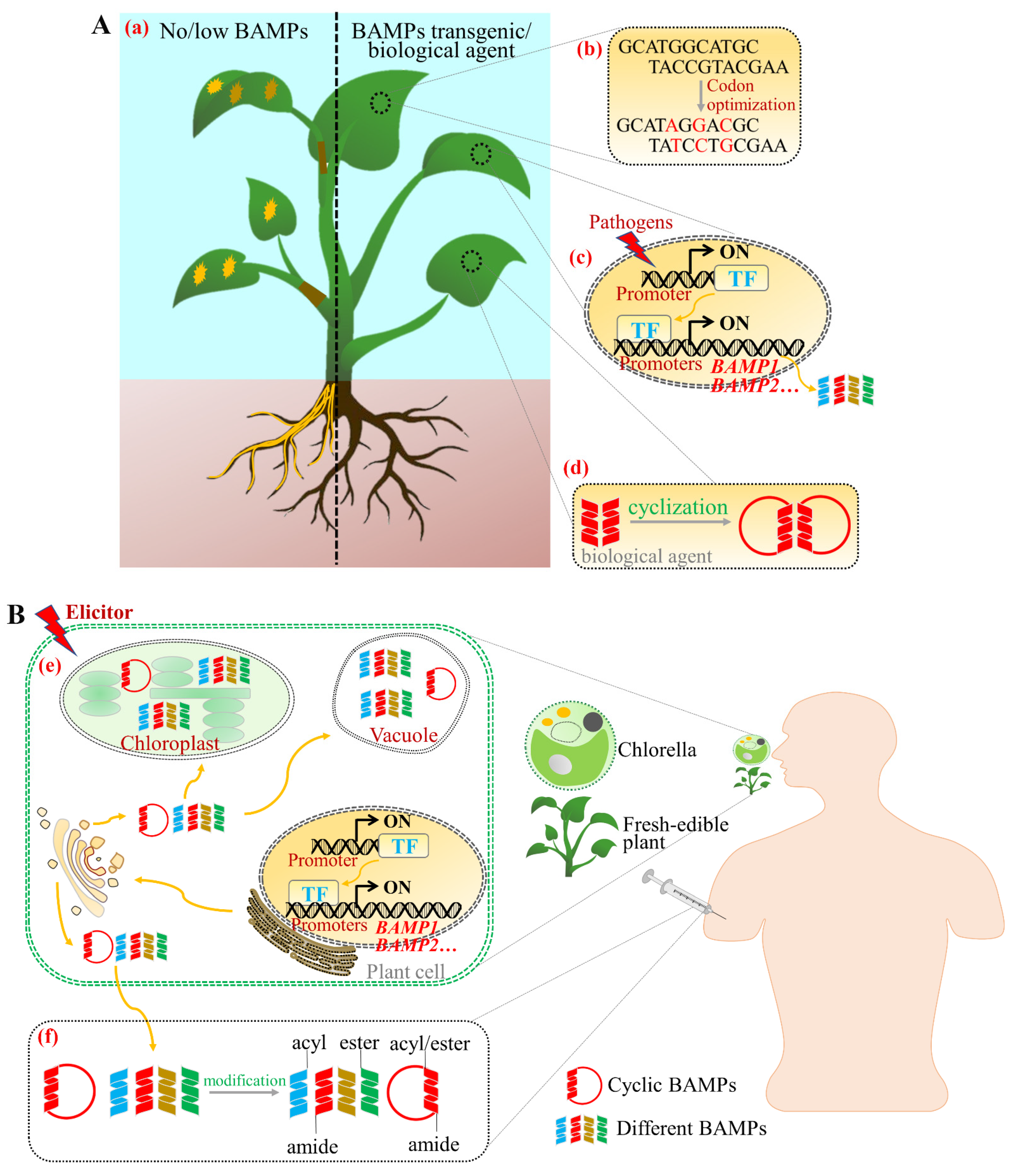
4. Applications of BAMPs for Agricultural Purposes

The in vitro and in vivo activities of many BAMPs are well-known and show clear potential for agricultural purposes. Plants expressing exogenous BAMP variants, synthetic BAMPs, and isogenous BAMPs are capable of producing resistance to a variety of pathogens [164,165]. Particularly, defensins have been heterologously expressed in many economically important crops to enhance resistance to pathogenic fungi. The radish defensin Rs-AFP2 was the first heterologous BAMP to be expressed in other plants, inducing protection against Alternaria longipes in tobacco and tomato [13], and against different fungi in wheat [14]. Other BAMPs also have been used to induce resistance in different plants, such as overexpression of Pn-AMP in tobacco and tomato enhances their resistance to Phytophthora nicotianae and Phytophthora capsici, respectively [166]. Further, overexpression of the barley LTP gene in A. thaliana enhances its resistance to Pseudomonas syringae and Staphylococcus griseus [167]. Compared with resistance enhancement by overexpression of a single gene, co-expression of multiple genes could incur stronger resistance. For example, co-expression of the defensins Dm-AMP1 and Rs-AFP2 in rice results in plants with stronger antifungal activity than the expression of either defensin alone [168]. In addition to their antimicrobial activity, BAMPs are able to promote plant growth and optimize crop traits. Therefore, combining their antimicrobial ability with growth-promoting effects can better control disease, improve crop yield, and ensure food security. Additionally, many non-plant AMPs also play an important role in plant protection in agriculture. For example, cecropin A and B from Hyalophora Cecropia are expressed in rice and tomato, enhancing the resistance of these crops to bacterial and fungal diseases [169,170,171]. The undecapeptide BP100 and its derivatives, which were identified from a library of synthetic cecropin A-melittin hybrids, can also improve the anti-pathogen activity of plants [172,173]. Unfortunately, due to their limited stability and activity conditions, BAMPs and non-plant AMPs have not yet been directly prepared as commercial biofungicides.
The rational, efficient, and innovative use of BAMPs will have a multiplier effect on the disease prevention and control. In summary, the following strategies are proposed (Figure 3A): (1) Exploring the expression regulation mode of BAMPs and optimizing BAMPs codons to increase their expression and activity. (2) Excluding the toxic effects of BAMP accumulation on host plants by using pathogens to induce high expression of BAMPs with high activity, multiple mechanisms of action, and low toxicity. (3) Inducing synergistic expression of BAMPs from different sources, with variable mechanisms of action to expand the range and variability of host resistance to pathogens. (4) Modifying BAMPs in vitro to enhance their stability and improve disease resistance for preparing them into biological agents for direct external application. Overall, the use of BAMPs for both molecular breeding and direct external application can reduce the use of chemical pesticides, control crop losses, and ensure food security. Combining multiple biotechnologies can better expand their application potential.

5. Applications of BAMPs for Human Health

BAMPs have emerged as important novel candidates for the treatment of various human infections due to their high efficiency, specific selectivity, broad range of targets, high permeability in tissues, low immunogenicity, low toxicity, and tissue accumulation. In addition to antimicrobial effects, some BAMPs have shown promise as anticancer agents, as their activity against target cells and/or induced immune responses can effectively control infections and reduce tumorigenesis [10]. Further, BAMPs have shown promising therapeutic effects in combination with conventional therapies [10]. The most well-studied BAMP, lunasin [137], has become the reference and standard for elucidating the anticancer mechanism of BAMPs, and provides a viable candidate for drug development. In addition, the reported anticancer mechanisms of BAMPs, such as viscotoxin B2 [144], ligatoxin B [145], NaD1 [143], CyO2 [140], MCo-PMI [141], and NTP [16], have indicated their potential to be developed as anticancer drugs. The numerous advantages of BAMPs have successfully attracted the attention of the pharmaceutical industry [174]. Currently, some BAMPs and their derivatives are in various clinical trial stages, such as Brilacidin (defensin mimetic), Surotomycin (cyclic lipopeptide), PAC-113 (12 amino acid antimicrobial peptide), and HB1275 (lipohexapeptide) [175]. Like other peptides of natural origin, BAMPs mostly exist at very low concentrations, and would need to be produced in large quantities for pharmaceutical or biological applications [10]. Although the development process is similar to that of traditional small molecule drugs, peptide drugs require unique process designs, preparation methods, structure confirmation, and production equipment. In particular, the large-scale commercial production of high-purity antimicrobial peptides is a practical demand for the treatment of diseases. The lack of a suitable manufacturing platform in terms of product yield, cost, and purity is a barrier to the medical use of BAMPs. Recent advances in biotechnology allow plants to be employed as bioreactors for BAMPs production, as mentioned above [176]. In addition, non-plant AMPs can also be expressed in plants for their use in a global health strategy. For example, retrocyclin-101 (RC101) and protegrin-1 (PG1) are two important antimicrobial peptides that can be used to treat bacterial and/or viral infections, especially those caused by HIV-1 or sexually transmitted bacteria. Lee et al. used plant molecular farming to achieve stable expression of these two AMPs in tobacco, which accounted for 38% and 26% of total soluble protein of chloroplast, respectively [177]. Similarly, lactostatin is an anti-hypercholesterolemic peptide derived from β-lactoglobulin in cow’s milk. Cabanos et al. realized that the expression of lactostatin in rice, and its content in dry seeds, reached 2 mg/g, which has potential clinical application value as an anti-high-cholesterol peptide drug [178]. Therefore, the establishment of transgenic plants expressing bioactive BAMPs and non-plant AMPs is a promising strategy for the production of therapeutic AMPs. In addition, peptides have a short half-life and are easily broken down by enzymes in the body, making them difficult to absorb through the digestive system. Accordingly, peptide drugs are often administered by injection.
Based on the limitations of peptide drug production and clinical delivery methods, the following solutions are proposed (Figure 3B): (1) Prioritizing cyclic peptides or cyclizing linear peptides with high antimicrobial activity and anticancer activity, since the head-to-tail cyclic backbone and the cysteine binding motif of cyclic peptides make them more stable and resistant to thermal or enzymatic degradation [179]. Additionally, the cyclization of linear peptides leads to a reduction in their conformational flexibility or actually decreases the number of hydrogen bonds formed by the peptide, thus increasing their ability to pass through biological membranes and their resistance to endopeptidases and exopeptidases [180]. (2) Using fresh-edible plants as bioreactors and BAMPs with medicinal value as a starting point, then, modify the N-terminus of BAMPs by compartmentalization and localization to allow targeted biosynthesis of BAMPs in organelles such as vesicles and chloroplasts, thereby adding a protective layer and reducing their degradation by the digestive tract during consumption. (3) Combined with synthetic biology, scaling up production of medicinal BAMPs using Chlorella pyrenoidosa as a bioreactor, which could be photoautotrophic and heterotrophic. C. pyrenoidosa is a single-celled algae that is rich in nutrients and protein content (more than 50% of dry weight), making it a new resource food with health value. Due to its fast growth rate, simple nutritional requirements, low cost, and that it can be cultivated via heterotrophic fermentation on a large scale, C. pyrenoidosa is an ideal chassis for biosynthesis. On the one hand, using C. pyrenoidosa as a chassis to synthesize BAMPs can directly treat diseases through oral administration. On the other hand, BAMPs could be synthesized and secreted outside the cells, enabling extraction from the fermentation broth and subsequent modification to enhance their stability for oral or intravenous administration. (4) Modifying the N- and/or C-terminus of the peptide sequence through N-acylation, N-esterification, or C-amidation using biotechnology to increase the stability of BAMPs and enhance their ability to pass through biological membranes [180].

6. Outlook

Researchers have gained a profound understanding of BAMPs in recent decades. To date, they have isolated and identified hundreds of BAMPs, analyzed their functions against plant pathogens, and elucidated the mechanisms underlying the antimicrobial action of some BAMPs. However, there remains a big knowledge gap for the development of BAMPs into anti/fungicidal agents for application in plant protection. Firstly, the mechanism of action remains unclear. Some studies have reported that BAMPs interact with the cell membrane and cell wall components of pathogens, causing membrane perforation and leakage of cell contents, which achieves antimicrobial efficacy. However, the detailed process of membrane perforation caused by BAMPs requires further study. Further, some studies have reported that BAMPs can be internalized into the cytoplasm and interact with intracellular targets to mediate cellular signaling pathways, leading to apoptosis of pathogenic cells. However, it is unknown how BAMPs are internalized into cells, which targets they interact with, and how they mediate cellular signaling in pathogen cells. The lag in mechanistic studies has hindered our in-depth understanding of BAMPs. Secondly, multiple BAMPs are upregulated to participate together in the immune response when plants are infected by pathogens. In practical applications, BAMPs’ synergism may be required to achieve optimal efficacy. Finally, external application of BAMPs faces limitations such as short half-lives, weaker resistance, and harsh activity conditions. Therefore, enhancing the stability of BAMPs is an inescapable problem that must be resolved prior to their industrial application.
The field of peptide therapeutics is rapidly growing. To date, more than 80 peptide drugs have been approved by the FDA or the EU EMA, and dozens of AMPs are currently being evaluated in clinical trials. Although some studies report BAMP efficacy in cancer inhibition, as well as inhibition of viral replication and propagation, no drugs developed with a BAMP backbone have yet to be marketed. Lunasin is one of the more thoroughly studied BAMPs, which not only targets the chromatin in cells, but also disrupts the formation of the mitotic complex in the N-terminal region of histones and blocks normal mitosis. Additionally, lunasin is able to target tumor cell integrin receptors specifically through its RGD domain and mediate the integrin signaling pathway, thereby inhibiting tumor cell proliferation. Most importantly, lunasin is resistant to digestion and can be absorbed directly after oral administration to reach specific tissue sites, which provides a reference for the development of therapeutic foods and oral BAMP-derived drugs, and provides insight for basic research on BAMPs. Is it possible for BAMPs to interfere with signaling pathways by targeting specific tumor cell surface receptors, while being internalized intracellularly, to interact with target proteins and thus induce apoptosis or other biological process in cancer cells? Moreover, due to their short half-lives and instability, peptides are currently administered locally by intravenous injection. Developing BAMPs for direct therapeutic feeding or oral administration would be a meaningful and promising alternative. Based on synthetic biotechnology, whole edible plants or edible microalgae can be used as bioreactors, and compartmentalized synthesis and amino acid modification can be used to synthesize BAMPs, thus optimizing the structural stability of BAMPs and solving scale-up issues for the industrial production of BAMPs.
Currently, the global market for peptide drugs has significant growth potential. Future in-depth research addressing the aforementioned limitations of BAMPs will enable their successful application in agriculture and human health. Due to the advantages of high yield, high quality, homogeneity, and post-translational modifications such as glycosylation and disulfide bond formation that are critical to BAMPs’ activity in plant bioreactors, a plant-based BAMPs’ production platform is more promising than other biological systems [176]. It is believed that plant molecular farms can play a greater role in the production of BAMPs if the yield and stability of the products can be further solved to meet the needs of the market, as well as the safety, quality, and effectiveness.

Author Contributions

Conceptualization, X.L. and M.R.; writing—original draft preparation, X.L. and W.W.; writing—review and editing, H.T. and M.R.; funding acquisition, X.L. and M.R.; project administration, L.F. All authors have read and agreed to the published version of the manuscript.

Funding

This work is funded by the National key R&D program of China (2017YFE0115500), the National Natural Science Foundation of China (31801911, 31972469), the Basic Research and Frontier Exploration Foundation of Chongqing (cstc2018jcyjAX0753), the Central Public-Interest Scientific Institution Basal Research Fund (Y2021XK05), and the Science and Technology Innovation Project of the Chinese Academy of Agricultural Sciences (No. 34-IUA-02).

Institutional Review Board Statement

Not applicable.

Informed Consent Statement

Not applicable.

Data Availability Statement

Not applicable.

Acknowledgments

We acknowledge all the members of the functional plant innovation team for their valuable comments on the manuscript.

Conflicts of Interest

The authors declare no competing interests.

References

  1. Goyal, R.K.; Mattoo, A.K. Multitasking antimicrobial peptides in plant development and host defense against biotic/abiotic stress. Plant Sci. 2014, 228, 135–149. [Google Scholar] [CrossRef]
  2. van der Weerden, N.L.; Bleackley, M.R.; Anderson, M.A. Properties and mechanisms of action of naturally occurring antifungal peptides. Cell. Mol. Life Sci. 2013, 70, 3545–3570. [Google Scholar] [CrossRef]
  3. Zasloff, M. Antimicrobial peptides of multicellular organisms. Nature 2002, 415, 389–395. [Google Scholar] [CrossRef] [PubMed]
  4. Yount, N.Y.; Yeaman, M.R. Immunocontinuum: Perspectives in antimicrobial peptide mechanisms of action and resistance. Protein Pept. Lett. 2005, 12, 49–67. [Google Scholar] [CrossRef] [PubMed]
  5. Alfred, R.L.; Palombo, E.A.; Panozzo, J.F.; Bhave, M. The antimicrobial domains of wheat puroindolines are cell-penetrating peptides with possible intracellular mechanisms of action. PLoS ONE 2013, 8, e75488. [Google Scholar] [CrossRef] [PubMed] [Green Version]
  6. Nanni, V.; Schumacher, J.; Giacomelli, L.; Brazzale, D.; Sbolci, L.; Moser, C.; Tudzynski, P.; Baraldi, E. VvAMP2, a grapevine flower-specific defensin capable of inhibiting Botrytis cinerea growth: Insights into its mode of action. Plant Pathol. 2014, 63, 899–910. [Google Scholar] [CrossRef] [Green Version]
  7. Struyfs, C.; Cammue, B.P.A.; Thevissen, K. Membrane-interacting antifungal peptides. Front. Cell Dev. Biol. 2021, 9, 706. [Google Scholar] [CrossRef]
  8. Rahnamaeian, M. Antimicrobial peptides modes of mechanism, modulation of defense responses. Plant Signal. Behav. 2011, 6, 1325–1332. [Google Scholar] [CrossRef]
  9. Hammami, R.; Ben Hamida, J.; Vergoten, G.; Fliss, I. PhytAMP: A database dedicated to antimicrobial plant peptides. Nucleic Acids Res. 2009, 37, D963–D968. [Google Scholar] [CrossRef] [Green Version]
  10. Candido, E.d.S.; Silva Cardoso, M.H.e.; Sousa, D.A.; Viana, J.C.; de Oliveira-Junior, N.G.; Miranda, V.; Franco, O.L. The use of versatile plant antimicrobial peptides in agribusiness and human health. Peptides 2014, 55, 65–78. [Google Scholar] [CrossRef]
  11. Tam, J.P.; Wang, S.; Wong, K.H.; Tan, W.L. Antimicrobial peptides from plants. Pharmaceuticals 2015, 8, 711–757. [Google Scholar] [CrossRef]
  12. Lay, F.T.; Anderson, M.A. Defensins-components of the innate immune system in plants. Curr. Protein Pept. Sci. 2005, 6, 85–101. [Google Scholar] [CrossRef]
  13. Terras, F.R.G.; Eggermont, K.; Kovaleva, V.; Raikhel, N.V.; Osborn, R.W.; Kester, A.; Rees, S.B.; Torrekens, S.; Vanleuven, F.; Vanderleyden, J.; et al. SMALL cysteine-rich antifungal proteins from radish-their role in host-defense. Plant Cell 1995, 7, 573–588. [Google Scholar] [CrossRef] [PubMed] [Green Version]
  14. Li, Z.; Zhou, M.; Zhang, Z.; Ren, L.; Du, L.; Zhang, B.; Xu, H.; Xin, Z. Expression of a radish defensin in transgenic wheat confers increased resistance to Fusarium graminearum and Rhizoctonia cerealis. Funct. Integr. Genom. 2011, 11, 63–70. [Google Scholar] [CrossRef] [PubMed]
  15. Ghag, S.B.; Shekhawat, U.K.S.; Ganapathi, T.R. Petunia floral defensins with unique prodomains as novel candidates for development of Fusarium wilt resistance in transgenic banana plants. PLoS ONE 2012, 7, e39557. [Google Scholar] [CrossRef] [PubMed]
  16. Ooi, L.S.M.; Tian, L.; Su, M.; Ho, W.S.; Sun, S.S.M.; Chung, H.Y.; Wong, H.N.C.; Ooi, V.E.C. Isolation, characterization, molecular cloning and modeling of a new lipid transfer protein with antiviral and antiproliferative activities from Naycissus tazetta. Peptides 2008, 29, 2101–2109. [Google Scholar] [CrossRef] [PubMed]
  17. Ilyas, H.; Datta, A.; Bhunia, A. An approach towards structure based antimicrobial peptide design for use in development of transgenic plants: A strategy for plant disease management. Curr. Med. Chem. 2017, 24, 1350–1364. [Google Scholar] [CrossRef]
  18. van Loon, L.C.; Rep, M.; Pieterse, C.M.J. Significance of inducible defense-related proteins in infected plants. Annu. Rev. Phytopathol. 2006, 44, 135–162. [Google Scholar] [CrossRef] [PubMed] [Green Version]
  19. Parisi, K.; Shafee, T.M.A.; Quimbar, P.; van der Weerden, N.L.; Bleackley, M.R.; Anderson, M.A. The evolution, function and mechanisms of action for plant defensins. Semin. Cell Dev. Biol. 2019, 88, 107–118. [Google Scholar] [CrossRef]
  20. Lay, F.T.; Poon, S.; McKenna, J.A.; Connelly, A.A.; Barbeta, B.L.; McGinness, B.S.; Fox, J.L.; Daly, N.L.; Craik, D.J.; Heath, R.L.; et al. The C-terminal propeptide of a plant defensin confers cytoprotective and subcellular targeting functions. BMC Plant Biol. 2014, 14, 55–67. [Google Scholar] [CrossRef] [Green Version]
  21. van der Weerden, N.L.; Anderson, M.A. Plant defensins: Common fold, multiple functions. Fungal Biol. Rev. 2013, 26, 121–131. [Google Scholar] [CrossRef]
  22. Wilmes, M.; Cammue, B.P.A.; Sahl, H.G.; Thevissen, K. Antibiotic activities of host defense peptides: More to it than lipid bilayer perturbation. Cheminform 2011, 28, 1350–1358. [Google Scholar] [CrossRef]
  23. Schneider, T.; Kruse, T.; Wimmer, R.; Wiedemann, I.; Sass, V.; Pag, U.; Jansen, A.; Nielsen, A.K.; Mygind, P.H.; Ravents, D.S.; et al. Plectasin, a fungal defensin, targets the bacterial cell wall precursor lipid II. Science 2010, 328, 1168–1172. [Google Scholar] [CrossRef] [PubMed] [Green Version]
  24. Thevissen, K.; Franois, I.E.J.A.; Takemoto, J.Y.; Ferket, K.K.A.; Meert, E.M.K.; Cammue, B.P.A. DmAMP1, an antifungal plant defensin from dahlia (Dahlia merckii), interacts with sphingolipids from Saccharomyces cerevisiae. FEMS Microbiol. Lett. 2003, 226, 169–173. [Google Scholar] [CrossRef]
  25. Thevissen, K.; Ferket, K.K.A.; Franois, I.E.J.A.; Cammue, B.P.A. Interactions of antifungal plant defensins with fungal membrane components. Peptides 2003, 24, 1705–1712. [Google Scholar] [CrossRef] [PubMed]
  26. Payne, J.A.E.; Bleackley, M.R.; Lee, T.H.; Shafee, T.M.A.; Poon, I.K.H.; Hulett, M.D.; Aguilar, M.I.; van der Weerden, N.L.; Anderson, M.A. The plant defensin NaD1 introduces membrane disorder through a specific interaction with the lipid, phosphatidylinositol 4,5 bisphosphate. Biomembranes 2016, 33, 1099–1109. [Google Scholar] [CrossRef]
  27. Poon, I.K.; Baxter, A.A.; Lay, F.T.; Mills, G.D.; Hulett, M.D. Phosphoinositide-mediated oligomerization of a defensin induces cell lysis. Elife Sci. 2014, 3, e01808. [Google Scholar] [CrossRef]
  28. Thevissen, K.; Cammue, B.; Lemaire, K.; Winderickx, J.; Dickson, R.; Lester, R.; Ferket, K.; van Even, F.; Parret, A.; Broekaert, W. A gene encoding a sphingolipid biosynthesis enzyme determines the sensitivity of Saccharomyces cerevisiae to an antifungal plant defensin from dahlia (Dahlia merckii). Proc. Natl. Acad. Sci. USA 2000, 97, 9531–9536. [Google Scholar] [CrossRef] [Green Version]
  29. Thevissen, K. Defensins from insects and plants interact with fungal glucosylceramides. J. Biol. Chem. 2004, 279, 3900–3905. [Google Scholar] [CrossRef] [Green Version]
  30. Aerts, A.M.; Fran, I.E.J.A.; Meert, E.M.K.; Li, Q.T.; Cammue, B.P.A.; Thevissen, K. The antifungal activity of RsAFP2, a plant defensin from Raphanus sativus, involves the induction of reactive oxygen species in Candida albicans. J. Mol. Microbiol. Biotechnol. 2007, 13, 243–247. [Google Scholar] [CrossRef]
  31. Thevissen, K.; Tavares, P.D.M.; Xu, D.; Blankenship, J.; Vandenbosch, D.; Idkowiak-Baldys, J.; Govaert, G.; Bink, A.; Rozental, S.; Groot, P.W.J.D. The plant defensin RsAFP2 induces cell wall stress, septin mislocalization and accumulation of ceramides in Candida albicans. Mol. Microbiol. 2012, 84, 166–180. [Google Scholar] [CrossRef] [PubMed] [Green Version]
  32. Vriens, K.; Cammue, B.; Thevissen, K. Antifungal plant defensins: Mechanisms of action and production. Molecules 2014, 19, 12280–12303. [Google Scholar] [CrossRef] [Green Version]
  33. Spelbrink, R.G. Differential antifungal and calcium channel-blocking activity among structurally related plant defensins. Plant Physiol. 2004, 135, 2055–2067. [Google Scholar] [CrossRef] [PubMed] [Green Version]
  34. Ramamoorthy, V.; Cahoon, E.B.; Li, J.; Thokala, M.; Shah, D.M. Glucosylceramide synthase is essential for alfalfa defensin-mediated growth inhibition but not for pathogenicity of Fusarium graminearum. Mol. Microbiol. 2010, 66, 771–786. [Google Scholar] [CrossRef] [PubMed]
  35. van der Weerden, N.L.; Hancock, R.E.W.; Anderson, M.A. Permeabilization of fungal hyphae by the plant defensin NaD1 occurs through a cell wall-dependent process. J. Biol. Chem. 2010, 285, 37513–37520. [Google Scholar] [CrossRef] [Green Version]
  36. Aerts, A.M.; Leen, B.; Gilmer, G.; Didac, C.G.; Frank, M.; Cammue, B.P.A.; Karin, T. The antifungal plant defensin HsAFP1 from Heuchera sanguinea induces apoptosis in Candida albicans. Front. Microbiol. 2011, 2, 47. [Google Scholar] [CrossRef] [Green Version]
  37. Lobo, D.S.; Pereira, I.B.; Fragel-Madeira, L.; Medeiros, L.N.; Kurtenbach, E. Antifungal Pisum sativum defensin 1 interacts with Neurospora crassa cyclin F related to the cell cycle. Biochemistry 2007, 46, 987–996. [Google Scholar] [CrossRef] [PubMed]
  38. Loon, L.C.V.; Pierpoint, W.S.; Boller, T.; Conejero, V. Recommendations for naming plant pathogenesis-related proteins. Plant Mol. Biol. Rep. 1994, 12, 245–264. [Google Scholar] [CrossRef]
  39. Loon, L.C.V.; Strien, E.A.V. The families of pathogenesis-related proteins, their activities, and comparative analysis of PR-1 type proteins. Physiol. Mol. Plant Pathol. 2002, 55, 85–97. [Google Scholar] [CrossRef]
  40. Sung, Y.C.; Outram, M.A.; Breen, S.; Wang, C.; Dagvadorj, B.; Winterberg, B.; Kobe, B.; Williams, S.J.; Solomon, P.S. PR1-mediated defence via C-terminal peptide release is targeted by a fungal pathogen effector. New Phytol. 2021, 229, 3467–3480. [Google Scholar] [CrossRef]
  41. Alexander, D.; Goodman, R.M.; Gut-Rella, M.; Glascock, C.; Weymann, K.; Friedrich, L.; Maddox, D.; Ahl-Goy, P.; Luntz, T.; Ward, E. Increased tolerance to two oomycete pathogens in transgenic tobacco expressing pathogenesis-related protein 1a. Proc. Natl. Acad. Sci. USA 1993, 90, 7327–7331. [Google Scholar] [CrossRef] [Green Version]
  42. Niderman, T.; Genetet, I.; Bruyere, T.; Gees, R.; Stintzi, A.; Legrand, M.; Fritig, B.; Mosinger, E. Pathogenesis-Related PR-1 proteins are antifungal (isolation and characterization of three 14-kilodalton proteins of tomato and of a basic PR-1 of tobacco with inhibitory activity against Phytophthora infestans). Plant Physiol. 1995, 108, 17–27. [Google Scholar] [CrossRef] [Green Version]
  43. Gamir, J.; Darwiche, R.; van‘t Hof, P.; Choudhary, V.; Stumpe, M.; Schneiter, R.; Mauch, F. The sterol-binding activity of pathogenesis-related protein 1 reveals the mode of action of an antimicrobial protein. Plant J. 2017, 89, 502–509. [Google Scholar] [CrossRef] [Green Version]
  44. Choudhary, V.; Schneiter, R. Pathogen-Related Yeast (PRY) proteins and members of the CAP superfamily are secreted sterol-binding proteins. Proc. Natl. Acad. Sci. USA 2012, 109, 16882–16887. [Google Scholar] [CrossRef] [Green Version]
  45. Lincoln, J.E.; Sanchez, J.P.; Zumstein, K.; Gilchrist, D.G. Plant and animal PR1 family members inhibit programmed cell death and suppress bacterial pathogens in plant tissues. Mol. Plant Pathol. 2018, 19, 2111–2123. [Google Scholar] [CrossRef] [Green Version]
  46. Chien, P.S.; Nam, H.G.; Chen, Y.R. A salt-regulated peptide derived from the CAP superfamily protein negatively regulates salt-stress tolerance in Arabidopsis. J. Exp. Bot. 2015, 66, 5301–5313. [Google Scholar] [CrossRef] [Green Version]
  47. Chen, Y.L.; Lee, C.Y.; Cheng, K.T.; Chang, W.H.; Huang, R.N.; Nam, H.G.; Chen, Y.R. Quantitative peptidomics study reveals that a wound-induced peptide from PR-1 regulates immune signaling in tomato. Plant Cell 2014, 26, 4135–4148. [Google Scholar] [CrossRef] [Green Version]
  48. Lu, S.; Faris, J.D.; Sherwood, R.; Friesen, T.L.; Edwards, M.C. A dimeric PR-1-type pathogenesis-related protein interacts with ToxA and potentially mediates ToxA-induced necrosis in sensitive wheat. Mol. Plant Pathol. 2014, 15, 650–663. [Google Scholar] [CrossRef] [PubMed]
  49. Breen, S.; Williams, S.J.; Winterberg, B.; Kobe, B.; Solomon, P.S. Wheat PR-1 proteins are targeted by necrotrophic pathogen effector proteins. Plant J. 2016, 88, 13–25. [Google Scholar] [CrossRef] [PubMed] [Green Version]
  50. Yang, G.; Tang, L.; Gong, Y.; Xie, J.; Fu, Y.; Jiang, D.; Li, G.; Collinge, D.B.; Chen, W.; Cheng, J. A cerato-platanin protein SsCP1 targets plant PR1 and contributes to virulence of Sclerotinia sclerotiorum. New Phytol. 2018, 217, 739–755. [Google Scholar] [CrossRef] [PubMed] [Green Version]
  51. Bohlmann, H.; Apel, K. Thionins. Annu. Rev. Plant Physiol. Plant Mol. Biol. 1991, 42, 227–240. [Google Scholar] [CrossRef]
  52. Stec, B. Plant thionins-the structural perspective. Cell. Mol. Life Sci. 2006, 63, 1370–1385. [Google Scholar] [CrossRef]
  53. Colilla, F.J.; Rocher, A.; Mendez, E. gamma-Purothionins: Amino acid sequence of two polypeptides of a new family of thionins from wheat endosperm. FEBS Lett. 1990, 270, 191–194. [Google Scholar] [CrossRef] [Green Version]
  54. Romero, A.; Alamillo, J.M.; Garcia-Olmedo, F. Processing of thionin precursors in barley leaves by a vacuolar proteinase. Eur. J. Biochem. 1997, 243, 202–208. [Google Scholar] [CrossRef] [Green Version]
  55. Epple, P.; Apel, K.; Bohlmann, H. Overexpression of an endogenous thionin enhances resistance of Arabidopsis against Fusarium oxysporum. Plant Cell 1997, 9, 509–520. [Google Scholar] [CrossRef] [PubMed] [Green Version]
  56. Chan, Y.L.; Prasad, V.; Sanjaya; Chen, K.H.; Liu, P.C.; Chan, M.T.; Chiu-Ping, C. Transgenic tomato plants expressing an Arabidopsis thionin (Thi2.1) driven by fruit-inactive promoter battle against phytopathogenic attack. Planta 2005, 221, 386–393. [Google Scholar] [CrossRef] [PubMed]
  57. Carmona, M.J.; Molina, A.; Fernandez, J.A.; Lopezfando, J.J.; Garciaolmedo, F. Expression of the alpha-thionin gene from barley in tobacco confers enhanced resistance to bacterial pathogens. Plant J. 1993, 3, 457–462. [Google Scholar] [CrossRef] [PubMed]
  58. Iwai, T.; Kaku, H.; Honkura, R.; Nakamura, S.; Ochiai, H.; Sasaki, T.; Ohashi, Y. Enhanced resistance to seed-transmitted bacterial diseases in transgenic rice plants overproducing an oat cell-wall-bound thionin. Mol. Plant Microbe Interact. 2002, 15, 515–521. [Google Scholar] [CrossRef] [PubMed]
  59. Richard, J.A.; Kelly, I.; Marion, D.; Pezolet, M.; Auger, M. Interaction between beta-purothionin and dimyristoylphosphatidylglycerol: A P-31-NMR and infrared spectroscopic study. Biophys. J. 2002, 83, 2074–2083. [Google Scholar] [CrossRef] [Green Version]
  60. Thevissen, K.; Terras, F.R.G.; Broekaert, W.F. Permeabilization of fungal membranes by plant defensins inhibits fungal growth. Appl. Environ. Microbiol. 1999, 65, 5451–5458. [Google Scholar] [CrossRef] [Green Version]
  61. Molina, A.; Segura, A.; Garciaolmedo, F. Lipid transfer proteins (NSLTPS) from barley and maize leaves are potent inhibitors of bacterial and fungal plant-pathogens. FEBS Lett. 1993, 316, 119–122. [Google Scholar] [CrossRef] [Green Version]
  62. Evans, J.; Wang, Y.D.; Shaw, K.P.; Vernon, L.P. Cellular responses to Pyrularia thionin are mediated by Ca2+ influx and phospholipase A2 activation and are inhibited by thionin tyrosine iodination. Proc. Natl. Acad. Sci. USA 1989, 86, 5849–5853. [Google Scholar] [CrossRef] [Green Version]
  63. Stein, G.M.; Pfuller, U.; Schietzel, M.; Bussing, A. Toxic proteins from European mistletoe (Viscum album L.: Increase of intracellular IL-4 but decrease of IFN-gamma in apoptotic cells. Anticancer Res. 2000, 20, 1673–1678. [Google Scholar]
  64. Richard, J.A.; Kelly, I.; Marion, D.; Auger, M.; Pezolet, M. Structure of beta-purothionin in membranes: A two-dimensional infrared correlation spectroscopy study. Biochemistry 2005, 44, 52–61. [Google Scholar] [CrossRef]
  65. Salminen, T.A.; Blomqvist, K.; Edqvist, J. Lipid transfer proteins: Classification, nomenclature, structure, and function. Planta 2016, 244, 971–997. [Google Scholar] [CrossRef] [Green Version]
  66. Castro, M.S.; Fontes, W. Plant defense and antimicrobial peptides. Protein Pept. Lett. 2005, 12, 13–18. [Google Scholar] [CrossRef]
  67. Berecz, B.; Mills, E.N.C.; Tamas, L.; Lang, F.; Shewry, P.R.; Mackie, A.R. Structural stability and surface activity of sunflower 2s albumins and nonspecific lipid transfer protein. J. Agric. Food Chem. 2010, 58, 6490–6497. [Google Scholar] [CrossRef]
  68. Edqvist, J.; Blomqvist, K.; Nieuwland, J.; Salminen, T.A. Plant lipid transfer proteins: Are we finally closing in on the roles of these enigmatic proteins? J. Lipid Res. 2018, 59, 1374–1382. [Google Scholar] [CrossRef] [Green Version]
  69. Carvalho, A.d.O.; Gomes, V.M. Role of plant lipid transfer proteins in plant cell physiology—A concise review. Peptides 2007, 28, 1144–1153. [Google Scholar] [CrossRef]
  70. Nieuwland, J.; Feron, R.; Huisman, B.A.H.; Fasolino, A.; Hilbers, C.W.; Derksen, J.; Mariani, C. Lipid transfer proteins enhance cell wall extension in tobacco. Plant Cell 2005, 17, 2009–2019. [Google Scholar] [CrossRef] [Green Version]
  71. Tsuboi, S.; Osafune, T.; Tsugeki, R.; Nishimura, M.; Yamada, M. Nonspecific lipid transfer protein in castor bean cotyledon cells subcellular localization and a possible role in lipid-metabolism. J. Biochem. 1992, 111, 500–508. [Google Scholar] [CrossRef]
  72. Jacq, A.; Pernot, C.; Martinez, Y.; Domergue, F.; Payre, B.; Jamet, E.; Burlat, V.; Pacquit, V.B. The Arabidopsis lipid transfer protein 2 (AtLTP2) is involved in cuticle-cell wall interface integrity and in etiolated hypocotyl permeability. Front. Plant Sci. 2017, 8, 263. [Google Scholar] [CrossRef] [PubMed] [Green Version]
  73. Deeken, R.; Saupe, S.; Klinkenberg, J.; Riedel, M.; Leide, J.; Hedrich, R.; Mueller, T.D. The nonspecific lipid transfer protein AtLtpI-4 is involved in suberin formation of Arabidopsis thaliana crown galls. Plant Physiol. 2016, 172, 1911–1927. [Google Scholar] [CrossRef] [PubMed] [Green Version]
  74. DeBono, A.; Yeats, T.H.; Rose, J.K.C.; Bird, D.; Jetter, R.; Kunst, L.; Samuelsa, L. Arabidopsis LTPG Is a glycosylphosphatidylinositol-anchored lipid transfer protein required for export of lipids to the plant surface. Plant Cell 2009, 21, 1230–1238. [Google Scholar] [CrossRef] [PubMed] [Green Version]
  75. Chassot, C.; Nawrath, C.; Metraux, J.P. Cuticular defects lead to full immunity to a major plant pathogen. Plant J. 2007, 49, 972–980. [Google Scholar] [CrossRef] [Green Version]
  76. Molina, A.; GarciaOlmedo, F. Enhanced tolerance to bacterial pathogens caused by the transgenic expression of barley lipid transfer protein LTP2. Plant J. 1997, 12, 669–675. [Google Scholar] [CrossRef] [Green Version]
  77. Diz, M.S.; Carvalho, A.O.; Ribeiro, S.F.F.; Da Cunha, M.; Beltramini, L.; Rodrigues, R.; Nascimento, V.V.; Machado, O.L.T.; Gomes, V.M. Characterisation, immunolocalisation and antifungal activity of a lipid transfer protein from chili pepper (Capsicum annuum) seeds with novel alpha-amylase inhibitory properties. Physiol. Plant. 2011, 142, 233–246. [Google Scholar] [CrossRef]
  78. Gonorazky, A.G.; Regente, M.C.; de la Canal, L. Stress induction and antimicrobial properties of a lipid transfer protein in germinating sunflower seeds. J. Plant Physiol. 2005, 162, 618–624. [Google Scholar] [CrossRef]
  79. Cammue Bruno, P.A.; Thevissen, K.; Hendriks, M.; Eggermont, K.; Goderis, I.J.; Proost, P.; Vandamme, J.; Osborn, R.W.; Guerbette, F.; Kader, J.C.; et al. A potent antimicrobial protein from onion seeds showing sequence homology to plant lipid transfer proteins. Plant Physiol. 1995, 109, 445–455. [Google Scholar] [CrossRef] [Green Version]
  80. Terras, F.R.; Goderis, I.J.; Van Leuven, F.; Vanderleyden, J.; Cammue, B.P.; Broekaert, W.F. In vitro antifungal activity of a radish (Raphanus sativus L.) seed protein homologous to nonspecific lipid transfer proteins. Plant Physiol. 1992, 100, 1055–1058. [Google Scholar] [CrossRef] [Green Version]
  81. Regente, M.C.; Giudici, A.M.; Villalain, J.; de la Canal, L. The cytotoxic properties of a plant lipid transfer protein involve membrane permeabilization of target cells. Lett. Appl. Microbiol. 2005, 40, 183–189. [Google Scholar] [CrossRef] [PubMed]
  82. Kader, J.C. Lipid-transfer proteins in plants. Annu. Rev. Plant Physiol. Plant Mol. Biol. 1996, 47, 627–654. [Google Scholar] [CrossRef] [PubMed] [Green Version]
  83. Glazebrook, J. Contrasting mechanisms of defense against biotrophic and necrotrophic pathogens. Annu. Rev. Phytopathol. 2005, 43, 205–227. [Google Scholar] [CrossRef]
  84. Sels, J.; Mathys, J.; De Coninck, B.M.A.; Cammue, B.P.A.; de Bolle, M.F.C. Plant pathogenesis-related (PR) proteins: A focus on PR peptides. Plant Physiol. Biochem. 2008, 46, 941–950. [Google Scholar] [CrossRef]
  85. Christeller, J.; Laing, W. Plant serine proteinase inhibitors. Protein Pept. Lett. 2005, 12, 439–447. [Google Scholar] [CrossRef]
  86. Haq, S.K.; Atif, S.M.; Khan, R.H. Protein proteinase inhibitor genes in combat against insects, pests, and pathogens: Natural and engineered phytoprotection. Arch. Biochem. Biophys. 2004, 431, 145–159. [Google Scholar] [CrossRef] [PubMed]
  87. Valueva, T.A.; Mosolov, V.V. Role of inhibitors of proteolytic enzymes in plant defense against phytopathogenic microorganisms. Biochem. Mosc. 2004, 69, 1305–1309. [Google Scholar] [CrossRef]
  88. Dunaevskii, Y.E.; Tsybina, T.A.; Belyakova, G.A.; Domash, V.I.; Sharpio, T.P.; Zabreiko, S.A.; Belozerskii, M.A. Proteinase inhibitors as antistress proteins in higher plants. Appl. Biochem. Microbiol. 2005, 41, 344–348. [Google Scholar] [CrossRef]
  89. Gutierrez-Campos, R.; Torres-Acosta, J.A.; Saucedo-Arias, L.J.; Gomez-Lim, M.A. The use of cysteine proteinase inhibitors to engineer resistance against potyviruses in transgenic tobacco plants. Nat. Biotechnol. 1999, 17, 1223–1226. [Google Scholar] [CrossRef]
  90. Vila, L.; Quilis, J.; Meynard, D.; Breitler, J.C.; Marfa, V.; Murillo, I.; Vassal, J.M.; Messeguer, J.; Guiderdoni, E.; San Segundo, B. Expression of the maize proteinase inhibitor (mpi) gene in rice plants enhances resistance against the striped stem borer (Chilo suppressalis): Effects on larval growth and insect gut proteinases. Plant Biotechnol. J. 2005, 3, 187–202. [Google Scholar] [CrossRef]
  91. Peng, J.H.; Black, L.L. Increased proteinase-inhibitor activity in response to infection of resistant tomato plants by Phytophthora infestans. Phytopathology 1976, 66, 958–963. [Google Scholar]
  92. Pautot, V.; Holzer, F.M.; Walling, L.L. Differential expression of tomato proteinase inhibitor-i and inhibitor-ii genes during bacterial pathogen invasion and wounding. Mol. Plant Microbe Interact. 1991, 4, 284–292. [Google Scholar] [CrossRef] [PubMed]
  93. Terras, F.R.G.; Schoofs, H.M.E.; Thevissen, K.; Osborn, R.W.; Vanderleyden, J.; Cammue, B.P.A.; Broekaert, W.F. Synergistic enhancement of the antifungal activity of wheat and barley thionins by radish and oilseed rape-2S albumins and by barley trypsin-inhibitors. Plant Physiol. 1993, 103, 1311–1319. [Google Scholar] [CrossRef] [PubMed] [Green Version]
  94. Vieira Bard, G.C.; Nascimento, V.V.; Ribeiro, S.F.F.; Rodrigues, R.; Perales, J.; Teixeira-Ferreira, A.; Carvalho, A.O.; Fernandes, K.V.S.; Gomes, V.M. Characterization of peptides from Capsicum annuum hybrid seeds with inhibitory activity against α-amylase, serine proteinases and fungi. Protein J. 2015, 34, 122–129. [Google Scholar] [CrossRef] [PubMed]
  95. ten Have, A.; Dekkers, E.; Kay, J.; Phylip, L.H.; van Kan, J.A.L. An aspartic proteinase gene family in the filamentous fungus Botrytis cinerea contains members with novel features. Microbiology 2004, 150, 2475–2489. [Google Scholar] [CrossRef] [PubMed]
  96. Lorito, M.; Peterbauer, T.C.; Hayes, C.K.; Harman, G.E. Synergistic interaction between fungal cell-wall degrading enzymes and different antifungal compounds enhances inhibition of spore germination. Microbiology 1994, 140, 623–629. [Google Scholar] [CrossRef] [PubMed] [Green Version]
  97. Machida, S.; Saito, M. Purification and characterization of membrane-bound chitin synthase. J. Biol. Chem. 1993, 268, 1702–1707. [Google Scholar] [CrossRef]
  98. Ramamoorthy, V.; Zhao, X.; Snyder, A.K.; Xu, J.; Shah, D.M. Two mitogen-activated protein kinase signalling cascades mediate basal resistance to antifungal plant defensins in Fusarium graminearum. Cell. Microbiol. 2010, 9, 1491–1506. [Google Scholar] [CrossRef] [PubMed]
  99. Rao, U.; Stec, B.; Teeter, M.M. Refinement of purothionins reveals solute particles important for lattice formation and toxicity. Part 1: Alpha1-purothionin revisited. Acta Crystallogr. Sect. D 1995, 51, 904–913. [Google Scholar] [CrossRef]
  100. Giudici, A.M.; Regente, M.C.; Villalain, J.; Pfuller, K.; Pfuller, U.; de la Canal, L. Mistletoe viscotoxins induce membrane permeabilization and spore death in phytopathogenic fungi. Physiol. Plant. 2004, 121, 2–7. [Google Scholar] [CrossRef]
  101. Fisher, M.C.; Henk, D.A.; Briggs, C.J.; Brownstein, J.S.; Madoff, L.C.; McCraw, S.L.; Gurr, S.J. Emerging fungal threats to animal, plant and ecosystem health. Nature 2012, 484, 186–194. [Google Scholar] [CrossRef] [PubMed]
  102. Shah, P.; Hsiao, F.S.H.; Ho, Y.H.; Chen, C.S. The proteome targets of intracellular targeting antimicrobial peptides. Proteomics 2016, 16, 1225–1237. [Google Scholar] [CrossRef] [PubMed]
  103. Jarva, M.; Lay, F.T.; Thanh Kha, P.; Humble, C.; Poon, I.K.H.; Bleackley, M.R.; Anderson, M.A.; Hulett, M.D.; Kvansakul, M. X-ray structure of a carpet-like antimicrobial defensin-phospholipid membrane disruption complex. Nat. Commun. 2018, 9, 1962. [Google Scholar] [CrossRef] [PubMed]
  104. Saito, K.; Takakuwa, N.; Ohnishi, M.; Oda, Y. Presence of glucosylceramide in yeast and its relation to alkali tolerance of yeast. Appl. Microbiol. Biotechnol. 2006, 71, 515–521. [Google Scholar] [CrossRef]
  105. Tavares, P.M.; Thevissen, K.; Cammue, B.P.A.; Francois, I.E.J.A.; Barreto-Bergter, E.; Taborda, C.P.; Marques, A.F.; Rodrigues, M.L.; Nimrichter, L. In vitro activity of the antifungal plant defensin RsAFP2 against Candida isolates and its in vivo efficacy in prophylactic murine models of candidiasis. Antimicrob. Agents Chemother. 2008, 52, 4522–4525. [Google Scholar] [CrossRef] [Green Version]
  106. Noble, S.M.; French, S.; Kohn, L.A.; Chen, V.; Johnson, A.D. Systematic screens of a Candida albicans homozygous deletion library decouple morphogenetic switching and pathogenicity. Nat. Genet. 2010, 42, 590–598. [Google Scholar] [CrossRef] [Green Version]
  107. Garcia-Rubio, R.; de Oliveira, H.C.; Rivera, J.; Trevijano-Contador, N. The fungal cell wall: Candida, Cryptococcus, and Aspergillus species. Front. Microbiol. 2020, 10, 2993. [Google Scholar] [CrossRef]
  108. Rodrigues, M.L.; Travassos, L.R.; Miranda, K.R.; Franzen, A.J.; Rozental, S.; de Souza, W.; Alviano, C.S.; Barreto-Bergter, E. Human antibodies against a purified glucosylceramide from Cryptococcus neoformans inhibit cell budding and fungal growth. Infect. Immun. 2000, 68, 7049–7060. [Google Scholar] [CrossRef] [Green Version]
  109. Koo, J.C.; Lee, B.; Young, M.E.; Koo, S.C.; Cooper, J.A.; Baek, D.; Lim, C.O.; Lee, S.Y.; Yun, D.J.; Cho, M.J. Pn-AMP1, a plant defense protein, induces actin depolarization in yeasts. Plant Cell Physiol. 2004, 45, 1669–1680. [Google Scholar] [CrossRef]
  110. Tsai, P.W.; Yang, C.Y.; Chang, H.T.; Lan, C.Y. Human antimicrobial peptide LL-37 inhibits adhesion of Candida albicans by interacting with yeast cell-wall carbohydrates. PLoS ONE 2011, 6, e17755. [Google Scholar] [CrossRef] [Green Version]
  111. Li, H.; Velivelli, S.L.S.; Shah, D.M. Antifungal potency and modes of action of a novel olive tree defensin against closely related ascomycete fungal pathogens. Mol. Plant Microbe Interact. 2019, 32, 1649–1664. [Google Scholar] [CrossRef]
  112. Combest, M.M.; Moroz, N.; Tanaka, K.; Rogan, C.J.; Anderson, J.C.; Thura, L.; Rakotondrafara, A.M.; Goyer, A. StPIP1, a PAMP-induced peptide in potato, elicits plant defenses and is associated with disease symptom severity in a compatible interaction with Potato virus Y. J. Exp. Bot. 2021, 72, 4472–4488. [Google Scholar] [CrossRef] [PubMed]
  113. Reyes, M.I.; Nash, T.E.; Dallas, M.M.; Ascencio-Ibanez, J.T.; Hanley-Bowdoin, L. Peptide aptamers that bind to geminivirus replication proteins confer a resistance phenotype to tomato yellow leaf curl virus and tomato mottle virus infection in tomato. J. Virol. 2013, 87, 9691–9706. [Google Scholar] [CrossRef] [PubMed] [Green Version]
  114. Mendoza-Figueroa, J.S.; Kvarnheden, A.; Mendez-Lozano, J.; Rodriguez-Negrete, E.A.; Arreguin-Espinosa de los Monteros, R.; Soriano-Garcia, M. A peptide derived from enzymatic digestion of globulins from amaranth shows strong affinity binding to the replication origin of Tomato yellow leaf curl virus reducing viral replication in Nicotiana benthamiana. Pestic. Biochem. Physiol. 2018, 145, 56–65. [Google Scholar] [CrossRef] [PubMed]
  115. Rudolph, C.; Schreier, P.H.; Uhrig, J.F. Peptide-mediated broad-spectrum plant resistance to tospoviruses. Proc. Natl. Acad. Sci. USA 2003, 100, 4429–4434. [Google Scholar] [CrossRef] [PubMed] [Green Version]
  116. Wang, H.X.; Ng, T.B. Ginkbilobin, a novel antifungal protein from Ginkgo biloba seeds with sequence similarity to embryo-abundant protein. Biochem. Biophys. Res. Commun. 2000, 279, 407–411. [Google Scholar] [CrossRef]
  117. Wang, H.X.; Ng, T.B. Ascalin, a new anti-fungal peptide with human immunodeficiency virus type 1 reverse transcriptase-inhibiting activity from shallot bulbs. Peptides 2002, 23, 1025–1029. [Google Scholar] [CrossRef]
  118. Wong, J.H.; Ng, T.B. Lunatusin, a trypsin-stable antimicrobial peptide from lima beans (Phaseolus lunatus L.). Peptides 2005, 26, 2086–2092. [Google Scholar] [CrossRef]
  119. Wong, J.H.; Ng, T.B. Vulgarinin, a broad-spectrum antifungal peptide from haricot beans (Phaseolus vulgaris). Int. J. Biochem. Cell Biol. 2005, 37, 1626–1632. [Google Scholar] [CrossRef]
  120. Wachsman, M.B.; Castilla, V.; Coto, C.E. Inhibition of foot and mouth disease virus (FMDV) uncoating by a plant-derived peptide isolated from Melia azedarach L leaves. Arch. Virol. 1998, 143, 581–590. [Google Scholar] [CrossRef]
  121. Panya, A.; Yongpitakwattana, P.; Budchart, P.; Sawasdee, N.; Krobthong, S.; Paemanee, A.; Roytrakul, S.; Rattanabunyong, S.; Choowongkomon, K.; Yenchitsomanus, P.T. Novel bioactive peptides demonstrating anti-dengue virus activity isolated from the Asian medicinal plant Acacia Catechu. Chem. Biol. Drug Des. 2019, 93, 100–109. [Google Scholar] [CrossRef]
  122. Panya, A.; Sawasdee, N.; Songprakhon, P.; Tragoolpua, Y.; Rotarayanont, S.; Choowongkomon, K.; Yenchitsomanus, P.T. A synthetic bioactive peptide derived from the asian medicinal plant Acacia Catechu binds to dengue virus and inhibits cell entry. Viruses Basel 2020, 12, 1267. [Google Scholar] [CrossRef]
  123. Yamaji, Y.; Maejima, K.; Komatsu, K.; Shiraishi, T.; Okano, Y.; Himeno, M.; Sugawara, K.; Neriya, Y.; Minato, N.; Miura, C.; et al. Lectin-mediated resistance impairs plant virus infection at the cellular level. Plant Cell 2012, 24, 778–793. [Google Scholar] [CrossRef] [Green Version]
  124. Yoshida, T.; Shiraishi, T.; Hagiwara-Komoda, Y.; Komatsu, K.; Maejima, K.; Okano, Y.; Fujimoto, Y.; Yusa, A.; Yamaji, Y.; Namba, S. The Plant noncanonical antiviral resistance protein JAX1 inhibits potexviral replication by targeting the viral RNA-dependent RNA polymerase. J. Virol. 2019, 93, e01506-18. [Google Scholar] [CrossRef] [Green Version]
  125. Li, F.; Zhang, C.; Li, Y.; Wu, G.; Hou, X.; Zhou, X.; Wang, A. Beclin1 restricts RNA virus infection in plants through suppression and degradation of the viral polymerase. Nat. Commun. 2018, 9, 1268. [Google Scholar] [CrossRef] [PubMed]
  126. Lin, J.Y.; Mendu, V.; Pogany, J.; Qin, J.; Nagy, P.D. The TPR domain in the host Cyp40-like cyclophilin binds to the viral replication protein and inhibits the assembly of the tombusviral replicase. PLoS Pathog. 2012, 8, e1002491. [Google Scholar] [CrossRef] [PubMed] [Green Version]
  127. Mookherjee, N.; Anderson, M.A.; Haagsman, H.P.; Davidson, D.J. Antimicrobial host defence peptides: Functions and clinical potential. Nat. Rev. Drug Discov. 2020, 19, 311–332. [Google Scholar] [CrossRef] [PubMed]
  128. Kota, S.; Sabbah, A.; Chang, T.H.; Harnack, R.; Xiang, Y.; Meng, X.; Bose, S. Role of human beta-defensin-2 during tumor necrosis factor-alpha/NF-kappa B- mediated innate antiviral response against human respiratory syncytial virus. J. Biol. Chem. 2008, 283, 22417–22429. [Google Scholar] [CrossRef] [Green Version]
  129. Robinson, W.E.; McDougall, B.; Tran, D.; Selsted, M.E. Anti-HIV-1 activity of indolicidin, an antimicrobial peptide from neutrophils. J. Leukoc. Biol. 1998, 63, 94–100. [Google Scholar] [CrossRef] [PubMed]
  130. Smith, J.G.; Nemerow, G.R. Mechanism of adenovirus neutralization by human alpha-defensins. Cell Host Microbe 2008, 3, 11–19. [Google Scholar] [CrossRef] [PubMed] [Green Version]
  131. Lehrer, R.I.; Jung, G.; Ruchala, P.; Andre, S.; Gabius, H.J.; Lu, W. Multivalent binding of carbohydrates by the human alpha-defensin, HD5. J. Immunol. 2009, 183, 480–490. [Google Scholar] [CrossRef] [Green Version]
  132. Wang, W.; Owen, S.M.; Rudolph, D.L.; Cole, A.M.; Hong, T.; Waring, A.J.; Lal, R.B.; Lehrer, R.I. Activity of alpha- and theta-defensins against primary isolates of HIV-1. J. Immunol. 2004, 173, 515–520. [Google Scholar] [CrossRef] [PubMed]
  133. Song, B.H.; Lee, G.C.; Moon, M.S.; Cho, Y.H.; Lee, C.H. Human cytomegalovirus binding to heparan sulfate proteoglycans on the cell surface and/or entry stimulates the expression of human leukocyte antigen class I. J. Gen. Virol. 2001, 82, 2405–2413. [Google Scholar] [CrossRef] [Green Version]
  134. Yasin, B.; Wang, W.; Pang, M.; Cheshenko, N.; Hong, T.; Waring, A.J.; Herold, B.C.; Wagar, E.A.; Lehrer, R.I. Theta defensins protect cells from infection by herpes simplex virus by inhibiting viral adhesion and entry. J. Virol. 2004, 78, 5147–5156. [Google Scholar] [CrossRef] [PubMed] [Green Version]
  135. Andersen, J.H.; Jenssen, H.; Sandvik, K.; Gutteberg, T.J. Anti-HSV activity of lactoferrin and lactoferricin is dependent on the presence of heparan sulphate at the cell surface. J. Med. Virol. 2004, 74, 262–271. [Google Scholar] [CrossRef]
  136. Sousa, F.H.; Casanova, V.; Findlay, F.; Stevens, C.; Svoboda, P.; Pohl, J.; Proudfoot, L.; Barlow, P.G. Cathelicidins display conserved direct antiviral activity towards rhinovirus. Peptides 2017, 95, 76–83. [Google Scholar] [CrossRef] [PubMed]
  137. de Lumen, B.O. Lunasin: A cancer-preventive soy peptide. Nutr. Rev. 2005, 63, 16–21. [Google Scholar] [CrossRef] [PubMed]
  138. Jeong, H.J.; Lam, Y.; de Lumen, B.O. Barley lunasin suppresses ras-induced colony formation and inhibits core histone acetylation in mammalian cells. J. Agric. Food Chem. 2002, 50, 5903–5908. [Google Scholar] [CrossRef] [PubMed]
  139. Jeong, H.J.; Jin, B.J.; Kim, D.S.; Park, J.H.; Lee, J.B.; Kweon, D.H.; Chung, G.Y.; Seo, E.W.; Lumen, B. The cancer preventive peptide lunasin from wheat inhibits core histone acetylation. Cancer Lett. 2007, 255, 42–48. [Google Scholar] [CrossRef]
  140. Gerlach, S.L.; Rathinakumar, R.; Chakravarty, G.; Goransson, U.; Wimley, W.C.; Darwin, S.P.; Mondal, D. Anticancer and Chemosensitizing Abilities of cycloviolacin O2 from Viola odorata and psyle cyclotides from Psychotria leptothyrsa. Biopolymers 2010, 94, 617–625. [Google Scholar] [CrossRef]
  141. Ji, Y.; Majumder, S.; Millard, M.; Borra, R.; Bi, T.; Elnagar, A.Y.; Neamati, N.; Shekhtman, A.; Camarero, J.A. In vivo activation of the p53 tumor suppressor pathway by an engineered cyclotide. J. Am. Chem. Soc. 2013, 135, 11623–11633. [Google Scholar] [CrossRef] [Green Version]
  142. Ding, X.; Bai, D.; Qian, J. Novel cyclotides from Hedyotis biflora inhibit proliferation and migration of pancreatic cancer cell in vitro and in vivo. Med. Chem. Res. 2014, 23, 1406–1413. [Google Scholar] [CrossRef]
  143. Bleackley, M.R.; Payne, J.A.E.; Hayes, B.M.E.; Durek, T.; Craik, D.J.; Shafee, T.M.A.; Poon, I.K.H.; Hulett, M.D.; van der Weerden, N.L.; Anderson, M.A. Nicotiana alata defensin chimeras reveal differences in the mechanism of fungal and tumor cell killing and an enhanced antifungal variant. Antimicrob. Agents Chemother. 2016, 60, 6302–6312. [Google Scholar] [CrossRef] [Green Version]
  144. Kong, J.L.; Du, X.B.; Fan, C.X.; Xu, J.F.; Zheng, X.J. Determination of primary structure of a novel peptide from mistletoe and its antitumor activity. Yao Xue Xue Bao Acta Pharm. Sin. 2004, 39, 813–817. [Google Scholar]
  145. Li, S.S.; Gullbo, J.; Lindholm, P.; Larsson, R.; Thunberg, E.; Samuelsson, G.; Bohlin, L.; Claeson, P. Ligatoxin B, a new cytotoxic protein with a novel helix-turn-helix DNA-binding domain from the mistletoe Phoradendron liga. Biochem. J. 2002, 366, 405–413. [Google Scholar] [CrossRef] [Green Version]
  146. van Meer, G.; Voelker, D.R.; Feigenson, G.W. Membrane lipids: Where they are and how they behave. Nat. Rev. Mol. Cell Biol. 2008, 9, 112–124. [Google Scholar] [CrossRef] [PubMed]
  147. Johansson, S.; Gullbo, J.; Lindholm, P.; Ek, B.; Thunberg, E.; Samuelsson, G.; Larsson, R.; Bohlin, L.; Claeson, P. Small, novel proteins from the mistletoe Phoradendron tomentosum exhibit highly, selective cytotoxicity to human breast cancer cells. Cell. Mol. Life Sci. 2003, 60, 165–175. [Google Scholar] [CrossRef] [PubMed]
  148. Loeza-Angeles, H.; Sagrero-Cisneros, E.; Lara-Zarate, L.; Villagomez-Gomez, E.; Lopez-Meza, J.E.; Ochoa-Zarzosa, A. Thionin Thi2.1 from Arabidopsis thaliana expressed in endothelial cells shows antibacterial, antifungal and cytotoxic activity. Biotechnol. Lett. 2008, 30, 1713–1719. [Google Scholar] [CrossRef] [PubMed]
  149. Wong, J.H.; Ng, T.B. Sesquin, a potent defensin-like antimicrobial peptide from ground beans with inhibitory activities toward tumor cells and HIV-1 reverse transcriptase. Peptides 2005, 26, 1120–1126. [Google Scholar] [CrossRef]
  150. Wong, J.H.; Ng, T.B. Limenin, a defensin-like peptide with multiple exploitable activities from shelf beans. J. Pept. Sci. 2006, 12, 341–346. [Google Scholar] [CrossRef] [PubMed]
  151. Ngai, P.H.K.; Ng, T.B. Coccinin, an antifungal peptide with antiproliferative and HIV-1 reverse transcriptase inhibitory activities from large scarlet runner beans. Peptides 2004, 25, 2063–2068. [Google Scholar] [CrossRef]
  152. Silva, O.N.; Porto, W.F.; Migliolo, L.; Mandal, S.M.; Gomes, D.G.; Holanda, H.H.S.; Silva, R.S.P.; Dias, S.C.; Costa, M.P.; Costa, C.R.; et al. Cn-AMP1: A new promiscuous peptide with potential for microbial infections treatment. Biopolymers 2012, 98, 322–331. [Google Scholar] [CrossRef] [PubMed]
  153. Mandal, S.M.; Migliolo, L.; Das, S.; Mandal, M.; Franco, O.L.; Hazra, T.K. Identification and characterization of a bactericidal and proapoptotic peptide from cycas revoluta seeds with DNA binding properties. J. Cell. Biochem. 2012, 113, 184–193. [Google Scholar] [CrossRef]
  154. Wele, A.; Zhang, Y.J.; Ndoye, I.; Brouard, J.P.; Pousset, J.L.; Bodo, B. A cytotoxic cyclic heptapeptide from the seeds of Annona cherimola. J. Nat. Prod. 2004, 67, 1577–1579. [Google Scholar] [CrossRef] [PubMed]
  155. Mishra, A.; Gauri, S.S.; Mukhopadhyay, S.K.; Chatterjee, S.; Das, S.S.; Mandal, S.M.; Dey, S. Identification and structural characterization of a new pro-apoptotic cyclic octapeptide cyclosaplin from somatic seedlings of Santalum album L. Peptides 2014, 54, 148–158. [Google Scholar] [CrossRef] [PubMed]
  156. Pinto, M.E.F.; Najas, J.Z.G.; Magalhaes, L.G.; Bobey, A.F.; Mendonca, J.N.; Lopes, N.P.; Leme, F.M.; Teixeira, S.P.; Trovo, M.; Andricopulo, A.D.; et al. Inhibition of breast cancer cell migration by cyclotides isolated from Pombalia calceolaria. J. Nat. Prod. 2018, 81, 1203–1208. [Google Scholar] [CrossRef]
  157. Chang, V.H.S.; Yang, D.H.A.; Lin, H.-H.; Pearce, G.; Ryan, C.A.; Chen, Y.C. IbACP, a sixteen-amino-acid peptide isolated from Ipomoea batatas leaves, induces carcinoma cell apoptosis. Peptides 2013, 47, 148–156. [Google Scholar] [CrossRef]
  158. Gaspar, D.; Veiga, A.S.; Castanho, M.R.B. From antimicrobial to anticancer peptides. A review. Front. Microbiol. 2013, 4. [Google Scholar] [CrossRef] [Green Version]
  159. Schweizer, F. Cationic amphiphilic peptides with cancer-selective toxicity. Eur. J. Pharmacol. 2009, 625, 190–194. [Google Scholar] [CrossRef]
  160. Wang, L.; Dong, C.; Li, X.; Han, W.; Su, X. Anticancer potential of bioactive peptides from animal sources (Review). Oncol. Rep. 2017, 38, 637–651. [Google Scholar] [CrossRef] [Green Version]
  161. Feire, A.L.; Koss, H.; Compton, T. Cellular integrins function as entry receptors for human cytomegalovirus via a highly conserved disintegrin-like domain. Proc. Natl. Acad. Sci. USA 2004, 101, 15470–15475. [Google Scholar] [CrossRef] [Green Version]
  162. Lin, P.; Wong, J.H.; Ng, T.B. A defensin with highly potent antipathogenic activities from the seeds of purple pole bean. Biosci. Rep. 2010, 30, 101–109. [Google Scholar] [CrossRef] [Green Version]
  163. Luna-Vital, D.A.; Liang, K.; Gonzalez de Mejia, E.; Loarca-Pina, G. Dietary peptides from the non-digestible fraction of Phaseolus vulgaris L. decrease angiotensin II-dependent proliferation in HCT116 human colorectal cancer cells through the blockade of the renin-angiotensin system. Food Funct. 2016, 7, 2409–2419. [Google Scholar] [CrossRef]
  164. Iqbal, A.; Khan, R.S.; Shehryar, K.; Imran, A.; Ali, F.; Attia, S.; Shah, S.; Mii, M. Antimicrobial peptides as effective tools for enhanced disease resistance in plants. Plant Cell Tissue Organ Cult. 2019, 139, 1–15. [Google Scholar] [CrossRef]
  165. Khan, R.S.; Iqbal, A.; Malak, R.; Shehryar, K.; Attia, S.; Ahmed, T.; Khan, M.A.; Arif, M.; Mii, M. Plant defensins: Types, mechanism of action and prospects of genetic engineering for enhanced disease resistance in plants. 3 Biotech 2019, 9, 192. [Google Scholar] [CrossRef]
  166. Lee, O.S.; Lee, B.; Park, N.; Koo, J.C.; Kim, Y.H.; Prasad, T.; Karigar, C.; Chun, H.J.; Jeong, B.R.; Kim, D.H.; et al. Pn-AMPs, the hevein-like proteins from Pharbitis nil confers disease resistance against phytopathogenic fungi in tomato, Lycopersicum esculentum. Phytochemistry 2003, 62, 1073–1079. [Google Scholar] [CrossRef]
  167. Jung, H.W.; Kim, K.D.; Hwang, B.K. Identification of pathogen-responsive regions in the promoter of a pepper lipid transfer protein gene (CALTPI) and the enhanced resistance of the CALTPI transgenic Arabidopsis against pathogen and environmental stresses. Planta 2005, 221, 361–373. [Google Scholar] [CrossRef]
  168. Jha, S.; Chattoo, B.B. Transgene stacking and coordinated expression of plant defensins confer fungal resistance in rice. Rice 2009, 2, 143–154. [Google Scholar] [CrossRef] [Green Version]
  169. Coca, M.; Peñas, G.; Gómez, J.; Campo, S.; Bortolotti, C.; Messeguer, J.; Segundo, B.S. Enhanced resistance to the rice blast fungus Magnaporthe grisea conferred by expression of a cecropin A gene in transgenic rice. Planta 2006, 223, 392–406. [Google Scholar] [CrossRef] [PubMed] [Green Version]
  170. Bundó, M.; Montesinos, L.; Izquierdo, E.; Campo, S.; Mieulet, D.; Guiderdoni, E.; Rossignol, M.; Badosa, E.; Montesinos, E.; Segundo, B.S.; et al. Production of cecropin A antimicrobial peptide in rice seed endosperm. BMC Plant Biol. 2014, 14, 102. [Google Scholar] [CrossRef] [PubMed] [Green Version]
  171. Jan, P.S.; Huang, H.Y.; Chen, H.M. Expression of a synthesized gene encoding cationic peptide cecropin B in transgenic tomato plants protects against bacterial diseases. Appllied Environ. Microbiol. 2010, 76, 769–775. [Google Scholar] [CrossRef] [PubMed] [Green Version]
  172. Company, N.; Nadal, A.; La Paz, J.L.; Martínez, S.; Rasche, S.; Schillberg, S.; Pla, M. The production of recombinant cationic α-helical antimicrobial peptides in plant cells induces the formation of protein bodies derived from the endoplasmic reticulum. Plant Biotechnol. J. 2014, 12, 81–92. [Google Scholar] [CrossRef]
  173. Nadal, A.; Montero, M.; Company, N.; Badosa, E.; Messeguer, J.; Montesinos, L.; Montesinos, E.; Pla, M. Constitutive expression of transgenes encoding derivatives of the synthetic antimicrobial peptide BP100: Impact on rice host plant fitness. BMC Plant Biol. 2012, 12, 159. [Google Scholar] [CrossRef] [Green Version]
  174. Nuti, R.; Goud, N.S.; Saraswati, A.P.; Alvala, R.; Alvala, M. Antimicrobial peptides: A promising therapeutic strategy in tackling antimicrobial resistance. Curr. Med. Chem. 2017, 24, 4303–4314. [Google Scholar] [CrossRef]
  175. Greber, K.E.; Dawgul, M. Antimicrobial peptides under clinical trials. Curr. Top. Med. Chem. 2017, 17, 620–628. [Google Scholar] [CrossRef]
  176. Holaskova, E.; Galuszka, P.; Frebort, I.; Oz, M.T. Antimicrobial peptide production and plant-based expression systems for medical and agricultural biotechnology. Biotechnol. Adv. 2015, 33, 1005–1023. [Google Scholar] [CrossRef] [PubMed]
  177. Lee, S.B.; Li, B.; Jin, S.; Daniell, H. Expression and characterization of antimicrobial peptides Retrocyclin-101 and Protegrin-1 in chloroplasts to control viral and bacterial infections. Plant Biotechnol. J. 2011, 9, 100–115. [Google Scholar] [CrossRef] [PubMed] [Green Version]
  178. Cabanos, C.; Ekyo, A.; Amari, Y.; Kato, N.; Kuroda, M.; Nagaoka, S.; Takaiwa, F.; Utsumi, S.; Maruyama, N. High-level production of lactostatin, a hypocholesterolemic peptide, in transgenic rice using soybean A1aB1b as carrier. Transgenic Res. 2013, 22, 621–629. [Google Scholar] [CrossRef] [PubMed]
  179. de Veer, S.J.; Kan, M.W.; Craik, D.J. Cyclotides: From structure to function. Chem. Rev. 2019, 119, 12375–12421. [Google Scholar] [CrossRef]
  180. Vlieghe, P.; Lisowski, V.; Martinez, J.; Khrestchatisky, M. Synthetic therapeutic peptides: Science and market. Drug Discov. Today 2010, 15, 40–56. [Google Scholar] [CrossRef]
Figure 1. 3D structures of BAMPs. (A) Plant defensin from Raphanus sativus (RsAFP2; PDB: 2N2R; Accession: 2N2R_A; pI: 8.70). (B) PR1 from Solanum lycopersicum (P14A; PDB: 1CFE; Accession: 1CFE_A; pI: 8.94). (C) Thionin from Viscum album (Viscotoxin B; PDB: 1JMP; Accession: 1JMP_A; pI: 8.77). (D) Lipid transfer protein from Oryza sativa (nsLTP2; PDB: 1L6H; Accession: 1L6H_A; pI: 8.72). (E) Proteinase-inhibitor from Capsicum annum (HyPep; PDB: 5ZFO; Accession: 5ZFO_A; pI: 6.15). N and C represent N-terminus and C-terminus, respectively. Green represents the amino acid skeleton. Red represents cysteines and disulfide bonds between cysteines.
Ijms 22 11363 g001
Figure 2. The possible mechanism of BAMPs in antifungal, antiviral, and anticancer activities. (A) The antifungal activity of BAMPs. The antifungal mechanisms of BAMPs are divided into the following categories: (a) BAMPs destroy the membrane permeability through electrostatic interaction with the membrane surface. (b) BAMPs affect membrane structure or in vivo signaling pathways by interacting with lipids such as phospholipids and sphingolipids on fungal membranes. (c) BAMPs interact with fungal cell wall components such as glucan and chitin, thus inhibiting cell wall formation and pathogen growth. (d) BAMPs act on intracellular targets and participate in cellular signaling pathways, including ROS production, programmed cell death, cell cycle arrest, autophagy, the CWI (cell wall integrity) pathway, the MAPK pathway, and so on. (B) The antiviral activity of BAMPs. The replication cycle of the virus during infection is roughly divided into four steps: (a) By recognition and binding to receptors, viruses attach to and fuse with the host membrane. (b) Virus uncoating and release of nucleic acid. (c) Viral genome replication and protein synthesis. (d) Assembly and release of virus particles. During virus replication, BAMPs inhibit virus proliferation and transmission by destroying virus envelope, inhibiting the interaction between capsid proteins with host cell surface receptors, blocking the expression of virus gene, and preventing the assembly and release of virus particles. (C) The anticancer activity of BAMPs. Obvious differences exist in normal and tumor cells, including the charge and receptors on the membrane. On the one hand, BAMPs can especially kill cancer through membrane dissolution; on the other hand, BAMPs regulate angiogenesis, apoptosis, autophagy, cell cycle, and other biological processes which are critical to tumor proliferation and migration after binding to receptors.
Ijms 22 11363 g002
Figure 3. The proposed application strategies of BAMPs for agricultural purposes and human health. (A) The application of BAMPs for plant disease control. Plants without or with low BAMPs are vulnerable to fungi and viruses (a), while transgenic expression of BAMPs in vivo or application of BAMPs’ biological agents in vitro can enhance the resistance of host plants to pathogens. Codon optimization (b) and multigene-induced expression (c) can elevate BAMPs expression and broaden host resistance to pathogens. Furthermore, modification of BAMPs in vitro, such as cyclization, can enhance their stability and promote antimicrobial activity, which can be directly used as a biological agent in the future (d). (B) The application of BAMPs for human health. Using fresh-edible plants or chlorella as a bioreactor to prepare oral BAMPs drugs can not only break through the restriction of intravenous injection, but also greatly increase the yield and decrease the cost (e). On the one hand, BAMPs can be biosynthesized in organelles such as vacuoles and chloroplasts to protect them from being degraded by digestive enzymes during oral administration. On the other hand, BAMPs can be directly secreted in the fermentation broth and be prepared for intravenous injection when chlorella is used as a bioreactor. Additionally, N- and/or C-terminus modification of BAMPs can increase their stability and their ability to pass through the biological membrane (f).
Ijms 22 11363 g003
Table 2. Representative botany-derived antimicrobial peptides (BAMPs) with antiviral activity and their modes of action.
Source of VirusRepresentative PeptideAntiviral ActivityMode of ActionReferences
Plant virusStPIP1Potato Y virus (PVY)StPIP1 induces the ROS production, callose deposition, and expression of defense-related genes in plants.[112]
A22 and A64Tomato Golden Mosaic virus (TGMV)Peptides interfere with virus replication by binding to the replication origin sequence (OriRep).[113]
AmPep1Tomato yellow leaf curl virus (TYLCV)
Pepper yellow vein virus (PHYVV)
[114]
Dominant transacting peptideTomato chlorotic spot virus (TCSV)
Groundnut ring spot virus (GRSV)
Chrysanthemum stem necrotic virus (CSNV)
Impatiens necrotic spot virus (INSV)
Iris macular spot virus (IYSV)
Physalis severe mottle virus (PSMV)
Watermelon silver mottle virus (WSMV)
The peptide interacts with the nucleocapsid proteins (N) of different tospoviruses and induces host immune responses.[115]
Animal virusNTPInfluenza A virus (H1N1)NTP inhibits virus proliferation by blocking the neuramidase on the virus envelope, and inhibits the cytopathic effect induced by H1N1.[16]
Respiratory syncytial virus (RSV)NTP prevents RSV entry into host cells and proliferation by binding to the viral glycoproteins or inhibiting viral replication and assembly.
GinkbilobinHIV-1Peptides suppress the activity of HIV-1 reverse transcriptase.[116,117,118,119]
Ascalin
Lunatusin
Vulgarinin
MeliacineFoot-and-mouth disease virus (FMDV)Meliacine prevents the hulling process of FMDV by inhibiting vacuolar acidification, and thus restrains the virus proliferation.[120]
Pep-RTYMDengue virus (DENV)Pep-RTYM binds to the DENV particles to prevent viral interaction with host cell receptors and the subsequent nucleic acid release.[121,122]
Publisher’s Note: MDPI stays neutral with regard to jurisdictional claims in published maps and institutional affiliations.

