Article

Increased Calbindin D28k Expression via Long-Term Alternate-Day Fasting Does Not Protect against Ischemia-Reperfusion Injury: A Focus on Delayed Neuronal Death, Gliosis and Immunoglobulin G Leakage

1
Department of Neurobiology, School of Medicine, Kangwon National University, Chuncheon, Gangwon 24341, Korea
2
Department of Biomedical Science and Research Institute for Bioscience and Biotechnology, Hallym University, Chuncheon, Gangwon 24252, Korea
3
Department of Emergency Medicine, Institute of Medical Sciences, Kangwon National University Hospital, School of Medicine, Kangwon National University, Chuncheon, Gangwon 24289, Korea
4
Department of Physical Therapy, College of Health Science, Youngsan University, Yangsan, Gyeongnam 50510, Korea
5
Department of Biochemistry and Molecular Biology, Research Institute of Oral Sciences, College of Dentis try, Gangnung-Wonju National University, Gangneung, Gangwon 25457, Korea
6
Department of Anatomy, College of Korean Medicine, Dongguk University, Gyeongju, Gyeongbuk 38066, Korea
7
Department of Food Science and Nutrition, College of Natural Sciences, Hallym University, Chuncheon, Gangwon 24252, Korea
8
Department of Physical Education, College of Natural Science, Hallym University, Chuncheon, Gangwon 24252, Korea
9
Department of Molecular and Cellular Biochemistry, School of Medicine, Kangwon National University, Chuncheon, Gangwon 24341, Korea
*
Authors to whom correspondence should be addressed.
These two authors have contributed equally to this work.
Int. J. Mol. Sci. 2021, 22(2), 644; https://doi.org/10.3390/ijms22020644
Submission received: 8 December 2020 / Revised: 6 January 2021 / Accepted: 6 January 2021 / Published: 11 January 2021
(This article belongs to the Section Molecular Biology)

Abstract

:
Calbindin-D28k (CB), a calcium-binding protein, mediates diverse neuronal functions. In this study, adult gerbils were fed a normal diet (ND) or exposed to intermittent fasting (IF) for three months, and were randomly assigned to sham or ischemia operated groups. Ischemic injury was induced by transient forebrain ischemia for 5 min. Short-term memory was examined via passive avoidance test. CB expression was investigated in the Cornu Ammonis 1 (CA1) region of the hippocampus via western blot analysis and immunohistochemistry. Finally, histological analysis was used to assess neuroprotection and gliosis (microgliosis and astrogliosis) in the CA1 region. Short-term memory did not vary significantly between ischemic gerbils with IF and those exposed to ND. CB expression was increased significantly in the CA1 pyramidal neurons of ischemic gerbils with IF compared with that of gerbils fed ND. However, the CB expression was significantly decreased in ischemic gerbils with IF, similarly to that of ischemic gerbils exposed to ND. The CA1 pyramidal neurons were not protected from ischemic injury in both groups, and gliosis (astrogliosis and microgliosis) was gradually increased with time after ischemia. In addition, immunoglobulin G was leaked into the CA1 parenchyma from blood vessels and gradually increased with time after ischemic insult in both groups. Taken together, our study suggests that IF for three months increases CB expression in hippocampal CA1 pyramidal neurons; however, the CA1 pyramidal neurons are not protected from transient forebrain ischemia. This failure in neuroprotection may be attributed to disruption of the blood–brain barrier, which triggers gliosis after ischemic insults.

Graphical Abstract

1. Introduction

Intermittent fasting (IF) entails alternate cycles of feeding and fasting to induce energy and dietary restriction [1]. A few studies using rodent models of focal cerebral ischemia have demonstrated that IF increases resistance to ischemia/reperfusion injury in rodent brains [1,2,3,4]. In these studies, IF attenuates tissue damage (infarction) and neurological deficit following focal brain ischemia, demonstrating that IF acts as a mild metabolic stressor in neurons or glial cells and effectively upregulates the expression of several neuroprotective antioxidant enzymes, inflammatory mediators, and calcium-binding proteins. However, recent studies suggest that IF in a gerbil model of 5-minute transient forebrain ischemia (TFI) does not protect neurons [5].
Irreversible neuronal death in the gerbil model occurs specifically in vulnerable subregions of the brain, including the striatum, neocortex and hippocampus [6,7]. In particular, pyramidal neurons in the hippocampal cornu ammonis 1 (CA1) are vulnerable to ischemic insults and are prone to die several days after 5-minute episodes of TFI, and this selective neuronal death is designated as “delayed neuronal death” [8,9]. It is well known that glial cells (microglia and astrocytes) proliferate with hypertrophied cell bodies, and this “reactive gliosis (microgliosis and astrogliosis)”, which occurs early after 5-minute TFI, is gradually enhanced with time until delayed neuronal death occurs [9,10]. In addition, gliosis in CNS insults is initiated after the disruption of the blood–brain barrier (BBB), allowing non-CNS molecules including blood and serum components to enter the brain parenchyma [11,12,13,14].
Among neuroprotective factors against brain insults, calbindin-D28k (CB), one of the major calcium-binding and buffering proteins, maintains intracellular calcium homeostasis and plays a critical role in protecting neurons against calcium-mediated neurotoxicity [15,16,17,18]. Ca2+ is an important intracellular messenger controlling cellular differentiation, growth, membrane excitability and synaptic activity [19]. Ischemic insults lead to an excessive intracellular influx of Ca2+ [1], which results in neuronal death [20]. In addition, CB-containing neurons play an important role in learning, memory, cognitive function, and long-term potentiation [21,22]. Hippocampal CB expression may influence memory function via neuronal calcium homeostasis [23].
Based on ongoing studies, IF and/or CB exhibit favorable effects on memory and cognitive function. To date, however, the mechanism of the IF-induced modulation of memory and cognitive function, and CB expression, is unclear. To the best of our knowledge, studies have yet to analyze these IF-induced effects on gerbil brains exposed to 5-minute TFI. Therefore, we investigated whether IF influenced passive avoidance tests, which are used to determine learning and short-term memory, CB expression, neuronal survival, reactive gliosis and BBB leakage (disruption) in the hippocampal CA1region, in which the pyramidal neurons are very vulnerable to transient ischemia in gerbils with 3-month IF.

2. Results

We previously presented a change in body weight caused by IF and normal diet for three months, showing that no significant difference in body weights was detected between IF-subjected gerbils and normal-dieted gerbils (data not shown) [11,24].

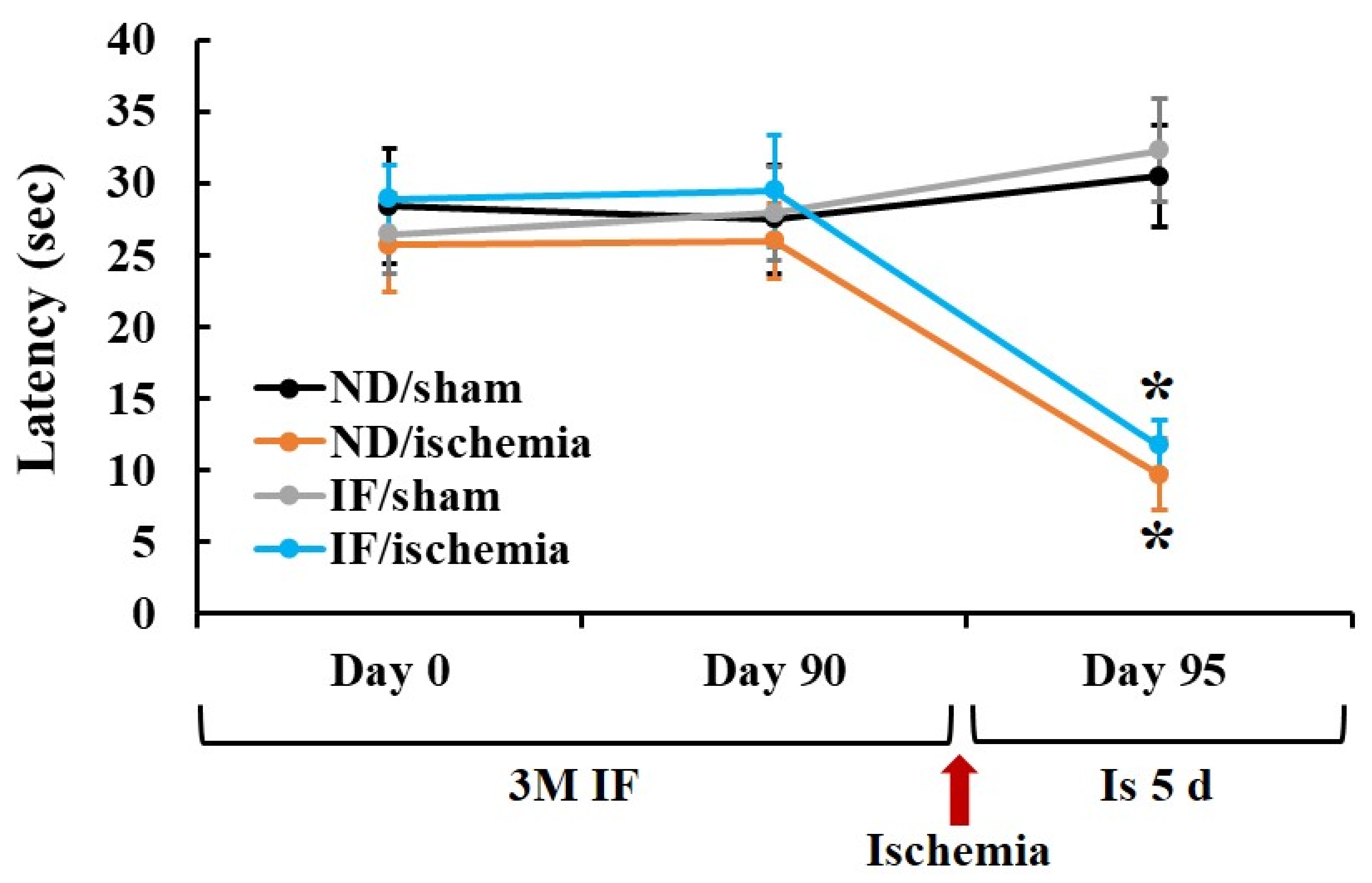
2.1. Passive Avoidance Test (PAT)

PAT was performed to examine the effect of IF on learning and memory following TFI (Figure 1). No significant difference was shown in the latency time after 3 months of IF between the normal diet (ND)/sham and the IF/sham groups. At 5 days after ischemia, a significant reduction (p < 0.001) in the latency time was shown in both ND/ischemia and IF/ischemia groups compared to that in the ND/sham group, showing that the latency time in both of the groups was similar. These results demonstrate that the short-term memory function in the IF/ischemia group was not different from that in the ND/ischemia group.

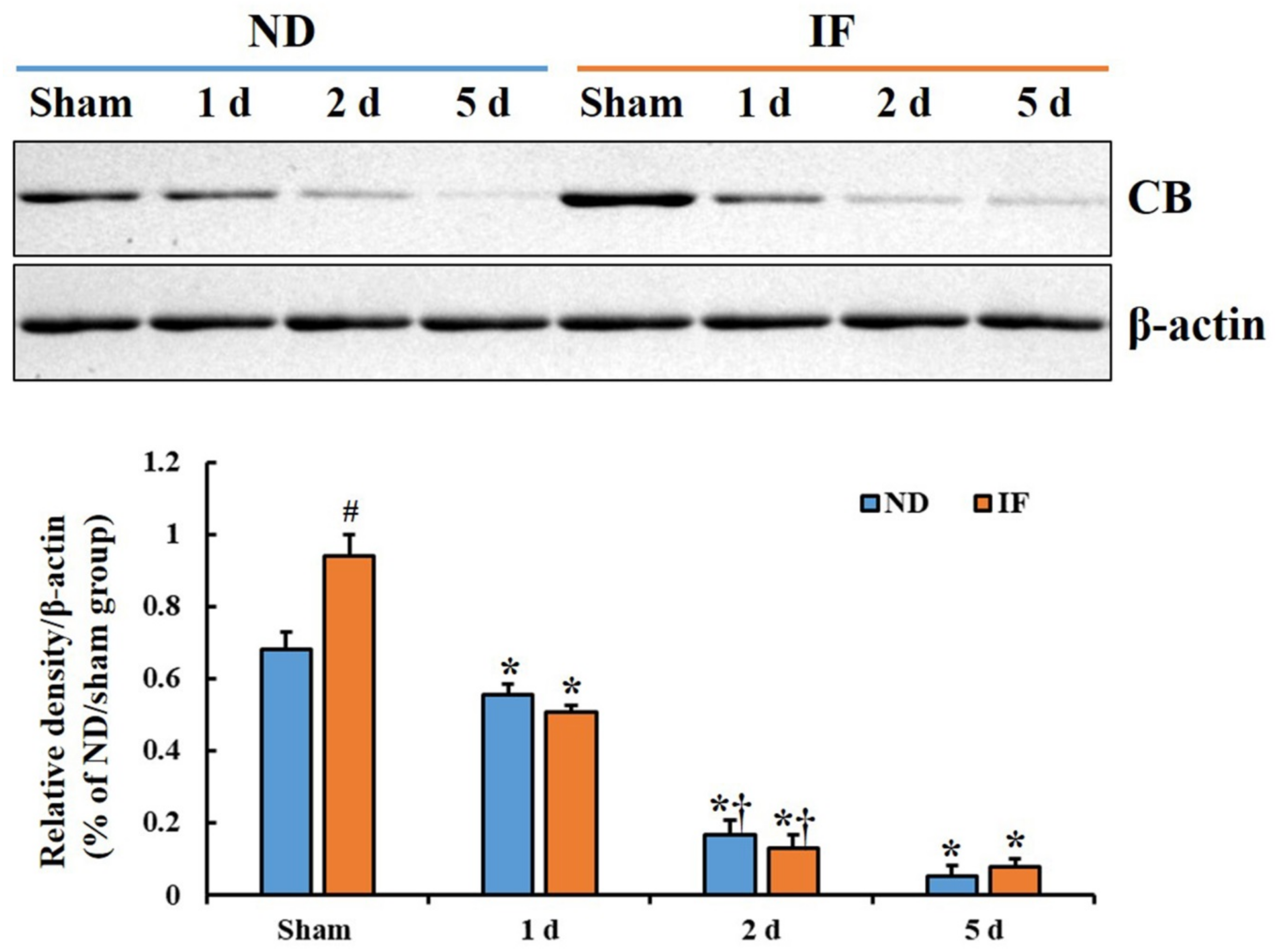
2.2. CB Protein Levels

The CB protein level in the hippocampal CA1 of the ND/ischemia group was significantly decreased 1 day after ischemia and gradually decreased until 5 days after ischemia (Figure 2). In the IF/sham group, the CB protein level was significantly increased (p < 0.001) after 3-month IF compared with that in the ND/sham group (Figure 2). However, the CB protein level in this group was significantly reduced (p < 0.001) from 1 day to 5 days after ischemia, showing that the change pattern after ischemia was similar to that of the ND/ischemia group (Figure 2).

2.3. CB Immunoreactivity

As shown in Figure 3A, CB immunoreactivity in the ND/sham group was shown in the stratum pyramidale (SP) of the hippocampal CA1. CB immunoreactivity in the ND/ischemia group was significantly and gradually decreased (p < 0.001) from 1 day to 5 days after ischemia, showing that relative optical density (ROD) was 70% at 1 day, 31% at 2 days, and 41% at 5 days post-ischemia compared with that in the ND/sham group (Figure 3B–D,I).
The CB immunoreactivity in the CA1 of the IF/sham group had almost doubled (about 200% of the ND/sham group) compared with that in the ND/sham group (Figure 3E). In the IF/ischemia group, the CB immunoreactivity in the SP of the CA1 region was also significantly decreased (p < 0.001) with time after ischemia (53% at 1 day, 33% at 2 days, and 36% at 5 days post-ischemia when compared with that in the ND/sham group) (Figure 3F–I), showing that the ROD pattern was similar to that found in the ND/ischemia group (Figure 3I). These finding indicate that IF for 3 months increased CB expression; however, the IF did not maintain CB expression in the CA1 after transient ischemia.

2.4. Neuroprotection

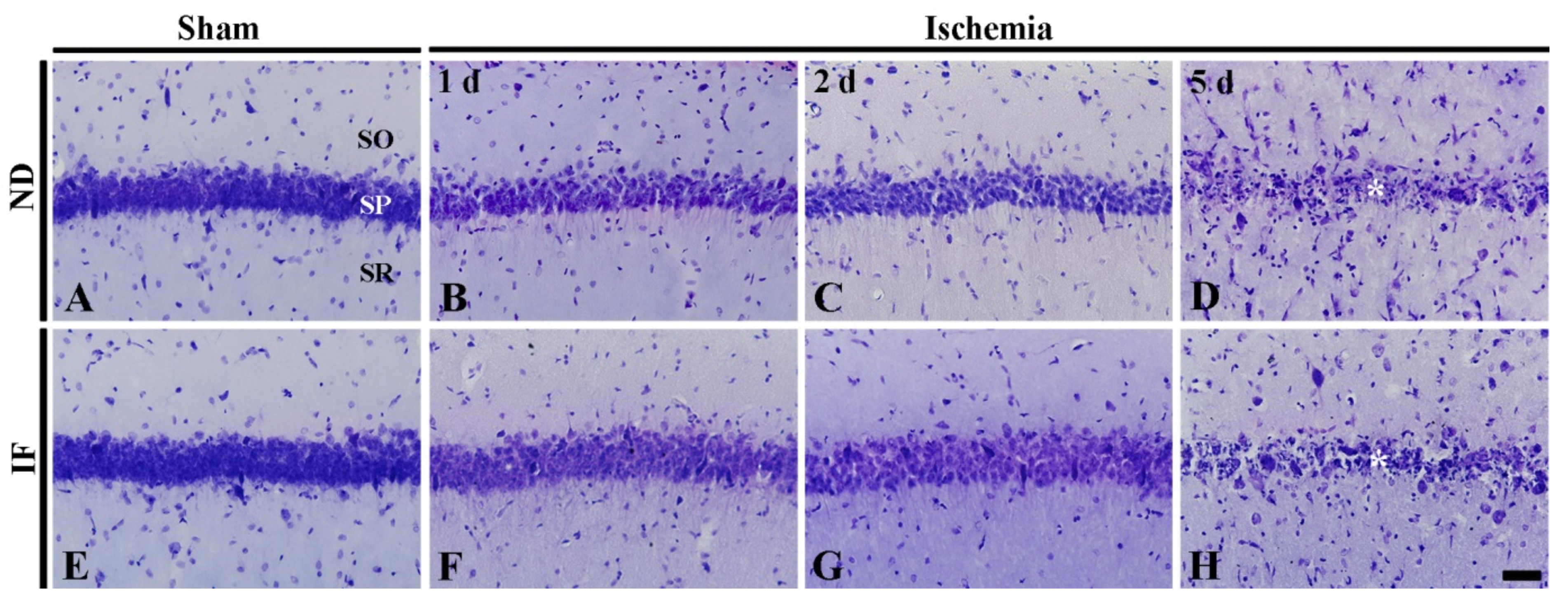
2.4.1. Cresyl Violet (CV) Positive Cells

CV staining was performed to investigate the distribution and morphology of cells located in the hippocampal CA1 region (Figure 4). In the ND/sham group, numerous CV-positive cells located in the stratum pyramidale were CA1 pyramidal cells (Figure 4A). In the ND/ischemia group, the patterns of CV-positive CA1 pyramidal cells were not altered at 1 day and 2 days after ischemia, although the CV stainability was decreased with time after ischemia (Figure 4B,C). At 5 days after ischemia, most of the CV-positive pyramidal cells were damaged at 5 days post-ischemia (Figure 4D).
In the IF/sham group, the distribution of CV-positive pyramidal cells was not different from that in the ND/ischemia group (Figure 4E). In the IF/ischemia group, the change pattern of CV-positive pyramidal cells was similar to that found in the ND/ischemia group (Figure 4F–H).

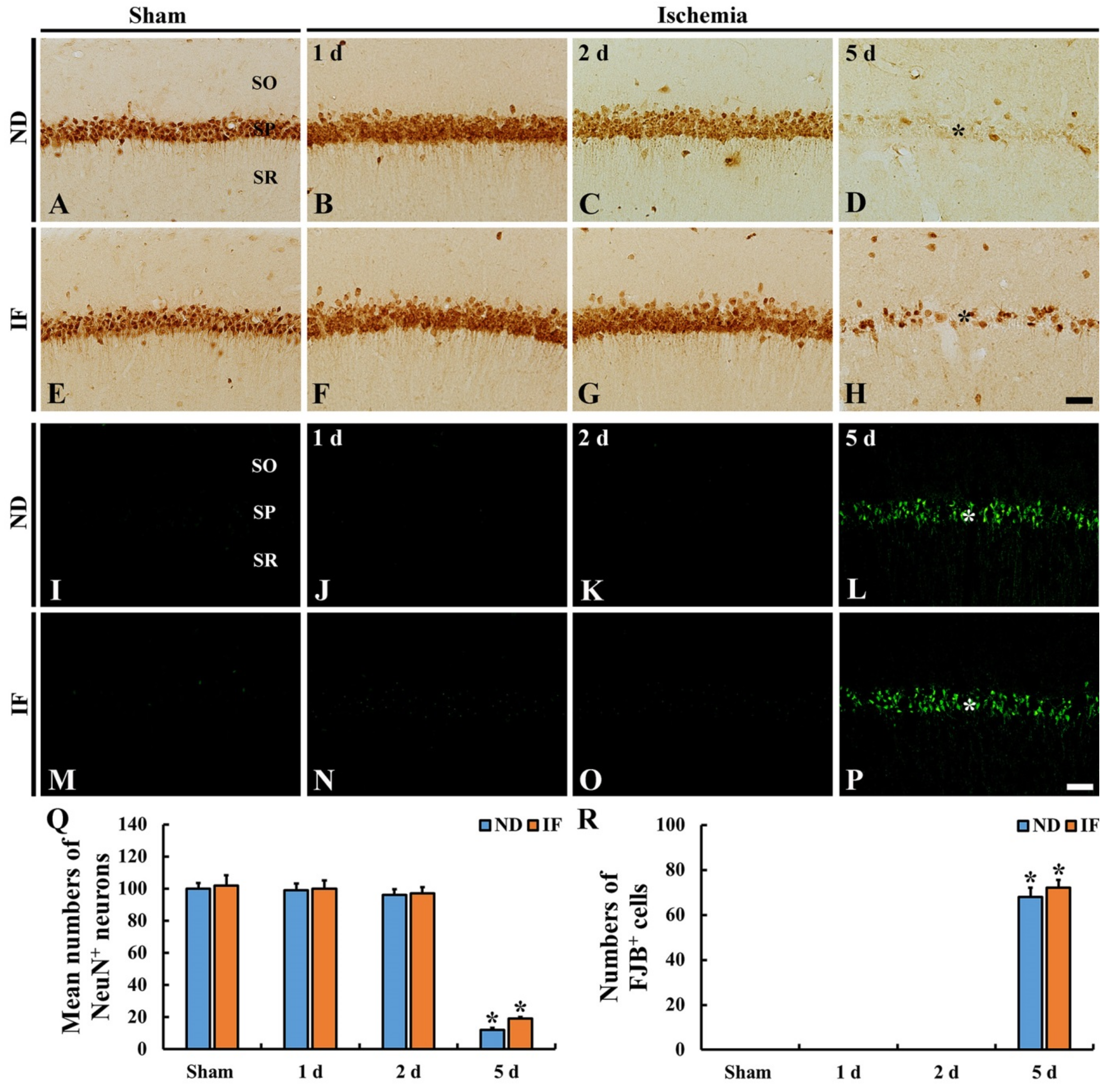
2.4.2. Neuronal Nuclear Antigen (NeuN) Immunoreactive Neurons

NeuN (a marker for neurons) immunoreactivity was observed in CA1 pyramidal neurons of the ND/sham groups (Figure 5A). In the ND/ischemia group, NeuN-immunoreactive neurons were not significantly altered (p < 0.001) in number at 1 day and 2 days (about 98% and 95% of the ND/sham group, respectively) after ischemia (Figure 5B,C,Q), showing that the intensity of NeuN immunoreactivity at 2 days post-ischemia was weaker than that in the ND/sham group (Figure 5C). At 5 days post-ischemia, however, the numbers of NeuN immunoreactive CA1 pyramidal neurons were dramatically reduced (p < 0.001) (about 12% of the ND/sham group) (Figure 5D,Q).
In the IF/sham group, the numbers and NeuN immunoreactivity in the CA1 pyramidal neurons did not differ from those in the ND/sham group (Figure 5E,Q). In the IF/ischemia group, the number of NeuN-immunoreactive CA1 pyramidal neurons at 1 day and 2 days post-ischemia was similar to that found in the ND/ischemia group (Figure 5F,G,Q), showing that, at 2 days post-ischemia, NeuN immunoreactivity was still strong (Figure 5G). At 5 days after ischemia, the number of NeuN immunoreactive CA1 pyramidal neurons was also markedly decreased (p < 0.001) (about 19% of the ND/sham group) (Figure 5H,Q).

2.4.3. Fluoro-Jade B (FJB)-Positive Cells
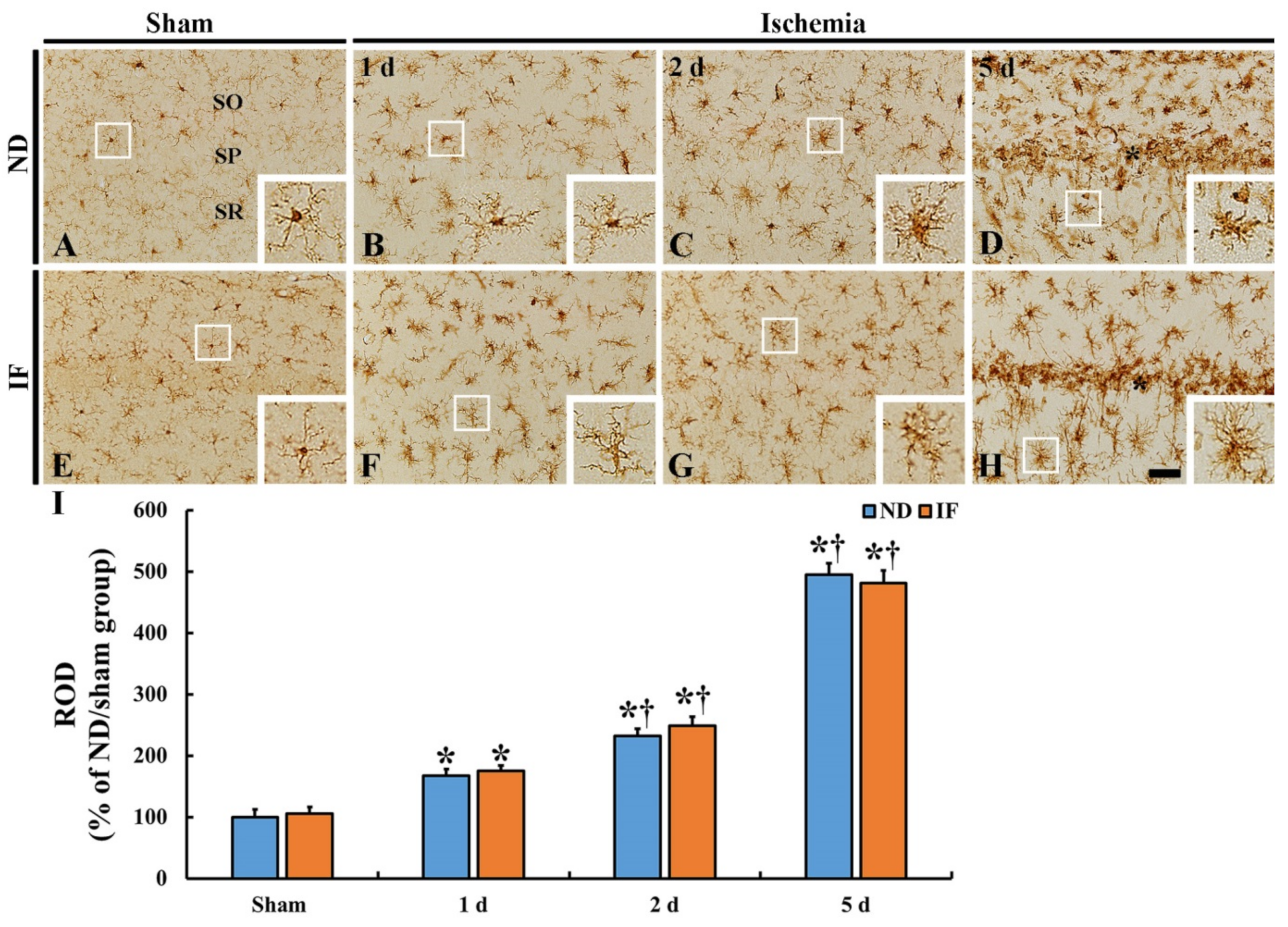
FJB (a high-affinity fluorescent marker for neuronal degradation)-positive cells were not observed in the ND/sham or IF/sham groups (Figure 5I,J). After ischemia in both of the groups, FJB-positive cells were not detected until 2 days after ischemia (Figure 5J,K,N,O,R). At 5 days following ischemia, FJB-positive cells were detected in both of the groups (Figure 5L,P), showing that there was no significant difference in the numbers of FJB-positive cells between the ND/ischemia and IF/ischemia groups (Figure 5R).

2.5. Microgliosis

Immunohistochemical staining for ionized calcium-binding adapter molecule 1 (Iba-1), a marker for microglia, was done to observe microglia activation in the CA1 region in the ND and IF groups with or without ischemia (Figure 6). In the ND/sham (Figure 6A) and IF/sham (Figure 6E) groups, there was no difference in the distribution of Iba-1 immunoreactive microglia between the two groups. Namely, they were evenly scattered in all layers of the CA1 region and identified as a resting form of microglia (Figure 6A,E). After ischemia in the two groups, the change patterns of the Iba-1 immunoreactive microglia were similar (Figure 6B–D,F–H). With time after ischemia, their cytoplasm was enlarged with thickened processes as the active form. In addition, no significant difference in the ROD of Iba-1 immunoreactivity was observed between the two groups. The ROD in the ND/ischemia group was gradually increased (167% at 1 day, 231% at 2 days, and 494% at 5 days compared to that of the ND/sham group) (Figure 6I). In the IF/ischemia group, the ROD was 175% at 1 day, 248% at 2 days, and 481% at 5 days, compared to that of the ND/sham group (Figure 6I).

2.6. Astrogliosis

Immunohistochemical staining for glial fibrillary acidic protein (GFAP), a marker for astrocytes, was carried out in order to investigate the activation of astrocytes in the CA1 region in the ND and IF groups with or without ischemia (Figure 7). GFAP immunoreactive astrocytes, as the resting form, had fine cellular processes in both ND/sham (Figure 7A) and IF/sham groups (Figure 7E). On the other hand, after ischemia, the pattern of changes in the GFAP immunoreactive astrocytes was similar between the ND and IF groups (Figure 7B–D,F–H). With time after ischemia, GFAP immunoreactive astrocytes became hypertrophied, and their cellular processes became thickened in both of the ND and IF/ischemia groups (Figure 7B–D,F–H). ROD in the ND/ischemia group was gradually increased (142% at 1 day, 214% at 2 days, and 316% at 5 days compared to that of the ND/sham group) after ischemia (Figure 7I). In the IF/ischemia group, the ROD was 140% at 1 day, 207% at 2 days, and 309% at 5 days compared to that of the ND/sham group (Figure 7I).

2.7. Immunoglobulin G (IgG) Immunoreactivity

To investigate BBB leakage due to the disruption of the BBB after ischemia, immunohistochemistry for IgG was conducted in the CA1 region in the ND and IF gerbils with or without ischemia (Figure 7). IgG immunoreactivity was fundamentally shown inside of the blood vessels in both the ND/sham (Figure 8A) and IF/sham groups (Figure 8E) (arrows). However, in both the ND and IF groups with ischemia, IgG immunoreactivity was shown near or outside the blood vessels (arrows), and was significantly enhanced (p < 0.001) (ND and IF, 183% and 181%, respectively) at 1 day, and a more significant increase (544% and 564%, respectively) was detected at 2 days compared to that in the ND/sham group (Figure 8B,C,F,G,I). At 5 days after ischemia, the IgG immunoreactivity was dramatically increased (ND and IF, 2027% and 1987%, respectively) in both the ND and IF/ischemia groups compared to that in the ND/sham group (Figure 8D,H,I).

3. Discussion

The three major mechanisms underlying the neuronal death induced by transient global brain or forebrain ischemia include the following: (1) oxidative stress induced by overproduction of reactive oxygen species (ROS), (2) inflammatory response by pro-inflammatory cytokines and immune cells, and (3) glutamate-induced excitotoxicity [25,26,27]. Accordingly, many studies have reported the factors underlying neuroprotection against TFI. The enhanced expression of antioxidant enzymes contributes to ROS scavenging, and the elevation of anti-inflammatory cytokines triggers inflammation in ischemic brains by suppressing the expression of pro-inflammatory cytokines, and the increased levels of calcium-binding proteins attenuate excitotoxicity by buffering glutamate influx [28,29]. Therefore, we investigated whether an IF-mediated increase in CB expression influenced neuronal survival, reactive gliosis and BBB leakage (disruption) in the hippocampal CA1 region.
The hippocampus is critical for memory and cognitive function [30]. IF increases the thickness of the hippocampal CA1 region in mice, suggesting enhanced learning and memory [31]. In addition, alternate-day IF has been shown to improve memory, sensory and motor skills [32,33,34]. In this study, we evaluated short-term memory after TFI with or without 3-month IF in gerbils, and found no significant difference in function between groups with ND/ischemia and IF/ischemia as well as groups exposed to ND/sham and IF/sham. These findings differ from those reported in foregoing studies. Based on our current study, the long-term (3 months) exposure of gerbils to alternate-day IF does not increase short-term memory. In this regard, we evaluated the expression of CB in cognitive and memory function [35], neuroprotection and reactive gliosis (microgliosis and astrogliosis) in the hippocampal CA1 region of the four groups (ND/sham, ND/ischemia, IF/sham and IF/ischemia) to determine the factors underlying the lack of short-term memory enhancement following IF.
The CB levels are decreased and the Ca2+ influx is increased in brains with aging, which influences memory and cognitive function decline [36,37,38]. Based on these studies, the increased CB expression enhances memory and cognitive function. In our current study, we found that CB expression was significantly increased in the IF/sham group after 3 months of IF compared with that of the ND/sham group; however, based on PAT, the learning and memory performance following 3-month IF was not enhanced. We also found that CB expression following ischemia declined gradually with time after ischemia in the IF/ischemia group, similar to that of the ND/sham group. This result indicates that the 3-month IF in gerbils does not sustain CB expression after ischemic insult despite the increased CB expression in CA1 pyramidal neurons following 3-month IF. Based on the findings of altered CB in the IF/ischemia group, we analyzed the degrees of neuroprotection and reactive gliosis in the IF/ischemia group. The findings indicate that the death (loss) of CA1 pyramidal neurons and reactive gliosis in the IF/ischemia group was similar to that of the ND/ischemia group, suggesting that IF for 3 months in gerbils does not protect against TFI.
Dietary restriction or caloric restriction in animal models of focal or global ischemia significantly reduces neurological damage [4,39,40,41,42]. In addition, a few studies have demonstrated that IF induces neuroprotective effects by attenuating cellular dysfunction, degeneration and death in the brain after experimental focal brain ischemia [43], in contrast to the transient forebrain ischemia induced in our study. We recently reported that IF for 3 months in a gerbil model of TFI increased the expression of endogenous antioxidant enzymes (SOD1, SOD2, and catalase) without protecting the hippocampal CA1 pyramidal neurons from ischemic injury [5]. Similarly, in our current study, the increased CB levels following IF failed to protect the CA1 pyramidal neurons in the IF/ischemia group against TFI.
Several studies have reported that CB levels play a key role in neuroprotection or neuronal survival in the brains of patients with neurodegenerative disorder or injury [44,45,46,47]. In the case of focal brain ischemia involving rabbits, the increased CB protects the brain tissue against focal ischemia, suggesting that the increased CB blocks intracellular calcium entry and protects the brain against focal ischemia [48,49]. In contrast, it has been reported that CB fails to protect hippocampal neurons from transient global brain ischemia in spite of its cytoplasmic calcium-buffering properties observed in CB knockout mice [44].
Ischemic insults trigger reactive gliosis in the brain [9,10]. Attenuated reactive gliosis in ischemic brains is a measure of neuroprotection against ischemic insults [10,50,51]. Our results showed the development of microgliosis and astrogliosis, which was enhanced with time in the CA1 region of both the ND and IF groups after 5 min of TFI, although the differences between the two groups were not statistically different. This finding indicates that ischemia-induced gliosis, which leads to neuronal damage/death, cannot be prevented by 3-month IF. Some studies have reported that gliosis following brain insults begins after the disruption of the BBB, which allows blood and serum components to enter the ischemic brain parenchyma [11,12,13,14]. We recently reported that 1-, 2- and 3-month IF did not prevent BBB leakage in the gerbil hippocampal CA1 region 5 days after 5-minute TFI [11]. In addition, we found that interleukin-13 (an anti-inflammatory cytokine), which plays a beneficial role in ischemic injury, was significantly increased in the ischemic CA1 region, but there was no neuroprotective effect against TFI [11]. Taken together, the BBB leakage in ischemic brains may fail to protect neurons from ischemic insults.
Our present study was performed using gerbils with a mean lifespan of 110 weeks in males and 139 weeks in females [52]. In this regard, the 3-month IF period in gerbils may translate to approximately 10 years in human beings. The 3-month duration should be adequate for gerbils to adapt to the new dietary protocol, which may explain the lack of neuroprotective effects following transient ischemia with 3-month IF. This finding suggests that the effects of long-term IF on brains may differ from those of dietary or caloric restriction following various brain injuries, in particular ischemic insults.
Ischemic stroke is a representative senile disease, and several studies have investigated various outcomes after ischemic insults using aged animals [50,51,53,54]. The current data, however, relate to adult animals, suggesting the need for additional and identical studies involving aged animals.
In brief, IF for 3 months increased the production of CB in hippocampal CA1 pyramidal neurons in gerbils. However, the 3-month IF did not prevent BBB disruption or reactive gliosis, which might lead to the death of CA1 pyramidal neurons after 5 min of TFI. Further studies with diverse and modified IF regimens are necessary to determine the neuroprotective effects after TFI, and to establish the type of IF protocol that prevents or protects against ischemic brain injury.

4. Materials and Methods

4.1. Experimental Animals

Male Mongolian gerbils (Meriones unguiculatus) at six months of age (body weight, 75 ± 5 g) were obtained from the Experimental Animal Center (Kangwon University, Chuncheon, Gangwon, Republic of Korea). They had been maintained at a constant temperature (23 ± 2 °C) and humidity (50 ± 5%) with a 12 h light/dark cycle. The care and handling of animals in this research complied with the “Guidelines of the Current International Laws and Policies” described in the “Guide for the Care and Use of Laboratory Animals” (The National Academies Press, 8th Ed., 2011). In addition, the protocol of this experiment was approved (approval number: KW-200113-1) by the committee of Institutional Animal Care and Use Committee at Kangwon National University.

4.2. IF and Experimental Groups

For IF, gerbils were allowed free access to ND every other day and no food on alternate days (24 h fasting and 24 h feeding), and IF was applied for three months according to the published method [3,32,55]. During the feeding period, food intake in all gerbils was controlled daily (10 g/day), and body weight was monitored every week.
For experimental groups, 96 male gerbils were used and randomly divided into four groups, as follows: (1) ND/sham group (n = 12)—gerbils were fed ND and received sham ischemia operation; (2) ND/ischemia group (n = 36)—animals were fed ND and received ischemia; (3) IF/sham group (n = 12)—gerbils had IF for three months and received sham operation; (4) IF/ischemia group (n = 36)—animals had IF and received ischemia. The animals in the ischemia group were sacrificed at 1 day, 2 days, and 5 days after ischemia to investigate the effects of IF on CB expression and neuroprotection following transient global forebrain ischemia. To decrease the number of gerbils, gerbils in the sham group were sacrificed only at 5 days after the sham ischemia operation.

4.3. Induction of Transient Forebrain Ischemia

As we previously described [5], all gerbils were anesthetized with a mixture of 2.5% isoflurane from Baxtor (Deerfield, IL, USA) in 67% nitrous oxide and 33% oxygen. Under anesthesia, the gerbils received an incision on the neck to find both common carotid arteries, and the arteries were occluded by non-traumatic aneurysm clips for 5 min and then re-perfused. The body temperature of the gerbils was controlled at normothermia (37 ± 0.5 °C) using a thermometric blanket during the surgery, monitoring the temperature using a rectal temperature probe (TR-100) (Fine Science Tools, Foster City, CA, USA). All animals were fed ND after the surgical procedure. The gerbils included in the sham group underwent identical surgical procedures without the ligation of common carotid arteries. Thereafter, the gerbils were kept in thermal incubators (temperature, 23 °C; humidity, 60%) to maintain body temperature at the normothermic level until they were euthanized. Until the gerbils were sacrificed, they were continually fed ND or IF.

4.4. PAT

Learning and memory was assayed through PAT according to the modification of a published method [56]. Shortly, the Gemini Avoidance System (GEM 392) (San Diego Instruments, San Diego, CA, USA) was used for this PAT, which consisted of two rooms (light and dark) with a grid floor. First, training was performed one day before IF, 29 days after IF and 4 days after ischemia, as follows. For the training, each gerbil was allowed to explore the environment of the two rooms for one min, while the grid floor was opened. After the exploration, the gerbil was permitted to enter the dark room when a light in the light room was turned on, while the floor was given an inescapable foot-shock (0.5 mA for 5 s). A substantive PAT test was done 24 h after the training. Each trained gerbil was put in the dark room, the light of the light room was turned on, and the floor was opened. Thereafter, we recorded the latency time, which is the time to enter the dark room, within 180 s.

4.5. Western Blotting

To examine changes of CB protein level in the hippocampal CA1, 40 gerbils (n = 5 at sham, 1, 2 and 5 days post-ischemia in each group) were used for the blotting according to a previously published method [57]. Shortly, at each point in time, five gerbils in each group were deeply anesthetized by an intraperitoneal injection of pentobarbital sodium (60 mg/kg) from JW Pharm (Seoul, South Korea), and their hippocampal CA1 tissues were collected. The tissues were lysed with RIPA buffer (Santa Cruz, CA, USA) and homogenized with an ultrasonic homogenizer for 5 min. These homogenates were then centrifuged at 12,000 rpm for 20 min at 4 °C, and the supernatants were collected. Next, protein concentrations were measured with a bicinchoninic acid kit from Thermo Fisher Scientific (Waltham, MA, USA). In detail, the proteins were separated by 10% sodium dodecyl sulfate–polyacrylamide gel and transferred to nitrocellulose membranes (Pall Corp., Pittsburgh, PA, USA). These membranes were blocked with 5% non-fat milk (in Tris-buffered saline/Tween, TBST) for 60 min on a shaker at room temperature and then incubated in primary antibodies (rabbit anti-CB) (diluted 1:1000) (Cell signaling technology, Danvers, MA, USA) and rabbit anti-β-actin (42 kDa) (1:2000, Sigma-Aldrich, St. Louis, MO, USA) overnight at 4 °C. The membranes were washed three times with TBST and incubated with peroxidase conjugated anti-rabbit IgG (1:4000, Santa Cruz, CA, USA) for 1 h at room temperature. After washing them with TBST, they were visualized by horseradish peroxidase (Millipore, Billerica, MA, USA). The band intensities were analyzed using ImageJ (ver. 1.52v, National Institutes of Health, Bethesda, MD, USA).

4.6. Preparation of Histological Sections

At each point in time after TFI, seven gerbils in each group were deeply anesthetized by intraperitoneal injection of 60 mg/kg of pentobarbital sodium (JW Pharm. Co., Ltd., Seoul, Korea) at designated times (1, 2 and 5 days after ischemia) following IF. Under anesthesia, the gerbils were transcardially washed with 0.1 M phosphate buffered saline (PBS) (pH 7.4) and fixed with 4% paraformaldehyde solution (in 0.1 M PB, pH 7.4). After fixation, their brains were removed, and cryoprotected in 30% sucrose solution. Finally, brain tissues containing the hippocampus were serially sectioned into coronal sections (30 μm thickness) in a cryostat of Leica (Wetzlar, Germany) and kept in plates containing PBS.

4.7. Immunohistochemistry

We used the following antibodies: rabbit anti-CB (diluted 1:1000; Cell signaling technology, Danvers, MA, USA), NeuN (diluted 1:800; Abcam, Cambridge, MA, USA), Iba-1 (1:800, Wako, Japan) and GFAP (1:1000, Chemicon, Temecula, CA, USA). For immunohistochemistry for each antibody, we carried it out according to our published method [38]. Shortly, the sections obtained at designated times after ischemia were incubated in 0.3% hydrogen peroxide (H2O2) solution for 30 min, followed by 10% normal goat serum solution (in 0.05 M PBS, pH 7.4) for 30 min. These sections were reacted with each antibody for 18 h at 4 °C. Continuously, they were exposed to biotinylated anti-rabbit IgG (diluted 1:200, Vector, Burlingame, CA, USA) and streptavidin peroxidase complex (diluted 1:200, Vector, Burlingame, CA, USA) for 2 h at room temperature, respectively. Finally, the reacted sections were visualized by incubating in a 0.05% solution of 3, 3′-diaminobenzidine tetrahydrochloride (DAB) (in 0.1 M Tris–HCl buffer, pH 7.2).

4.8. CV Histochemistry

CV histochemical staining was performed to examine the distribution and morphology of cells in the hippocampus. In short, according to a published method [58], 1% CV acetate (Sigma, St. Louis, MO, USA) solution (in distilled water) was prepared, and glacial acetic acid was added to this solution. To stain the sections, they were reacted in the CV solution for 1 h at room temperature (about 23 °C). After washing the sections with distilled water, they were dehydrated with serial ethanol. Finally, the stained sections were prepared as permeant slides.

4.9. FJB Histofluorescence Staining

FJB is a fluorescent derivative used to detect degenerating cells. In this study, FJB histofluorescence staining was done to examine the damage/death of hippocampal cells after ischemia. As described previously [55,59,60], in short, the sections were immersed in a 0.06% solution of potassium permanganate and reacted with 0.0004% solution of FJB (Histochem, Jefferson, AR, USA).

4.10. Data Analysis

Changes in CB, Iba-1 and GFAP immunoreactivity were quantitatively analyzed according to our published method [38]. In brief, we selected seven sections at 120 μm intervals within the antero-posterior from −1.4 to −2.2 mm according to the gerbil brain atlas in each gerbil. Images of each immunoreactivity were taken from the corresponding area (250 µm2) under 20× primary magnification in the hippocampus with an AxioM1 light microscope from Carl Zeiss (Göttingen, Germany) equipped with a digital camera from Axiocam (Carl Zeiss, Germany), which was connected to a PC monitor. The captured images were calibrated into an array of 512 × 512 pixels, and each immunoreactivity was evaluated by optical density (OD). The OD was obtained after the transformation of the mean grey level using a formula (OD = log (256/mean grey level). The background density was subtracted, and the OD ratio was calibrated as the percent of relative OD (ROD) using Adobe Photoshop version 8.0. Finally, ROD was analyzed with the Image J 1.46 software from the National Institutes of Health (Bethesda, MD, USA).
Numbers of NeuN-and FJB-positive cells were counted according to our published method [61]. In brief, we selected seven sections via the above-mentioned method. Images of NeuN-positive cells were captured with an AxioM1 light microscope (Carl Zeiss, Göttingen, Germany). Images of FJB-positive cells were taken with an epifluorescent microscope from Carl Zeiss (Göttingen, Germany) equipped with 450–490 nm of blue excitation light and a barrier filter. The digital images of cells positive for NeuN and FJB were counted in a 250 × 250 μm square applied at the center of the CA1 region using an image analyzing software (Optimas 6.5) from CyberMetrics (Scottsdale, AZ, USA).

4.11. Statistical Analysis

Data are expressed as the mean±SEM (standard error of the mean). The differences in the ROD or mean numbers of the respective immunoreactive structures obtained for each group were statistically analyzed with one-way analysis of variance followed by a post hoc Tukey’s test using GraphPad Instat from Instat Statistics (GraphPad Software Inc., La Jolla, CA, USA). A p value of <0.05 was considered statistically significant.

Author Contributions

Conceptualization, M.-H.W.; methodology, T.-K.L., D.W.K. and J.-C.L.; software, J.H.A.; validation, Y.P., J.H.P., S.-S.L. and J.-S.P.; investigation, H.S., B.K. and Y.H.Y.; data curation, Y.P., M.C.S., J.H.C., Y.-M.K. and I.J.K.; writing—original draft preparation, Y.H.Y.; writing—review and editing M.-H.W.; supervision, M.-H.W.; project administration, M.-H.W.; funding acquisition, T.-K.L., M.-H.W. and I.J.K. All authors have read and agreed to the published version of the manuscript.

Funding

This work was supported by the Basic Science Research Program through the National Research Foundation of Korea (NRF) funded by the Ministry of Education (NRF-2020R1I1A1A01070897, NRF-2020R1F1A1052380 and NRF-2020R1F1A1071973).

Institutional Review Board Statement

The care and handling of animals in this research complied with the “Guidelines of the Current International Laws and Policies” described in the “Guide for the Care and Use of Laboratory Animals” (The National Academies Press, 8th Ed., 2011). In addition, the protocol of this experiment was approved (approval number: KW-200113-1) by the committee of Institutional Animal Care and Use Committee at Kangwon National University.

Informed Consent Statement

Not applicable.

Data Availability Statement

The data presented in this study are available on request from the corresponding author.

Acknowledgments

This work was supported by the Basic Science Research Program through the National Research Foundation of Korea (NRF) funded by the Ministry of Education (NRF-2020R1I1A1A01070897, NRF-2020R1F1A1052380 and NRF-2020R1F1A1071973).

Conflicts of Interest

The authors in the present study have declared that there are no conflicts of interest.

Abbreviations

BBBBlood–Brain Barrier
CACornu Ammonis
CBCalbindin D28K
CVCresyl Violet
FJBFluoro Jade B
GFAPGlial fibrillary Acidic Protein
Iba-1Ionized Calcium Binding Adapter Molecule 1
IFIntermittent Fasting
NDNormal Diet
NeuNNeuronal Nuclear Antigen
PATPassive Avoidance Test
RODRelative Optical Density
SOStratum Oriens
SPStratum Pyramidale
SRStratum Radiatum
TFITransient Forebrain Ischemia

References

  1. Fann, D.Y.; Ng, G.Y.; Poh, L.; Arumugam, T.V. Positive effects of intermittent fasting in ischemic stroke. Exp. Gerontol. 2017, 89, 93–102. [Google Scholar] [CrossRef] [PubMed]
  2. Mitchell, J.R.; Verweij, M.; Brand, K.; van de Ven, M.; Goemaere, N.; van den Engel, S.; Chu, T.; Forrer, F.; Muller, C.; de Jong, M.; et al. Short-term dietary restriction and fasting precondition against ischemia reperfusion injury in mice. Aging Cell 2010, 9, 40–53. [Google Scholar] [CrossRef] [Green Version]
  3. Jeong, J.H.; Yu, K.S.; Bak, D.H.; Lee, J.H.; Lee, N.S.; Jeong, Y.G.; Kim, D.K.; Kim, J.J.; Han, S.Y. Intermittent fasting is neuroprotective in focal cerebral ischemia by minimizing autophagic flux disturbance and inhibiting apoptosis. Exp. Ther. Med. 2016, 12, 3021–3028. [Google Scholar] [CrossRef] [PubMed] [Green Version]
  4. Varendi, K.; Airavaara, M.; Anttila, J.; Vose, S.; Planken, A.; Saarma, M.; Mitchell, J.R.; Andressoo, J.O. Short-term preoperative dietary restriction is neuroprotective in a rat focal stroke model. PLoS ONE 2014, 9, e93911. [Google Scholar] [CrossRef] [PubMed] [Green Version]
  5. Ahn, J.H.; Noh, Y.; Shin, B.N.; Kim, S.S.; Park, J.H.; Lee, T.K.; Song, M.; Kim, H.; Lee, J.C.; Yong, J.H.; et al. Intermittent fasting increases SOD2 and catalase immunoreactivities in the hippocampus but does not protect from neuronal death following transient ischemia in gerbils. Mol. Med. Rep. 2018, 18, 4802–4812. [Google Scholar] [CrossRef] [PubMed] [Green Version]
  6. Ahn, J.H.; Shin, B.N.; Park, J.H.; Lee, T.K.; Park, Y.E.; Lee, J.C.; Yang, G.E.; Shin, M.C.; Cho, J.H.; Lee, K.C.; et al. Pre-and post-treatment with novel antiepileptic drug oxcarbazepine exerts neuroprotective effect in the hippocampus in a gerbil model of transient global cerebral ischemia. Brain Sci. 2019, 9, 279. [Google Scholar] [CrossRef] [PubMed] [Green Version]
  7. Kirino, T.; Tamura, A.; Sano, K. Selective vulnerability of the hippocampus to ischemia—Reversible and irreversible types of ischemic cell damage. Prog. Brain Res. 1985, 63, 39–58. [Google Scholar]
  8. Kirino, T. Delayed neuronal death. Neuropathol. Off. J. Jpn. Soc. Neuropathol. 2000, 20, S95–S97. [Google Scholar] [CrossRef]
  9. Lee, T.K.; Kim, H.; Song, M.; Lee, J.C.; Park, J.H.; Ahn, J.H.; Yang, G.E.; Kim, H.; Ohk, T.G.; Shin, M.C.; et al. Time-course pattern of neuronal loss and gliosis in gerbil hippocampi following mild, severe, or lethal transient global cerebral ischemia. Neural Regen. Res. 2019, 14, 1394–1403. [Google Scholar]
  10. Burda, J.E.; Sofroniew, M.V. Reactive gliosis and the multicellular response to cns damage and disease. Neuron 2014, 81, 229–248. [Google Scholar] [CrossRef] [Green Version]
  11. Lee, T.K.; Park, Y.; Kim, B.; Lee, J.C.; Shin, M.C.; Ohk, T.G.; Park, C.W.; Cho, J.H.; Park, J.H.; Lee, C.H.; et al. Long-term alternating fasting increases interleukin-13 in the gerbil hippocampus, but does not protect bbb and pyramidal neurons from ischemia-reperfusion injury. Neurochem. Res. 2020, 45, 2352–2363. [Google Scholar] [CrossRef] [PubMed]
  12. Ghosh, A.; Birngruber, T.; Sattler, W.; Kroath, T.; Ratzer, M.; Sinner, F.; Pieber, T.R. Assessment of blood-brain barrier function and the neuroinflammatory response in the rat brain by using cerebral open flow microperfusion (cofm). PLoS ONE 2014, 9, e98143. [Google Scholar] [CrossRef] [PubMed]
  13. Jiang, X.; Andjelkovic, A.V.; Zhu, L.; Yang, T.; Bennett, M.V.L.; Chen, J.; Keep, R.F.; Shi, Y. Blood-brain barrier dysfunction and recovery after ischemic stroke. Prog. Neurobiol. 2018, 163–164, 144–171. [Google Scholar] [CrossRef] [PubMed]
  14. Silver, J.; Miller, J.H. Regeneration beyond the glial scar. Nat. Rev. Neurosci. 2004, 5, 146–156. [Google Scholar] [CrossRef] [PubMed]
  15. Airaksinen, M.S.; Thoenen, H.; Meyer, M. Vulnerability of midbrain dopaminergic neurons in calbindin-d28k-deficient mice: Lack of evidence for a neuroprotective role of endogenous calbindin in mptp-treated and weaver mice. Eur. J. Neurosci. 1997, 9, 120–127. [Google Scholar] [CrossRef] [PubMed]
  16. Baimbridge, K.G.; Celio, M.R.; Rogers, J.H. Calcium-binding proteins in the nervous system. Trends Neurosci. 1992, 15, 303–308. [Google Scholar] [CrossRef]
  17. Heizmann, C.W. Calcium signaling in the brain. Acta Neurobiol. Exp. 1993, 53, 15–23. [Google Scholar]
  18. Nagerl, U.V.; Mody, I.; Jeub, M.; Lie, A.A.; Elger, C.E.; Beck, H. Surviving granule cells of the sclerotic human hippocampus have reduced ca(2+) influx because of a loss of calbindin-d(28k) in temporal lobe epilepsy. J. Neurosci. Off. J. Soc. Neurosci. 2000, 20, 1831–1836. [Google Scholar] [CrossRef] [Green Version]
  19. Arundine, M.; Tymianski, M. Molecular mechanisms of calcium-dependent neurodegeneration in excitotoxicity. Cell Calcium 2003, 34, 325–337. [Google Scholar] [CrossRef]
  20. Wojda, U.; Salinska, E.; Kuznicki, J. Calcium ions in neuronal degeneration. Iubmb Life 2008, 60, 575–590. [Google Scholar] [CrossRef]
  21. Chung, T.H.; Choi, H.S.; Lee, C.H. Change of calbindin d-28k protein expression in the mice hippocampus after lipopolysaccharide treatment. J. Vet. Med Sci. 2015, 77, 349–352. [Google Scholar] [CrossRef] [PubMed] [Green Version]
  22. Molinari, S.; Battini, R.; Ferrari, S.; Pozzi, L.; Killcross, A.S.; Robbins, T.W.; Jouvenceau, A.; Billard, J.M.; Dutar, P.; Lamour, Y.; et al. Deficits in memory and hippocampal long-term potentiation in mice with reduced calbindin d28k expression. Proc. Natl. Acad. Sci. USA 1996, 93, 8028–8033. [Google Scholar] [CrossRef] [PubMed] [Green Version]
  23. Soontornniyomkij, V.; Risbrough, V.B.; Young, J.W.; Soontornniyomkij, B.; Jeste, D.V.; Achim, C.L. Hippocampal calbindin-1 immunoreactivity correlate of recognition memory performance in aged mice. Neurosci. Lett. Shannon Clare Irel. 2012, 516, 161–165. [Google Scholar] [CrossRef] [Green Version]
  24. Ahn, J.H.; Shin, B.N.; Song, M.; Kim, H.; Park, J.H.; Lee, T.K.; Park, C.W.; Park, Y.E.; Lee, J.C.; Yong, J.H.; et al. Intermittent fasting increases the expressions of sods and catalase in granule and polymorphic cells and enhances neuroblast dendrite complexity and maturation in the adult gerbil dentate gyrus. Mol. Med. Rep. 2019, 19, 1721–1727. [Google Scholar] [CrossRef] [PubMed]
  25. Jayaraj, R.L.; Azimullah, S.; Beiram, R.; Jalal, F.Y.; Rosenberg, G.A. Neuroinflammation: Friend and foe for ischemic stroke. J. Neuroinflammation 2019, 16, 142. [Google Scholar] [CrossRef] [PubMed] [Green Version]
  26. Khoshnam, S.E.; Winlow, W.; Farzaneh, M.; Farbood, Y.; Moghaddam, H.F. Pathogenic mechanisms following ischemic stroke. Neurol. Sci. Off. J. Ital. Neurol. Soc. Ital. Soc. Clin. Neurophysiol. 2017, 38, 1167–1186. [Google Scholar] [CrossRef] [PubMed]
  27. Shirley, R.; Ord, E.N.; Work, L.M. Oxidative stress and the use of antioxidants in stroke. Antioxidants 2014, 3, 472–501. [Google Scholar] [CrossRef] [Green Version]
  28. Lee, J.C.; Won, M.H. Neuroprotection of antioxidant enzymes against transient global cerebral ischemia in gerbils. Anat. Cell Biol. 2014, 47, 149–156. [Google Scholar] [CrossRef] [Green Version]
  29. Lee, T.K.; Kang, I.J.; Kim, B.; Sim, H.J.; Kim, D.W.; Ahn, J.H.; Lee, J.C.; Ryoo, S.; Shin, M.C.; Cho, J.H.; et al. Experimental pretreatment with chlorogenic acid prevents transient ischemia-induced cognitive decline and neuronal damage in the hippocampus through anti-oxidative and anti-inflammatory effects. Molecules 2020, 25, 3578. [Google Scholar] [CrossRef]
  30. Hitti, F.L.; Siegelbaum, S.A. The hippocampal ca2 region is essential for social memory. Nature 2014, 508, 88–92. [Google Scholar] [CrossRef]
  31. Li, L.; Wang, Z.; Zuo, Z. Chronic intermittent fasting improves cognitive functions and brain structures in mice. PLoS ONE 2013, 8, e66069. [Google Scholar] [CrossRef] [PubMed] [Green Version]
  32. Longo, V.D.; Mattson, M.P. Fasting: Molecular mechanisms and clinical applications. Cell Metab. 2014, 19, 181–192. [Google Scholar] [CrossRef] [PubMed] [Green Version]
  33. Fontana, L.; Klein, S. Aging, adiposity, and calorie restriction. JAMA 2007, 297, 986–994. [Google Scholar] [CrossRef] [PubMed]
  34. Singh, R.; Lakhanpal, D.; Kumar, S.; Sharma, S.; Kataria, H.; Kaur, M.; Kaur, G. Late-onset intermittent fasting dietary restriction as a potential intervention to retard age-associated brain function impairments in male rats. Age 2012, 34, 917–933. [Google Scholar] [CrossRef] [PubMed] [Green Version]
  35. Toescu, E.C.; Verkhratsky, A. The importance of being subtle: Small changes in calcium homeostasis control cognitive decline in normal aging. Aging Cell 2007, 6, 267–273. [Google Scholar] [CrossRef]
  36. Villa, A.; Podini, P.; Panzeri, M.C.; Racchetti, G.; Meldolesi, J. Cytosolic ca2+ binding proteins during rat brain ageing: Loss of calbindin and calretinin in the hippocampus, with no change in the cerebellum. Eur. J. Neurosci. 1994, 6, 1491–1499. [Google Scholar] [CrossRef] [PubMed]
  37. De Jong, G.I.; Naber, P.A.; Van der Zee, E.A.; Thompson, L.T.; Disterhoft, J.F.; Luiten, P.G. Age-related loss of calcium binding proteins in rabbit hippocampus. Neurobiol. Aging 1996, 17, 459–465. [Google Scholar] [CrossRef] [Green Version]
  38. Bae, E.J.; Chen, B.H.; Shin, B.N.; Cho, J.H.; Kim, I.H.; Park, J.H.; Lee, J.C.; Tae, H.J.; Choi, S.Y.; Kim, J.D.; et al. Comparison of immunoreactivities of calbindin-d28k, calretinin and parvalbumin in the striatum between young, adult and aged mice, rats and gerbils. Neurochem. Res. 2015, 40, 864–872. [Google Scholar] [CrossRef] [PubMed]
  39. Yu, Z.F.; Mattson, M.P. Dietary restriction and 2-deoxyglucose administration reduce focal ischemic brain damage and improve behavioral outcome: Evidence for a preconditioning mechanism. J. Neurosci. Res. 1999, 57, 830–839. [Google Scholar] [CrossRef]
  40. Ran, M.; Li, Z.; Yang, L.; Tong, L.; Zhang, L.; Dong, H. Calorie restriction attenuates cerebral ischemic injury via increasing sirt1 synthesis in the rat. Brain Res. 2015, 1610, 61–68. [Google Scholar] [CrossRef]
  41. Ciobanu, O.; Elena Sandu, R.; Tudor Balseanu, A.; Zavaleanu, A.; Gresita, A.; Petcu, E.B.; Uzoni, A.; Popa-Wagner, A. Caloric restriction stabilizes body weight and accelerates behavioral recovery in aged rats after focal ischemia. Aging Cell 2017, 16, 1394–1403. [Google Scholar] [CrossRef] [PubMed] [Green Version]
  42. Roberge, M.C.; Hotte-Bernard, J.; Messier, C.; Plamondon, H. Food restriction attenuates ischemia-induced spatial learning and memory deficits despite extensive ca1 ischemic injury. Behav. Brain Res. 2008, 187, 123–132. [Google Scholar] [CrossRef] [PubMed]
  43. Arumugam, T.V.; Phillips, T.M.; Cheng, A.; Morrell, C.H.; Mattson, M.P.; Wan, R. Age and energy intake interact to modify cell stress pathways and stroke outcome. Ann. Neurol. 2010, 67, 41–52. [Google Scholar] [CrossRef] [PubMed] [Green Version]
  44. Klapstein, G.J.; Vietla, S.; Lieberman, D.N.; Gray, P.A.; Airaksinen, M.S.; Thoenen, H.; Meyer, M.; Mody, I. Calbindin-d28k fails to protect hippocampal neurons against ischemia in spite of its cytoplasmic calcium buffering properties: Evidence from calbindin-d28k knockout mice. Neuroscience 1998, 85, 361–373. [Google Scholar] [CrossRef]
  45. Cheng, B.; Christakos, S.; Mattson, M.P. Tumor necrosis factors protect neurons against metabolic-excitotoxic insults and promote maintenance of calcium homeostasis. Neuron 1994, 12, 139–153. [Google Scholar] [CrossRef]
  46. Ho, B.K.; Alexianu, M.E.; Colom, L.V.; Mohamed, A.H.; Serrano, F.; Appel, S.H. Expression of calbindin-d28k in motoneuron hybrid cells after retroviral infection with calbindin-d28k cdna prevents amyotrophic lateral sclerosis igg-mediated cytotoxicity. Proc. Natl. Acad. Sci. USA 1996, 93, 6796–6801. [Google Scholar] [CrossRef] [Green Version]
  47. Iacopino, A.; Christakos, S.; German, D.; Sonsalla, P.K.; Altar, C.A. Calbindin-d28k-containing neurons in animal models of neurodegeneration: Possible protection from excitotoxicity. Brain Res. Mol. Brain Res. 1992, 13. [Google Scholar] [CrossRef]
  48. Rothman, S.M.; Olney, J.W. Excitotoxicity and the nmda receptor—Still lethal after eight years. Trends Neurosci. 1995, 18, 57–58. [Google Scholar]
  49. Perez-Pinzon, M.A.; Yenari, M.A.; Sun, G.H.; Kunis, D.M.; Steinberg, G.K. Snx-111, a novel, presynaptic n-type calcium channel antagonist, is neuroprotective against focal cerebral ischemia in rabbits. J. Neurol. Sci. 1997, 153, 25–31. [Google Scholar] [CrossRef]
  50. Petcu, E.B.; Sfredel, V.; Platt, D.; Herndon, J.G.; Kessler, C.; Popa-Wagner, A. Cellular and molecular events underlying the dysregulated response of the aged brain to stroke: A mini-review. Gerontology 2008, 54, 6–17. [Google Scholar] [CrossRef]
  51. Buga, A.M.; Di Napoli, M.; Popa-Wagner, A. Preclinical models of stroke in aged animals with or without comorbidities: Role of neuroinflammation. Biogerontology 2013, 14, 651–662. [Google Scholar] [CrossRef] [PubMed]
  52. Troup, G.M.; Smith, G.S.; Walford, R.L. Life span, chronologic disease patterns, and age-related changes in relative spleen weights for the mongolian gerbil (meriones unguiculatus). Exp. Gerontol. 1969, 4, 139–143. [Google Scholar] [CrossRef]
  53. Popa-Wagner, A.; Dinca, I.; Yalikun, S.; Walker, L.; Kroemer, H.; Kessler, C. Accelerated delimitation of the infarct zone by capillary-derived nestin-positive cells in aged rats. Curr. Neurovascular Res. 2006, 3, 3–13. [Google Scholar] [CrossRef] [PubMed]
  54. Badan, I.; Platt, D.; Kessler, C.; Popa-Wagner, A. Temporal dynamics of degenerative and regenerative events associated with cerebral ischemia in aged rats. Gerontology 2003, 49, 356–365. [Google Scholar] [CrossRef] [PubMed]
  55. Ahn, J.H.; Kim, D.W.; Park, J.H.; Lee, T.K.; Lee, H.A.; Won, M.H.; Lee, C.H. Expression changes of CX3CL1 and CX3CR1 proteins in the hippocampal CA1 field of the gerbil following transient global cerebral ischemia. Int. J. Mol. Med. 2019, 44, 939–948. [Google Scholar] [CrossRef] [PubMed] [Green Version]
  56. Ahn, J.H.; Choi, J.H.; Park, J.H.; Kim, I.H.; Cho, J.H.; Lee, J.C.; Koo, H.M.; Hwangbo, G.; Yoo, K.Y.; Lee, C.H.; et al. Long-term exercise improves memory deficits via restoration of myelin and microvessel damage, and enhancement of neurogenesis in the aged gerbil hippocampus after ischemic stroke. Neurorehabilit. Neural Repair 2016, 30, 894–905. [Google Scholar] [CrossRef] [Green Version]
  57. Ahn, J.H.; Hong, S.; Park, J.H.; Kim, I.H.; Cho, J.H.; Lee, T.K.; Lee, J.C.; Chen, B.H.; Shin, B.N.; Bae, E.J.; et al. Immunoreactivities of calbindin-D28k, calretinin and parvalbumin in the somatosensory cortex of rodents during normal aging. Mol. Med. Rep. 2017, 16, 7191–7198. [Google Scholar] [CrossRef] [PubMed] [Green Version]
  58. Ahn, J.H.; Shin, B.N.; Park, J.H.; Kim, I.H.; Cho, J.H.; Chen, B.; Lee, T.K.; Tae, H.J.; Lee, J.C.; Cho, J.H.; et al. Long-term observation of neuronal degeneration and microgliosis in the gerbil dentate gyrus after transient cerebral ischemia. J. Neurol. Sci. 2016, 363, 21–26. [Google Scholar] [CrossRef]
  59. Hong, S.; Ahn, J.Y.; Cho, G.S.; Kim, I.H.; Cho, J.H.; Ahn, J.H.; Park, J.H.; Won, M.H.; Chen, B.H.; Shin, B.N.; et al. Monocarboxylate transporter 4 plays a significant role in the neuroprotective mechanism of ischemic preconditioning in transient cerebral ischemia. Neural Regen. Res. 2015, 10, 1604–1611. [Google Scholar]
  60. Kim, M.J.; Cho, J.H.; Cho, J.H.; Park, J.H.; Ahn, J.H.; Tae, H.J.; Cho, G.S.; Yan, B.C.; Hwang, I.K.; Lee, C.H.; et al. Impact of hyperthermia before and during ischemia-reperfusion on neuronal damage and gliosis in the gerbil hippocampus induced by transient cerebral ischemia. J. Neurol. Sci. 2015, 348, 101–110. [Google Scholar] [CrossRef]
  61. Park, J.H.; Shin, B.N.; Ahn, J.H.; Cho, J.H.; Kim, I.H.; Kim, D.W.; Won, M.H.; Hong, S.; Cho, J.H.; Lee, C.H. Ischemia-induced changes of PRAS40 and p-PRAS40 immunoreactivities in the gerbil hippocampal CA1 region after transient cerebral ischemia. Cell. Mol. Neurobiol. 2016, 36, 821–828. [Google Scholar] [CrossRef] [PubMed]
Figure 1. Short-term memory tests using passive avoidance test in the ND/sham, IF/sham, ND/ischemia and IF/ischemia groups. The latency time before and after 3-month IF in both sham groups is not altered. In addition, there is no difference in latency time at 5 days post-ischemia between the ND/ischemia and IF/ischemia groups (n = 7 per group; * p < 0.05, vs. ND/sham group by post hoc Tukey’s test). Bars indicate means ± SEM. IF, intermittent fasting; ND, normal diet.
Ijms 22 00644 g001
Figure 2. Representative blot images and quantitative analysis of CB protein in the hippocampal CA1 region of the ND/sham, IF/sham, ND/ischemia and IF/ischemia groups at sham, 1 day, 2 days, and 5 days after ischemia (n = 5 at each point in time in each group, * p < 0.05 vs. sham group, # p < 0.05 vs. corresponding time point group of ND group, p < 0.05 vs. pre-time point group of each group by post hoc Tukey’s test). Bars indicate means ± SEM. IF, intermittent fasting; ND, normal diet.
Ijms 22 00644 g002
Figure 3. Calbindin D28K (CB) immunohistochemistry in the CA1 region of the ND (upper columns) and the IF (lower columns) groups at sham (A,E), 1 day (B,F), 2 days (C,G) and 5 days (D,H) after ischemia CB immunoreactivity is shown in CA1 pyramidal neurons (arrows). CB immunoreactivity in the CA1 pyramidal neurons (asterisk) in the IF/sham group is significantly higher than that in the ND/sham group. However, the CB immunoreactivities in both of the groups decreases gradually and similarly with time after ischemia. Scale bar = 50 µm. (I) relative optical density (ROD) of CB immunoreactivity as percent value in the CA1 region (n = 7 in each group, * p < 0.05 vs. ND/sham group, # p < 0.05 vs. corresponding time point group of ND group, p < 0.05 vs. pre-time point group of each group by post hoc Tukey’s test). Bars indicate the means ± SEM. IF, intermittent fasting; ND, normal diet; SO, stratum oriens; SP, stratum pyramidale; SR, stratum radiatum.
Ijms 22 00644 g003
Figure 4. Cresyl violet (CV) staining in the CA1 region of the ND (upper column) and IF (lower column) groups at sham (A,E), 1 day (B,F), 2 days (C,G) and 5 days (D,H) after ischemia. Numerous CV-positive cells are found in stratum pyramidale (SP). CV stainability of cells in the SP is gradually decreased after ischemia in both of the ND/ischemia and IF/ischemia groups, and, at 5 days after ischemia, CV-positive cells in the SP of the groups are damaged (asterisks). Scale bar = 50 µm. IF, intermittent fasting; ND, normal diet; SO, stratum oriens; SR, stratum radiatum.
Ijms 22 00644 g004
Figure 5. Neuronal nuclei antigen (NeuN) immunohistochemistry (AH) and fluoro-Jade B (FJB) fluorescence staining (IP) in the CA1 region of the ND and IF groups at sham (A,E,I,M), 1 day (B,F,J,N), 2 days (C,G,K,O) and 5 days (D,H,L,P) after ischemia. The NeuN immunoreactive pyramidal neurons in both of the groups show immunoreactivity until 2 days post-ischemia; however, at 5 days post-ischemia, few NeuN immunoreactive pyramidal neurons (asterisk) are shown in both of the groups. In addition, in both of the groups, numerous FJB-positive neurons are shown in the stratum pyramidale (SP) at 5 days post-ischemia. Scale bar = 50 µm. (Q,R) Mean numbers of NeuN immunoreactive (Q) and FJB-positive (R) cells in 250 μm2 at the center of the CA1 region (n = 7 in each group, * p < 0.05 vs. ND/sham group by post hoc Tukey’s test). Bars indicate the means ± SEM. IF, intermittent fasting; ND, normal diet; SO, stratum oriens; SR, stratum radiatum.
Ijms 22 00644 g005
Figure 6. Ionized calcium-binding adapter molecule 1 (Iba-1) immunohistochemistry in the CA1 region of the ND (upper column) and IF (lower column) groups at sham (A,E), 1 day (B,F), 2 days (C,G) and 5 days (D,H) after ischemia. In both of the groups, Iba-1 immunoreactivity is significantly increased with time following ischemia, showing no difference in the immunoreactivity between the two groups. Note that Iba-1 immunoreactive cells are numerous near the stratum pyramidale (SP) (asterisk), in which pyramidal neurons are dead. Scale bar = 50 µm. (I) Relative optical density (ROD) of Iba-1 immunoreactivity as percent values in the CA1 region (n = 7 in each group, * p < 0.05 vs. ND/sham group, p < 0.05 vs. pre-time point group of each group by post hoc Tukey’s test). Bars indicate the means ± SEM. IF, intermittent fasting; ND, normal diet; SO, stratum oriens; SR, stratum radiatum.
Ijms 22 00644 g006
Figure 7. Glial fibrillary acidic protein (GFAP) immunohistochemistry in the CA1 region of the ND (upper column) and IF (lower column) groups at sham (A,E), 1 day (B,F), 2 days (C,G) and 5 days (D,H) after ischemia. In both of the groups, the GFAP immunoreactivity is significantly increased with time following ischemia, showing no difference in the immunoreactivity between the two groups. Scale bar = 50 µm. (I) Relative optical density (ROD) of GFAP immunoreactivity as percent values in the CA1 region (n = 7 in each group, * p < 0.05 vs. ND/sham group, p < 0.05 vs. pre-time point group of each group by post hoc Tukey’s test). Bars indicate the means ± SEM. IF, intermittent fasting; ND, normal diet; SO, stratum oriens; SP, stratum pyramidale; SR, stratum radiatum.
Ijms 22 00644 g007
Figure 8. Immunoglobulin G (IgG) immunohistochemistry in the CA1 region of the ND (upper column) and IF (lower column) groups at sham (A,E), 1 day (B,F), 2 days (C,G) and 5 days (D,H) after ischemia. In the two groups, IgG immunoreactivity is significantly increased with time after ischemia, showing that there is no difference in the immunoreactivity between the two groups. Scale bar = 50 µm. (I) Relative optical density (ROD) of IgG immunoreactivity as percent values in the CA1 region (n = 7 in each group, * p < 0.05 vs. ND/sham group, p < 0.05 vs. pre-time point group of each group by post hoc Tukey’s test). Bars indicate the means ± SEM. IF, intermittent fasting; ND, normal diet; SO, stratum oriens; SP, stratum pyramidale; SR, stratum radiatum.
Ijms 22 00644 g008
Publisher’s Note: MDPI stays neutral with regard to jurisdictional claims in published maps and institutional affiliations.

