Review

The Involvement of Ubiquitination Machinery in Cell Cycle Regulation and Cancer Progression

School of Life Sciences, Chongqing University, Chongqing 401331, China
*
Author to whom correspondence should be addressed.
Int. J. Mol. Sci. 2021, 22(11), 5754; https://doi.org/10.3390/ijms22115754
Submission received: 30 April 2021 / Revised: 12 May 2021 / Accepted: 26 May 2021 / Published: 27 May 2021

Abstract

:
The cell cycle is a collection of events by which cellular components such as genetic materials and cytoplasmic components are accurately divided into two daughter cells. The cell cycle transition is primarily driven by the activation of cyclin-dependent kinases (CDKs), which activities are regulated by the ubiquitin-mediated proteolysis of key regulators such as cyclins, CDK inhibitors (CKIs), other kinases and phosphatases. Thus, the ubiquitin-proteasome system (UPS) plays a pivotal role in the regulation of the cell cycle progression via recognition, interaction, and ubiquitination or deubiquitination of key proteins. The illegitimate degradation of tumor suppressor or abnormally high accumulation of oncoproteins often results in deregulation of cell proliferation, genomic instability, and cancer occurrence. In this review, we demonstrate the diversity and complexity of the regulation of UPS machinery of the cell cycle. A profound understanding of the ubiquitination machinery will provide new insights into the regulation of the cell cycle transition, cancer treatment, and the development of anti-cancer drugs.

1. Introduction

The cell cycle is a ubiquitous, complex, and highly regulated process that is involved in the sequential events during which a cell duplicates its genetic materials, grows, and divides into two daughter cells. Many cellular proteins engage in controlling progression through the four cell cycle phases: G1, S (DNA synthesis), G2, and M (duplication of genetic materials and even distribution). Research during the past several decades has unraveled that the transition of cell cycle phases is mainly driven by phosphorylation of many target proteins via cyclin-dependent kinase (CDK)–cyclin complexes [1,2,3] (Figure 1). CDK activity is closely related to its regulatory subunit cyclins, which are expressed periodically during cell cycle progression [3]. In contrast to most cyclins driving CDK activity, cyclin-dependent kinase inhibitors (CKIs) suppress CDK activity [4]. Thus, close cooperation between this trio is necessary to ensure orderly progression through the cell cycle. In addition to binding cyclins, phosphorylation and dephosphorylation events also govern the CDK activity [5]. CDK7, together with cyclin H and MAT1, forms the CDK-activating kinase (CAK), which allows the T-loop phosphorylation required for activation of CDKs 1,2, 4 and 6, thus driving cell cycle progression [6,7,8,9]. Furthermore, other kinase and phosphatase families also play important roles in the cell cycle, such as Aurora kinase, Polo-like kinase (PLK), Wee1 kinase, Spindle Assembly Checkpoint (SAC) kinase, Never-in-mitosis Aspergillus (NIMA)-related kinases (NEK), checkpoint kinases and CDC25 phosphatase. Aurora kinases (Auroras A, B and C) were initially identified as protein kinases essential for error-free chromosome segregation during mitosis and meiosis. In humans, the most studied of the PLKs is surely PLK1, which directs multiple aspects of the cell cycle process. Van et al. found that PLK1 controls recovery from a G2 DNA damage-induced arrest in mammalian cells [10]. In contrast, PLK1 is dispensable for mitotic entry in unperturbed cell cycles [10]. Of note, Macůrek et al. revealed that PLK1 is activated by aurora A to promote checkpoint recovery [11]. Wee1 is a tyrosine kinase that plays a critical role in the G2–M cell cycle checkpoint arrest for DNA repair before mitosis entry. It directly phosphorylates CDK1 and CDK2 and inhibits their activities [12,13]. The CDC25 (CDC25A, -B and -C) phosphatases are key regulators of normal cell division and the cellular response to DNA damage. The inhibition of Wee1 kinase on CDC2 can be reversed by the human CDC25C protein, which catalyzed the dephosphorylation of CDC2 [13]. SAC is a cell cycle surveillance pathway that inhibits chromosome–segregation in response to unattached kinetochores. BUB1 and BUBR1, the central component of SAC kinases, participate in this correct chromosome-spindle attachment [14], while MPS1 and APC form a feedback loop to allow cells to irreversibly inactivate the checkpoint during anaphase [15]. NEKs, have been identified in most eukaryotes, including humans where eleven genetically distinct proteins named NEK1 to NEK11 are expressed. It is worth noting that NEK2, NEK6, NEK7 and NEK9 contribute to the establishment of the microtubule-based mitotic spindle, whereas NEK1, NEK10 and NEK11 have been implicated in the DNA damage response [16]. Checkpoint kinases CHK1 and CHK2 are serine/threonine kinases which are activated in response to diverse genotoxic insults (reviewed in [17,18]). Cellular responses to DNA damage are mainly regulated by two distinct kinase signaling cascades, the ATR-CHK1 and ATM-CHK2 pathways, which are activated by DNA double-strand breaks (DSBs) and single-stranded DNA respectively (reviewed in [19]).
The post-translational modification (PTM) of the cell cycle-related proteins is predominantly achieved by two types of protein modifications—phosphorylation and ubiquitination, for qualitative and quantitative control, respectively [20]. Indeed, in recent years, increasing studies have reported that the highly conserved ubiquitin-proteasome system (UPS) plays a critical role in various kinds of cellular processes, including cell cycle control, DNA replication, and DNA damage response [21,22]. Highly specific protein degradation provides direction, order, and proper timing of cell cycle events. Importantly, ubiquitin-mediated proteasome degradation can be reversed by deubiquitylating enzymes (also known as deubiquitinases, DUBs) by removing polyubiquitin chains from substrate proteins. Ubiquitylation and deubiquitylation can regulate the stability, localization, and function of target substrates.
In this review, we focus on and summarize the studies about E3 ubiquitin ligases and DUBs and their functional roles in cell cycle regulation. We illustrate how the UPS regulates cell cycle transition, along with its impact on cancer progression.

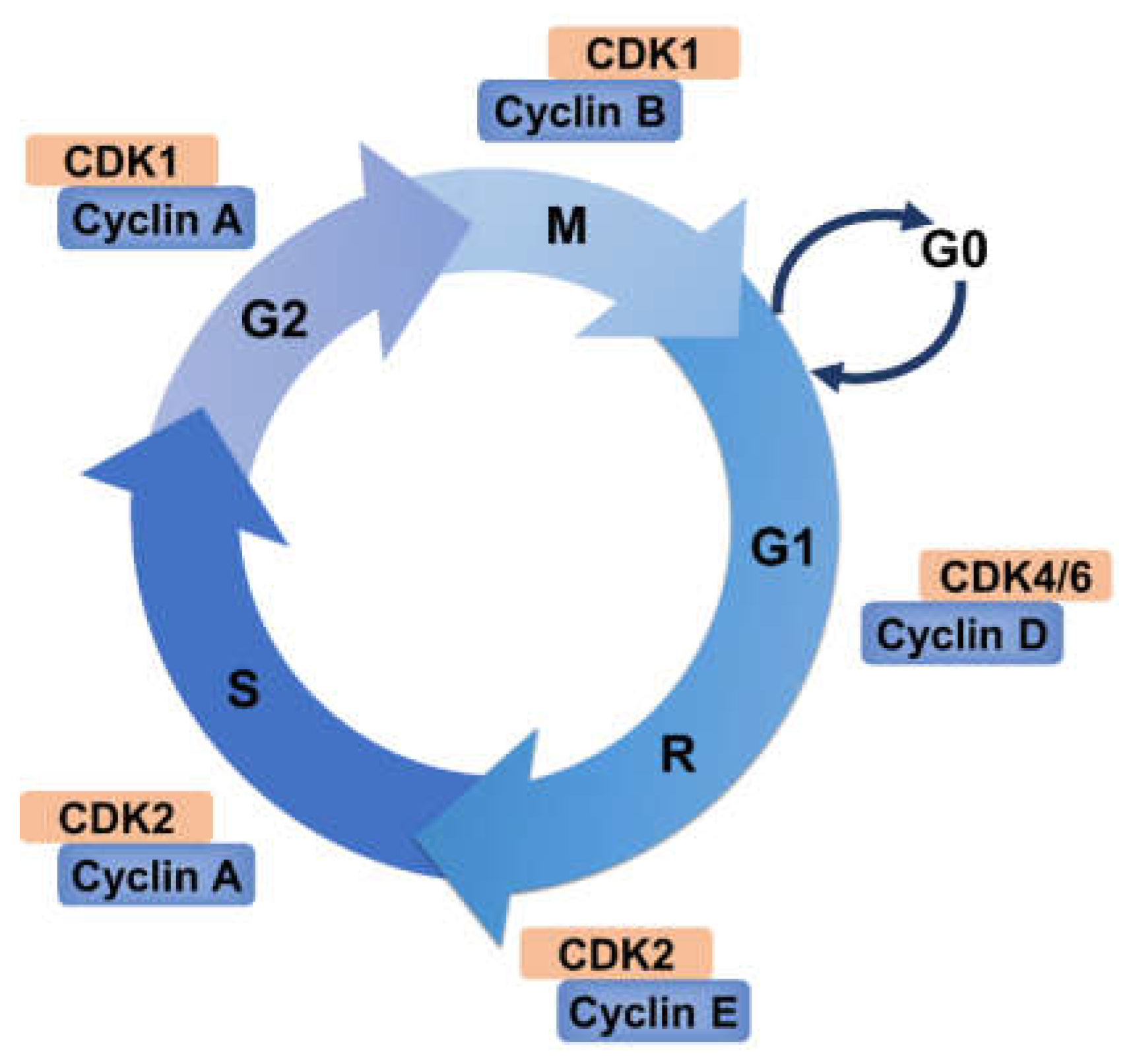
2. Cell Cycle and Its Regulation

A decisive characteristic of life is the reproductive capacity of cells, which it does through a collection of highly complex and ordered regulatory process commonly known as the cell cycle [23]. The cell cycle combines DNA replication with chromosomal segregation in an oscillatory manner [24]. In this way, the cell cycle coordinates the precise replication of the genome through specific events to ensure that the duplicated genetic material is distributed equally to each daughter cell. The repetition of this process leads to the exponential proliferation of cells. This process is classically described as interphase and mitosis (M) phase. Most of the cell cycle is in interphase, which encompasses Gap 1 (G1), synthesis (S), and Gap 2 (G2) phases. During the interphase, the cell grows, replicates genetic materials, and repairs DNA damage and replication errors. M phase, a relatively short period, consists of prophase, metaphase, anaphase, and telophase, which completes the equal distribution of genome and cytoplasmic components [1]. Following interphase, most nondividing cells exit the cell cycle at G1 and go into G0 phase (quiescence) [25]. G0 was originally used to describe cells that are not in the cell cycle but with the potential for division [1]. The rate of cell cycling varies with the developmental stage and cell type [26]. In general, the cell cycle is most active during development, as cells in early embryos can proliferate and differentiate to form tissues and organs [27]. The cell cycle involves numerous life processes, and it is closely related to the growth and proliferation of eukaryotic cells, development of organisms, regulation of DNA damage repair [28], and occurrence of diseases [28,29].

2.1. Cyclin–CDK Complexes Regulation

Studies in the past few decades have revealed that the cell cycle is tightly regulated by complexes containing CDKs and cyclin proteins. CDKs belong to a highly conserved family of serine/threonine protein kinases whose activity relies on a regulatory subunit, a cyclin. The original member of the CDKs family, CDK1 (also known as CDC2), was discovered in genetic screens for Schizosaccharomyces pombe and Saccharomyces cerevisiae [30]. The proteins of cyclin family which vary with cell cycle progression, are classified by the existence of a CDK-binding domain called the cyclin-box [31,32]. Cyclins were first identified by Tim Hunt in developing sea urchin eggs, and they were synthesized during interphase and then rapidly destroyed at each cleavage division [33]. Up to now, studies showed that fungi contain 6–8 CDKs and 9–15 cyclins, flies and Echinodermata include 11 CDKs and 14 cyclins, and humans have 20 CDKs and 29 cyclins [34]. Many of these 29 cyclins lack known CDK partners, and not all cyclins are involved in cell cycle control, such as cyclin I [35].
Different stages of the cell cycle require different cyclins. CDK4 and CDK6 are frequently regarded together as promoters of G1 progression. Initially, mitogenic signaling induces synthesis and assembly of the D-type cyclins, the proper folding of CDK4 and/or CDK6 and their transport to the nucleus [36]. CDK4 or CDK6 is activated after binding to D-type cyclins (D1, D2, and D3). Active complexes of CDK4/6 and D-type cyclins phosphorylate members of the retinoblastoma protein (pRb) family, which includes Rb/p105, p107, and Rb2/p130. Rb protein is a tumor suppressor which plays a key role in the negative regulation of cell cycle and tumor progression. It has been demonstrated that pRb is responsible for preventing S phase entry and cell growth. Phosphorylation of Rb protein leads to their functional inactivation [37]. CDK2 associates with cyclin E during late G1 phase and subsequently binds to cyclin A during early S phase for DNA synthesis and replication [38]. Therefore, both the D- and E-type cyclins and their associated kinases are considered to be pivotal for entry into G1 and G1/S phase transition. As the cell cycle progresses, Cyclin A subsequently interacts with and activates CDK1 in late G2 phase to facilitate the onset of M phase. At last, CDK1 is activated by B-type cyclins, thus triggering chromosome condensation and mitotic entry after nuclear lamina breakdown [30].

2.2. CKIs Regulation

The activity of each CDK is not only controlled by the availability of its cyclin partner, but it is also negatively regulated by the expression of a specific CDK inhibitor (CKI). CKIs bind to free CDKs or cyclin–CDK complexes to regulate CDK activity. CKIs are classified into two families according to their structures and CDK targets: (1) the CIP/KIP proteins, including p21CIP1, p27KIP1, and p57KIP2, extensively influence the activity of cyclin D-, cyclin E-, and cyclin A-dependent kinase complexes; (2) the INK4 proteins, P16INK4a, p15INK4b, p18INK4c, and p19INK4d, specifically target CDK4 and CDK6 [4,39,40]. The lack of these proteins leads to loss of function in growth control, DNA replication, cell apoptosis, and wound repair. Therefore, CKIs are considered as potential targets for cancer treatment.

2.3. The Restriction Point and Checkpoints

In fact, Arthur Pardee discovered as early as 1974 that a normal cell needs exogenous mitogenic stimulation only during the first two-thirds of the G1 phase. After undergoing continuous mitogenic stimuli during this period, the cell could decide to continue and complete its cell cycle in the absence of mitogenic signals. This phenomenon suggests the existence of a key point at the mid/late G1 phase, which was termed the “restriction point” (R point) by Pardee [41,42]. Therefore, the R point is the time after which the cell is committed to entering the cell cycle. The R point is considered to be regulated largely by pRb [43].
Furthermore, these regulatory roles of cyclin–CDK complexes allow for checkpoints during the cell cycle, which are inhibitory pathways. Cell cycle checkpoints have been defined as “biochemical pathways that ensure dependence of one process upon another process that is otherwise biochemically unrelated.” Checkpoints can control cell cycle progression and ensure high-fidelity completion of crucial events such as DNA replication and chromosome segregation [44]. Regulatory checkpoints include the DNA damage and DNA replication checkpoints and spindle assembly checkpoint. The DNA damage checkpoint is responsible for detecting damaged DNA and generating arrest in G1, S, and G2 phase of the cell cycle. The G1 arrest provides time for DNA repair before replication, while the block in G2 allows repair before chromosome separation in M phase. Cells can also be arrested in S phase, slowing down DNA synthesis [1]. The DNA replication checkpoint ensures that mitosis does not take place until DNA replication is complete, otherwise cells face the risk of chromosome mutation [45]. The spindle assembly checkpoint is one of the critical mechanisms to ensure the correct and even distribution of genetic material into two daughter cells. It monitors the binding of chromosomes and spindle microtubules as well as the combination of chromosomes and spindle microtubules [46]. Recently, a growing body of evidences suggested that checkpoint dysfunctions result in genomic instability which have been related to cancer.

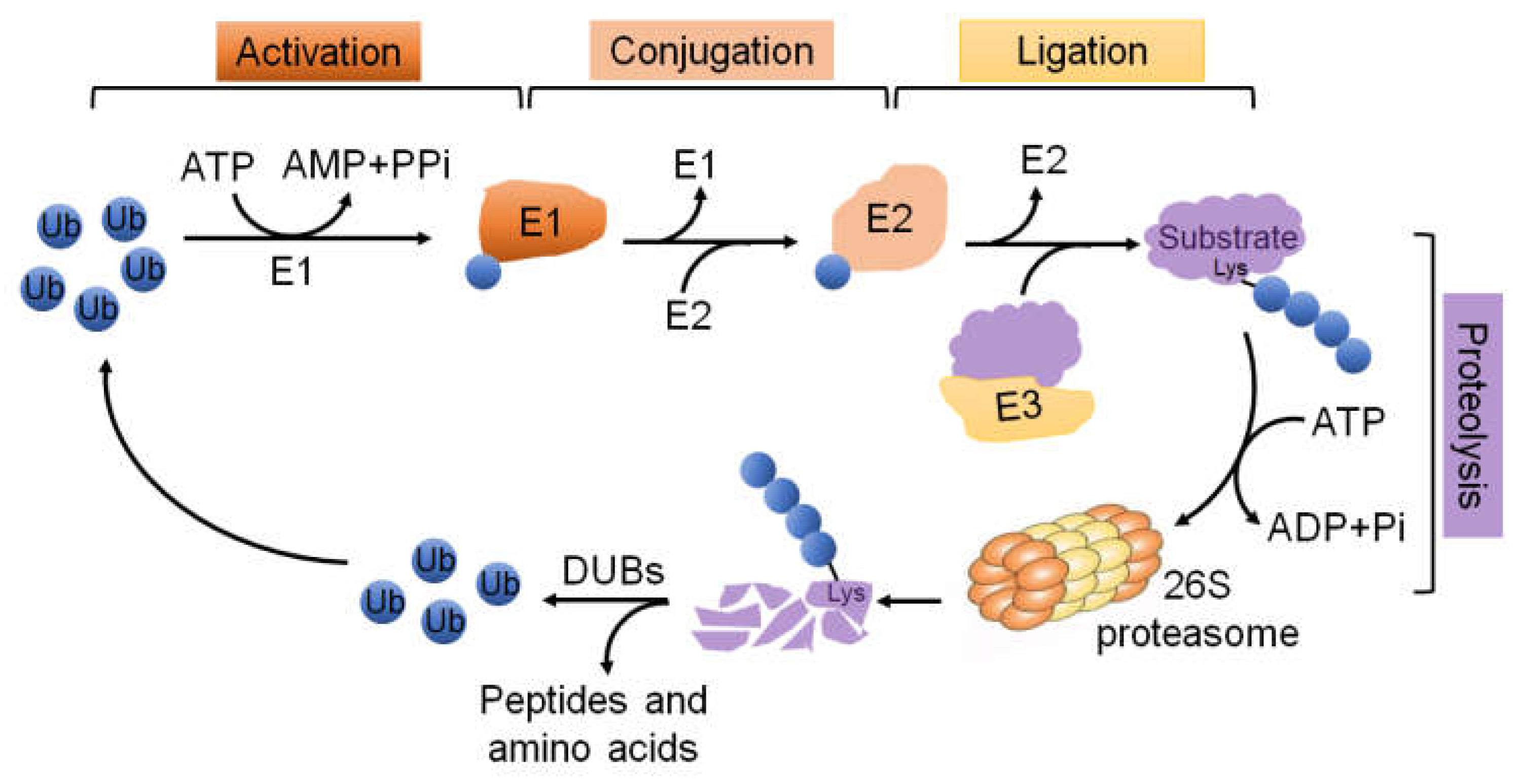
3. The UPS Molecular Machinery

The expression levels of cyclins during cell division are periodic. This is the result of a constant synthetic rate coupled with specific proteolysis, which is executed by the UPS. In the cell cycle, ubiquitination plays a central role in cell cycle transitions and checkpoints by establishing the strict temporal control of proteins such as cyclins, CDKs, CKIs, other kinases and phosphatases [21]. The UPS is one of the protein degradation pathways in eukaryotic cells, and most proteins rely on this system for degradation. The UPS consists of ubiquitin, E1 ubiquitin-activating enzymes, E2 ubiquitin-conjugating enzymes, E3 ubiquitin–ligase enzymes, deubiquitinating enzymes (DUBs), and 26S proteasome [47]. Degradation of target proteins requires a multistep reaction (Figure 2). The first step is to covalently conjugate a single ubiquitin or several ubiquitin molecules to the substrate protein mediated by E1, E2, and E3 enzymes. Then, the 26S proteasome selectively degrades based on different ubiquitin connection types [48]. Ubiquitination is a reversible modification, and DUBs can remove the ubiquitin chain from the substrate protein [49]. For instance, K11- or K48-polyubiquitin chains mediate proteasome degradation [50], whereas K63-polyubiquitin conjugates are involved in regulating cellular signal transduction [51]. In addition, mono-ubiquitination has been shown to be implicated in the endocytosis of plasma membrane receptors [52,53]. Many studies have demonstrated that ubiquitination can participate in cell cycle regulation, cell apoptosis and autophagy, ER stress, and protein localization and activity regulation. Of relevance, the regulation of the cell cycle is one of the cellular processes which have to be loyally coordinated and regulated by ubiquitination [54] (Figure 3). For example, the principal ubiquitin ligase families involved in cell cycle regulation are the SCF and anaphase promoting complex/cyclosome (APC/C) complexes [3,20,21]. The human APC/C consists of 19 core subunits and is an evolutionarily conserved multi-subunit cullin-RING E3 ubiquitin ligase which is primarily known for its ability to trigger the transition from metaphase to anaphase during mitosis [55]. APC/C is controlled by two major activators, Cdc20 in metaphase and Cdh1 in telophase. Either one can activate APC/C to target the degradation of specific substrates, including cyclins, mitotic kinases (such as Polo-like kinases, NIMA-related kinases, and Aurora kinases), spindle assembly factors, and DNA replication proteins during different time of the cell cycle [3,56,57,58]. Binding of Cdc20 or Cdh1 to the APC/C induces a conformational change to the active forms, APC/CCdc20 and APC/CCdh1, respectively. APC/CCdc20 exists in the onset of mitosis until the beginning of anaphase. In early mitosis, Cdc20 becomes active and associates with phosphorylated APC/C by CDK1, resulting in the degradation of prometaphase substrates, such as cyclin A [56,59,60]. Once all chromosomes achieve proper positions in metaphase, APC/CCdc20 degrades Securin and cyclin B to facilitate cellular transition to anaphase. Securin is an anaphase inhibitor and is essential for chromosomal separation [61]. Therefore, APC/CCdc20 mediates the separation of sister chromatids and is the target of regulatory mechanisms whose role is to ensure that daughter cells inherit complete copies of the genome [62], whereas APC/CCdh1 is present in the late mitosis to late G1, promoting efficient mitotic exit and maintenance of the G1 state. Similarly, APC/CCdh1 also mediates the degradation of mitotic cyclins [63]. Furthermore, APC/CCdh1 induces the degradation of SKP2, which, in turn, targets for the degradation of the CKIs such as p27, p21, and p57 [64,65].
APC/C is mainly active in mitosis and G1 phase of the cell cycle and acts as the key antagonist of mitotic CDKs [62], while SCF complexes is always active, executing a variety of functions at most phases of the cell cycle and beyond. The SCF complex is comprised of the invariable components SKP1, CUL1, and RBX1 as well as one variable component, known as an F-box protein, that binds to SKP1 through its F-box motif and is responsible for substrate recognition [66]. To date, about 70 F-box proteins have been identified in humans, and these proteins are divided into three categories: those with WD40 repeats (FBXW), leucine-rich repeats (FBXL), or other domains (FBXO) [67]. Some of them, e.g., FBXW8 [68], FBXO25 [69], Fbw7 [70], and SKP2 [71,72,73,74], can directly regulate cyclins or CKIs. More importantly, not only APC/C and SCF cooperate in the regulation of common substrates, but also their activities are regulated by each other. As noted above, APC/CCdh1 ubiquitinates SKP2 during G1 phase, allowing for the post-mitotic down-modulation of SCF [64]. Furthermore, Emi1 is a protein able to inhibit the activity of APC/C in S and G2 phases [75]. Emi1 is phosphorylated by Cdc2 and is subsequently recognized by the SCFβ-TRCP ubiquitin ligase and destroyed in prophase, thus promoting APC/C activation [76].

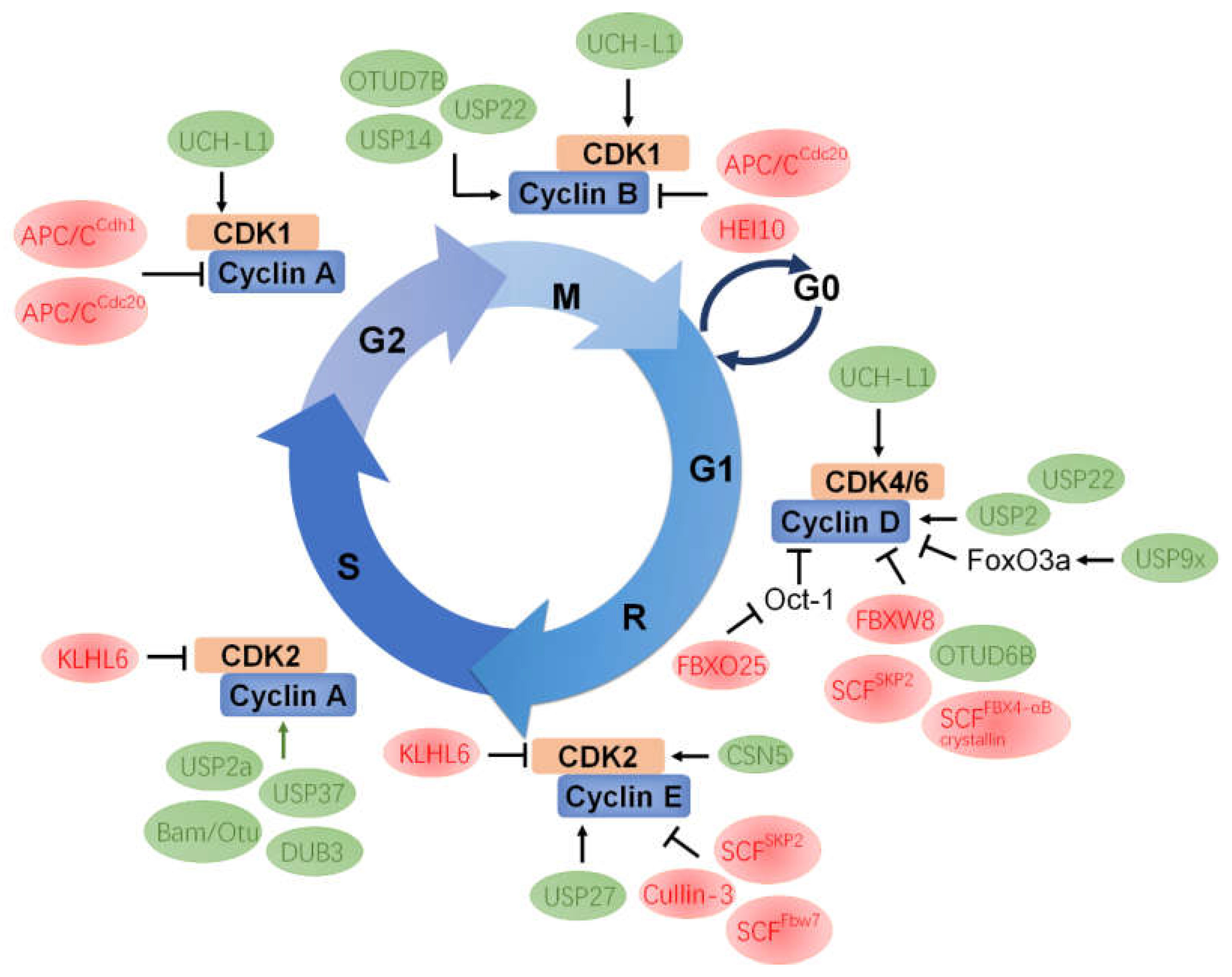
4. The Functional Roles of UPS in Cyclins

Ubiquitin-dependent degradation plays an essential role in many important cellular processes such as cell cycle regulation and signal transduction. In the cell cycle, ubiquitin-dependent proteolysis provides a rapid control for modulating the protein expression levels of cyclins. The accumulation or degradation of cyclins will lead to different cell fates (Table 1).

4.1. Cyclin A

Cyclin A plays a fundamental role in regulating cell cycle events because it can activate CDK2 and CDK1, and has functions in both S and M phase [77]. A study has found that human cyclin A is relatively stable during S and G2 phase, but it is rapidly degraded when cells enter metaphase of mitosis [78]. There are two types of cyclin A (cyclin A1 and cyclin A2) in Xenopus [79,80], mice [81], and humans [82], both of which can form complexes with either CDK1 or CDK2. Cyclin A1 is only expressed in meiosis and very early embryos, whereas cyclin A2 exists in proliferating somatic cells [78]. A previous study has shown that the APC/C is an evolutionarily conserved multi-subunit E3 ubiquitin ligase composed of 14 proteins, which can target cyclin A for ubiquitin-mediated degradation. The degradation of cyclin A by Cdc20 or Cdh1-activated APC/C is necessary for the onset of anaphase and exit from mitosis, in contrast to nondegradable cyclin A arrests cells in anaphase [55,78]. However, the ubiquitination of cyclin A can be reversed by USP37. USP37 binds, deubiquitinates, and stabilizes cyclin A, thereby regulating the G1/S transition [83]. In the identification of other DUBs of cyclin A, it was found that USP2a can bind to and negatively stabilize cyclin A, resulting in enhanced oncogenic characteristics in bladder cancer cells [84]. Subsequent research in lung cancer cells has identified DUB3 (also termed USP17) as a new regulator of cyclin A. DUB3 binds to cyclin A and promotes its deubiquitination and stabilization. Compared with the control cells, the percentage of S phase cells was significantly decreased in DUB3 knockdown A549 cells, whereas this effect can be rescued by overexpression of cyclin A in DUB3 depletion cells. These results suggest that the DUB3–cyclin A axis plays a key role in G1/S transition during cell cycle progression and at least partially explain the mechanism of DUB3 which promotes the proliferation of NSCLC cells, providing a promising target for NSCLC treatment [85].
In Drosophila, it has been demonstrated that fate determination of germ-line stem cells (GSCs) (such as differentiation or regeneration) is tightly controlled by a specific cell cycle program [86]. Previous studies have revealed that mutation or dysregulated expression of cyclins, such as cyclin A and cyclin E, leads to defects of GSC maintenance, differentiation, and/or an abnormal cell number of cyst [87,88,89]. Thus, correct expression of cyclins is important for GSC fate determination and the normal development. A recent study in Drosophila uncovered that Bag of marbles (Bam) can form a deubiquitinase complex with Otu to deubiquitinate and stabilize cyclin A, thereby balancing GSC self-renewal and differentiation [90].

4.2. Cyclin B

On the one hand, cyclin B is the key regulatory protein controlling mitosis in all eukaryotes and forms a complex with CDK1 to initiate the mitotic program through phosphorylation of selected proteins. On the other hand, destruction of cyclin B due to ubiquitin-mediated proteolysis is necessary for mitotic exit [106]. Therefore, the abundance of cyclin B is critical to cell cycle progression. Interestingly, the APC/C protein complex can target both cyclin A and cyclin B. In the late 20th century, scientists proved that degradation of B-type cyclins depends on ubiquitination which is mediated by the activity of the APC/CCdc20 complex [91,107]. Later in 2003, human enhancer of invasion 10 (HEI10) was first identified as a RING-finger family ubiquitin ligase that regulates cell cycle by interacting with cyclin B1 and promoting its degradation, thereby specifically functioning in progression through G2/M [92].
Researchers have also explored the mechanism of deubiquitination of cyclin B. Studies have found that the deubiquitinating enzyme USP14 seems to be closely related to cyclin B. USP14, belonging to the USP family, which binds reversibly to the 26S proteasome [108], limits proteasomal degradation of ubiquitinated proteins [109,110]. Lee et al. reported that USP14 shows a dramatic preference for ubiquitin–cyclin B conjugates that carry more than one ubiquitin modification or chain [110]. A recent study further verified that USP14 can interact with cyclin B1, promoting its deubiquitination and stabilization. Knocking down USP14 with siRNAs significantly inhibited the proliferation and migration of breast cancer cells. Thus, these results provide a solid theoretical basis for the development of anti-cancer drugs targeting USP14 [97]. Cyclin B is also regulated by multiple DUBs. USP22 can interact with cyclin B1, promoting its deubiquitination and stabilization. Interestingly, USP22 is also phosphorylated by CDK1, and this phosphorylation enhances its activity in deubiquitinating cyclin B1. Compared with the wild-type control, knockdown of usp22 gene in colon cancer HCT116 cells or in mouse embryonic fibroblast cells leads to decreased cell proliferation [98]. Thus, USP22 is involved in cell proliferation possibly through promoting cyclin B1 stabilization. In addition, another study has also found that ovarian tumor domain-containing 7B (OTUD7B) is a cell cycle-regulated deubiquitinase that antagonizes the degradation of APC/C substrates. OTUD7B can deubiquitinate cyclin B by specifically removing the K11-linked ubiquitin chain from cyclin B, thereby protecting cyclin B from 26S proteasome-mediated degradation. Further observations unraveled that OTUD7B contributes to mitotic exit and that its function is crucial for cells to properly progress through the cell cycle and proliferate [99].

4.3. Cyclin D

It is well known that D-type cyclin regulates G1 progression and is overexpressed in various human cancers [111,112]. Therefore, cyclin D is considered as one of the potential therapeutic targets for cancer [113]. SKP1–CUL1 (Cdc53 in yeast)–F-box-protein (SCF) is one of the important E3 ubiquitin ligases that regulate D-type cyclins, and its substrate specificity is determined largely by F-box protein (FBP) [67]. As early as 1998, Zhang’s team identified that human Cullin 1 (CUL-1) associates with SKP1/SKP2 to form the SCFSKP2 complex. This complex may play function as a conserved E3 ubiquitin ligase, which is similar to yeast SKP1–CDC53–F-box protein complex. They proposed that this complex is likely to target cyclin D and p21 for ubiquitin-dependent degradation, although this view needs further biochemical analysis to support [71]. Okabe et al. found that FBXW8, the F-box protein, could specifically associate with cyclin D1 in a Thr286 phosphorylation-dependent manner and regulate its stability through the UPS pathway. Interestingly, depletion of FBXW8 leads to a significant accumulation of cyclin D1, while preventing cyclin D1 from entering the nucleus and resulting in a slower rate of cell proliferation [68]. A recent study has shown that another F-box protein FBXO25 also regulates cyclin D1, but this regulation is not achieved through the direct interaction between FBXO25 and cyclin D1. FBXO25 directly interacts with the cyclin D1 repressor Oct-1 and downregulates its expression [69]. It is well known that Oct-1 as transcription factor binds to the promoter of cyclin D1 and leads to its inactivation [93]. Therefore, the decrease of Oct-1 is conducive to stabilize cyclin D1, thereby promoting tumor growth and metastasis [69]. In another study, SCFFBX4-αB crystallin complex was identified as an E3 ubiquitin ligase of cyclin D1, promoting ubiquitin-dependent degradation of Thr286 phosphorylated cyclin D1. Knockdown of SCFFBX4-αB crystallin ligase can increase the accumulation of cyclin D and accelerate G1 phase progression [94].
Ubiquitination of proteins is a dynamic process which can be reversed by removal of ubiquitin by DUBs. Thus, cyclin D1 is also regulated through a specific DUB. In 2009, a novel regulator of cyclin D1 was described by Shan et al., who identified USP2 as a deubiquitinase responsible for cyclin D1 stabilization. The accumulation of cyclin D1 accelerates the G1 to S phase progression. Of note, restraint of USP2 function induced growth suppression only in the cancer cells addicted to cyclin D1 expression, whereas there was no major effect on cell growth of normal human fibroblasts [100]. This different effect of USP2 in normal and cancer cells suggests that USP2 may be used as a new drug target to block its activity and reduce the proliferation of cancer cells in a safe and non-toxic way. In 2017, Magiera et al. found that lithocholic acid hydroxyamide (LCAHA) destabilizes cyclin D1 and induces G0/G1 arrest by inhibiting activity of USP2 deubiquitinase [114]. In addition, three distinct classes of small-molecule inhibitors of USP2 have been reported: NSC 632839 [115], chalcones [116], and the compound ML364 [117]. It has been reported that the cell cycle arrest induced by FoxO forkhead transcription factors (such as FoxO4, FoxO3a, and FoxO1a) involves the reduced protein expression of D-type cyclins [102]. Thus, these transcription factors are negative regulators of D-type cyclins. Zheng et al. found that USP9x can interact with FoxO3a and reduce the ubiquitination level of FoxO3a and stabilize it. At the same time, FoxO3a is also regulated by prolyl hydroxylase EgIN2. Prolyl-hydroxylated FoxO3a prevents the binding of USP9x, thereby promoting the proteasomal degradation of FoxO3a and the accumulation of cyclin D1 [101]. Another study provided evidence that OTUD6B probably participates in cell cycle progression of B lymphocytes by regulating cyclin D2. Meanwhile, the deubiquitinating activity of OTUD6B on cyclin D2 was not observed [103]. Therefore, OTUD6B is likely to be involved in the regulation of cyclin D2 through an intermediate, but it is not yet clear. Apart from that, a recent study has revealed that USP22 regulates cell cycle by directly deubiquitylating cyclin D1, thus protecting cyclin D1 from proteasome-mediated degradation. Loss of USP22 in cancer cells results in defective G1/S cell cycle transition, whereas ectopic cyclin D1 protein partially rescues the aberrant cell proliferation phenotype. It suggests that USP22 plays a significant role in cell cycle progression via stabilization of cyclin D1 [104].

4.4. Cyclin E

Cyclin E binds and activates CDK2 and promotes the G1/S transition of the cell cycle. The amount of cyclin E is tightly regulated by ubiquitin-mediated proteolysis. Cyclin E accumulation can lead to accelerated S phase entry [118], genetic instability [119], and tumorigenesis [120]. Cullin-3 (Cul-3) is a member of the cullin family of E3 ubiquitin–protein ligases. A study has shown that Cul-3 can bind to free cyclin E but not to the cyclin E/CDK2 complex in mammalian cells, and it specifically increased ubiquitination of cyclin E. Deletion of the Cul-3 gene in mice caused increased accumulation of cyclin E. In the extraembryonic ectoderm and ectoplacental cone of the Cul-3−/− embryos, there was a great increase of S phase cells [95]. A recent study further proved that Cul-3 regulates cyclin E1 protein abundance via a degron located within the N-terminal region of cyclin E. However, cyclin E2 lacks the Cul-3 degron and is not targeted for degradation by Cul-3 [121]. Cyclin E is also regulated by multiple E3 ubiquitin ligases. Koepp et al. identified that the E3 ubiquitin ligase complex SCFFbw7 is responsible for cyclin E ubiquitination and demonstrated that it is functionally conserved in yeast, flies, and mammals. Fbw7, as the FBP component of SCF protein complex, can specifically associate with cyclin E and promote its ubiquitination. Deletion of Fbw7 results in the accumulation and stabilization of endogenous cyclin E in human and Drosophila melanogaster cells [70]. Thus, Fbw7 is a negative regulator of cyclin E and a potential tumor suppressor. In another study in the same year, it was also proved that another important F-box protein SKP2 can negatively regulate the stability of cyclin E [96]. It has been previously shown that phosphorylation of threonine 380 (Thr380) at the carboxy terminus of cyclin E provides a signal for the ubiquitin-dependent proteolysis of cyclin E [122,123]. They identified that SKP2 specifically binds to cyclin E, which is phosphorylated at Thr380 and mediates its ubiquitin-dependent degradation, thus contributing to the control of S phase progression [96].
Cyclin E abundance is also regulated by a specific DUB. In 2018, USP27 was identified as the DUB of cyclin E [105]. Both the C-terminal UCH domain of USP27 and the N terminus of Cyclin E are required for this interaction. USP27 can reduce the ubiquitination level of cyclin E, thereby promoting cyclin E stabilization. Furthermore, knockdown of USP27 suppresses cell cycle progression by increasing the percentage of cells in G0/G1 phase, which can be reversed by the recovery of cyclin E expression [105].

5. Mutual Regulation between CDK and UPS Components

Protein phosphorylation is a key regulatory mechanism for cell cycle control in eukaryotes. From yeast to humans, cell cycle progression and cell division require the activation of CDKs. CDKs are activated by associating with a cyclin, then initiating and coordinating these processes by orderly phosphorylation of their targets. Therefore, CDK is not only regulated by ubiquitination and/or deubiquitination, but also exerts kinase activity to phosphorylate substrate proteins. In this section, we pay attention to the regulation of CDKs by UPS and discuss the phosphorylation regulation of some deubiquitinases and E3 ligases by CDKs (Table 2).

5.1. CDK1

The onset of mitosis requires increased association activity of CDK1 with cyclin A and cyclin B. The CDK1/cyclin B complex is the dominant regulator of the G2/M transition and has maximal activity during metaphase. Yang et al. reported that CDK1 phosphorylates the histone H2A deubiquitinase Ubp-M at serine 552 (S552P), and, importantly, this phosphorylation is required for cell cycle progression. Ubp-M (S552P) destroys Ubp-M interaction with chromosome maintenance protein 1 (CRM1), allowing Ubp-M to be retained in the nucleus and deubiquitinate nucleosomal uH2A, which is required for chromosome condensation and cell cycle progression [124]. In addition, Ubiquitin C-terminal Hydrolase L1 (UCH-L1/PGP9.5) is a deubiquitinating enzyme which hydrolyzes C-terminal esters and amides of ubiquitin [125]. Kabuta et al. proved that UCH-L1 physically interacts with CDK1, CDK4, and CDK5 and enhances CDKs activity. To investigate the mechanism how UCH-L1 enhances the kinase activity of CDK, they used UCH-L1I93M and UCH-L1C90S mutants in cell-based kinase assays. Their observations indicated that the enhancement of CDK activities by UCH-L1 is independent of the known ubiquitin-related functions of UCH-L1 but correlates with interaction levels between UCH-L1 and CDKs. Besides that, overexpression of UCH-L1 enhances cell proliferation, and knockdown of UCH-L1 reduces tumor growth and CDK kinase activity [126]. The findings provide us with a promising strategy by targeting UCH-L1 to treat cancer.

5.2. CDK2

CDK2 plays a pivotal part in cell cycle regulation and is involved in a range of biological processes. It is also controlled by both ubiquitination and deubiquitination. The COP9 signalosome (CSN) is a DUB of the JAMM family composed of eight subunits (CSN1-8) and well-conserved in all eukaryotes from yeast to humans [127,128]. CSN5 is the only CSN subunit possessing the JAMM motif, and it was assumed that CSN5 has a deubiquitylating activity on its own [129]. Yoshida et al. found that CSN5 specifically interacts with CDK2 and positively regulates CDK2 stabilization. Interestingly, the expression level of cyclin E increased in accordance with CDK2’s upregulation, suggesting that CSN5 also regulates the level of cyclin E through CDK2. In addition, G1 progression of the cell cycle is markedly hampered in CSN5-depleted cells [127]. In a later study, it was verified that Kelch-like protein 6 (KLHL6), as an E3 ubiquitin ligase, plays an important role in acute myeloid leukemia (AML) by regulating the expression of CDK2. KLHL6 is the E3 ligase that binds to CDK2 and mediates its degradation [130].

5.3. CDK4/6

CDK4/6 are activated by the D-type cyclins, thereby phosphorylating pRb to promote G1/S transition. A recent study revealed that the CDK4/6–DUB3–SNAIL1 axis functions as an important regulatory mechanism of breast cancer metastasis [131]. SNAIL1 is deemed to be a key factor in the aggressive expression of tumors for its critical role in the epithelial–mesenchymal transition (EMT) pathway associated with tumor metastasis [132]. Liu et al. identified DUB3 as a new target of CDK4/6, and CDK4/6 phosphorylates DUB3 at serine 41. As a linker between CDK4/6 and SNAIL1, DUB3 activation by CDK4/6 is essential to deubiquitinate SNAIL1. As a bona fide SNAIL1 deubiquitinase, DUB3 interacts with SNAIL1, and then specifically removes the K48-linked ubiquitin chain to stabilize SNAIL1. Furthermore, knockdown of either DUB3 or CDK4 or CDK6 markedly decreased the expression level of SNAIL1, thus regulating cell migration and cancer metastasis [131].

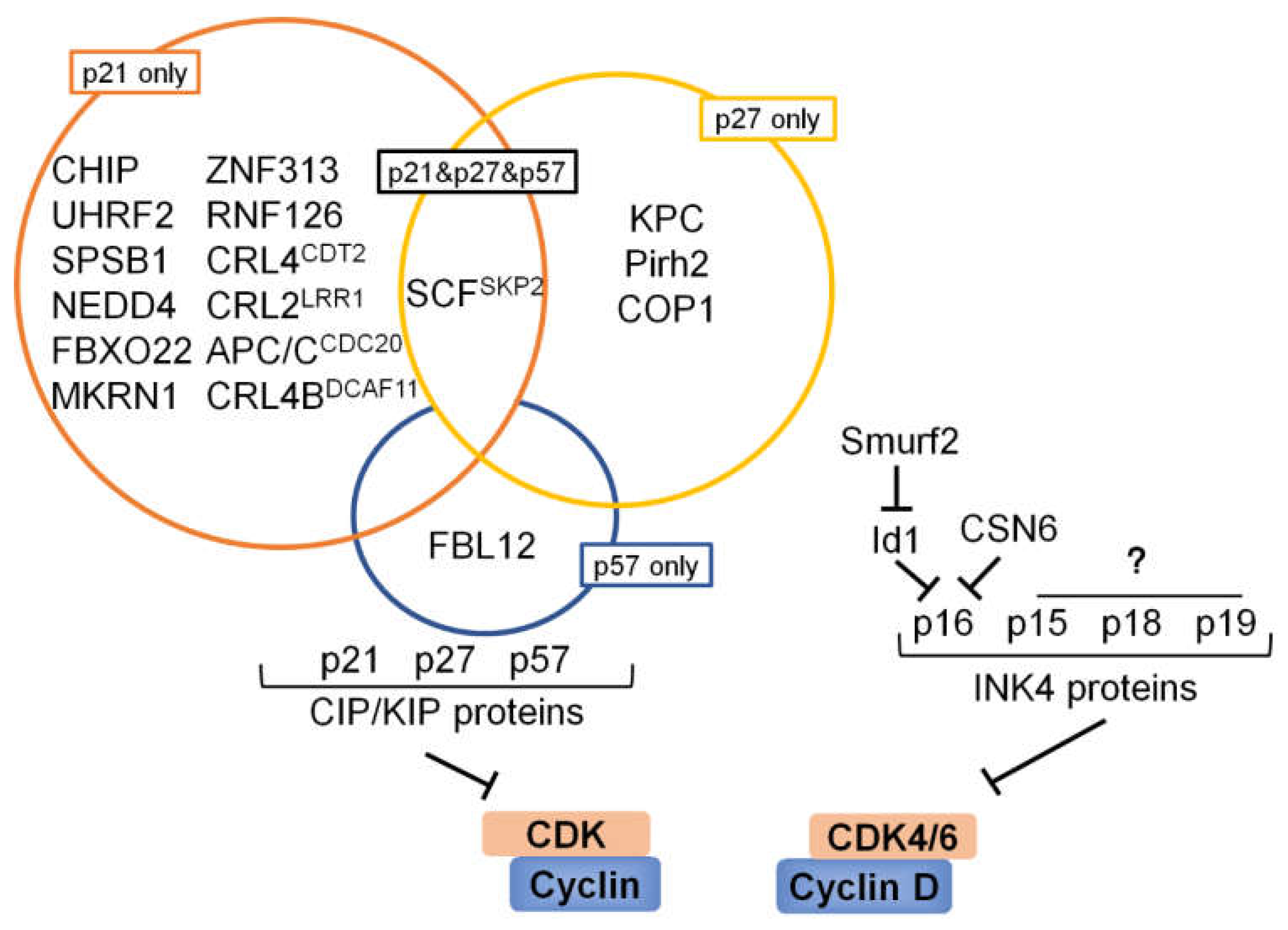
6. Ubiquitination and Deubiquitination Involved in CKIs

The kinase activity of cyclin–CDK complexes is tightly regulated by a plethora of CKIs which serve as brakes to halt cell cycle progression under adverse conditions. Similar to cyclins and CDKs, the expression level of CKIs is also balanced by ubiquitination and deubiquitination (Table 3 and Figure 4).

6.1. p53 and Its Downstream Target p21

As one of the most well-known and important tumor suppressors, p53 has received widespread attention and study. One central role of the tumor suppressor p53 is to arrest the cell cycle, as p53 indirectly downregulates the expression of many genes which are essential for progression through the cell division cycle [133]. Notably, many studies cemented that p53 is commonly mutated in most tumor types, with conservative estimates indicating a mutation rate of 50% across all tumors [134,135,136,137]. In normal cells, p53 is maintained at very low, often at undetectable levels. Murine double minute 2 (MDM2) is the main negative regulator of p53 activity and stability. Several studies have revealed that diverse signals regulate p53 levels by regulating the interaction between MDM2 and p53 [138,139]. First, MDM2 binds the transcriptional activation domain of wild-type and mutant p53, and blocks its ability to regulate target genes [140,141]. Second, MDM2 can act as an E3 ubiquitin ligase and interact with p53 to promote its ubiquitination and 26S proteasome-mediated degradation [142]. Importantly, p53 activates the expression of mdm2 gene in an autoregulatory feedback loop which aims to maintain low cellular p53 levels in the absence of stress [140,143]. The inhibitory effect of MDM2 on p53 can be reversed by E3 ligase of MDM2, such as PCAF [144], SCFβ-TRCP [145], XIAP [146], RFP2 [147], NAT10 [148], and MARCH7 [149]. These E3 ubiquitin ligases increase the stability of p53 by reducing p53 ubiquitination through ubiquitination and degradation of MDM2. In contrast, several deubiquitinating enzymes, such as HAUSP (also known as USP7) [150], USP2a [151], and USP15 [152], are able to stabilize MDM2 by removing its polyubiquitin chains. Intriguingly, HAUSP was initially identified as a deubiquitinase of p53. HAUSP interacts with p53 and removes the ubiquitin from p53, resulting in p53 stabilization. It has been found that the overexpression of HAUSP stabilizes p53 even in the presence of excess MDM2, and the downregulation of HAUSP expression makes the endogenous p53 unstable [153]. Compared with p53, the binding between MDM2 and HAUSP is much stronger [154,155]. Thus, it has also been observed that the disruption of the HAUSP gene stabilizes p53. With the in-depth development of research, more and more ubiquitin ligases that regulate p53 have been discovered, including Pirh2 [156], ICP0 [157], COP1 [158], Topors [159], CHIP [160], ARF-BP1 [161], TRIM24 [162], HRD1 [163], MKRN1 [164], WWP1 [165], TRIM39 [166], TRAF7 [167], Cullin4B [168], TRIM71 [169], RING1 [170], FBW7α [171], and TRIM69 [172]. All of these ubiquitin ligases could target p53 for proteasomal degradation. Great progress has been made in identifying the regulation of p53 ubiquitination; in addition, diverse DUBs that positively regulate p53 stability have also been discovered. HAUSP is the first identified deubiquitinating enzyme for p53, and it is considered to have a dual role in the p53-MDM2 pathway because it deubiquitinates both p53 and MDM2. In a subsequent study, USP10 was also identified as a p53 deubiquitinase which can interact with and stabilize p53. In unstressed cells, USP10 localizes in the cytoplasm and regulates p53 homeostasis. Following DNA damage, a fraction of USP10 translocates to the nucleus and contributes to p53 activation. Moreover, USP10, through its regulation of p53, appears to play an important role in inhibiting cancer cell growth [173]. In addition, Sun et al. revealed that Otub1 interacts with DNA-binding domain of p53 via its N and C termini and positively regulates stability of p53. Moreover, the DUB activity of Otub1 on p53 may depend on its Asp88. Overexpression of Otub1 drastically induces p53-dependent apoptosis and inhibition of cell proliferation [174]. Besides that, a series of studies has demonstrated that USP29 [175], USP42 [176], USP11 [177], USP9x [178], USP24 [179,180], and Ataxin-3 [181] can participate in p53-mediated multiple functions through deubiquitination and stabilization of p53.
p21CIP1/WAF1 (p21 herein after) is a potent, tight-binding inhibitor of CDKs and can suppress the phosphorylation of Rb by cyclin A–CDK2, cyclin E–CDK2, cyclin D1–CDK4, and cyclin D2–CDK4 complexes, resulting in G1 cell cycle arrest [182]. Thus, p21−/− cells show an impaired G1 checkpoint and do not arrest upon DNA damage and other insults [183,184]. Moreover, p21 gene is one of the most important downstream targets of p53 gene and a p53-binding site was identified in 2.4 kb upstream of p21 coding sequences. Ever since the initial identification of p21 as a direct p53 transcriptional target capable of inducing cell cycle arrest, this protein has been considered an important mediator of p53-dependent tumor suppression [185]. Therefore, both p21 and p53 are negative regulators of the cell cycle, and their stability is crucial for cell cycle progression. We previously discussed that p53 is the target of multifarious E3 ubiquitin ligases and DUBs. Much of the regulation of p21 levels is achieved by transcriptional control mechanisms. However, it appears likely that post-transcriptional processes may also regulate p21 levels [72]. Three different E3 ubiquitin ligases have been identified in targeting p21 degradation: SCFSKP2 [71,72,73], APC/CCDC20 [186], and CRL4CDT2 [187,188,189]. In subsequent research, MKRN1 was reported as an E3 ubiquitin ligase for both p53 and p21, and ablation of MKRN1 induces cell cycle arrest by stabilizing p53 and p21 [164]. In 2010, Starostina et al. demonstrated that the CRL2LRR1 complex also targets p21 for degradation in C. elegans to promote cell cycle progression in germ cells. However, the degradation of p21 mediated by human CRL2LRR1 complex does not appreciably affect cell cycle progression [190]. Up to date, ZNF313 [191], RNF126 [192], CHIP [193], CRL4BDCAF11 [194], UHRF2 [195], SPSB1 [196], NEDD4 [197], and FBXO22 [198] have also been added to the growing list of E3 ubiquitin ligases that control the abundance of p21 protein. Compared with the ubiquitination studies of p21, the identified deubiquitinases targeting p21 are much less. Until 2018, Deng et al. identified USP11 as the first deubiquitylase that directly reverses p21 polyubiquitylation and stabilizes the p21 protein. Loss of USP11 causes the destabilization of p21 and induces the G1/S transition in unperturbed cells. Furthermore, DNA damage-mediated p21 accumulation is completely abolished in cells depleted of USP11, which leads to abrogation of the G2 checkpoint and induction of apoptosis [199].
Although the rapid degradation of p53 and p21 are largely achieved through the ubiquitin proteasome pathway, both p53 and p21 are regulated by a ubiquitin-independent process mediated by 20S proteasome [200,201]. In a series of studies, Asher et al. demonstrated that NAD(P)H quinone oxidoreductase 1 (NQO1) could stabilize p53 and revealed that (NQO1) is associated with the 20S proteasomes and interacts with p53 in an NADH-dependent manner to protect p53 from 20S proteasomal degradation [200,202,203]. Another study has shown that proteasome activator REG-γ (also known as PA28, PSME3, and Ki antigen), which binds and activates 20S proteasome [204], plays an important role in regulating p21. They suggested that REGγ directly mediates degradation of free p21 in an ATP- and ubiquitin-independent manner. Overexpression of REGγ reduced p21 level, while depletion of REGγ enhanced the p21 level in multiple cell lines. In addition, depletion of REGγ in a thyroid carcinoma cell line results in cell cycle and proliferative alterations [201]. In conclusion, the stability regulation of p53 and p21 is a very complicated process and is affected by many factors.

6.2. p27

The p27 gene is located on chromosome 12p13. p27 protein binds to cyclin (A, B, D, and E)–CDK (1, 2, 4, and 6) complexes and leads to the failure of Rb phosphorylation, which ultimately results in standstill of G1/S transition [205]. Therefore, degradation of mammalian p27 is required for the cellular transition from quiescence to the proliferative state. Previous studies have demonstrated that nuclear SCFSKP2 E3 ligase specifically recognizes p27 in a phosphorylation-dependent manner that is characteristic of an F-box-protein–substrate interaction, thus promoting ubiquitin-mediated degradation [74]. Most importantly, phosphorylation of p27 at Thr187 was essential for its degradation [206]. Complete knockdown of SKP2 partially prevented p27 degradation. Furthermore, SKP2 is important in the progression from quiescence to S phase and that the ability of SKP2 to promote p27 degradation is a key aspect of its S-phase-inducing function [207]. However, proteolysis of p27 at the G0/G1 transition proceeds normally in skp2-/- cells [208]. Kamura et al. proposed that the existence of a SKP2-independent pathway for the degradation of p27 at G1 phase. They described that the cytoplasmic ubiquitin ligase KPC consisting of KPC1 and KPC2 interacts with and ubiquitinates p27. Moreover, the nuclear export of p27 by CRM1 seems to be necessary for KPC-mediated proteolysis. Overexpression of KPC promoted the degradation of p27, whereas a dominant-negative mutant of KPC1 delayed p27 degradation, and knockdown of KPC1 by RNA interference also inhibited p27 degradation. Further exploration suggested that KPC controls not only the progression from G0 to S phase but also the growth of continuously cycling cells during long-term culture [209]. Subsequently, a study has identified that a RING-H2-type ubiquitin ligase Pirh2 interacts with p27 and facilitates its proteasomal degradation. Depletion of Pirh2 induced accumulation of p27 in both the nucleus and cytoplasm and resulted in an inhibition of cell cycle progression at G1/S transition in a p53-independent manner [210]. It has been reported that COP1 also enhances ubiquitin-mediated degradation of p27 to promote cancer growth [211]. Although CSN is involved in regulating various biological processes, individual subunits of COP9 have been suggested to modulate different physiological functions [212]. Different from CSN5 as mentioned above, subunit 6 of the COP9 signalosome complex (CSN6) usually collaborates with other E3 ligase to regulate target proteins, thereby governing cancer development. One study demonstrated that the CSN6–COP1 axis plays a critical role in controlling the stability of p27 [213]. Depletion of CSN6 results in reduction of COP1 and concurrent elevation of p27. Mechanistic studies showed that CSN6 interacts with p27 and enhances ubiquitin-mediated degradation of p27. CSN6-mediated p27 degradation depends on the nuclear export of p27, which is regulated through COP1 nuclear exporting signal [213]. A previous study has reported that CSN6 associates with COP1 and positively regulates its stability through inhibition of ubiquitin-mediated COP1 proteasomal degradation [214]. Therefore, p27, COP1, and CSN6 possibly form a complex, and CSN6 increases the degradation of p27 by stabilizing COP1. However, it seems that USP37 is the only deubiquitinase directly related to p27. USP37 formed a complex with p27, promoted its deubiquitination and stabilization, and blocked cell proliferation [215].

6.3. p57

The cyclin/CDK inhibitor p57Kip2 is the least studied member of the Cip/Kip family [216]. As one of the most important E3 ligases in cell cycle regulation, the SCFSKP2 complex targets multiple substrate proteins including p57 for ubiquitylation and proteolysis [217]. Then, Chiba’s team identified that FBL12, a new F-box protein induced by TGF-β1, could directly ubiquitinate p57 and lead to its degradation. Impairment of FBL12 and overexpression of p57 enhanced osteoblast cell differentiation, indicating the importance of p57 degradation in the TGF-β1-mediated inhibition of osteoblast cell differentiation [218]. Subsequently, it was also proved that p57 can be negatively regulated by CSN6 [219]. CSN6 negatively regulates p57 stability via increased ubiquitination levels of p57, thereby reducing p57′s steady-state expression. Further mechanistic studies have indicated that CSN6 associates with p57 and SKP2, which, in turn, facilitates SKP2-mediated protein ubiquitination of p57. Loss of SKP2 hinders CSN6-mediated p57 destabilization, suggesting collaboration between SKP2 and CSN6 in controlling degradation of p57. Furthermore, downregulation of p57 expression by CSN6 leads to faster cell growth, deregulated cell cycle, and potentiated transformational activity [219]. However, until now, the deubiquitinase of p57 has not been reported.

6.4. INK4 Proteins

The members of INK4 family, including P16INK4a, p15INK4b, p18INK4c, and p19INK4d, block the progression of the cell cycle by binding to either CDK4 or CDK6 and inhibiting the action of cyclin D [222]. Logically, the stable expression of a CKI would be incompatible with the cell cycle progression and might instead prove important for cell cycle entry or exit, and for maintaining differentiated cells in a quiescent, post-mitotic state. In general, P16INK4a and p15INK4b act to inhibit the cell cycle in response to various forms of oncogenic stress [223], whereas p18INK4c and p19INK4d are engaged in regulating organismal development [224,225,226,227]. All INK4 proteins are stably expressed except p19INK4d. One feature of p19INK4d, which appears distinct from the remaining INK4 CKIs is the pronounced periodic accumulation of its protein during the cell cycle [228,229]. Although it has been pointed out that the INK4 proteins are also degraded by proteasome, up to now, there are few reports on the post transcriptional modification of INK4 proteins [229].
p16 is encoded by the INK4a/ARF locus, which is situated on human chromosome 9p21. This locus encodes two physically linked tumor suppressor proteins, INK4a and ARF, in humans [230]. A recent study demonstrated that CSN6 associates with and downregulates p16 stability via the 20S proteasome ubiquitin-independent degradation pathway, thus promoting cell cycle progression and cancer-cell proliferation [231]. However, we do not know yet which intermediate protein mediates the degradation of p16. It is well known that the inhibitor of DNA binding protein 1 (Id1) represses p16 expression [232,233]. Previous study has shown that Smurf2 can induce p16 expression via mediating ubiquitin-degradation of Id1, providing a mechanistic link between Smurf2 and p16 during senescence [221]. At present, there are no other E3 enzymes or deubiquitinating enzymes targeting INK4 proteins, and more exploration is needed in the future.

7. Discussion

The UPS, through ubiquitination and deubiquitination, coordinates cell cycle progression between each phase, ensuring a unidirectional way to the cell cycle. In this review, we not only discuss the regulation of cell cycle by the two major types of ubiquitin ligases but also summarize the effects of other E3 ligases and DUBs on the cell cycle. Due to the complexity of the UPS mechanism and the diversity of cell cycle-related proteins, we only discuss the UPS regulation involved in cyclins, CDKs and CKIs here. Actually, many other kinases also play important roles in cell cycle regulation. For instance, Aurora A and PLK1 are centrosomal kinases involved in centrosome maturation and spindle assembly [234]. Wee1 is a tyrosine kinase that plays a crucial role in the G2/M cell cycle checkpoint arrest for DNA repair before mitotic entry [235]. Many E3 ligases and DUBs targeting cyclins or CKIs have been identified, but there are too few reports about E3 ligases or DUBs targeting CDKs. On the one hand, a CDK is activated by a corresponding cyclin, and the abnormal accumulation or degradation of cyclins will directly affect the activity of CDK. On the other hand, the regulation of CKIs by UPS also influences the roles of CDKs in the cell cycle. Moreover, CKIs have received increased attention, but there are few studies on INK4 protein at present, and the research on UPS regulation of INK4 protein needs to be further explored. The cell cycle is a very complex process influenced by many aspects, and there are many kinds of proteins involved in cell cycle regulation. For example, SCFSKP2 not only targets the degradation of cyclin B but also mediates the proteasomal degradation of CKIs including p21, p27, and p57. Both cyclin A and cyclin D are the substrates of USP2. In the regulation of cell cycle, researchers pay more attention to the tumor suppressor p53, which is the substrate protein of many E3 ligases and DUBs (Table 4). It is noteworthy that ubiquitin-mediated proteolysis plays a significant role in many peripheral cell cycle-linked processes beyond regulating major cell cycle phase transitions, such as maintenance and assembly of the mitotic spindle, chromosome condensation, DNA damage response, and centrosome duplication.
Cancer is regarded as a disease of deregulated cell proliferation. Each of the pathways that constrains the proliferative response in normal cells is perturbed in most cancers [29]. As a negative regulator of the cell cycle, targeting a CKI has always been considered as a promising strategy of cancer treatment. For CKI, the DUB that makes it stable is generally a tumor suppressor, while the E3 ligase that promotes its degradation is generally an oncoprotein, and vice versa for CDKs and cyclins. For a long time, countless studies have been devoted to finding new targets for cancer treatment. Attempts to develop proteasome inhibitors for cancer therapy have been questioned because suppression of the proteasome is likely to be highly toxic to normal cells. In addition, proteasome inhibitors have dramatically improved outcomes, but relapses are frequent and acquired resistance to treatment eventually emerges [237]. Although ubiquitination events are considered to be promising targets for cancer therapy, the identification of efficient and specific ubiquitin ligase inhibitors has not been successful due to the complexity of the enzymatic cascade involved in ubiquitin binding. However, since deubiquitination is a simpler process similar to proteolysis, DUBs may prove to be better targets to develop enzyme inhibitors for cancer therapy.

Author Contributions

T.Z. wrote the manuscript. Z.L. provided constructive suggestions and reviewed and revised the manuscript. All authors have read and agreed to the published version of the manuscript.

Funding

This work was supported by Natural Science Foundation Project of CQ CSTC (Grant cstc2020jcyj-msxmX0154) and the National Natural Science Foundation of China (Grant No. 31571454).

Conflicts of Interest

The authors declare no conflict of interest.

References

  1. Schafer, K.A. The cell cycle: A review. Vet. Pathol. 1998, 35, 461–478. [Google Scholar] [CrossRef]
  2. Poon, R.Y. Cell Cycle Control: A System of Interlinking Oscillators. Methods Mol. Biol. 2016, 1342, 3–19. [Google Scholar]
  3. Teixeira, L.K.; Reed, S.I. Ubiquitin ligases and cell cycle control. Annu. Rev. Biochem. 2013, 82, 387–414. [Google Scholar] [CrossRef] [PubMed]
  4. Lim, S.; Kaldis, P. Cdks, cyclins and CKIs: Roles beyond cell cycle regulation. Development 2013, 140, 3079–3093. [Google Scholar] [CrossRef] [PubMed] [Green Version]
  5. Morgan, D.O. Principles of CDK regulation. Nature 1995, 374, 131–134. [Google Scholar] [CrossRef]
  6. Schachter, M.M.; Fisher, R.P. The CDK-activating kinase Cdk7: Taking yes for an answer. Cell Cycle 2013, 12, 3239–3240. [Google Scholar] [CrossRef] [PubMed] [Green Version]
  7. Larochelle, S.; Merrick, K.A.; Terret, M.E.; Wohlbold, L.; Barboza, N.M.; Zhang, C.; Shokat, K.M.; Jallepalli, P.V.; Fisher, R.P. Requirements for Cdk7 in the assembly of Cdk1/cyclin B and activation of Cdk2 revealed by chemical genetics in human cells. Mol. Cell 2007, 25, 839–850. [Google Scholar] [CrossRef] [PubMed] [Green Version]
  8. Schachter, M.M.; Merrick, K.A.; Larochelle, S.; Hirschi, A.; Zhang, C.; Shokat, K.M.; Rubin, S.M.; Fisher, R.P. A Cdk7-Cdk4 T-loop phosphorylation cascade promotes G1 progression. Mol. Cell 2013, 50, 250–260. [Google Scholar] [CrossRef] [Green Version]
  9. Bisteau, X.; Paternot, S.; Colleoni, B.; Ecker, K.; Coulonval, K.; De Groote, P.; Declercq, W.; Hengst, L.; Roger, P.P. CDK4 T172 phosphorylation is central in a CDK7-dependent bidirectional CDK4/CDK2 interplay mediated by p21 phosphorylation at the restriction point. PLoS Genet. 2013, 9, e1003546. [Google Scholar] [CrossRef] [PubMed] [Green Version]
  10. Van Vugt, M.A.; Bras, A.; Medema, R.H. Polo-like kinase-1 controls recovery from a G2 DNA damage-induced arrest in mammalian cells. Mol. Cell 2004, 15, 799–811. [Google Scholar] [CrossRef] [PubMed]
  11. Macurek, L.; Lindqvist, A.; Lim, D.; Lampson, M.A.; Klompmaker, R.; Freire, R.; Clouin, C.; Taylor, S.S.; Yaffe, M.B.; Medema, R.H. Polo-like kinase-1 is activated by aurora A to promote checkpoint recovery. Nature 2008, 455, 119–123. [Google Scholar] [CrossRef] [PubMed]
  12. McGowan, C.H.; Russell, P. Human Wee1 kinase inhibits cell division by phosphorylating p34cdc2 exclusively on Tyr15. EMBO J. 1993, 12, 75–85. [Google Scholar] [CrossRef]
  13. Parker, L.L.; Piwnica-Worms, H. Inactivation of the p34cdc2-cyclin B complex by the human WEE1 tyrosine kinase. Science 1992, 257, 1955–1957. [Google Scholar] [CrossRef] [PubMed]
  14. Bernard, P.; Hardwick, K.; Javerzat, J.P. Fission yeast bub1 is a mitotic centromere protein essential for the spindle checkpoint and the preservation of correct ploidy through mitosis. J. Cell Biol. 1998, 143, 1775–1787. [Google Scholar] [CrossRef] [PubMed]
  15. Palframan, W.J.; Meehl, J.B.; Jaspersen, S.L.; Winey, M.; Murray, A.W. Anaphase inactivation of the spindle checkpoint. Science 2006, 313, 680–684. [Google Scholar] [CrossRef] [Green Version]
  16. Fry, A.M.; O’Regan, L.; Sabir, S.R.; Bayliss, R. Cell cycle regulation by the NEK family of protein kinases. J. Cell Sci. 2012, 125, 4423–4433. [Google Scholar] [CrossRef] [PubMed] [Green Version]
  17. Bartek, J.; Falck, J.; Lukas, J. CHK2 kinase--a busy messenger. Nat. Rev. Mol. Cell Biol. 2001, 2, 877–886. [Google Scholar] [CrossRef] [PubMed]
  18. McGowan, C.H. Checking in on Cds1 (Chk2): A checkpoint kinase and tumor suppressor. Bioessays 2002, 24, 502–511. [Google Scholar] [CrossRef] [PubMed]
  19. Smith, J.; Tho, L.M.; Xu, N.; Gillespie, D.A. The ATM-Chk2 and ATR-Chk1 pathways in DNA damage signaling and cancer. Adv. Cancer Res. 2010, 108, 73–112. [Google Scholar]
  20. Fasanaro, P.; Capogrossi, M.C.; Martelli, F. Regulation of the endothelial cell cycle by the ubiquitin-proteasome system. Cardiovasc Res. 2010, 85, 272–280. [Google Scholar] [CrossRef] [Green Version]
  21. Nakayama, K.I.; Nakayama, K. Ubiquitin ligases: Cell-cycle control and cancer. Nat. Rev. Cancer 2006, 6, 369–381. [Google Scholar] [CrossRef] [PubMed]
  22. Kouranti, I.; Peyroche, A. Protein degradation in DNA damage response. Semin. Cell Dev. Biol. 2012, 23, 538–545. [Google Scholar] [CrossRef]
  23. Coffman, J.A. Cell cycle development. Dev. Cell 2004, 6, 321–327. [Google Scholar] [CrossRef] [Green Version]
  24. Bai, J.; Li, Y.; Zhang, G. Cell cycle regulation and anticancer drug discovery. Cancer Biol. Med. 2017, 14, 348–362. [Google Scholar]
  25. Norbury, C.; Nurse, P. Animal-Cell Cycles and Their Control. Annu. Rev. Biochem. 1992, 61, 441–470. [Google Scholar] [CrossRef] [PubMed]
  26. Figel, S.; Fenstermaker, R.A. Chapter 18 Cell-Cycle Regulation. In Handbook of Brain Tumor Chemotherapy, Molecular Therapeutics, and Immunotherapy, 2nd ed.; Academic Press: Cambridge, MA, USA, 2018; pp. 257–269. [Google Scholar]
  27. Massague, J. G1 cell-cycle control and cancer. Nature 2004, 432, 298–306. [Google Scholar] [CrossRef] [PubMed]
  28. Kastan, M.B.; Bartek, J. Cell-cycle checkpoints and cancer. Nature 2004, 432, 316–323. [Google Scholar] [CrossRef]
  29. Evan, G.I.; Vousden, K.H. Proliferation, cell cycle and apoptosis in cancer. Nature 2001, 411, 342–348. [Google Scholar] [CrossRef]
  30. Malumbres, M.; Barbacid, M. Mammalian cyclin-dependent kinases. Trends Biochem. Sci. 2005, 30, 630–641. [Google Scholar] [CrossRef]
  31. Chotiner, J.Y.; Wolgemuth, D.J.; Wang, P.J. Functions of cyclins and CDKs in mammalian gametogenesisdagger. Biol. Reprod. 2019, 101, 591–601. [Google Scholar] [CrossRef]
  32. Pines, J. Cyclins and Their Associated Cyclin-Dependent Kinases in the Human Cell-Cycle. Biochem. Soc. T 1993, 21, 921–925. [Google Scholar] [CrossRef] [PubMed] [Green Version]
  33. Evans, T.; Rosenthal, E.T.; Youngblom, J.; Distel, D.; Hunt, T. Cyclin: A protein specified by maternal mRNA in sea urchin eggs that is destroyed at each cleavage division. Cell 1983, 33, 389–396. [Google Scholar] [CrossRef]
  34. Cao, L.; Chen, F.; Yang, X.; Xu, W.; Xie, J.; Yu, L. Phylogenetic analysis of CDK and cyclin proteins in premetazoan lineages. BMC Evol. Biol. 2014, 14, 10. [Google Scholar] [CrossRef] [Green Version]
  35. Nakamura, T.; Sanokawa, R.; Sasaki, Y.F.; Ayusawa, D.; Oishi, M.; Mori, N. Cyclin I: A new cyclin encoded by a gene isolated from human brain. Exp. Cell Res. 1995, 221, 534–542. [Google Scholar] [CrossRef] [PubMed]
  36. Hitomi, M.; Stacey, D.W. Cyclin D1 production in cycling cells depends on ras in a cell-cycle-specific manner. Curr. Biol. 1999, 9, 1075–1084. [Google Scholar] [CrossRef] [Green Version]
  37. Giacinti, C.; Giordano, A. RB and cell cycle progression. Oncogene 2006, 25, 5220–5227. [Google Scholar] [CrossRef] [PubMed] [Green Version]
  38. Ohtsubo, M.; Theodoras, A.M.; Schumacher, J.; Roberts, J.M.; Pagano, M. Human cyclin E, a nuclear protein essential for the G1-to-S phase transition. Mol. Cell Biol. 1995, 15, 2612–2624. [Google Scholar] [CrossRef] [Green Version]
  39. Sherr, C.J.; Roberts, J.M. CDK inhibitors: Positive and negative regulators of G1-phase progression. Genes Dev. 1999, 13, 1501–1512. [Google Scholar] [CrossRef] [PubMed] [Green Version]
  40. Nabel, E.G. CDKs and CKIs: Molecular targets for tissue remodelling. Nat. Rev. Drug Discov. 2002, 1, 587–598. [Google Scholar] [CrossRef]
  41. Lundberg, A.S.; Weinberg, R.A. Control of the cell cycle and apoptosis. Eur. J. Cancer 1999, 35, 531–539. [Google Scholar] [CrossRef]
  42. Pardee, A.B. A restriction point for control of normal animal cell proliferation. Proc. Natl. Acad. Sci. USA 1974, 71, 1286–1290. [Google Scholar] [CrossRef] [PubMed] [Green Version]
  43. Zetterberg, A.; Larsson, O.; Wiman, K.G. What is the restriction point? Curr. Opin. Cell Biol. 1995, 7, 835–842. [Google Scholar] [CrossRef]
  44. Elledge, S.J. Cell cycle checkpoints: Preventing an identity crisis. Science 1996, 274, 1664–1672. [Google Scholar] [CrossRef]
  45. Barnum, K.J.; O’Connell, M.J. Cell cycle regulation by checkpoints. Methods Mol. Biol. 2014, 1170, 29–40. [Google Scholar] [PubMed] [Green Version]
  46. Bharadwaj, R.; Yu, H. The spindle checkpoint, aneuploidy, and cancer. Oncogene 2004, 23, 2016–2027. [Google Scholar] [CrossRef] [PubMed] [Green Version]
  47. Wang, Y.; Le, W.D. Autophagy and Ubiquitin-Proteasome System. Adv. Exp. Med. Biol. 2019, 1206, 527–550. [Google Scholar]
  48. Grumati, P.; Dikic, I. Ubiquitin signaling and autophagy. J. Biol. Chem. 2018, 293, 5404–5413. [Google Scholar] [CrossRef] [Green Version]
  49. Amerik, A.Y.; Hochstrasser, M. Mechanism and function of deubiquitinating enzymes. Biochim. Biophys. Acta 2004, 1695, 189–207. [Google Scholar] [CrossRef] [PubMed] [Green Version]
  50. Wang, D.; Ma, L.; Wang, B.; Liu, J.; Wei, W. E3 ubiquitin ligases in cancer and implications for therapies. Cancer Metastasis Rev. 2017, 36, 683–702. [Google Scholar] [CrossRef] [PubMed]
  51. Bulatov, E.; Valiullina, A.; Sayarova, R.; Rizvanov, A. Promising new therapeutic targets for regulation of inflammation and immunity: RING-type E3 ubiquitin ligases. Immunol. Lett. 2018, 202, 44–51. [Google Scholar] [CrossRef] [PubMed]
  52. Deshaies, R.J.; Joazeiro, C.A. RING domain E3 ubiquitin ligases. Annu. Rev. Biochem. 2009, 78, 399–434. [Google Scholar] [CrossRef] [PubMed]
  53. Komander, D.; Rape, M. The ubiquitin code. Annu. Rev. Biochem. 2012, 81, 203–229. [Google Scholar] [CrossRef] [PubMed] [Green Version]
  54. Fournane, S.; Krupina, K.; Kleiss, C.; Sumara, I. Decoding ubiquitin for mitosis. Genes Cancer 2012, 3, 697–711. [Google Scholar] [CrossRef] [Green Version]
  55. Schrock, M.S.; Stromberg, B.R.; Scarberry, L.; Summers, M.K. APC/C ubiquitin ligase: Functions and mechanisms in tumorigenesis. Semin. Cancer Biol. 2020, 67, 80–91. [Google Scholar] [CrossRef] [PubMed]
  56. Kramer, E.R.; Scheuringer, N.; Podtelejnikov, A.V.; Mann, M.; Peters, J.M. Mitotic regulation of the APC activator proteins CDC20 and CDH1. Mol. Biol. Cell 2000, 11, 1555–1569. [Google Scholar] [CrossRef] [PubMed] [Green Version]
  57. Pesin, J.A.; Orr-Weaver, T.L. Regulation of APC/C activators in mitosis and meiosis. Annu. Rev. Cell Dev. Biol. 2008, 24, 475–499. [Google Scholar] [CrossRef] [PubMed] [Green Version]
  58. Vodermaier, H.C. APC/C and SCF: Controlling each other and the cell cycle. Curr. Biol. 2004, 14, R787–R796. [Google Scholar] [CrossRef] [Green Version]
  59. Kraft, C.; Herzog, F.; Gieffers, C.; Mechtler, K.; Hagting, A.; Pines, J.; Peters, J.M. Mitotic regulation of the human anaphase-promoting complex by phosphorylation. EMBO J. 2003, 22, 6598–6609. [Google Scholar] [CrossRef] [PubMed] [Green Version]
  60. Fujimitsu, K.; Grimaldi, M.; Yamano, H. Cyclin-dependent kinase 1-dependent activation of APC/C ubiquitin ligase. Science 2016, 352, 1121–1124. [Google Scholar] [CrossRef] [PubMed] [Green Version]
  61. Luo, S.; Tong, L. Structural biology of the separase-securin complex with crucial roles in chromosome segregation. Curr. Opin. Struct. Biol. 2018, 49, 114–122. [Google Scholar] [CrossRef] [PubMed]
  62. Zachariae, W.; Nasmyth, K. Whose end is destruction: Cell division and the anaphase-promoting complex. Genes Dev. 1999, 13, 2039–2058. [Google Scholar] [CrossRef] [PubMed]
  63. Zachariae, W.; Schwab, M.; Nasmyth, K.; Seufert, W. Control of cyclin ubiquitination by CDK-regulated binding of Hct1 to the anaphase promoting complex. Science 1998, 282, 1721–1724. [Google Scholar] [CrossRef] [PubMed]
  64. Bashir, T.; Dorrello, N.V.; Amador, V.; Guardavaccaro, D.; Pagano, M. Control of the SCF(Skp2-Cks1) ubiquitin ligase by the APC/C(Cdh1) ubiquitin ligase. Nature 2004, 428, 190–193. [Google Scholar] [CrossRef] [PubMed]
  65. Wei, W.; Ayad, N.G.; Wan, Y.; Zhang, G.J.; Kirschner, M.W.; Kaelin, W.G., Jr. Degradation of the SCF component Skp2 in cell-cycle phase G1 by the anaphase-promoting complex. Nature 2004, 428, 194–198. [Google Scholar] [CrossRef]
  66. Deshaies, R.J. SCF and Cullin/Ring H2-based ubiquitin ligases. Annu. Rev. Cell Dev. Biol. 1999, 15, 435–467. [Google Scholar] [CrossRef] [PubMed] [Green Version]
  67. Jin, J.; Cardozo, T.; Lovering, R.C.; Elledge, S.J.; Pagano, M.; Harper, J.W. Systematic analysis and nomenclature of mammalian F-box proteins. Genes Dev. 2004, 18, 2573–2580. [Google Scholar] [CrossRef] [PubMed] [Green Version]
  68. Okabe, H.; Lee, S.H.; Phuchareon, J.; Albertson, D.G.; McCormick, F.; Tetsu, O. A critical role for FBXW8 and MAPK in cyclin D1 degradation and cancer cell proliferation. PLoS ONE 2006, 1, e128. [Google Scholar] [CrossRef] [PubMed] [Green Version]
  69. Kuzmanov, A.; Johansen, P.; Hofbauer, G. FBXO25 Promotes Cutaneous Squamous Cell Carcinoma Growth and Metastasis through Cyclin D1. J. Investig. Dermatol. 2020, 140, 2496–2504. [Google Scholar] [CrossRef]
  70. Koepp, D.M.; Schaefer, L.K.; Ye, X.; Keyomarsi, K.; Chu, C.; Harper, J.W.; Elledge, S.J. Phosphorylation-dependent ubiquitination of cyclin E by the SCFFbw7 ubiquitin ligase. Science 2001, 294, 173–177. [Google Scholar] [CrossRef] [PubMed]
  71. Yu, Z.K.; Gervais, J.L.; Zhang, H. Human CUL-1 associates with the SKP1/SKP2 complex and regulates p21(CIP1/WAF1) and cyclin D proteins. Proc. Natl. Acad. Sci. USA 1998, 95, 11324–11329. [Google Scholar] [CrossRef] [Green Version]
  72. Bornstein, G.; Bloom, J.; Sitry-Shevah, D.; Nakayama, K.; Pagano, M.; Hershko, A. Role of the SCFSkp2 ubiquitin ligase in the degradation of p21Cip1 in S phase. J. Biol. Chem. 2003, 278, 25752–25757. [Google Scholar] [CrossRef] [Green Version]
  73. Wang, W.; Nacusi, L.; Sheaff, R.J.; Liu, X. Ubiquitination of p21Cip1/WAF1 by SCFSkp2: Substrate requirement and ubiquitination site selection. Biochemistry 2005, 44, 14553–14564. [Google Scholar] [CrossRef]
  74. Carrano, A.C.; Eytan, E.; Hershko, A.; Pagano, M. SKP2 is required for ubiquitin-mediated degradation of the CDK inhibitor p27. Nat. Cell Biol. 1999, 1, 193–199. [Google Scholar] [CrossRef] [PubMed]
  75. Reimann, J.D.; Freed, E.; Hsu, J.Y.; Kramer, E.R.; Peters, J.M.; Jackson, P.K. Emi1 is a mitotic regulator that interacts with Cdc20 and inhibits the anaphase promoting complex. Cell 2001, 105, 645–655. [Google Scholar] [CrossRef] [Green Version]
  76. Margottin-Goguet, F.; Hsu, J.Y.; Loktev, A.; Hsieh, H.M.; Reimann, J.D.; Jackson, P.K. Prophase destruction of Emi1 by the SCF(betaTrCP/Slimb) ubiquitin ligase activates the anaphase promoting complex to allow progression beyond prometaphase. Dev. Cell 2003, 4, 813–826. [Google Scholar] [CrossRef] [Green Version]
  77. Pagano, M.; Pepperkok, R.; Verde, F.; Ansorge, W.; Draetta, G. Cyclin A is required at two points in the human cell cycle. EMBO J. 1992, 11, 961–971. [Google Scholar] [CrossRef] [PubMed]
  78. Geley, S.; Kramer, E.; Gieffers, C.; Gannon, J.; Peters, J.M.; Hunt, T. Anaphase-promoting complex/cyclosome-dependent proteolysis of human cyclin A starts at the beginning of mitosis and is not subject to the spindle assembly checkpoint. J. Cell Biol. 2001, 153, 137–148. [Google Scholar] [CrossRef]
  79. Minshull, J.; Golsteyn, R.; Hill, C.S.; Hunt, T. The A- and B-type cyclin associated cdc2 kinases in Xenopus turn on and off at different times in the cell cycle. EMBO J. 1990, 9, 2865–2875. [Google Scholar] [CrossRef]
  80. Howe, J.A.; Howell, M.; Hunt, T.; Newport, J.W. Identification of a developmental timer regulating the stability of embryonic cyclin A and a new somatic A-type cyclin at gastrulation. Genes Dev. 1995, 9, 1164–1176. [Google Scholar] [CrossRef] [Green Version]
  81. Sweeney, C.; Murphy, M.; Kubelka, M.; Ravnik, S.E.; Hawkins, C.F.; Wolgemuth, D.J.; Carrington, M. A distinct cyclin A is expressed in germ cells in the mouse. Development 1996, 122, 53–64. [Google Scholar] [CrossRef]
  82. Yang, R.; Morosetti, R.; Koeffler, H.P. Characterization of a second human cyclin A that is highly expressed in testis and in several leukemic cell lines. Cancer Res. 1997, 57, 913–920. [Google Scholar] [PubMed]
  83. Huang, X.; Summers, M.K.; Pham, V.; Lill, J.R.; Liu, J.; Lee, G.; Kirkpatrick, D.S.; Jackson, P.K.; Fang, G.; Dixit, V.M. Deubiquitinase USP37 is activated by CDK2 to antagonize APC(CDH1) and promote S phase entry. Mol. Cell 2011, 42, 511–523. [Google Scholar] [CrossRef]
  84. Kim, J.; Kim, W.J.; Liu, Z.; Loda, M.; Freeman, M.R. The ubiquitin-specific protease USP2a enhances tumor progression by targeting cyclin A1 in bladder cancer. Cell Cycle 2012, 11, 1123–1130. [Google Scholar] [CrossRef] [PubMed] [Green Version]
  85. Hu, B.; Deng, T.; Ma, H.; Liu, Y.; Feng, P.; Wei, D.; Ling, N.; Li, L.; Qiu, S.; Zhang, L.; et al. Deubiquitinase DUB3 Regulates Cell Cycle Progression via Stabilizing Cyclin A for Proliferation of Non-Small Cell Lung Cancer Cells. Cells 2019, 8, 297. [Google Scholar] [CrossRef] [Green Version]
  86. Wang, Z.; Lin, H. The division of Drosophila germline stem cells and their precursors requires a specific cyclin. Curr. Biol. 2005, 15, 328–333. [Google Scholar] [CrossRef] [PubMed] [Green Version]
  87. Lilly, M.A.; de Cuevas, M.; Spradling, A.C. Cyclin A associates with the fusome during germline cyst formation in the Drosophila ovary. Dev. Biol. 2000, 218, 53–63. [Google Scholar] [CrossRef]
  88. Chen, D.; Wang, Q.; Huang, H.; Xia, L.; Jiang, X.; Kan, L.; Sun, Q.; Chen, D. Effete-mediated degradation of Cyclin A is essential for the maintenance of germline stem cells in Drosophila. Development 2009, 136, 4133–4142. [Google Scholar] [CrossRef] [PubMed] [Green Version]
  89. Ables, E.T.; Drummond-Barbosa, D. Cyclin E controls Drosophila female germline stem cell maintenance independently of its role in proliferation by modulating responsiveness to niche signals. Development 2013, 140, 530–540. [Google Scholar] [CrossRef] [PubMed] [Green Version]
  90. Ji, S.; Li, C.; Hu, L.; Liu, K.; Mei, J.; Luo, Y.; Tao, Y.; Xia, Z.; Sun, Q.; Chen, D. Bam-dependent deubiquitinase complex can disrupt germ-line stem cell maintenance by targeting cyclin A. Proc. Natl. Acad. Sci. USA 2017, 114, 6316–6321. [Google Scholar] [CrossRef] [PubMed] [Green Version]
  91. Irniger, S.; Nasmyth, K. The anaphase-promoting complex is required in G1 arrested yeast cells to inhibit B-type cyclin accumulation and to prevent uncontrolled entry into S-phase. J. Cell Sci. 1997, 110, 1523–1531. [Google Scholar] [CrossRef] [PubMed]
  92. Toby, G.G.; Gherraby, W.; Coleman, T.R.; Golemis, E.A. A novel RING finger protein, human enhancer of invasion 10, alters mitotic progression through regulation of cyclin B levels. Mol. Cell Biol. 2003, 23, 2109–2122. [Google Scholar] [CrossRef] [PubMed] [Green Version]
  93. Brockman, J.L.; Schuler, L.A. Prolactin signals via Stat5 and Oct-1 to the proximal cyclin D1 promoter. Mol. Cell Endocrinol. 2005, 239, 45–53. [Google Scholar] [CrossRef] [PubMed] [Green Version]
  94. Lin, D.I.; Barbash, O.; Kumar, K.G.; Weber, J.D.; Harper, J.W.; Klein-Szanto, A.J.; Rustgi, A.; Fuchs, S.Y.; Diehl, J.A. Phosphorylation-dependent ubiquitination of cyclin D1 by the SCF(FBX4-alphaB crystallin) complex. Mol. Cell 2006, 24, 355–366. [Google Scholar] [CrossRef] [Green Version]
  95. Singer, J.D.; Gurian-West, M.; Clurman, B.; Roberts, J.M. Cullin-3 targets cyclin E for ubiquitination and controls S phase in mammalian cells. Genes Dev. 1999, 13, 2375–2387. [Google Scholar] [CrossRef] [Green Version]
  96. Yeh, K.H.; Kondo, T.; Zheng, J.; Tsvetkov, L.M.; Blair, J.; Zhang, H. The F-box protein SKP2 binds to the phosphorylated threonine 380 in cyclin E and regulates ubiquitin-dependent degradation of cyclin E. Biochem. Biophys. Res. Commun. 2001, 281, 884–890. [Google Scholar] [CrossRef] [PubMed]
  97. Liu, B.; Liu, Y.; Wang, Y.; Xie, C.; Gan, M.; Han, T.; Cao, J.; Wang, J. CyclinB1 deubiquitination by USP14 regulates cell cycle progression in breast cancer. Pathol. Res. Pract. 2019, 215, 152592. [Google Scholar] [CrossRef] [PubMed]
  98. Lin, Z.; Tan, C.; Qiu, Q.; Kong, S.; Yang, H.; Zhao, F.; Liu, Z.; Li, J.; Kong, Q.; Gao, B.; et al. Ubiquitin-specific protease 22 is a deubiquitinase of CCNB1. Cell Discov. 2015, 1, 15028. [Google Scholar] [CrossRef] [PubMed]
  99. Bonacci, T.; Suzuki, A.; Grant, G.D.; Stanley, N.; Cook, J.G.; Brown, N.G.; Emanuele, M.J. Cezanne/OTUD7B is a cell cycle-regulated deubiquitinase that antagonizes the degradation of APC/C substrates. EMBO J. 2018, 37, e98701. [Google Scholar] [CrossRef]
  100. Shan, J.; Zhao, W.; Gu, W. Suppression of cancer cell growth by promoting cyclin D1 degradation. Mol. Cell 2009, 36, 469–476. [Google Scholar] [CrossRef] [Green Version]
  101. Zheng, X.; Zhai, B.; Koivunen, P.; Shin, S.J.; Lu, G.; Liu, J.; Geisen, C.; Chakraborty, A.A.; Moslehi, J.J.; Smalley, D.M.; et al. Prolyl hydroxylation by EglN2 destabilizes FOXO3a by blocking its interaction with the USP9x deubiquitinase. Genes Dev. 2014, 28, 1429–1444. [Google Scholar] [CrossRef] [Green Version]
  102. Schmidt, M.; Fernandez de Mattos, S.; van der Horst, A.; Klompmaker, R.; Kops, G.J.; Lam, E.W.; Burgering, B.M.; Medema, R.H. Cell cycle inhibition by FoxO forkhead transcription factors involves downregulation of cyclin D. Mol. Cell Biol. 2002, 22, 7842–7852. [Google Scholar] [CrossRef] [PubMed] [Green Version]
  103. Xu, Z.; Zheng, Y.; Zhu, Y.; Kong, X.; Hu, L. Evidence for OTUD-6B participation in B lymphocytes cell cycle after cytokine stimulation. PLoS ONE 2011, 6, e14514. [Google Scholar] [CrossRef]
  104. Gennaro, V.J.; Stanek, T.J.; Peck, A.R.; Sun, Y.; Wang, F.; Qie, S.; Knudsen, K.E.; Rui, H.; Butt, T.; Diehl, J.A.; et al. Control of CCND1 ubiquitylation by the catalytic SAGA subunit USP22 is essential for cell cycle progression through G1 in cancer cells. Proc. Natl. Acad. Sci. USA 2018, 115, E9298–E9307. [Google Scholar] [CrossRef] [PubMed] [Green Version]
  105. Dong, L.; Yu, L.; Bai, C.; Liu, L.; Long, H.; Shi, L.; Lin, Z. USP27-mediated Cyclin E stabilization drives cell cycle progression and hepatocellular tumorigenesis. Oncogene 2018, 37, 2702–2713. [Google Scholar] [CrossRef] [Green Version]
  106. Petri, E.T.; Errico, A.; Escobedo, L.; Hunt, T.; Basavappa, R. The crystal structure of human cyclin B. Cell Cycle 2007, 6, 1342–1349. [Google Scholar] [CrossRef] [PubMed]
  107. Lara-Gonzalez, P.; Moyle, M.W.; Budrewicz, J.; Mendoza-Lopez, J.; Oegema, K.; Desai, A. The G2-to-M Transition Is Ensured by a Dual Mechanism that Protects Cyclin B from Degradation by Cdc20-Activated APC/C. Dev. Cell 2019, 51, 313–325.e10. [Google Scholar] [CrossRef] [PubMed]
  108. Lee, B.H.; Lee, M.J.; Park, S.; Oh, D.C.; Elsasser, S.; Chen, P.C.; Gartner, C.; Dimova, N.; Hanna, J.; Gygi, S.P.; et al. Enhancement of proteasome activity by a small-molecule inhibitor of USP14. Nature 2010, 467, 179–184. [Google Scholar] [CrossRef] [Green Version]
  109. Chen, P.C.; Bhattacharyya, B.J.; Hanna, J.; Minkel, H.; Wilson, J.A.; Finley, D.; Miller, R.J.; Wilson, S.M. Ubiquitin homeostasis is critical for synaptic development and function. J. Neurosci. 2011, 31, 17505–17513. [Google Scholar] [CrossRef] [PubMed] [Green Version]
  110. Lee, B.H.; Lu, Y.; Prado, M.A.; Shi, Y.; Tian, G.; Sun, S.; Elsasser, S.; Gygi, S.P.; King, R.W.; Finley, D. USP14 deubiquitinates proteasome-bound substrates that are ubiquitinated at multiple sites. Nature 2016, 532, 398–401. [Google Scholar] [CrossRef]
  111. Malumbres, M.; Barbacid, M. To cycle or not to cycle: A critical decision in cancer. Nat. Rev. Cancer 2001, 1, 222–231. [Google Scholar] [CrossRef] [PubMed]
  112. Lee, J.T.; Shan, J.; Gu, W. Targeting the degradation of cyclin D1 will help to eliminate oncogene addiction. Cell Cycle 2010, 9, 857–858. [Google Scholar] [CrossRef] [PubMed] [Green Version]
  113. Yu, Q.; Geng, Y.; Sicinski, P. Specific protection against breast cancers by cyclin D1 ablation. Nature 2001, 411, 1017–1021. [Google Scholar] [CrossRef]
  114. Magiera, K.; Tomala, M.; Kubica, K.; De Cesare, V.; Trost, M.; Zieba, B.J.; Kachamakova-Trojanowska, N.; Les, M.; Dubin, G.; Holak, T.A.; et al. Lithocholic Acid Hydroxyamide Destabilizes Cyclin D1 and Induces G0/G1 Arrest by Inhibiting Deubiquitinase USP2a. Cell Chem. Biol. 2017, 24, 458–470. [Google Scholar] [CrossRef] [PubMed] [Green Version]
  115. Nicholson, B.; Leach, C.A.; Goldenberg, S.J.; Francis, D.M.; Kodrasov, M.P.; Tian, X.; Shanks, J.; Sterner, D.E.; Bernal, A.; Mattern, M.R.; et al. Characterization of ubiquitin and ubiquitin-like-protein isopeptidase activities. Protein. Sci. 2008, 17, 1035–1043. [Google Scholar] [CrossRef] [Green Version]
  116. Issaenko, O.A.; Amerik, A.Y. Chalcone-based small-molecule inhibitors attenuate malignant phenotype via targeting deubiquitinating enzymes. Cell Cycle 2012, 11, 1804–1817. [Google Scholar] [CrossRef] [PubMed] [Green Version]
  117. Davis, M.I.; Pragani, R.; Fox, J.T.; Shen, M.; Parmar, K.; Gaudiano, E.F.; Liu, L.; Tanega, C.; McGee, L.; Hall, M.D.; et al. Small Molecule Inhibition of the Ubiquitin-specific Protease USP2 Accelerates cyclin D1 Degradation and Leads to Cell Cycle Arrest in Colorectal Cancer and Mantle Cell Lymphoma Models. J. Biol. Chem. 2016, 291, 24628–24640. [Google Scholar] [CrossRef] [PubMed] [Green Version]
  118. Ohtsubo, M.; Roberts, J.M. Cyclin-dependent regulation of G1 in mammalian fibroblasts. Science 1993, 259, 1908–1912. [Google Scholar] [CrossRef] [PubMed]
  119. Spruck, C.H.; Won, K.A.; Reed, S.I. Deregulated cyclin E induces chromosome instability. Nature 1999, 401, 297–300. [Google Scholar] [CrossRef] [PubMed]
  120. Keyomarsi, K.; Conte, D., Jr.; Toyofuku, W.; Fox, M.P. Deregulation of cyclin E in breast cancer. Oncogene 1995, 11, 941–950. [Google Scholar]
  121. Davidge, B.; Rebola, K.G.O.; Agbor, L.N.; Sigmund, C.D.; Singer, J.D. Cul3 regulates cyclin E1 protein abundance via a degron located within the N-terminal region of cyclin E. J. Cell Sci. 2019, 132, jcs233049. [Google Scholar] [CrossRef]
  122. Clurman, B.E.; Sheaff, R.J.; Thress, K.; Groudine, M.; Roberts, J.M. Turnover of cyclin E by the ubiquitin-proteasome pathway is regulated by cdk2 binding and cyclin phosphorylation. Genes Dev. 1996, 10, 1979–1990. [Google Scholar] [CrossRef] [PubMed] [Green Version]
  123. Won, K.A.; Reed, S.I. Activation of cyclin E/CDK2 is coupled to site-specific autophosphorylation and ubiquitin-dependent degradation of cyclin E. EMBO J. 1996, 15, 4182–4193. [Google Scholar] [CrossRef]
  124. Xu, Y.; Yang, H.; Joo, H.Y.; Yu, J.H.; Smith, A.D.t.; Schneider, D.; Chow, L.T.; Renfrow, M.; Wang, H. Ubp-M serine 552 phosphorylation by cyclin-dependent kinase 1 regulates cell cycle progression. Cell Cycle 2013, 12, 3219–3227. [Google Scholar] [CrossRef] [Green Version]
  125. Larsen, C.N.; Krantz, B.A.; Wilkinson, K.D. Substrate specificity of deubiquitinating enzymes: Ubiquitin C-terminal hydrolases. Biochemistry 1998, 37, 3358–3368. [Google Scholar] [CrossRef] [PubMed]
  126. Kabuta, T.; Mitsui, T.; Takahashi, M.; Fujiwara, Y.; Kabuta, C.; Konya, C.; Tsuchiya, Y.; Hatanaka, Y.; Uchida, K.; Hohjoh, H.; et al. Ubiquitin C-terminal hydrolase L1 (UCH-L1) acts as a novel potentiator of cyclin-dependent kinases to enhance cell proliferation independently of its hydrolase activity. J. Biol. Chem. 2013, 288, 12615–12626. [Google Scholar] [CrossRef] [Green Version]
  127. Yoshida, A.; Yoneda-Kato, N.; Kato, J.Y. CSN5 specifically interacts with CDK2 and controls senescence in a cytoplasmic cyclin E-mediated manner. Sci. Rep. 2013, 3, 1054. [Google Scholar] [CrossRef] [PubMed] [Green Version]
  128. Dubiel, W.; Chaithongyot, S.; Dubiel, D.; Naumann, M. The COP9 Signalosome: A Multi-DUB Complex. Biomolecules 2020, 10, 1082. [Google Scholar] [CrossRef] [PubMed]
  129. Groisman, R.; Polanowska, J.; Kuraoka, I.; Sawada, J.; Saijo, M.; Drapkin, R.; Kisselev, A.F.; Tanaka, K.; Nakatani, Y. The ubiquitin ligase activity in the DDB2 and CSA complexes is differentially regulated by the COP9 signalosome in response to DNA damage. Cell 2003, 113, 357–367. [Google Scholar] [CrossRef] [Green Version]
  130. Ying, M.; Shao, X.; Jing, H.; Liu, Y.; Qi, X.; Cao, J.; Chen, Y.; Xiang, S.; Song, H.; Hu, R.; et al. Ubiquitin-dependent degradation of CDK2 drives the therapeutic differentiation of AML by targeting PRDX2. Blood 2018, 131, 2698–2711. [Google Scholar] [CrossRef]
  131. Liu, T.; Yu, J.; Deng, M.; Yin, Y.; Zhang, H.; Luo, K.; Qin, B.; Li, Y.; Wu, C.; Ren, T.; et al. CDK4/6-dependent activation of DUB3 regulates cancer metastasis through SNAIL1. Nat. Commun. 2017, 8, 13923. [Google Scholar] [CrossRef] [PubMed]
  132. Szynglarewicz, B.; Kasprzak, P.; Donizy, P.; Biecek, P.; Halon, A.; Matkowski, R. Biological Aggressiveness of Subclinical No-Mass Ductal Carcinoma In Situ (DCIS) Can Be Reflected by the Expression Profiles of Epithelial-Mesenchymal Transition Triggers. Int. J. Mol. Sci. 2018, 19, 3941. [Google Scholar] [CrossRef] [PubMed] [Green Version]
  133. Engeland, K. Cell cycle arrest through indirect transcriptional repression by p53: I have a DREAM. Cell Death Differ. 2018, 25, 114–132. [Google Scholar] [CrossRef] [PubMed] [Green Version]
  134. Baker, S.J.; Fearon, E.R.; Nigro, J.M.; Hamilton, S.R.; Preisinger, A.C.; Jessup, J.M.; vanTuinen, P.; Ledbetter, D.H.; Barker, D.F.; Nakamura, Y.; et al. Chromosome 17 deletions and p53 gene mutations in colorectal carcinomas. Science 1989, 244, 217–221. [Google Scholar] [CrossRef]
  135. Hollstein, M.; Sidransky, D.; Vogelstein, B.; Harris, C.C. p53 mutations in human cancers. Science 1991, 253, 49–53. [Google Scholar] [CrossRef] [PubMed] [Green Version]
  136. Lawrence, M.S.; Stojanov, P.; Mermel, C.H.; Robinson, J.T.; Garraway, L.A.; Golub, T.R.; Meyerson, M.; Gabriel, S.B.; Lander, E.S.; Getz, G. Discovery and saturation analysis of cancer genes across 21 tumour types. Nature 2014, 505, 495–501. [Google Scholar] [CrossRef] [PubMed] [Green Version]
  137. Bouaoun, L.; Sonkin, D.; Ardin, M.; Hollstein, M.; Byrnes, G.; Zavadil, J.; Olivier, M. TP53 Variations in Human Cancers: New Lessons from the IARC TP53 Database and Genomics Data. Hum. Mutat. 2016, 37, 865–876. [Google Scholar] [CrossRef]
  138. Haupt, Y.; Maya, R.; Kazaz, A.; Oren, M. Mdm2 promotes the rapid degradation of p53. Nature 1997, 387, 296–299. [Google Scholar] [CrossRef] [PubMed]
  139. Kubbutat, M.H.; Jones, S.N.; Vousden, K.H. Regulation of p53 stability by Mdm2. Nature 1997, 387, 299–303. [Google Scholar] [CrossRef] [PubMed]
  140. Picksley, S.M.; Lane, D.P. The p53-mdm2 autoregulatory feedback loop: A paradigm for the regulation of growth control by p53? Bioessays 1993, 15, 689–690. [Google Scholar] [CrossRef] [PubMed]
  141. Chen, J.; Lin, J.; Levine, A.J. Regulation of transcription functions of the p53 tumor suppressor by the mdm-2 oncogene. Mol. Med. 1995, 1, 142–152. [Google Scholar] [CrossRef] [Green Version]
  142. Honda, R.; Tanaka, H.; Yasuda, H. Oncoprotein MDM2 is a ubiquitin ligase E3 for tumor suppressor p53. FEBS Lett. 1997, 420, 25–27. [Google Scholar] [CrossRef] [Green Version]
  143. Moll, U.M.; Petrenko, O. The MDM2-p53 interaction. Mol. Cancer Res. 2003, 1, 1001–1008. [Google Scholar] [PubMed]
  144. Linares, L.K.; Kiernan, R.; Triboulet, R.; Chable-Bessia, C.; Latreille, D.; Cuvier, O.; Lacroix, M.; Le Cam, L.; Coux, O.; Benkirane, M. Intrinsic ubiquitination activity of PCAF controls the stability of the oncoprotein Hdm2. Nat. Cell Biol. 2007, 9, 331–338. [Google Scholar] [CrossRef] [PubMed]
  145. Inuzuka, H.; Tseng, A.; Gao, D.; Zhai, B.; Zhang, Q.; Shaik, S.; Wan, L.; Ang, X.L.; Mock, C.; Yin, H.; et al. Phosphorylation by casein kinase I promotes the turnover of the Mdm2 oncoprotein via the SCF(beta-TRCP) ubiquitin ligase. Cancer Cell 2010, 18, 147–159. [Google Scholar] [CrossRef] [PubMed] [Green Version]
  146. Huang, X.; Wu, Z.; Mei, Y.; Wu, M. XIAP inhibits autophagy via XIAP-Mdm2-p53 signalling. EMBO J. 2013, 32, 2204–2216. [Google Scholar] [CrossRef] [Green Version]
  147. Joo, H.M.; Kim, J.Y.; Jeong, J.B.; Seong, K.M.; Nam, S.Y.; Yang, K.H.; Kim, C.S.; Kim, H.S.; Jeong, M.; An, S.; et al. Ret finger protein 2 enhances ionizing radiation-induced apoptosis via degradation of AKT and MDM2. Eur. J. Cell Biol. 2011, 90, 420–431. [Google Scholar] [CrossRef] [PubMed]
  148. Liu, X.; Tan, Y.; Zhang, C.; Zhang, Y.; Zhang, L.; Ren, P.; Deng, H.; Luo, J.; Ke, Y.; Du, X. NAT10 regulates p53 activation through acetylating p53 at K120 and ubiquitinating Mdm2. EMBO Rep. 2016, 17, 349–366. [Google Scholar] [CrossRef]
  149. Zhao, K.; Yang, Y.; Zhang, G.; Wang, C.; Wang, D.; Wu, M.; Mei, Y. Regulation of the Mdm2-p53 pathway by the ubiquitin E3 ligase MARCH7. EMBO Rep. 2018, 19, 305–319. [Google Scholar] [CrossRef]
  150. Li, M.; Brooks, C.L.; Kon, N.; Gu, W. A dynamic role of HAUSP in the p53-Mdm2 pathway. Mol. Cell 2004, 13, 879–886. [Google Scholar] [CrossRef]
  151. Stevenson, L.F.; Sparks, A.; Allende-Vega, N.; Xirodimas, D.P.; Lane, D.P.; Saville, M.K. The deubiquitinating enzyme USP2a regulates the p53 pathway by targeting Mdm2. EMBO J. 2007, 26, 976–986. [Google Scholar] [CrossRef] [Green Version]
  152. Zou, Q.; Jin, J.; Hu, H.; Li, H.S.; Romano, S.; Xiao, Y.; Nakaya, M.; Zhou, X.; Cheng, X.; Yang, P.; et al. USP15 stabilizes MDM2 to mediate cancer-cell survival and inhibit antitumor T cell responses. Nat. Immunol. 2014, 15, 562–570. [Google Scholar] [CrossRef] [PubMed] [Green Version]
  153. Li, M.; Chen, D.; Shiloh, A.; Luo, J.; Nikolaev, A.Y.; Qin, J.; Gu, W. Deubiquitination of p53 by HAUSP is an important pathway for p53 stabilization. Nature 2002, 416, 648–653. [Google Scholar] [CrossRef]
  154. Hu, M.; Gu, L.; Li, M.; Jeffrey, P.D.; Gu, W.; Shi, Y. Structural basis of competitive recognition of p53 and MDM2 by HAUSP/USP7: Implications for the regulation of the p53-MDM2 pathway. PLoS Biol. 2006, 4, e27. [Google Scholar] [CrossRef] [PubMed]
  155. Sheng, Y.; Saridakis, V.; Sarkari, F.; Duan, S.; Wu, T.; Arrowsmith, C.H.; Frappier, L. Molecular recognition of p53 and MDM2 by USP7/HAUSP. Nat. Struct. Mol. Biol. 2006, 13, 285–291. [Google Scholar] [CrossRef] [PubMed]
  156. Leng, R.P.; Lin, Y.; Ma, W.; Wu, H.; Lemmers, B.; Chung, S.; Parant, J.M.; Lozano, G.; Hakem, R.; Benchimol, S. Pirh2, a p53-induced ubiquitin-protein ligase, promotes p53 degradation. Cell 2003, 112, 779–791. [Google Scholar] [CrossRef] [Green Version]
  157. Boutell, C.; Everett, R.D. The herpes simplex virus type 1 (HSV-1) regulatory protein ICP0 interacts with and Ubiquitinates p53. J. Biol. Chem. 2003, 278, 36596–36602. [Google Scholar] [CrossRef] [Green Version]
  158. Dornan, D.; Wertz, I.; Shimizu, H.; Arnott, D.; Frantz, G.D.; Dowd, P.; O’Rourke, K.; Koeppen, H.; Dixit, V.M. The ubiquitin ligase COP1 is a critical negative regulator of p53. Nature 2004, 429, 86–92. [Google Scholar] [CrossRef] [PubMed]
  159. Rajendra, R.; Malegaonkar, D.; Pungaliya, P.; Marshall, H.; Rasheed, Z.; Brownell, J.; Liu, L.F.; Lutzker, S.; Saleem, A.; Rubin, E.H. Topors functions as an E3 ubiquitin ligase with specific E2 enzymes and ubiquitinates p53. J. Biol. Chem. 2004, 279, 36440–36444. [Google Scholar] [CrossRef] [PubMed] [Green Version]
  160. Esser, C.; Scheffner, M.; Hohfeld, J. The chaperone-associated ubiquitin ligase CHIP is able to target p53 for proteasomal degradation. J. Biol. Chem. 2005, 280, 27443–27448. [Google Scholar] [CrossRef] [Green Version]
  161. Chen, D.; Kon, N.; Li, M.; Zhang, W.; Qin, J.; Gu, W. ARF-BP1/Mule is a critical mediator of the ARF tumor suppressor. Cell 2005, 121, 1071–1083. [Google Scholar] [CrossRef] [Green Version]
  162. Allton, K.; Jain, A.K.; Herz, H.M.; Tsai, W.W.; Jung, S.Y.; Qin, J.; Bergmann, A.; Johnson, R.L.; Barton, M.C. Trim24 targets endogenous p53 for degradation. Proc. Natl. Acad. Sci. USA 2009, 106, 11612–11616. [Google Scholar] [CrossRef] [PubMed] [Green Version]
  163. Yamasaki, S.; Yagishita, N.; Sasaki, T.; Nakazawa, M.; Kato, Y.; Yamadera, T.; Bae, E.; Toriyama, S.; Ikeda, R.; Zhang, L.; et al. Cytoplasmic destruction of p53 by the endoplasmic reticulum-resident ubiquitin ligase ’Synoviolin’. EMBO J. 2007, 26, 113–122. [Google Scholar] [CrossRef]
  164. Lee, E.W.; Lee, M.S.; Camus, S.; Ghim, J.; Yang, M.R.; Oh, W.; Ha, N.C.; Lane, D.P.; Song, J. Differential regulation of p53 and p21 by MKRN1 E3 ligase controls cell cycle arrest and apoptosis. EMBO J. 2009, 28, 2100–2113. [Google Scholar] [CrossRef] [PubMed] [Green Version]
  165. Laine, A.; Ronai, Z. Regulation of p53 localization and transcription by the HECT domain E3 ligase WWP1. Oncogene 2007, 26, 1477–1483. [Google Scholar] [CrossRef] [PubMed]
  166. Zhang, L.; Huang, N.J.; Chen, C.; Tang, W.; Kornbluth, S. Ubiquitylation of p53 by the APC/C inhibitor Trim39. Proc. Natl. Acad. Sci. USA 2012, 109, 20931–20936. [Google Scholar] [CrossRef] [PubMed] [Green Version]
  167. Wang, L.; Wang, L.; Zhang, S.; Qu, G.; Zhang, D.; Li, S.; Liu, S. Downregulation of ubiquitin E3 ligase TNF receptor-associated factor 7 leads to stabilization of p53 in breast cancer. Oncol. Rep. 2013, 29, 283–287. [Google Scholar] [CrossRef]
  168. Thirunavukarasou, A.; Singh, P.; Govindarajalu, G.; Bandi, V.; Baluchamy, S. E3 ubiquitin ligase Cullin4B mediated polyubiquitination of p53 for its degradation. Mol. Cell Biochem. 2014, 390, 93–100. [Google Scholar] [CrossRef] [PubMed]
  169. Nguyen, D.T.T.; Richter, D.; Michel, G.; Mitschka, S.; Kolanus, W.; Cuevas, E.; Wulczyn, F.G. The ubiquitin ligase LIN41/TRIM71 targets p53 to antagonize cell death and differentiation pathways during stem cell differentiation. Cell Death Differ. 2017, 24, 1063–1078. [Google Scholar] [CrossRef] [Green Version]
  170. Shen, J.; Li, P.; Shao, X.; Yang, Y.; Liu, X.; Feng, M.; Yu, Q.; Hu, R.; Wang, Z. The E3 Ligase RING1 Targets p53 for Degradation and Promotes Cancer Cell Proliferation and Survival. Cancer Res. 2018, 78, 359–371. [Google Scholar] [CrossRef] [Green Version]
  171. Tripathi, V.; Kaur, E.; Kharat, S.S.; Hussain, M.; Damodaran, A.P.; Kulshrestha, S.; Sengupta, S. Abrogation of FBW7alpha-dependent p53 degradation enhances p53’s function as a tumor suppressor. J. Biol. Chem. 2019, 294, 13224–13232. [Google Scholar] [CrossRef]
  172. Rong, X.; Rao, J.; Li, D.; Jing, Q.; Lu, Y.; Ji, Y. TRIM69 inhibits cataractogenesis by negatively regulating p53. Redox. Biol. 2019, 22, 101157. [Google Scholar] [CrossRef]
  173. Yuan, J.; Luo, K.; Zhang, L.; Cheville, J.C.; Lou, Z. USP10 regulates p53 localization and stability by deubiquitinating p53. Cell 2010, 140, 384–396. [Google Scholar] [CrossRef] [PubMed] [Green Version]
  174. Sun, X.X.; Challagundla, K.B.; Dai, M.S. Positive regulation of p53 stability and activity by the deubiquitinating enzyme Otubain 1. EMBO J. 2012, 31, 576–592. [Google Scholar] [CrossRef] [PubMed]
  175. Liu, J.; Chung, H.J.; Vogt, M.; Jin, Y.; Malide, D.; He, L.; Dundr, M.; Levens, D. JTV1 co-activates FBP to induce USP29 transcription and stabilize p53 in response to oxidative stress. EMBO J. 2011, 30, 846–858. [Google Scholar] [CrossRef] [PubMed]
  176. Hock, A.K.; Vigneron, A.M.; Carter, S.; Ludwig, R.L.; Vousden, K.H. Regulation of p53 stability and function by the deubiquitinating enzyme USP42. EMBO J. 2011, 30, 4921–4930. [Google Scholar] [CrossRef]
  177. Ke, J.Y.; Dai, C.J.; Wu, W.L.; Gao, J.H.; Xia, A.J.; Liu, G.P.; Lv, K.S.; Wu, C.L. USP11 regulates p53 stability by deubiquitinating p53. J. Zhejiang Univ. Sci. B 2014, 15, 1032–1038. [Google Scholar] [CrossRef] [PubMed] [Green Version]
  178. Liu, H.; Chen, W.; Liang, C.; Chen, B.W.; Zhi, X.; Zhang, S.; Zheng, X.; Bai, X.; Liang, T. WP1130 increases doxorubicin sensitivity in hepatocellular carcinoma cells through usp9x-dependent p53 degradation. Cancer Lett. 2015, 361, 218–225. [Google Scholar] [CrossRef] [PubMed]
  179. Zhang, L.; Gong, F. Involvement of USP24 in the DNA damage response. Mol. Cell Oncol. 2016, 3, e1011888. [Google Scholar] [CrossRef] [PubMed] [Green Version]
  180. Zhang, L.; Nemzow, L.; Chen, H.; Lubin, A.; Rong, X.; Sun, Z.; Harris, T.K.; Gong, F. The deubiquitinating enzyme USP24 is a regulator of the UV damage response. Cell Rep. 2015, 10, 140–147. [Google Scholar] [CrossRef] [PubMed] [Green Version]
  181. Liu, H.; Li, X.; Ning, G.; Zhu, S.; Ma, X.; Liu, X.; Liu, C.; Huang, M.; Schmitt, I.; Wullner, U.; et al. The Machado-Joseph Disease Deubiquitinase Ataxin-3 Regulates the Stability and Apoptotic Function of p53. PLoS Biol. 2016, 14, e2000733. [Google Scholar] [CrossRef] [PubMed] [Green Version]
  182. Harper, J.W.; Adami, G.R.; Wei, N.; Keyomarsi, K.; Elledge, S.J. The p21 Cdk-interacting protein Cip1 is a potent inhibitor of G1 cyclin-dependent kinases. Cell 1993, 75, 805–816. [Google Scholar] [CrossRef]
  183. Brugarolas, J.; Chandrasekaran, C.; Gordon, J.I.; Beach, D.; Jacks, T.; Hannon, G.J. Radiation-induced cell cycle arrest compromised by p21 deficiency. Nature 1995, 377, 552–557. [Google Scholar] [CrossRef] [PubMed]
  184. Deng, C.; Zhang, P.; Harper, J.W.; Elledge, S.J.; Leder, P. Mice lacking p21CIP1/WAF1 undergo normal development, but are defective in G1 checkpoint control. Cell 1995, 82, 675–684. [Google Scholar] [CrossRef] [Green Version]
  185. El-Deiry, W.S.; Tokino, T.; Velculescu, V.E.; Levy, D.B.; Parsons, R.; Trent, J.M.; Lin, D.; Mercer, W.E.; Kinzler, K.W.; Vogelstein, B. WAF1, a potential mediator of p53 tumor suppression. Cell 1993, 75, 817–825. [Google Scholar] [CrossRef]
  186. Amador, V.; Ge, S.; Santamaria, P.G.; Guardavaccaro, D.; Pagano, M. APC/C(Cdc20) controls the ubiquitin-mediated degradation of p21 in prometaphase. Mol. Cell 2007, 27, 462–473. [Google Scholar] [CrossRef] [PubMed] [Green Version]
  187. Abbas, T.; Sivaprasad, U.; Terai, K.; Amador, V.; Pagano, M.; Dutta, A. PCNA-dependent regulation of p21 ubiquitylation and degradation via the CRL4Cdt2 ubiquitin ligase complex. Genes Dev. 2008, 22, 2496–2506. [Google Scholar] [CrossRef] [PubMed] [Green Version]
  188. Kim, Y.; Starostina, N.G.; Kipreos, E.T. The CRL4Cdt2 ubiquitin ligase targets the degradation of p21Cip1 to control replication licensing. Genes Dev. 2008, 22, 2507–2519. [Google Scholar] [CrossRef] [PubMed] [Green Version]
  189. Nishitani, H.; Shiomi, Y.; Iida, H.; Michishita, M.; Takami, T.; Tsurimoto, T. CDK inhibitor p21 is degraded by a proliferating cell nuclear antigen-coupled Cul4-DDB1Cdt2 pathway during S phase and after UV irradiation. J. Biol. Chem. 2008, 283, 29045–29052. [Google Scholar] [CrossRef] [PubMed] [Green Version]
  190. Starostina, N.G.; Simpliciano, J.M.; McGuirk, M.A.; Kipreos, E.T. CRL2(LRR-1) targets a CDK inhibitor for cell cycle control in C. elegans and actin-based motility regulation in human cells. Dev. Cell 2010, 19, 753–764. [Google Scholar] [CrossRef] [PubMed] [Green Version]
  191. Han, J.; Kim, Y.L.; Lee, K.W.; Her, N.G.; Ha, T.K.; Yoon, S.; Jeong, S.I.; Lee, J.H.; Kang, M.J.; Lee, M.G.; et al. ZNF313 is a novel cell cycle activator with an E3 ligase activity inhibiting cellular senescence by destabilizing p21(WAF1.). Cell Death Differ. 2013, 20, 1055–1067. [Google Scholar] [CrossRef] [PubMed] [Green Version]
  192. Zhi, X.; Zhao, D.; Wang, Z.; Zhou, Z.; Wang, C.; Chen, W.; Liu, R.; Chen, C. E3 ubiquitin ligase RNF126 promotes cancer cell proliferation by targeting the tumor suppressor p21 for ubiquitin-mediated degradation. Cancer Res. 2013, 73, 385–394. [Google Scholar] [CrossRef] [PubMed] [Green Version]
  193. Biswas, K.; Sarkar, S.; Du, K.; Brautigan, D.L.; Abbas, T.; Larner, J.M. The E3 Ligase CHIP Mediates p21 Degradation to Maintain Radioresistance. Mol. Cancer Res. 2017, 15, 651–659. [Google Scholar] [CrossRef] [PubMed] [Green Version]
  194. Chen, Z.; Wang, K.; Hou, C.; Jiang, K.; Chen, B.; Chen, J.; Lao, L.; Qian, L.; Zhong, G.; Liu, Z.; et al. CRL4B(DCAF11) E3 ligase targets p21 for degradation to control cell cycle progression in human osteosarcoma cells. Sci. Rep. 2017, 7, 1175. [Google Scholar] [CrossRef] [PubMed] [Green Version]
  195. Wang, Y.; Yan, X.; Zeng, S.; Zhang, T.; Cheng, F.; Chen, R.; Duan, C. UHRF2 promotes DNA damage response by decreasing p21 via RING finger domain. Biotechnol. Lett. 2018, 40, 1181–1188. [Google Scholar] [CrossRef] [PubMed]
  196. Kim, H.J.; Kim, H.J.; Kim, M.K.; Bae, M.K.; Sung, H.Y.; Ahn, J.H.; Kim, Y.H.; Kim, S.C.; Ju, W. SPSB1 enhances ovarian cancer cell survival by destabilizing p21. Biochem. Biophys. Res. Commun. 2019, 510, 364–369. [Google Scholar] [CrossRef] [PubMed]
  197. Zhang, S.; Yu, C.; Yang, X.; Hong, H.; Lu, J.; Hu, W.; Hao, X.; Li, S.; Aikemu, B.; Yang, G.; et al. N-myc downstream-regulated gene 1 inhibits the proliferation of colorectal cancer through emulative antagonizing NEDD4-mediated ubiquitylation of p21. J. Exp. Clin. Cancer Res. 2019, 38, 490. [Google Scholar] [CrossRef] [PubMed] [Green Version]
  198. Zhang, L.; Chen, J.; Ning, D.; Liu, Q.; Wang, C.; Zhang, Z.; Chu, L.; Yu, C.; Liang, H.F.; Zhang, B.; et al. FBXO22 promotes the development of hepatocellular carcinoma by regulating the ubiquitination and degradation of p21. J. Exp. Clin. Cancer Res. 2019, 38, 101. [Google Scholar] [CrossRef] [Green Version]
  199. Deng, T.; Yan, G.; Song, X.; Xie, L.; Zhou, Y.; Li, J.; Hu, X.; Li, Z.; Hu, J.; Zhang, Y.; et al. Deubiquitylation and stabilization of p21 by USP11 is critical for cell-cycle progression and DNA damage responses. Proc. Natl. Acad. Sci. USA 2018, 115, 4678–4683. [Google Scholar] [CrossRef] [Green Version]
  200. Asher, G.; Tsvetkov, P.; Kahana, C.; Shaul, Y. A mechanism of ubiquitin-independent proteasomal degradation of the tumor suppressors p53 and p73. Genes Dev. 2005, 19, 316–321. [Google Scholar] [CrossRef] [Green Version]
  201. Li, X.; Amazit, L.; Long, W.; Lonard, D.M.; Monaco, J.J.; O’Malley, B.W. Ubiquitin- and ATP-independent proteolytic turnover of p21 by the REGgamma-proteasome pathway. Mol. Cell 2007, 26, 831–842. [Google Scholar] [CrossRef] [PubMed]
  202. Asher, G.; Lotem, J.; Cohen, B.; Sachs, L.; Shaul, Y. Regulation of p53 stability and p53-dependent apoptosis by NADH quinone oxidoreductase 1. Proc. Natl. Acad. Sci. USA 2001, 98, 1188–1193. [Google Scholar] [CrossRef]
  203. Asher, G.; Lotem, J.; Sachs, L.; Kahana, C.; Shaul, Y. Mdm-2 and ubiquitin-independent p53 proteasomal degradation regulated by NQO1. Proc. Natl. Acad. Sci. USA 2002, 99, 13125–13130. [Google Scholar] [CrossRef] [PubMed] [Green Version]
  204. Dubiel, W.; Pratt, G.; Ferrell, K.; Rechsteiner, M. Purification of an 11 S regulator of the multicatalytic protease. J. Biol. Chem. 1992, 267, 22369–22377. [Google Scholar] [CrossRef]
  205. Sgambato, A.; Cittadini, A.; Faraglia, B.; Weinstein, I.B. Multiple functions of p27(Kip1) and its alterations in tumor cells: A review. J. Cell Physiol. 2000, 183, 18–27. [Google Scholar] [CrossRef]
  206. Tsvetkov, L.M.; Yeh, K.H.; Lee, S.J.; Sun, H.; Zhang, H. p27(Kip1) ubiquitination and degradation is regulated by the SCF(Skp2) complex through phosphorylated Thr187 in p27. Curr. Biol. 1999, 9, 661–664. [Google Scholar] [CrossRef] [Green Version]
  207. Sutterluty, H.; Chatelain, E.; Marti, A.; Wirbelauer, C.; Senften, M.; Muller, U.; Krek, W. p45SKP2 promotes p27Kip1 degradation and induces S phase in quiescent cells. Nat. Cell Biol. 1999, 1, 207–214. [Google Scholar] [CrossRef] [PubMed]
  208. Auld, C.A.; Fernandes, K.M.; Morrison, R.F. Skp2-mediated p27(Kip1) degradation during S/G2 phase progression of adipocyte hyperplasia. J. Cell Physiol. 2007, 211, 101–111. [Google Scholar] [CrossRef] [PubMed]
  209. Kamura, T.; Hara, T.; Matsumoto, M.; Ishida, N.; Okumura, F.; Hatakeyama, S.; Yoshida, M.; Nakayama, K.; Nakayama, K.I. Cytoplasmic ubiquitin ligase KPC regulates proteolysis of p27(Kip1) at G1 phase. Nat. Cell Biol. 2004, 6, 1229–1235. [Google Scholar] [CrossRef] [PubMed]
  210. Hattori, T.; Isobe, T.; Abe, K.; Kikuchi, H.; Kitagawa, K.; Oda, T.; Uchida, C.; Kitagawa, M. Pirh2 promotes ubiquitin-dependent degradation of the cyclin-dependent kinase inhibitor p27Kip1. Cancer Res. 2007, 67, 10789–10795. [Google Scholar] [CrossRef] [Green Version]
  211. Choi, H.H.; Phan, L.; Chou, P.C.; Su, C.H.; Yeung, S.C.; Chen, J.S.; Lee, M.H. COP1 enhances ubiquitin-mediated degradation of p27Kip1 to promote cancer cell growth. Oncotarget 2015, 6, 19721–19734. [Google Scholar] [CrossRef] [PubMed] [Green Version]
  212. Zhao, R.; Yeung, S.C.; Chen, J.; Iwakuma, T.; Su, C.H.; Chen, B.; Qu, C.; Zhang, F.; Chen, Y.T.; Lin, Y.L.; et al. Subunit 6 of the COP9 signalosome promotes tumorigenesis in mice through stabilization of MDM2 and is upregulated in human cancers. J. Clin. Investig. 2011, 121, 851–865. [Google Scholar] [CrossRef] [Green Version]
  213. Choi, H.H.; Guma, S.; Fang, L.; Phan, L.; Ivan, C.; Baggerly, K.; Sood, A.; Lee, M.H. Regulating the stability and localization of CDK inhibitor p27(Kip1) via CSN6-COP1 axis. Cell Cycle 2015, 14, 2265–2273. [Google Scholar] [CrossRef] [Green Version]
  214. Choi, H.H.; Gully, C.; Su, C.H.; Velazquez-Torres, G.; Chou, P.C.; Tseng, C.; Zhao, R.; Phan, L.; Shaiken, T.; Chen, J.; et al. COP9 signalosome subunit 6 stabilizes COP1, which functions as an E3 ubiquitin ligase for 14-3-3sigma. Oncogene 2011, 30, 4791–4801. [Google Scholar] [CrossRef] [PubMed] [Green Version]
  215. Das, C.M.; Taylor, P.; Gireud, M.; Singh, A.; Lee, D.; Fuller, G.; Ji, L.; Fangusaro, J.; Rajaram, V.; Goldman, S.; et al. The deubiquitylase USP37 links REST to the control of p27 stability and cell proliferation. Oncogene 2013, 32, 1691–1701. [Google Scholar] [CrossRef] [PubMed] [Green Version]
  216. Creff, J.; Besson, A. Functional Versatility of the CDK Inhibitor p57(Kip2). Front. Cell Dev. Biol. 2020, 8, 584590. [Google Scholar] [CrossRef] [PubMed]
  217. Kamura, T.; Hara, T.; Kotoshiba, S.; Yada, M.; Ishida, N.; Imaki, H.; Hatakeyama, S.; Nakayama, K.; Nakayama, K.I. Degradation of p57Kip2 mediated by SCFSkp2-dependent ubiquitylation. Proc. Natl. Acad. Sci. USA 2003, 100, 10231–10236. [Google Scholar] [CrossRef] [PubMed] [Green Version]
  218. Kim, M.; Nakamoto, T.; Nishimori, S.; Tanaka, K.; Chiba, T. A new ubiquitin ligase involved in p57KIP2 proteolysis regulates osteoblast cell differentiation. EMBO Rep. 2008, 9, 878–884. [Google Scholar] [CrossRef] [PubMed] [Green Version]
  219. Chen, B.; Zhao, R.; Su, C.H.; Linan, M.; Tseng, C.; Phan, L.; Fang, L.; Yang, H.Y.; Yang, H.; Wang, W.; et al. CDK inhibitor p57 (Kip2) is negatively regulated by COP9 signalosome subunit 6. Cell Cycle 2012, 11, 4633–4641. [Google Scholar] [CrossRef] [PubMed] [Green Version]
  220. Yamauchi, Y.; Nita, A.; Nishiyama, M.; Muto, Y.; Shimizu, H.; Nakatsumi, H.; Nakayama, K.I. Skp2 contributes to cell cycle progression in trophoblast stem cells and to placental development. Genes Cells 2020, 25, 427–438. [Google Scholar] [CrossRef] [PubMed]
  221. Kong, Y.; Cui, H.; Zhang, H. Smurf2-mediated ubiquitination and degradation of Id1 regulates p16 expression during senescence. Aging Cell 2011, 10, 1038–1046. [Google Scholar] [CrossRef] [PubMed] [Green Version]
  222. Canepa, E.T.; Scassa, M.E.; Ceruti, J.M.; Marazita, M.C.; Carcagno, A.L.; Sirkin, P.F.; Ogara, M.F. INK4 proteins, a family of mammalian CDK inhibitors with novel biological functions. IUBMB Life 2007, 59, 419–426. [Google Scholar] [CrossRef] [PubMed]
  223. Latres, E.; Malumbres, M.; Sotillo, R.; Martin, J.; Ortega, S.; Martin-Caballero, J.; Flores, J.M.; Cordon-Cardo, C.; Barbacid, M. Limited overlapping roles of P15(INK4b) and P18(INK4c) cell cycle inhibitors in proliferation and tumorigenesis. EMBO J. 2000, 19, 3496–3506. [Google Scholar] [CrossRef] [PubMed] [Green Version]
  224. Franklin, D.S.; Godfrey, V.L.; Lee, H.; Kovalev, G.I.; Schoonhoven, R.; Chen-Kiang, S.; Su, L.; Xiong, Y. CDK inhibitors p18(INK4c) and p27(Kip1) mediate two separate pathways to collaboratively suppress pituitary tumorigenesis. Genes Dev. 1998, 12, 2899–2911. [Google Scholar] [CrossRef] [PubMed] [Green Version]
  225. Zindy, F.; Cunningham, J.J.; Sherr, C.J.; Jogal, S.; Smeyne, R.J.; Roussel, M.F. Postnatal neuronal proliferation in mice lacking Ink4d and Kip1 inhibitors of cyclin-dependent kinases. Proc. Natl. Acad. Sci. USA 1999, 96, 13462–13467. [Google Scholar] [CrossRef] [PubMed] [Green Version]
  226. Zindy, F.; den Besten, W.; Chen, B.; Rehg, J.E.; Latres, E.; Barbacid, M.; Pollard, J.W.; Sherr, C.J.; Cohen, P.E.; Roussel, M.F. Control of spermatogenesis in mice by the cyclin D-dependent kinase inhibitors p18(Ink4c) and p19(Ink4d). Mol. Cell Biol. 2001, 21, 3244–3255. [Google Scholar] [CrossRef] [PubMed] [Green Version]
  227. Chen, P.; Zindy, F.; Abdala, C.; Liu, F.; Li, X.; Roussel, M.F.; Segil, N. Progressive hearing loss in mice lacking the cyclin-dependent kinase inhibitor Ink4d. Nat. Cell Biol. 2003, 5, 422–426. [Google Scholar] [CrossRef] [PubMed]
  228. Hirai, H.; Roussel, M.F.; Kato, J.Y.; Ashmun, R.A.; Sherr, C.J. Novel INK4 proteins, p19 and p18, are specific inhibitors of the cyclin D-dependent kinases CDK4 and CDK6. Mol. Cell Biol. 1995, 15, 2672–2681. [Google Scholar] [CrossRef] [Green Version]
  229. Thullberg, M.; Bartek, J.; Lukas, J. Ubiquitin/proteasome-mediated degradation of p19INK4d determines its periodic expression during the cell cycle. Oncogene 2000, 19, 2870–2876. [Google Scholar] [CrossRef] [PubMed]
  230. Sharpless, N.E. INK4a/ARF: A multifunctional tumor suppressor locus. Mutat. Res. 2005, 576, 22–38. [Google Scholar] [CrossRef]
  231. Du, W.; Liu, Z.; Zhu, W.; Li, T.; Zhu, Z.; Wei, L.; Song, J.; Pei, D. CSN6 promotes tumorigenesis of gastric cancer by ubiquitin-independent proteasomal degradation of p16(INK4a). Cancer Biol. Med. 2019, 16, 514–529. [Google Scholar]
  232. Alani, R.M.; Young, A.Z.; Shifflett, C.B. Id1 regulation of cellular senescence through transcriptional repression of p16/Ink4a. Proc. Natl. Acad. Sci. USA 2001, 98, 7812–7816. [Google Scholar] [CrossRef] [PubMed] [Green Version]
  233. Ohtani, N.; Zebedee, Z.; Huot, T.J.; Stinson, J.A.; Sugimoto, M.; Ohashi, Y.; Sharrocks, A.D.; Peters, G.; Hara, E. Opposing effects of Ets and Id proteins on p16INK4a expression during cellular senescence. Nature 2001, 409, 1067–1070. [Google Scholar] [CrossRef] [PubMed]
  234. De Luca, M.; Lavia, P.; Guarguaglini, G. A functional interplay between Aurora-A, Plk1 and TPX2 at spindle poles: Plk1 controls centrosomal localization of Aurora-A and TPX2 spindle association. Cell Cycle 2006, 5, 296–303. [Google Scholar] [CrossRef] [Green Version]
  235. Matheson, C.J.; Backos, D.S.; Reigan, P. Targeting WEE1 Kinase in Cancer. Trends Pharmacol. Sci. 2016, 37, 872–881. [Google Scholar] [CrossRef] [PubMed]
  236. Lin, L.; Ozaki, T.; Takada, Y.; Kageyama, H.; Nakamura, Y.; Hata, A.; Zhang, J.H.; Simonds, W.F.; Nakagawara, A.; Koseki, H. topors, a p53 and topoisomerase I-binding RING finger protein, is a coactivator of p53 in growth suppression induced by DNA damage. Oncogene 2005, 24, 3385–3396. [Google Scholar] [CrossRef] [PubMed] [Green Version]
  237. Manasanch, E.E.; Orlowski, R.Z. Proteasome inhibitors in cancer therapy. Nat. Rev. Clin. Oncol. 2017, 14, 417–433. [Google Scholar] [CrossRef] [PubMed]
Figure 1. A brief overview of the cell cycle. The normal cell cycle consists of four phases: Gap 1 (G1), DNA synthesis (S), Gap 2 (G2), and mitosis (M). Most nondividing cells exit from the cell cycle and go into quiescent state (G0) but also can re-enter the cell cycle under appropriate stimulation. A cell is committed to entering the cell cycle after passing the restriction point (R). Each phase in cell cycle progression is driven by the activities of cyclin–CDK complexes.
Ijms 22 05754 g001
Figure 2. Overview of the ubiquitin-proteasome system. Ubiquitination is a multistep enzyme cascade. The first step is that free ubiquitin (Ub) is activated by an E1 ubiquitin-activating enzyme in an ATP-dependent manner. Then, the activated ubiquitin is transferred to an E2 ubiquitin-conjugating enzyme and is attached to a substrate protein through the actions of an E3 ubiquitin-ligase enzyme, which identified substrate specifically. Finally, the ubiquitinated substrate protein is subsequently recognized by the 26S proteasome and degraded into small peptides and amino acids. Deubiquitinating enzymes (DUBs) reverse the ubiquitination process through removing polyubiquitin chains from proteins to maintain intracellular ubiquitin levels.
Ijms 22 05754 g002
Figure 3. E3 ubiquitin ligases and DUBs regulate cell cycle by targeting cyclins and CDKs. E3 ubiquitin ligases (red ovals) trigger the ubiquitylation and proteolysis of cyclins and CDKs at specific stages of the cell cycle. Most deubiquitinating enzymes (green ovals) deubiquitinates and stabilizes cyclins and CDKs, promoting cell cycle progression. However, OTUD6B is likely to participate in the downregulation of cyclin D2 through an intermediate, but the mechanism is not yet clear. UCH-L1 does not control the cell cycle by stabilizing substrate proteins, but physically interacts with CDK1, CDK4, and CDK5 and enhances CDKs activity. Refer to the main text for details.
Ijms 22 05754 g003
Figure 4. Summary of E3 ubiquitin ligases involved in the regulation of CKIs. Summary of E3 ligases that regulate cell cycle progression by targeting p21, p27, p57, or p16. p21, p27, and p57 are all controlled by SCFSKP2. Currently, the identification of the E3 ligase of the INK4 protein family is still insufficient.
Ijms 22 05754 g004
Table 1. Regulation of UPS machinery on cyclins and its functional roles in the cell cycle.
E3 Ligase/DUBTargetFunctionReference
E3 ligase
APC/CCdc20
APC/CCdh1
cyclin APromotes the onset of anaphase and exit from Mitosis[78]
APC/CCdc20cyclin BInhibits B-type cyclin accumulation and to prevent uncontrolled entry into S phase[91]
HEI10cyclin B1Functions in progression through G2/M[92]
SCFSKP2cyclin DRegulates the mammalian G1/S transition[71]
FBXW8cyclin D1Promotes cancer cell proliferation[68]
FBXO25Oct-1Promotes tumor growth and metastasis[69,93]
SCFFBX4-αB crystallincyclin D1Knockdown of the SCFFBX4-αB crystallin ligase accelerates G1 phase progression[94]
Cullin-3cyclin E1Controls S phase in mammalian cells[95]
SCFFbw7cyclin EInhibits cancer cell proliferation[70]
SCFSKP2cyclin EPlays a role in the S phase progression[96]
DUB
USP37cyclin AAccelerates S phase entry[83]
USP2acyclin A1Increases cell proliferation[84]
DUB3 (USP17)cyclin ABe critical for G1/S transition[85]
Bam/Otu complexcyclin AContributes to germ-line stem cell fate determination[90]
USP14cyclin B1Facilitates G2/M phase transition[97]
USP22cyclin B1USP22 knockdown leads to slower cell growth[98]
OTUD7Bcyclin BContributes to mitotic exit[99]
USP2cyclin D1Accelerates the G1 to S phase progression[100]
USP9xFoxO3aSuppresses cell proliferation by downregulating expression of cyclin D[101,102]
OTUD6B?Blocks cell proliferation by arresting cells in G1 phase[103]
USP22cyclin D1Promotes G1/S cell cycle transition[104]
USP27cyclin EIncreases the percentage of cells in G2/M phase and decreases the percentage of cells in G0/G1 phase[105]
Table 2. CDK and its related UPS regulation.
E3 Ligase/DUBTargetFunctionReference
E3 ligase
KLHL6CDK2Drives granulocytic differentiation in human AML cells[130]
DUB
Ubp-MuH2AFacilitates chromosome condensation and cell cycle progression[124]
UCH-L1CDK1/4/5Enhances CDKs activity and promotes cell proliferation[126]
CSN5CDK2Promotes cell cycle progression[127]
DUB3SNAIL1Regulates cell migration and cancer metastasis[131]
Table 3. Regulation of ubiquitination and deubiquitination involved in CKIs.
SubstrateE3 Ligase/DUBFunctionReference
E3 ligase
p21CIP1/WAF1SCFSKP2Regulates the mammalian G1/S transition[71,72,73]
APC/CCDC20Contributes to the full activation of Cdk1 necessary for mitotic events and prevents mitotic slippage during spindle checkpoint activation[186]
CRL4CDT2Promotes DNA repair by promoting the degradation of p21[187,188,189]
MKRN1Depletion of MKRN1 induces cell cycle arrest by activating p53 and p21[164]
CRL2LRR1Nematode CRL2LRR1 ensures G1-phase cell cycle progression in germ cells, whereas human CRL2LRR−1 has no significant effect on cell cycle[190]
ZNF313Activates cell cycle progression and inhibits cellular senescence[191]
RNF126Facilitates cell cycle G1/S progression and cell proliferation[192]
CHIPKnockdown of CHIP results in enhanced cellular senescence and increased sensitivity of lung cancer cells to ionizing radiation[193]
CRL4BDCAF11Promotes S phase entry and osteosarcoma cell proliferation[194]
UHRF2Promotes DNA damage response[195]
SPSB1SPSB1 knockdown induced cell cycle arrest and apoptosis[196]
NEDD4Promotes cell growth[197]
FBXO22Promotes cell growth, and affects cell cycle and apoptosis[198]
p27KIP1SCFSKP2Induces S phase in quiescent cells[74,206,207,208]
KPCControls cell cycle progression from G0 to S phase[209]
Pirh2Depletion of Pirh2 induces an inhibition of cell cycle progression at G1/S transition[210]
COP1Cell cycle progression is delayed with COP1 deficiency[211]
p57KIP2SCFSKP2Contributes to cell cycle progression[217,220]
FBL12Regulates osteoblast cell differentiation[218]
Id1Smurf2Provides a mechanistic link between Smurf2 and p16 during senescence[221]
DUB
p21CIP1/WAF1USP11Regulates G1/S transition and the DNA damage response[199]
p27KIP1USP37Controls cell proliferation[215]
Table 4. E3 ligases and DUBs that directly regulate the p53 protein.
E3 Ligase/DUBFunctionReference
E3 ligase
MDM2Blocks p53-dependent transcription, and promotes the rapid degradation of p53[140,141,142]
Pirh2Represses p53-dependent transactivation and cell cycle arrest[156]
ICP0Inhibits the apoptotic response to DNA damage in irradiated U2OS cells[157]
COP1Inhibits p53-dependent transcription, apoptosis, and cell cycle arrest[158]
ToporsActs as a coactivator of p53 in response to DNA damage[159,236]
CHIPInfluences p53-mediated transcription[160]
ARE-BP1Inhibits p53-dependent apoptosis[161]
TRIM24Inhibits p53-dependent apoptosis[162]
HRD1Directly regulates p53-dependent apoptotic pathway in Drosophila fly[163]
MKRN1Leads cells to p53-dependent apoptosis by suppressing p21[164]
WWP1Increases p53 stability, and decreases p53 transcriptional activities[165]
TRIM39Promotes G1/S transition and cell proliferation[166]
TRAF7Promotes cell proliferation[167]
Cullin4BPromotes cell proliferation[168]
TRIM71Antagonizes p53-dependent pro-apoptotic and pro-differentiation responses.[169]
RING1Promotes cancer cell proliferation and survival[170]
FBW7αResponds to DNA damage[171]
TRIM69Decreases cell apoptosis and ROS production after UVB irradiation[172]
DUB
HAUSP (USP7)Plays a dual role in the regulation of p53 function[153,154,155]
USP10Potentiates p53-dependent transcription activity and apoptosis[173]
Otub1Results in apoptosis and inhibition of cell proliferation in a p53-dependent manner[174]
USP29Stabilizes p53 in response to oxidative stress[175]
USP42Contributes to the repair and recovery of cells from mild or transient damage[176]
USP11Promotes p53 activation in response to DNA damage[177]
USP9xStabilizes p53 and increases p53-dependent apoptosis[178]
USP24Regulates the DNA damage response[179,180]
Ataxin-3Regulates the functions of p53 in transactivation and apoptosis[181]
Publisher’s Note: MDPI stays neutral with regard to jurisdictional claims in published maps and institutional affiliations.

