Article

Inhibition of C. albicans Dimorphic Switch by Cobalt(II) Complexes with Ligands Derived from Pyrazoles and Dinitrobenzoate: Synthesis, Characterization and Biological Activity

by
Daniela Fonseca
1,
Sandra M. Leal-Pinto
2,
Martha V. Roa-Cordero
2,
José D. Vargas
2,
Erika M. Moreno-Moreno
3,
Mario A. Macías
1,
Leopoldo Suescun
4,
Álvaro Muñoz-Castro
5 and
John J. Hurtado
1,*
1
Department of Chemistry, Universidad de los Andes, Carrera 1 No. 18A-12, 111711 Bogotá, Colombia
2
Grupo de Investigación en Manejo Clínico-CLINIUDES, Facultad de Ciencias de la Salud, Universidad de Santander, 680002 Bucaramanga, Colombia
3
Grupo de Investigación en Biotecnología Agroambiente y Salud-MICROBIOTA, Facultad de Ciencias de la Salud, Universidad de Santander, 680002 Bucaramanga, Colombia
4
Cryssmat-Lab, DETEMA, Facultad de Química, Universidad de la República, Av. 18 de Julio 1824-1850, 11200 Montevideo, Uruguay
5
Grupo de Química Inorgánica y Materiales Moleculares, Universidad Autónoma de Chile, El Llano Subercaseaux, Santiago 2801, Chile
*
Author to whom correspondence should be addressed.
Int. J. Mol. Sci. 2019, 20(13), 3237; https://doi.org/10.3390/ijms20133237
Submission received: 23 April 2019 / Revised: 12 June 2019 / Accepted: 18 June 2019 / Published: 1 July 2019
(This article belongs to the Section Molecular Microbiology)

Abstract

:
Seven cobalt(II) complexes of pyrazole derivatives and dinitrobenzoate ligands were synthesized and characterized. The single-crystal X-ray diffraction structure was determined for one of the ligands and one of the complexes. The analysis and spectral data showed that all the cobalt complexes had octahedral geometries, which was supported by DFT calculations. The complexes and their free ligands were evaluated against fungal strains of Candida albicans and emerging non-albicans species and epimastigotes of Trypanosoma cruzi. We obtained antifungal activity with a minimum inhibitory concentration (MIC) ranging from 31.3 to 250 µg mL−1. The complexes were more active against C. krusei, showing MIC values between 31.25 and 62.5 µg mL−1. In addition, some ligands (L1L6) and complexes (5 and Co(OAc)2 · 4H2O) significantly reduced the yeast to hypha transition of C. albicans at 500 µg mL−1 (inhibition ranging from 30 to 54%). Finally, the complexes and ligands did not present trypanocidal activity and were not toxic to Vero cells. Our results suggest that complexes of cobalt(II) with ligands derived from pyrazoles and dinitrobenzoate may be an attractive alternative for the treatment of diseases caused by fungi, especially because they target one of the most important virulence factors of C. albicans.

Graphical Abstract

1. Introduction

Microorganisms, such as fungi and parasites, cause diseases that have become a public health problem worldwide. Fungal infections caused by opportunistic yeasts of the genus Candida are becoming more frequent and are associated with high morbidity and mortality [1]. C. albicans is the most frequently isolated species in patients with candidiasis; however, the emergence of non-albicans strains, such as C. tropicalis, C. parasilopsis, C. krusei, and C. glabrata, among others, has become a serious problem due to the varied sensitivity of these strains to antifungals and to a lack of timely diagnosis [2,3]. Azoles, amphotericin B and echinocandins have been used as common antifungals for many years, but adverse effects, prolonged therapies and resistance, among others, limit treatment options [1].
On the other hand, the T. cruzi parasite causes Chagas disease, a zoonotic parasitosis that is predominantly vectorially transmitted through a hematophagous insect [4]. Treatment of this disease is limited because since the 70s only two drugs, Nifurtimox and Benznidazole, have been used, both of which cause serious side effects. In the context of the compounds used for the treatment of these diseases, azoles stand out [5]. Azoles inhibit the enzyme lanosterol 14 α-demethylase, which participates in the biosynthesis of ergosterol. In the same way, benzoate ligands play an important role in bioinorganic chemistry because the carboxylate group is very versatile, and they have high antifungal activity. The study of azole chemistry represents a promising option, and the development of ligands derived from azoles bound to transition metals has been of particular interest due to the increased biological activity of the complexes relative to that of the free azoles [6,7]. Our research group previously studied the antibacterial and antifungal activities of cobalt(II), chromium(III) and copper(II) complexes derived from benzotriazole and triazole, which showed higher antimicrobial activities than the free ligands [8]. Likewise, studies of the trypanocidal activity of cobalt(II), copper(II), zinc(II), nickel(II) and chromium(III) complexes with pyrazole derivatives were also carried out, which showed higher activity with respect to the corresponding ligands. Similarly, Batista et al. reported that manganese(II), cobalt(II) and copper(II)-1,10-phenanthroline complexes derived from norfloxacin (NOR) and sparfloxacin (SPAR) had higher trypanocidal activity than free NOR and SPAR drugs [9]. The increase in the biological activity of the complexes may be due to polarity reduction, which is generated due to the metal–ligand union, to the overlap of the orbitals of the ligand and the metal and the contribution of electrons of the donor groups on the deficient metal. That results in an increase in its lipophilic character, favoring permeation through the lipid membrane and affecting certain normal processes inside the cell. In this work, we report the synthesis and characterization of new cobalt(II) complexes derived from pyrazole ligands and 3,5-dinitrobenzoic acid and their antifungal, trypanocidal and cytotoxic activity [10,11].

2. Results and Discussion

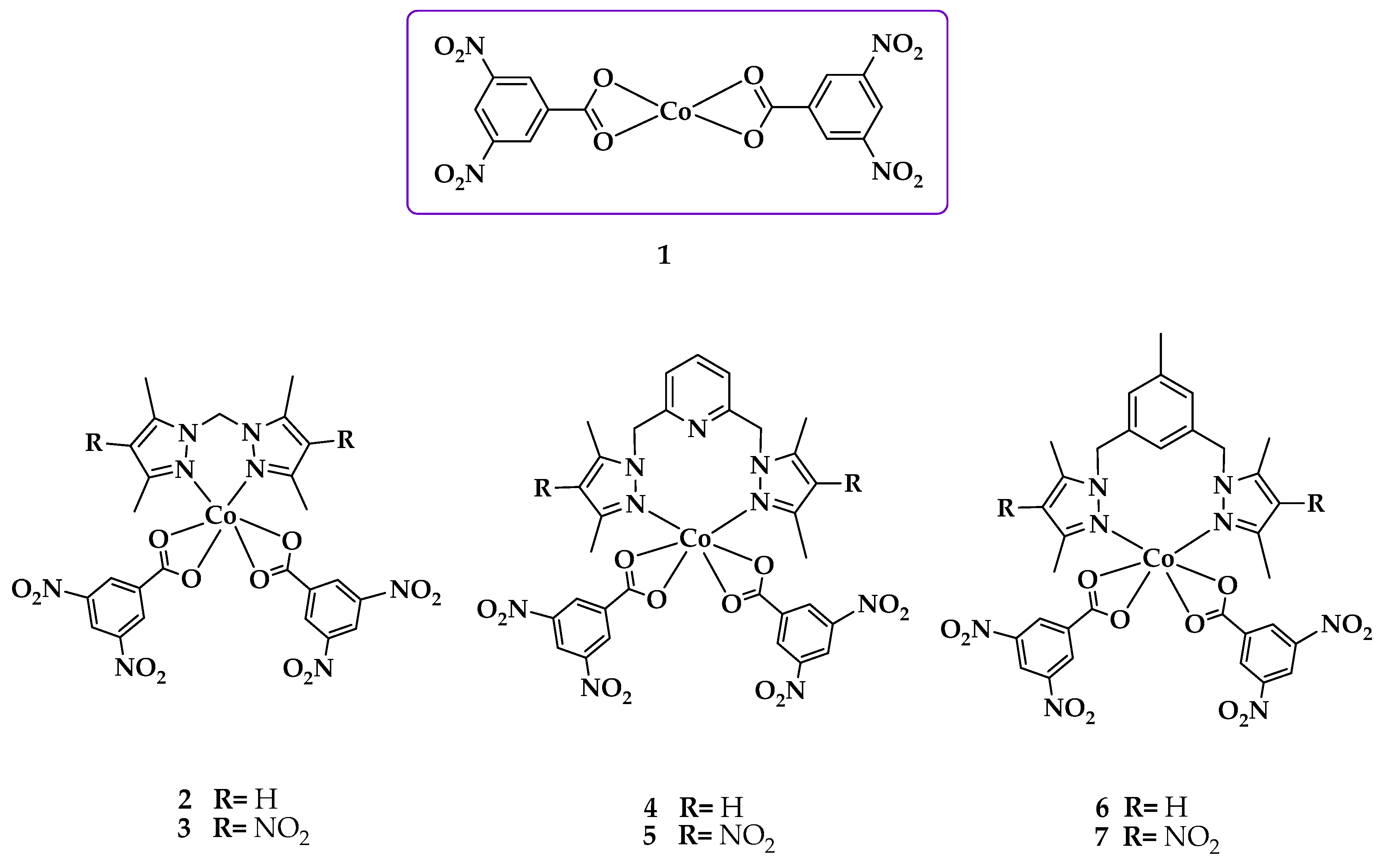
A systematic study of the structure and biological activities of the azole ligands (L1L6) in Figure 1 was synthesized as described in the literature, and their Co(II) complexes prepared under different conditions were performed. The synthesis of azole L6 was not previously reported so we synthesized and characterized it.

2.1. Synthesis and Characterization of 2,6 bis(4-nitro-3,5-dimethylpyrazol-1-ylmethyl)pyridine L6

The synthesis of L6 was performed by a modified literature procedure [12]. L6 was synthesized by the reaction between 4-nitro-3,5-dimethylpyrazole and 3,5-bis(bromomethyl)toluene in toluene. L6 was isolated as an air-stable white solid with an 80% yield by crystallization from dichloromethane: ethyl ether. The ligand was characterized by elemental analysis, Raman, infrared, ultraviolet/visible, RMN spectroscopy and mass spectrometry (Figures S1–S35 in the Supplementary Materials). The FT-IR spectra showed bands for asymmetric and symmetric stretching of the NO2 group at 1485 and 1350 cm−1, respectively. The methylene group was observed at δ 53.31 ppm (Figure S7 in the Supplementary Materials). The 13C and 1H chemical shifts were assigned with the aid of a heteronuclear single quantum correlation (HSQC) experiment. The 1H NMR spectra showed six singlets, and the signal corresponding to the proton in the pyrazole ring was not observed, indicating that substitution of H for NO2 had occurred (Figure S6 in the Supplementary Materials). Additionally, the signals were observed at a lower field than the corresponding singlets in L5 [13], which is expected because the NO2 group is an electron withdrawing substituent. The crystal structure of L5 is discussed later in this manuscript.

2.2. Synthesis and Characterization of the Cobalt(II) Complexes

The precursor complex [Co(dnb)2] (1) was synthesized by the reaction between 3,5-dinitrobenzoic acid and Co(OAc)2 ∙ 4H2O in MeOH at a 2:1 molar ratio, respectively. In the case of complexes 27, the respective ligand (L1L6) was mixed with 1 using two solvents in which both were dissolved. However, 2 was the only complex synthesized using only one solvent in the reaction. The cobalt complexes (Figure 2) were stable in air at room temperature and were isolated in high yields. Table 1 shows the elemental analysis, melting point, and color of complexes.
The results allowed us to propose a 1:2 [Metal(M):Co-ligand(dnb)] relative stoichiometry for 1. Complexes 16 showed a 1:1:2 [M:azole ligand(L):(dnb)] ratio, resulting in a [M(L)(dnb)2] type stoichiometry.

2.2.1. FT-IR and Raman Spectroscopy

The complexes were analyzed by infrared spectroscopy to observe the shifts of defined bands relative to those of the free ligands following coordination to the metal center. The bands corresponding to the vibrations of the most representative bonds of L1L6 and the precursor complex [Co(dnb)2] (1) were compared with those observed in the FT-IR spectra of their respective complexes 27. Table 2 summarizes the assignments of the most significant vibration bands.
Ligands L1, L3 and L5 are ligands that do not have the nitro group in their structure, whereas L2, L4 and L6 do have it (Figure 1). When comparing the FT-IR spectra of 27 with those of L1–L6 (Figures S1–S5, S11 and Table S1 in the Supplementary Materials), it was observed that the band associated with the vibration of the asymmetric stretch υ(C–H) of the methyl groups, was present in all azole ligands and complexes in the same position. However, the band corresponding to the vibration of the stretch υ(C=C) of the pyrazole ring was observed as shifted to a lower wave number in all complexes compared to free ligands [14]. This is because the bonds are less rigid; therefore, they require less energy to vibrate. The band associated with the vibration of the stretch of the bond υ(C=N) of the pyrazole ring appears in 27 displaced towards greater number of waves, which implies that the bonds vibrate to greater energy; this means that they have a greater rigidity in the complexes. This result suggests that the ligands are being coordinated to the metallic center by the N-pyrazole. Another band identified in the complexes was associated with the bending vibration in the pyrazole ring plane in the range of 802 to 825 cm−1 [14].
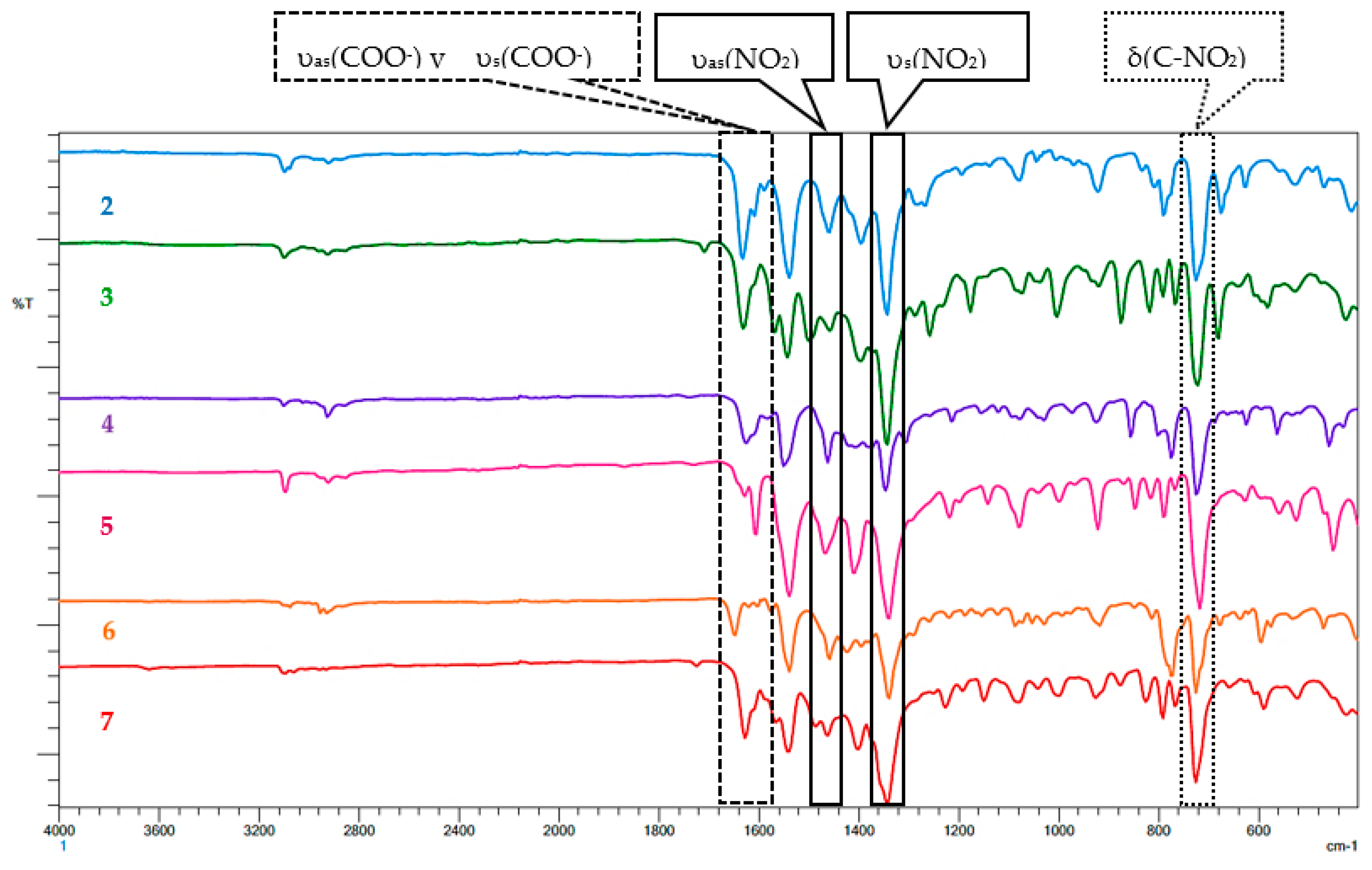
The asymmetric stretching υas(NO2) between 1458 and 1500 cm−1 and symmetric stretching υs(NO2) between 1338 and 1346 cm −1 corroborate the presence of NO2 in all the complexes and suggest that NO2 is not directly involved in coordination [12]. In the case of 3 and 4, the bands corresponding to the υ(C-H) vibration of the pyridine ring at 1045 (3) and 1041 (4) cm−1 do not show shifts relative to the free ligands L3 and L4, which indicates that the nitrogen of the pyridinic ring is not coordinated to the metallic center [15]. In addition, in the FT-IR spectra of 17, two bands associated with asymmetric and symmetric stretching of the carboxylate group were observed. To determine the coordination form of the carboxylate in the complexes (monodentate, bridged and/or bidentate), Δυ = υas(COO) − υs(COO) values were calculated. In all the complexes, these Δυ values were very low, which indicates bidentate chelation of carboxylates [16]. The mentioned bands for the carboxylate group and nitro group are indicated in Figure 3. However, the band associated with the vibration υs (COO) was not explicitly indicated in Figure 3, due to its low intensity. However, it appears in the range of 1604 to 1566 cm−1 in all complexes.
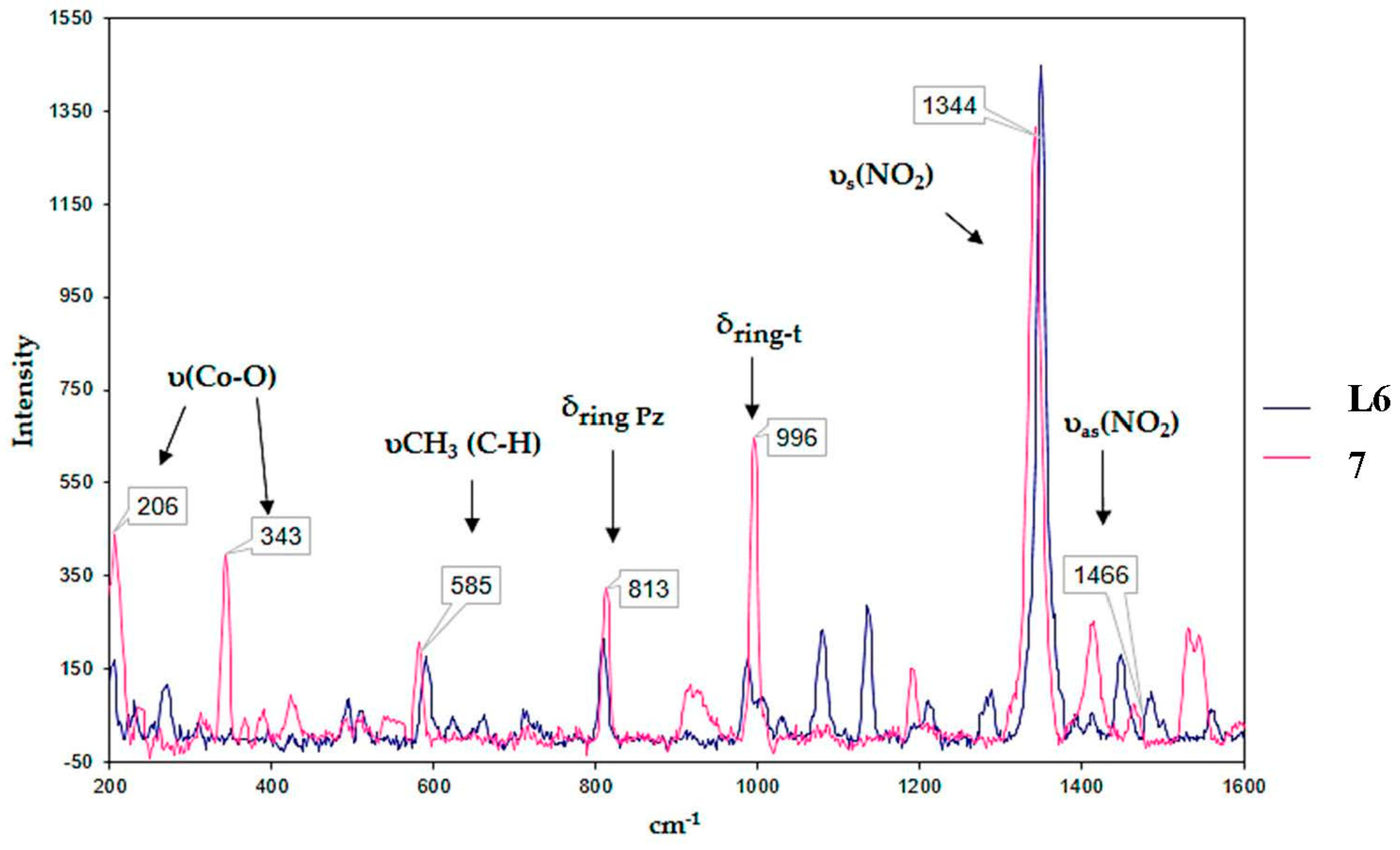
In the Raman spectra of the complexes, 27 can be bands that come from the azole ligand, in addition to bands corresponding to the coordination of this ligand to the metallic center. As an example, Figure 4 shows the comparison of the Raman spectrum of 7 with ligand L6. Two bands corresponding to the azole ligands were identified. The band of the stretching vibration υ(C–H) of the methyl groups between 527 and 600 cm−1 and the band corresponding to the bending vibration in the pyrazole ring plane in the range of 811 to 820 cm−1. Additionally, bands related to 1 were observed. Two bands corresponding to the υas(NO2) asymmetric stretching between 1400 and 1462 cm−1 and υs(NO2) symmetric stretching between 1344 and 1365 cm−1 were present. In addition, the band corresponding to the 1,3,5-trisubstituted benzene ring was observed between 993 and 996 cm−1. Finally, the band assigned to the stretching vibration of the υ(Co–O) bond was observed in a range of 201 to 348 cm−1. The presence of these bands confirms coordination of the benzoate ligands with the metallic center, corroborating the formation of 27, as described above.

2.2.2. UV/Vis Spectroscopy

The UV-Vis spectra of 1 was recorded in MeOH, and two bands are observed in the ultraviolet region, which can be attributed to intraligand transitions: the first band at 205 nm corresponding to transitions between the n-π* orbitals, and the second at 233 nm corresponding to transition of the π-π* orbitals of the dinitrobenzoate ligand. However, the transfer bands are not evident due to an overlap with the absorption bands characteristic of the ligand dnb. In addition, the UV-Vis spectra of 26 were recorded in DMSO (dimethyl sulfoxide), and a band between 200 and 350 nm in the ultraviolet region was observed corresponding to transitions between the π–π*orbitals of the ligand. Charge transfer bands were also observed in this zone. Charge transfer transitions of the metal to the ligand mostly occur because the complexes contain ligands with π* orbitals of low energy, which can accept electron density from the metal. An important feature found was the absence of n–π * bands in all the complexes due to the formation of the N–M bond that stabilizes the pair of electrons in the nitrogen atoms. Additionally, all complexes 17 showed other bands corresponding to dd transitions in a range of 400 to 700 nm. These bands correspond to the 4T1g (F) 4A2g (F) electronic transition characteristic of d7 metal complexes.
The UV-Vis spectra of 26 are not directly comparable to the spectrum of 1 because they were recorded in different solvents. In the spectra of 26, a bathochromic shift was observed relative to the spectrum of 1. In comparing the spectra of complexes 2, 4 and 6 to those of 3, 5 and 7, respectively, a bathochromic shift was observed, likely due to insertion of the nitro group that extends the conjugation of the aromatic ring and generates a shift to lower energy due to the unsubstituted ring.
To verify the stability of complexes (13) in DMSO solution, the electronic (UV-Vis) spectra of these complexes were recorded as a function of time. No significant variations were observed, suggesting that the formed species of complexes in DMSO solution are stable in the assayed conditions [17].

2.2.3. X-Ray Structural Determination of L5 and 1

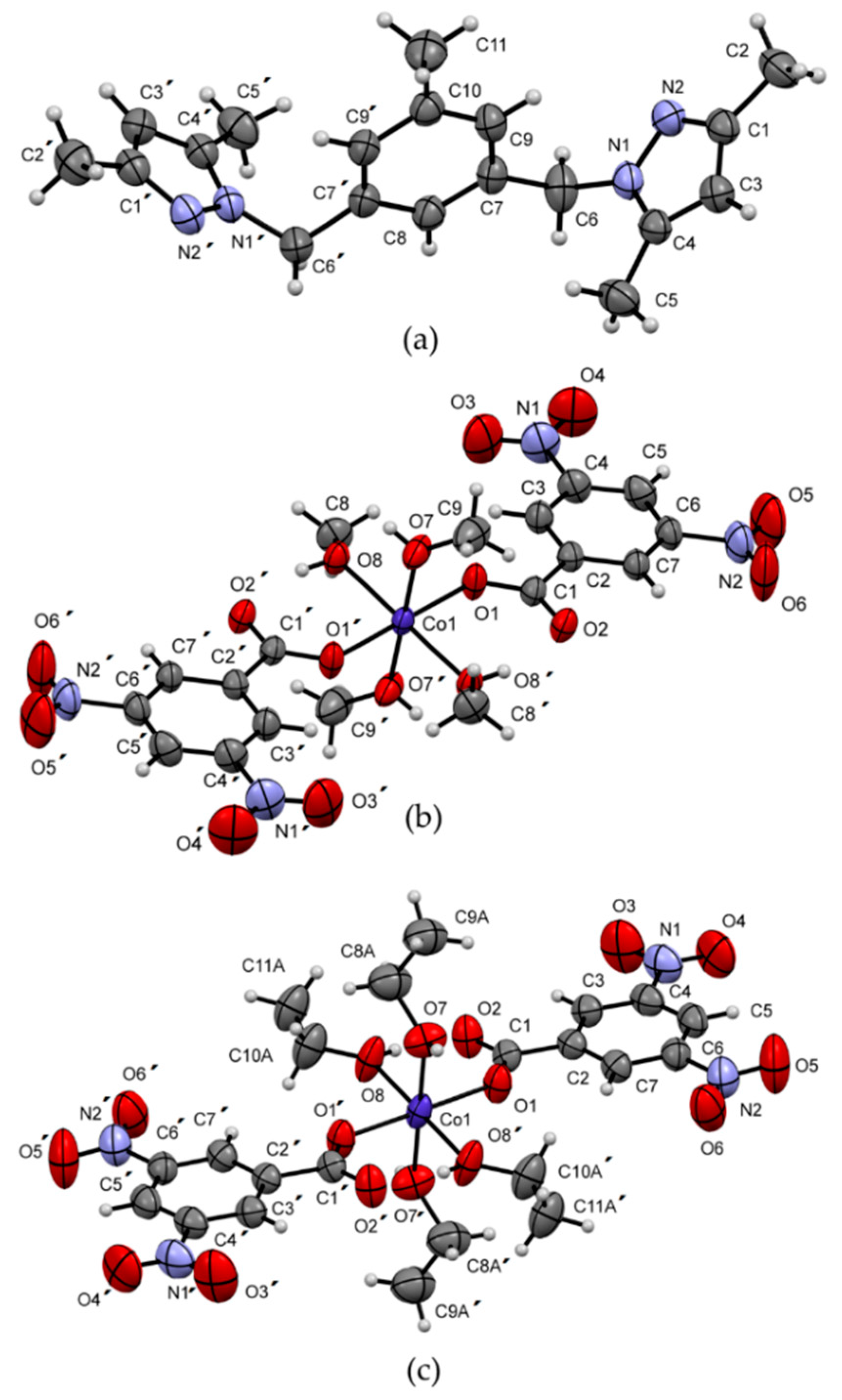
Figure 5 shows the molecules of ligand L5 and complex 1 coordinated by methanol and ethanol, and Table 3 shows the crystal data and experimental details. The molecular structure of L5 shows two methyl substituted pyrazole rings joined through a ‒CH2‒ group to a central toluene fragment. The structure, which crystallizes in the Monoclinic C2/c space group, has a Z’ value of 0.5 due to a coincident symmetry of the molecule with the two-fold rotation axis along the [010] direction that falls in the center of the molecule at (0, y, 1/4). This axis makes the two pyrazole fragments symmetrically equal and oriented in a cis‒conformation, forming a dihedral angle of 88.61(6)° with the central six-membered ring, which is different from the trans-conformation observed in the organic-inorganic hybrid (LH)+(FeCl4), where L = 3,5-bis(3,5-dimethylpyrazole-1-ylmethyl)toluene [18]. In the supramolecular structure, no classical hydrogen bonds were found. Instead, a combination of weak C11‒H11B···N2 hydrogen interactions (distance C11···N2 of 3.865(2) Å) connecting molecules along the [−101] direction and van der Waals forces are responsible for the observed three-dimensional network.
The crystal structure of complex 1 was also analyzed by X-ray crystallography. Interestingly, in our attempts to obtain crystals of suitable quality, two different crystal structures were found. Recrystallization in methanol (1m) and acetone: ethanol (1e) gave the crystal structures shown in Figure 5b,c, with methanol and ethanol coordinating the central Co atom, respectively. In both structures, a mononuclear six-coordinate complex was observed. The coordination sphere contains six oxygen atoms from four alcohol molecules and two dinitrobenzoate ligands in an octahedral geometry with average volumes of 12.044 Å3 and 12.018 Å3, bond angle variances of 2.05° and 1.71° for 1m and 1e, respectively, and identical mean quadratic elongations of λ = 1.001 [19]. Complexes 1m and 1e crystallize in the triclinic P−1 and monoclinic P21/n space groups, respectively, with Z´ values of 0.5 due to a coincident symmetry of the molecules with an inversion center. The dinitrobenzoate groups act as monodentate ligands with the O2 atoms connected by intramolecular O8‒H8···O2 (H···O distances of 1.89(2) Å and 1.85(3) Å for 1m and 1e, respectively) hydrogen bonds to the coordinating alcohol molecules. This form of coordination results differently from that observed in the FT-IR analysis considering that, in this case, the crystallographic study was performed on a recrystallized sample. The supramolecular structure is dominated by strong pairs of equivalent O7‒H7···O2 hydrogen bonds (H···O distances of 1.90(3) Å/1.90(2) Å and symmetry codes 1 − x, 1 − y and −z/1 + x, y, z for 1m and 1e, respectively) connecting pairs of inversion-related molecules to form chains running along the [100] direction in both structures. The three-dimensional networks are completed through van der Waals forces. Interestingly, in both crystal structures, the molecules have similar arrangements and intermolecular interactions. During refinements, half of the ethanol molecules showed positional disorder, which presumably breaks the 2-fold symmetry axes and the glide planes observed in the monoclinic structure (methanol), making the unit cell of 1m nearly double the volume of that of 1e.
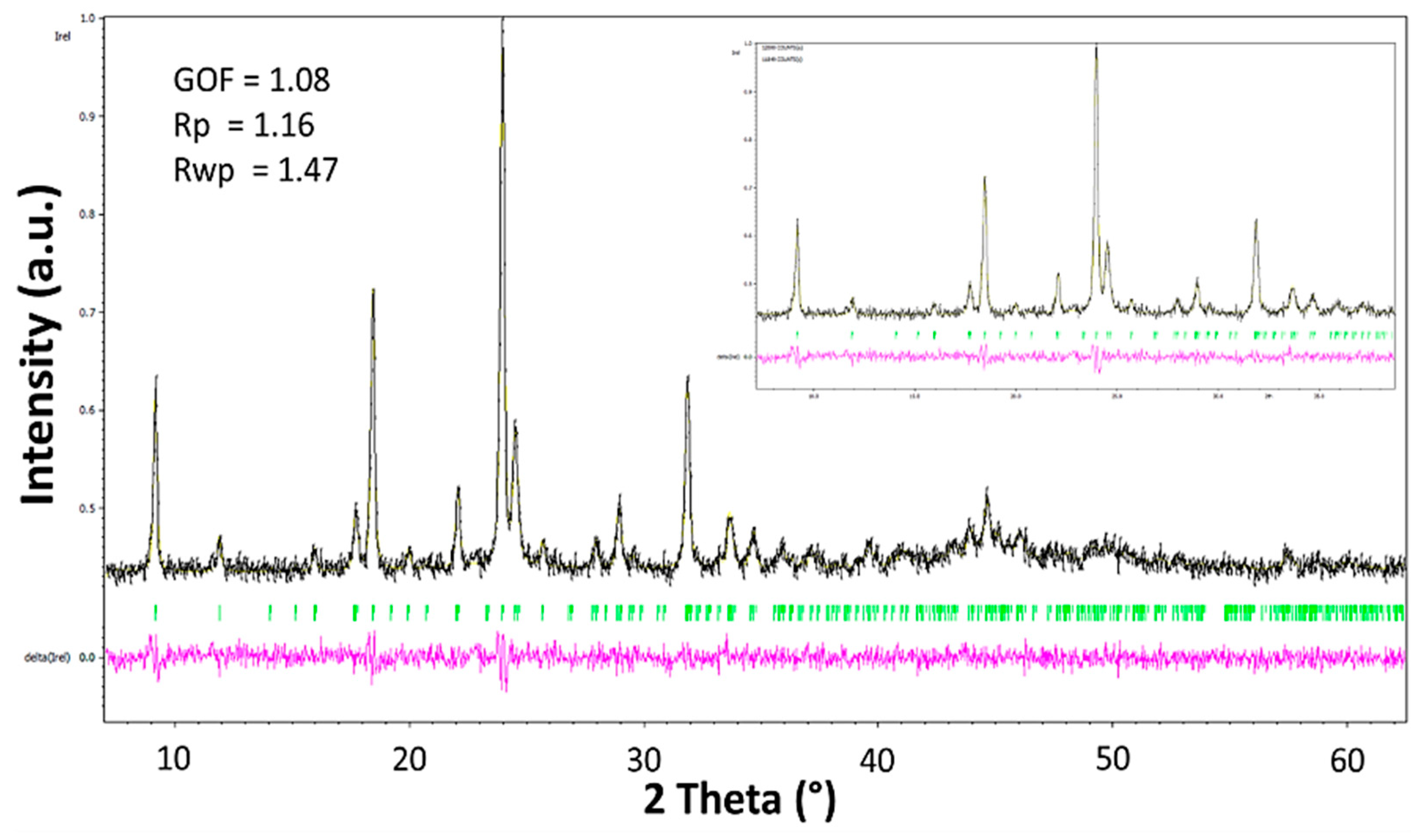
Thermal treatment of complex 1 (pink) at 130 °C for 8 h produced a purple powder. Several attempts to grow single crystals from this powder sample were unsuccessful. Regardless of the solvent used, the metallic center always adopted an octahedral geometry involving solvent molecules. In this sense, the square planar geometry of the cobalt complex in the purple sample was lost during recrystallization. Therefore, X-ray powder diffraction analysis was carried out. Efforts to resolve the structure from powder data were fruitless. However, autoindexing of the diffraction peaks and unit cell refinement by Le Bail analysis [20] showed that the whole diffractogram for this complex is explained by the unit cell a = 9.6586 (17) Å, b = 11.6717 (14) Å, c = 6.3204 (9) Å and β = 96.286 °, with the most likely space group P21/m (Figure 6) in a monoclinic symmetry. It is important to observe that the purple powder corresponds to a pure sample since extra peaks from the secondary phases were not observed.

2.3. Quantum-Chemical Calculations

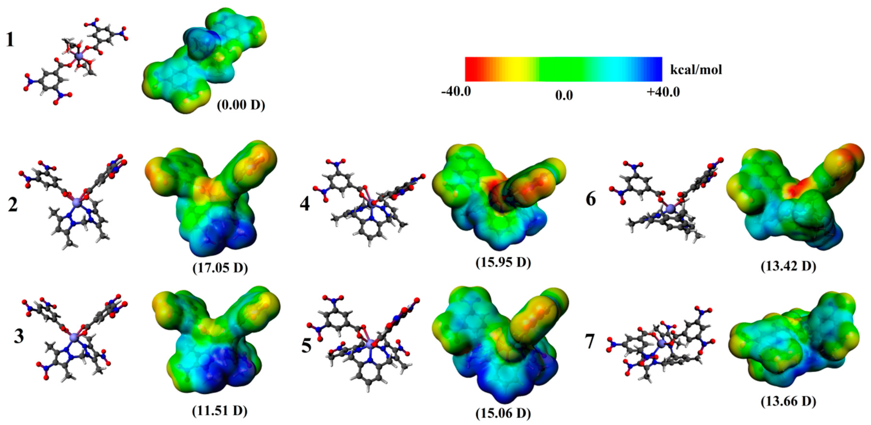
To explore relevant physicochemical properties along the series and to rationalize differences in their biological activity, density functional theory (DFT) calculations were carried out at the dispersion corrected TZ2P/BP86-D3 level of theory [21] with all-electron basis sets via the ADF code [22]. The obtained relaxed structures exhibited an octahedral coordination sphere for the studied systems, where the calculated structure for 1 agrees with characterization via X-ray measurements. In this concern, the permanent molecular dipole moment (µ) has appeared to be a useful parameter in drug–receptor interaction within a quantitative structure–activity relationship (QSAR) framework since early works [23] since it plays a crucial role in promoting long-range electrostatic interactions for supramolecular structure stabilization and drug-site interactions in biomolecules [19].
Our results denote differences in the calculated molecular dipole moment, increasing from 1, with a zero dipole moment owing to its centrosymmetric structure, to values larger than 11 Debyes, which is accounted by the charge distribution on an electron density surface of 0.001 a.u. (electrons/Bohr3) showing a van der Waals surface of each molecule [24,25] as given by the molecular electrostatic potential (MEP) energy surface (Figure 7). Previously, we qualitatively correlated the lowest MIC values with larger permanent dipole moments (µ values) in the series [8] when the isolated ligands did not exhibit relevant biological activity. In this series, all of the Co(II) species showed mild biological activity against C. albicans, C. tropicalis and C. krusei (Table 4). Introduction of nitro-groups into isolated ligands and Co(II) complexes enables rather similar biological activity owing to their inherent enzymatic reduction function [26]. Thus, a similar mechanism involving the enzymatic reduction role of –NO2 groups is expected.

2.4. Biological Activity

The discovery of new compounds with an anti-Candida effect is pivotal to combat candidiasis, a major opportunistic fungal infection with increasing morbidity and mortality worldwide [1]. Although C. albicans is the most frequent etiology, there has been an alarming global emergence of non-albicans strains due to their resistance to the common antifungals used [3]. First, we tested cytotoxic activity using an MTT assay, which determines mitochondrial function in cells by measuring mitochondrial enzymes, indicators of cell viability [27]. The compounds in this study were not toxic to Vero cells. We obtained CC50 values ranging from 103.97 to >300 µg mL−1, except L5 (CC50 = 44.95 µg mL−1), which showed cytotoxic activity (Table 4). Itraconazole used as reference drug showed toxicity higher than 93% of the compounds assayed.
Then, the biological activity was confirmed with fungal and parasite cells to examine therapeutic potential, considering that some cobalt complexes have been studied as antimicrobial agents and have shown potential activity against different strains of fungi and T. cruzi [5,12,28,29]. The complexes analyzed exhibited antifungal activity against at least two of the three Candida species utilized in this study. We obtained MIC values ranging from 31.3 to 250 µg mL−1. By contrast, the free ligands were not active against these strains (MIC > 2000 µg mL−1) (Table 4). The increase in the biological activity of the complexes may be due to increases in their lipophilicity, causing polarity reduction of the fungal membrane and enhancing antimicrobial activity [8,30,31].
The compounds were more active against C. krusei. In this case, 3 showed fungicidal activity at 31.25 µg mL−1 and the best selective index (SI = 9.55), followed by 6 and 7 (SI = 4.79 and 4.13, respectively). Although multidrug resistance is uncommon in non-albicans Candida species, recently, it has been reported with more frequency [1,2,3]. In particular, C. krusei is known to be intrinsically resistant to Fluconazole, the main drug used to treat Candida infection [32], which probably increases their prevalence as etiological agents of invasive candidiasis and candidemia, as well as non-albicans isolates less susceptible to commonly used antifungal agents. Thus, the anti-Candida profile of the complexes synthesized shows promise for further development of preclinical assays, and it is necessary to elucidate their effect on virulence and pathogenicity factors.
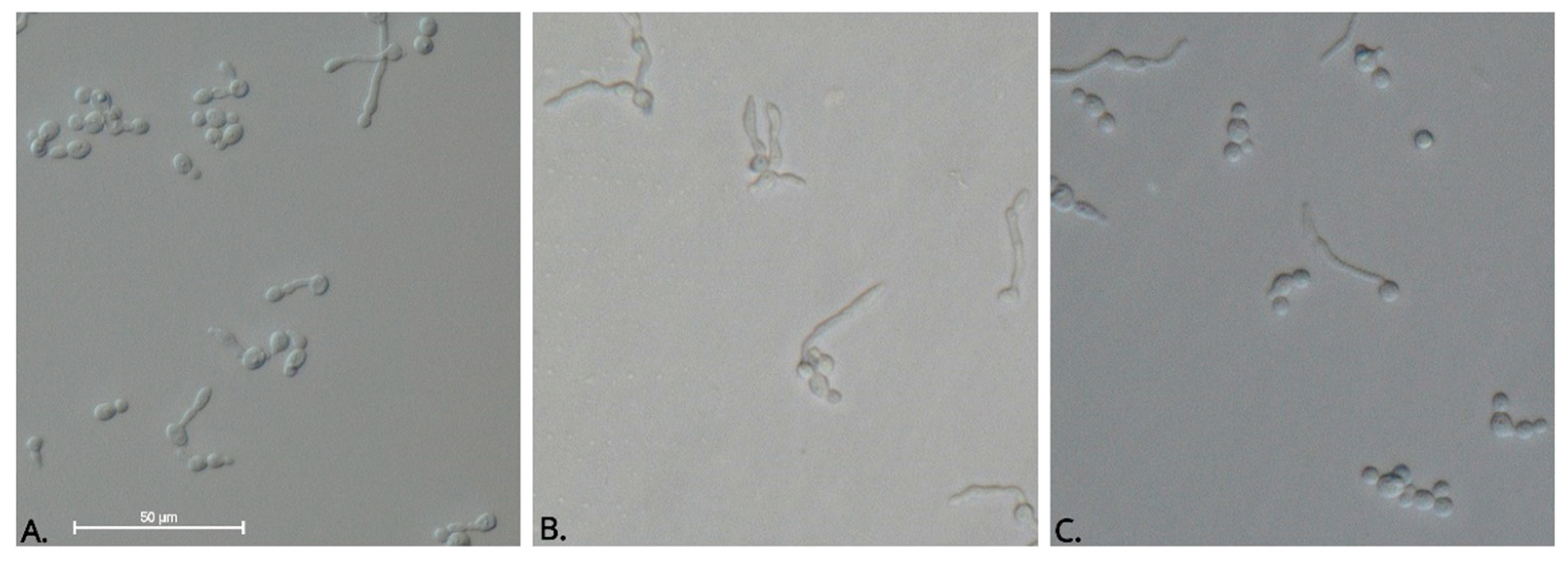
On the other hand, we tested the inhibitory activity of the complexes and their ligands against dimorphic switch of C. albicans. The morphological switch to hyphae is critical to pathogenesis, especially because the hyphal form has been shown to be more invasive than the yeast form due to the expression of cell-wall proteins that facilitate adhesion and tissue invasion [33,34]. Interestingly, we found that 83.3% of free ligands and 25% of the complexes synthesized inhibited tube germ formation (Table 5). Complexes 4 and 7 showed less than 20% inhibitory activity at the maximum concentration assayed. The remaining complexes (50%) were not active against the yeast to hypha transition (data not shown). Itraconazole was tested as the reference drug. In our hands, the average percent of filamentation inhibition that it presented was 44% at 1.0 µg mL−1. However, there were no significant differences between the range of concentrations evaluated in the positive control (p = 1.00). One-way ANOVA followed by Tukey’s post hoc test established that the effect of L1 was better than the inhibition produced by Itraconazole at 0.5 and 0.25 µg mL−1 (p < 0.01) and equivalent to that produced by Itraconazole at 1 µg mL−1 (p = 0.051) (Figure 8). Although antifungal susceptibility experiments suggested that the free ligands did not inhibit yeast cell growth, they inhibited filamentous growth, which could provide evidence concerning the therapeutic potential of these compounds because strains unable to perform the phase transition are less virulent [35,36]. The signal transduction pathways and key transcription factors that modulate the dimorphic transition in C. albicans have been previously established [37,38,39,40,41,42,43]. In this study, we used human serum as an inducer of the morphological change from yeast to hyphae, and thus the inhibitory activity observed might suggest that cobalt(II) complexes with ligands derived from pyrazoles and dinitrobenzoate impact the activity of the Ras-mediated signal transduction pathway, which is involved in serum-induced filamentous growth [44]. However, to investigate the effect of the free ligands and complexes on hyphae-inducing signaling pathways, further experiments are required.
In addition, we tested the effect of the compounds against T. cruzi epimastigotes. Although previous studies conducted by our group with similar molecules (metal complex derivatives of bis (pyrazol-1-yl) methane ligands) suggested antiparasitic potential of these compounds [12], in this study, we did not observe trypanocidal activity at the maximum concentration assayed (IC50 > 100 µg mL−1). Benznidazole showed an IC50 value of 18.4 µg mL−1 + 1.3.

3. Materials and Methods

3.1. General Information

The starting compounds 3,5-dinitrobenzoic acid (ADNB), cobalt(II) acetate tetrahydrate Co(OAc)2 ∙ 4H2O, 3,5-bis(bromomethyl)toluene (BBMT), and tetrabutylammonium bromide (TBAB) were used as received from Alfa Aesar (Ward Hill, MA, USA). The solvents that were required to be anhydrous were dried, distilled and stored on 3 Å molecular sieves under a nitrogen atmosphere before use. Elemental analyses (C, H and N) were performed with a Thermo Scientific FLASH 2000 CHNS/O Analyzer (Thermo Fisher Scientific, Waltham, MA, USA). Fourier transform infrared (FT-IR) spectra were recorded on a Shimadzu IR Tracer-100 spectrophotometer (Shimadzu Corporation, Kyoto, Japan) in a range of 400–4000 cm−1 using KBr pellets. Melting points were recorded with a capillary Mel-Temp® 1101D Electrothermal apparatus in open capillary tubes and are uncorrected (Staffordshire, UK). Nuclear magnetic resonance (NMR) spectra were recorded on a Bruker Ascend-400 spectrometer (Bruker, Billerica, MA, USA) at 295 K (400.13 MHz for 1H; 100.61 MHz for 13C) in the solvent CDCl3. 1H and 13C NMR chemical shifts (δ) are reported in parts per million (ppm) with the residual solvent peak used as an internal reference. High-resolution mass spectra (HRMS) were recorded on an Agilent Technologies 1260 (Q-TOF 6520) spectrometer (Agilent Technologies, Santa Clara, CA, USA) via electrospray ionization (ESI) in positive ion mode. The mass spectra were recorded on a Thermo Scientific™ TRACE™ 1300 Gas Chromatograph via electronic impact (Waltham, MA, USA). The electronic UV/ Vis absorption spectra were measured from 200 to 800 nm in DMSO solution in a quartz cuvette with a 1-cm optical path length using a Varian Cary 100 spectrophotometer from Agilent Technologies (Agilent Technologies, Santa Clara, CA, USA). Raman spectroscopy was performed in a HORIBA Scientific spectrometer (HORIBA Scientific, Kyoto, Japan) using a 785-nm laser in a range of 200–1600 cm−1. Recrystallization of L5 was carried out by slow evaporation from a toluene solution. Complex 1 was recrystallized from solutions of methanol and a mixture of acetone: ethanol (1:1). These procedures afforded crystals of suitable size and quality for single-crystal X-ray diffraction. The data collection carried out using a Bruker D8 Venture/Photon 100 CMOS diffractometer (Madison, WI, USA) and refinement details are summarized in Table 3. In the refinements, all the nonhydrogen atoms were anisotropically treated, and the hydrogen atoms were generated geometrically, placed at geometrically suitable calculated positions (C–H = 0.93–0.97 Å) and refined by applying isotropic displacement parameters set at 1.2–1.5 times the Ueq value of the parent atom. The H atoms belonging to methanol and ethanol molecules in the coordination sphere of the Co atom in 1 were located in the difference Fourier maps and freely refined. The crystal structures were refined using the SHELXL2014 program [45]. The graphic material was prepared using Mercury 3.10.3 software [46].
A powder sample of complex 1, thermally treated, was analyzed using X-ray powder diffraction at room temperature with an Empyrean-Panalytical X-ray diffractometer (Malvern Panalytical, Almelo, Netherlands) working in Bragg–Brentano geometry with Cu-Kα1,2 (1.5406 and 1.54439 Å) wavelengths. The diffractometer was operated over an angular range of 2θ = 2°–70° with a step size of 0.02° (2θ). Data analysis was performed by the Le Bail method using the Jana-2006 program [47]. The process of refinement was carried out assuming a pseudo-Voigt function for peak shape and a calculated background using a linear interpolation between a set of fixed points.
The ligands bis(3,5-dimethyl-1-pyrazolyl)methane (L1) [48], bis(3,5-dimethyl-4-nitro-1-pyrazolyl)methane (L2), 2,6-bis(3,5-dimethyl-1-pyrazolyl)pyridine (L3), 2,6-bis(3,5-dimethyl-4-nitro-1-pyrazolyl)pyridine (L4), and 3,5-bis(3,5-dimethylpyrazol-1-ylmethyl)toluene (L5) [12,48,49] were synthesized as described in the literature.

3.2. Synthesis of 3,5-bis(3,5-dimethyl-4-nitropyrazol-1-ylmethyl)toluene L6

This ligand was prepared by a modified literature procedure [12]; 4-nitro-3,5-dimethylpyrazole (1.8 mmol, 0.254 g), KOH (3.517 mmol, 208.43 g) and water (1 mL) were stirred at room temperature (rt) for 30 min, and then, tetrabutylammonium bromide (TBAB) (0.055 mmol; 0.018 g), 3,5-bis(bromomethyl)toluene (0.9 mmol, 0.250 g) and 30 mL of toluene were added. The mixture was heated to reflux for 26 h. The resulting mixture was evaporated to dryness, and the solid residue was extracted with CH2Cl2: water. The organic layer was collected and dried with anhydride sodium sulfate and evaporated to dryness. The white solid was crystallized from dichloromethane (DCM): ethyl ether. Yield: 0.286 g (80%). M.p.: 187–186 °C. IR (KBr, υ/ cm−1): 2924, 1562, 1485, 1400, 1350, 999. Raman (υ/ cm−1): 1448, 1351, 1137, 1079, 990, 808, 589. 1H NMR (400 MHz, CDCl3): δ 6.86 (s, 2H), 6.70 (s, 1H), 5.18 (s), 4H), 2.54 (s, 6H), 2.53 (s, 6H), 2.31 (s, 3H). 13C NMR (100 MHz, CDCl3): δ 146.3, 140.3, 140.0, 135.9, 131.5, 127.4, 122.5, 53.3, 21.4, 14.1, 11.7. DEPT-135: 127.47, 122.50, 53.31, 21.42, 14.19, 11.74. Anal. calcd. for C19H22N6O4: C, 57.28; H, 5.57; N, 21.09%; found: C, 57.28; H, 5.56; N, 21.07%. HRMS (ESI+) m/z: calcd. for [C19H22N6O4 + H] + 399.1780; found: 399.1836 [M + H] +. UV-Vis (DMSO) [(concentration, M) λmáx, nm (Log ε, M−1 cm−1)]: (9.89 × 10⁻⁵) 260 (4.28). MS (EI) m/z calcd. for [C14H16N3O2]: 258.1; found [C14H16N3O2]: 258.1.

3.3. Synthesis of the Cobalt(II) Complexes (17)

3.3.1. Synthesis of bis(dinitrobenzoate-O,O′) of Co(II) ([Co(dnb)2]) (1)

A solution of cobalt(II) acetate tetrahydrate Co(OAc)2∙4H2O (1 mmol, 0.25 g) in methanol (MeOH) (10 mL) was added to a solution of 3,5-dinitrobenzoic acid (DNBA) (2 mmol, 0.43 g) in MeOH (10 mL). The mixture was stirred at rt for 30 min. The solvent was evaporated to dryness to give a pink solid, which was recrystallized from MeOH. The solid obtained was dried at 130 °C for 8 h. Yield: 0.458 g (95%). M.p.: >400 °C. IR (KBr, υ/cm−1): 3097, 1624 1346, 725. Raman (υ/cm−1): 1539, 1350, 1190, 999, 918, 817, 346, 206. Anal. calcd. for C14H6CoN4O12: C, 34.95; H, 1.26; N, 11.64%; found: C, 34.90; H, 1.25; N, 11.51%. HRMS (ESI+) m/z: calculated for [C14H8N4O12Co + NH4] + 500.9814; found: 500.9129 [M + NH4] +. UV-Vis (MeOH) [(concentration, M) λmáx, nm (Log ε, M−1 cm−1)]: (3.25 × 10⁻⁴) 205 (3.58), 233 (3.45); (2 × 10⁻⁴) 518 (1.01).

3.3.2. Synthesis of Dinitrobenzoate [bis(3,5-dimethylpyrazol-1-yl)methane] of Co(II) ([Co(L1)(dnb)2]) (2)

A solution of 1 (0.42 mmol, 0.20 g) in MeOH (10 mL) was added to a solution of L1 (0.40 mmol, 0.08 g) in toluene (10 mL). The mixture was stirred at rt for 12 h. The solvent was evaporated to dryness to give a pink solid, which was washed with ethanol (EtOH) and ethyl ether and dried at 100 °C for 8 h. Yield: 0.251 g (92%). IR (KBr, υ/cm−1): 3097, 2920, 1631, 1539, 1396, 1342, 1006, 725. Raman (υ/cm−1): 1537, 1348, 1187, 1050, 996, 915, 811, 588, 339, 201. Anal. calcd. for C25H22CoN8O12: C, 43.81; H, 3.24; N, 16.35%; Found: C, 43.80; H, 3.22; N, 16.33%. UV-Vis (DMSO) [(concentration, M) λmáx, nm (Log ε, M−1 cm−1)]: (3.32 × 10⁻⁴) 245 (3.42); (9.95 × 10⁻⁴) 543 (1.36).

3.3.3. Synthesis of Dinitrobenzoate[bis(3,5-dimethyl-4-nitro-pyrazol-1-yl)methane] of Co(II) ([Co(L2)(dnb)2]) (3)

A solution of 1 (0.42 mmol, 0.20 g) in acetone (10 mL) was added to a solution of L2 (0.40 mmol; 0.1 g) in acetone (10 mL). The mixture was heated to reflux for 8 h. The solvent was evaporated to dryness to give a pink solid, which was washed with acetone: pentane (20:10) and then with EtOH and ethyl ether and dried at 100 °C for 8 h. Yield: 0.123 g (90%). IR (KBr, υ/cm−1): 3079, 2924, 1631, 1543, 1500, 1373, 1342, 1002, 721. Raman (υ/cm−1): 1540, 1465, 1351, 1189, 998, 813, 600, 341, 208. Anal. calcd. for C25H20CoN10O16: C, 38.72; H, 2.60; N, 18.06%; Found: C, 38.64; H, 2.60; N, 17.99%. UV-Vis in DMSO [(concentration, M) λmáx, nm (Log ε, M−1 cm−1)]: (3.45 × 10⁻⁴) 265 (3.28); (9.77 × 10⁻⁴) 552 (1.70).

3.3.4. Synthesis of Dinitrobenzoate[2,6-bis(3,5-dimethylpyrazol-1-ylmethyl)pyridine] of Co(II) ([Co(L3)(dnb)2]) (4)

A solution of 1 (0.36 mmol, 0.17 g) in ethyl acetate (10 mL) was added to a solution of L3 (0.34 mmol, 0.1 g) in DCM (10 mL). The mixture was heated to reflux for 7 h. The solvent was evaporated to dryness to give a pink solid, which was washed with CHCl3: n-heptane (50:50) and then ethyl acetate: n-heptane (40:10) and finally with ethyl ether and dried at 100 °C for 8 h. Yield: 0.208 g (80%). IR (KBr, υ/cm−1): 3093, 2962, 2873, 1643, 1608, 1573, 1462, 1427, 1396, 1369, 1342, 999, 725. Raman (υ/cm−1): 1535, 1460, 1344, 1044, 996, 816, 758, 582, 335, 212. Anal. calcd. for C31H27CoN9O12: C, 47.95; H, 3.50; N, 16.23%; found: C, 47.83; H, 3.48; N, 16.18%. UV-Vis (DMSO) [(concentration, M) λmáx, nm (Log ε, M−1 cm−1)]: (3.50 × 10⁻⁴) 267 (3.38); (9.88 × 10⁻⁴) 541 (1.12).

3.3.5. Synthesis of Dinitrobenzoate[2,6-bis(3,5-dimethyl-4-nitro-pyrazol-1-ylmethyl)pyridine] of Co(II) ([Co(L4)(dnb)2]) (5)

A solution of 1 (0.28 mmol, 0.13 g) in ethyl acetate (10 mL) was added to a solution of L4 (0.26 mmol, 0.1 g) in toluene (10 mL). The mixture was heated to reflux for 7 h. The solvent was evaporated to dryness to give a pink solid, which was washed with CHCl3: n-heptane (50:50), ethyl acetate: cyclohexene (20:30) and then with ethyl ether and dried at 100 °C for 8 h. Yield: 0.183 g (82%). IR (KBr, υ/cm−1): 3093, 2931, 1627, 1566, 1543, 1485, 1462, 1400, 1342, 999, 725. Raman (υ/cm−1): 1535, 1460, 1344, 1044, 996, 816, 758, 582, 335, 212. Anal. calcd. for C31H27CoN11O16: C, 42.97; H, 2.91; N, 17.78%; found: C, 42.69; H, 2.90; N, 17.73%. UV-Vis (DMSO) [(concentration, M) λmáx, nm (Log ε, M−1 cm−1)]: (3.42 × 10⁻⁴) 268 (3.50); (9.80 × 10⁻⁴) 544 (1.07).

3.3.6. Synthesis of Dinitrobenzoate[3,5-bis(3,5-dimethylpyrazol-1-ylmethyl)toluene] of Co(II) ([Co(L5)(dnb)2]) (6)

A solution of 1 (0.42 mmol, 0.20 g) in ethyl acetate (10 mL) was added to a solution of L5 (0.40 mmol; 0.12 g) in DCM (10 mL). The mixture was refluxed for 5 h. The solvent was evaporated to dryness to give a purple solid, which was washed with ethyl acetate: cyclohexene (20:30) and then with ethyl ether and dried at 100 °C for 8 h. Yield: 0.222 g (87%). IR (KBr, υ/cm−1): 3101, 2927, 1624, 1550, 1346, 1313, 925, 725. Raman (υ/cm−1): 1548, 1345, 1192, 1049, 996, 815, 579, 345, 207. Anal. calcd. for C33H30CoN8O12: C, 50.20; H, 3.83; N, 14.19%; Found: C, 49.96; H, 3.79; N, 14.16%. UV-Vis (DMSO) [(concentration, M) λmáx, nm (Log ε, M−1 cm−1)]: (3.37 × 10⁻⁴) 261 (3.46); (1.01 × 10⁻³) 538 (1.49).

3.3.7. Synthesis of Dinitrobenzoate[3,5-bis(3,5-dimethyl-4-nitro-pyrazol-1-ylmethyl)toluene] of Co(II) ([Co(L6)(dnb)2]) (7)

A solution of 1 (0.50 mmol, 0.240 g) in ethyl acetate (10 mL) was added to a solution of L6 (0.40 mmol, 0.16 g) in toluene (10 mL). The mixture was heated to reflux for 7 h. The solvent was evaporated to dryness to give a blue solid, which was washed with CHCl3: n-heptane (30:20) and then with ethyl ether and dried at 100 °C for 8 h. Yield: 0.290 g (84%). IR (KBr, υ/cm−1): 3093, 2924, 1627, 1539, 1408, 1465, 1338, 999, 717. Raman (υ/cm−1): 1534, 1416, 1345, 1190, 998, 815, 345. Anal. calcd. for C33H28CoN10O16: C, 45.06; H, 3.21; N, 15.92%; Found: C, 45.05; H, 3.20; N, 15.90%. UV-Vis (DMSO) [(concentration, M) λmáx, nm (Log ε, M−1 cm−1)]: (3.43 × 10⁻⁴) 269 (3.13); (1.03 × 10⁻³) 549 (2.03).

3.4. Biological Studies

3.4.1. In Vitro Anti T. cruzi Activity

T. cruzi (SYLVIO-X10, ATCC (American Type Culture Collection)) were obtained from M. López-Casillas, Fundación Cardiovascular de Colombia. Epimastigotes were cultivated to the exponential growth phase in 96-well plates at a concentration of 5 × 10⁵ parasites/mL in LIT (liver infusion triptose) medium supplemented with 10% SFBi (serum bovine fetal inactivated) and incubated at 28 °C. Subsequently, the parasites were exposed to the different compound concentrations (100, 33.3, 11.1, 3.7 µg mL−1) for 72 h and evaluated in triplicate. Untreated parasites and parasites treated with benznidazole maintained under the same conditions were used as negative and positive controls, respectively. Benznidazole (purified by L.Y. Vargas, Universidad Santo Tomás, Santander, Colombia) was used as the reference drug. Growth inhibition was determined by optical microscopy using Trypan Blue (Sigma-Aldrich, Saint Louis, MO, USA). The antiparasitic activity of the compounds evaluated is expressed as the concentration required to inhibit 50% of parasites (IC50).

3.4.2. In Vitro Antifungal Susceptibility Testing

The antifungal effect of the compounds against Candida albicans (ATCC® 10231), C. tropicalis (ATCC® 20366) and C. krusei (ATCC® 14243) was determined. Strains were subcultured on Sabouraud dextrose and grown at 37 °C for 24 h prior to assays. Experiments were performed using the broth microdilution method according to the Clinical and Laboratory Standards Institute (CLSI) M27A-3 and M07-A10 protocols. Stock solutions (100 times higher than the highest tested concentration) were prepared using dimethyl sulfoxide (DMSO; Merck, Darmstadt, Alemania) to dissolve each compound. Intermedial dilutions were made in order to reduce the final solvent concentration to <1% using RPMI 1640 medium (Gibco, Life Technology, Carlsbad, CA, USA) supplemented with 3-(N-morpholino)propanesulfonic acid (MOPS, Sigma-Aldrich, Saint Louis, MO, USA). Twofold dilutions in the rank of 1.95–2000 µg mL−1 of each compound was tested in 96-well plates. Untreated controls were similarly evaluated. Itraconazole was purchased from Sigma-Aldrich (St. Louis, MI, USA) and used as the reference drug. Inoculum containing between 2500 and 5000 cells/mL of each strain of Candida was added to each well of the plate and incubated at 37 °C for 24 h. The MIC and fungicidal endpoint were determined as described previously (Murcia, R.A., et al., 2018). Triplicate measurements of each of the compound concentrations were obtained in two independent assays.

3.4.3. Germ Tube Inhibition Assay

The effect of the complexes and their respective ligands on germ tube formation of Candida albicans was tested. The strain was subcultured on Sabouraud dextrose agar and grown at 37 °C for 24 h prior to assays. A loopful of inoculum was added into pooled human serum at a final concentration of 1 × 10⁶ yeast/mL. Further two-fold dilutions were prepared to inoculate each well of a 96-well plate containing 100 µL of pooled human serum with or without different concentrations of the compounds ranging from 500 to 62.5 µg mL−1. The test plates were incubated for 3 h at 37 °C. After the indicated time, the presence of a tube germ was investigated by counting at least 200 cells with a hemocytometer under the 40× objective lens. The positive control was established by supplementing human serum with Itraconazole (range of concentrations: 1.0–0.25 µg mL−1). The results are expressed as inhibition percentages of tube germ formation using the following formula: Control mycelium percent—mycelium percent of the treatment/Control mycelium percent × 100. For each concentration, five repetitions were performed.

3.4.4. Cytotoxicity in Mammalian Cells

Epithelial cells derived from African green monkey kidney (Vero, ATCC, CCL-81) were obtained from M. López-Casillas, Fundación Cardiovascular de Colombia, and cultured in 96-well plates in Dulbecco’s modified Eagle medium (DMEM) (Life Technology, Carlsbad, CA, USA) supplemented with 10% Inactive Fetal Bovine Serum (SFBi) and incubated at 37 °C, 5% CO2 and 95% humidity for 24 h until monolayer formation. Subsequently, the cells were exposed to the different compounds for 72 h; four concentrations were evaluated in triplicate (300, 100, 33.3 and 11.1 µg mL−1). Cell viability was determined by using an MTT (3-(4,5-dimethylthiazol-2-yl)-2,5-diphenyltetrazole) colorimetric test as previously described (Leal-Pinto et al., 2019). The cytotoxicity percentage was calculated by the following equation: Cytotoxicity (%) = ((DO control group − DO treated group)/DO control group) × 100. The results are expressed as Cytotoxic Concentration 50 (CC50) and 90 (CC90), calculated by sigmoidal regression using the statistical software Msxlfit™, ID Business Solution, (Guildford, UK).

3.4.5. Statistical Analysis

The values obtained in the germ tube inhibition assays were analyzed by oneway ANOVA followed by Tukey’s post hoc test for multiple comparisons. A significance level of 5% was adopted.

In Vitro anti-T. cruzi Activity

T. cruzi epimastigotes in the exponential growth phase from the SYLVIO-X10 (ATCC) strain were cultivated in 96-well plates at a concentration of 5 × 10⁵ epimastigotes/mL in LIT (liver infusion triptose) medium supplemented with 10% SFBi (serum bovine fetal inactivated) and incubated at 28 °C. Subsequently, the parasites were exposed for 72 h to the compounds at different concentrations (100, 33.3, 11.1, 3.7 µg mL−1), with triplicate evaluations performed. Untreated parasites and parasites treated with benznidazole maintained under the same conditions were used as negative and positive controls, respectively. Benznidazole was used as the reference drug. Growth inhibition was determined by optical microscopy using Trypan Blue (Gibco). The antiparasitic activity of the compounds evaluated is expressed as the concentration required to inhibit 50% of parasites (IC50).

Cytotoxicity in Mammalian Cells

Epithelial cells derived from African green monkey kidney (Vero, ATCC (American Type Culture Collection)) and CCL-81 (Cercopithecus aethiops) cells were cultured in 96-well plates in DMEM (Dulbecco’s modified Eagle’s medium) (Life Technology, CA, USA) supplemented with 10% of Inactive Bovine Fetal Serum (SFBi), 1000 µg mL−1 of penicillin and 100 µg mL−1 of streptomycin and incubated at 37 °C, with 5% CO2 and 95% humidity for 24 h until the monolayer formation. Subsequently, the cells were exposed to the different compounds for 72 h, and four concentrations were evaluated in triplicate (300, 100, 33.3 and 11.1 μg/mL). Cell viability was determined using an MTT colorimetric test (3-(4,5-dimethylthiazol-2-yl)-2,5-diphenyltetrazole)). The treated cells were reincubated for 4 h with MTT reagent (5 mg/mL), and, subsequently, the reduced formazan crystals were dissolved in DMSO. The optical density was determined via spectrophotometry at a wavelength of 595 nm, and the cytotoxicity percentage was calculated by the following equation: Cytotoxicity (%) = ((DO control group − DO treated group)/DO control group) × 100. From the inhibition percentages and respective concentrations, the results are expressed as Cytotoxic Concentration 50 (CC50), calculated by sigmoidal regression using the statistical software Msxlfit™, ID Business Solution (Guildford, UK).

4. Conclusions

In this paper, we report the synthesis and characterization of seven cobalt(II) complexes with ligands derived from pyrazoles and dinitrobenzoate. The ligand bis(3,5-dimethyl-4-nitro-1H-pyrazol-1-yl)methane (L6) and the metal complexes 17 are new. Ligand bonding to metal ions was confirmed by elemental analysis and Raman, infrared, ultraviolet/visible and mass spectrometry studies. The structures of L5 and 1 were confirmed by X-ray diffraction analysis. The analyses’ spectral data and DFT calculations showed that complexes 27 had a 1:1:2 [M(L)(dnb)2] stoichiometry and octahedral geometries, while 1 had a 1:2 (M:dnb) stoichiometry. In general, the complexes showed higher antifungal activity than the free ligands under planktonic conditions. However, most ligands and 3 and Co(OAc)2 ∙4H2O complexes inhibited C. albicans filamentation in a dose-dependent manner. In addition, none of the tested compounds were toxic to Vero cells. These results indicate that the compounds are promising alternative inhibitors of important virulence factors during candidiasis. On the other hand, the complexes did not present trypanocidal activity. Studies to further elucidate their structure–activity relationship are in progress.

Supplementary Materials

The following are available online at https://www.mdpi.com/1422-0067/20/13/3237/s1, CCDC 1907575-1907577 contain supplementary crystallographic data for L5, 1m and 1e, respectively. These data can be obtained free of charge via http://www.ccdc.cam.ac.uk/conts/retrieving.html or the Cambridge Crystallographic Data Centre, 12 Union Road, Cambridge CB2 1EZ, UK; fax: (+44) 1223-336-033; or e-mail: [email protected].

Author Contributions

S.M.L.-P., M.V.R.-C., J.D.V. and E.M.M.-M performed the in vitro experiments. D.F. synthesized and characterized the ligands and complexes. M.A.M. and L.S. performed X-ray structural determinations. A.M.-C. carried out the DFT computational studies. All authors contributed with crucial discussions and constructive reviews. J.J.H. is the corresponding author.

Funding

The authors express their thanks to the Universidad de los Andes for financial support from the Faculty of Sciences (project no. INV-2018-50-1354) and Departamento Administrativo de Ciencia, Tecnología e Innovación, COLCIENCIAS, Contract 761-2018, grant 129980763078. A.M.-C. acknowledges funding from FONDECYT 1180683.

Acknowledgments

The authors wish to thank the Department of Chemistry and the School of Science of the Universidad de los Andes for financial support. The authors thank Beatriz Guerra-Sierra and Liliana Garcia-Sánchez for allowing use of their laboratories and for the T. cruzi and Vero cells, respectively. We also thank Lenka V. Tamayo for assistance with UV/Vis spectroscopy. We thank the reviewers and editor for their useful comments.

Conflicts of Interest

The authors declare no conflict of interest.

References

  1. Arendrup, M.C.; Patterson, T.F. Multidrug-Resistant Candida: Epidemiology, Molecular Mechanisms, and Treatment. J. Infect. Dis. 2017, 216, 445–451. [Google Scholar] [CrossRef] [PubMed]
  2. Kljun, J.; Scott, A.J.; Lanišnik Rižner, T.; Keiser, J.; Turel, I. Synthesis and biological evaluation of organoruthenium complexes with azole antifungal agents. First crystal structure of a tioconazole metal complex. Organometallics 2014, 33, 1594–1601. [Google Scholar] [CrossRef]
  3. Dadar, M.; Tiwari, R.; Karthik, K.; Chakraborty, S.; Shahali, Y.; Dhama, K. Candida albicansBiology, molecular characterization, pathogenicity, and advances in diagnosis and control—An update. Microb. Pathog. 2018, 117, 128–138. [Google Scholar] [CrossRef] [PubMed]
  4. Bonney, K.M. Chagas disease in the 21st Century: A public health success or an emerging threat? Parasite 2014, 21, 11. [Google Scholar] [CrossRef] [PubMed]
  5. Sánchez-Delgado, R.A.; Navarro, M.; Lazardi, K.; Atencio, R.; Capparelli, M.; Vargas, F.; Urbina, J.A.; Bouillez, A.; Noels, A.F.; Masi, D. Toward a Novel Metal Based Chemotherapy against Tropical Diseases 4. Synthesis and Characterization of New Metal-Clotrimazole Complexes and Evaluation of Their Activity against Trypanosoma Cruzi. Inorganica Chim. Acta 1998, 275, 528–540. [Google Scholar] [CrossRef]
  6. Bello-Vieda, N.J.; Pastrana, H.F.; Garavito, M.F.; Ávila, A.G.; Celis, A.M.; Muñoz-Castro, A.; Restrepo, S.; Hurtado, J.J. Antibacterial activities of azole complexes combined with silver nanoparticles. Molecules 2018, 23, 361. [Google Scholar] [CrossRef]
  7. Urbina, J.A. Ergosterol biosynthesis and drug development for Chagas disease. Mem. Inst. Oswaldo Cruz 2009, 104, 311–318. [Google Scholar] [CrossRef] [PubMed] [Green Version]
  8. Murcia, R.; Leal, S.; Roa, M.; Nagles, E.; Muñoz-Castro, A.; Hurtado, J. Development of Antibacterial and Antifungal Triazole Chromium(III) and Cobalt(II) Complexes: Synthesis and Biological Activity Evaluations. Molecules 2018, 23, 2013. [Google Scholar] [CrossRef]
  9. Batista, D.D.G.J.; Da Silva, P.B.; Stivanin, L.; Lachter, D.R.; Silva, R.S.; Felcman, J.; Louro, S.R.W.; Teixeira, L.R.; Soeiro, M.D.N.C. Co(II), Mn(II) and Cu(II) complexes of fluoroquinolones: Synthesis, spectroscopical studies and biological evaluation against Trypanosoma cruzi. Polyhedron 2011, 30, 1718–1725. [Google Scholar] [CrossRef]
  10. Amalanathan, M.; Rastogi, V.K.; Hubert Joe, I.; Palafox, M.A.; Tomar, R. Density functional theory calculations and vibrational spectral analysis of 3,5-(dinitrobenzoic acid). Spectrochim. Acta Part A Mol. Biomol. Spectrosc. 2011, 78, 1437–1444. [Google Scholar] [CrossRef]
  11. Zhang, C.X.; Zhang, Y.Y.; Sun, Y.Q. Synthesis, structures and magnetic properties of copper (II) and cobalt (II) complexes containing pyridyl-substituted nitronyl nitroxide and 3, 5-dinitrobenzoate. Polyhedron 2010, 29, 1387–1392. [Google Scholar] [CrossRef]
  12. Fonseca, D.; Páez, C.; Ibarra, L.; García-Huertas, P.; Macías, M.A.; Triana-Chávez, O.; Hurtado, J.J. Metal complex derivatives of bis (pyrazol-1-yl) methane ligands: Synthesis, characterization and anti-Trypanosoma cruzi activity. Transit. Met. Chem. 2018, 44, 135–144. [Google Scholar] [CrossRef]
  13. Hurtado, J.; Portaluppi, M.; Quijada, R.; Rojas, R.; Valderrama, M. Synthesis, characterization, and reactivity studies in ethylene polymerization of cyclometalated palladium(II) complexes containing terdentate ligands with N,C,N-donors. J. Coord. Chem. 2009, 62, 2772–2781. [Google Scholar] [CrossRef]
  14. Sundaraganesan, N.; Kavitha, E.; Sebastian, S.; Cornard, J.P.; Martel, M. Experimental FTIR, FT-IR (gas phase), FT-Raman and NMR spectra, hyperpolarizability studies and DFT calculations of 3,5-dimethylpyrazole. Spectrochim. Acta Part A Mol. Biomol. Spectrosc. 2009, 74, 788–797. [Google Scholar] [CrossRef] [PubMed]
  15. Tankov, I.; Mitkova, M.; Nikolova, R.; Veli, A. N-Butyl Acetate Synthesis in the Presence of Pyridinium-Based Acidic Ionic Liquids: Influence of the Anion Nature. Catal. Lett. 2017, 147, 2279–2289. [Google Scholar] [CrossRef]
  16. Bal, S.; Köytepe, S.; Connolly, J.D. Synthesis of carbazole-derived ligands and their metal complexes: Characterization, thermal, catalytic, and electrochemical features. Mon. Fur Chem. 2016, 147, 2061–2071. [Google Scholar] [CrossRef]
  17. Tamayo, L.V.; Santos, A.F.; Ferreira, I.P.; Santos, V.G.; Lopes, M.T.P.; Beraldo, H. Silver(I) complexes with chromone-derived hydrazones: Investigation on the antimicrobial and cytotoxic effects. BioMetals 2017, 30, 379–392. [Google Scholar] [CrossRef]
  18. Hernández, L.; Quevedo-Acosta, Y.; Vázquez, K.; Gómez-Treviño, A.; Zarate-Ramos, J.J.; Macías, M.A.; Hurtado, J.J. Study of the Effect of Metal Complexes on Morphology and Viability of Embryonated Toxocara canis Eggs. Vector Borne Zoonotic Dis. 2018, 18, 548–553. [Google Scholar] [CrossRef]
  19. Hurtado, J.; Ibarra, L.; Yepes, D.; García-huertas, P.; Macías, M.A. Synthesis, crystal structure, catalytic and anti-Trypanosoma cruzi activity of a new chromium (III) complex containing bis (3,5-dimethylpyrazol-1-yl) methane. J. Mol. Struct. 2017, 1146, 365–372. [Google Scholar] [CrossRef]
  20. Le Bail, A.; Duroy, H.F. Ab-initio structure determination. Mat. Res. Bul. 1988, 23, 447–452. [Google Scholar] [CrossRef]
  21. Grimme, S.; Chemie, T.O.; Münster, O.I.D.U. Semiempirical GGA-Type Density Functional Constructed with a Long-Range Dispersion Correction. Wiley Intersci. 2006, 16, 1787–1799. [Google Scholar] [CrossRef] [PubMed]
  22. Velde, G.T.E.; Bickelhaupt, F.M.; Baerends, E.J.; Guerra, C.F.; Gisbergen, S.J.A.V.A.N. Chemistry with ADF. J. Comput. Chem. 2001, 22, 931–967. [Google Scholar] [CrossRef]
  23. Lienx, E.J.; Guo, Z.; Li, R.; Su, C. Dipole Moment as a Parameter. J. Pharm. Sci. 1982, 71, 641–655. [Google Scholar]
  24. Cheeseright, T.; Mackey, M.; Rose, S.; Vinter, A. Molecular Field Extrema as Descriptors of Biological Activity: Definition and Validation. J. Chem. Inf. Model. 2006, 46, 665–676. [Google Scholar] [CrossRef] [PubMed]
  25. Bulat, F.A.; Toro-labbé, A.; Brinck, T.; Murray, J.S.; Politzer, P. Quantitative analysis of molecular surfaces: Areas, volumes, electrostatic potentials and average local ionization energies. J. Mol. Model. 2010, 16, 1679–1691. [Google Scholar] [CrossRef] [PubMed]
  26. Olender, D.; Zwawiak, J.; Zaprutko, L. Multidirectional Efficacy of Biologically Active Nitro Compounds Included in Medicines. Pharmaceuticals 2018, 11, 54. [Google Scholar] [CrossRef] [PubMed]
  27. Stone, V.; Johnston, H.; Schins, R.P.F. Development of in vitro systems for nanotoxicology: Methodological considerations. Crit. Rev. Toxicol. 2009, 39, 613–626. [Google Scholar] [CrossRef] [PubMed]
  28. Chohan, Z.H.; Hanif, M. Design, synthesis, and biological properties of triazole derived compounds and their transition metal complexes. J. Enzym. Inhib. Med. Chem. 2010, 25, 737–749. [Google Scholar] [CrossRef] [PubMed] [Green Version]
  29. Chohan, Z.H.; Hanif, M. Antibacterial and antifungal metal based triazole Schiff bases. J. Enzym. Inhib. Med. Chem. 2013, 28, 944–953. [Google Scholar] [CrossRef]
  30. Allen, D.; Wilson, D.; Drew, R.; Perfect, J. Azole antifungals: 35 years of invasive fungal infection management. Expert Rev. Anti Infect. Ther. 2015, 13, 787–798. [Google Scholar] [CrossRef]
  31. Heerding, D.A.; Chan, G.; DeWolf, W.E.; Fosberry, A.P.; Janson, C.A.; Jaworski, D.D.; McManus, E.; Miller, W.H.; Moore, T.D.; Payne, D.J.; et al. 1,4-Disubstituted imidazoles are potential antibacterial agents functioning as inhibitors of enoyl acyl carrier protein reductase (FabI). Bioorganic Med. Chem. Lett. 2001, 11, 2061–2065. [Google Scholar] [CrossRef]
  32. Sanguinetti, M.; Posteraro, B. Antifungal drug resistance among Candida species: Mechanisms and clinical impact. Mycoses 2015, 58, 2–13. [Google Scholar] [CrossRef] [PubMed]
  33. Taylor, P.; Mayer, F.L.; Wilson, D.; Hube, B.; Mayer, F.L.; Wilson, D.; Hube, B. Candida albicans pathogenicity mechanisms. Virulence 2013, 4, 119–128. [Google Scholar]
  34. Berman, J.; Sudbery, P.E. Candida albicans: A molecular revolution built on lessons from budding yeast. Nat. Rev. Genet. 2002, 3, 918–930. [Google Scholar] [CrossRef] [PubMed]
  35. Pukkila-Worley, R.; Peleg, A.Y.; Tampakakis, E.; Mylonakis, E. Candida albicans Hyphal Formation and Virulence Assessed Using a Caenorhabditis elegans Infection Model. Eukaryote Cell 2009, 8, 1750–1758. [Google Scholar] [CrossRef]
  36. Lo, H.; Ko, J.R.; Didomenico, B.; Loebenberg, D.; Cacciapuoti, A.; Fink, G.R. Nonfilamentous C. albicans Mutants Are Avirulent. Cell 2015, 90, 939–949. [Google Scholar] [CrossRef]
  37. Taylor, P. Regulation of phenotypic transitions in the fungal Regulation of phenotypic transitions in the fungal pathogen Candida albicans. Virulence 2012, 3, 37–41. [Google Scholar]
  38. Rocha, C.R.C.; Schro, K.; Harcus, D.; Marcil, A.; Dignard, D.; Taylor, B.N.; Thomas, D.Y.; Leberer, E. Signaling through Adenylyl Cyclase Is Essential for Hyphal Growth and Virulence in the Pathogenic Fungus Candida albicans. Mol. Biol. Cell 2001, 12, 3631–3643. [Google Scholar] [CrossRef]
  39. Harcus, D.; Marcil, A.; Rigby, T.; Whiteway, M. Transcription Profiling of Cyclic AMP Signaling in. Mol. Biol. Cell 2004, 15, 4490–4499. [Google Scholar] [CrossRef]
  40. Bockmu, D.P.; Ernst, J.F. A Potential Phosphorylation Site for an A-Type Kinase in the Efg1 Regulator Protein Contributes to Hyphal Morphogenesis of Candida albicans. Genetics 2001, 157, 1523–1530. [Google Scholar]
  41. Cao, F.; Lane, S.; Raniga, P.P.; Lu, Y.; Zhou, Z.; Ramon, K.; Chen, J.; Liu, H. The Flo8 Transcription Factor Is Essential for Hyphal Development and Virulence in Candida albicans. Mol. Biol. Cell 2006, 17, 295–307. [Google Scholar] [CrossRef] [PubMed]
  42. Leberer, E.; Harcus, D.; Dignard, D.; Johnson, L.; Ushinsky, S.; Thomas, D.Y.; Schro, K. Ras links cellular morphogenesis to virulence by regulation of the MAP kinase and cAMP signalling pathways in the pathogenic fungus Candida albicans. Mol. Microbiol. 2001, 42, 673–687. [Google Scholar] [CrossRef]
  43. Lane, S.; Zhou, S.; Pan, T.; Dai, Q.; Liu, H. The Basic Helix-Loop-Helix Transcription Factor Cph2 Regulates Hyphal Development in Candida albicans Partly via Tecmolecular. Cell. Biol. 2001, 21, 6418–6428. [Google Scholar]
  44. Feng, Q.; Summers, E.; Guo, B.; Fink, G.; Acteriol, J.B. Ras Signaling Is Required for Serum-Induced Hyphal Differentiation in Candida albicans. J. Bacteriol. 1999, 181, 6339–6346. [Google Scholar] [PubMed]
  45. Sheldrick, G.M. Crystal structure refinement with SHELXL. Acta Crystallogr. Sect. C Struct. Chem. 2015, 71, 3–8. [Google Scholar] [CrossRef]
  46. Macrae, C.F.; Bruno, I.J.; Chisholm, J.A.; Edgington, P.R.; Mccabe, P.; Pidcock, E.; Rodriguez-monge, L.; Taylor, R.; Streek, J.V.D.; Wood, P.A. Mercury CSD 2.0—New features for the visualization and investigation of crystal structures. J. Appl. Crystallogr. 2008, 41, 466–470. [Google Scholar] [CrossRef]
  47. Article, R. Crystallographic Computing System JANA2006: General features. Zeitschrift für Kristallographie-Crystalline Materials 2014, 229, 345–352. [Google Scholar]
  48. Castillo, K.F.; Bello-Vieda, N.J.; Nuñez-Dallos, N.G.; Pastrana, H.F.; Celis, A.M.; Restrepo, S.; Hurtado, J.J.; Ávila, A.G. Metal complex derivatives of azole: A study on their synthesis, characterization, and antibacterial and antifungal activities. J. Braz. Chem. Soc. 2016, 27, 2334–2347. [Google Scholar] [CrossRef]
  49. Hurtado, J.; Nuñez-Dallos, N.; Movilla, S.; Pietro Miscione, G.; Peoples, B.C.; Rojas, R.; Valderrama, M.; Fröhlich, R. Chromium(III) complexes bearing bis(benzotriazolyl)pyridine ligands: Synthesis, characterization and ethylene polymerization behavior. J. Coord. Chem. 2017, 70, 803–818. [Google Scholar] [CrossRef]
Figure 1. Azole ligands under study.
Ijms 20 03237 g001
Figure 2. Possible structure of the complexes under study.
Ijms 20 03237 g002
Figure 3. Comparison of absorption spectra in the infrared region of complexes 27.
Ijms 20 03237 g003
Figure 4. Comparison of absorption spectra in the Raman region of L6 and 7.
Ijms 20 03237 g004
Figure 5. Molecular structures of (a) L5 and complex 1 recrystallized in (b) methanol and (c) acetone: ethanol, showing anisotropic displacement ellipsoids drawn at the 50% probability level. H atoms are shown as small spheres of arbitrary radii. Selected bond lengths (Å) for (b): Co(1)‒O(1) 2.0671(14), Co(1)‒O(7) 2.0710(15), Co(1)‒O(8) 2.1119(13) and (c): Co(1)‒O(1) 2.0612(18), Co(1)‒O(7) 2.098(2), Co(1)‒O(8) 2.086(2). Selected bond angles (°) for (b): O(1)‒Co(1)‒O(7) 88.77(6), O(1)‒Co(1)‒O(8) 90.75(5) and for (c): O(1)‒Co(1)‒O(7) 90.40(8), O(1)‒Co(1)‒O(8) 89.75(8).
Ijms 20 03237 g005
Figure 6. Graphical result of LeBail analysis after heat treatment of complex 1 at 130 °C.
Ijms 20 03237 g006
Figure 7. Relaxed structures, molecular electrostatic potential (MEP) energy surface and calculated dipole moment on a surface set at 0.001 a.u.
Ijms 20 03237 g007
Figure 8. Effect of L1 on C. albicans germ tube formation. Pooled human serum samples were inoculated with C. albicans yeast and incubated for 3 h at 37 °C. Morphology was assessed and photographed with a Nikon Eclipse Ni phase-contrast microscope (40×). A. Itraconazole; B. Negative control; C. L1 at 500 µg mL−1.
Ijms 20 03237 g008
Table 1. Analytical and physical data of the complexes.
Compound (Formula)Molecular Weight/(g mol−1)Color (Yield)/%Melting Point/°CCalculated (Found)/%
CHN
1
(C14H6N4O12Co)
481.15Purple
(98)
300–30234.95
(34.90)
1.26
(1.25)
11.46
(11.51)
2
(C25H22N8O12Co)
685.43Light pink
(92)
324–32643.81 (43.80)3.24
(3.22)
16.35
(16.33)
3
(C25H20N10O16Co)
775.42Dark pink
(90)
347–34938.72
(38.64)
2.60
(2.60)
18.06
(17.99)
4
(C33H30N8O12Co)
789.58Purple
(87)
353–35547.95
(47.83)
3.50
(3.48)
16.23
(16.18)
5
(C33H28N10O16Co)
879.57Dark blue
(84)
362–36442.97
(42.69)
2.91
(2.90)
17.78
(17.73)
6
(C31H27N9O12Co)
776.54Pink
(80)
378–38050.20
(49.96)
3.83
(3.79)
14.19
(14.16)
7
(C31H25N11O16Co)
866.54Lilac
(82)
384–38645.06
(45.05)
3.21
(3.20)
15.92
(15.90)
Table 2. Infrared and Raman spectral bands of 17. υ: stretching; s: symmetric; as: asymmetric; δ: flexion; t: tri-substituted.
ComplexFT-IR (cm−1)Raman (cm−1)
υas(NO2)υs(NO2)υas(COO)υs(COO)δring tυ(Co-O)
11462134616241577999346204
21458134216311589996339201
31500134216311570998341208
41462134616241581996345207
51465133816271604996343206
61458133816431573993348212
71485134216271566996348207
Table 3. Crystal data and experimental details.
ComplexL51m (Methanol)1e (Acetone: Ethanol)
Crystal Data
Chemical formulaC19H24N4C18H22CoN4O16C22H30CoN4O16
Mr308.42609.33665.43
Crystal system, space groupMonoclinic, C2/cTriclinic, P 1 ¯ Monoclinic, P21/n
Temperature (K)298273298
a, b, c (Å)15.0005 (8), 8.4989 (6), 13.7852 (8)6.4119 (13), 8.7752 (19), 12.154 (2)6.7234 (4), 9.0108 (6), 24.6830 (13)
α, β, γ (°)90.0, 96.778 (3), 90.090.444 (7), 100.387 (7), 102.212 (8)90.0, 93.3622 (19), 90.0
V3)1745.16 (18)656.7 (2)1492.80 (15)
Z412
Radiation typeMo KαMo KαMo Kα
µ (mm−1)0.070.740.65
Crystal size (mm)0.48 × 0.43 × 0.340.27 × 0.17 × 0.110.29 × 0.14 × 0.10
Data Collection
DiffractometerBruker D8 Venture/Photon 100 CMOS diffractometer
Absorption correctionMulti-scan SADABS; Bruker, 2016
Tmin, Tmax0.715, 0.7470.700, 0.7460.674, 0.746
No. of measured, independent and observed [I > 2σ(I)] reflections39,902, 3342, 203624,349, 3987, 330132,804, 3050, 2439
Rint0.0520.0370.046
(sin θ/λ)max−1)0.7710.7140.625
Refinement
R[F2 > 2σ(F2)], wR(F2), S0.057, 0.186, 1.030.042, 0.113, 1.030.045, 0.127, 1.09
No. of reflections334239873050
No. of parameters109186244
No. of restraints02480
H-atom treatmentH-atom parameters constrainedH atoms treated with a mixture of independent and constrained refinementH atoms treated with a mixture of independent and constrained refinement
Δρmax, Δρmin (e Å−3)0.23 (0.91 Å from H2B), −0.24 (1.01 Å from N1)0.53 (0.98 Å from O5), −0.39 (0.35 Å from O5)0.33 (0.10 Å from C10A), −0.37 (0.19 Å from C11A)
Table 4. Antifungal and cytotoxic activity *.
CompoundC. albicansC. tropicalisC. kruseiVero Cells
MICMFCMICMFCMICMFCCC50 ± SDCC90 ± SD
L11000>20001000>20001000>2000157.38 ± 3.1>300
L2>2000>2000>2000>2000>2000>2000>300>300
L32000>20002000>2000>2000>2000121.94 ± 2.1>300
L4>2000>2000>2000>2000>2000>2000111.43 ± 1.4>300
L52000>20002000>20002000>200044.95 ± 1.8>300
L6>2000>2000>2000>2000>2000>2000>300>300
1125250125>50062.5>2000197.05 ± 1.4>300
2125250125>50062.5>1000162.22 ± 2.5>300
3125500250>200031.2531.25298.73 ± 3.6>300
4250500250>200062.5>2000>300>300
5250250250100062.5>2000>300>300
6250>2000125>50062.5500299.87 ± 3.2>300
712550012550062.5250258.45 ± 1.4>300
Co(OAc)2∙ 4H2O12512512525031.25>1000103.97 ± 3.7>300
Itraconazole1.04.0>16ND0.51.059.84 ± 4.5>300
* MIC: Minimal inhibitory concentration; MFC: Minimum fungicidal concentration; CC50: Cytotoxic concentration 50. CC90: Cytotoxic concentration 90; SD = Standard deviation; ND: Not determined. Measurements are expressed in µg mL−1 and represent the average of two independent experiments with three repetitions.
Table 5. Inhibitory activity against Candida albicans germ tube formation.
CompoundConcentrations (µg mL−1) ± SD
50025012562.5
L154.16 *,† ± 6.8216.35 ± 5.169.87 ± 1.48NI
L243.47 * ± 7.1839.55 * ± 2.867.82 ± 3.30NI
L345.24 * ± 5.1621.66 ± 3.83NINI
L447.32 * ± 5.8844.92 * ± 6.5527.57 ** ± 4.43NI
L533.28 * ± 7.0330.65 ** ± 4.5928.09 ** ± 5.9925.21 ± 2.30
417.3 ± 1.64NININI
530.06 * ± 2.86NININI
712.01 ± 2.655.99 ± 2.214.56± 3.154.19 ± 3.18
Co(OAc)2 ∙ 4H2O37.73 * ± 4.10NININI
Concentrations (µg mL−1) ± SD
Reference Compound10.50.250.125
Itraconazole44.34 ± 6.1539.91 ± 5.9336.45 ± 2.80ND
NI: Not inhibitory; ND: Not detected; SD: Standard deviation. The data represent the mean value of five replicates. * The effect of these compounds was equivalent to that of Itraconazole at 1 µg mL−1 (p > 0.05); ** the effect of these compounds on tube germ formation was statistically equivalent to that produced by Itraconazole at 0.5 µg mL−1 and 0.25 µg mL−1 (p > 0.05); the effect of L1 was better than that of the positive control at 0.5 and 0.25 µg mL−1 (p < 0.01).