Review

ROS-Activated Ion Channels in Plants: Biophysical Characteristics, Physiological Functions and Molecular Nature

by
Vadim Demidchik
1,2,3
1
Department of Horticulture, School of Food Science and Engineering, Foshan University, Foshan 528000, China
2
Department of Plant Cell Biology and Bioengineering, Biological Faculty, Belarusian State University, 4 Independence Avenue, 220030 Minsk, Belarus
3
Russian Academy of Sciences, Komarov Botanical Institute, 2 Professora Popova Street, 197376 St. Petersburg, Russia
Int. J. Mol. Sci. 2018, 19(4), 1263; https://doi.org/10.3390/ijms19041263
Submission received: 3 December 2017 / Revised: 2 April 2018 / Accepted: 3 April 2018 / Published: 23 April 2018
(This article belongs to the Special Issue Plasma-Membrane Transport)

Abstract

:
Ion channels activated by reactive oxygen species (ROS) have been found in the plasma membrane of charophyte Nitella flixilis, dicotyledon Arabidopsis thaliana, Pyrus pyrifolia and Pisum sativum, and the monocotyledon Lilium longiflorum. Their activities have been reported in charophyte giant internodes, root trichoblasts and atrichoblasts, pollen tubes, and guard cells. Hydrogen peroxide and hydroxyl radicals are major activating species for these channels. Plant ROS-activated ion channels include inwardly-rectifying, outwardly-rectifying, and voltage-independent groups. The inwardly-rectifying ROS-activated ion channels mediate Ca2+-influx for growth and development in roots and pollen tubes. The outwardly-rectifying group facilitates K+ efflux for the regulation of osmotic pressure in guard cells, induction of programmed cell death, and autophagy in roots. The voltage-independent group mediates both Ca2+ influx and K+ efflux. Most studies suggest that ROS-activated channels are non-selective cation channels. Single-channel studies revealed activation of 14.5-pS Ca2+ influx and 16-pS K+ efflux unitary conductances in response to ROS. The molecular nature of ROS-activated Ca2+ influx channels remains poorly understood, although annexins and cyclic nucleotide-gated channels have been proposed for this role. The ROS-activated K+ channels have recently been identified as products of Stellar K+ Outward Rectifier (SKOR) and Guard cell Outwardly Rectifying K+ channel (GORK) genes.

1. Introduction

Ion channels are proteinaceous pores in membranes that play indispensable roles in plant physiology. Originally, they were explored as systems for uptake of minerals in ionic form for nutritional and osmotic needs and generation of the electric potential difference across the membrane [1]. Over the last two to three decades, ion channels have been shown to be involved in much more than initially expected [2,3,4,5]. New functions included elongation growth of root cells [6] and pollen tube [7], regulation of programmed cell death [8], sophisticated stomatal behavior [9,10,11], control of stress-induced signaling and electrolyte leakage [12,13,14], and even the regulation of photosynthesis [15,16]. One of the recent findings is that ion channels are involved in redox-dependent processes and are capable of sensing reactive oxygen species (ROS), which are abundantly synthesized by plant cells during expansive growth, and response to hormones and stresses [10,12,13,17,18,19,20,21].
ROS-activated ionic conductances in plant membranes were originally reported in relation to Cu2+ toxicity in the charophyte algae Nitella flexilis by Demidchik et al. [17,22]. We now know that their functions are much broader. Modern schemes of plant regulatory networks, such as the action of signaling molecules, stress perception, and stimulation of elongation growth have included the ROS-activated channels as key components [23,24]. The interest in these systems is in linking transmembrane fluxes of Ca+, K+ and other ions to the production of ROS. Both phenomena are ubiquitous and crucial for plant life. Acting in concert, ROS-generating, and Ca2+- and K+-transporting systems can control most physiological and pathophysiological reactions. The purpose of this review is to summarize available data on ROS-activated channels in plants with an emphasis on their biophysical properties, physiological functions, and genetic background.

2. First Observations of ROS-Activated Ion Fluxes: Electrolyte and K+ Leakage in Response to Stresses

The first evidence for stimulation of ion fluxes in plant tissues treated with ROS was obtained from measurements of so-called electrolyte leakage caused by a transition metal copper ions—Cu+/2+, which is a free radical capable of donating and accepting electrons from hydrogen peroxide (H2O2), superoxide anion radicals (O2•−), nitric oxide (NO) and other ROS and reactive nitrogen species (RNS) [14,25]. Cu+ reduces oxygen in H2O2 molecule to extremely reactive hydroxyl radical (HO), in the Haber-Weiss cycle based on Fenton-like reactions [25,26,27,28,29]. McBrien and Hassall [30] treated Chlorella vulgaris cells with copper ions and found a dramatic loss of K+ from these cells, accounting for 94% of all cell K+ at approximately 0.3 mM CuSO4. Detectable K+ activities in medium were measured in the few minutes after the addition of copper ions. Very similar rapid K+ release was found in tests on two species of marine algae, Dunaliella tertiolecta and Phaeodactylum tricornutum treated with copper ions [31]. Studying the sensitivity of Chlorella cells to heavy metal ions and oxidants, De Filippis [32] showed that not only Cu2+, but also Hg2+, as well as methylmercuric chloride and phenylmercuric acetate, can induce K+ efflux [32]. All these agents are redox-active and can produce ROS.
The first evidence for ROS-induced ion fluxes in higher plants was probably obtained in the study of electrolyte leakage from Cu2+-treated Agrostis tenuis plants [33]. In these experiments, root growth inhibition and K+ efflux were measured and compared in Cu2+- and Zn2+-treated Agrostis tenuis populations with different sensitivities to these heavy metal ions. Experiments demonstrated that Zn2+ was without effect on the release of K+ from the root tips, but all the concentrations of Cu2+ tested (0.01–1 mM) caused dramatic efflux of K+, although the effect was less in the case of the Cu-tolerant populations than for the others. Similar results were obtained for Cu2+-treated Silene cucubalus roots [34] and Avena sativa leaves [35]. These tests were additionally accompanied by treatment with SH-oxidising and ROS-generating reagents, such as N-ethylmaleimide, cumene hydroperoxide and p-chloromercuribenzoate, which all caused effects similar to Cu2+, demonstrating that Cu2+-induced ROS, but not the specific characteristics of copper, are the reason of K+ efflux from cells. An involvement of potassium channels in K+ release was finally demonstrated by Murphy and Taiz [36] and Murphy et al. [37] working on Arabidopsis thaliana varieties with different copper sensitivity. They found that K+ loss from Arabidopsis thaliana roots is a good indicator of general sensitivity of plants to Cu2+, and that it is inhibited by a specific K+ channel blocker—tetraethylammonium ions (TEA+). Further studies of K+ efflux from Arabidopsis thaliana root demonstrated that Cu2+ ions can induce hydroxyl radical production in intact roots which increases upon addition of H2O2 and ascorbate [12]. Using Microelectrode Ion Flux Estimation™ (MIFE™) technique, Demidchik et al. [12,13] showed that the hydroxyl radical-generating mixture containing Cu2+, H2O2, and ascorbate causes massive and reproducible TEA+-sensitive K+ efflux from Arabidopsis, pea, corn, spinach, wheat, and clover roots, which was lost in the Arabidopsis K+ channel knockout mutant Atgork1-1, lacking K+ efflux channel AtGORK (Arapidopsis thaliana Guard cell Outwardly Rectifying K+ channel).
ROS generation is ubiquitous in most stresses, such as salinity, freezing, pathogen attack, drought, heavy metals, hypoxia, ultraviolet, ozone, and many others [29]. Intriguingly, cation channel-mediated electrolyte leakage always accompanies these stresses [14]. It can be speculated that K+ leakage during stress response is induced by ROS (in addition to commonly observed depolarisation). The idea of an ion channel mechanism of stress-induced electrolyte/K+ efflux was proposed by Palta et al. (1977) [38], based on studies of freezing tolerance, which has now been shown to be dependent on ROS generation [39]. Atkinson et al. (1985, 1990, 1996) found the sensitivity of pathogen-induced K+ efflux to cation channel blockers, such as La3+, Gd3+, and Co2+, and concluded that there was an involvement of cation channels [40,41,42]. It is becoming clear that stresses caused by pathogens rely on de novo ROS synthesis and sometimes severe oxidative stress [43]. Electrolyte and K+ leakage induced by high levels of NaCl was observed in the 1960s [44]. Originally, this effect was attributed to the non-specific membrane damage. However, in the 1970s, Nassery (1975, 1979) provided evidence that high levels of NaCl specifically induces efflux of K+, but not other ions. Analyzing K+ efflux from wheat, barley, bean, and chick pea roots, Nassery (1975, 1979) demonstrated that this reaction was sensitive to K+ channel blockers and that it was not induced by osmotic stress [45,46]. Similar reactions have been found in drought- and heat-treated plants, which demonstrated dramatic electrolyte/K+ efflux [47,48]. Recent findings showed that NaCl can trigger production of hydroxyl radicals in roots of higher plants, which stimulate K+ efflux via K+ channel GORK activation [13].
In parallel to K+ release studies, Price [49] and Price et al. [50], using Commelina communis leaf epidermal strips and aequorin-transformed Nicotiana plumbaginirolia seedlings, demonstrated that exogenously applied ROS or ROS-generating chemicals (0.05–1 mM H2O2 and 0.1 mM paraquat) can induce influx of Ca2+ from extracellular space to the cytosol transiently increasing cytosolic free Ca2+. Effects were inhibited by verapamil, a Ca2+ channel antagonist, which is widely used in animal ion channel studies, pointing to involvement of ROS-activated Ca2+-permeable ion channels. Clayton et al. [51] demonstrated very similar elevation of cytosolic Ca2+ in response to high levels of O3, which is a critically important abiotic stress factor and, at the same time, it is a ROS. This increase in cytosolic Ca2+ was inhibited by La3+, which is a non-specific inhibitor of cation channels in plants. Demidchik et al. [12,18], utilising Ca2+-aequorin luminometry and MIFE techniques, found that HO-generating mixtures (Cu2+, H2O2 and ascorbate) induced Ca2+ influx, which was sensitive to the range of cation channel blockers and antioxidants.

3. Cu2+-Activated Non-Selective Cation Conductances in Charophyte Algae

Following the observations of Cu2+-induced ion leakage from alga cells [30,31,32], Demidchik et al. [17,22] examined effects of Cu2+ on major types of ionic currents in charophyte Nitella flexilis and discovered Cu2+-activated cation conductances, which were rapidly activated after the addition of Cu2+ to the bathing solution. This microelectrode voltage-clamp study had an advantage in the preservation of the cell wall, where H2O2 and ascorbate can catalyse HO generation in the presence of Cu2+ [29], with minimal cell damage since it was impaled just by one microelectrode (compared to patch-clamp techniques). Demidchik et al. [17,22,52] showed that exposure of intact Nitella flexilis cells to redox-active transition metals, Cu2+ and Fe3+ (5–100 µM), induced voltage-independent rapidly activating conductance, which showed permeation of different cations, but did not allow passage of anions [17,22,52]. This conductance was blocked by nifedipine (Ca2+ channel blocker) and had temperature coefficient Q10 between 1.2 and 1.6, suggesting the involvement of an ion channel-based mechanism rather that of active transporters, which have higher Q10. Cu2+-activated cation conductance in Nitella flexilis explained the mechanism of high heavy metal toxicity in green alga. Increase in non-specific cation efflux caused by Cu2+ resulted in irreversible collapse of the electric potential across the plasma membrane and stable loss of cell turgor. Thus alga cells died due to activation of the cation channels by very low concentrations of Cu2+ (10–30 µM). The effect of cell death was prevented by addition to the bathing solution of cation channels blockers within 20–30 min after Cu2+ application. These studies also demonstrated that Cu2+ blocked Nitella anion channels and H+-ATPase, demonstrating specificity of the effect on the non-selective conductance [17,22].

4. Ca2+ Influx Activated by H2O2 in Leaves

The first report on ROS-activated cation channel in leaves of higher plants was published in 2000 by Pei et al. [10] who studied Ca2+-permeable channels in guard cells in relation to the mechanism of stomata closure. These authors have found that exogenously applied H2O2, which was probably produced in response to abscisic acid, activated Ca2+-permeable non-selective cation channels in the guard cell plasma membrane. The channels were sensitive to phosphorylation status [53]. In abi2-1 protein phosphatase mutants, the H2O2-mediated activation of guard cell Ca2+-permeable channels was suppressed and was insensitive to abscisic acid [24]. Further biochemical analyses of phosphatase 2C (PP2C) showed that this protein is directly inhibited by H2O2 and could be one of the prime targets for H2O2 in guard cells [54]. Another possible scenario is that PP2C dephosphorylates some intermediate regulators controlling gating of ROS-activated Ca2+-permeable channels [55]. Moreover, the phosphatase can block NADPH oxidase activation and ROS production evoked by abscisic acid, resulting in no ROS generation and no channel activation [24].
The function of ROS-activated Ca2+-permeable channels in guard cell plasma membrane relates to the need for decreasing the turgor pressure when stomata close under drought stress. Ca2+ influx increases the cytosolic Ca2+, which activates anion channels, efflux of anions and water loss from guard cells needed for stomatal closure [53]. The guard cell ROS-activated Ca2+-permeable channels are activated by NADPH oxidase-generated ROS while the NADPH oxidase, in turn, is stimulated by the drought stress phytohormone, abscisic acid (reviewed by Murata et al. [24]). Moreover, salicylic acid can also participate in this reaction but via peroxidase-dependent ROS production, resulting in very similar Ca2+ influx-mediated phenomena (reviewed by Sierla et al. [56]). Intriguingly, Wu et al. [57] have recently shown that leaf mesophyll ROS production for activation of K+ efflux can be catalyzed by disruption of electron flow in the chloroplast electron transport chains. This suggests a different pattern of regulation comparing to the guard cells.

5. ROS-Activated Cation Channels in Roots

In 2003, Demidchik et al. [12] and Foreman et al. [6] provided the first evidence that ROS-activated cation channels exist in roots of higher plants. The idea that, similar to charophytes, Cu2+ and hydroxyl radicals can activate ionic conductances in the plasma membrane of higher plants was tested using Arabidopsis thaliana roots [6,12,13,18,58]. The protoplasts, which are used in patch-clamp studies, lack apoplastic H2O2 and ascorbate (because they do not contain cell walls), which can be required for hydroxyl radical production by Cu2+ [59]. Therefore, Cu2+ was applied to root epidermal protoplasts together with ascorbate (Cu/Asc) or as three-component mixture of Cu2+, ascorbate and H2O2 (Cu/Asc/H2O2). These mixtures are used by animal physiologists studying ROS-activated conductances [60]. Demidchik et al. [12] demonstrated that Cu/Asc activated two types of conductances in protoplasts isolated from root mature epidermis, including Ca2+-permeable inwardly-directed and K+-selective outwardly-directed conductances, respectively. The Ca2+-permeable conductance was voltage-independent and non-selective, showing the following permeability series: K+ (1.00) ≈ NH4+ (0.91) ≈ Na+ (0.71) ≈ Cs+ (0.67) > Ba2+ (0.32) ≈ Ca2+ (0.24) > TEA+ (0.09). The K+ efflux conductance demonstrated typical Shaker-like outward rectification with relatively high selectivity for K+: K+ (1.00) > Na+ (0.31) >> Ba2+ (0.06) >TEA+ (0.05). These selectivity series were typical for Arabidopsis thaliana ‘K+ outward rectifiers’ (KOR) reported by [61]. Surprisingly, the kinetics of activation over time for Arabidopsis cation currents after Cu/Asc addition was similar to the time-dependence of Cu2+-induced conductances in Nitella, suggesting the involvement of a similar activation mechanism. Interestingly, Cu2+ added without ascorbate did not activate conductances in protoplasts [6,12] although it caused elevation of cytosolic free Ca2+ in intact roots [12] and induced currents in intact Nitella cells [17]. This indicates that apoplastic ascorbate and H2O2 are probably required for HO generation via the Haber-Weiss cycle in the cell wall. Hydrogen peroxide for these reactions is likely to be produced by NADPH oxidases because, when cell wall peroxidases were removed by the protoplast isolation procedure, Cu/Asc was still capable of activating currents [6]. Moreover, knockout mutants lacking NADPH oxidase RBOHC, were deficient in Ca2+ flux needed for root cell elongation [6]. Peroxidase and oxidase sources of ROS in the apoplastic space can be important during stress responses, but the evidence that this source is involved in activation of ion channels is still missing [29].
Based on the higher ROS-activated Ca2+ conductance in growing cells compared to mature non-growing cells, and inhibition of root hair growth by cation channel antagonists, a mechanism of cell expansive and polar growth, has been proposed by [6]. According to this mechanism, the localised elevation of ROS-activated Ca2+ influx in growing cell parts leads to up to 10 times higher Ca2+ activity in the cytosol, which stimulates exocytosis and the delivery of new cell structural material [6,62,63,64]. NADPH oxidase provides most ROS for this Ca2+ loading [6,62]. This mechanism has been confirmed in other species, for example in young roots of Mesembryanthemum crystallinum [65] and Salix nigra [66]. ROS-activated Ca2+ influx in growing root cells is up-regulated by auxin (a major hormone inducing root growth) via stimulation of expression of NADPH oxidases and peroxidases by transcription factor RSL4, which in turn is transcriptionally regulated by auxin through several auxin response factors [67]. Apart from stimulation of root cell elongation, this mechanism is also involved in formation the lysigenous aerenchyma in rice roots in hypoxia conditions [68]. NADPH oxidases may not be the only system for ROS generation in root cell growth. Peroxidases can also participate in this process [69].
Outer layers of cells in different plants organs, such as guard cells, leaf and root epidermis, and particularly in the root tip, are the first to sense new environments. While the root elongates, the root tip explores new areas providing first contact with stresses as well. Internal tissues, such as the pericycle or vascular system, do not have contact with the soil and may not be involved in primary stress signalling. Supporting this, HO-activated Ca2+ influx currents in root elongation zone and tips of root hairs was larger than in other tissues [12]. For example, it was 10 to 12 times larger in elongation zone cells than in cells of pericycle [12]. This indicates that Ca2+-permeable conductances responding to HO are necessary for both growth and sensing a new environment. Sidedness of ROS application, developmental stage of cells and potential source of generation are major factors altering activation of cation-permeable conductances in the root cell plasma membrane [5,18]. In contrast to the guard cells, H2O2 was unable to activate currents in mature Arabidopsis thaliana root epidermal cells when it was applied inside and outside the pipette in a whole-cell patch-clamp configuration or added outside in excised outside-out patches [12,18]. However, H2O2 activated Ca2+-permeable channels when it was applied to excised outside-out patches at the cytoplasmic side. This shows that H2O2 should be delivered directly to the channel inside mature epidermal cells. Nevertheless, in protoplasts from young cells, exogenously applied H2O2 was capable of activating Ca2+ currents [18]. This suggests that ROS-activated channels in different tissues/cells may be encoded by different genes or that younger root cells (and guard cells) may have a higher density of H2O2-permeable aquaporins which facilitate H2O2 delivery to the cytosol [70,71,72,73]. Hypothetically, elongating root cells and guard cells can have higher catalytic activities of transition metal ions converting H2O2 to hydroxyl radicals than mature root epidermal cells.
Apart from Arabidopsis thaliana, ROS-activated Ca2+-permeable channels were investigated in detail in Pisum sativum mature root cells [21]. In patch-clamped root protoplasts of this species, hydroxyl radicals but not H2O2 activated a Ca2+-permeable nonselective conductance, which was sensitive to cation channel blockers (Gd3+, nifedipine and verapamil) as well as to anion channel blockers (5-nitro-2(3-phenylpropylamino)-benzoate and niflumate). Interestingly, the same pharmacology was seen on K+ efflux and Ca2+ influx of HO-treated intact pea roots (analysed by MIFE system). These authors also tested effect of polyamines on HO-activated K+ and Ca2+ conductances. Polyamines are key endogenous stress protectants in higher plants. Their levels increase under stress conditions, such as salinity or drought and facilitate adaptation [74]. Intriguingly, polyamines (spermine, spermidine, and putrescine) stimulated the HO-induced Ca2+ and K+ currents and fluxes in pea roots [21]. Authors of this study suggested that polyamines directly (chemically) interact with ROS increasing their reactivity. Hypothetically, polyamines can interact with ROS-activated channels by penetrating the pore and keeping it in open state that can be important for ROS-induced activation. Recent data showed that ROS-sensing groups in K+ channels activated by ROS become accessible to extracellular substances only when the channel is activated by depolarisation [75]. In the case of ROS-activated Ca2+-permeable channels reported by Zepeda-Jazo et al. [21], the accessibility of ROS-sensing group probably increases, when polyamines interact with the pore of the channel complex. Moreover, it should also be considered that polyamine radicals could have a longer half-life than HO.
Single-channel characteristics of ROS-activated cation channels have been reported twice [13,18]. Hydrogen peroxide-activated Ca2+ influx unitary conductance was 14.5 pS (bath: 20 mM CaCl2; pipette: 25 mM Kgluconate, 5 mM KCl, 1 mM H2O2) [18]. The ROS-activated K+ channel unitary conductance was 16 pS (bath: 1 mM KCl, 0.3 mM CaCl2; pipette: 70 mM Kgluconate, 10 mM KCl) [13]. These data indicate that ROS activated one Ca2+-permeable channel and one K+-permeable channel. The gene of K+ channel activated by ROS was identified as GORK.

6. Structure and Function of ROS-Activated K+ Efflux Channels

Although major focus of ROS signaling in relation to ion channel activities has always been the Ca2+ influx, which is involved in growth, signalling and stress responses, another group of ROS-evoked events, which are related to ROS-activated K+ efflux channels, may be as important. The activation of K+ efflux channels by HO was demonstrated under salt stress conditions and pathogen attack [13]. Tests with electron paramagnetic resonance spectroscopy and the range of electrophysiological techniques showed that treatment of intact roots by high (NaCl) (100 and 250 mM) stimulated HO production leading to K+ channel activation and K+ efflux from roots [13]. Hydroxyl radicals and NaCl caused programmed cell death (PCD) and collapse of membrane potential in root cells of Arabidopsis thaliana in a K+-dependent manner (blocked by TEA+). These effects were delayed in plants, lacking functional K+ efflux channel AtGORK (Atgork1-1). Atgork1-1 plants showed no K+ efflux (measured by MIFE), nor K+ outwardly-directed currents (measure by patch-clamp) in response to HO. They also demonstrated much smaller K+ efflux after exposure to high NaCl levels, pathogen elicitors, hypoxia and other treatments [13,76]. GORK transcription was up-regulated upon onset of drought, salt stress and cold [77]. This effect was probably related to ROS production caused by stresses, because the level of GORK channel transcript significantly increased in the presence of O2•− leading to an increased activity of this channel [78]. Identification of root HO-activated K+ efflux channels (GORK) has linked stress induced ROS generation and electrolyte/K+ leakage [12,13]. Electrolyte leakage is a hallmark of stress in plant physiology [14]. However, until recently the mechanism of this phenomenon was not understood. According to the recently proposed metabolic adjustment hypothesis, K+ leakage mediated by GORK could also play the role of a ‘metabolic switch’, which decreases the rate of anabolic reactions and stimulates catabolic processes, causing the release of energy for adaptation and repair needs [14].
The HO-activated K+ efflux mediated by GORK channels also explains how stress-induced ROS generation leading to K+/electrolyte leakage triggers PCD and autophagy. Demidchik et al. [13] demonstrated that K+ loss by root cells dramatically enhances the activities of endonucleases and proteases. This was accompanied by a set of cytological PCD symptoms. In animal cells, K+-dependent caspases and endonucleases are major players in PCD [79]. They are directly inhibited by high cytosolic K+ concentrations, but K+ loss through K+-permeable ion channels can release their activities [80]. Similar to plants, the cytoplasmic K+/Na+ ratio is a major parameter regulating animal PCD [81,82]. Sodium ions cannot substitute for K+ in its protease inhibition reaction therefore high Na+ levels ultimately lead to onset of PCD [80,81]. Various death factors can stimulate animal K+ efflux channels promoting increased protease and endonuclease activities [83]. Identification of ROS-activated K+ efflux channels in plants and characterisation of its role in PCD demonstrates that a similar mechanism of hydrolase activation exists in plants. K+-modulated PCD is also shared by fungi [84,85].

7. Pollen Tube ROS-Activated Channels

Pollen tubes elongate using similar ion channel-mediated mechanism as root hairs (Figure 1). This mechanism is based on polar (apical) ROS-activated loading of Ca2+, which induces cytoskeleton rearrangement facilitating exocytosis and delivery of new cell wall material to the growing part of the cell [2,67,86]. Interestingly, growth-related gene expression is shared between pollen tubes and root hairs [77]. Similar to root hairs, a steep Ca2+ activity gradient in the tip of elongating pollen tubes is regulated via ROS produced by the NADPH oxidase [87]. Wu et al. [57] demonstrated that exogenous H2O2 rapidly activated plasma membrane inwardly-rectifying Ca2+-permeable conductance in pear pollen tube protoplasts. Breygina et al. [88] found very similar H2O2-activated conductances in lily pollen grain protoplasts. Breygina et al. [88] also demonstrated activation of weak K+ efflux current by H2O2 in lily pollen tube, also similar to root cells. Among the putative Ca2+-permeable ion channels, cyclic nucleotide-gated channels (CNGC7, CNGC8, CNGC16, and CNGC18), ionotropic glutamate receptors (GLR1.2, GLR1.4, GLR3.4, GLR3.7), ‘Reduced hyperosmolarity-induced [Ca2+]i increase 1’ (OSCA1) and ‘Mechanosensitive channel of small conductance-like 8’ (MSL8) are expressed in pollen tube [86]. Moreover, a number of K+ channels, potentially including stellar K+ outward rectifier (SKOR), which can be activated by ROS, are expressed in the pollen tube [86]. Nevertheless, a direct link between gene and function is still missing in the case of ROS-activated ion channels in this important model. Recent studies showed that the mechanosensitive OSCA1 channel, which is responsible for Ca2+ entry to the cytosol due to osmolarity changes, is insensitive to H2O2 [89].

8. Annexins as Potential ROS-Activated Ion Channels

Annexins are proteins, which are normally located in the cytosol. However, in the presence of Ca2+, they are capable of association with membrane phospholipids and changing their characteristics of biomembranes. Eight putative genes encoding annexins have been identified in Arabidopsis thaliana, and 11 and 25 genes were found in barley and wheat, respectively [90]. Plant annexins consist of repeated annexin domains with a conserved endonexin fold binding Ca2+. Their structure significantly differs from animal annexins. Some authors proposed that annexins can form Ca2+ influx channels, which are regulated by ROS [91,92,93]. This is based on the observation that an addition of purified plant annexin protein induced elevation of cytosolic free Ca2+ in Arabidopsis root epidermal protoplasts, indicative of formation of ion channel-like Ca2+-permeable pores [91]. Laohavisit et al. [92] demonstrated that Ca2+ influx current activated by HO were lost in annexin KO mutants (ann1) lacking functional annexin. However, in should be noted that ann1 plants are dwarfs with dramatically changed morphology and physiology. It is expected that characteristics of ion channels can be different in these plants compared to the wild type.
In animals, annexins are peripheral membrane proteins interacting with membranes in a Ca2+-dependent manner [94]. Ion channel activity of various animal annexins has been shown in artificial lipid bilayers while in vivo evidence for this is still missing. Animal annexin monomers cannot expand the lipid bilayer of the membrane. Animal annexins A7 and A5 trigger Ca2+ influx activity in the plasma membrane and endomembranes. One hypothetical mechanism suggests that annexins can assembly to a seven- or four-domain structure forming a transmembrane hydrophilic cation-permeable channel. At the same time, an alternative hypothesis proposed that a membrane destabilisation and electroporation as well as membrane flippage and resealing due to peripheral interaction with phospholipids underlie annexin function [94,95]. Annexins are usually associated with the onset of Ca2+-dependent apoptosis in different animal tissues. However recent studies showed that annexin action on apoptosis is induced by inhibition of K+ efflux channels [96]. In resting conditions, annexin blocked Ca2+-activated cytoplasmic site of K+ efflux channels, but increased cytosolic Ca2+ causes relocation of annexin to the extracellular side triggering K+ efflux channel activation by cytosolic Ca2+.
Wang et al. (2015) have recently found that Annexin1 was expressed in vesicles derived from the endoplasmic reticulum [97]. This expression was rapidly up-regulated by heat shock. KO plants lacking microsomal annexin showed abolished heat stress-induced [Ca2+]cyt. elevation and an increased heat sensitivity. Consistent with this, some animal annexins act as scaffolding proteins in microsomes. They anchor other proteins to the cell membrane and make connections between membranes. Plant annexins probably share this function and can hypothetically mediate intracellular trafficking of Ca2+-permeable channels [98]. Annexin knock-out lines may have less channel subunits delivered to the plasma membrane, demonstrating compromised Ca2+ signals [98]. Supporting this hypothesis, annexin knock-out mutants are dwarfed, having stunted growth and dramatically modified morphology [99]. Qiao et al. (2015) recently demonstrated that plant annexins, which have peroxidase domain, also generated H2O2 inside the cell and reacted with Ca2+-dependent protein kinases, promoting heat stress tolerance [100]. Thus, apart from the proposed direct insertion into the plasma membrane [91,92,101], intracellular annexins are likely affecting the delivery of Ca2+-permeable channels to the plasma membrane [98].

9. Mechanisms of Ion Channel Activation by ROS

The study by Garcia-Mata et al. [75] has shed light on the molecular mechanism of ROS-induced activation of K+ channels. Using heterologous expression systems (HEK293 cells and Xenopus oocytes), these authors demonstrated that the K+ channel SKOR, which has very similar structure to GORK, is activated by H2O2 via targeted oxidation of Cys168 at the S3 α-helix within channel’s voltage sensor. This residue is exposed to the outside when the GORK channel is in the open conformation. Substitution of this amino acid abolished SKOR’s sensitivity to H2O2. A corresponding Cys residue exists in GORK [75].
A mechanism of HO generation from H2O2 in the cation channel (leading to the channel activation) was proposed by Demidchik et al. [14]. Using Metal Detector ver. 2.0 software (Universities of Florence and Trento, Florence, Italy) the putative Cu/Fe binding sites in CNGC19 and CNGC20 were identified. Cys 102, 107 and 110 of CNGC19 and Cys 133, 138 and 141 of CNCG20 can co-ordinate transition metals assembling into the metal-binding sites with a probability close to 100%. These sites potentially generate HO from H2O2 within a channel complex, which is crucial, considering that HO is extremely short-lived and can therefore act at a distance not more than 1 nm from the point of its generation. Notably, cysteine residues have been shown to be responsible for direct HO-induced activation of animal Ca2+-permeable channels [102] and transcription factors [103].

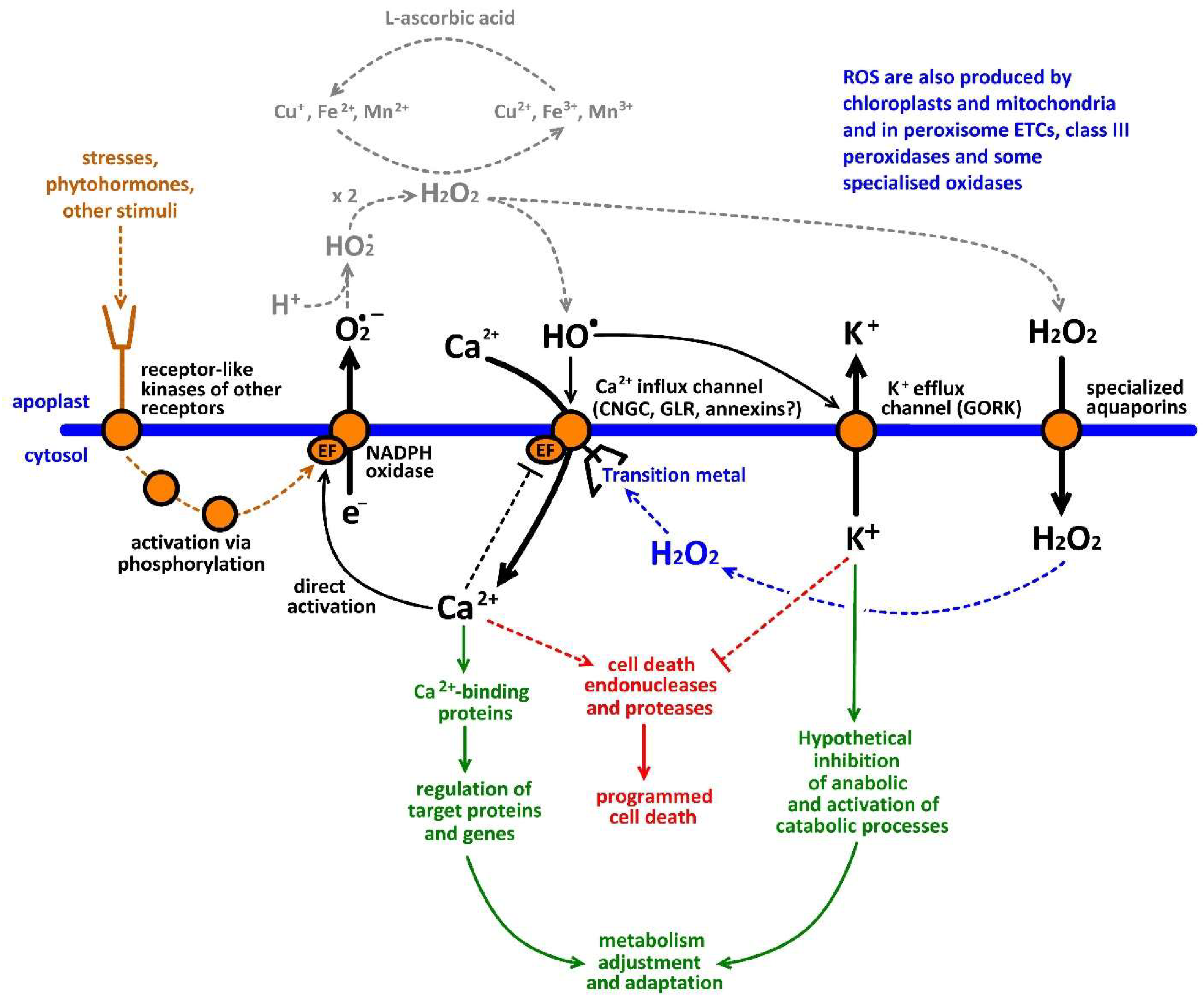
10. The Hypothesis of a ROS-Ca2+ Hub for Amplification of Redox and Ca2+ Signals at the Plant-Environment Interface

ROS are critically important for stress and hormonal signalling, polar and gravitropic growth, autophagy, PCD and other functions of higher plants [29,104]. ROS and cytosolic Ca2+ signals simultaneously control the same physiological processes and cross-talk via reciprocal stimulation—Ca2+-dependent activation of NADPH oxidase and ROS activation of Ca2+-permeable channels (Table 1).
The hypothesis of a ‘ROS-Ca2+ hub’ (Figure 1) assumes that the Ca2+-activated NADPH oxidases (encoded by RBOH genes) act together with ROS-activated Ca2+-permeable channels to amplify stress-induced Ca2+ and ROS signals [2,12,18,29,104]. Elevation of cytosolic Ca2+ level causes an increase in superoxide production and vice versa, superoxide, via generation HO, activates ROS-activated Ca2+ influx channels (Figure 1). This forms a self-amplification positive feedback loop stimulating duration and amplitude of initially weak signals and transforming them into the large-scale responses [58]. Moreover, this also involves higher activation of GORK-mediated K+ efflux [13]. The analysis of recent literature demonstrated that ROS-Ca2+ hub is involved in root and pollen tube growth, hypersensitive response to pathogen, transduction of hormonal and other regulatory chemical signals, responses to abiotic stresses, water balance, autophagy, programmed cell death and mineral nutrition [5]. The ROS-Ca2+ has a sophisticated system for control and regulation in plants, which include self-inactivation of Ca2+-permeable channels, induction of Ca2+-ATPase through Ca2+-calmodulin binding to specific binding sites of these enzymes, suppression of Rac/Rop GTPases, and Ca2+-Dependent Protein Kinase (CDPK)-catalysed phosphorylation of ion channels or Botrytis-Induced Kinase (BIK)1-catalysed phosphorylation of NADPH oxidase [5,104,105,106,107,108,109,110,111,112].
The concept of ROS-Ca2+ hub allows to predict the ion channel subunits involved in activation by ROS [5]. Most ion channels activated by ROS have not yet been identified genetically or electrophysiologically. However, there is indirect evidence that they function in concert with some specific NADPH oxidases (Table 1). An involvement of both NADPH oxidase and channels points to potential activation of channels by ROS. One of the widely acknowledged examples of ‘ROS-Ca2+ hub’ function is the loading of Ca2+ for elongation growth of root cells [6,12] and pollen tubes [87]. This process is driven by extremely high polar NADPH oxidase activity leading to activation of Ca2+-permeable channels. ROS-Ca2+ hub maintains [Ca2+]cyt in growing part of the cell, such as tips root hair of pollen tube, at constantly high level, supporting longitudinal cytoskeleton bundles and tip-directed exocytosis [105]. Arabidopsis CNGC3 [113], annexins [92] and RbohC [6] probably form the root hair ROS-Ca2+ hub. Ionotropic glutamate receptors GLR1.2 and GLR3.7 [86] and CNGC18 [114], jointly with RbohH and RbohJ [115] could function as a ROS-Ca2+ hub in Arabidopsis pollen tube.
The ROS-Ca2+ hub is also involved in plant cell hypersensitive response during pathogen attack, leading to a massive PCD around the infection site and probably preventing spread of the disease [116]. AtrbohD and AtrbohF [117,118] may cross-talk with AtCNGC2, 4, 11 and 12 [119] to form ‘ROS-Ca2+ hub’ mediating this reaction. The following hormones are potentially involved in the ROS-Ca2+ hub: abscisic acid (AtrbohD and AtrbohF cross-talking with AtCNGC5 and AtCNGC6) [53], auxins (AtRbohD/AtCNGC14) [120,121,122], methyl jasmonate (AtrbohD and AtrbohF/AtCNGC2) [123,124] and salicylic acid (AtrbohD/AtGLR3.3) [125,126,127]. Drought induces stomata closure via abscisic acid-mediated activation of AtrbohD and AtrbohF/AtCNGC5 and AtCNGC6 couples [128]. Gémes et al. (2016) have shown that NtRbohD and NtRbohF are crucial for NaCl-induced ROS production and activation of ROS-Ca2+ hub [129], while Guo et al. (2008, 2010) demonstrated that Ca2+ entry in this case is mediated by AtCNGC10 [130,131]. High temperatures are directly sensed via membrane fluidity changes by CNGC6 [113]. At the same time the heat tolerance is impaired in AtrbohD and AtrbohB KO lines, suggesting a clash with CNGC6 [132]. Overall, these data strongly suggest that the ‘ROS-Ca2+ hub’ is an important and ubiquitous mechanism, which is potentially involved in a multitude of physiological processes.

11. Summary and Concluding Remarks

Key biophysical and functional properties of ROS-activated ion channels are summarized in Table 2. Conductances mediated by these channels have been measured in the plasma membrane of Charophyte Nitella flexilis, three dicotyledous higher plants, including Arabidopsis thaliana, Pyrus pyrifolia and Pisum sativum, and one monocotyledon Lilium longiflorum. They exist in root hairs, root mature and elongation zone epidermal cells, pollen tubes, and guard cells. Activating species include H2O2 and hydroxyl radicals. Prevalent intracellular activating species is H2O2 while, in the extracellular medium, it is the hydroxyl radicals. ROS-activated ion channels can be divided into the following three groups by the shape of current-voltage curves: inwardly-rectifying, voltage-independent and outwardly-rectifying. Channels of inwardly-rectifying group conducts Ca2+-influx currents for growth and signaling needs, while the outwardly-rectifying members transport K+ from the cell to extracellular medium to regulate osmotic pressure for guard cell closure, induce programmed cell death or autophagy, or hypothetically adjust metabolism for reparation needs [14]. The voltage-independent group is less studied, but it mediates both Ca2+-influx and K+ efflux, co-existing with other types of conductances in same patches. Selectivity studies are contradicting, showing that ROS-activated channels could be fully nonselective, including permeation to anions, or can specifically conduct only cations. Most reports suggest that ROS-activated channels are cation-selective. However further studies on selectivity are definitely needed. Arabidopsis Ca2+-permeable ROS-activated channels are blocked by lanthanides, verapamil and nifedipine, while the same channels in pea are also sensitive to some anion channel blockers, 5-nitro-2(3-phenylpropylamino)-benzoate and niflumate. ROS-activated K+-permeable channels are blocked by TEA+. Time-dependence of activation can also vary including rapidly- and slowly-activated channels. Both Ca2+ influx and K+ efflux ROS-activated conductances include rapidly- and slowly-activated current components. Few preparations showed single-channel characteristics revealing 14.5-pS Ca2+ influx and 16-pS K+ efflux ROS-activated unitary conductances. Molecular nature of ROS-activated conductances is defined only for K+ efflux channels, which are encoded by SKOR and GORK genes, both having ROS sensing cysteine moieties. Moreover, more genes can be predicted from the analysis of NADPH oxidase-mediated ROS-Ca2+-hubs. Established physiological functions of ROS-activated conductances include root cell and pollen tube growth, regulation of osmotic balance and stomata aperture, responses to major stresses, amplification of external stress and phytohormonal signals via ROS-Ca2+-hub, programmed cell death, and autophagy. Undoubtedly, this list will be extended in further studies on ROS-activated ion channels.

Acknowledgments

This study was supported by the Russian Science Foundation grant#15-14-30008 to Vadim Demidchik.

Conflicts of Interest

The author declares no conflict of interest.

References

  1. Tester, M. Plant ion channels: Whole-cell and single channel studies. New Phytol. 1990, 114, 305–340. [Google Scholar] [CrossRef]
  2. Demidchik, V.; Maathuis, F.J.M. Physiological roles of nonselective cation channels in plants: From salt stress to signalling and development. New Phytol. 2007, 175, 387–405. [Google Scholar] [CrossRef] [PubMed]
  3. Kollist, H.; Jossier, M.; Laanemets, K.; Thomine, S. Anion channels in plant cells. FEBS J. 2011, 278, 4277–4292. [Google Scholar] [CrossRef] [PubMed]
  4. Hedrich, R. Ion channels in plants. Physiol. Rev. 2012, 92, 1777–1811. [Google Scholar] [CrossRef] [PubMed]
  5. Demidchik, V.; Shabala, S. Mechanisms of cytosolic calcium elevation in plants: The role of ion channels, calcium extrusion systems and NADPH oxidase-mediated ‘ROS-Ca2+ Hub’. Funct. Plant Biol. 2017, 45, 9–27. [Google Scholar] [CrossRef]
  6. Foreman, J.; Demidchik, V.; Bothwell, J.H.F.; Mylona, P.; Miedema, H.; Torres, M.A.; Linstead, P.; Costa, S.; Brownlee, C.; Jones, J.D.; et al. Reactive oxygen species produced by NADPH oxidase regulate plant cell growth. Nature 2003, 422, 442–446. [Google Scholar] [CrossRef] [PubMed]
  7. Wang, Y.F.; Fan, L.M.; Zhang, W.Z.; Zhang, W.; Wu, W.H. Ca2+-permeable channels in the plasma membrane of Arabidopsis pollen are regulated by actin microfilaments. Plant Physiol. 2004, 136, 3892–3904. [Google Scholar] [CrossRef] [PubMed]
  8. Jurkowski, G.I.; Smith, R.K., Jr.; Yu, I.C.; Ham, J.H.; Sharma, S.B.; Klessig, D.F.; Fengler, K.A.; Bent, A.F. Arabidopsis DND2, a second cyclic nucleotide-gated ion channel gene for which mutation causes the “defense, no death” phenotype. Mol. Plant Microbe Interact. 2004, 17, 511–520. [Google Scholar] [CrossRef] [PubMed]
  9. Ache, P.; Becker, D.; Ivashikina, N.; Dietrich, P.; Roelfsema, M.R.; Hedrich, R. GORK, a delayed outward rectifier expressed in guard cells of Arabidopsis thaliana, is a K+-selective, K+-sensing ion channel. FEBS Lett. 2000, 486, 93–98. [Google Scholar] [CrossRef]
  10. Pei, Z.M.; Murata, Y.; Benning, G.; Thomine, S.; Klusener, B.; Allen, G.J.; Grill, E.; Schroeder, J.I. Calcium channels activated by hydrogen peroxide mediate abscisic acid signalling in guard cells. Nature 2000, 406, 731–734. [Google Scholar] [CrossRef] [PubMed]
  11. Hosy, E.; Vavasseur, A.; Mouline, K.; Dreyer, I.; Gaymard, F.; Poree, F.; Boucherez, J.; Lebaudy, A.; Bouchez, D.; Very, A.A.; et al. The Arabidopsis outward K+ channel GORK is involved in regulation of stomatal movements and plant transpiration. Proc. Natl. Acad. Sci. USA 2003, 100, 5549–5554. [Google Scholar] [CrossRef] [PubMed]
  12. Demidchik, V.; Shabala, S.N.; Coutts, K.B.; Tester, M.A.; Davies, J.M. Free oxygen radicals regulate plasma membrane Ca2+- and K+-permeable channels in plant root cells. J. Cell Sci. 2003, 116, 81–88. [Google Scholar] [CrossRef] [PubMed]
  13. Demidchik, V.; Cuin, T.A.; Svistunenko, D.; Smith, S.J.; Miller, A.J.; Shabala, S.; Sokolik, A.; Yurin, V. Arabidopsis root K+ efflux conductance activated by hydroxyl radicals: Single-channel properties, genetic basis and involvement in stress-induced cell death. J. Cell Sci. 2010, 123, 1468–1479. [Google Scholar] [CrossRef] [PubMed]
  14. Demidchik, V.; Straltsova, D.; Medvedev, S.S.; Pozhvanov, G.A.; Sokolik, A.; Yurin, V. Stress-induced electrolyte leakage: The role of K+-permeable channels and involvement in programmed cell death and metabolic adjustment. J. Exp. Bot. 2014, 65, 1259–1270. [Google Scholar] [CrossRef] [PubMed]
  15. Herdean, A.; Teardo, E.; Nilsson, A.K.; Pfeil, B.E.; Johansson, O.N.; Ünnep, R.; Nagy, G.; Zsiros, O.; Dana, S.; Solymosi, K.; et al. A voltage-dependent chloride channel fine-tunes photosynthesis in plants. Nat. Commun. 2016, 24, 11654. [Google Scholar] [CrossRef] [PubMed]
  16. Checchetto, V.; Teardo, E.; Carraretto, L.; Formentin, E.; Bergantino, E.; Giacometti, G.M.; Szabo, I. Regulation of photosynthesis by ion channels in cyanobacteria and higher plants. Biophys. Chem. 2013, 182, 5157. [Google Scholar] [CrossRef] [PubMed]
  17. Demidchik, V.; Sokolik, A.; Yurin, V. The effect of Cu2+ on ion transport systems of the plant cell plasmalemma. Plant Physiol. 1997, 114, 1313–1325. [Google Scholar] [CrossRef] [PubMed]
  18. Demidchik, V.; Shabala, S.; Davies, J. Spatial variation in H2O2 response of Arabidopsis thaliana root epidermal Ca2+ flux and plasma membrane Ca2+ channels. Plant J. 2007, 49, 377–386. [Google Scholar] [CrossRef] [PubMed]
  19. Demidchik, V.; Shang, Z.; Shin, R.; Shabala, S.; Davies, J.M. Receptor-like activity evoked by extracellular ADP in Arabidopsis thaliana root epidermal plasma membrane. Plant Physiol. 2011, 156, 1375–1385. [Google Scholar] [CrossRef] [PubMed]
  20. Yuan, F.; Yang, H.; Xue, Y.; Kong, D.; Ye, R.; Li, C.; Zhang, J.; Theprungsirikul, L.; Shrift, T.; Krichilsky, B.; et al. OSCA1 mediates osmotic-stress-evoked Ca2+ increases vital for osmosensing in Arabidopsis. Nature 2014, 514, 367–371. [Google Scholar] [CrossRef] [PubMed]
  21. Zepeda-Jazo, I.; Velarde-Buendía, A.M.; Enríquez-Figueroa, R.; Bose, J.; Shabala, S.; Muñiz-Murguía, J.; Pottosin, I.I. Polyamines interact with hydroxyl radicals in activating Ca2+ and K+ transport across the root epidermal plasma membranes. Plant Physiol. 2011, 157, 2167–2180. [Google Scholar] [CrossRef] [PubMed]
  22. Demidchik, V.; Sokolik, A.; Yurin, V. Characteristics of non-specific permeability and H+-ATPase inhibition induced in the plasma membrane of Nitella flexilis by excessive Cu2+. Planta 2001, 212, 583–590. [Google Scholar] [CrossRef] [PubMed]
  23. Demidchik, V.; Maathuis, F.J.M. (Eds.) Ion Channels and Plant Stress Responses; Springer-Verlag: Berlin/Heidelberg, Germany; New York, NY, USA, 2010; ISBN 3-642-10494-7. [Google Scholar]
  24. Murata, Y.; Mori, I.C.; Munemasa, S. Diverse stomatal signaling and the signal integration mechanism. Annu. Rev. Plant Biol. 2015, 66, 369–392. [Google Scholar] [CrossRef] [PubMed]
  25. Halliwell, B.; Gutteridge, J.M.C. Free Radicals in Biology and Medicine, 5th ed.; Oxford University Press: New York, NY, USA, 2015; ISBN 9780198717478. [Google Scholar]
  26. Fenton, H.J.H. Oxidation of tartaric acid in presence of iron. J. Chem. Soc. Trans. 1894, 65, 899–911. [Google Scholar] [CrossRef]
  27. Haber, F.; Weiss, J. On the catalysis of hydroperoxide. Naturwissenschaften 1932, 20, 948–950. [Google Scholar] [CrossRef]
  28. Goldstein, S.; Meyerstein, D.; Czapski, G. The Fenton reagents. Free Radic. Biol. Med. 1993, 15, 435–445. [Google Scholar] [CrossRef]
  29. Demidchik, V. Mechanisms of oxidative stress in plants: From classical chemistry to cell biology. Environ. Exp. Bot. 2015, 109, 212–228. [Google Scholar] [CrossRef]
  30. McBrien, D.C.H.; Hassall, K.A. Loss of cell potassium by Chlorella vulgaris after contact with toxic amounts of copper sulphate. Physiol. Plant 1965, 18, 1059–1065. [Google Scholar] [CrossRef]
  31. Overnell, J. The effect of heavy metals on photosynthesis and loss of cell potassium in two species of marine algae, Dunaliella tertiolecta and Phaeodactylum tricomutum. Mar. Biol. 1975, 29, 99–103. [Google Scholar] [CrossRef]
  32. De Filippis, L.F. The effect of heavy metal compounds on the permeability of Chlorella cells. Z. Pflanzenphysiol. 1979, 92, 39–49. [Google Scholar] [CrossRef]
  33. Wainwright, S.J.; Woolhouse, H.W. Some physiological aspects of copper and zinc tolerance in Agrostis tenuis Sibth: Cell elongation and membrane damage. J. Exp. Bot. 1977, 28, 1029–1036. [Google Scholar] [CrossRef]
  34. De Vos, C.; Schat, H.; Vooijs, R.; Ernst, W. Copper induced damage to the permeability barrier in roots of Silene cucubalus. J. Plant Physiol. 1989, 135, 164–165. [Google Scholar] [CrossRef]
  35. Luna, C.; Gonzalez, C.; Trippi, V. Oxidative damage caused by an excess of copper in oat leaves. Plant Cell Physiol. 1994, 35, 11–15. [Google Scholar] [CrossRef]
  36. Murphy, A.; Taiz, L. Correlation between potassium efflux and copper sensitivity in ten Arabidopsis ecotypes. New Phytol. 1997, 136, 211–222. [Google Scholar] [CrossRef]
  37. Murphy, A.S.; Eisinger, W.R.; Shaff, J.E.; Kochian, L.V.; Taiz, L. Early copper-induced leakage of K+ from Arabidopsis seedlings is mediated by ion channels and coupled to citrate efflux. Plant Physiol. 1999, 121, 1375–1382. [Google Scholar] [CrossRef] [PubMed]
  38. Palta, J.P.; Levitt, J.; Stadelmann, E.J. Freezing injury in onion bulb cells. I. Evaluation of the conductivity method and analysis of ion and sugar efflux from injured cells. Plant Physiol. 1977, 60, 393–397. [Google Scholar] [CrossRef] [PubMed]
  39. Shabala, S. Plant Stress Physiology, 2nd ed.; CABI: Boston, MA, USA, 2017; ISBN 9781780647302. [Google Scholar]
  40. Atkinson, M.M.; Midland, S.L.; Sims, J.J.; Keen, N.T. Syringolide 1 triggers Ca2+ influx, K+ efflux, and extracellular alkalization in soybean cells carrying the disease-resistance gene Rpg4. Plant Physiol. 1996, 112, 297–302. [Google Scholar] [CrossRef] [PubMed]
  41. Atkinson, M.M.; Huang, J.S.; Knopp, J.A. The hypersensitive response of tobacco to Pseudomonas syringae pv. pisi: Activation of a plasmalemma K+/H+ exchange mechanism. Plant Physiol. 1985, 79, 843–847. [Google Scholar] [CrossRef] [PubMed]
  42. Atkinson, M.M.; Keppler, L.D.; Orlandi, E.W.; Baker, C.J.; Mischke, C.F. Involvement of plasma membrane calcium influx in bacterial induction of the K+/H+ and hypersensitive responses in tobacco. Plant Physiol. 1990, 92, 215–221. [Google Scholar] [CrossRef] [PubMed]
  43. Van Dongen, J.T.; Licausi, F. Oxygen sensing and signaling. Annu. Rev. Plant Biol. 2015, 66, 345–367. [Google Scholar] [CrossRef] [PubMed]
  44. Levitt, J. Responses of Plants to Environmental Stresses: Chilling, Freezing and High Temperature Stresses v. 1 (Physiological ecology), 1st ed.; Academic Press Inc.: London, UK, 1972; ISBN 9780124455603. [Google Scholar]
  45. Nassery, H. The effect of salt and osmotic stress on the retention of Potassium by excised barley and bean roots. New Phytol. 1975, 75, 63–67. [Google Scholar] [CrossRef]
  46. Nassery, H. Salt-induced loss of potassium from plant roots. New Phytol. 1979, 83, 23–27. [Google Scholar] [CrossRef]
  47. Blum, A.; Ebercon, A. Cell membrane stability as a measure of drought and heat tolerance in wheat. Crop Sci. 1979, 21, 43–47. [Google Scholar] [CrossRef]
  48. Leopold, A.C.; Musgrave, M.E.; Williams, K.M. Solute leakage resulting from leaf desiccation. Plant Physiol. 1981, 68, 1222–1225. [Google Scholar] [CrossRef] [PubMed]
  49. Price, A.H. A possible role for calcium in oxidative plant stress. Free Radic. Res. Commun. 1990, 10, 345–349. [Google Scholar] [CrossRef] [PubMed]
  50. Price, A.H.; Taylor, A.; Ripley, S.J.; Griffiths, A.; Trewavas, A.J.; Knight, M.R. Oxidative signals in tobacco increase cytosolic calcium. Plant Cell 1994, 6, 1301–1310. [Google Scholar] [CrossRef] [PubMed]
  51. Clayton, H.; Knight, M.R.; Knight, H.; McAinsh, M.R.; Hetherington, A.M. Dissection of the ozone-induced calcium signature. Plant J. 1999, 17, 575–579. [Google Scholar] [CrossRef] [PubMed]
  52. Demidchik, V.V.; Sokolik, A.I.; Yurin, V.M. Mechanisms of conductance modification in plant cell membranes under the action of trivalent iron ions. Doklady Akademii Nauk Belarusi 1996, 41, 83–87. [Google Scholar]
  53. Mori, I.C.; Schroeder, J.I. Reactive oxygen species activation of plant Ca2+ channels. a signaling mechanism in polar growth, hormone transduction, stress signaling, and hypothetically mechanotransduction. Plant Physiol. 2004, 135. [Google Scholar] [CrossRef] [PubMed]
  54. Meinhard, M.; Rodriguez, P.L.; Grill, E. The sensitivity of ABI2 to hydrogen peroxide links the abscisic acid-response regulator to redox signaling. Planta 2002, 214, 775–782. [Google Scholar] [CrossRef] [PubMed]
  55. Meinhard, M.; Grill, E. Hydrogen peroxide is a regulator of ABI1, a protein phosphatase 2C from Arabidopsis. FEBS Lett. 2001, 508, 443–446. [Google Scholar] [CrossRef]
  56. Sierla, M.; Waszczak, C.; Vahisalu, T.; Kangasjärvi, J. Reactive oxygen species in the regulation of stomatal movements. Plant Physiol. 2016, 171, 1569–1580. [Google Scholar] [CrossRef] [PubMed]
  57. Wu, H.; Shabala, L.; Zhou, M.; Shabala, S. Chloroplast-generated ROS dominate NaCl induced K+ efflux in wheat leaf mesophyll. Plant Signal. Behav. 2015, 10, e1013793. [Google Scholar] [CrossRef] [PubMed]
  58. Demidchik, V.; Shang, Z.; Shin, R.; Thompson, E.; Rubio, L.; Laohavisit, A.; Mortimer, J.C.; Chivasa, S.; Slabas, A.R.; Glover, B.J.; et al. Plant extracellular ATP signaling by plasma membrane NADPH oxidase and Ca2+ channels. Plant J. 2009, 58, 903–913. [Google Scholar] [CrossRef] [PubMed]
  59. Fry, S.C.; Miller, J.G.; Dumville, J.C. A proposed role for copper ions in cell wall loosening. Plant Soil 2002, 247, 57–67. [Google Scholar] [CrossRef]
  60. Bogeski, I.; Niemeyer, B.A. Redox regulation of ion channels. Antioxid Redox Signal 2014, 21, 859–862. [Google Scholar] [CrossRef] [PubMed]
  61. Lebaudy, A.; Véry, A.A.; Sentenac, H. K+ channel activity in plants: Genes, regulations and functions. FEBS Lett. 2007, 581, 2357–2366. [Google Scholar] [CrossRef] [PubMed]
  62. Monshausen, G.B.; Bibikova, T.N.; Messerli, M.A.; Shi, C.; Gilroy, S. Oscillations in extracellular pH and reactive oxygen species modulate tip growth of Arabidopsis root hairs. Proc. Natl. Acad. Sci. USA 2007, 104, 20996–21001. [Google Scholar] [CrossRef] [PubMed]
  63. Coelho, S.M.B.; Brownlee, C.; Bothwell, J.H.F. A tip-high, Ca2+-interdependent, reactive oxygen species gradient is associated with polarized growth in Fucus serratus zygotes. Planta 2008, 227, 1037–1046. [Google Scholar] [CrossRef] [PubMed]
  64. Mendrinna, A.; Persson, S. Root hair growth: It’s a one way street. F1000Prime Rep. 2015, 7, 23. [Google Scholar] [CrossRef] [PubMed]
  65. Libik-Konieczny, M.; Kozieradzka-Kiszkurno, M.; Desel, C.; Michalec-Warzecha, Ż.; Miszalski, Z.; Konieczny, R. The localization of NADPH oxidase and reactive oxygen species in in vitro-cultured Mesembryanthemum crystallinum L. hypocotyls discloses their differing roles in rhizogenesis. Protoplasma 2015, 252, 477–487. [Google Scholar] [CrossRef] [PubMed]
  66. Causin, H.F.; Roqueiro, G.; Petrillo, E.; Láinez, V.; Pena, L.B.; Marchetti, C.F.; Gallego, S.M.; Maldonado, S.I. The control of root growth by reactive oxygen species in Salix nigra Marsh. seedlings. Plant Sci. 2012, 183, 197–205. [Google Scholar] [CrossRef] [PubMed]
  67. Mangano, S.; Denita-Juarez, S.P.; Choi, H.S.; Marzol, E.; Hwang, Y.; Ranocha, P.; Velasquez, S.M.; Borassi, C.; Barberini, M.L.; Aptekmann, A.A.; et al. Molecular link between auxin and ROS-mediated polar growth. Proc. Natl. Acad. Sci. USA 2017, 114, 5289–5294. [Google Scholar] [CrossRef] [PubMed]
  68. Yamauchi, T.; Yoshioka, M.; Fukazawa, A.; Mori, H.; Nishizawa, N.K.; Tsutsumi, N.; Yoshioka, H.; Nakazono, M. An NADPH Oxidase RBOH functions in rice roots during Lysigenous aerenchyma formation under oxygen-deficient conditions. Plant Cell 2017, 4, 775–790. [Google Scholar] [CrossRef] [PubMed]
  69. Dunand, C.; Crèvecoeur, M.; Penel, C. Distribution of superoxide and hydrogen peroxide in Arabidopsis root and their influence on root development: Possible interaction with peroxidases. New Phytol. 2007, 174, 332–341. [Google Scholar] [CrossRef] [PubMed]
  70. Eisenbarth, D.A.; Weig, A.R. Dynamics of aquaporins and water relations during hypocotyl elongation in Ricinus communis L. seedlings. J. Exp. Bot. 2005, 56, 1831–1842. [Google Scholar] [CrossRef] [PubMed]
  71. Bienert, G.P.; Møller, A.L.; Kristiansen, K.A.; Schulz, A.; Møller, I.M.; Schjoerring, J.K.; Jahn, T.P. Specific aquaporins facilitate the diffusion of hydrogen peroxide across membranes. J. Biol. Chem. 2007, 282, 1183–1192. [Google Scholar] [CrossRef] [PubMed]
  72. Bienert, G.P.; Schjoerring, J.K.; Jahn, T.P. Membrane transport of hydrogen peroxide. Biochim. Biophys. Acta 2006, 1758, 994–1003. [Google Scholar] [CrossRef] [PubMed]
  73. Vieceli Dalla Sega, F.; Zambonin, L.; Fiorentini, D.; Rizzo, B.; Caliceti, C.; Landi, L.; Hrelia, S.; Prata, C. Specific aquaporins facilitate Nox-produced hydrogen peroxide transport through plasma membrane in leukaemia cells. Biochim. Biophys. Acta 2014, 1843, 806–814. [Google Scholar] [CrossRef] [PubMed]
  74. Pottosin, I.; Shabala, S. Polyamines control of cation transport across plant membranes: Implications for ion homeostasis and abiotic stress signaling. Front Plant Sci. 2014, 23, 154. [Google Scholar] [CrossRef] [PubMed]
  75. Garcia-Mata, C.; Wang, J.; Gajdanowicz, P.; Gonzalez, W.; Hills, A.; Donald, N.; Riedelsberger, J.; Amtmann, A.; Dreyer, I.; Blatt, M.R. A minimal cysteine motif required to activate the SKOR K+ channel of Arabidopsis by the reactive oxygen species H2O2. J. Biol. Chem. 2010, 285, 29286–29294. [Google Scholar] [CrossRef] [PubMed]
  76. Wang, F.; Chen, Z.H.; Liu, X.; Colmer, T.D.; Shabala, L.; Salih, A.; Zhou, M.; Shabala, S. Revealing the roles of GORK channels and NADPH oxidase in acclimation to hypoxia in Arabidopsis. J. Exp. Bot. 2016, 68, 3191–3204. [Google Scholar] [CrossRef] [PubMed]
  77. Becker, D.; Hoth, S.; Ache, P.; Wenkel, S.; Roelfsema, M.R.; Meyerhoff, O.; Hartung, W.; Hedrich, R. Regulation of the ABA-sensitive Arabidopsis potassium channel gene GORK in response to water stress. FEBS Lett. 2003, 554, 119–126. [Google Scholar] [CrossRef]
  78. Tran, D.; El-Maarouf-Bouteau, H.; Rossi, M.; Biligui, B.; Briand, J.; Kawano, T.; Mancuso, S.; Bouteau, F. Post-transcriptional regulation of GORK channels by superoxide anion contributes to increases in outward-rectifying K+ currents. New Phytol. 2013, 198, 1039–1048. [Google Scholar] [CrossRef] [PubMed]
  79. Remillard, C.V.; Yuan, J.X.-J. Activation of K+ channels: An essential pathway in programmed cell death. Am. J. Physiol. Lung Cell Mol. Physiol. 2004, 286, L49–L67. [Google Scholar] [CrossRef] [PubMed]
  80. Kondratskyi, A.; Kondratska, K.; Skryma, R.; Prevarskaya, N. Ion channels in the regulation of apoptosis. Biochim. Biophys. Acta 2015, 1848(10PtB), 2532–2546. [Google Scholar] [CrossRef] [PubMed]
  81. Orlov, S.; Hamet, P. JIntracellular monovalent ions as second messengers. Membr. Biol. 2006, 210, 161–172. [Google Scholar] [CrossRef] [PubMed]
  82. Bortner, C.D.; Gómez-Angelats, M.; Cidlowski, J.A. Plasma membrane depolarization without repolarization is an early molecular event in anti-fas-induced apoptosis. J. Biol. Chem. 2001, 276, 4304–4314. [Google Scholar] [CrossRef] [PubMed]
  83. Yu, S.P. Regulation and critical role of potassium homeostasis in apoptosis. Prog. Neurobiol. 2003, 70, 363–386. [Google Scholar] [CrossRef]
  84. Lauff, D.B.; Santa-María, G.E. Potassium deprivation is sufficient to induce a cell death program in Saccharomyces cerevisiae. FEMS Yeast Res. 2010, 10, 497–507. [Google Scholar] [CrossRef] [PubMed]
  85. Yun, J.; Lee, D.G. Role of potassium channels in chlorogenic acid-induced apoptotic volume decrease and cell cycle arrest in Candida albicans. Biochim. Biophys. Acta 2017, 1861, 585–592. [Google Scholar] [CrossRef] [PubMed]
  86. Michard, E.; Simon, A.A.; Tavares, B.; Wudick, M.M.; Feijó, J.A. Signaling with ions: The keystone for apical cell growth and morphogenesis in pollen tubes. Plant Physiol. 2017, 173, 91–111. [Google Scholar] [CrossRef] [PubMed]
  87. Potocky, M.; Jones, M.A.; Bezvoda, R.; Smirnoff, N.; Zarsky, V. Reactive oxygen species produced by NADPH oxidase are involved in pollen tube growth. New Phytol. 2007, 174, 742–751. [Google Scholar] [CrossRef] [PubMed]
  88. Breygina, M.A.; Abramochkin, D.V.; Maksimov, N.M.; Yermakov, I.P. Hydrogen peroxide affects ion channels in lily pollen grain protoplasts. Plant Biol. 2016, 18, 761–767. [Google Scholar] [CrossRef] [PubMed]
  89. Makavitskaya, M.; Svistunenko, D.; Navaselsky, I.; Hryvusevich, P.; Mackievic, V.; Rabadanova, C.; Tyutereva, E.; Samokhina, V.; Straltsova, D.; Sokolik, A.; et al. Novel roles of ascorbate in plants: induction of cytosolic Ca2+ signals and efflux from cells via anion channels. J. Exp. Bot. 2018, ery056. [Google Scholar] [CrossRef] [PubMed]
  90. Xu, L.; Tang, Y.; Gao, S.; Su, S.; Hong, L.; Wang, W.; Fang, Z.; Li, X.; Ma, J.; Quan, W.; et al. Comprehensive analyses of the annexin gene family in wheat. BMC Genom. 2016, 28, 415. [Google Scholar] [CrossRef] [PubMed]
  91. Laohavisit, A.; Mortimer, J.C.; Demidchik, V.; Coxon, K.M.; Stancombe, M.A.; Macpherson, N.; Brownlee, C.; Hofmann, A.; Webb, A.A.; Miedema, H.; et al. Zea mays annexins modulate cytosolic free Ca2+ and generate a Ca2+-permeable conductance. Plant Cell 2009, 21, 479–493. [Google Scholar] [CrossRef] [PubMed]
  92. Laohavisit, A.; Shang, Z.; Rubio, L.; Cuin, T.A.; Véry, A.A.; Wang, A.; Mortimer, J.C.; Macpherson, N.; Coxon, K.M.; Battey, N.H.; et al. Arabidopsis annexin1 mediates the radical-activated plasma membrane Ca2+- and K+-permeable conductance in root cells. Plant Cell 2012, 24, 1522–1533. [Google Scholar] [CrossRef] [PubMed]
  93. Baucher, M.; Pérez-Morga, D.; El Jaziri, M. Insight into plant annexin function: From shoot to root signaling. Plant Signal Behav. 2012, 7, 524–528. [Google Scholar] [CrossRef] [PubMed]
  94. Lizarbe, M.A.; Barrasa, J.I.; Olmo, N.; Gavilanes, F.; Turnay, J. Annexin-phospholipid interactions. Functional implication. Int. J. Mol. Sci. 2013, 14, 2652–2683. [Google Scholar] [CrossRef] [PubMed]
  95. Carmeille, R.; Degrelle, S.A.; Plawinski, L.; Bouvet, F.; Gounou, C.; Evain-Brion, D.; Brisson, A.R.; Bouter, A. Annexin-A5 promotes membrane resealing in human trophoblasts. Biochim. Biophys. Acta 2015, 1853, 2033–2044. [Google Scholar] [CrossRef] [PubMed]
  96. Brazier, S.P.; Telezhkin, V.; Kemp, P.J. Functional Interactions between BKCaα-subunit and annexin A5: Implications in apoptosis. Oxid. Med. Cell. Longev. 2016, 1607092. [Google Scholar] [CrossRef] [PubMed]
  97. Wang, X.; Ma, X.; Wang, H.; Li, B.; Clark, G.; Guo, Y.; Roux, S.; Sun, D.; Tang, W. Proteomic study of microsomal proteins reveals a key role for Arabidopsis annexin 1 in mediating heat stress-induced increase in intracellular calcium levels. Mol. Cell. Proteom. 2015, 14, 686–694. [Google Scholar] [CrossRef] [PubMed]
  98. Konopka-Postupolska, D.; Clark, G. Annexins as overlooked regulators of membrane trafficking in plant cells. Int. J. Mol. Sci. 2017, 18, 863. [Google Scholar] [CrossRef] [PubMed]
  99. Konopka-Postupolska, D.; Clark, G.; Goch, G.; Debski, J.; Floras, K.; Cantero, A.; Fijolek, B.; Roux, S.; Hennig, J. The role of annexin 1 in drought stress in Arabidopsis. Plant Physiol. 2009, 150, 1394–1410. [Google Scholar] [CrossRef] [PubMed]
  100. Qiao, B.; Zhang, Q.; Liu, D.; Wang, H.; Yin, J.; Wang, R.; He, M.; Cui, M.; Shang, Z.; Wang, D.; Zhu, Z. A calcium-binding protein, rice annexin OsANN1, enhances heat stress tolerance by modulating the production of H2O2. J. Exp. Bot. 2015, 66, 5853–5866. [Google Scholar] [CrossRef] [PubMed]
  101. Laohavisit, A.; Richards, S.L.; Shabala, L.; Chen, C.; Colaço, R.D.; Swarbreck, S.M.; Shaw, E.; Dark, A.; Shabala, S.; Shang, Z.; et al. Salinity-induced calcium signaling and root adaptation in Arabidopsis require the calcium regulatory protein annexin. Plant Physiol. 2013, 163, 253–262. [Google Scholar] [CrossRef] [PubMed]
  102. Simon, L.; Gauvin, F.; Amre, D.K.; Saint-Louis, P.; Lacroix, J. Serum procalcitonin and C-reactive protein levels as markers of bacterial infection: A systematic review and meta-analysis. Clin. Infect Dis. 2004, 39, 206–217. [Google Scholar] [CrossRef] [PubMed]
  103. Dubbs, J.M.; Mongkolsuk, S. Peroxide-sensing transcriptional regulators in bacteria. J. Bacteriol. 2012, 194, 5495–5503. [Google Scholar] [CrossRef] [PubMed]
  104. Shabala, S.; Wu, H.H.; Bose, J. Salt stress sensing and early signalling events in plant roots: Current knowledge and hypothesis. Plant Sci. 2015, 241, 109–119. [Google Scholar] [CrossRef] [PubMed]
  105. Noctor, G.; Reichheld, J.P.; Foyer, C.H. ROS-related redox regulation and signaling in plants. Semin. Cell Dev. Biol. 2017. [Google Scholar] [CrossRef] [PubMed]
  106. Baxter-Burrell, A.; Yang, Z.; Springer, P.S.; Bailey-Serres, J. RopGAP4-dependent Rop GTPase rheostat control of Arabidopsis oxygen deprivation tolerance. Science 2002, 296, 2026–2028. [Google Scholar] [CrossRef] [PubMed]
  107. Tidow, H.; Poulsen, L.R.; Andreeva, A.; Knudsen, M.; Hein, K.L.; Wiuf, C.; Palmgren, M.G.; Nissen, P. A bimodular mechanism of calcium control in eukaryotes. Nature 2012, 491, 468–472. [Google Scholar] [CrossRef] [PubMed]
  108. Zhou, J.; Wang, J.; Li, X.; Xia, X.J.; Zhou, Y.H.; Shi, K. H2O2 mediates the crosstalk of brassinosteroid and abscisic acid in tomato responses to heat and oxidative stresses. J. Exp. Bot. 2014, 65, 4371–4383. [Google Scholar] [CrossRef] [PubMed]
  109. Ben-Johny, M.; Yue, D.N.; Yue, D.T. Detecting stoichiometry of macromolecular complexes in live cells using FRET. Nat. Commun. 2016, 7, 13709–13719. [Google Scholar] [CrossRef] [PubMed]
  110. Kadota, Y.; Shirasu, K.; Zipfel, C. Regulation of the NADPH Oxidase RBOHD during Plant Immunity. Plant Cell Physiol. 2015, 56, 1472–1480. [Google Scholar] [CrossRef] [PubMed]
  111. DeFalco, T.A.; Moeder, W.; Yoshioka, K. Opening the Gates: Insights into Cyclic Nucleotide-Gated Channel-Mediated Signaling. Trends Plant Sci. 2016, 11, 903–906. [Google Scholar] [CrossRef] [PubMed]
  112. Rounds, C.M.; Bezanilla, M. Growth mechanisms in tip-growing plant cells. Annu. Rev. Plant Biol. 2013, 64, 243–265. [Google Scholar] [CrossRef] [PubMed]
  113. Gobert, A.; Park, G.; Amtmann, А.; Sanders, D.; Maathuis, F.J.M. Arabidopsis thaliana cyclic nu leotide gated channel 3 forms a non-selective ion transporter involved in germination and cation transport. J. Exp. Bot. 2006, 57, 791–800. [Google Scholar] [CrossRef] [PubMed]
  114. Gao, Q.F.; Gu, L.L.; Wang, H.Q.; Fei, C.F.; Fang, X.; Hussain, J.; Sun, S.J.; Dong, J.Y.; Liu, H.; Wang, Y.F. Cyclic nucleotide-gated channel 18 is an essential Ca2+ channel in pollen tube tips for pollen tube guidance to ovules in Arabidopsis. Proc. Natl. Acad. Sci. USA 2016, 113, 3096–3101. [Google Scholar] [CrossRef] [PubMed]
  115. Kaya, A.; Lobanov, A.V.; Gerashchenko, M.V.; Koren, A.; Fomenko, D.E.; Koc, A.; Gladyshev, V.N. Thiol peroxidase deficiency leads to increased mutational load and decreased fitness in Saccharomyces cerevisiae. Genetics 2014, 198, 905–917. [Google Scholar] [CrossRef] [PubMed]
  116. Levine, A.; Pennell, R.I.; Alvarez, M.E.; Palmer, R.; Lamb, C. Calcium mediated apoptosis in a plant hypersensitive disease resistance response. Curr. Biol. 1996, 6, 427–437. [Google Scholar] [CrossRef]
  117. Dubiella, U.; Seybold, H.; Durian, G.; Komander, E.; Lassig, R.; Witte, C.P.; Schulze, W.X.; Romeis, T. Calcium-dependent protein kinase/NADPH oxidase activation circuit is required for rapid defense signal propagation. Proc. Natl. Acad. Sci. USA 2013, 110, 8744–8749. [Google Scholar] [CrossRef] [PubMed]
  118. Torres, M.A.; Dangl, J.L.; Jones, J.D.G. Arabidopsis gp91phox homologues, AtrbohD and AtrbohF are required for accumulation of reactive oxygen intermediates in the plant defense response. Proc. Natl. Acad. Sci. USA 2002, 99, 517–522. [Google Scholar] [CrossRef] [PubMed]
  119. Moeder, W.; Urquhart, W.; Ung, H.; Yoshioka, K. The role of cyclic nucleotide-gated ion channels in plant immunity. Mol. Plant 2011, 4, 442–452. [Google Scholar] [CrossRef] [PubMed]
  120. Shih, H.W.; DePew, C.L.; Miller, N.D.; Monshausen, G.B. The cyclic nucleotide-gated channel CNGC14 regulates root gravitropism in Arabidopsis thaliana. Curr. Biol. 2015, 25, 3119–3125. [Google Scholar] [CrossRef] [PubMed]
  121. Nguyen, H.T.; Umemura, K.; Kawano, T. Indole-3-acetic acid-induced oxidative burst and an increase in cytosolic calcium ion concentration in rice suspension culture. Biosci. Biotechnol. Biochem. 2016, 80, 1546–1554. [Google Scholar] [CrossRef] [PubMed]
  122. Peer, W.A.; Cheng, Y.; Murphy, A.S. Evidence of oxidative attenuation of auxin signalling. J. Exp. Bot. 2013, 64, 2629–2639. [Google Scholar] [CrossRef] [PubMed]
  123. Maruta, T.; Inoue, T.; Tamoi, M.; Yabuta, Y.; Yoshimura, K.; Ishikawa, T.; Shigeoka, S. Arabidopsis NADPH oxidases, AtrbohD and AtrbohF, are essential for jasmonic acid-induced expression of genes regulated by MYC2 transcription factor. Plant Sci. 2011, 180, 655–660. [Google Scholar] [CrossRef] [PubMed]
  124. Lu, M.; Zhang, Y.; Tang, S.; Pan, J.; Yu, Y.; Han, J.; Li, Y.; Du, X.; Nan, Z.; Sun, Q. AtCNGC2 is involved in jasmonic acid-induced calcium mobilization. J. Exp. Bot. 2016, 67, 809–819. [Google Scholar] [CrossRef] [PubMed]
  125. Kawano, T.; Sahashi, N.; Takahashi, K.; Uozumi, N.; Muto, S. Salicylic acid induces extracellular superoxide generation followed by an increase in cytosolic calcium ion in tobacco suspension culture: The earliest events in salicylic acid signal transduction. Plant Cell Physiol. 1998, 39, 721–730. [Google Scholar] [CrossRef]
  126. Manzoor, H.; Kelloniemi, J.; Chiltz, A.; Wendehenne, D.; Pugin, A.; Poinssot, B.; Garcia-Brugger, A. Involvement of the glutamate receptor AtGLR3.3 in plant defense signaling and resistance to Hyaloperonospora arabidopsidis. Plant J. 2013, 76, 466–480. [Google Scholar] [CrossRef] [PubMed]
  127. Jayakannan, M.; Bose, J.; Babourina, O.; Rengel, Z.; Shabala, S. Salicylic acid in plant salinity stress signalling and tolerance. Plant Growth Regul. 2015, 76, 25–40. [Google Scholar] [CrossRef]
  128. Wang, Y.F.; Munemasa, S.; Nishimura, N.; Ren, H.M.; Robert, N.; Han, M.; Puzõrjova, I.; Kollist, H.; Lee, S.; Mori, I.; Schroeder, J.I. Identification of cyclic GMP-activated nonselective Ca2+-permeable cation channels and associated CNGC5 and CNGC6 genes in Arabidopsis guard cells. Plant Physiol. 2013, 163, 578–590. [Google Scholar] [CrossRef] [PubMed]
  129. Gémes, K.; Kim, Y.J.; Park, K.Y.; Moschou, P.N.; Andronis, E.; Valassaki, C.; Roussis, A.; Roubelakis-Angelakis, K.A. An NADPH-oxidase/polyamine oxidase feedback loop controls oxidative burst under salinity. Plant Physiol. 2016, 172, 1418–1431. [Google Scholar] [CrossRef] [PubMed]
  130. Guo, K.M.; Babourina, O.; Christopher, D.A.; Borsics, T.; Rengel, Z. The cyclic nucleotide-gated channel, AtCNGC10, influences salt tolerance in Arabidopsis. Physiol. Plant 2008, 134, 499–507. [Google Scholar] [CrossRef] [PubMed]
  131. Guo, K.M.; Babourina, O.; Christopher, D.A.; Borsics, T.; Rengel, Z. The cyclic nucleotide-gated channel AtCNGC10 transports Ca2+ and Mg2+ in Arabidopsis. Physiol. Plant 2010, 139, 303–312. [Google Scholar] [CrossRef] [PubMed]
  132. Larkindale, J.; Hall, J.D.; Knight, M.R.; Vierling, E. Heat stress phenotypes of Arabidopsis mutants implicate multiple signaling pathways in the acquisition of thermotolerance. Plant Physiol. 2005, 138, 882–897. [Google Scholar] [CrossRef] [PubMed]
  133. Guo, J.; Zeng, W.; Chen, Q.; Lee, C.; Chen, L.; Yang, Y.; Cang, C.; Ren, D.; Jiang, Y. Structure of the voltage-gated two-pore channel TPC1 from Arabidopsis thaliana. Nature 2016, 531, 196–201. [Google Scholar] [CrossRef] [PubMed]
Figure 1. The concept of reactive oxygen species (ROS)-Ca2+-hub in the plasma membrane of higher plants. Stresses and regulatory stimuli can react with cell surface receptors leading to the activation of NADPH-oxidases (or directly activate ion channels). NADPH oxidases produce superoxide (O2•−), which binds H+ and forms hydroperoxyl radicals (HO2), which undergo dismutation reaction forming H2O2. H2O2 can be reduced in Haber-Weiss cycle using electrons from transition metals, which can be reduced by l-ascorbic acid. H2O2 reduction leads for formation of HO in the close proximity (clusters) to ROS-activated Ca2+-permeable cation channels. Cell wall transition metals are required for Haber-Weiss cycle in the apoplast. Channels are activated in response to increased HO production and mediate Ca2+ influx and K+ efflux. Hydrogen peroxide can hypothetically activate channels from inside via reaction with transition metal binding sites (via generation of HO). ROS-induced cytosolic Ca2+ elevation results in activation of signaling and regulatory cascades, stimulation of exocytosis and growth as well as induction of programmed cell death. ROS-activated efflux of K+ can lead to stimulation of programmed cell death, autophagy or metabolic adjustment, which is required for changing plant metabolism during stress responses and releasing energy for reparation needs [14].
Ijms 19 01263 g001
Table 1. NADPH oxidases and cation channels, which can potentially function as ROS-Ca2+ hubs in key physiological reactions (see hypothesis on ROS-Ca2+ hubs in the text).
Physiological ProcessGenes of Cation Channels with Predicted or Demonstrated Ca2+ PermeabilityGenes Encoding NADPH Oxidase Producing ROS for Activation of Ca2+-Permeable ChannelsReferences
Growth and development
Root cell elongation AtCNGC3AtRBOHC[6,113]
Pollen tube elongationAtCNGC18, AtGLR1.2, AtGLR3.7AtRBOHH, AtRBOHJ[86,115,133]
Phytohormonal regulation
Effects of auxin AtCNGC14AtRBOHD[120,121,122]
Abscisic acid signallingAtCNGC5, AtCNGC6AtRBOHD, AtRBOHF[7,122]
Methyl jasmonate-induced reactions AtCNGC2AtRBOHD, AtRBOHF[123,124]
Action of salicylic acidAtGLR3.3AtRBOHD[125,126,127]
Stress responses
Hypersensitive response (massive PCD around the spot of infection, preventing spread of the disease)AtCNGC2, AtCNGC4, AtCNGC11, AtCNGC12AtRBOHD, AtRBOHF[117,118,119]
Response to salinityAtCNGC10NtRBOHD, NtRBOHF[128,130]
Drought-induced stomata closureAtCNGC5, AtCNGC6AtRBOHD, AtRBOHF[128]
Response to extreme temperaturesAtCNGC6AtRBOHD, AtRBOHB[114,132]
Table 2. Biophysical and functional characteristics of ROS-activated ion channels in plant plasma membranes.
Preparation Activating ROS Selectivity and Kinetics of Activation Blockers, ModulatorsFunctionReferences
Nitella flexilis
- intermodal cellsCu2+, HONS *, VI *, RA *Lanthanides, verapamil, nifedipineSensing transition metals, copper toxicity [17,22]
Arabidopsis thaliana
- root hairsHONS, IR *, SA *LanthanidesGrowth[6,12]
- root mature epidermisHO, H2O2NS, IR, SALanthanides, verapamil
TEA+
Stress response[12,18]
- root elongation zoneHOKS *, OR *, SA
NS, VI, RA
Lanthanides, verapamilGrowth, stress response[12]
- guard cellsH2O2 H2O2KS, OR, SA
NS, VI, RA
NS, IR
Lanthanides
Lanthanides,
verapamil
ABA signaling, stomata closure[10]
Pyrus pyrifolia
- pollen tubeH2O2IRActivation by polyaminesPollen tube growth[57]
Pisum sativum
- rootHONS, VI, RAStimulation by polyamines [21]
Lilium longiflorum
- pollen tubeH2O2NS, IR, SA
KS, OR, SA
Lanthanides, nifedipine
TEA+
Pollen tube growth[88]
* NS—non-selective, RA—rapidly-activating, SA—slowly-activating, VI—voltage-independent, OR—outwardly-rectifying, IR—inwardly-rectifying, KS—K+-selective.

