Article

Divergent Expression Patterns and Function of Two cxcr4 Paralogs in Hermaphroditic Epinephelus coioides

1
State Key Laboratory of Freshwater Ecology and Biotechnology, Institute of Hydrobiology, Chinese Academy of Sciences, Wuhan 430072, China
2
University of Chinese Academy of Sciences, Beijing 100049, China
3
Key Laboratory of Mariculture & Stock Enhancement in North China Sea, Ministry of Agriculture and Rural Affairs, Dalian Ocean University, Dalian 116023, China
4
State Key Laboratory of Biocontrol, Guangdong Province Key Laboratory for Aquatic Economic Animals, and Institute of Aquatic Economic Animals, School of Life Sciences, Sun Yat-Sen University, Guangzhou 510275, China
*
Authors to whom correspondence should be addressed.
Int. J. Mol. Sci. 2018, 19(10), 2943; https://doi.org/10.3390/ijms19102943
Submission received: 7 August 2018 / Revised: 11 September 2018 / Accepted: 18 September 2018 / Published: 27 September 2018
(This article belongs to the Section Biochemistry)

Abstract

:
Chemokine receptor Cxcr4 evolved two paralogs in the teleost lineage. However, cxcr4a and cxcr4b have been characterized only in a few species. In this study, we identified two cxcr4 paralogs from the orange-spotted grouper, Epinephelus coioides. The phylogenetic relationship and gene structure and synteny suggest that the duplicated cxcr4a/b should result from the teleost-specific genome duplication (Ts3R). The teleost cxcr4 gene clusters in two paralogous chromosomes exhibit a complementary gene loss/retention pattern. Ec_cxcr4a and Ec_cxcr4b show differential and biased expression patterns in grouper adult tissue, gonads, and embryos at different stages. During embryogenesis, Ec_cxcr4a/b are abundantly transcribed from the neurula stage and mainly expressed in the neural plate and sensory organs, indicating their roles in neurogenesis. Ec_Cxcr4a and Ec_Cxcr4b possess different chemotactic migratory abilities from the human SDF-1α, Ec_Cxcl12a, and Ec_Cxcl12b. Moreover, we uncovered the N-terminus and TM5 domain as the key elements for specific ligand–receptor recognition of Ec_Cxcr4a-Ec_Cxcl12b and Ec_Cxcr4b-Ec_Cxcl12a. Based on the biased and divergent expression patterns of Eccxcr4a/b, and specific ligand–receptor recognition of Ec_Cxcl12a/b–Ec_Cxcr4b/a, the current study provides a paradigm of sub-functionalization of two teleost paralogs after Ts3R.

Graphical Abstract

1. Introduction

Chemokine receptors belong to the largest rhodopsin family of G-protein coupled receptors (GPCR) [1,2] and are considered an evolutionary innovations in vertebrate [3]. As key regulators of cell migration, they interact with ligand chemokines and exert pivotal roles in diverse physiological processes, including inflammatory and immune responses, angiogenesis and hematopoiesis, neurological development and neuroendocrine regulation, organogenesis, and germ cell migration [4,5,6,7,8,9]. Depending on the chemokine subfamily they bind, chemokine receptors are classified into the CXCR, CCR, CX3CR and XCR subfamilies [9]. Accompanied by the emergence of thymopoiesis in jawed vertebrates, CXCL12-CXCR4 is one of the most ancient chemokine ligand–receptor in vertebrates [3,10,11] and it participates in angiogenesis, lymphopoiesis, neurogenesis, myelopoiesis, germ cell development, and so on [12,13,14,15,16,17,18].
In the teleost lineage, both cxcr4 and cxcl12 have evolved two paralogs [19,20,21,22] in the course of fish-specific whole genome duplication [23,24]. Cxcr4a and cxcr4b have been identified in zebrafish (Danio rerio) [25,26], Asian swamp eel (Monopterus albus) [27], and channel catfish (Ictalurus punctatus) [28]. In addition, one paralog of cxcr4 has been characterized in several telesots [29,30,31,32,33,34,35,36]: medaka (Oryzias latipes) [29], turbot (Scophthalmus maximus) [30], orange-spotted grouper (Epinephelus coioides) [31], and so on. The expression and function of cxcl12s-cxcr4s have been well documented in zebrafish [4]. Additional studies have focused on the dynamic expression changes in response to pathogens or hypoxia stress [28,30,31,32,33,34,35,36,37,38]. However, detailed research on comparative synteny and evolution analysis, and divergent expression patterns and function of the two cxcr4 paralogs in teleost is scarce.
Orange-spotted grouper is a protogynous hermaphrodite teleost with high commercial value, widely cultured in Southeast Asia [39,40,41,42,43,44]. The repertoire of chemokine receptors varies in each species owing to their rapid evolution through species-specific gene duplications [3]. In a previous study, one paralog of cxcr4, gCXCR4, was identified in orange-spotted grouper and revealed its up-regulated expression in response to lipopolysaccharide (LPS) and nervous necrosis virus (NNV) infection [31]. However, whether another paralog of cxcr4 exists in the grouper genome, and the divergent expression pattern and function between the two paralogs in grouper are unclear. In this study, we characterized two paralogs of cxcr4 in orange-spotted grouper, and revealed their expression in grouper adult tissue, embryos, and gonads at different developmental stages. Moreover, we identified two paralogs of cxcl12 and revealed the key elements for specific ligand–receptor recognition of grouper cxcr4a/b-cxcl12a/b.

2. Results

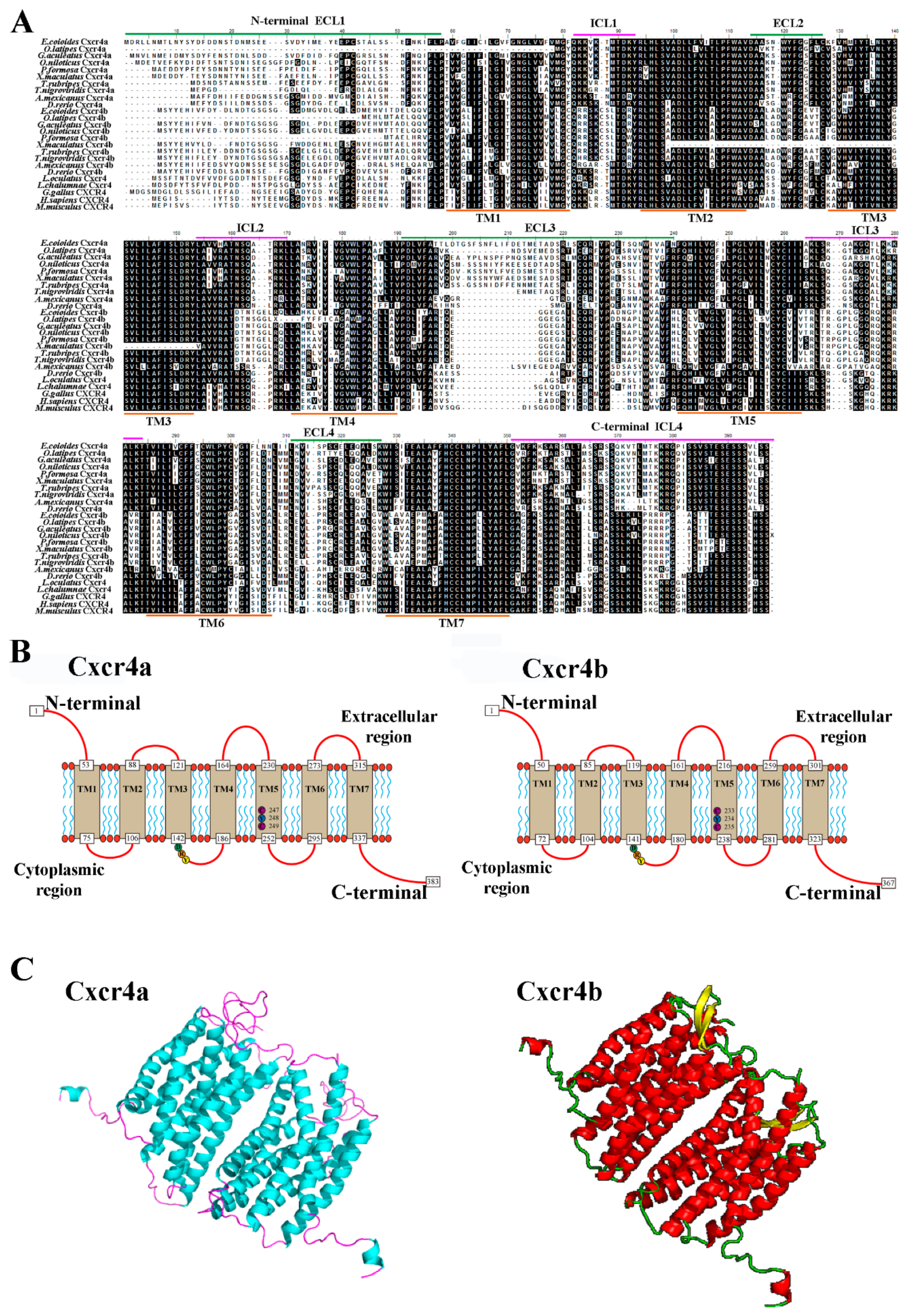
2.1. Molecular Characterization of Two cxcr4 Paralogs in Orange-Spotted Grouper

Two cxcr4 genes (EPI_005803 and EPI_021273) were cloned from orange-spotted grouper. The full-length cDNA of EPI_005803 is 1794 bp, consisting of a 116-bp 5’-terminal untranslated region (UTR), a 1152-bp open-reading frame (ORF), and a 526-bp 3’-UTR (Figure S1). The EPI_021273 cDNA is 1293 bp in length, and encodes 367 amino acids (Figure S2). To clarify the structural features of orange-spotted grouper cxcr4a/b, 23 cxcr4 genes from 14 species were downloaded from the Ensembl and NCBI databases, and we performed multiple sequence alignments with Ec_cxcr4a and Ec_cxcr4b (Figure 1A). Tetrapods, coelacanth (Latimeria chalumnae) and spotted gar (Lepisosteus oculatus) possess only one cxcr4 gene, while other teleosts have two cxcr4 genes. EPI_005803 and EPI_021273 share about 60% and 50% identities with homologs of tetrapod, coelacanth and spotted gar, respectively, so we named them as Ec_cxcr4a (EPI_005803) and Ec_cxcr4b (EPI_021273). The identity between Ec_Cxcr4a and Ec_cxcr4b is 51.1%. Both Ec_Cxcr4a and Ec_Cxcr4b consist of seven hydrophobic transmembrane (TM) domains, four extracellular loops (ECL) and four intracellular loops (ICL), and an extracellular N-terminus and an intracellular C-terminus (Figure 1A,B). By using the SWISS-MODEL, 3D protein-structure models of Ec_Cxcr4a and Ec_Cxcr4b were predicted (Figure 1C). Both Ec_Cxcr4a and Ec_Cxcr4b are homodimers and contain seven α-helices in each homolog that correspond to seven TM domains. Additionally, Ec_Cxcr4b possesses two β-sheets in each homolog.

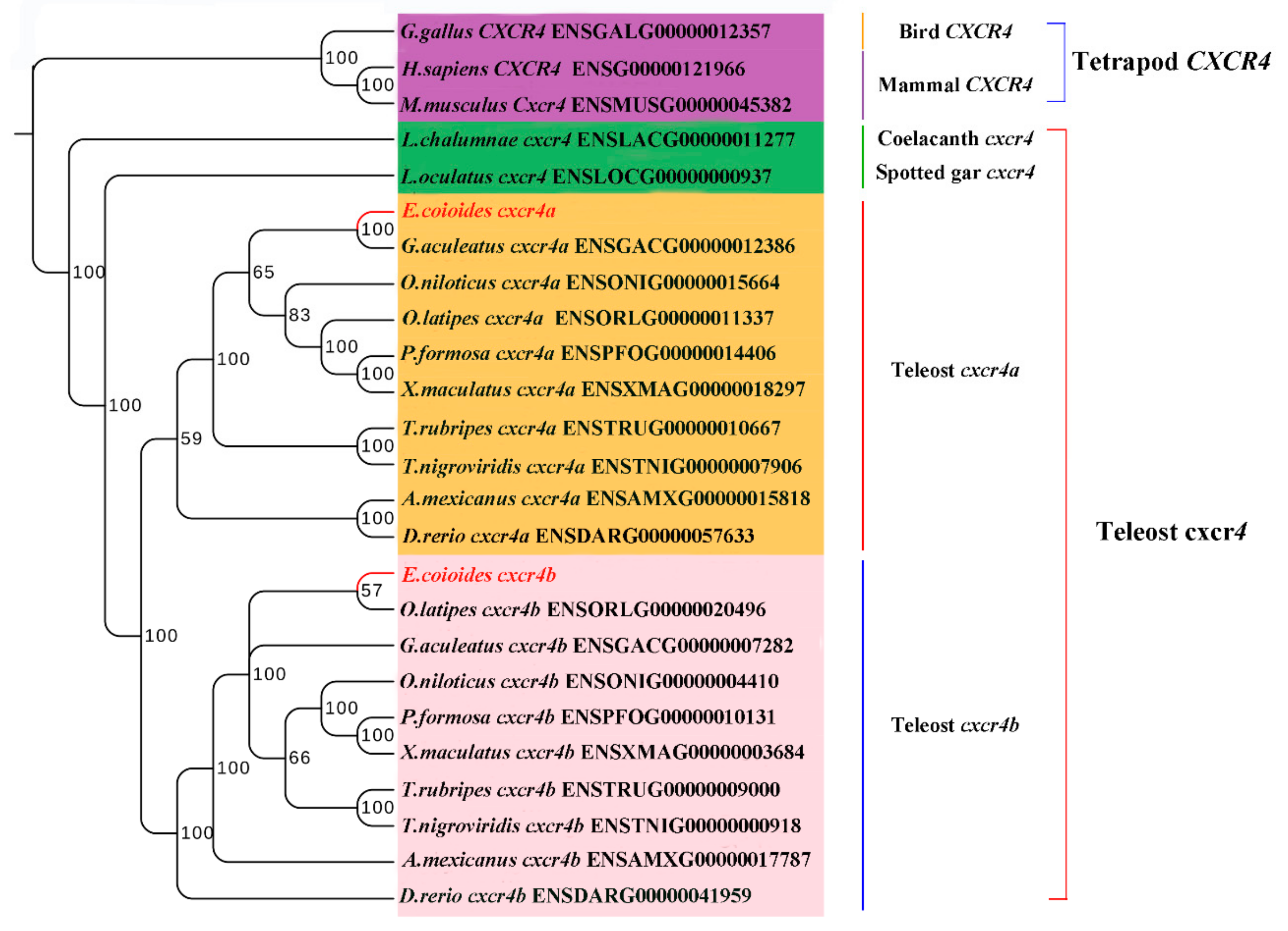
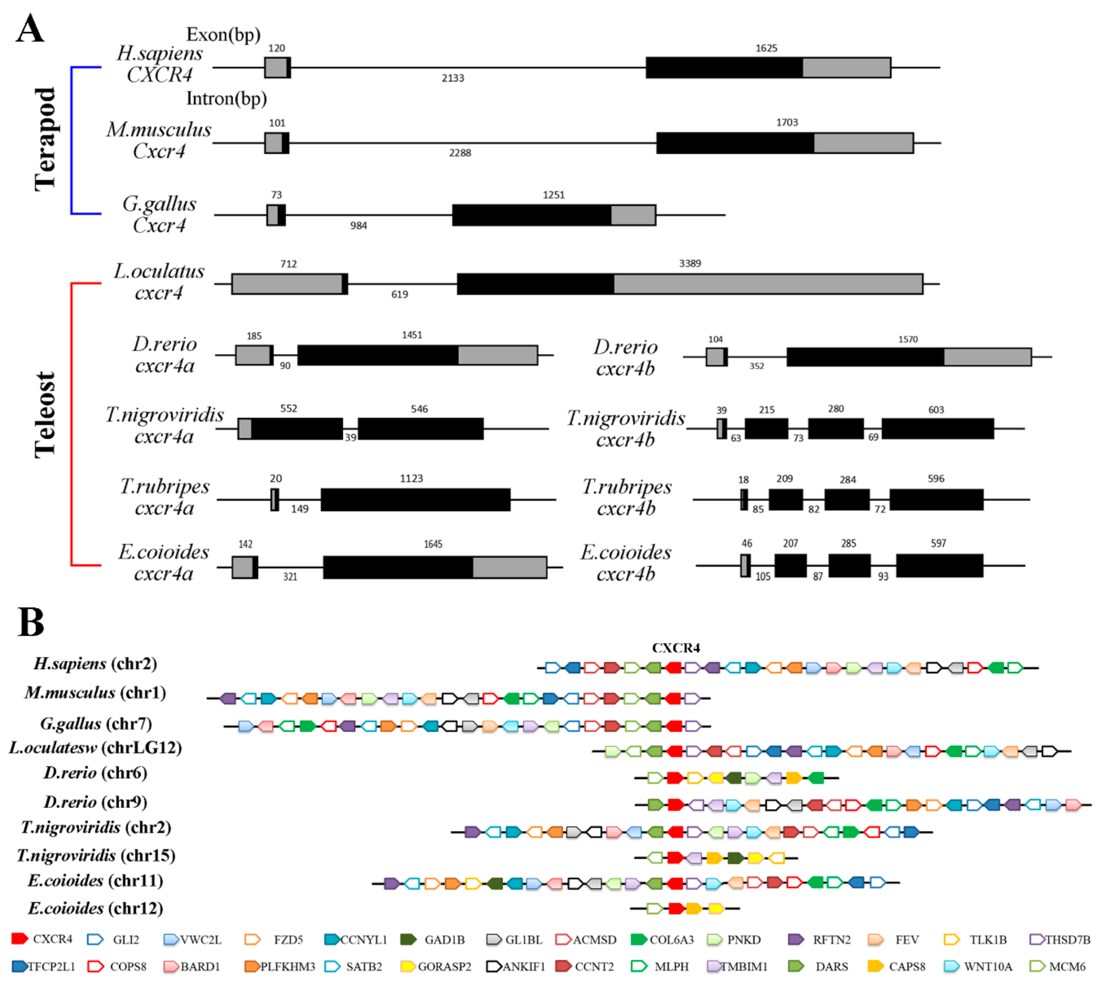
2.2. Phylogenetic Relationship, Gene Structure and Gene Synteny of Teleost and Tetrapod cxcr4s

To confirm the nomenclature and resolve the origin of the duplicated Ec_cxcr4a/b, a phylogenetic tree was constructed (Figure 2). Vertebrate Cxcr4s clustered largely consistent with the accepted species phylogeny and were divided into two clearly distinct clusters of the teleost and tetrapod Cxcr4. Ec_Cxcr4a and Ec_Cxcr4b are clustered into teleost Cxcr4a and teleost Cxcr4b branch, respectively. Teleost Cxcr4a and Cxcr4b branches are first grouped together and clustered with coelacanth and spotted gar Cxcr4, and then clustered with the tetrapod Cxcr4 branch.
Subsequently, the genomic sequences of around cxcr4 genes from seven species were downloaded and their gene structure and syntenic alignment were analyzed (Figure 3). Ec_cxcr4a has a bi-exonic structure and its ORF is interrupted by an intron, same as tetrapod cxcr4s [34], spotted gar cxcr4, and other teleost cxcr4as. Ec_cxcr4b is composed of four exons and three introns, consistent with other teleost cxcr4bs except zebrafish cxcr4b (Figure 3A). Ec_cxcr4a and Ec_cxcr4b are located on chromosome 11 and 12 of the orange-spotted grouper, respectively. Although identical genes were found in human (Homo sapiens) chromosome 2 (chr2), mouse (Mus musculus) chr1, chicken (Gallus gallus) chr7, and spotted gar chrLG12, the different locations of identical genes among these chromosomes showed that a large number of rearrangements occurred during evolution. However, a block of a gene cluster (MCM6-DARS-CXCR4-THSD7β) around cxcr4 loci presents conserved synteny in tetrapods and spotted gar (Figure 3B). Compared with the genes in tetrapod and spotted gar chromosomes, a complementary loss/retention pattern of genes exists between two teleost paralogous chromosomes. Conserved gene clusters (dars-cxcr4a-thsd7β) and (mcm6-cxcr4b) are found in two paralogous Tetraodon and orange-spotted grouper chromosomes, respectively (Figure 3B). Compared to the genes in spotted gar chrLG12, not only a large number of rearrangements, but also many gene inversions occurred in orange-spotted grouper chr11 and chr12.

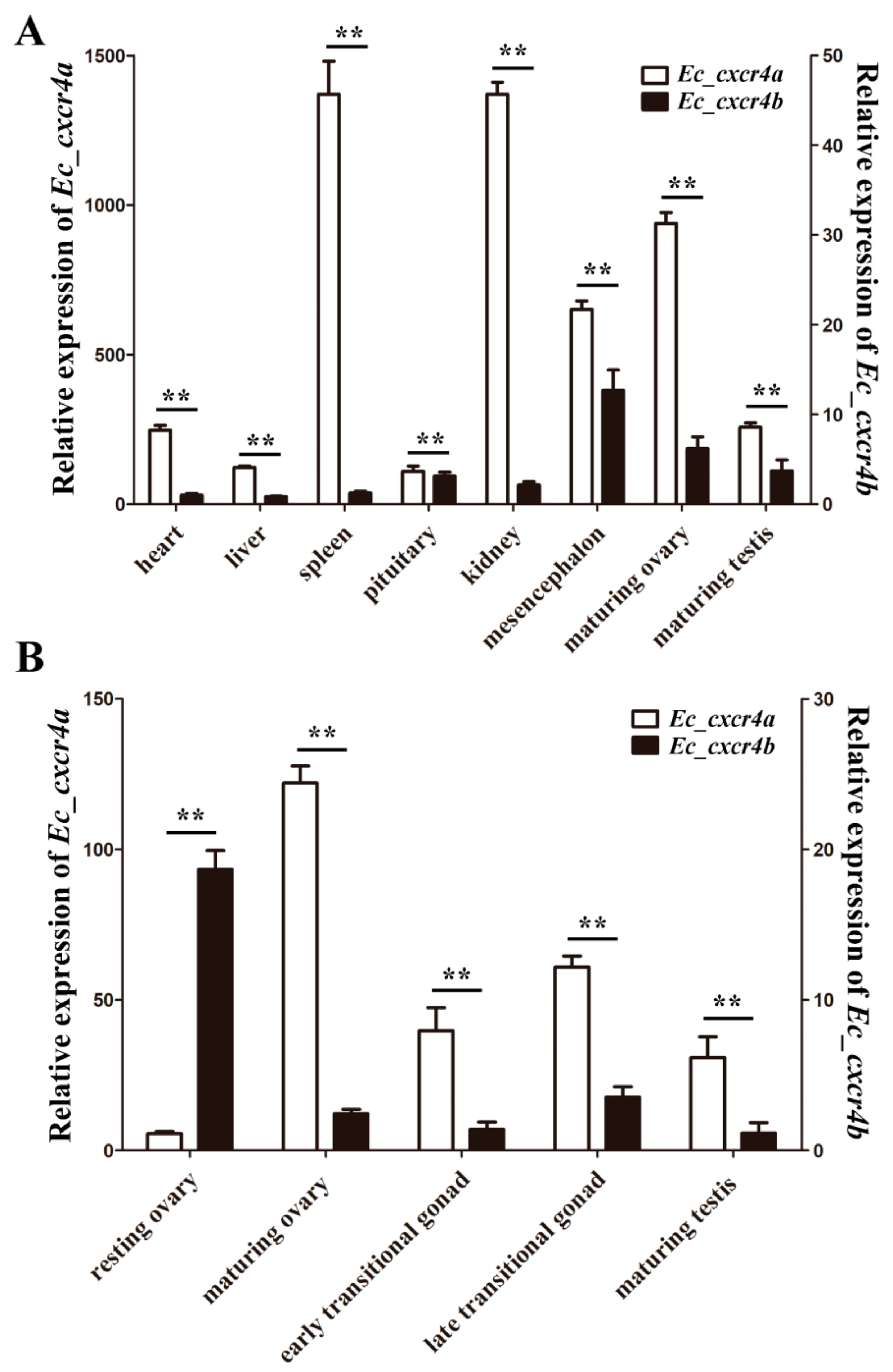
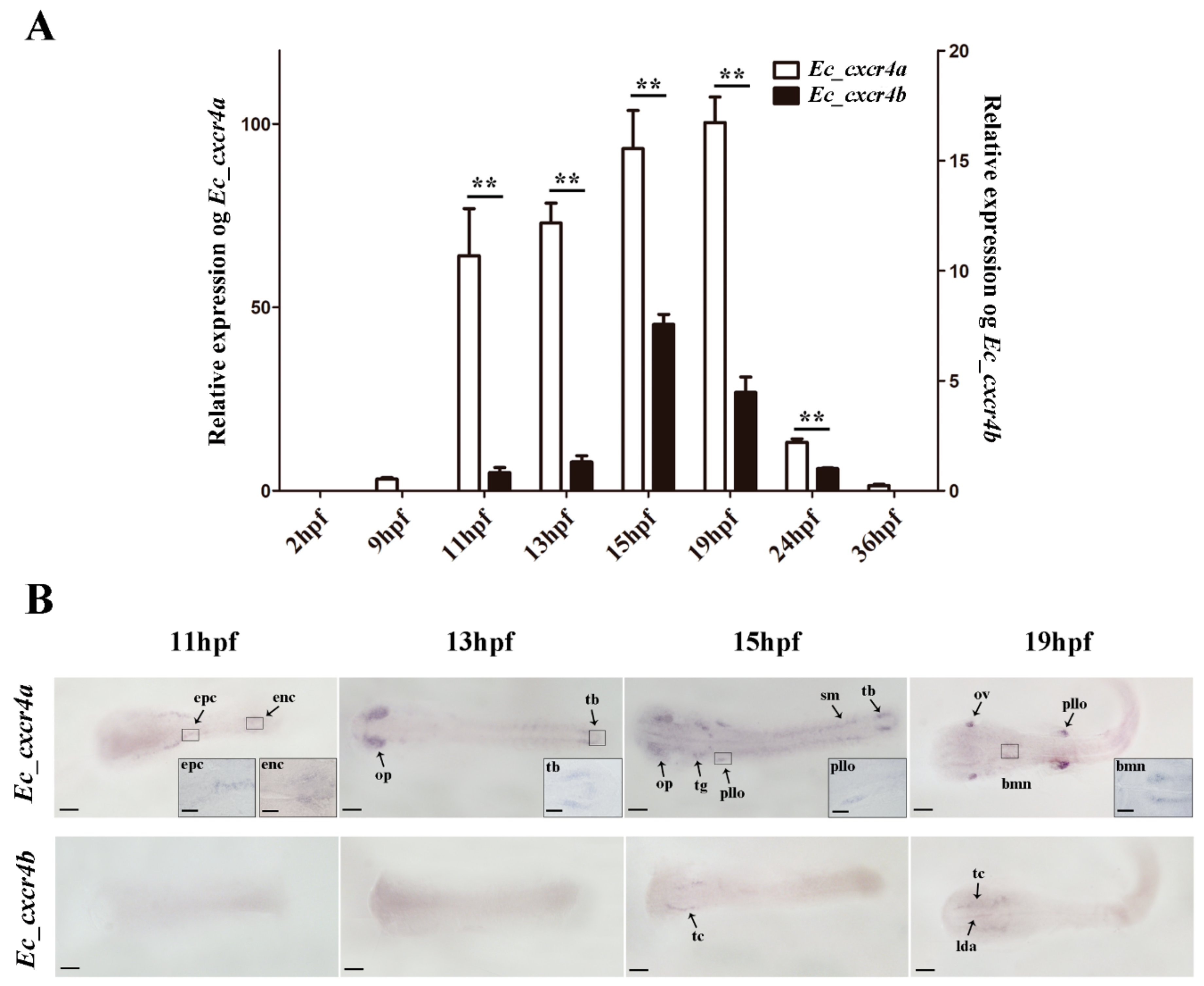
2.3. Biased and Differential Expression Patterns of Two cxcr4 Paralogs in Orange-Spotted Grouper

The distributions of Ec_cxcr4a and Ec_cxcr4b in adult orange-spotted grouper tissue were analyzed by quantitative RT-PCR (qPCR). Ec_cxcr4a and Ec_cxcr4b were detected in all eight examined tissues. The expression levels of Eccxcr4a in adult tissue are all remarkably higher (51–1093 folds) compared to Ec_cxcr4b (p < 0.01), indicating significantly biased expression of Ec_cxcr4a and Ec_cxcr4b in adult tissue. In addition, Ec_cxcr4b is most abundantly expressed in the mesencephalon, while Ec_cxcr4a is highly expressed in the spleen and kidney, suggesting Ec_cxcr4a might play an important role in immune response (Figure 4A).
Owing to differential expression of Eccxcr4a/b in maturing ovary and testis (Figure 4A), we also evaluated the expression of Ec_cxcr4a and Ec_cxcr4b in the gonads at five developmental stages (Figure 4B). A resting ovary contains a large number of primary-growth oocytes (PO) and previtellogenic oocytes (PVO). In maturing ovaries, many vitellogenic oocytes (VO), PVO, and PO fill the gonad. The degenerating primary oocytes (DPO), PO, spermatocytes (SPC), and spermatids (SPD) are mixed in lobules of early transitional gonads. Along with sex transition, many spermatogonia (SPG), SPC, and SPD arise, and only a few PO scatter in the lobules of late transitional gonads. In mature testis, male germ cells at all spermatogenic stages scatter within the cysts [45,46]. Ec_cxcr4b transcripts are highly expressed in the resting ovary, and sharply decrease by 86.8% in the maturing ovary. However, Ec_cxcr4a exhibit a distinctly differential expression pattern from Ec_cxcr4b. Ec_cxcr4a expression is relatively lower in the resting ovary, and sharply increase up to 22-folds in the maturing ovary. When the ovary begins to cause sex transition to the testis, Ec_cxcr4a expression decrease but still keeps a relative higher level (17–28 folds) in transitional gonads and maturing testis compared to those of Eccxcr4b (p < 0.01).
The dynamic expression changes of Ec_cxcr4a and Ec_cxcr4b during embryogenesis were analyzed by qPCR and whole-mount in situ hybridization (WISH) (Figure 5). Ec_cxcr4a was transcribed at 9 h post fertilization (hpf), sharply increasing expression up to 20-fold at 11 hpf, and then kept a high level until 24 hpf. Ec_cxcr4b expression was hardly detected until 11 hpf, gradually increased, reached a peak at 15 hpf, and then gradually decreased during 19–24 hpf. Ec_cxcr4a and Ec_cxcr4b also showed an apparently biased expression pattern during embryogenesis (Figure 5A). Ec_cxcr4a transcripts were localized in epithelial cells and endothelial cells at 11 hpf and were predominantly expressed in the olfactory placode at 13 hpf. In addition, positive signals were also observed in tail buds). At 15 hpf, Ec_cxcr4a transcripts were located in the primordium lateral line organ, trigeminal ganglion and somites in addition to the olfactory placode and tail bud. Along with the development progress of embryos, positive signals were concentrated in the otic vesicle, brachiomotor neurons and primordium lateral line organ (Figure 5B). Faint Ec_cxcr4b signals were observed in the neural plate at 13 hpf and concentrated in the telencephalon at 15 hpf. In addition, positive signals emerged in the telencephalon and lateral dorsal aorta at 19 hpf.

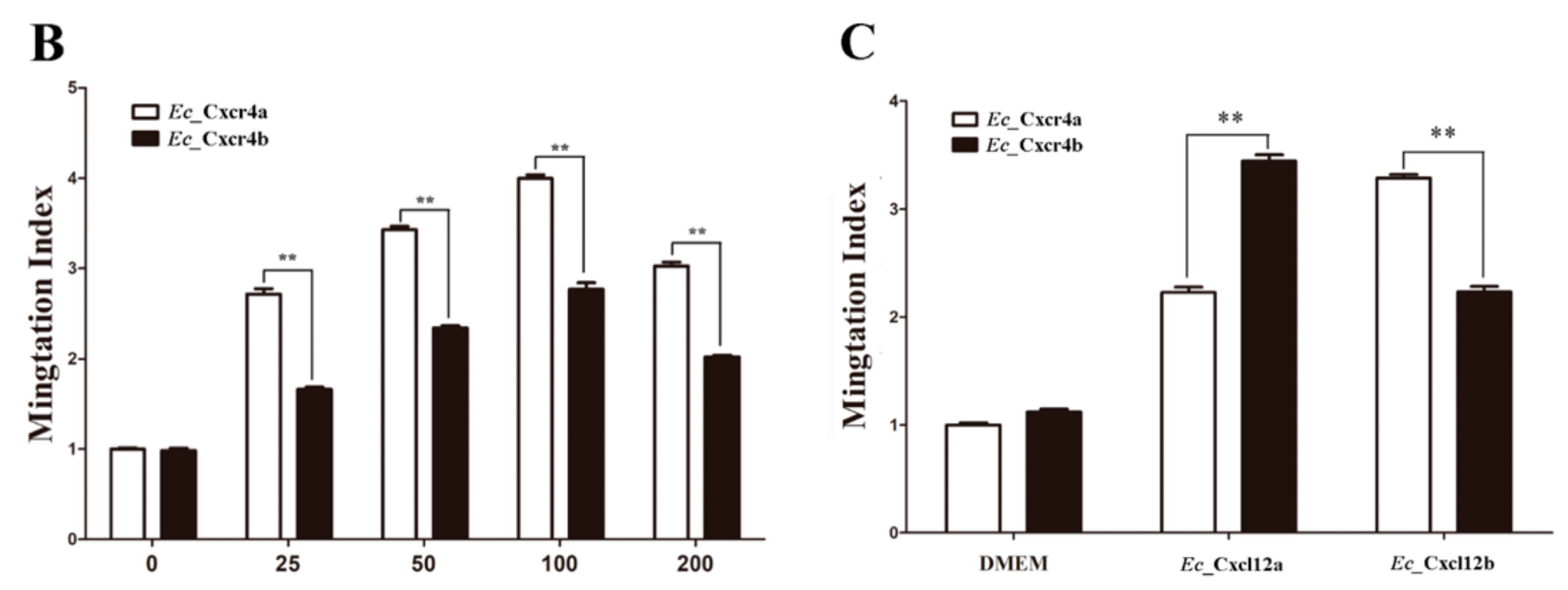
2.4. Differential Chemotactic Migratory Abilities of HEK293T Cells Transfected with Cxcr4a/b Induced by Cxcl12a/b

Cells that strongly express human CXCR4 can migrate towards CXCL12 (also known as SDF-1) in vitro [47]. To evaluate the difference of chemotactic migratory abilities between Ec_cxcr4a and Ec_cxcr4b, wild-type Ec_cxcr4a or Ec_cxcr4b (WTs) were linked to GFP (Ec_cxcr4a-GFP and Ec_cxcr4b-GFP) and transfected into HEK293T cells, respectively. Both Ec_Cxcr4a-GFP and Ec_Cxcr4b-GFP mainly localized in the cell membrane and some region of the internal membrane system (Figure 6A). A transwell migration assay was used to quantitatively evaluate the chemotactic migration ability of HEK293T cells overexpressing Ec_Cxcr4a-GFP (Ec_Cxcr4a-HEK293T) or Ec_Cxcr4b-GFP (Ec_Cxcr4b-HEK293T) in response to human SDF-1α in vitro. In comparison with serum-free DMEM, both of the average percentages of Ec_Cxcr4a-HEK293T and Ec_Cxcr4b-HEK293T that migrated toward human SDF-1α significantly increased in a dose-dependent manner (Figure 6B), indicating the chemotaxis of SDF-1 on Ec_Cxcr4a and Ec_Cxcr4b. The overexpression of Ec_Cxcr4a could make remarkably more HEK293T cells (1.4–1.6 fold) migrate to human SDF-1α than that induced by the overexpression of Ec_Cxcr4b (p < 0.01).
To test the differential chemotactic migratory abilities between EcCxcr4a and EcCxcr4b in response to their ligands, cxcl12 cDNAs were cloned from orange-spotted grouper by RACE PCR based on the conserved sequences among teleost cxcl12s. Two divergent cDNAs of cxcl12 were also identified (Figures S3–S6). Subsequently, Ec_Cxcl12a-HEK293T or Ec_Cxcl12b-HEK293T was placed in the lower chamber, and Ec_Cxcr4a-HEK293T and Ec_Cxcr4b-HEK293T were placed in the upper chamber. The migration ability of Ec_Cxcr4a-HEK293T toward Ec_Cxcl12b-HEK293T increased to 3.3-fold after 24 cultured hours, while that of Ec_Cxcr4a-HEK293T toward Ec_Cxcl12a-HEK293T rose up 2.2-fold (Figure 6C), indicating stronger chemotaxis of Ec_Cxcl12b on Ec_Cxcr4a (p < 0.01). Similarly, Ec_Cxcr4b showed higher chemotactic response to Ec_Cxcl12a.

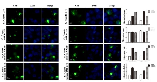
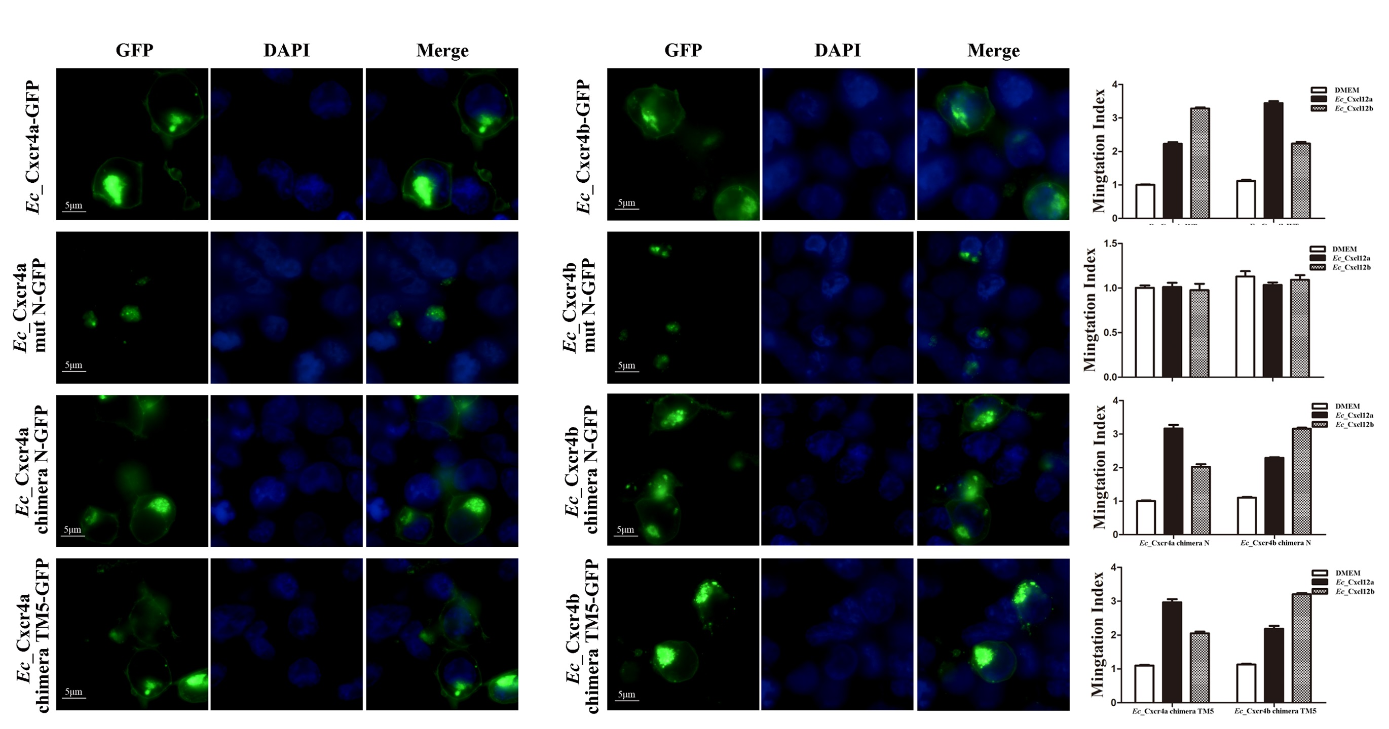
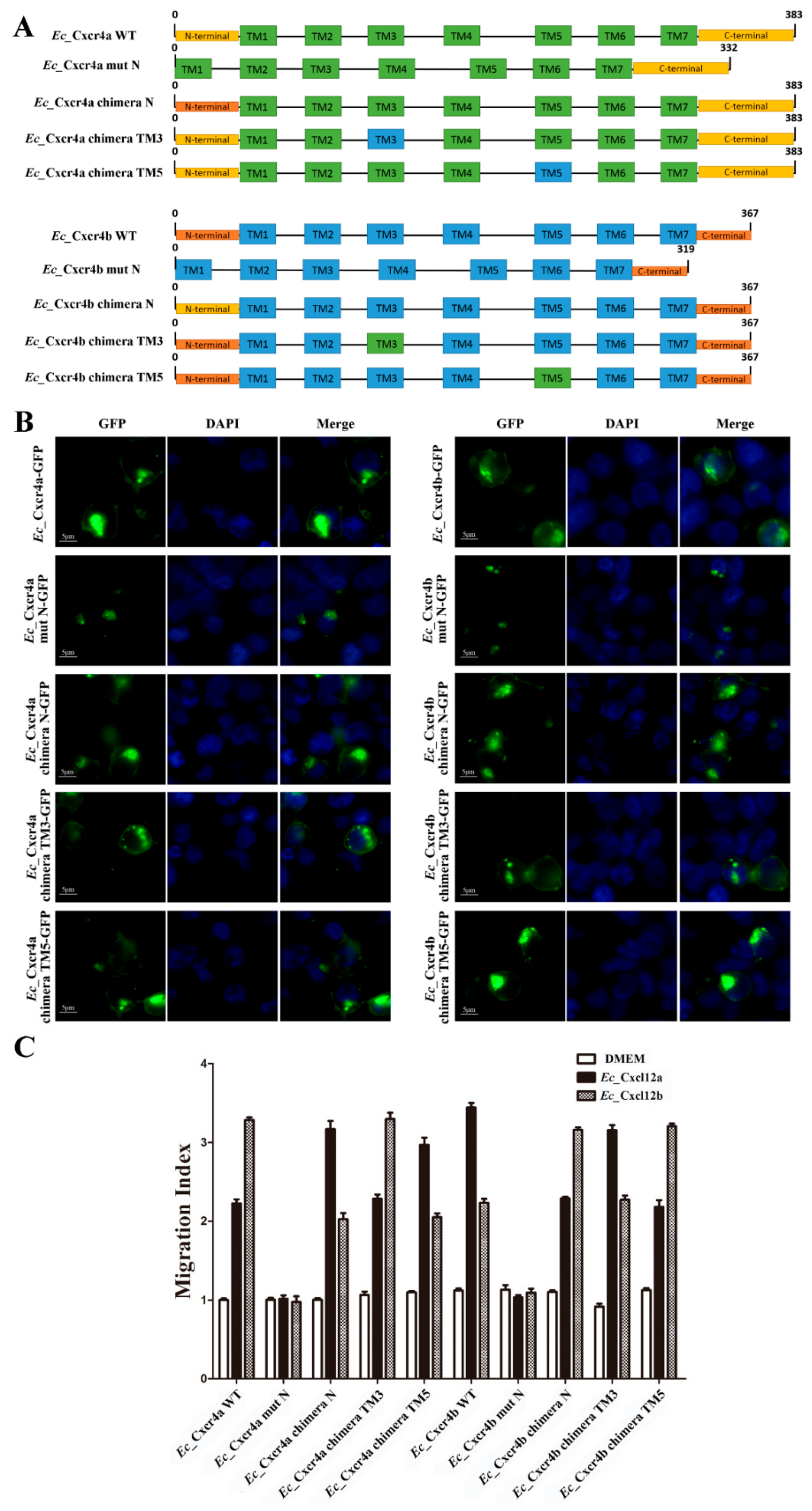
2.5. Specific Key Domains for Chemotactic Interaction of Ec_Cxcr4a/b-Ec_Cxcl12a/b

In mammals, the extracellular N-terminus, ICL2, and ECL3 have shown to be important to the CXCR4-CXCL12 interaction and activation of downstream pathways [48,49]. To reveal the specific key domains for the chemotactic interaction of Ec_Cxcr4b-Ec_Cxcl12a and Ec_Cxcr4a-Ec_Cxcl12b, respectively, we generated eight mutants by truncating the N-terminus of Ec_Cxcr4a and Ec_Cxcr4b, or by replacing the corresponding N-terminus, TM3 and TM5 of Ec_Cxcr4a and Ec_Cxcr4b (Figure 7A). Both Ec_Cxcr4a mut N-GFP and Ec_Cxcr4b mut N-GFP were mainly distributed in cytoplasm, not in cell membrane. The GFP signals of the six other mutants were still localized to the cell membrane and inner membrane, similar to WT Ec_Cxcr4a-GFP and Ec_Cxcr4b-GFP (Figure 7B). The results indicate that the N-terminus is crucial for the membrane localization of Ec_Cxcr4a/b.
Then, we assessed the chemotaxis abilities of these mutants in response to their ligand chemokines, Ec_Cxcl12a or Ec_Cxcl12b, in comparison with WTs. Ec_Cxcl12a-HEK293T or Ec_Cxcl12b-HEK293T were placed in the lower chamber and HEK293T cells expressing mutants were placed in the upper chamber. After 24 cultured hours, Ec_Cxcr4a mut N-GFP and Ec_Cxcr4b mut N-GFP lost their chemotaxis abilities. The chemotaxis abilities of the Ec_Cxcr4a chimera TM3-GFP and Ec_Cxcr4b chimera TM3-GFP to Ec_Cxcl12a-GFP and Ec_Cxcl12b-GFP were identical to the WTs, suggesting that the replacement of the TM3 domain did not influence the specific chemotactic interactions of Ec_Cxcr4b-Ec_Cxcl12a and Ec_Cxcr4a-Ec_Cxcl12b. In contrast, HEK293T cells expressing the Ec_Cxcr4a chimera N-GFP showed higher chemotaxis to Ec_Cxcl12a-GFP (3.2 fold) than that of Ec_Cxcr4a-GFP to Ec_Cxcl12a-GFP (2.0 fold), and lower chemotaxis to Ec_Cxcl12b-GFP (2.2 fold) than that of Ec_Cxcr4a-GFP to Ec_Cxcl12b-GFP (3.3 fold) (Figure 7C). The switch of chemotaxis effects was also observed in the Ec_Cxcr4b chimera N-GFP, Ec_Cxcr4a chimera TM5, and Ec_Cxcr4b chimera TM5.

3. Discussion

In addition to the two rounds of whole genome duplication (WGD) that occurred at the root of vertebrate lineage, the fish-specific genome duplication that occurred in a teleost ancestor gave dramatic rise and rapid radiation to teleost fish [50,51,52,53,54,55]. The phylogenetic tree showed that teleost Cxcr4 separated into two groups (Cxcr4a and Cxcr4b). As with tetrapod, coelacanth and spotted gar diverged from teleosts before Ts3R [51,56], and have only a single cxcr4 gene that falls outside the branches of teleost cxcr4s (Figure 2). The results suggest that the duplicated cxcr4a/b should have been produced through Ts3R, which is also supported by the duplication of neighboring genes surrounding cxcr4a/b (Figure 3B). Together with highly conserved gene synteny among homologous chromosomes containing teleost cxcr4a and tetrapod cxcr4 (Figure 3B), we supposed that the tetrapod cxcr4 cannot come from only one of the two teleost duplicated genes. The rapid rediploidization process was demonstrated to cause complicated and non-Mendelian genomic changes just following WGD events, including duplicated gene loss, additional gene duplication, gene inversion and large genomic reorganization [57,58,59,60,61,62]. The gene is ancestral. One of the duplicated genes kept more similar features with one of the ohnologous copies [62,63]. Through gene synteny analysis, we not only revealed a complementary gene loss/retention pattern, but also observed many gene inversions, genomic rearrangements and gene translocations in two teleost paralogous chromosomes (Figure 3B), indicating divergent evolution occurring in the radiation of teleost.
During rediploidization, pseudogenized genes lost function, or underwent sub-functionalization to partition ancestral gene function, or neo-functionalization to obtain a new function [57,58,60,63]. Two paralogs generally showed a dominant, biased or silencing expression pattern [64,65,66,67,68,69,70,71,72,73,74,75,76]. In this study, we revealed a significantly biased expression of Eccxcr4a and Ec_cxcr4b in adult tissue, gonads and embryos at different stages. The biased expression of two cxcr4 paralogs had been observed in zebrafish embryogenesis [25] and several Asian swamp eel adult tissue [27]. Teleost cxcr4s are constitutively expressed in adult tissue, and highly constitutive and up-regulated in immune tissue after pathogen infection [27,30,32,33,34,36,38,77,78], which indicates their role in immune surveillance and response. Ec_cxcr4a is highly expressed in analyzed tissues, while Ec_cxcr4b is mainly expressed in the mesencephalon (Figure 4A). Thus, it seems that Ec_cxcr4a acts the main role to execute Cxcr4 functions in orange-spotted grouper. Interestingly, we observed distinctly differential expression patterns between Ec_cxcr4a and Ec_cxcr4b during sex differentiation and sex reversal, indicating Ec_cxcr4a/b might play a role in gonad differentiation (Figure 4B). Cxcr4 has been proven to guide the migration of primordial germ cells (PGCs) [26,77,79,80,81]. However, the function of cxcr4 in gonad differentiation is still mostly unknown, owing to the severe defects and perinatal death of Cxcr4 knockout mice [82,83]. The function of Ec_cxcr4a/b in grouper sex differentiation awaits further investigation.
Previous studies have shown that zebrafish cxcr4s are essential for normal development, including the migration of PGCs [17,26,29] and endodermal cells [84], muscle formation [85], and sensory development and retinal growth [86,87]. The expression pattern of cxcr4a/b during embryogenesis has been finely described only in zebrafish [25]. Different from zebrafish, the transcripts of Ec_cxcr4a and Ec_cxcr4b were detected until the gastrula stage (9 hpf) and neurula stage (11 hpf), respectively (Figure 5A). It was supposed that two zebrafish paralogs partitioned ancestral functions, of which cxcr4a maintained most of murine Cxcr4 functions [88], while cxcr4b acquired a novel early function [25]. However, the distribution of Ec_cxcr4a in grouper embryos was more reminiscent of zebrafish cxcr4b. They are both expressed in structures associated with sensory organs or escape reactions (Figure 5B). In addition, Ec_cxcr4b and zebrafish cxcr4b were co-localized in the telencephalon. CXCL12-CXCR4 signaling is required for neuronal specification, migration, and outgrowth [88,89,90,91,92]. Therefore, the abundant expression of Ec_cxcr4a/b in the neural plate and sensory organs suggests that they might play a role in grouper neurogenesis.
Cxcr4 is conserved in most vertebrates. Both Ec_Cxcr4a and Ec_Cxcr4b possess conserved domains in chemokine receptors, containing an extracellular N-terminus, seven TMs, four ECLs, four ICLs, and an intracellular C-terminus (Figure 1). Previous studies have proposed a two-step/two-site binding model and demonstrated key elements for the human SDF-1-CXCR4 interaction [48,49,93,94,95]. SDF-1 binds the CXCR4 N-terminus, and subsequently interacts with the TM region to trigger receptor activation. Using Ec_Cxcr4a/b chimeras and mutants (Figure 7), we determined that the N-terminus is crucial for the membrane localization and ligand binding of Ec_Cxcr4a/b. The best-characterized teleost chemokine ligand/receptor is zebrafish Cxcl12-Cxcr4 [4]. The zebrafish Cxcl12a–Cxcr4b and Cxcl12b–Cxcr4a interactions have been proved and the aa N33 of zebrafish Cxcl12a is responsible for the specific recognition of zebrafish Cxcr4b [96]. In this study, we confirmed the stronger interactions of Ec_Cxcl12a–Ec_Cxcr4b and Ec_Cxcl12b–Ec_Cxcr4a (Figure 6). Moreover, we found that the N-terminus contributed to the specific recognition of Ec_Cxcr4a-Ec_Cxcl12b and Ec_Cxcr4b-Ec_Cxcl12a. TM3 forms a disulfide bond with ECL2, which stabilizes the seven-trans-membrane helix bundle of chemokine receptors [97], and has been proposed to interact with G-proteins [39]. CXCR4 forms dimers for proper physiological activity [98] and TM5 is supposed to maintain the dimer motif [99]. In this study, we demonstrated that TM5 is needed for specific recognition and TM3 is not involved in the Cxcr4-Cxcl12 interaction.
In conclusion, current studies represent an example of sub-functionalization of two teleost paralogs after Ts3R, which in this case is based on biased and divergent expression patterns of Ec_cxcr4a and Ec_cxcr4b, and specific ligand–receptor recognition of Ec_Cxcl12a–Ec_Cxcr4b and Ec_Cxcl12b–Ec_Cxcr4a.

4. Materials and Methods

4.1. Experimental Fish

Adult orange-spotted groupers were bought from markets in Wuhan, China. The samples of orange-spotted groupers embryos and juveniles were obtained from the Marine Fisheries Development Center of Guangdong Province, China. All experiments and animal treatments were approved by the Institute of Hydrobiology Institutional Animal Care and Use Committee (Approval ID: keshuizhuan 0829, 2012).

4.2. RNA Extraction and qPCR

Total RNAs were extracted from different types of tissues, including heart, liver, spleen, pituitary, kidney, telencephalon and gonads using an SV Total RNA Isolation System (Promaga Z3100, Madison, WI, USA) according to the manufacturer’s protocol. In addition, 50 embryos (n = 3) at different development stages, including 2 hpf (32-cell stage), 9 hpf (gastrula stage), 11 hpf (neurula stage), 13 hpf (bud stage), 15 hpf (heart and liver stage), 19 hpf (muscle differentiation), 24 hpf (hatching stage), and 36 hpf were collected to extract total RNAs. Total RNA (1 μg) was used to synthesize first-strand cDNAs in a 20 μL reaction volume following the protocol of GoScript™ Reverse Transcription System (Promega A5000, Madison, WI, USA).
qPCR was performed on a CFX96TM Real-Time PCR System (Bio-Rad) using an iTaqTM Universal SYBR Green Supermix (Bio-Rad, California, USA). The reactions, protocols, and selection of internal controls were performed as described previously [45,46]. Specificity of amplification for each reaction was analyzed by dissociation curves using CFX manager software (Bio-Rad) and sequencing. Gene-specific primers (Table S1) were designed with http://biotools.nubic.northwestern.edu/OligoCalc.html. All samples were analyzed in triplicate, and relative expression levels of target genes were calculated using the 2−ΔΔCT method. SPSS software (SPSS Inc.) was used for statistical analysis. A probability (p) of <0.05 was considered statistically significant.

4.3. Sequence and Phylogenetic Analyses

Orange-spotted grouper spleen SMART cDNA library was constructed according to the SMARTer PCR cDNA Synthesis Kit User Manual (Clontech 634,923). Ec_cxcr4a/b and Ec_cccl12a/b cDNAs were amplified by 3′ and 5′ RACE. The complete cDNA sequences of Ec_cxcr4a and Ec_cxcr4b were deposited in GenBank (accession numbers MH716017 and MH716018, respectively). The sequences of Ec_cccl12a and Ec_cxcl12b were obtained as Ec_cxcr4a and Ec_cxcr4b, and deposited in GenBank (accession numbers MH716019 and MH716020, respectively). Multiple amino acid sequence alignments were performed by Clustal W. Sequence identities were calculated using DNAStar 5.0. Phylogenetic analysis was conducted using Bayesian inference (BI) in MRBAYES 3.1.2 [100]. For BI tree, four independent Markov chain Monte Carlo (MCMC) chains were simultaneously run for 700,000 generations with a sample frequency of 1000 generations. The first 25% of the trees were discarded as burn-ins and the remaining tree samples were used to generate a consensus tree. Transmembrane-domain prediction was detected with SMART (http://smart.embl-heidelberg.de/) and TMHMM (http://www.cbs.dtu.dk/services/TMHMM/). Protein secondary structure predictions were made by SWISS-MODEL (https://swissmodel.expasy.org/).
The exon–intron structure was determined by the aligning mRNA and genomic sequences of Ec_cxcr4a and Ec_cxcr4b. To compare the genomic organization of other vertebrate cxcr4 sequences, exon–intron data were obtained using the Ensembl Genome browser (http://asia.ensembl.org/index.html). Syntenic analysis was also carried out using information extracted from the Ensembl (http://asia.ensembl.org/index.html) genome and the chromosomal regions around cxcr4s genes in H. sapiens chr2, M. musculus chr1, G. gallus chr7, L. oculatus LG12, D. rerio chr6 and chr9, Tetraodon. nigroviridis chr2 and chr15, and E. coioides chr11 and chr12.

4.4. Probe Synthesis and In Situ Hybridization

For antisense-probe synthesis, a T7 RNA polymerase promoter was added to the end of reverse primers and a DIG RNA Labeling Kit (Roche, Mannheim, Germany) was used according to protocol. In brief, a 558-bp cDNA fragment of Ec_cxcr4a and an 821-bp cDNA fragment of Ec_cxcr4b were amplified by specific primers (Table S1). For WISH, orange-spotted grouper embryos were collected at 11 hpf, 13 hpf, 15 hpf, and 19 hpf, fixed in 4% PFA at 4 °C overnight, and stored in 100% methanol at −20 °C. WISH was performed as previously described [101,102]. Digital images were taken on a Leica S8 APO microscope.

4.5. Plasmid Constructs

For subcellular localization and chemotaxis assay, the full-length ORF of Ec_Cxcr4a, Ec_Cxcr4b, Ec_Cxcl12a or Ec_Cxcl12b was sub-cloned into Hind III/Kpn I sites of pEGFP-N3 vector (BD Biosciences Clontech). The N-terminus- truncated mutations or the corresponding chimeras by replacing the N-terminus, TM3 and TM5 between Ec_Cxcr4a and Ec_Cxcr4b, were produced by overlap PCR. The mutants of Ec_Cxcr4a and Ec_Cxcr4b were also sub-cloned into the pEGFP-N3 vector. All the plasmids were verified by sequence analysis.

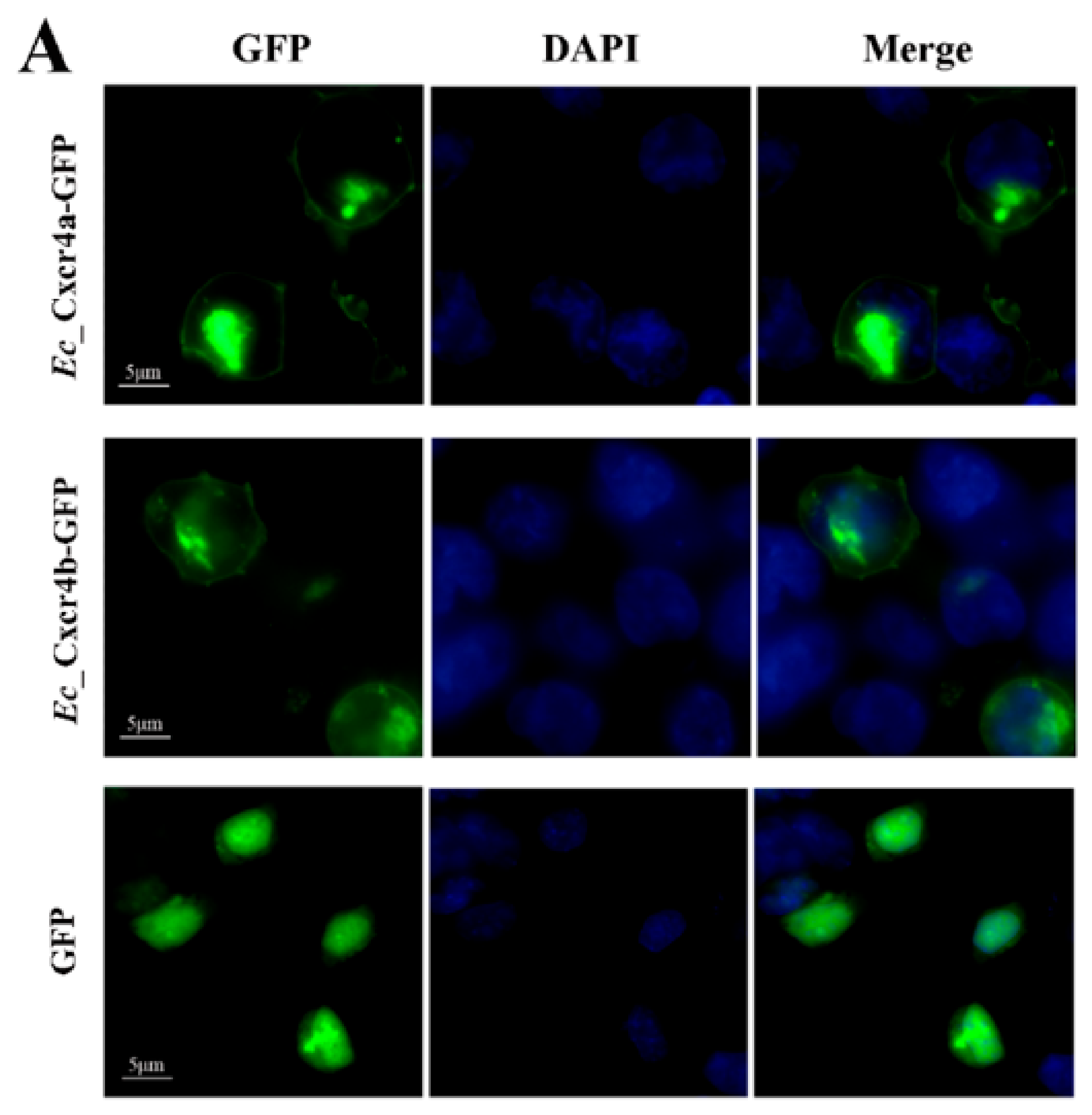
4.6. Subcellular Localization

HEK293T cells were grown overnight to 80% confluence on microscopy cover-glasses in 6-well plates, and then transfected with 1 μg corresponding plasmids. After 24 h post transfection, the transfected cells on the cover-glasses were fixed with 4% paraformaldehyde for 20 min at room temperature, washed, incubated with 0.2% Triton X-100 for 30 min, and finally stained with 4’, 6-diamidino-2-phenylindole (DAPI) (50 ng/mL) for 10 min, followed by an examination on a fluorescence microscope (Leica CTR6) according to previous reports [103]. The magnification of all images is 400×.

4.7. Chemotaxis Assay

HEK293T cells were seeded in 6 cm plates overnight, and transiently transfected with 1 μg corresponding expression plasmids using a FuGENE HD Transfection Reagent (Promega , Madison, WI, USA) and cultured for 24h.Transmigration assay of the HEK293T cells was done in a 24-well chemotaxis chamber (8.0 mm diameter; Corning Inc, Corning, USA). Cell density was adjusted to 1 × 10⁶/mL with DMEM out of bovine serum (Gibco, Melbourne, Australia).
For the transmigration assay, 300 μL adjusted Eccxcr4a/b-overexpression cells were placed in the upper chambers, and 500 μL recombinant human SDF-1α with different concentrations (0 ng/mL, 50 ng/mL, 100 ng/mL and 200 ng/mL) in serum-free DMEM media was placed in the lower chamber. The other groups were constructed by the following setting: (1–2) 300 μL Ec_Cxcr4a-HEK293T or Ec_Cxcr4b-HEK293T in the upper and 500 μL serum-free DMEM in the lower chamber; (3–4) 300 μL Ec_Cxcr4a-HEK293T or Ec_Cxcr4b-HEK293T in the upper and 500 μL Ec_Cxcl12a-HEK293T in the lower chamber; and (5–6) 300 μL Ec_Cxcr4a-HEK293T or Ec_Cxcr4b-HEK293T in upper and 500 μL Ec_Cxcl12b-HEK293T in lower chamber. The transwell plates were kept at 37 °C in 5% CO2 for 24 h, the polycarbonate membrane was put out, fixed with 4% paraformaldehyde for 15 min and Crystal violet (C0121, Beyotime, Wuhan, China) staining for 10 min, then observed and counted the HEK293T cells under the fluorescence Leica microscope. The chemotaxis experiments of chimeras and mutants were also constructed as described above.

Supplementary Materials

Supplementary Materials can be found at https://www.mdpi.com/1422-0067/19/10/2943/s1. Figure S1. Nucleotide sequence and deduced amino acid sequence of Eccxcr4a. Figure S2. Nucleotide sequence and deduced amino acid sequence of Eccxcr4b. Figure S3. Nucleotide sequence and deduced amino acid sequence of Eccxcl12a. Figure S4. Nucleotide sequence and deduced amino acid sequence of Eccxcl12b. Figure S5. Multiple amino acid sequence alignment of EcCxcl12a/b protein and other vertebrate Cxcl12 proteins. Figure S6. Phylogenetic tree of vertebrate Cxcl12. Table S1. Primers and adapters used in this study. Table S2. Protein sequences similarities among vertebrate Cxcr4s. Table S3. Protein sequences similarities among of vertebrate Cxcl12s.

Author Contributions

Y.W., L.Z. and W.-J.L. conceived and designed the experiments; W.-J.L., F.-X.G., Z.-H.S., and Z.L. performed the experiments; W.-J.L. and L.Z. analyzed the data; X.-C.L. and S.-S.L. contributed materials; and Y.W., L.Z. and J-F.G. wrote the paper.

Funding

This research was funded by the National Natural Science Foundation of China (Grant No. U1401213) and the Key Program of Frontier Sciences of the Chinese Academy of Sciences (Grant No. QYZDY-SWS-SMC025).

Acknowledgments

We thank Hai-Fa Zhang and the members of the Marine Fisheries Development Center of Guandong Province for helping with the artificial propagation of the orange-spotted grouper. This study was supported by the National Natural Science Foundation of China (Grant No. U1401213) and the Key Program of Frontier Sciences of the Chinese Academy of Sciences (Grant No. QYZDY-SWS-SMC025).

Conflicts of Interest

The authors declare no conflict of interest. The founding sponsors had no role in the design of the study; in the collection, analyses, or interpretation of data; in the writing of the manuscript, and in the decision to publish the results.

References

  1. Fredriksson, R.; Lagerstrom, M.C.; Lundin, L.G.; Schioth, H.B. The g-protein-coupled receptors in the human genome form five main families. Phylogenetic analysis, paralogon groups, and fingerprints. Mol. Pharmacol. 2003, 63, 1256–1272. [Google Scholar] [CrossRef] [PubMed]
  2. Viola, A.; Luster, A.D. Chemokines and their receptors: Drug targets in immunity and inflammation. Annu. Rev. Pharmacol. 2008, 48, 171–197. [Google Scholar] [CrossRef] [PubMed]
  3. Bajoghli, B. Evolution and function of chemokine receptors in the immune system of lower vertebrates. Eur. J. Immunol. 2013, 43, 1686–1692. [Google Scholar] [CrossRef] [PubMed] [Green Version]
  4. Bussmann, J.; Raz, E. Chemokine-guided cell migration and motility in zebrafish development. EMBO J. 2015, 34, 1309–1318. [Google Scholar] [CrossRef] [PubMed] [Green Version]
  5. Calderon, L.; Boehm, T. Three chemokine receptors cooperatively regulate homing of hematopoietic progenitors to the embryonic mouse thymus. Proc. Natl. Acad. Sci. USA 2011, 108, 7517–7522. [Google Scholar] [CrossRef] [PubMed] [Green Version]
  6. Griffith, J.W.; Sokol, C.L.; Luster, A.D. Chemokines and chemokine receptors: Positioning cells for host defense and immunity. Annu. Rev. Immunol. 2014, 32, 659–702. [Google Scholar] [CrossRef] [PubMed]
  7. Nagarsheth, N.; Wicha, M.S.; Zou, W.P. Chemokines in the cancer microenvironment and their relevance in cancer immunotherapy. Nat. Rev. Immunol. 2017, 17, 559–572. [Google Scholar] [CrossRef] [PubMed] [Green Version]
  8. Schall, T.J.; Proudfoot, A.E.I. Overcoming hurdles in developing successful drugs targeting chemokine receptors. Nat. Rev. Immunol. 2011, 11, 355–363. [Google Scholar] [CrossRef] [PubMed]
  9. Zlotnik, A.; Yoshie, O. The chemokine superfamily revisited. Immunity 2012, 36, 705–716. [Google Scholar] [CrossRef] [PubMed]
  10. Bajoghli, B.; Aghaallaei, N.; Hess, I.; Rode, I.; Netuschil, N.; Tay, B.H.; Venkatesh, B.; Yu, J.K.; Kaltenbach, S.L.; Holland, N.D.; et al. Evolution of genetic networks underlying the emergence of thymopoiesis in vertebrates. Cell 2009, 138, 186–197. [Google Scholar] [CrossRef] [PubMed]
  11. Huising, M.O.; Stolte, E.; Flik, G.; Savelkoul, H.F.J.; Verburg-van Kemenade, B.M.L. Cxc chemokines and leukocyte chemotaxis in common carp (Cyprinus carpio L.). Dev. Comp. Immunol. 2003, 27, 875–888. [Google Scholar] [CrossRef]
  12. Cencioni, C.; Capogrossi, M.C.; Napolitano, M. The sdf-1/cxcr4 axis in stem cell preconditioning. Cardiovasc. Res. 2012, 94, 400–407. [Google Scholar] [CrossRef] [PubMed]
  13. Liu, H.B.; Liu, S.B.; Li, Y.; Wang, X.H.; Xue, W.J.; Ge, G.Q.; Luo, X.H. The role of sdf-1-cxcr4/cxcr7 axis in the therapeutic effects of hypoxia-preconditioned mesenchymal stem cells for renal ischemia/reperfusion injury. PLoS ONE 2012. [Google Scholar] [CrossRef] [PubMed]
  14. Nagasawa, T.; Hirota, S.; Tachibana, K.; Takakura, N.; Nishikawa, S.; Kitamura, Y.; Yoshida, N.; Kikutani, H.; Kishimoto, T. Defects of b-cell lymphopoiesis and bone-marrow myelopoiesis in mice lacking the cxc chemokine pbsf/sdf-1. Nature 1996, 382, 635–638. [Google Scholar] [CrossRef] [PubMed]
  15. Tachibana, K.; Hirota, S.; Iizasa, H.; Yoshida, H.; Kawabata, K.; Kataoka, Y.; Kitamura, Y.; Matsushima, K.; Yoshida, N.; Nishikawa, S.; et al. The chemokine receptor cxcr4 is essential for vascularization of the gastrointestinal tract. Nature 1998, 393, 591–594. [Google Scholar] [CrossRef] [PubMed]
  16. Zou, Y.R.; Kottmann, A.H.; Kuroda, M.; Taniuchi, I.; Littman, D.R. Function of the chemokine receptor cxcr4 in haematopoiesis and in cerebellar development. Nature 1998, 393, 595–599. [Google Scholar] [CrossRef] [PubMed]
  17. Knaut, H.; Werz, C.; Geisler, R.; Nusslein-Volhard, C. A zebrafish homologue of the chemokine receptor cxcr4 is a germ-cell guidance receptor. Nature 2003, 421, 279–282. [Google Scholar] [CrossRef] [PubMed]
  18. Katsumoto, K.; Kume, S. The role of cxcl12-cxcr4 signaling pathway in pancreatic development. Theranostics 2013, 3, 11–17. [Google Scholar] [CrossRef] [PubMed]
  19. Alejo, A.; Tafalla, C. Chemokines in teleost fish species. Dev. Comp. Immunol. 2011, 35, 1215–1222. [Google Scholar] [CrossRef] [PubMed]
  20. Bird, S.; Tafalla, C. Teleost chemokines and their receptors. Biology 2015, 4, 756–784. [Google Scholar] [CrossRef] [PubMed] [Green Version]
  21. Peatman, E.; Liu, Z.J. Evolution of cc chemokines in teleost fish: A case study in gene duplication and implications for immune diversity. Immunogenetics 2007, 59, 613–623. [Google Scholar] [CrossRef] [PubMed]
  22. Zou, W.P.; Max, S.W. Chemokines and cellular plasticity of ovarian cancer stem cells. Oncoscience 2015. [Google Scholar] [CrossRef] [PubMed]
  23. Amores, A.; Force, A.; Yan, Y.L.; Joly, L.; Amemiya, C.; Fritz, A.; Ho, R.K.; Langeland, J.; Prince, V.; Wang, Y.L.; et al. Zebrafish hox clusters and vertebrate genome evolution. Science 1998, 282, 1711–1714. [Google Scholar] [CrossRef] [PubMed]
  24. Postlethwait, J.H.; Yan, Y.L.; Gates, M.A.; Horne, S.; Amores, A.; Brownlie, A.; Donovan, A.; Egan, E.S.; Force, A.; Gong, Z.Y.; et al. Vertebrate genome evolution and the zebrafish gene map. Nat. Genet. 1998, 18, 345–349. [Google Scholar] [CrossRef] [PubMed]
  25. Chong, S.W.; Emelyanov, A.; Gong, Z.Y.; Korzh, V. Expression pattern of two zebrafish genes, cxcr4a and cxcr4b. Mech. Dev. 2001, 109, 347–354. [Google Scholar] [CrossRef]
  26. Doitsidou, M.; Reichman-Fried, M.; Stebler, J.; Koprunner, M.; Dorries, J.; Meyer, D.; Esguerra, C.V.; Leung, T.; Raz, E. Guidance of primordial germ cell migration by the chemokine sdf-1. Cell 2002, 111, 647–659. [Google Scholar] [CrossRef]
  27. Gao, W.H.; Fang, L.; Yang, D.Q.; Ai, K.T.; Luo, K.; Tian, G.M.; Zhou, J.W.; Hu, W.; Yuan, H.W.; Xu, Q.Q. Cloning and expression of Asian swamp eel (Monopterus albus) cxcr4 paralogues, and their modulation by pathogen infection. Aquaculture 2016, 457, 50–60. [Google Scholar] [CrossRef]
  28. Fu, Q.; Yang, Y.J.; Li, C.; Zeng, Q.F.; Zhou, T.; Li, N.; Liu, Y.; Liu, S.K.; Liu, Z.J. The cc and cxc chemokine receptors in channel catfish (Ictalurus punctatus) and their involvement in disease and hypoxia responses. Dev. Comp. Immunol. 2017, 77, 241–251. [Google Scholar] [CrossRef] [PubMed]
  29. Kurokawa, H.; Saito, D.; Nakamura, S.; Katoh-Fukui, Y.; Ohta, K.; Baba, T.; Morohashi, K.I.; Tanaka, M. Germ cells are essential for sexual dimorphism in the medaka gonad. Proc. Natl. Acad. Sci. USA 2007, 104, 16958–16963. [Google Scholar] [CrossRef] [PubMed] [Green Version]
  30. Jia, A.R.; Zhang, X.H. Molecular cloning, characterization, and expression analysis of the cxcr4 gene from turbot: Scophthalmus maximus. J. Biomed. Biotechnol. 2009, 2009. [Google Scholar] [CrossRef] [PubMed]
  31. Lin, C.Y.; Chen, Y.M.; Hsu, H.H.; Shiu, C.T.; Kuo, H.C.; Chen, T.Y. Grouper (Epinephelus coioides) cxcr4 is expressed in response to pathogens infection and early stage of development. Dev. Comp. Immunol. 2012, 36, 112–120. [Google Scholar] [CrossRef] [PubMed]
  32. Liu, X.X.; Kang, L.S.; Liu, W.; Lou, B.; Wu, C.W.; Jiang, L.H. Molecular characterization and expression analysis of the large yellow croaker (Larimichthys crocea) chemokine receptors cxcr2, cxcr3, and cxcr4 after bacterial and poly I:C challenge. Fish Shellfish Immun. 2017, 70, 228–239. [Google Scholar] [CrossRef] [PubMed]
  33. Priyathilaka, T.T.; Oh, M.; Bathige, S.D.N.K.; De Zoysa, M.; Lee, J. Two distinct cxc chemokine receptors (cxcr3 and cxcr4) from the big-belly seahorse hippocampus abdominalis: Molecular perspectives and immune defensive role upon pathogenic stress. Fish Shellfish Immun. 2017, 65, 59–70. [Google Scholar] [CrossRef] [PubMed]
  34. Thulasitha, W.S.; Umasuthan, N.; Revathy, K.S.; Whang, I.; Lee, J. Molecular characterization, genomic structure and expressional profiles of a cxc chemokine receptor 4 (cxcr4) from rock bream oplegnathus fasciatus. Fish Shellfish Immun. 2015, 44, 471–477. [Google Scholar] [CrossRef] [PubMed]
  35. Wang, N.; Luo, H.J.; Yin, G.B.; Dong, C.R.; Xu, M.; Chen, G.G.; Liu, Z.M. Overexpression of hif-2 alpha, twist, and cxcr4 is associated with lymph node metastasis in papillary thyroid carcinoma. Clin. Dev. Immunol. 2013, 2013. [Google Scholar] [CrossRef] [PubMed]
  36. Xu, T.J.; Zhu, Z.H.; Sun, Y.N.; Ren, L.P.; Wang, R.X. Characterization and expression of the cxcr1 and cxcr4 in miiuy croaker and evolutionary analysis shows the strong positive selection pressures imposed in mammal cxcr1. Dev. Comp. Immunol. 2014, 44, 133–144. [Google Scholar] [CrossRef] [PubMed]
  37. Yang, J.Y.; Zhang, B.; Lin, Y.; Yang, Y.; Liu, X.Q.; Lu, F. Breast cancer metastasis suppressor 1 inhibits sdf-1 alpha-induced migration of non-small cell lung cancer by decreasing cxcr4 expression. Cancer Lett. 2008, 269, 46–56. [Google Scholar] [CrossRef] [PubMed]
  38. Zhang, J.; Wei, X.L.; Chen, L.P.; Chen, N.; Li, Y.H.; Wang, W.M.; Wang, H.L. Sequence analysis and expression differentiation of chemokine receptor cxcr4b among three populations of megalobrama amblycephala. Dev. Comp. Immunol. 2013, 40, 195–201. [Google Scholar] [CrossRef] [PubMed]
  39. Alabyev, B.Y.; Najakshin, A.M.; Mechetina, L.V.; Taranin, A.V. Cloning of a cxcr4 homolog in chondrostean fish and characterization of the cxcr4-specific structural features. Dev. Comp. Immunol. 2000, 24, 765–770. [Google Scholar] [CrossRef]
  40. Ko, C.F.; Chiou, T.T.; Chen, T.T.; Wu, J.L.; Chen, J.C.; Lu, J.K. Molecular cloning of myostatin gene and characterization of tissue-specific and developmental stage-specific expression of the gene in orange spotted grouper, epinephelus coioides. Mar. Biotechnol. 2007, 9, 20–32. [Google Scholar] [CrossRef] [PubMed]
  41. Lo, W.Y.; Chang, C.F.; Song, Y.L. Evaluation of dorsal aorta cannulation for immunological studies of grouper (Epinephelus malabaricus). Fish Shellfish Immun. 2003, 14, 289–303. [Google Scholar] [CrossRef]
  42. Yao, B.; Zhou, L.; Wang, Y.; Xia, W.; Gui, J.F. Differential expression and dynamic changes of sox3 during gametogenesis and sex reversal in protogynous hermaphroditic fish. J. Exp. Zool. Part A 2007, 307, 207–219. [Google Scholar] [CrossRef] [PubMed]
  43. Yeh, S.L.; Dai, Q.C.; Chu, Y.T.; Kuo, C.M.; Ting, Y.Y.; Chang, C.F. Induced sex change, spawning and larviculture of potato grouper, epinephelus tukula. Aquaculture 2003, 228, 371–381. [Google Scholar] [CrossRef]
  44. Zhou, L.; Gui, J.F. Molecular mechanisms underlying sex change in hermaphroditic groupers. Fish Physiol. Biochem. 2010, 36, 181–193. [Google Scholar] [CrossRef] [PubMed]
  45. Sun, Z.H.; Zhou, L.; Li, Z.; Liu, X.C.; Li, S.S.; Wang, Y.; Gui, J.F. Sexual dimorphic expression of dnd in germ cells during sex reversal and its requirement for primordial germ cell survival in protogynous hermaphroditic grouper. Comp. Biochem. Phys. B 2017, 208, 47–57. [Google Scholar] [CrossRef] [PubMed]
  46. Sun, Z.H.; Wang, Y.; Lu, W.J.; Li, Z.; Liu, X.C.; Li, S.S.; Zhou, L.; Gui, J.F. Divergent expression patterns and function implications of four nanos genes in a hermaphroditic fish, epinephelus coioides. Int. J. Mol. Sci. 2017, 18, 685. [Google Scholar] [CrossRef] [PubMed]
  47. Son, B.R.; Marquez-Curtis, L.A.; Kucia, M.; Wysoczynski, M.; Turner, A.R.; Ratajczak, J.; Ratajczak, M.Z.; Janowska-Wieczorek, A. Migration of bone marrow and cord blood mesenchymal stem cells in vitro is regulated by stromal-derived factor-1-cxcr4 and hepatocyte growth factor-c-met axes and involves matrix metalloproteinases. Stem Cells 2006, 24, 1254–1264. [Google Scholar] [CrossRef] [PubMed]
  48. Doranz, B.J.; Orsini, M.J.; Turner, J.D.; Hoffman, T.L.; Berson, J.F.; Hoxie, J.A.; Peiper, S.C.; Brass, L.F.; Doms, R.W. Identification of cxcr4 domains that support coreceptor and chemokine receptor functions. J. Virol. 1999, 73, 2752–2761. [Google Scholar] [PubMed]
  49. Roland, J.; Murphy, B.J.; Ahr, B.; Robert-Hebmann, W.; Delauzun, V.; Nye, K.E.; Devaux, C.; Biard-Piechaczyk, M. Role of the intracellular domains of cxcr4 in sdf-1-mediated signaling. Blood 2003, 101, 399–406. [Google Scholar] [CrossRef] [PubMed]
  50. Amores, A.; Catchen, J.; Ferrara, A.; Fontenot, Q.; Postlethwait, J.H. Genome evolution and meiotic maps by massively parallel DNA sequencing: Spotted gar, an outgroup for the teleost genome duplication. Genetics 2011, 188, 799–808. [Google Scholar] [CrossRef] [PubMed]
  51. Braasch, I.; Gehrke, A.R.; Smith, J.J.; Kawasaki, K.; Manousaki, T.; Pasquier, J.; Amores, A.; Desvignes, T.; Batzel, P.; Catchen, J.; et al. The spotted gar genome illuminates vertebrate evolution and facilitates human-teleost comparisons. Nat. Genet. 2016, 48, 427–437. [Google Scholar] [CrossRef] [PubMed] [Green Version]
  52. Jaillon, O.; Aury, J.M.; Brunet, F.; Petit, J.L.; Stange-Thomann, N.; Mauceli, E.; Bouneau, L.; Fischer, C.; Ozouf-Costaz, C.; Bernot, A.; et al. Genome duplication in the teleost fish tetraodon nigroviridis reveals the early vertebrate proto-karyotype. Nature 2004, 431, 946–957. [Google Scholar] [CrossRef] [PubMed]
  53. Kasahara, M.; Naruse, K.; Sasaki, S.; Nakatani, Y.; Qu, W.; Ahsan, B.; Yamada, T.; Nagayasu, Y.; Doi, K.; Kasai, Y.; et al. The medaka draft genome and insights into vertebrate genome evolution. Nature 2007, 447, 714–719. [Google Scholar] [CrossRef] [PubMed] [Green Version]
  54. Meyer, A.; Van de Peer, Y. From 2r to 3r: Evidence for a fish-specific genome duplication (fsgd). In BioEssays: News and Reviews in Molecular, Cellular and Developmental Biology; Wiley: Hoboken, NJ, USA, 2005; Volume 27, pp. 937–945. [Google Scholar]
  55. Zhou, L.; Gui, J. Natural and artificial polyploids in aquaculture. Aquac. Fish. 2017, 2, 103–111. [Google Scholar] [CrossRef]
  56. Amemiya, C.T.; Alfoldi, J.; Lee, A.P.; Fan, S.H.; Philippe, H.; MacCallum, I.; Braasch, I.; Manousaki, T.; Schneider, I.; Rohner, N.; et al. The african coelacanth genome provides insights into tetrapod evolution. Nature 2013, 496, 311–316. [Google Scholar] [CrossRef] [PubMed] [Green Version]
  57. Wapinski, I.; Pfeffer, A.; Friedman, N.; Regev, A. Natural history and evolutionary principles of gene duplication in fungi. Nature 2007, 449, 54–61. [Google Scholar] [CrossRef] [PubMed]
  58. Scannell, D.R.; Byrne, K.P.; Gordon, J.L.; Wong, S.; Wolfe, K.H. Multiple rounds of speciation associated with reciprocal gene loss in polyploid yeasts. Nature 2006, 440, 341–345. [Google Scholar] [CrossRef] [PubMed] [Green Version]
  59. Markov, A.V.; Kaznacheev, I.S. Evolutionary consequences of polyploidy in prokaryotes and the origin of mitosis and meiosis. Biol. Direct 2016. [Google Scholar] [CrossRef] [PubMed]
  60. Otto, S.P. The evolutionary consequences of polyploidy. Cell 2007, 131, 452–462. [Google Scholar] [CrossRef] [PubMed]
  61. Lien, S.; Koop, B.F.; Sandve, S.R.; Miller, J.R.; Kent, M.P.; Nome, T.; Hvidsten, T.R.; Leong, J.S.; Minkley, D.R.; Zimin, A.; et al. The atlantic salmon genome provides insights into rediploidization. Nature 2016, 533, 200–205. [Google Scholar] [CrossRef] [PubMed]
  62. Xiong, J.; Lu, X.Y.; Lu, Y.M.; Zeng, H.H.; Yuan, D.X.; Feng, L.F.; Chang, Y.; Bowen, J.; Gorovsky, M.; Fu, C.J.; et al. Tetrahymena gene expression database (tged): A resource of microarray data and co-expression analyses for tetrahymena. Sci. China Life Sci. 2011, 54, 65–67. [Google Scholar] [CrossRef] [PubMed]
  63. Comai, L. The advantages and disadvantages of being polyploid. Nat. Rev. Genet. 2005, 6, 836–846. [Google Scholar] [CrossRef] [PubMed]
  64. Buggs, R.J.A.; Chamala, S.; Wu, W.; Tate, J.A.; Schnable, P.S.; Soltis, D.E.; Soltis, P.S.; Barbazuk, W.B. Rapid, repeated, and clustered loss of duplicate genes in allopolyploid plant populations of independent origin. Curr. Biol. 2012, 22, 248–252. [Google Scholar] [CrossRef] [PubMed]
  65. Doyle, J.J.; Flagel, L.E.; Paterson, A.H.; Rapp, R.A.; Soltis, D.E.; Soltis, P.S.; Wendel, J.F. Evolutionary genetics of genome merger and doubling in plants. Annu. Rev. Genet. 2008, 42, 443–461. [Google Scholar] [CrossRef] [PubMed]
  66. Grover, C.E.; Gallagher, J.P.; Szadkowski, E.P.; Yoo, M.J.; Flagel, L.E.; Wendel, J.F. Homoeolog expression bias and expression level dominance in allopolyploids. New Phytol. 2012, 196, 966–971. [Google Scholar] [CrossRef] [PubMed] [Green Version]
  67. Hu, G.J.; Koh, J.; Yoo, M.J.; Chen, S.X.; Wendel, J.F. Gene-expression novelty in allopolyploid cotton: A proteomic perspective. Genetics 2015, 200, 91–104. [Google Scholar] [CrossRef] [PubMed]
  68. Jackson, S.; Chen, Z.J. Genomic and expression plasticity of polyploidy. Curr. Opin. Plant Biol. 2010, 13, 153–159. [Google Scholar] [CrossRef] [PubMed] [Green Version]
  69. Koh, J.; Chen, S.X.; Zhu, N.; Yu, F.H.; Soltis, P.S.; Soltis, D.E. Comparative proteomics of the recently and recurrently formed natural allopolyploid Tragopogon mirus (asteraceae) and its parents. New Phytol. 2012, 196, 292–305. [Google Scholar] [CrossRef] [PubMed]
  70. Lashermes, P.; Hueber, Y.; Combes, M.C.; Severac, D.; Dereeper, A. Inter-genomic DNA exchanges and homeologous gene silencing shaped the nascent allopolyploid coffee genome (Coffea arabica L.). G3-Genes Genom. Genet. 2016, 6, 2937–2948. [Google Scholar] [CrossRef] [PubMed]
  71. Page, J.T.; Liechty, Z.S.; Alexander, R.H.; Clemons, K.; Hulse-Kemp, A.M.; Ashrafi, H.; Van Deynze, A.; Stelly, D.M.; Udall, J.A. DNA sequence evolution and rare homoeologous conversion in tetraploid cotton. PLoS Genet. 2016. [Google Scholar] [CrossRef]
  72. Pala, I.; Coelho, M.M.; Schartl, M. Dosage compensation by gene-copy silencing in a triploid hybrid fish. Curr. Biol. 2008, 18, 1344–1348. [Google Scholar] [CrossRef] [PubMed]
  73. Salmon, A.; Flagel, L.; Ying, B.; Udall, J.A.; Wendel, J.F. Homoeologous nonreciprocal recombination in polyploid cotton. New Phytol. 2010, 186, 123–134. [Google Scholar] [CrossRef] [PubMed]
  74. Wang, X.F.; Dong, Q.L.; Li, X.C.; Yuliang, A.Z.; Yu, Y.N.; Li, N.; Liu, B.; Gong, L. Cytonuclear variation of rubisco in synthesized rice hybrids and allotetraploids. Plant Genome 2017. [Google Scholar] [CrossRef] [PubMed]
  75. Yang, J.H.; Liu, D.Y.; Wang, X.W.; Ji, C.M.; Cheng, F.; Liu, B.N.; Hu, Z.Y.; Chen, S.; Pental, D.; Ju, Y.H.; et al. The genome sequence of allopolyploid brassica juncea and analysis of differential homoeolog gene expression influencing selection. Nat. Genet. 2016, 48, 1225–1232. [Google Scholar] [CrossRef] [PubMed]
  76. Yoo, M.J.; Szadkowski, E.; Wendel, J.F. Homoeolog expression bias and expression level dominance in allopolyploid cotton. Heredity 2013, 110, 171–180. [Google Scholar] [CrossRef] [PubMed]
  77. Amat-Fernandez, J.; Hammond, M.J.; Liang, D.; Wang, T.F.; Ventura, T.; Elizur, A.; Cummins, S.F. Molecular characterization of sdf1 and cxcr4 in the mozambique tilapia, oreochromis mossambicus. Anim. Reprod. Sci. 2017, 176, 51–63. [Google Scholar] [CrossRef] [PubMed]
  78. Paredes-Zuniga, S.; Morales, R.A.; Munoz-Sanchez, S.; Munoz-Montecinos, C.; Parada, M.; Tapia, K.; Rubilar, C.; Allende, M.L.; Pena, O.A. Cxcl12a/cxcr4b acts to retain neutrophils in caudal hematopoietic tissue and to antagonize recruitment to an injury site in the zebrafish larva. Immunogenetics 2017, 69, 341–349. [Google Scholar] [CrossRef] [PubMed]
  79. Boldajipour, B.; Mahabaleshwar, H.; Kardash, E.; Reichman-Fried, M.; Blaser, H.; Minina, S.; Wilson, D.; Xu, Q.; Raz, E. Control of chemokine-guided cell migration by ligand sequestration. Cell 2008, 132, 463–473. [Google Scholar] [CrossRef] [PubMed]
  80. Fernandez, J.A.; Bubner, E.J.; Takeuchi, Y.; Yoshizaki, G.; Wang, T.; Cummins, S.F.; Elizur, A. Primordial germ cell migration in the yellowtail kingfish (Seriola lalandi) and identification of stromal cell-derived factor 1. Gen. Comp. Endocrinol. 2015, 213, 16–23. [Google Scholar] [CrossRef] [PubMed]
  81. Li, H.T.; Liang, R.; Lu, Y.N.; Wang, M.X.; Li, Z.D. Rtn3 regulates the expression level of chemokine receptor cxcr4 and is required for migration of primordial germ cells. Int. J. Mol. Sci. 2016. [Google Scholar] [CrossRef] [PubMed]
  82. Ma, Q.; Jones, D.; Borghesani, P.R.; Segal, R.A.; Nagasawa, T.; Kishimoto, T.; Bronson, R.T.; Springer, T.A. Impaired B-lymphopoiesis, myelopoiesis, and derailed cerebellar neuron migration in cxcr4- and sdf-1-deficient mice. Proc. Natl. Acad. Sci. USA 1998, 95, 9448–9453. [Google Scholar] [CrossRef] [PubMed]
  83. Power, C.A. Knock out models to dissect chemokine receptor function in vivo. J. Immunol. Methods 2003, 273, 73–82. [Google Scholar] [CrossRef]
  84. Mizoguchi, T.; Verkade, H.; Heath, J.K.; Kuroiwa, A.; Kikuchi, Y. Sdf1/cxcr4 signaling controls the dorsal migration of endodermal cells during zebrafish gastrulation. Development 2008, 135, 2521–2529. [Google Scholar] [CrossRef] [PubMed]
  85. Chong, S.W.; Nguyet, L.M.; Jiang, Y.J.; Korzh, V. The chemokine sdf-i and its receptor cxcr4 are required for formation of muscle in zebrafish. BMC Dev. Biol. 2007, 7. [Google Scholar] [CrossRef] [PubMed]
  86. Li, Q.; Shirabe, K.; Kuwada, J.Y. Chemokine signaling regulates sensory cell migration in zebrafish. Dev. Biol. 2004, 269, 123–136. [Google Scholar] [CrossRef] [PubMed]
  87. Li, Q.; Shirabe, K.; Thisse, C.; Thisse, B.; Okamoto, H.; Masai, I.; Kuwada, J.Y. Chemokine signaling guides axons within the retina in zebrafish. J. Neurosci. 2005, 25, 1711–1717. [Google Scholar] [CrossRef] [PubMed]
  88. Abe, P.; Molnar, Z.; Tzeng, Y.S.; Lai, D.M.; Arnold, S.J.; Stumm, R. Intermediate progenitors facilitate intracortical progression of thalamocortical axons and interneurons through cxcl12 chemokine signaling. J. Neurosci. 2015, 35, 13053–13063. [Google Scholar] [CrossRef] [PubMed]
  89. Mithal, D.S.; Banisadr, G.; Miller, R.J. Cxcl12 signaling in the development of the nervous system. J. Neuroimmune Pharm. 2012, 7, 820–834. [Google Scholar] [CrossRef] [PubMed]
  90. Nash, B.; Meucci, O. Functions of the chemokine receptor cxcr4 in the central nervous system and its regulation by mu-opioid receptors. Int. Rev. Neurobiol. 2014, 118, 105–128. [Google Scholar] [PubMed]
  91. Patel, J.R.; McCandless, E.E.; Dorsey, D.; Klein, R.S. Cxcr4 promotes differentiation of oligodendrocyte progenitors and remyelination. Proc. Natl. Acad. Sci. USA 2010, 107, 11062–11067. [Google Scholar] [CrossRef] [PubMed]
  92. Zhu, Y.; Murakami, F. Chemokine cxcl12 and its receptors in the developing central nervous system: Emerging themes and future perspectives. Dev. Neurobiol. 2012, 72, 1349–1362. [Google Scholar] [CrossRef] [PubMed]
  93. Crump, M.P.; Spyracopoulos, L.; Lavigne, P.; Kim, K.S.; Clark-Lewis, I.; Sykes, B.D. Backbone dynamics of the human cc chemokine eotaxin: Fast motions, slow motions, and implications for receptor binding. Protein Sci. 1999, 8, 2041–2054. [Google Scholar] [CrossRef] [PubMed] [Green Version]
  94. Kofuku, Y.; Yoshiura, C.; Ueda, T.; Terasawa, H.; Hirai, T.; Tominaga, S.; Hirose, M.; Maeda, Y.; Takahashi, H.; Terashima, Y.; et al. Structural basis of the interaction between chemokine stromal cell-derived factor-1/cxcl12 and its g-protein-coupled receptor cxcr4. J. Biol. Chem. 2009, 284, 35240–35250. [Google Scholar] [CrossRef] [PubMed]
  95. Wu, C.H.; Pan, C.H. Salvianolic acid b inhibits stromal cell-derived factor-1/cxcr4 axis and promotes apoptosis on vascular smooth muscle cells. FASEB J. 2010, 24. [Google Scholar]
  96. Boldajipour, B.; Doitsidou, M.; Tarbashevich, K.; Laguri, C.; Yu, S.R.; Ries, J.; Dumstrei, K.; Thelen, S.; Dorries, J.; Messerschmidt, E.M.; et al. Cxcl12 evolution—Subfunctionalization of a ligand through altered interaction with the chemokine receptor. Development 2011, 138, 2909–2914. [Google Scholar] [CrossRef] [PubMed]
  97. Szpakowska, M.; Bercoff, D.P.; Chevigne, A. Closing the ring: A fourth extracellular loop in chemokine receptors. Sci. Signal. 2014. [Google Scholar] [CrossRef] [PubMed]
  98. Vila-Coro, A.J.; Rodriguez-Frade, J.M.; De Ana, A.M.; Moreno-Ortiz, M.C.; Martinez, C.; Mellado, M. The chemokine sdf-1 alpha triggers cxcr4 receptor dimerization and activates the jak/stat pathway. FASEB J. 1999, 13, 1699–1710. [Google Scholar] [CrossRef] [PubMed]
  99. Pluhackova, K.; Gahbauer, S.; Kranz, F.; Wassenaar, T.A.; Bockmann, R.A. Dynamic cholesterol-conditioned dimerization of the g protein coupled chemokine receptor type 4. PLoS Comput. Biol. 2016. [Google Scholar] [CrossRef] [PubMed]
  100. Ronquist, F.; Huelsenbeck, J.P. Mrbayes 3: Bayesian phylogenetic inference under mixed models. Bioinformatics 2003, 19, 1572–1574. [Google Scholar] [CrossRef] [PubMed]
  101. Wang, Y.; Zhou, L.; Li, Z.; Li, W.H.; Gui, J.F. Apolipoprotein c1 regulates epiboly during gastrulation in zebrafish. Sci. China Life Sci. 2013, 56, 975–984. [Google Scholar] [CrossRef] [PubMed]
  102. Wang, Y.; Sun, Z.H.; Zhou, L.; Li, Z.; Gui, J.F. Grouper tsh beta promoter-driven transgenic zebrafish marks proximal kidney tubule development. PLoS ONE 2014. [Google Scholar] [CrossRef]
  103. Shi, J.; Zhang, Y.B.; Liu, T.K.; Sun, F.; Gui, J.F. Subcellular localization and functional characterization of a fish irf9 from crucian carp carassius auratus. Fish Shellfish Immun. 2012, 33, 258–266. [Google Scholar] [CrossRef] [PubMed]
Figure 1. Molecular characterization of orange-spotted grouper Cxcr4a and Cxcr4b. (A) Multiple amino acid sequence alignment of Ec_Cxcr4a/b protein and other vertebrate Cxcr4 proteins. TM, Transmembrane domains; ECL, extracellular loop; ICL, intracellular loop. (B) Diagrammatic representation of the domain structure and the topology of transmembrane regions of Ec_Cxcr4a/b. TM helices are shown as light brown barrels, and ECLs and ICLs are shown as red lines. Specific conserved motifs, such as DRY and CYC, are also indicated. (C) The 3-D structures of EcCxcr4a/b. Alpha helices are shown as blue or red helices and beta sheets are shown as yellow arrows.
Ijms 19 02943 g001
Figure 2. Phylogenetic tree of vertebrate Cxcr4. Phylogenetic analysis showed that teleost Cxcr4 separated into two groups (Cxcr4a and Cxcr4b). Bayesian posterior probability values are indicated.
Ijms 19 02943 g002
Figure 3. Genomic structure and gene synteny of vertebrate cxcr4. (A) Genomic structure of cxcr4. Exons and introns are shown by boxes and horizontal lines, respectively. Open-reading frames (ORFs) are highlighted by black boxes. Exon and intron size are indicated above or below as bp. (B) Syntenic alignment of chromosomal regions around vertebrate cxcr4 genes. Chromosome segments are represented as thick lines. Conserved gene blocks are shown in matching colors and transcription orientations are indicated by arrows. Chr, chromosome.
Ijms 19 02943 g003
Figure 4. Ec_cxcr4a and Ec_cxcr4b expression: in adult tissue (A); and gonads at different developmental stages (B). β-actin was used as control. Each bar represents mean ± standard deviation (SD) (n = 3). Asterisks (**) indicate the significant differences between Ec_cxcr4a and Ec_cxcr4b (p < 0.01). Data were acquired from three independent experiments.
Ijms 19 02943 g004
Figure 5. Ec_cxcr4a and Ec_cxcr4b expression during embryogenesis. (A) Quantitative RT-PCR (qPCR) detection of Ec_cxcr4a and Ec_cxcr4b transcripts from 2 hpf to 36 hpf. EF1α was used as control. Each bar represents mean ± SD (n = 3). The asterisks (**) indicate the significant differences between Ec_cxcr4a and Ec_cxcr4b (p < 0.01). (B) Whole-mount in situ hybridization (WISH) detection of Ec_cxcr4a and Ec_cxcr4b in orange-spotted grouper embryos. Development stages are marked on top of panels. Enc, endothelial cell; epc, epithelial cell; op, olfactory placode; tb, tail bud; pllo, primordium lateral line organ; tg, trigeminal ganglion; sm, somites; bmn, brachiomotor neurons; ov, otic vesicle; tc, telencephalon; lda, lateral dorsal aorta. Bar: 50 μm; magnification bar: 10 μm.
Ijms 19 02943 g005
Figure 6. Subcellular localization and chemotaxis ability of Ec_cxcr4a and Ec_cxcr4b. (A) Ec_Cxcr4a and Ec_Cxcr4b localization in HEK293T cells. Bar = 5 μm. (B,C) The chemotactic migration of Ec_cxcr4a-HEK293T and Ec_cxcr4b-HEK293T toward human SDF-1 (B), or Ec_Cxcl12a-HEK293T or Ec_Cxcl12ab-HEK293T (C) Each bar represents mean ± SD (n = 3). Asterisks (**) indicate the significant differences between Ec_cxcr4a and Ec_cxcr4b (p < 0.01). DMEM, Dulbecco’s Modified Eagle Medium. Serum-free DMEM was placed in the lower chamber as a control.
Ijms 19 02943 g006aIjms 19 02943 g006b
Figure 7. Subcellular localization and chemotaxis ability of Ec_Cxcr4a/b chimeras and mutants. (A) Schematic diagrams of wild-type (WT) and mutated Ec_Cxcr4a and Ec_Cxcr4b structures. Transmembrane domains (TMs) are illustrated by green or blue boxes, and the N-terminal and C-terminal are represented by yellow or orange boxes, respectively. Numbers refer to amino acid residues. (B) Localization of mutated Ec_Cxcr4a and Ec_Cxcr4b in HEK293T cells. Bar = 5 μm. (C) Chemotactic migration of HEK293T cells over-expressing mutated Ec_Cxcr4a or Ec_Cxcr4b toward Ec_Cxcl12a-HEK293T or Ec_Cxcl12ab-HEK293T. Serum-free DMEM was placed in the lower chamber as a control.
Ijms 19 02943 g007

