Review

Where Do They Come from and Where Do They Go: Candidates for Regulating Extracellular Vesicle Formation in Fungi

by
Débora L. Oliveira
1,*,
Juliana Rizzo
2,
Luna S. Joffe
2,
Rodrigo M. C. Godinho
2 and
Marcio L. Rodrigues
2,3,*
1
The Michael Smith Laboratories, Department of Microbiology and Immunology, University of British Columbia, Vancouver, BC V6T1Z4, Canada
2
Instituto de Microbiologia Paulo de Góes, Federal University of Rio de Janeiro (UFRJ), Rio de Janeiro 21941-902, Brazil
3
Oswaldo Cruz Foundation (FIOCRUZ), Centre for Technological Development in Health (CDTS), Rio de Janeiro 21040-900, Brazil
*
Authors to whom correspondence should be addressed.
Int. J. Mol. Sci. 2013, 14(5), 9581-9603; https://doi.org/10.3390/ijms14059581
Submission received: 29 March 2013 / Revised: 11 April 2013 / Accepted: 17 April 2013 / Published: 2 May 2013
(This article belongs to the Special Issue Regulation of Membrane Trafficking and Its Potential Implications)

Abstract

:
In the past few years, extracellular vesicles (EVs) from at least eight fungal species were characterized. EV proteome in four fungal species indicated putative biogenesis pathways and suggested interesting similarities with mammalian exosomes. Moreover, as observed for mammalian exosomes, fungal EVs were demonstrated to be immunologically active. Here we review the seminal and most recent findings related to the production of EVs by fungi. Based on the current literature about secretion of fungal molecules and biogenesis of EVs in eukaryotes, we focus our discussion on a list of cellular proteins with the potential to regulate vesicle biogenesis in the fungi.

1. Introduction

Fungi are eukaryotic organisms that use complex intracellular trafficking pathways for sorting proteins and lipids to their final cellular sites. Extracellular sites are the final destination of cell wall components, digestive enzymes and, in the pathogenic species, many virulence factors [13]. In eukaryotes, the classical secretory pathway has been widely investigated and proteins that undergo this route are synthesized at the endoplasmic reticulum. However, it has been shown that typical intracellular proteins produced by fungi are also found at the extracellular space, playing additional biological functions [47]. Efforts to understand how such molecules are driven to the extracellular space led to the finding of a novel secretory mechanism: the release of extracellular vesicles (EVs) by fungal cells. As discussed below, the current literature about fungal EVs still does not allow accurate differentiation between microvesicles, exosomes, ectosomes, apoptotic bodies and other extracellular, vesicle-like structures. Therefore, in this manuscript, we use the term EV to generally define extracellular compartments composed of lipid bilayers forming compartments ranging from 50 to 400 nm in diameter that can be isolated from fungal cultures and/or infected host cells [811].
Fungal EVs have been analyzed by a number of approaches that revealed important morphological features and complex composition [6,913]. Models of stimulation of host cells in vitro and in vivo with fungal EVs clearly demonstrated that these compartments are immunologically active and have the potential to regulate key pathogenic steps during fungal infections [6,10,11,14]. On the other hand, mechanisms required for biogenesis of fungal EVs still consist of an open field with more questions than answers [11,15,16]. In the next sections, we summarize the current knowledge on the structural and functional properties of fungal EVs. We also discuss pathways with the potential to regulate EV biogenesis in fungi, with focus on three classes of proteins with still poorly explored physiological roles in pathogenic fungi: endosomal proteins, flippases and Golgi reassembly and stacking protein (Figure 1).

2. An Overview on Molecular Traffic and EV Release

EV export has been proven to be essential to a variety of cellular systems. For instance, in the Prokaryotes, the ability of Gram-negative bacteria to produce outer membrane-derived vesicles (OMVs) carrying virulence factors has been documented in detail [1719]. Gram-positive bacteria including Staphylococcus aureus and Bacillus anthracis were also demonstrated to produce EVs related to virulence, although their cellular origin is so far poorly understood [20,21]. Comparative genomic analysis revealed that members of the Archaea domain produce proteins responsible for membrane remodeling and vesicle formation [22]. In fact, EVs were isolated from members of the Sulfolobales and Thermococci orders as part of the response to viral infections [23,24].
In mammalians, the release of vesicles to the extracellular space has been widely studied for the past two decades. Mammalian cells produce different types of EVs, including secretory lysosomes, multi-vesicular bodies (MVB)- derived exosomes or microvesicles shed from the plasma membrane [2527]. Exosomes are probably the most well studied type of mammalian EVs. These compartments were originally reported to be involved in reticulocyte maturation [28], but it is now clear that they are involved in multiple biological events. Exosomes are required for antigen presentation [29,30], neuronal communication [31] viral spreading [32,33], defense against microbial pathogens and cancer cells, [34,35] and tumor metastasis [36]. Exosome-like EVs were also described as pathogenesis-related transport vehicles in helminthes [37] and protozoans [38,39]. Finally, plant cells have also been described as producers of exosome-like vesicles [40]. Together, these studies indicate that the release of membranous vesicles to the extracellular space is a biological process conserved among a variety of cell types and a common property of cellular systems in nature.

3. Fungal EVs

In the Fungi, production of EVs was initially suggested in early reports [41,42], but fully characterized in the pathogenic yeast Cryptococcus neoformans only five years ago [8]. From the seminal discovery in the C. neoformans until now, fungal EVs were identified in at least eight additional fungal species [911,14]. The hypothesis that EVs by are produced by additional fungal species is supported by different reports describing vesicle-like structures as components of regular physiologic events [43] or products of viral infections [44].
The most remarkable feature of C. neoformans is the presence of a polysaccharide capsule coating the cell surface. The capsule is mostly composed of glucuronoxylomannan (GXM), a high molecular weight fibrillar polysaccharide that corresponds to the main virulence factor of C. neoformans [4547]. The mechanisms by which GXM contribute to C. neoformans virulence potentially involve its effective immunossupressive and anti-phagocytic properties [48].
Differently from other cell wall polysaccharides produced by microbes, GXM synthesis occurs at the intracellular level, in processes that are apparently Golgi-dependent [49]. The Golgi apparatus is the organelle responsible for the sorting of proteins to different cellular compartments, depending on specific glycosylation signatures [50]. Indeed, all molecules that engage the classical biosynthetic secretory pathway ought to pass through the Golgi apparatus in order to reach the extracellular space [50]. In C. neoformans, GXM was distributed into Golgi-derived intracellular vesicles [49]. Independent studies have shown, through the use of different electron microscopy techniques, that vesicle-like, GXM-containing compartments are present in the periplasm and through the cell wall [8,13,41,42,51]. GXM-containing vesicles were also isolated from culture supernatants, using protocols that were adapted from studies with mammalian exosomes [8]. Similar approaches allowed the identification of fungal EVs in Histoplasma capsulatum, Saccharomyces cerevisiae, Sporotrix shenkii, Candida albicans, Candida glabrata, Paracoccidioides brasiliensis, and Malasezzia sympodialis [911,14].
Compositional studies revealed that fungal EVs carry a complex mixture of different macromolecules. Proteomic characterization of vesicle fractions in C. neoformans, H. capsulatum, S. cerevisiae and P. brasiliensis [6,9,11,52] revealed proteins common to all four species. Strikingly, the EV proteome revealed cytoplasmic, plasma membrane, mitochondrial, vacuolar and even nuclear proteins [6,9,11,52]. This great variety in protein composition and functions supported a vast literature related to the presence in the Fungi of proteins localized to different cellular compartments [2,53]. Biologically active enzymes [6,54], sterols [8,11,16], phospholipids [9,13,16], polysaccharides [8,10] and pigments [6,54] were also characterized as components of fungal EVs. This great molecular diversity suggested that fungal EVs would likely interfere with the host immune response, as previously described for mammalian exosomes and EVs produced by parasites [34,38,39,55]. In fact, P. brasiliensis vesicles were demonstrated to contain highly immunogenic-galactosyl epitopes [10]. Vesicular proteins produced by H. capsulatum and C. neoformans were recognized by antibodies in sera from infected patients, but not from healthy individuals [6,9].
The results described above in combination with the description of virulence-related molecules in fungal EVs [6,810,52] were suggestive that these compartments might interfere with the physiology of host cells. Indeed, in vitro studies using macrophage-like cells and peripheral blood mononuclear cells (PBMCs), as well as the evaluation of the role of fungal EVs during mice cryptococcosis, supported this hypothesis [11,14]. C. neoformans and M. sympodialis EVs activated effector immune cells, ultimately inducing cytokine production [11,14]. The activation of macrophages by C. neoformans vesicles was accompanied by an augment in the phagocytosis index of yeast cells, as well as by a more effective fungicidal activity [11]. C. neoformans EVs also promoted enhanced passage across the blood-brain barrier by the fungus in vitro and increased the efficacy of cryptococci to colonize the brain of infected mice [56]. Together, these results suggest that EVs might actively participate in fungal pathogenesis.
The studies summarized above clearly demonstrate the advances during the last five years in the understanding of compositional and immunobiological aspects of fungal EVs. An analogous analysis of the progress made on the understanding of how fungal EVs are formed, however, suggests a completely different scenario. Studies on vesicle biogenesis, so far, addressed the involvement of both conventional and unconventional secretory pathways in formation of fungal EVs, but they were all unsuccessful in connecting the typical regulators of secretion with EV production by fungi [11,57,58]. In studies with C. neoformans and S. cerevisiae, the conventional secretory regulators Sec4, Sec1, Sec6 and Sec32, as well as the vacuolar protein-sorting (Vps) proteins Vps23 and Snf7, were all demonstrated to partially affect EV composition and /or their kinetics of extracellular release, but none of these proteins were consistently associated with blocking of EV formation [11,57]. In addition, density gradient fractionations demonstrated the existence of vesicle sub-populations differing in their molecular content, suggesting that more than one pathway might be implicated in vesicle generation [13]. Therefore, it is now clear that additional regulators of vesicle formation might be investigated to improve the understanding of the mechanisms involved in EV biogenesis in fungi. Based on the current literature of EV formation in mammalian cells, we will discuss here different and still unexplored cellular pathways with the potential to participate in the biogenesis of fungal EV.

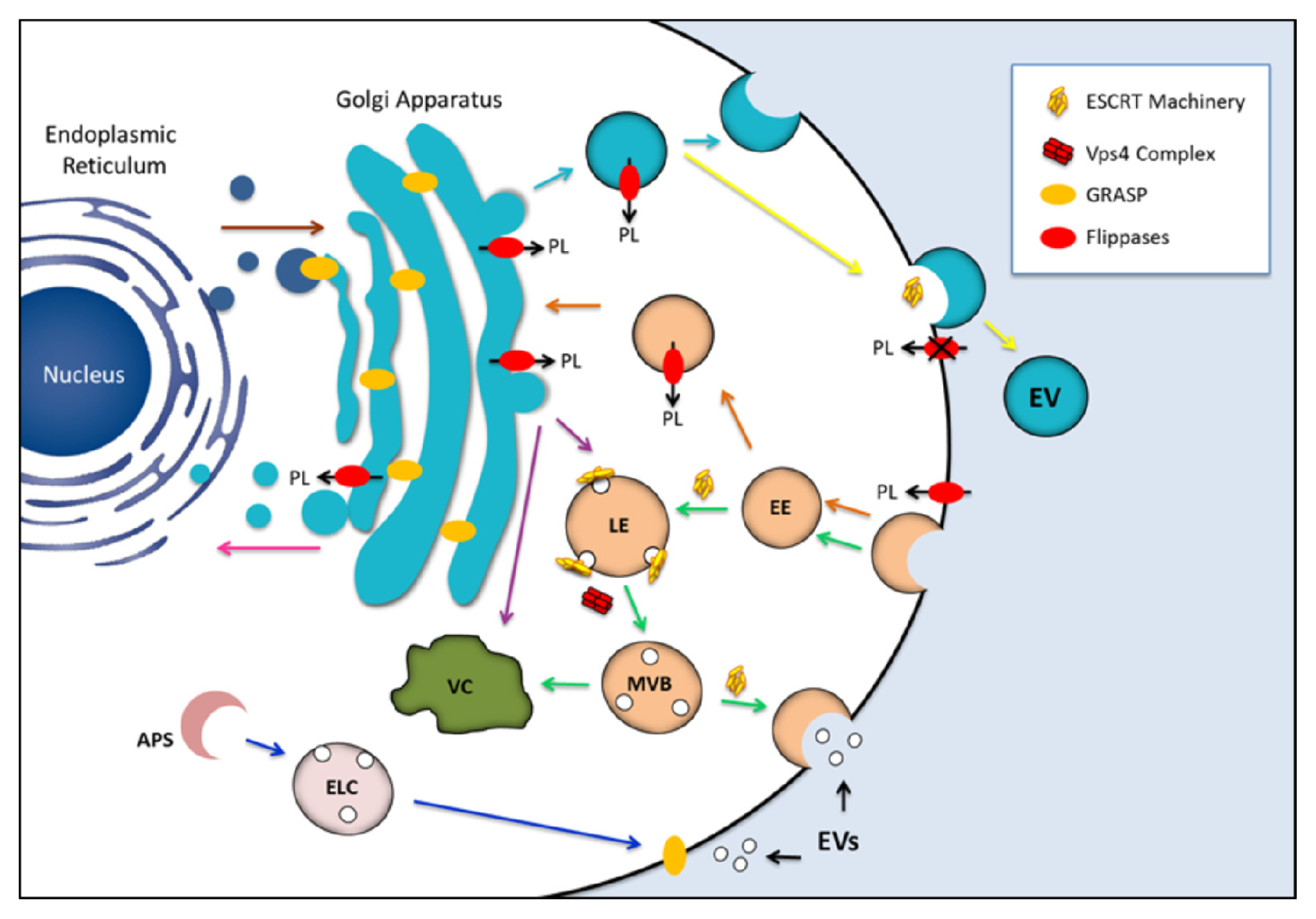
4. Candidates for Regulating the Formation of Fungal EVs

4.1. Exosome Regulators

The term exosome was first used by Trams and colleagues to describe 50 nm diameter vesicles observed in several cultures of normal and neoplasic cells [59]. Harding and colleagues further used electron microscopy techniques to demonstrate that small, luminal vesicles of endosomal compartments could be released to the extracellular space upon fusion between endosomal and plasma membranes [60,61].
Exosomes can be identified by morphological and biochemical criteria. Their dimensions usually correspond to those observed for the vesicles present in the endosomal compartments from where they originate (40–100 nm diameter). Exosomes are usually isolated from culture supernatants using ultracentrifugation procedures [62]. This method, however, frequently fails to distinguish exosomes from other small vesicles and macromolecular aggregates, implying that additional approaches are required for their characterization. These approaches may include immunoblotting analysis, mass spectrometry, and different imaging techniques [63]. Size distribution analysis by dynamic light scattering and high throughput flow cytometric approaches [6466] have also been successfully applied for exosome characterization. Exosomal membrane composition reflects mechanisms of biogenesis. Proteins of exosomal membranes contain endosomal molecules, including Rab GTPases, SNAREs, anexin and flotilin, in addition to plasma membrane proteins, including tetraspanins [67] and lipid raft-related proteins [68,69]. Lipidomic studies of exosomal membranes indicated cholesterol, sphingomyelin, and hexosylceramides as key hydrophobic components [6972].
As mentioned before, exosomes are originated from the membrane of endosomal compartments. The invagination of the late endosome membrane forms small vesicles within its lumen giving origin to what was first described by Porter and Palade as “large vesicles with smaller vesicles inside”[73]. A few years later, Sotelo and Porter [74] proposed classification of these structures as multivesicular bodies (MVB). It is now well known that MVB formation relies on the functions of the endosomal sorting complex required for transport (ESCRT), which refers to a series of cytosolic protein complexes called ESCRT-0, I, II and III. The ESCRT machinery was first described in the model yeast S. cerevisiae [7577]. In this organism the products of the vacuolar protein sorting (vps) class E genes assemble into five different units, the above-mentioned ESCRT-0, I, II and III and Vps4 [7580]. Each one of these protein complexes plays specific tasks not only in MVB formation but also in cell shrinking and viral budding. These latter aspects were reviewed elsewhere [81,82]. We will discuss below the molecular aspects of MVB formation and its possible relationship with the formation of EVs in fungi.
Although the ESCRT machinery has been studied in detail in the last two decades, many of the aspects by which it regulates formation of intraluminal vesicles (ILVs) are still obscure. It is generally accepted that the process begins with the recognition of ubiquitinated cargo proteins by ESCRT-0. This protein complex is composed by two subunits, Vps27 and Hse1, both capable of cargo recognition via ubiquitin interaction motif (UIM) and VHS domains (acronym derived from occurrence in the proteins Vps27, Hrs and STAM). The presence of a zinc finger motif on Vps27 ensures the affinity of the complex by lipid membranes through interaction with phosphatidylinositol 3-phosphate (PtdIns3P) [78,83].
The next step toward MVB formation is the recruitment of the second protein complex, ESCRT-I. This complex, which is composed by the subunits Vps23, Vps28, Vps37 and Mvb12, interacts with the ESCRT-0 PTAP-like motif (formed by aminoacids proline, threonine, and alanine), contained on Vps27 via its Vps23 subunit [75,84,85]. ESCRT-I is also capable of cargo recognition as it contains an ubiquitin E2 variant (UEV) domain on Vps23 and also an ubiquitin-binding domain (UBD) on MVB12 [75,84,86,87].
ESCRT-II is composed of Vps22, Vps25 and Vps36 [76,88]. The last protein is responsible for the interaction between this complex via its GRAM-like ubiquitin-binding in Eap45 (GLUE) domain on ESCRT-1 Vps28 C-terminus [89,90]. ESCRT-II is “Y” shaped and formed by copies of Vps25, which interact with high affinity with Vps20, forming a multiple arm-like structure [91,92].
Vps20 is one of the constituents of ESCRT-III complex, along with Snf7, Vps24 and Vps2 [77]. This is the only ESCRT component that does not form stable cytoplasmic complexes. Upon interaction of Vps20 with Vps25, the former begins recruiting monomers of Snf7, which interact with each other by forming long filament-like polymers that are thought to be capped by molecules of Vps24. When elongation reaches the endosome, Vps24 recruits Vps2, completing formation of ESCRT-III [9294]. The accessory molecules Bro1/Alix (BCK1-like resistance to osmotic shock protein-1/apoptosis-linked gene-2 interacting protein X) and Doa4 (degradation of alpha-4), which are both recruited by Snf7, regulate cargo deubiquitination [95,96].
Vps4, a component of the ESCRT-III machinery, is a class I AAA ATPase [79,97]. The dissociation of ESCRT-III from the membrane of the MVB requires energy that is provided by the Vps4-Vta1 complex [98]. This complex associates with the MIM (missing in metastasis) domains in ESCRT-III via N-terminal MIT (microtubule interacting and trafficking) domains [79,99]. Another possible function for the Vps4-Vta1 complex is the modulation of ESCRT-III activity through its multiple interactions via MIT domains with several Snf7 molecules and accessory proteins, like the Did2/Ist1 complex, possibly acting as an endosomal anchor for the whole Vps4 machinery [100,101].
Although the steps of ESCRT recruitment are well known, the mechanisms by which invagination of the endosomal membrane results in vesicle luminal release are still obscure. Two models explaining this phenomenon have been proposed. In the first model, nominated Snf7 oligomerization, each Vps25 arm of ESCRT-II can recruit one Vps20, which recruits two Snf7 molecules. The filament resulting from the elongation of the Snf7 oligomer associated with Vps24 and Vps2 would induce membrane deformation, causing ILV release [102]. Accordingly, systems at which mutations generating a “one armed” ESCRT-II were induced showed Snf7 elongation but failure to generate ILVs. On the other hand, Snf7 overexpression led to the formation of ILVs with larger, abnormal diameter (up to 360 nm) [103]. Apparently, the control of Snf7 oligomerization relies on the activity of Vps24 and Vps2, which stops Snf7 addition and recruits the Vps4-Vta1 complex for ESCRT-III release prior to ILV formation [81,103]. The second model proposed to explain the release of ILVs has been nominated the Snf7 dome. According to this model, Vps24-Vps2 spirals of decreasing diameter would distribute inside the forming ILV neck limited by Snf7 oligomers, forming a protein cap that prevents the loss of ILV cargo. The created membrane curvature would then facilitate membrane scission and vesicle release [104,105].
Although the involvement of the ESCRT machinery with MVB/vesicle formation is clear, the existence of other mechanisms with similar functions cannot be discarded. For example, in mammalian cells, ILV formation is unaffected even in the absence of central ESCRT components [106,107], suggesting alternative, ESCRT-independent mechanisms of MVB formation. In fact, in vitro studies demonstrated that lysobisphosphatidic acid (LBPA) drives vesicle budding inside LBPA-containing liposomes in processes that involve pH gradients across the membrane [108]. In addition, ceramide formation has been also linked to membrane deformation and budding of ILVs [107].
MVB-like structures fusing with the plasma membrane have been observed in C. neoformans [41,109]. In addition, fungal EVs manifested morphological characteristics and protein composition with similarity to that of mammalian exosomes. Deletion of SNF7 in S. cerevisiae resulted in altered composition of EVs [11], suggesting that the MVB-related machinery might me related to the biogenesis of exosome-like structures in fungi. Therefore, most of the above-mentioned proteins required for MVB formation could represent important regulators of formation of fungal EV.

4.2. Flippases

Lipid asymmetry is critical to many cellular functions, including regulation of membrane curvature and budding of vesicles [110112]. The correct membrane asymmetry relies on the activity of transmembrane proteins belonging to type 4 subfamily of P-type ATPases, the flippases. P-type ATPases represent an ubiquitous family of proteins that facilitate the ATP-dependent transport of ions and heavy metals across biological membranes [113]. Based on phylogenetic analysis, this large family is divided into five major subfamilies, each of them with unique affinities for their specific substrates [114]. Within the P-type ATPase family, P4-ATPases are responsible for phospholipid transport. Based on this biological activity, they were nominated aminophospholipid translocases (APTs or APLTs) [113118].
The primary biological activity of APTs is to promote lipid translocation. APTs preferentially transfer phosphatidylserine (PS) and phosphatidylethanolamine (PE) from the external face (extracytoplasmic) of the plasma membrane to its inner (cytoplasmic) side [115,119121]. An important and conserved feature of P4-ATPases is their ability to form heteromeric complexes with members of the Cdc50 protein family [122125]. The exact role played by Cdc50p in association to P4-ATPases is not yet fully understood. It has been proposed, however, that molecules belonging to the Cdc50 protein family participate in the substrate specificity of flippases [126].
Multiple members of the P4-ATPase family are distributed into eukaryotic genomes [127]. Prokaryotes, however, lack P4-ATPases. In S. cerevisiae, five aminophospholipid translocases (Drs2p, Dnf1p, Dnf2p, Dnf3p and Neo1p) have been described and all of them are directly involved with vesicular traffic of macromolecules. Drs2p and Dnf3p localize to the late Golgi compartment and potentially to endosomal membranes, whereas Dnf1p and Dnf2p localize primarily to the plasma membrane [128130]. Neo1p is apparently distributed into cellular sites that include both endosomes and the Golgi apparatus, which might explain its participation in the retrograde transport pathway [131,132].
A direct association between yeast flippases and vesicular mechanisms of traffic has been established after the observation that mutants with defects in the activity of Dnf1p, Dnf2p and Drs2p lack functionality in both endocytic and exocytic pathways [130,133136]. In Caenorhabditis elegans, the putative flippases TAT- 1and TAT-5 were demonstrated to regulate endocytic pathways [137] and formation of extracellular vesicles [138], respectively. Studies in other organisms including Arabidopsis thaliana, Leishmania donovani, and also mammals indicated that flippases have essential activities in the early mechanisms of vesicle biogenesis, vesicular traffic and/or maintenance of membrane integrity [111,123,125]. In erythrocytes, stimulation of the activity of APTs induced formation of endocytic vesicles [139,140]. A generally accepted hypothesis supporting the findings described above is that flippases might affect vesicle formation as a consequence of enrichment of specific phospholipids in one side of bilayered membranes [112], leading to membrane curvature. This mechanism has been nominated as the “bilayer couple mechanism” of membrane reshaping [141,142].
Small vesicles and tubular structures mediate protein transport between organelles of the secretory and endocytic pathways [50]. Several proteins control mechanisms of biogenesis and traffic of these vesicles, including those belonging to the coat protein complex (COP), adaptor proteins (APs), ADP-ribosylation factor (ARF), and its guanine-nucleotide-exchange factors (GEFs), among others. Basically, COPII-coated vesicles bud from the endoplasmic reticulum (ER) to deliver cargo to the Golgi, and COPI-coated vesicles mediate retrograde transport from the Golgi back to the ER. Clathrin associates with organelle-specific adaptor proteins (APs) to form vesicles from the trans-Golgi network (TGN), endosomes and the plasma membrane, mediating multiple transport pathways between these compartments [50]. The small, GTP binding protein ADP-ribosylation factor (ARF), and its guanine-nucleotide-exchange factors (GEFs) and GTPase activating proteins, regulate the assembly of COPI, clathrin-adaptor and AP-3 coat proteins on the Golgi and/or endosomes [143].
Genetic studies in yeast have implicated Drs2p, a flippase found in the late trans-Golgi network (TGN) of S. cerevisiae, in ARF and clathrin-dependent formation of transport vesicles [128,144]. The connection between Drs2p with Arfp suggests that this protein is important in processes of vesicular budding at the TGN, indicating that P4-ATPases might be part of the normal protein trafficking machinery. Indeed, studies with S. cerevisiae revealed that proteins whose traffic is related to Arf and/or clathrin vesicles in the TGN had their secretory efficacy altered after flippase deletion. Chitin synthase III (Chs3p) is a cargo protein in yeast that requires AP-1/clathrin for trafficking in the TGN–endosomal system. Loss of Drs2p affects Chs3p trafficking in a process that culminates with altered cell surface expression and increased trafficking of the protein into the late endosomes [136,145]. Drs2 mutants also exhibited a defect in generating exocytic vesicles carrying invertase and acid phosphatase [144]. These vesicles require clathrin for their formation and at least a portion of them are clathrin-coated [146,147]. A drs2 Δdnf1Δ double mutant exhibited an even lower efficacy in transport pathways required for the export of alkaline phosphatase protein and carboxypeptidase Y [129]. This observation suggests that Drs2p and Dnf1p are functionally redundant in their ability to regulate pathways of vacuolar protein transport. Moreover, a dnf1Δ dnf2Δ dnf3Δ triple deletion mutant exhibited defects in the endosome to TGN recycling pathway [129]. ARF, clathrin, and/or adaptins (AP-1, AP-3 and GGAs) have been implicated in each of the pathways requiring Drs2/Dnf proteins for normal functioning. Finally, Neo1p is required for protein transport in early mechanisms of the conventional secretory pathway, as well as in Golgi-dependent glycosylation [131]. For instance, Rer1, a protein that cycles between the ER and Golgi complex in COPI and COPII vesicles, is mislocalized to the vacuole in neo1 temperature sensitive mutants [131]. Altogether, these results suggest that flippases are deeply involved in different secretion mechanisms requiring vesicles.
Flippases have been reported to regulate important physiological and pathogenic events in different eukaryotic models. In Magnaporthe grisea, a rice fungal pathogen, two putative APTs, MgAPT2 and MgPDE1, were implicated in the development of invasive hyphae and also in vesicular export of pathogenesis-related proteins [148,149]. In the fungal pathogen C. neoformans, mutants lacking expression of the APT1 flippase manifested attenuated virulence and defects in responding to oxidative and nitrosative stresses. The apt1 mutants had increased sensitivity to brefeldin A and monensin, which interfere with the ER-Golgi trafficking [150]. These findings are probably related to the fact that C. neoformans uses a vesicular network of traffic to export polysaccharides [8,49], which is determinant for pathogenicity. In plant studies using the Arabidopsis thaliana model, deletion of the putative flippase ALA3 affected polysaccharide secretion and formation of TGN-derived vesicles [123]. These results strongly suggest that flippases might be an important part of the regulation of vesicular export mechanisms of proteins and polysaccharides in eukaryotic cells.

4.3. Golgi Reassembly Stacking Protein (GRASP)

The Golgi apparatus is typically formed by one or more layers of associated membranous discs called cisternae [151]. As previously discussed by Ramirez and colleagues, the individual layers of the Golgi are dispersed in the cytoplasm of fungi, plants and lower eukaryotes [152]. In most vertebrates, these layers are laterally connected to form a complex and bulky structure.
To maintain the flow of proteins that are continuously transported inside the Golgi apparatus, great membrane flexibility is required, but the existence of mechanisms that maintain the basic structure of the organelle are also essential. Early studies demonstrated protein connections between the cisternae, a seminal observation that has been confirmed in more recent studies [27,152]. GRASP (Golgi reassembly and stacking protein) is supposed to be one of these proteins [153]. Two isoforms of GRASP (GRASP55 and GRASP65) are present in vertebrates and a single gene is found in other eukaryotes, except plants [154]. GRASPs were also suggested to participate in the tethering of vesicles destined to fuse with the Golgi apparatus [153,155]. Other studies, however, suggested that GRASPs might have additional, non-structural roles that are independent of their primary functions. For instance, depletion of GRASP in Drosophila, S. cerevisiae and mammalian cells had no significant effects on cisternal stacking or general protein secretion [153,156160]. Plant cells, which lack GRASP-related genes, have perfectly stacked Golgi cisternae. On the other hand, GRASP has been described as a key regulator of unconventional mechanisms of protein secretion [161]. These observations suggest that, although the most essential functions of GRASP in eukaryotes are still obscure, this class of proteins might be essential for non-classic mechanisms of protein secretion.
In Dictyostelium discoideum, deletion of the single gene encoding GRASP resulted in defective secretion of acyl-coenzyme A binding protein (AcbA), which lacks the signal peptide required for engagement in the conventional secretory pathway [161]. AcbA-containing vesicles accumulated beneath the plasma membrane, suggesting a role for GRASP in unconventional, vesicular mechanisms of secretion of this cytoplasmic protein [162]. Similar findings were described in S. cerevisiae and Pichia pastoris [163,164]. In the former, GRASP has been found to interact with the COPII coat proteins Sec23 and Sec24, suggesting a role in the docking or fusion of COPII-coated vesicles at the Golgi, thus facilitating anterograde transport through the early secretory pathway [156].
The alternative roles of GRASP proposed in different studies suggest complex functions that would require additional cellular sites for the protein, besides the Golgi apparatus. It has been proposed that GRASP might mediate direct transport of AcbA across the plasma membrane by still unknown mechanisms that might require a plasma membrane distribution [154]. As a plasma membrane protein, GRASP could also act as a tether for endosomal or lysosomal compartments that have engulfed cytoplasmic AcbA in exosomes, or to which AcbA had been delivered by an autophagy-like process [165]. This cellular distribution and related functions would be in agreement with the observation that, during D. melanogaster development, GRASP is required for the delivery of α-integrin to the plasma membrane through a Golgi-independent manner [166]. Although most of these hypotheses still require experimental proof, a realistic perspective is that GRASP might be required for the vesicular traffic of a number of molecules in eukaryotic cells.
The functions of GRASP in eukaryotes apparently include the traffic of polysaccharides to the extracellular space, as demonstrated in the C. neoformans model [58]. Polysaccharide secretion in this fungus was initially linked to proteins with homology to Sec4 and Sec6 [49,57], two key regulators of conventional mechanisms of exocytosis in other models [167]. Although polysaccharide secretion was affected in the mutants lacking expression of the SEC4 and SEC6 orthologs, capsule assembly appeared normal in those cells [49,57]. This observation led to the hypothesis that polysaccharide secretion might also require regulators of unconventional secretion pathways. The fact that extracellular vesicles produced by C. neoformans are loaded with capsular polysaccharides [8,13] and the possibility that GRASP is required for mechanisms of secretion involving EVs in yeast cells [154,163,164] support this hypothesis. In this regard, a general analysis of putative defects in protein secretion in the graspΔ mutant of C. neoformans and its potential correlation with polysaccharide export might be a promising approach to correlate unconventional mechanisms of secretions of glycans and proteins in this fungus.
Deletion of the single GRASP ortholog of C. neoformans did not affect the traffic of pigments and urease [58], which are surface and extracellular molecules regulating key pathogenic steps in this fungus [168,169]. Deletion of the GRASP ortholog, however, profoundly affected polysaccharide secretion and, consequently, capsule formation [58]. The defective polysaccharide secretion in the mutant lacking GRASP resulted in a hypovirulent phenotype, associated with an increased susceptibility to the antimicrobial activity of macrophages [58,168]. It remains unknown why lack of GRASP in C. neoformans results in decreased polysaccharide secretion. GRASP could regulate the export of polysaccharide-containing extracellular vesicles to the milieu by the mechanisms proposed for unconventional secretory steps in S. cerevisiae, D. discoideum, D. melanogaster and P. pastoris [155,160,162165]. An alternative possibility would derive from the fact that the glycosyltransferases required for polysaccharide and glycoprotein synthesis in eukaryotes are essentially Golgi-associated. Since the C. neoformans mutant lacking the GRASP gene showed abnormal Golgi morphology [58], dysfunctions in intracellular glycan synthesis might be expected. C. neoformans GRASP could be also required for loading GXM into secretory vesicles, although this hypothesis still requires experimental proof. Finally, considering that the GRASP orthologs of S. cerevisiae and P. pastoris are supposed to be functional at the tER-Golgi interface [170], we speculate that lack of GRASP could also affect the functionality of cellular sites playing central functions within the secretory machinery.

5. Concluding Remarks

Studies based on the characterization of fungal EVs produced by mutants with defects in conventional or unconventional secretory pathways have failed in demonstrating the mechanisms required for biogenesis of these extracellular membrane compartments [11,167]. This observation makes clear the need of identification of additional targets with the potential to regulate EV biogenesis in the Fungi. Based on the current literature, we propose that proteins related to mechanisms of EV biogenesis in other eukaryotes (ESCRT machinery, flippases and GRASP) whose functions in EV formation in fungi are still unexplored could be excellent targets for future studies aiming at understanding some of the mechanisms required for regulation of this interesting phenomenon, as summarized in Figure 1.

Acknowledgments

DLO, JRB, LSJ, RMCG and MLR are supported by grants from Coordenação de Aperfeiçoamento de Pessoal de Nível Superior (CAPES, Brazil), Conselho Nacional de Desenvolvimento Científico e Tecnológico (CNPq, Brazil), Fundação de Amparo a Pesquisa do Estado de São Paulo (FAPESP, Brazil) and Fundação de Amparo a Pesquisa do Estado do Rio de Janeiro (FAPERJ, Brazil). We thank Leonardo Nimrichter for constant collaboration in EV studies developed by our group. The funders had no role in study design, data collection and analysis, decision to publish, or preparation of the manuscript. The authors declare no reported conflicts of interest.

References

  1. Latge, J.P.; Mouyna, I.; Tekaia, F.; Beauvais, A.; Debeaupuis, J.P.; Nierman, W. Specific molecular features in the organization and biosynthesis of the cell wall of Aspergillus fumigatus. Med. Mycol 2005, 43, S15–S22. [Google Scholar]
  2. Nimrichter, L.; Rodrigues, M.L.; Rodrigues, E.G.; Travassos, L.R. The multitude of targets for the immune system and drug therapy in the fungal cell wall. Microbes Infect 2005, 7, 789–798. [Google Scholar]
  3. Mitchell, A.P. Cryptococcal virulence: Beyond the usual suspects. J. Clin. Invest 2006, 116, 1481–1483. [Google Scholar]
  4. Nosanchuk, J.D.; Steenbergen, J.N.; Shi, L.; Deepe, G.S., Jr; Casadevall, A. Antibodies to a cell surface histone-like protein protect against Histoplasma capsulatum. J. Clin. Invest 2003, 112, 1164–1175. [Google Scholar]
  5. Barbosa, M.S.; Bao, S.N.; Andreotti, P.F.; de Faria, F.P.; Felipe, M.S.; dos Santos Feitosa, L.; Mendes-Giannini, M.J.; Soares, C.M. Glyceraldehyde-3-phosphate dehydrogenase of Paracoccidioides brasiliensis is a cell surface protein involved in fungal adhesion to extracellular matrix proteins and interaction with cells. Infect. Immun 2006, 74, 382–289. [Google Scholar]
  6. Rodrigues, M.L.; Nakayasu, E.S.; Oliveira, D.L.; Nimrichter, L.; Nosanchuk, J.D.; Almeida, I.C.; Casadevall, A. Extracellular vesicles produced by Cryptococcus neoformans contain protein components associated with virulence. Eukaryot Cell 2008, 7, 58–67. [Google Scholar]
  7. Batista, W.L.; Matsuo, A.L.; Ganiko, L.; Barros, T.F.; Veiga, T.R.; Freymuller, E.; Puccia, R. The PbMDJ1 gene belongs to a conserved MDJ1/LON locus in thermodimorphic pathogenic fungi and encodes a heat shock protein that localizes to both the mitochondria and cell wall of Paracoccidioides brasiliensis. Eukaryot Cell 2006, 5, 379–390. [Google Scholar]
  8. Rodrigues, M.L.; Nimrichter, L.; Oliveira, D.B.L.; Frases, S.; Miranda, K.; Zaragoza, O.; Alvarez, M.; Nakouzi, A.; Feldmesser, M.; Casadevall, A. Vesicular polysaccharide export in Cryptococcus neoformans is a eukaryotic solution to the problem of fungal trans-cell wall transport. Am. Soc. Microbiol 2007, 6, 48–59. [Google Scholar]
  9. Albuquerque, P.C.; Nakayasu, E.S.; Rodrigues, M.L.; Frases, S.; Casadevall, A.; Zancope-Oliveira, R.M.; Almeida, I.C.; Nosanchuk, J.D. Vesicular transport in Histoplasma capsulatum: An effective mechanism for trans-cell wall transfer of proteins and lipids in ascomycetes. Cell Microbiol 2008, 10, 1695–1710. [Google Scholar]
  10. Vallejo, M.C.; Matsuo, A.L.; Ganiko, L.; Medeiros, L.C.; Miranda, K.; Silva, L.S.; Freymuller-Haapalainen, E.; Sinigaglia-Coimbra, R.; Almeida, I.C.; Puccia, R. The pathogenic fungus Paracoccidioides brasiliensis exports extracellular vesicles containing highly immunogenic alpha-Galactosyl epitopes. Eukaryot Cell 2011, 10, 343–351. [Google Scholar]
  11. Oliveira, D.L.; Nakayasu, E.S.; Joffe, L.S.; Guimaraes, A.J.; Sobreira, T.J.; Nosanchuk, J.D.; Cordero, R.J.; Frases, S.; Casadevall, A.; Almeida, I.C.; et al. Characterization of yeast extracellular vesicles: Evidence for the participation of different pathways of cellular traffic in vesicle biogenesis. PloS One 2010, 5, e11113. [Google Scholar]
  12. Barbosa, F.M.; Daffre, S.; Maldonado, R.A.; Miranda, A.; Nimrichter, L.; Rodrigues, M.L. Gomesin, a peptide produced by the spider Acanthoscurria gomesiana, is a potent anticryptococcal agent that acts in synergism with fluconazole. FEMS Microbiol. Lett 2007, 274, 279–286. [Google Scholar]
  13. Oliveira, D.B.L.; Nimrichter, L.; Miranda, K.; Frases, S.; Faull, K.F.; Casadevall, A.; Rodrigues, M.L. Cryptococcus neoformans cryoultramicrotomy and vesicle fractionation reveals an intimate association between membrane lipids and glucuronoxylomannan. Fungal Genet. Biol 2009, 46, 956–963. [Google Scholar]
  14. Gehrmann, U.; Qazi, K.R.; Johansson, C.; Hultenby, K.; Karlsson, M.; Lundeberg, L.; Gabrielsson, S.; Scheynius, A. Nanovesicles from Malassezia sympodialis and host exosomes induce cytokine responses—Novel mechanisms for host-microbe interactions in atopic eczema. PLoS One 2011, 6, e21480. [Google Scholar]
  15. Oliveira, D.L.; Freire-de-Lima, C.G.; Nosanchuk, J.D.; Casadevall, A.; Rodrigues, M.L.; Nimrichter, L. Extracellular vesicles from Cryptococcus neoformans modulate macrophage functions. Infect. Immun 2010, 78, 1601–1609. [Google Scholar]
  16. Vallejo, M.C.; Nakayasu, E.S.; Longo, L.V.; Ganiko, L.; Lopes, F.G.; Matsuo, A.L.; Almeida, I.C.; Puccia, R. Lipidomic analysis of extracellular vesicles from the pathogenic phase of Paracoccidioides brasiliensis. PLoS One 2012, 7, e39463. [Google Scholar]
  17. Kuehn, M.J.; Kesty, N.C. Bacterial outer membrane vesicles and the host-pathogen interaction. Genes Dev 2005, 19, 2645–2655. [Google Scholar]
  18. Bomberger, J.M.; Maceachran, D.P.; Coutermarsh, B.A.; Ye, S.; O’Toole, G.A.; Stanton, B.A. Long-distance delivery of bacterial virulence factors by Pseudomonas aeruginosa outer membrane vesicles. PLoS Pathog 2009, 5, e1000382. [Google Scholar]
  19. Yonezawa, H.; Osaki, T.; Kurata, S.; Fukuda, M.; Kawakami, H.; Ochiai, K.; Hanawa, T.; Kamiya, S. Outer membrane vesicles of Helicobacter pylori TK1402 are involved in biofilm formation. BMC Microbiol 2009, 9, 197. [Google Scholar]
  20. Lee, E.Y.; Choi, D.Y.; Kim, D.K.; Kim, J.W.; Park, J.O.; Kim, S.; Kim, S.H.; Desiderio, D.M.; Kim, Y.K.; Kim, K.P.; et al. Gram-positive bacteria produce membrane vesicles: Proteomics-based characterization of Staphylococcus aureus-derived membrane vesicles. Proteomics 2009, 9, 5425–5436. [Google Scholar]
  21. Rivera, J.; Cordero, R.J.; Nakouzi, A.S.; Frases, S.; Nicola, A.; Casadevall, A. Bacillus anthracis produces membrane-derived vesicles containing biologically active toxins. Proc. Natl Acad Sci USA 2010, 107, 19002–19007. [Google Scholar]
  22. Makarova, K.S.; Yutin, N.; Bell, S.D.; Koonin, E.V. Evolution of diverse cell division and vesicle formation systems in Archaea. Nat. Rev. Microbiol 2010, 8, 731–741. [Google Scholar]
  23. Ellen, A.F.; Albers, S.V.; Huibers, W.; Pitcher, A.; Hobel, C.F.; Schwarz, H.; Folea, M.; Schouten, S.; Boekema, E.J.; Poolman, B.; et al. Proteomic analysis of secreted membrane vesicles of archaeal Sulfolobus species reveals the presence of endosome sorting complex components. Extremophiles 2009, 13, 67–79. [Google Scholar]
  24. Soler, N.; Marguet, E.; Verbavatz, J.M.; Forterre, P. Virus-like vesicles and extracellular DNA produced by hyperthermophilic archaea of the order Thermococcales. Res. Microbiol 2008, 159, 390–399. [Google Scholar]
  25. Rabouille, C.; Malhotra, V.; Nickel, W. Diversity in unconventional protein secretion. J. Cell Sci 2012, 125, 5251–5255. [Google Scholar]
  26. Nickel, W. Pathways of unconventional protein secretion. Curr. Opin. Biotechnol 2010, 21, 621–626. [Google Scholar]
  27. Nickel, W.; Rabouille, C. Mechanisms of regulated unconventional protein secretion. Nat. Rev. Mol. Cell Biol 2009, 10, 148–155. [Google Scholar]
  28. Johnstone, R.M.; Adam, M.; Pan, B.T. The fate of the transferrin receptor during maturation of sheep reticulocytes in vitro. Can. J. Biochem. Cell Biol 1984, 62, 1246–1254. [Google Scholar]
  29. Raposo, G.; Nijman, H.W.; Stoorvogel, W.; Liejendekker, R.; Harding, C.V.; Melief, C.J.; Geuze, H.J. B lymphocytes secrete antigen-presenting vesicles. J. Exp. Med 1996, 183, 1161–1172. [Google Scholar]
  30. Zitvogel, L.; Regnault, A.; Lozier, A.; Wolfers, J.; Flament, C.; Tenza, D.; Ricciardi-Castagnoli, P.; Raposo, G.; Amigorena, S. Eradication of established murine tumors using a novel cell-free vaccine: Dendritic cell-derived exosomes. Nat. Med 1998, 4, 594–600. [Google Scholar]
  31. Marzesco, A.M.; Janich, P.; Wilsch-Brauninger, M.; Dubreuil, V.; Langenfeld, K.; Corbeil, D.; Huttner, W.B. Release of extracellular membrane particles carrying the stem cell marker prominin-1 (CD133) from neural progenitors and other epithelial cells. J. Cell Sci 2005, 118, 2849–2858. [Google Scholar]
  32. Gould, S.J.; Booth, A.M.; Hildreth, J.E. The Trojan exosome hypothesis. Proc. Natl. Acad. Sci. USA 2003, 100, 10592–10597. [Google Scholar]
  33. Fang, Y.; Wu, N.; Gan, X.; Yan, W.; Morrell, J.C.; Gould, S.J. Higher-order oligomerization targets plasma membrane proteins and HIV gag to exosomes. PLoS Biol 2007, 5, e158. [Google Scholar]
  34. Bhatnagar, S.; Shinagawa, K.; Castellino, F.J.; Schorey, J.S. Exosomes released from macrophages infected with intracellular pathogens stimulate a proinflammatory response in vitro and in vivo. Blood 2007, 110, 3234–3244. [Google Scholar]
  35. Beatty, W.L.; Russell, D.G. Identification of mycobacterial surface proteins released into subcellular compartments of infected macrophages. Infect. Immun 2000, 68, 6997–7002. [Google Scholar]
  36. Park, J.E.; Tan, H.S.; Datta, A.; Lai, R.C.; Zhang, H.; Meng, W.; Lim, S.K.; Sze, S.K. Hypoxic tumor cell modulates its microenvironment to enhance angiogenic and metastatic potential by secretion of proteins and exosomes. Mol. Cell. Proteomics 2010, 9, 1085–1099. [Google Scholar]
  37. Marcilla, A.; Trelis, M.; Cortes, A.; Sotillo, J.; Cantalapiedra, F.; Minguez, M.T.; Valero, M.L.; Sanchez del Pino, M.M.; Munoz-Antoli, C.; Toledo, R.; et al. Extracellular vesicles from parasitic helminths contain specific excretory/secretory proteins and are internalized in intestinal host cells. PLoS One 2012, 7, e45974. [Google Scholar]
  38. Silverman, J.M.; Clos, J.; de’Oliveira, C.C.; Shirvani, O.; Fang, Y.; Wang, C.; Foster, L.J.; Reiner, N.E. An exosome-based secretion pathway is responsible for protein export from Leishmania and communication with macrophages. J. Cell Sci 2010, 123, 842–852. [Google Scholar]
  39. Clayton, C.; Estevez, A. The exosomes of trypanosomes and other protists. Adv. Exp. Med. Biol 2011, 702, 39–49. [Google Scholar]
  40. Regente, M.; Corti-Monzon, G.; Maldonado, A.M.; Pinedo, M.; Jorrin, J.; de la Canal, L. Vesicular fractions of sunflower apoplastic fluids are associated with potential exosome marker proteins. FEBS Lett 2009, 583, 3363–3366. [Google Scholar]
  41. Takeo, K.; Uesaka, I.; Uehira, K.; Nishiura, M. Fine structure of Cryptococcus neoformans grown in vitro as observed by freeze-etching. J. Bacteriol 1973, 113, 1442–1448. [Google Scholar]
  42. Rodrigues, M.L.; Travassos, L.R.; Miranda, K.R.; Franzen, A.J.; Rozental, S.; de Souza, W.; Alviano, C.S.; Barreto-Bergter, E. Human antibodies against a purified glucosylceramide from Cryptococcus neoformans inhibit cell budding and fungal growth. Infect. Immun 2000, 68, 7049–7060. [Google Scholar]
  43. Anderson, J.; Mihalik, R.; Soll, D.R. Ultrastructure and antigenicity of the unique cell wall pimple of the Candida opaque phenotype. J. Bacteriol 1990, 172, 224–235. [Google Scholar]
  44. Bogo, M.R.; Queiroz, M.V.; Silva, D.M.; Giménez, M.P.; Azevedo, J.L.; Schrank, A. Double-stranded RNA and isometric virus-like particles in the entomopathogenic fungus Metarhizium anisopliae. Mycol. Res 1996, 100, 1468–1472. [Google Scholar]
  45. Chang, Y.C.; Penoyer, L.A.; Kwon-Chung, K.J. The second capsule gene of Cryptococcus neoformans, CAP64, is essential for virulence. Infect. Immun 1996, 64, 1977–1983. [Google Scholar]
  46. Chang, Y.C.; Kwon-Chung, K.J. Isolation of the third capsule-associated gene, CAP60, required for virulence in Cryptococcus neoformans. Infect. Immun 1998, 66, 2230–2236. [Google Scholar]
  47. Chang, Y.C.; Kwon-Chung, K.J. Isolation, characterization, and localization of a capsule-associated gene, CAP10, of Cryptococcus neoformans. J. Bacteriol 1999, 181, 5636–5643. [Google Scholar]
  48. Feldmesser, M.; Kress, Y.; Novikoff, P.; Casadevall, A. Cryptococcus neoformans is a facultative intracellular pathogen in murine pulmonary infection. Infect. Immun 2000, 68, 4225–37. [Google Scholar]
  49. Yoneda, A.; Doering, T.L. A eukaryotic capsular polysaccharide is synthesized intracellularly and secreted via exocytosis. Mol. Biol. Cell 2006, 17, 5131–5140. [Google Scholar]
  50. Bonifacino, J.S.; Glick, B.S. The mechanisms of vesicle budding and fusion. Cell 2004, 116, 153–166. [Google Scholar]
  51. Feldmesser, M.; Kress, Y.; Casadevall, A. Dynamic changes in the morphology of Cryptococcus neoformans during murine pulmonary infection. Microbiology 2001, 147, 2355–2365. [Google Scholar]
  52. Vallejo, M.C.; Nakayasu, E.S.; Matsuo, A.L.; Sobreira, T.J.; Longo, L.V.; Ganiko, L.; Almeida, I.C.; Puccia, R. Vesicle and vesicle-free extracellular proteome of Paracoccidioides brasiliensis: Comparative analysis with other pathogenic fungi. J. Proteome Res 2012, 11, 1676–85. [Google Scholar]
  53. Jeffery, C.J. Moonlighting proteins. Trends Biochem. Sci 1999, 24, 8–11. [Google Scholar]
  54. Eisenman, H.C.; Frases, S.; Nicola, A.M.; Rodrigues, M.L.; Casadevall, A. Vesicle-associated melanization in Cryptococcus neoformans. Microbiology 2009, 155, 3860–3867. [Google Scholar]
  55. Schorey, J.S.; Bhatnagar, S. Exosome function: From tumor immunology to pathogen biology. Traffic 2008, 9, 871–881. [Google Scholar]
  56. Huang, S.H.; Wu, C.H.; Chang, Y.C.; Kwon-Chung, K.J.; Brown, R.J.; Jong, A. Cryptococcus neoformans-derived microvesicles enhance the pathogenesis of fungal brain infection. PloS One 2012, 7, e48570. [Google Scholar]
  57. Panepinto, J.; Komperda, K.; Frases, S.; Park, Y.D.; Djordjevic, J.T.; Casadevall, A.; Williamson, P.R. Sec6-dependent sorting of fungal extracellular exosomes and laccase of Cryptococcus neoformans. Mol. Microbiol 2009, 71, 1165–1176. [Google Scholar]
  58. Kmetzsch, L.; Joffe, L.S.; Staats, C.C.; de Oliveira, D.L.; Fonseca, F.L.; Cordero, R.J.; Casadevall, A.; Nimrichter, L.; Schrank, A.; Vainstein, M.H.; et al. Role for Golgi reassembly and stacking protein (GRASP) in polysaccharide secretion and fungal virulence. Mol. Microbiol 2011, 81, 206–218. [Google Scholar]
  59. Trams, E.G.; Lauter, C.J.; Salem, N., Jr; Heine, U. Exfoliation of membrane ecto-enzymes in the form of micro-vesicles. Biochim. Biophys. Acta 1981, 645, 63–70. [Google Scholar]
  60. Pan, B.T.; Teng, K.; Wu, C.; Adam, M.; Johnstone, R.M. Electron microscopic evidence for externalization of the transferrin receptor in vesicular form in sheep reticulocytes. J. Cell Biol 1985, 101, 942–948. [Google Scholar]
  61. Harding, C.; Heuser, J.; Stahl, P. Endocytosis and intracellular processing of transferrin and colloidal gold-transferrin in rat reticulocytes: Demonstration of a pathway for receptor shedding. Eur. J. Cell Biol 1984, 35, 256–263. [Google Scholar]
  62. Thery, C.; Amigorena, S.; Raposo, G.; Clayton, A. Isolation and characterization of exosomes from cell culture supernatants and biological fluids. Curr. Protoc. Cell Biol. 2006. [Google Scholar] [CrossRef]
  63. Raposo, G.; Stoorvogel, W. Extracellular vesicles: Exosomes, microvesicles, and friends. J. Cell Biol 2013, 200, 373–383. [Google Scholar]
  64. Soo, C.Y.; Song, Y.; Zheng, Y.; Campbell, E.C.; Riches, A.C.; Gunn-Moore, F.; Powis, S.J. Nanoparticle tracking analysis monitors microvesicle and exosome secretion from immune cells. Immunology 2012, 136, 192–197. [Google Scholar]
  65. Hoen, E.N.; van der Vlist, E.J.; Aalberts, M.; Mertens, H.C.; Bosch, B.J.; Bartelink, W.; Mastrobattista, E.; van Gaal, E.V.; Stoorvogel, W.; Arkesteijn, G.J.; et al. Quantitative and qualitative flow cytometric analysis of nanosized cell-derived membrane vesicles. Nanomedicine 2012, 8, 712–720. [Google Scholar]
  66. Van der Vlist, E.J.; Nolte-’t Hoen, E.N.; Stoorvogel, W.; Arkesteijn, G.J.; Wauben, M.H. Fluorescent labeling of nano-sized vesicles released by cells and subsequent quantitative and qualitative analysis by high-resolution flow cytometry. Nat. Protoc 2012, 7, 1311–1326. [Google Scholar]
  67. Escola, J.M.; Kleijmeer, M.J.; Stoorvogel, W.; Griffith, J.M.; Yoshie, O.; Geuze, H.J. Selective enrichment of tetraspan proteins on the internal vesicles of multivesicular endosomes and on exosomes secreted by human B-lymphocytes. J. Biol. Chem 1998, 273, 20121–20127. [Google Scholar]
  68. Thery, C.; Regnault, A.; Garin, J.; Wolfers, J.; Zitvogel, L.; Ricciardi-Castagnoli, P.; Raposo, G.; Amigorena, S. Molecular characterization of dendritic cell-derived exosomes. Selective accumulation of the heat shock protein hsc73. J. Cell Biol 1999, 147, 599–610. [Google Scholar]
  69. Wubbolts, R.; Leckie, R.S.; Veenhuizen, P.T.; Schwarzmann, G.; Mobius, W.; Hoernschemeyer, J.; Slot, J.W.; Geuze, H.J.; Stoorvogel, W. Proteomic and biochemical analyses of human B cell-derived exosomes. Potential implications for their function and multivesicular body formation. J. Biol. Chem 2003, 278, 10963–10972. [Google Scholar]
  70. Laulagnier, K.; Motta, C.; Hamdi, S.; Roy, S.; Fauvelle, F.; Pageaux, J.F.; Kobayashi, T.; Salles, J.P.; Perret, B.; Bonnerot, C.; et al. Mast cell- and dendritic cell-derived exosomes display a specific lipid composition and an unusual membrane organization. Biochem. J 2004, 380, 161–171. [Google Scholar]
  71. Subra, C.; Laulagnier, K.; Perret, B.; Record, M. Exosome lipidomics unravels lipid sorting at the level of multivesicular bodies. Biochimie 2007, 89, 205–212. [Google Scholar]
  72. Brouwers, J.F.; Aalberts, M.; Jansen, J.W.A.; van Niel, G.; Wauben, M.H.; Stout, T.A.E.; Helms, J.B.; Stoorvogel, W. Distinct lipid compositions of two types of human prostasomes. Proteomics 2013. [Google Scholar] [CrossRef]
  73. Palade, G.E. Studies on the endoplasmic reticulum. II. Simple dispositions in cells in situ. J. Biophys. Biochem. Cytol 1955, 1, 567–582. [Google Scholar]
  74. Sotelo, J.R.; Porter, K.R. An electron microscope study of the rat ovum. J. Biophys. Biochem. Cytol 1959, 5, 327–342. [Google Scholar]
  75. Katzmann, D.J.; Babst, M.; Emr, S.D. Ubiquitin-dependent sorting into the multivesicular body pathway requires the function of a conserved endosomal protein sorting complex, ESCRT-I. Cell 2001, 106, 145–155. [Google Scholar]
  76. Babst, M.; Katzmann, D.J.; Snyder, W.B.; Wendland, B.; Emr, S.D. Endosome-associated complex, ESCRT-II, recruits transport machinery for protein sorting at the multivesicular body. Dev. Cell 2002, 3, 283–289. [Google Scholar]
  77. Babst, M.; Katzmann, D.J.; Estepa-Sabal, E.J.; Meerloo, T.; Emr, S.D. Escrt-III: An endosome-associated heterooligomeric protein complex required for mvb sorting. Dev. Cell 2002, 3, 271–282. [Google Scholar]
  78. Asao, H.; Sasaki, Y.; Arita, T.; Tanaka, N.; Endo, K.; Kasai, H.; Takeshita, T.; Endo, Y.; Fujita, T.; Sugamura, K. Hrs is associated with STAM, a signal-transducing adaptor molecule. Its suppressive effect on cytokine-induced cell growth. J. Biol. Chem 1997, 272, 32785–32791. [Google Scholar]
  79. Babst, M.; Sato, T.K.; Banta, L.M.; Emr, S.D. Endosomal transport function in yeast requires a novel AAA-type ATPase, Vps4p. EMBO J 1997, 16, 1820–1831. [Google Scholar]
  80. Shih, S.C.; Katzmann, D.J.; Schnell, J.D.; Sutanto, M.; Emr, S.D.; Hicke, L. Epsins and Vps27p/Hrs contain ubiquitin-binding domains that function in receptor endocytosis. Nat. Cell Biol 2002, 4, 389–393. [Google Scholar]
  81. Henne, W.M.; Buchkovich, N.J.; Emr, S.D. The ESCRT pathway. Dev. Cell 2011, 21, 77–91. [Google Scholar]
  82. Schmidt, O.; Teis, D. The ESCRT machinery. Curr. Biol 2012, 22, R116–R120. [Google Scholar]
  83. Raiborg, C.; Bremnes, B.; Mehlum, A.; Gillooly, D.J.; D'Arrigo, A.; Stang, E.; Stenmark, H. FYVE and coiled-coil domains determine the specific localisation of Hrs to early endosomes. J. Cell Sci 2001, 114, 2255–2263. [Google Scholar]
  84. Katzmann, D.J.; Stefan, C.J.; Babst, M.; Emr, S.D. Vps27 recruits ESCRT machinery to endosomes during MVB sorting. J. Cell Biol 2003, 162, 413–423. [Google Scholar]
  85. Kostelansky, M.S.; Sun, J.; Lee, S.; Kim, J.; Ghirlando, R.; Hierro, A.; Emr, S.D.; Hurley, J.H. Structural and functional organization of the ESCRT-I trafficking complex. Cell 2006, 125, 113–26. [Google Scholar]
  86. Bache, K.G.; Raiborg, C.; Mehlum, A.; Stenmark, H. STAM and Hrs are subunits of a multivalent ubiquitin-binding complex on early endosomes. J. Biol. Chem 2003, 278, 12513–12521. [Google Scholar]
  87. Lu, Q.; Hope, L.W.; Brasch, M.; Reinhard, C.; Cohen, S.N. TSG101 interaction with HRS mediates endosomal trafficking and receptor down-regulation. Proc. Natl Acad Sci USA 2003, 100, 7626–7631. [Google Scholar]
  88. Langelier, C.; von Schwedler, U.K.; Fisher, R.D.; De Domenico, I.; White, P.L.; Hill, C.P.; Kaplan, J.; Ward, D.; Sundquist, W.I. Human ESCRT-II complex and its role in human immunodeficiency virus type 1 release. J. Virol 2006, 80, 9465–9480. [Google Scholar]
  89. Gill, D.J.; Teo, H.; Sun, J.; Perisic, O.; Veprintsev, D.B.; Emr, S.D.; Williams, R.L. Structural insight into the ESCRT-I/-II link and its role in MVB trafficking. EMBO J 2007, 26, 600–612. [Google Scholar]
  90. Teo, H.; Gill, D.J.; Sun, J.; Perisic, O.; Veprintsev, D.B.; Vallis, Y.; Emr, S.D.; Williams, R.L. ESCRT-I core and ESCRT-II GLUE domain structures reveal role for GLUE in linking to ESCRT-I and membranes. Cell 2006, 125, 99–111. [Google Scholar]
  91. Hierro, A.; Sun, J.; Rusnak, A.S.; Kim, J.; Prag, G.; Emr, S.D.; Hurley, J.H. Structure of the ESCRT-II endosomal trafficking complex. Nature 2004, 431, 221–225. [Google Scholar]
  92. Teo, H.; Perisic, O.; Gonzalez, B.; Williams, R.L. ESCRT-II, an endosome-associated complex required for protein sorting: crystal structure and interactions with ESCRT-III and membranes. Dev. Cell 2004, 7, 559–569. [Google Scholar]
  93. Saksena, S.; Emr, S.D. ESCRTs and human disease. Biochem. Soc. Trans 2009, 37, 167–172. [Google Scholar]
  94. Teis, D.; Saksena, S.; Emr, S.D. Ordered assembly of the ESCRT-III complex on endosomes is required to sequester cargo during MVB formation. Dev. Cell 2008, 15, 578–589. [Google Scholar]
  95. Luhtala, N.; Odorizzi, G. Bro1 coordinates deubiquitination in the multivesicular body pathway by recruiting Doa4 to endosomes. J. Cell Biol 2004, 166, 717–729. [Google Scholar]
  96. Odorizzi, G.; Katzmann, D.J.; Babst, M.; Audhya, A.; Emr, S.D. Bro1 is an endosome-associated protein that functions in the MVB pathway in Saccharomyces cerevisiae. J. Cell Sci 2003, 116, 1893–1903. [Google Scholar]
  97. Finken-Eigen, M.; Rohricht, R.A.; Kohrer, K. The VPS4 gene is involved in protein transport out of a yeast pre-vacuolar endosome-like compartment. Curr. Genet 1997, 31, 469–480. [Google Scholar]
  98. Babst, M.; Wendland, B.; Estepa, E.J.; Emr, S.D. The Vps4p AAA ATPase regulates membrane association of a Vps protein complex required for normal endosome function. EMBO J 1998, 17, 2982–2993. [Google Scholar]
  99. Scott, A.; Gaspar, J.; Stuchell-Brereton, M.D.; Alam, S.L.; Skalicky, J.J.; Sundquist, W.I. Structure and ESCRT-III protein interactions of the MIT domain of human VPS4A. Proc. Natl. Acad. Sci. USA 2005, 102, 13813–13818. [Google Scholar]
  100. Rue, S.M.; Mattei, S.; Saksena, S.; Emr, S.D. Novel Ist1-Did2 complex functions at a late step in multivesicular body sorting. Mol. Biol. Cell 2008, 19, 475–484. [Google Scholar]
  101. Shestakova, A.; Hanono, A.; Drosner, S.; Curtiss, M.; Davies, B.A.; Katzmann, D.J.; Babst, M. Assembly of the AAA ATPase Vps4 on ESCRT-III. Mol. Biol. Cell 2010, 21, 1059–1071. [Google Scholar]
  102. Henne, W.M.; Buchkovich, N.J.; Zhao, Y.; Emr, S.D. The endosomal sorting complex ESCRT-II mediates the assembly and architecture of ESCRT-III helices. Cell 2012, 151, 356–371. [Google Scholar]
  103. Teis, D.; Saksena, S.; Judson, B.L.; Emr, S.D. ESCRT-II coordinates the assembly of ESCRT-III filaments for cargo sorting and multivesicular body vesicle formation. EMBO J 2010, 29, 871–83. [Google Scholar]
  104. Lata, S.; Schoehn, G.; Jain, A.; Pires, R.; Piehler, J.; Gottlinger, H.G.; Weissenhorn, W. Helical structures of ESCRT-III are disassembled by VPS4. Science 2008, 321, 1354–1357. [Google Scholar]
  105. Fabrikant, G.; Lata, S.; Riches, J.D.; Briggs, J.A.; Weissenhorn, W.; Kozlov, M.M. Computational model of membrane fission catalyzed by ESCRT-III. PLoS Comput. Biol 2009, 5, e1000575. [Google Scholar]
  106. Theos, A.C.; Truschel, S.T.; Tenza, D.; Hurbain, I.; Harper, D.C.; Berson, J.F.; Thomas, P.C.; Raposo, G.; Marks, M.S. A lumenal domain-dependent pathway for sorting to intralumenal vesicles of multivesicular endosomes involved in organelle morphogenesis. Dev. Cell 2006, 10, 343–354. [Google Scholar]
  107. Trajkovic, K.; Hsu, C.; Chiantia, S.; Rajendran, L.; Wenzel, D.; Wieland, F.; Schwille, P.; Brugger, B.; Simons, M. Ceramide triggers budding of exosome vesicles into multivesicular endosomes. Science 2008, 319, 1244–1247. [Google Scholar]
  108. Matsuo, H.; Chevallier, J.; Mayran, N.; Le Blanc, I.; Ferguson, C.; Faure, J.; Blanc, N.S.; Matile, S.; Dubochet, J.; Sadoul, R.; et al. Role of LBPA and Alix in multivesicular liposome formation and endosome organization. Science 2004, 303, 531–534. [Google Scholar]
  109. Takeo, K.; Uesaka, I.; Uehira, K.; Nishiura, M. Fine structure of Cryptococcus neoformans grown in vivo as observed by freeze-etching. J. Bacteriol 1973, 113, 1449–1454. [Google Scholar]
  110. Farge, E.; Devaux, P.F. Shape changes of giant liposomes induced by an asymmetric transmembrane distribution of phospholipids. Biophys. J 1992, 61, 347–357. [Google Scholar]
  111. Farge, E.; Ojcius, D.M.; Subtil, A.; Dautry-Varsat, A. Enhancement of endocytosis due to aminophospholipid transport across the plasma membrane of living cells. Am. J. Physiol 1999, 276, C725–C733. [Google Scholar]
  112. Graham, T.R.; Kozlov, M.M. Interplay of proteins and lipids in generating membrane curvature. Curr. Opin. Cell Biol 2010, 22, 430–436. [Google Scholar]
  113. Catty, P.; de Kerchove d’Exaerde, A.; Goffeau, A. The complete inventory of the yeast Saccharomyces cerevisiae P-type transport ATPases. FEBS Lett 1997, 409, 325–332. [Google Scholar]
  114. Axelsen, K.B.; Palmgren, M.G. Evolution of substrate specificities in the P-type ATPase superfamily. J. Mol. Evol 1998, 46, 84–101. [Google Scholar]
  115. Auland, M.E.; Roufogalis, B.D.; Devaux, P.F.; Zachowski, A. Reconstitution of ATP-dependent aminophospholipid translocation in proteoliposomes. Proc. Natl. Acad. Sci. USA 1994, 91, 10938–10942. [Google Scholar]
  116. Paulusma, C.C.; Oude Elferink, R.P. The type 4 subfamily of P-type ATPases, putative aminophospholipid translocases with a role in human disease. Biochim. Biophys. Acta 2005, 1741, 11–24. [Google Scholar]
  117. Pomorski, T.; Holthuis, J.C.; Herrmann, A.; van Meer, G. Tracking down lipid flippases and their biological functions. J. Cell Sci 2004, 117, 805–813. [Google Scholar]
  118. Tang, X.; Halleck, M.S.; Schlegel, R.A.; Williamson, P. A subfamily of P-type ATPases with aminophospholipid transporting activity. Science 1996, 272, 1495–1497. [Google Scholar]
  119. Alder-Baerens, N.; Lisman, Q.; Luong, L.; Pomorski, T.; Holthuis, J.C. Loss of P4 ATPases Drs2p and Dnf3p disrupts aminophospholipid transport and asymmetry in yeast post-Golgi secretory vesicles. Mol. Biol. Cell 2006, 17, 1632–1642. [Google Scholar]
  120. Coleman, J.A.; Kwok, M.C.; Molday, R.S. Localization, purification, and functional reconstitution of the P4-ATPase Atp8a2, a phosphatidylserine flippase in photoreceptor disc membranes. J. Biol. Chem 2009, 284, 32670–32679. [Google Scholar]
  121. Seigneuret, M.; Devaux, P.F. ATP-dependent asymmetric distribution of spin-labeled phospholipids in the erythrocyte membrane: Relation to shape changes. Proc. Natl. Acad. Sci. USA 1984, 81, 3751–3755. [Google Scholar]
  122. Lenoir, G.; Williamson, P.; Puts, C.F.; Holthuis, J.C. Cdc50p plays a vital role in the ATPase reaction cycle of the putative aminophospholipid transporter Drs2p. J. Biol. Chem 2009, 284, 17956–17967. [Google Scholar]
  123. Poulsen, L.R.; Lopez-Marques, R.L.; McDowell, S.C.; Okkeri, J.; Licht, D.; Schulz, A.; Pomorski, T.; Harper, J.F.; Palmgren, M.G. The Arabidopsis P4-ATPase ALA3 localizes to the golgi and requires a beta-subunit to function in lipid translocation and secretory vesicle formation. Plant Cell 2008, 20, 658–676. [Google Scholar]
  124. Saito, K.; Fujimura-Kamada, K.; Furuta, N.; Kato, U.; Umeda, M.; Tanaka, K. Cdc50p, a protein required for polarized growth, associates with the Drs2p P-type ATPase implicated in phospholipid translocation in Saccharomyces cerevisiae. Mol. Biol. Cell 2004, 15, 3418–3432. [Google Scholar]
  125. Weingartner, A.; Drobot, B.; Herrmann, A.; Sanchez-Canete, M.P.; Gamarro, F.; Castanys, S.; Gunther Pomorski, T. Disruption of the lipid-transporting LdMT-LdRos3 complex in Leishmania donovani affects membrane lipid asymmetry but not host cell invasion. PLoS One 2010, 5, e12443. [Google Scholar]
  126. Van der Velden, L.M.; van de Graaf, S.F.; Klomp, L.W. Biochemical and cellular functions of P4 ATPases. Biochem. J 2010, 431, 1–11. [Google Scholar]
  127. Sebastian, T.T.; Baldridge, R.D.; Xu, P.; Graham, T.R. Phospholipid flippases: Building asymmetric membranes and transport vesicles. Biochim. Biophys. Acta 2012, 1821, 1068–1077. [Google Scholar]
  128. Chen, C.Y.; Ingram, M.F.; Rosal, P.H.; Graham, T.R. Role for Drs2p, a P-type ATPase and potential aminophospholipid translocase, in yeast late Golgi function. J. Cell Biol 1999, 147, 1223–1236. [Google Scholar]
  129. Hua, Z.; Fatheddin, P.; Graham, T.R. An essential subfamily of Drs2p-related P-type ATPases is required for protein trafficking between Golgi complex and endosomal/vacuolar system. Mol. Biol. Cell 2002, 13, 3162–3177. [Google Scholar]
  130. Pomorski, T.; Lombardi, R.; Riezman, H.; Devaux, P.F.; van Meer, G.; Holthuis, J.C. Drs2p-related P-type ATPases Dnf1p and Dnf2p are required for phospholipid translocation across the yeast plasma membrane and serve a role in endocytosis. Mol. Biol. Cell 2003, 14, 1240–1254. [Google Scholar]
  131. Hua, Z.; Graham, T.R. Requirement for neo1p in retrograde transport from the Golgi complex to the endoplasmic reticulum. Mol. Biol. Cell 2003, 14, 4971–4983. [Google Scholar]
  132. Wicky, S.; Schwarz, H.; Singer-Kruger, B. Molecular interactions of yeast Neo1p, an essential member of the Drs2 family of aminophospholipid translocases, and its role in membrane trafficking within the endomembrane system. Mol. Cell. Biol 2004, 24, 7402–7418. [Google Scholar]
  133. Furuta, N.; Fujimura-Kamada, K.; Saito, K.; Yamamoto, T.; Tanaka, K. Endocytic recycling in yeast is regulated by putative phospholipid translocases and the Ypt31p/32p-Rcy1p pathway. Mol. Biol. Cell 2007, 18, 295–312. [Google Scholar]
  134. Kishimoto, T.; Yamamoto, T.; Tanaka, K. Defects in structural integrity of ergosterol and the Cdc50p-Drs2p putative phospholipid translocase cause accumulation of endocytic membranes, onto which actin patches are assembled in yeast. Mol. Biol. Cell 2005, 16, 5592–5609. [Google Scholar]
  135. Liu, K.; Hua, Z.; Nepute, J.A.; Graham, T.R. Yeast P4-ATPases Drs2p and Dnf1p are essential cargos of the NPFXD/Sla1p endocytic pathway. Mol. Biol. Cell 2007, 18, 487–500. [Google Scholar]
  136. Liu, K.; Surendhran, K.; Nothwehr, S.F.; Graham, T.R. P4-ATPase requirement for AP-1/clathrin function in protein transport from the trans-Golgi network and early endosomes. Mol. Biol. Cell 2008, 19, 3526–3535. [Google Scholar]
  137. Ruaud, A.F.; Nilsson, L.; Richard, F.; Larsen, M.K.; Bessereau, J.L.; Tuck, S. The C. elegans P4-ATPase TAT-1 regulates lysosome biogenesis and endocytosis. Traffic 2009, 10, 88–100. [Google Scholar]
  138. Wehman, A.M.; Poggioli, C.; Schweinsberg, P.; Grant, B.D.; Nance, J. The P4-ATPase TAT-5 inhibits the budding of extracellular vesicles in C. elegans embryos. Curr. Biol. CB 2011, 21, 1951–1959. [Google Scholar]
  139. Birchmeier, W.; Lanz, J.H.; Winterhalter, K.H.; Conrad, M.J. ATP-induced endocytosis in human erythrocyte ghosts. Characterization of the process and isolation of the endocytosed vesicles. J. Biol. Chem 1979, 254, 9298–9304. [Google Scholar]
  140. Muller, P.; Pomorski, T.; Herrmann, A. Incorporation of phospholipid analogues into the plasma membrane affects ATP-induced vesiculation of human erythrocyte ghosts. Biochem. Biophys. Res. Commun 1994, 199, 881–887. [Google Scholar]
  141. Devaux, P.F. Is lipid translocation involved during endo- and exocytosis? Biochimie 2000, 82, 497–509. [Google Scholar]
  142. Sheetz, M.P.; Singer, S.J. Biological membranes as bilayer couples. A molecular mechanism of drug-erythrocyte interactions. Proc. Natl. Acad. Sci. USA 1974, 71, 4457–4461. [Google Scholar]
  143. Cox, R.; Mason-Gamer, R.J.; Jackson, C.L.; Segev, N. Phylogenetic analysis of Sec7-domain-containing Arf nucleotide exchangers. Mol. Biol. Cell 2004, 15, 1487–1505. [Google Scholar]
  144. Gall, W.E.; Geething, N.C.; Hua, Z.; Ingram, M.F.; Liu, K.; Chen, S.I.; Graham, T.R. Drs2p-dependent formation of exocytic clathrin-coated vesicles in vivo. Curr. Biol. CB 2002, 12, 1623–7. [Google Scholar]
  145. Valdivia, R.H.; Baggott, D.; Chuang, J.S.; Schekman, R.W. The yeast clathrin adaptor protein complex 1 is required for the efficient retention of a subset of late Golgi membrane proteins. Dev. Cell 2002, 2, 283–294. [Google Scholar]
  146. Gurunathan, S.; David, D.; Gerst, J.E. Dynamin and clathrin are required for the biogenesis of a distinct class of secretory vesicles in yeast. EMBO J 2002, 21, 602–614. [Google Scholar]
  147. Harsay, E.; Schekman, R. A subset of yeast vacuolar protein sorting mutants is blocked in one branch of the exocytic pathway. J. Cell Biol 2002, 156, 271–285. [Google Scholar]
  148. Balhadere, P.V.; Talbot, N.J. PDE1 encodes a P-type ATPase involved in appressorium-mediated plant infection by the rice blast fungus Magnaporthe grisea. Plant Cell 2001, 13, 1987–2004. [Google Scholar]
  149. Gilbert, M.J.; Thornton, C.R.; Wakley, G.E.; Talbot, N.J. A P-type ATPase required for rice blast disease and induction of host resistance. Nature 2006, 440, 535–539. [Google Scholar]
  150. Hu, G.; Kronstad, J.W. A putative P-type ATPase, Apt1, is involved in stress tolerance and virulence in Cryptococcus neoformans. Eukaryotic Cell 2010, 9, 74–83. [Google Scholar]
  151. Glick, B.S.; Malhotra, V. The curious status of the Golgi apparatus. Cell 1998, 95, 883–889. [Google Scholar]
  152. Ramirez, I.B.; Lowe, M. Golgins and GRASPs: Holding the Golgi together. Semin. Cell Dev. Biol 2009, 20, 770–779. [Google Scholar]
  153. Shorter, J.; Watson, R.; Giannakou, M.-E.; Clarke, M.; Warren, G.; Barr, F.A. GRASP55, a second mammalian GRASP protein involved in the stacking of Golgi cisternae in a cell-free system. EMBO J 1999, 18, 4949–4960. [Google Scholar]
  154. Levi, S.K.; Glick, B.S. GRASPing unconventional secretion. Cell 2007, 130, 407–409. [Google Scholar]
  155. Barr, F.A.; Puype, M.; Vandekerckhove, J.L.; Warren, G. GRASP65, a protein involved in the stacking of Golgi cisternae. Cell 1997, 91, 253–262. [Google Scholar]
  156. Behnia, R.; Barr, F.A.; Flanagan, J.J.; Barlowe, C.; Munro, S. The yeast orthologue of GRASP65 forms a complex with a coiled-coil protein that contributes to ER to Golgi traffic. J. Cell Biol 2007, 176, 255–261. [Google Scholar]
  157. Short, B.; Preisinger, C.; Körner, R.; Kopajtich, R.; Byron, O.; Barr, F.A. A GRASP55-rab2 effector complex linking Golgi structure to membrane traffic. J. Cell Biol 2001, 155, 877–884. [Google Scholar]
  158. Sutterlin, C.; Polishchuk, R.; Pecot, M.; Malhotra, V. The Golgi-associated protein GRASP65 regulates spindle dynamics and is essential for cell division. Mol. Biol. Cell 2005, 16, 3211–3222. [Google Scholar]
  159. Puthenveedu, M.A.; Bachert, C.; Puri, S.; Lanni, F.; Linstedt, A.D. GM130 and GRASP65-dependent lateral cisternal fusion allows uniform Golgi-enzyme distribution. Nat. Cell Biol 2006, 8, 238–248. [Google Scholar]
  160. Kondylis, V.; Spoorendonk, K.M.; Rabouille, C. dGRASP localization and function in the early exocytic pathway in Drosophila S2 cells. Am. Soc. Cell Biol 2005, 16, 4061–4072. [Google Scholar]
  161. Kinseth, M.A.; Anjard, C.; Fuller, D.; Guizzunti, G.; Loomis, W.F.; Malhotra, V. The Golgi-associated protein GRASP is required for unconventional protein secretion during development. Cell 2007, 130, 524–534. [Google Scholar]
  162. Cabral, M.; Anjard, C.; Malhotra, V.; Loomis, W.F.; Kuspa, A. Unconventional secretion of AcbA in Dictyostelium discoideum through a vesicular intermediate. Eukaryot Cell 2010, 9, 1009–1017. [Google Scholar]
  163. Duran, J.M.; Anjard, C.; Stefan, C.; Loomis, W.F.; Malhotra, V. Unconventional secretion of Acb1 is mediated by autophagosomes. J. Cell Biol 2010, 188, 527–536. [Google Scholar]
  164. Manjithaya, R.; Anjard, C.; Loomis, W.F.; Subramani, S. Unconventional secretion of Pichia pastoris Acb1 is dependent on GRASP protein, peroxisomal functions, and autophagosome formation. J. Cell Biol 2010, 188, 537–546. [Google Scholar]
  165. Abrahamsen, H.; Stenmark, H. Protein secretion: Unconventional exit by exophagy. Curr. Biol. CB 2010, 20, R415–R418. [Google Scholar]
  166. Schotman, H.; Karhinen, L.; Rabouille, C. dGRASP-mediated noncanonical integrin secretion is required for Drosophila epithelial remodeling. Dev. Cell 2008, 14, 171–182. [Google Scholar]
  167. Rodrigues, M.L.; Nosanchuk, J.D.; Schrank, A.; Vainstein, M.H.; Casadevall, A.; Nimrichter, L. Vesicular transport systems in fungi. Future Microbiol 2011, 6, 1371–1381. [Google Scholar]
  168. Casadevall, A.; Rosas, A.L.; Nosanchuk, J.D. Melanin and virulence in Cryptococcus neoformans. Curr. Opin. Microbiol 2000, 3, 354–358. [Google Scholar]
  169. Rodrigues, M.L.; Alviano, C.S.; Travassos, L.R. Pathogenicity of Cryptococcus neoformans: Virulence factors and immunological mechanisms. Microbes Infect 1999, 1, 293–301. [Google Scholar]
  170. Levi, S.K.; Bhattacharyya, D.; Strack, R.L.; Austin, J.R., II; Glick, B.S. The yeast GRASP Grh1 colocalizes with COPII and is dispensable for organizing the secretory pathway. Traffic 2010, 11, 1168–1179. [Google Scholar]
Figure 1. Potential participation of components of the endosomal sorting complex required for transport (ESCRT) machinery, GRASP and flippases in the biogenesis of fungal extracellular vesicles (EVs). The similarities between EVs produced by fungi and mammalian exosomes suggest that ESCRT machinery is required for formation of the fungal compartments (green arrows). Maturation of the late endosome (LE) is accompanied by membrane invagination, giving origin to small intraluminal vesicles and multivesicular bodies (MVB). The ESCRT machinery is recycled through the activity of the Vps4 protein complex. MVB may be directed to vacuolar (VC) degradation pathways, but also to fusion with the plasma membrane, releasing exosomes to the extracellular milieu now receiving the name exosomes. GRASP, a regulator of unconventional secretion by mechanisms that are putatively linked to EV release, was first identified as a structural component of the Golgi cisternae. Alternative roles included tethering activity for endosomal or lysosomal compartments and/or regulation of autophagy-related mechanisms (blue arrows). GRASP may also localize to the plasma membrane, mediating the release of exosomes to the extracellular space. Finally, GRASP can also participate in docking or fusion events involving vesicles originating at the Golgi, thus facilitating anterograde transport through the early secretory pathway (brown arrow). Flippases are involved in vesicle biogenesis through phospholipid translocation across the lipid bilayers. These enzymes can regulate endocytosis at the plasma membrane level (orange arrows) and also drive the formation of exocytic vesicles (light blue arrows). Flippases can also participate in protein trafficking between the trans-Golgi network and endosomal compartment or between the trans-Golgi network and vacuoles (purple arrows). It has been also proposed that flippases may regulate the retrograde transport pathway from the Golgi apparatus to the ER (pink arrow), as well as vesicle budding at the plasma membrane level (yellow arrow). The possibility that cellular pathways regulated by endosomal proteins, GRASPs and flippases are interconnected cannot be ruled out, as previously described for other unconventional secretory pathways [165]. Most of the mechanisms proposed here have been implicated with the physiology of yeast cells, although they also participate in pathways required for molecular degradation and / or export in other eukaryotes. (PL) phospholipid; (EE) early endosome; (LE) late endosome; (MVB) multivesicular bodies; (APS) autophagosome; (ELC) endosomal/lysosomal compartment; (VC) vacuole.
Ijms 14 09581f1

