Abstract
The NMR chemical shift, i.e., the π-electron density of the double bond, of acrylates and methacrylates is related to the reactivity of their monomers. We investigated quantitative structure-property relationships (QSPRs) between the base-catalyzed hydrolysis rate constants (k1) or the rate constant with glutathione (GSH) (log kGSH) for acrylates and methacrylates and the 13C NMR chemical shifts of their α,β-unsaturated carbonyl groups (δCα and δCβ) or heat of formation (Hf) calculated by the semi-empirical MO method. Reported data for the independent variables were employed. A significant linear relationship between k1 and δCβ, but not δCα, was obtained for methacrylates (r2 = 0.93), but not for acrylates. Also, a significant relationship between k1 and Hf was obtained for both acrylates and methacrylates (r2 = 0.89). By contrast, log kGSH for acrylates and methacrylates was linearly related to their δCβ (r2 = 0.99), but not to Hf. These findings indicate that the 13C NMR chemical shifts and calculated Hf values for acrylates and methacrylates could be valuable for estimating the hydrolysis rate constants and GSH reactivity of these compounds. Also, these data for monomers may be an important tool for examining mechanisms of reactivity.
1. Introduction
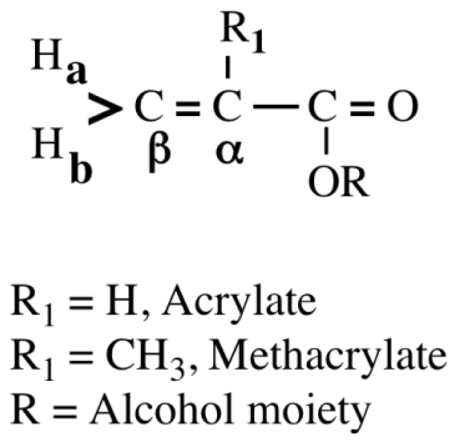
Acrylates and methacrylates (Figure 1) are widely used in the formation of polymeric materials for medical, dental and industrial applications.

Figure 1.
The structure of acrylates and methacrylates.
Many studies have investigated the hydrolysis reaction of acrylates and methacrylates [1–6] because the hydrolysis of monomers and decomposition of cured polymers are implicated in environmental pollution and toxicity. The hydrolysis reaction is one of the major sources of degradants (or metabolites) in in vitro and in vivo biodegradation tests stipulated in the Chemical Substance Control Law [7]. Therefore, reliable and economical methods are needed for predicting the hydrolysis rates of the large number of these chemicals. Freidig et al. have previously reported that when acrylates and methacrylates with α,β-unsaturated carbonyl groups undergo hydrolysis in alkaline media, their electrophile (carbon: α-carbon, β-carbon, carbonyl carbon (>C=O) may be preferentially attacked by a nucleophile (water, hydroxyl anion (OH−), glutathione (GSH) thiyl radical (GS−) [6]. Double bonds (C=C) in acrylates and methacrylates act as an active center for Michael addition and radical scavenging oxidation because HOMO (Highest Occupied Molecular Orbital) and LUMO (Lowest Unoccupied Molecular Orbital) in methacrylate molecules exist in the double bonds [8]. Also, since it is well known that the alkaline-mediated hydrolysis of acrylate polymers is due to an attack of OH− on the intermolecular carbonyl carbon in the ester group, the hydrolysis rate constant of a monomer is probably linked to that of the corresponding polymer and their reactions proceed via a similar mechanism. Therefore, we hypothesized that the OH−-catalyzed hydrolysis of acrylates and methacrylates may have an influence on the NMR-chemical shift of their β-carbon (δCβ), because OH− could attack the β-carbon in a monomer molecule in the initial stage through a hydrolysis reaction. Subsequently, through a reaction involving the BAC2 mechanism (i.e., through nucleophilic attack on the >C=O group) in most cases, acrylates and methacrylates are decomposed into acrylic acid and methacrylic acid together with alcohols, respectively, through hydrolysis. The resonance stabilization of methacrylates is dependent on their reactivity with these compounds. Kuznetsova et al. [9] previously investigated the hydrolysis of N,N-dimethylaminoethyl methacrylate via the AAC2 and BAC2 mechanism, and found that the monomer was hydrolyzed to methacrylic acid and amino alcohol in an anion, pH, and surfactant-dependent manner [9]. Acrylate and methacrylate esters are more highly hydrolyzed with a base than with an acid [1,6,9], but their hydrolysis rate constants have been reported to be considerably small under alkaline conditions at 20 °C or 30 °C [1–4,6]. There are a few QSPRs for acid- or base-catalyzed ester hydrolysis of methacrylates [2,4,6], however the limited number of monomers have been chosen for QSARs study [1–4,6] because the hydrolysis rate for many hydrophobic homologous acrylate and methacrylate esters may be below the detection limit of assays. We previously reported that the glutathione (GSH) reactivity of acrylates and methacrylates was significantly related to their δCβ, possibly due to the high reactivity of GS− with Cβ of these monomers [10–12].
Mallik and Das [2] previously investigated the kinetics of alkaline hydrolysis for methyl and ethyl acrylates and their corresponding methacrylates, and revealed a possible relationship between the hydrolysis rate constants and the experimentally determined activation energy for these compounds. Also, Nakajima et al. [13] investigated the relationship between the acid-catalyzed hydrolysis rates for acetates and acrylates and their activation energy calculated using the MOPAC program, and found a good relationship between the hydrolysis rate constant and calculated activation energy for these compounds, except for formates and methacrylates, which showed a large discrepancy between the experimental and calculated values. The authors mentioned that recalculation for formates and methacrylates was desirable whenever possible. If a more precise calculation for monomers is required, this could be one way of utilizing a higher level of theory, such as density function theory or ab initio MO calculation.
In order to clarify the base-catalyzed hydrolysis reaction for acrylates and methacrylates in the light of currently available data, we investigated the relationship between the base-catalyzed hydrolysis rate constants or reaction rate constants with the GSH and 13C NMR chemical shifts (δCβ) or heat of formation (Hf) for these compounds. Previously reported data [14,15] for the independent variables were used.
2. Results and Discussion
2.1. Hydrolysis
2.1.1. δCβ Parameter
Previously reported data for base-catalyzed hydrolysis constants (k1, k3) are shown in Tables 1 and 2. 13C NMR chemical shifts for the α,β-unsaturated carbonyl group of acrylates and methacrylates are shown in Table 1. The 13C NMR chemical shift of the β-carbon (δCβ) of monomers is also quantitatively related to the π-electron density. The higher the π-electron density on the β-carbon, the higher the magnetic field where the NMR peak is observed; that is, as the π-electron density increases, the chemical shift value (δ) becomes smaller [14]. Therefore, if OH− attacks the Cβ in the monomer molecule, it should be possible to correlate the magnitude of the shift with the reactivity of the monomers.

Table 1.
Base-catalyzed hydrolysis rate constants (k1, k3), reaction rate constants with glutathione (GSH) for acrylates and methacrylates and the 13C NMR chemical shifts of their β-carbon (δCβ) and α-carbon (δCα).
Table 2.
1H NMR chemical shifts for acrylates and methacrylates and the charge density of their carbonyl carbon.
We investigated the relationship between k1 and δCβ or δCα for both acrylates and methacrylates. There was no relationship between k1 and δCβ or δCα for both acrylates and methacrylates. By contrast, a significant relationship between k1 and δCβ, but not δCα, was obtained when acrylates and methacrylates were separated, particularly in the latter situation. Equation (1) for methacrylates is given as follows:
The k1 value for methacrylates was calculated using this equation. The results are also shown in Table 1. The calculated k1 values for isoPMA and isoBMA with the branched substituent were less than the corresponding experimental values. By contrast, the calculated k1 values for MMA and allyl MA were larger than the experimental values. Also, the k1 value for nBMA could not be determined using Equation (1), and the value of δCβ was below the limit of detection using that equation. A base-catalyzed second-order hydrolysis rate constant of 2.7 × 10⁻³ L/mole-s was estimated using a structure estimation method [16]. The hydrolysis rate constant for nBMA appeared to be less than that for isoBMA, as the δCβ for nBMA was smaller than that for isoBMA. We predicted the k1 value using Equation (1) for some monomers that have been used as medical, dental and industrial materials. The k1 values (mol−1s−1) for ethyleneglycol dimethacrylate (EGDMA), triethyleneglycol dimethacrylate (TEGDMA) and MMA were 0.130, 0.062 and 0.034, respectively. Note that the δCβ values (ppm) for EGDMA, TEGDMA and MMA were 125.9, 125.5, and 125.2, respectively [11]. Dimethacrylates such as EGDMA and TEGDMA appeared to be more hydrolyzed than MMA, a monomethacrylate. This may be due to the fact that dimethacrylates possess two double bonds in the molecule that has an affinity to OH−. We previously investigated the changes in the 13C NMR chemical shift for methacrylates induced by their interaction with the phospholipid liposome system in deuterium oxide (D2O) at 30 °C, and it was found that the changes in chemical shifts of electrophiles (carbons: β-carbon, and carbonyl carbon (>C=O)) for TEGDMA and EGDMA, particularly for the latter, was 3–5 fold larger than those for the corresponding MMA; the chemical shifts of β- and carbonyl carbon in the dimethacrylate molecule shifted to a higher field in liposomes in D2O, compared to those in MMA molecule [18]. Although these finding may not be directly related to hydrolysis of methacrylates, it has been assumed from the shielding of chemical shift of β- and carbonyl carbon in D2O that the hydrolysis rate of dimethacrylates is greater than that of MMA. The higher the reactivity, the higher the field in which β-carbon resonates.
On the other hand, there was no relationship between k1 or k3 and δCβ for both acrylates and methacrylates. In general, the electron-releasing methyl group was expected to reduce the rate of alkaline hydrolysis. Indeed, the k1 value for isoBA was greater than that for isoBMA. Similarly, the k1 value for EA was greater than for the corresponding EMA (Table 1).
The chemical shifts of the proton attached to the β-carbon of monomers are linearly related to the corresponding value of δCβ, where Ha represents the proton trans to the substituent and Hb the proton cis to that. The δHa for acrylates and methacrylates was related to their δCβ much more than the δHb. An understanding of the chemical shift difference between Ha and Hb, |δHa – δHb|, may be necessary to clarify the difference in the hydrolysis rate constants for acrylates and methacrylates. Table 2 shows Ha, Hb and |δHa – δHb|. We investigated the relationship between |δHa – δHb| and δCβ for the separation of the acrylates and methacrylates. There was a significant relationship between |δHa – δHb| and δCβ for acrylates (n = 7, r2 = 0.850, p < 0.01), whereas there was no such relationship for methacrylates when the training set shown in Table 2 was used. Because of the electron-withdrawing character of the carbonyl group in monomers, resonance stabilization increases the electron density at the carbonyl carbon (>C=O). As an example, MMA and its resonance form are shown in Figure 2. The π-electron density of the α,β-unsaturated carbonyl groups could be responsible for the resonance stabilization.
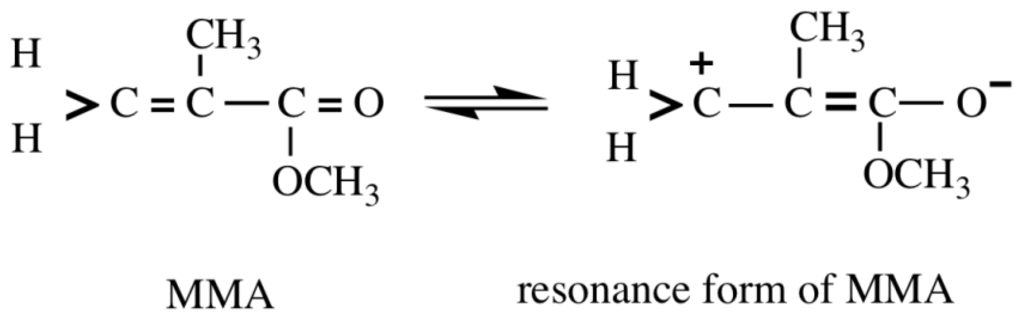
Figure 2.
The resonance form of MMA [14]. The double bonds of MMA monomers change to single bonds, and the diamagnetic anisotropy effect of the carbonyl group at the vinylidene proton is enhanced. As the resonance effect becomes important, the double bond character of CH2=C decreases.
As shown in Table 2, the charge density and |δHa – δHb| value for acrylates were greater than those for methacrylates, suggesting a possible relationship between the electron density at the carbonyl carbon and the |δHa – δHb| value. These findings can be interpreted in terms of a relationship between the base-catalyzed hydrolysis reaction and the δCβ value for methacrylates and acrylates. For the chemical shifts (Ha, Hb) of the proton attached to β-carbon, the coefficient is much closer to unity for the chemical shift of Ha than that of Hb, probably due to the fact that the chemical shift of Hb is more strongly affected by the diamagnetic anisotropy of the substituent. From the geometrical consideration on the molecular model, differences in the deshielding effects of the C=O group are larger at Hb than at Ha [14]. As the resonance stabilization becomes more important, the |δHa – δHb| becomes larger; the |δHa – δHb| is related to Q which is a measure of resonance stabilization. Q is a parameter concerning a monomer based on the Q–e scheme. As compared with the hydrolysis rate constant (k1) between the acrylate esters and the corresponding methacrylate esters (Table 1), the rate constant for EA is greater than the EMA one, and similarly, that for isoBA is greater than the isoBMA one.
Next, we investigated the relationships between the base-catalyzed hydrolysis rate constant (k3) and δCα or δCβ for the test compounds, acrylates (methyl acrylate, ethyl acrylate, butyl acrylate) and methacrylate (MMA) (Table 1). With the exception of MMA, significant linear relationships of k3 with δCα and δCβ were found, the correlation being r2 = 0.97 and r2 = 0.98, respectively. An increase of k3 occurred when δCβ increased, whereas k3 increased as δCα decreased. Although the hydrolysis rates were determined for only a limited number of acrylates (n = 3), this finding indicated that k3 increased along with the π-electron density for Cα. Thus in acrylates, the preferential site of attack of OH− may be both the Cα and Cβ, which is possibly due to the absence of α-methyl substituent in the acrylate molecule. However, further studies will be needed to clarify the details of ester hydrolysis of acrylates in alkaline media.
The hydrolysis rates of chemicals are reportedly correlated with Hammet σ, the Taft parameters, σ* and Taft steric property parameter, E(s), and the van der Waals radius [6,19]. Freidig et al. reported previously that the base-catalyzed hydrolysis of six methacrylates was able to establish a linear free energy relationship as a function of the Taft σ* but not of the Taft E(s) [6]. Therefore, we examined the relationships for the δCβ vs. Taft σ* for the six monomers, and it was found that a good relationship between the two descriptors was obtained (r2 = 0.9, p < 0.01) (data not shown). This clearly indicated that the attack of OH− on the β-carbon of methacrylates may be also associated with the electronic effects of the substituents (R, alcohol moiety as shown in Figure 1). The chemical shifts δCβ for methacrylates likely reflect their electronic effects of the substituents. The π density shown in the NMR spectra is thus considered to be a useful parameter.
2.1.2. Hf Parameter
Table 3.
Reported data for base-catalyzed hydrolysis rate constants (k1, k2) and heat of formation (Hf, ΔHf°) for acrylates and methacrylates and their rate constants calculated using the relevant Equations.
Next we investigated the relationship between k1 and Hf, and found that it was significant (r2 = 0.89, p < 0.05). Equation (2) is given as follows:
Data on the physical and thermodynamic properties of vinyl monomers are needed for the design and operation of industrial chemical processes. Vatani et al. previously predicted the standard enthalpy of formation using a QSPR model [21]. The application of this model may be useful for evaluating the biological activity of various vinyl monomers used for medical, dental and industrial materials. We investigated the relationship between the calculated Hf and the ΔHf° reported in DIPPR 9801 [20] for acrylates and methacrylates using the data set shown in Table 3, and found a good linear relationship between the two independent variables (n = 10, r2 = 0.992, p < 0.001).
The relationship between k1 and ΔHf° for both acrylates and methacrylates is given as Equation (3) as follows:
The k1 value in the ΔHf° term for acrylates and methacrylates was calculated from Equation (3), and the results are shown in Table 3. The experimentally determined and calculated values of k1 for each monomer were similar. We then investigated the relationship between the rate constant (k2) and Hf for MA, EA, MMA and EMA. Equation (4) is given as follows:
The calculated k2 value is also shown in Table 3. These findings indicated that the k1 or k2 value for both acrylate and methacrylate esters was related to their Hf. In the present study, the hydrolysis rate constant for acrylate esters with the simple alkyl substituent was higher than for the corresponding methacrylate esters. It was concluded that the Hf (or ΔHf°) value for acrylates and methacrylates is valuable for the estimation of the rate constants. As the Hf (or ΔHf°) value for monomers increased, their hydrolysis rate constant increased. The hydrolysis rate-determining process for acrylates and methacrylates may be controlled by their Hf (or ΔHf°) value.
2.2. GSH Reactivity
Acrylate and methacrylate esters are important chemicals in the polymer industry, and their induction of toxicity is considered to involve alkylation of crucial cellular nucleophiles through the Michael reaction [22]. We investigated the relationship between kGSH and δCα or δCβ for both acrylates and methacrylates, and obtained a good correlation for δCβ. For the δCβ, Equation (5) is given as follows:
By contrast, there was a weak correlation for δCα (r2 = 0.65). It was assumed from this that the preferential site of GS− attack for these monomers would likely be the β-carbon. We calculated log kGSH using Equation (5) and the result is also shown in Table 1. As the δCβ for monomers increased, their log kGSH increased. This showed that π-electron density of β-carbon for both acrylates and methacrylates may play an important role in the rate-determining process in GSH reactivity. GSH reactivity for acrylates was considerably higher than for methacrylates. It is interesting to note that the log kGSH for both acrylates and methacrylates was not related to their Hf. Freidig et al. reported that hydrolysis of acrylates does not interfere with the GSH reactivity assay [6]. In the present study, the base-catalyzed hydrolysis rate constant for acrylates and methacrylates was correlated with their Hf value (Equations (3) and (4)), whereas conversely, their GSH reactivity was not correlated with the Hf value. These findings support those of the above study [6].
Putz et al. [23] determined the actual quantitative-structure activity relationships (QSARs) for biological activity using the parameters recommended by the Hansch group [24] (hydrophobicity, polarizability and total energy) and special reactivity indices (electronegativity (χ) and chemical hardness (η)) employing computational chemistry. The need for these parameters has potentially been met by QSARs and quantitative structure-property relationships (QSPRs), and the total enthalpy of formation as an independent variable is useful for kinetic studies of the hydrolysis reaction of vinyl monomers. We recently reported a good QSAR for biological activities vs. theoretical parameters for vinyl monomers [12] and phenolic compounds [25]. On the other hand, to develop the phenolic carbonate ester prodrugs, Østergaard and Larsen investigated the water, acid and base catalyzed hydrolysis rate constants for various carbonate esters with fatty acid-like structures, bioreversible derivatives of phenols [26]. They reported that the hydrolysis rate constant of such chemicals may be useful for estimating their physical properties favorable for drug transport to the target site within the body.
3. Experimental Section
3.1. Monomers
The monomers used are abbreviated as follows. Acrylates: methyl acrylate (MA), ethyl acrylate (EA), n-butyl acrylate (nBA), isobutyl acrylate (isoBA), hexyl acrylate (Hexyl A); Methacrylates: methyl methacrylate (MMA), ethyl methacrylate (EMA), isopropyl methacrylate (isoPMA), isobutyl methacrylate (isoBMA), allyl methacrylate (allyl MA), benzyl methacrylate (benzyl MA).
3.2. NMR Spectra
The 13C NMR chemical shift data for various monomers in chloroform-d (CDCl3) were taken from the literature [14]. Briefly, the chemical shifts of the indicated monomers were measured in CDCl3 at 35 °C at 125 and/or 500 MHz, respectively, using tetramethylsilane (TMS) as an internal standard.
3.3. Hydrolysis
The hydrolysis rate constants (k1) for acrylate EA, HA, isoBA and methacrylates (MMA, allyl MA, benzyl MA, isoPMA, isoBMA) under alkaline conditions (pH 10) were taken from Freidig et al. [6]. The hydrolysis rate measurements were carried out using an HPLC method at 20 °C. Also, the alkaline hydrolysis rate constant k2 (second-order rate constant) for MA, EA, MMA and EMA at 30 °C was taken from Mallik and Das [2]. Furthermore, the second-order rate constant (k3) data for base-catalyzed acrylates (MA, EA, BA) and methacrylates (MMA) obtained using a diffusion method were taken from Sharma and Sharma [4].
3.4. Heats of Formation (Hf) and Standard Enthalpy of Formation (ΔHf°)
For calculation of the molecular HF descriptor, the optimized chemical structures of compounds are needed. We have previously calculated the Hf values for acrylates and methacrylates, and those data were used in the present work [10,11,15]. Briefly, calculations of Hf were performed using the PM3/CONFLEX method. To obtain fine geometry details in the present study, initial geometry optimization was performed using CONFLEX5 (Conflex, Tokyo, Japan), then calculations using the PM3 method in the MOPAC 2000 program were carried out on a Tektronix CAChe workstation (Fujitsu Ltd., Tokyo, Japan). Also, the ΔHf° values for acrylates and methacrylates were taken from the literature [20,21].
3.5. GSH Reactivity
Data for GSH reactivity were taken from the literature [6]. The reaction rate constant (kGSH) was measured at 20 °C at pH 8.8.
3.6. Multi-Regression Analysis
The multi-regression equations were calculated using StatMate III (ATMS Co., Ltd., Tokyo, Japan).
4. Conclusions
The present study has shown that the base-catalyzed hydrolysis rate constant for methacrylates is significantly related to δCβ. By contrast, there was no relationship between two independent variables for acrylates. A good relationship for the hydrolysis rate constant vs. heat of formation was also obtained for both acrylates and methacrylates. The GSH reactivity for both acrylates and methacrylates was related to δCβ, but not to Hf. The NMR spectra and heat of formation for acrylates and methacrylates could be used to estimate the base-catalyzed hydrolysis rate constants and GSH reactivity of these compounds, and also may be an important tool for examining the mechanism of their reactivity.
Acknowledgements
We thank M. Ishihara of Meikai University for the semi-empirical calculations.
References
- Mabey, W.; Mill, T. Critical review of hydrolysis of organic compounds in water under environmental conditions. J. Phys. Chem. Ref. Data 1978, 7, 383–415. [Google Scholar]
- Mallik, K.L.; Das, M.N. Alkaline hydrolysis of acrylic and methacrylic esters in 60% aqueous ethanol. Naturwissenschaften 1964, 51. [Google Scholar] [CrossRef]
- Sharma, R.C.; Sharma, M.M. Kinetics of fast alkaline hydrolysis of esters. J. Appl. Chem 1969, 19, 162–166. [Google Scholar]
- Sharma, R.C.; Sharma, M.M. Kinetics of alkaline hydrolysis of esters. II. Unsaturated esters and oxalic Esters. Bull. Chem. Soc. Jpn 1970, 43, 642–645. [Google Scholar]
- Kadoma, Y.; Tanaka, M. Acid and base-catalyzed hydrolysis of bisphenol A-related compounds. Dent. Mater. J 2000, 19, 139–152. [Google Scholar]
- Freidig, A.P.; Verhaar, H.J.M.; Hermen, J.L.M. Quantitative structure-property relationships for the chemical reactivity of acrylates and methacrylates. Environ. Toxicol. Chem 1999, 18, 1133–1139. [Google Scholar]
- Chemical Substance Control Law. Available online: http://www.safe.nite.go.jp/english/db.html accessed on 12 December 2011.
- Fujisawa, S.; Kadoma, Y.; Ishihara, M.; Atsumi, T.; Yokoe, I. Dipalmitoylphosphatidylcholine (DPPC) and DPPC/cholesterol liposomes as predictors of the cytotoxicity of bis-GMA related compounds. J. Liposome Res 2004, 14, 39–49. [Google Scholar]
- Kuznetsova, N.A.; Kazantsev, O.A.; Shirshin, K.V.; Khokhlova, T.A.; Malyshev, A.P. Hydrolysis of N,N-dimethylaminoethyl methacrylate and its salts in concentrated aqueous solution. Russ. J. Appl. Chem 2003, 7, 1117–1120. [Google Scholar]
- Ishihara, M.; Fujisawa, S. A structure-activity relationship study on the mechanisms of methacrylate-induced toxicity using NMR chemical shift of β-carbon, RP-HPLC log P and semiempirical molecular descriptor. Dent. Mater. J 2009, 28, 113–120. [Google Scholar]
- Fujisawa, S.; Kadoma, Y. Prediction of the reduced glutathione (GSH) reactivity of dental methacrylate monomers using NMR spectra—Relationship between toxicity and GSH reactivity. Dent. Mater. J 2009, 28, 722–729. [Google Scholar]
- Fujisawa, S.; Kadoma, Y. Mechanism of action of (meth)acrylayes in hemolytic activity, in vivo toxicity and dipalmitoylphosphatidylcholine (DPPC) liposomes determined using NMR spectroscopy. Int. J. Mol. Sci 2012, 13, 758–773. [Google Scholar]
- Nakajima, M.; Sakuratani, Y.; Noguchi, Y.; Yamada, J.; Hori, K. Development of hydrolysis prediction system using reaction analysis with quantum chemical calculation. J. Comput. Aided Chem 2007, 18, 103–113. [Google Scholar]
- Hatada, K.; Kitayama, T.; Nishiura, T.; Shibuya, W. Relation between reactivities of vinyl monomers and their NMR spectra. Curr. Org. Chem 2002, 6, 121–153. [Google Scholar]
- Ishihara, M.; Fujisawa, S. Quantum-chemical descriptors for estimating hemolytic activity of aliphatic and aromatic methacrylates. Chemosphere 2008, 70, 1898–1902. [Google Scholar]
- Mill, T.; Haag, W.; Penwell, P.; Pettit, T.; Johnson, H. Environmental Fate and Exposure Studies Development of a PC-SAR for Hydrolysis: Esters, Alkyl Halides and Epoxides; EPA Contract No. 68-02-4254; SRI International: Menlo Park, CA, USA, 1987. [Google Scholar]
- Lawrence, W.H.; Bass, G.E.; Purcell, W.P.; Autian, J. Use of mathematical models in the study of structure-toxicity relationships of dental compounds: I. Esters of acrylic and methacrylic acids. J. Dent. Res 1972, 51, 526–535. [Google Scholar]
- Fujisawa, S.; Kadoma, Y.; Komoda, Y. Changes in NMR chemical shifts of methacrylates induced by their interactions with the phospholipid and the phospholipid/cholesterol liposome system. Dent. Mater. J 1990, 9, 100–107. [Google Scholar]
- Collett, T.W. Ester hydrolysis rate constant prediction from infrared interferograms. Environ. Sci. Technol 1990, 24, 1671–1676. [Google Scholar]
- Design Institute for Physical Properties Research (DIPPR). American Institute of Chemical Engineers, Project 801. 2006. Available online: http://www.aiche.org/dippr/products/801.aspx accessed on 12 December 2011.
- Vatani, A.; Mehrpooya, M.; Gharagheizi, F. Prediction of standard enthalpy of formation by a QSAR model. Int. J. Mol. Sci 2003, 8, 407–432. [Google Scholar]
- McCarthy, T.J.; Hayes, E.P.; Schwartz, C.S.; Witz, G. The reactivity of selected acrylate esters toward gluthatione and deoxyribonucleosides in vitro structure-activity relationships. Fundam. Appl. Toxicol 1994, 22, 543–548. [Google Scholar]
- Putz, M.V.; Ionaşcu, C.; Putz, A.M.; Ostafe, V. Alert QSAR. Implication for electrophilic theory of chemical carcinogenesis. Int. J. Mol. Sci 2011, 12, 5098–5134. [Google Scholar]
- Hansch, C.; Kurup, A.; Garg, R.; Gao, H. Chem-bioinformatics and QSAR: A review of QSAR lacking positive hydrophobic terms. Chem. Rev 2001, 101, 619–672. [Google Scholar]
- Fujisawa, S.; Kadoma, Y. Relationship between phenol-induced cytotoxicity and experimental inhibition rate constants or theoretical parameters. Mini Rev. Med. Chem 2012, in press. [Google Scholar]
- Østergaard, J.; Larsen, C. Bioreversible derivatives of phenol. 2. Reactivity of carbonate esters with fatty acid-like structures towards hydrolysis in aqueous solutions. Molecules 2007, 12, 2396–2412. [Google Scholar]

© 2012 by the authors; licensee Molecular Diversity Preservation International, Basel, Switzerland. This article is an open-access article distributed under the terms and conditions of the Creative Commons Attribution license (http://creativecommons.org/licenses/by/3.0/).