Betulin Complex in γ-Cyclodextrin Derivatives: Properties and Antineoplasic Activities in In Vitro and In Vivo Tumor Models


 and
and
Abstract
:1. Introduction
2. Results
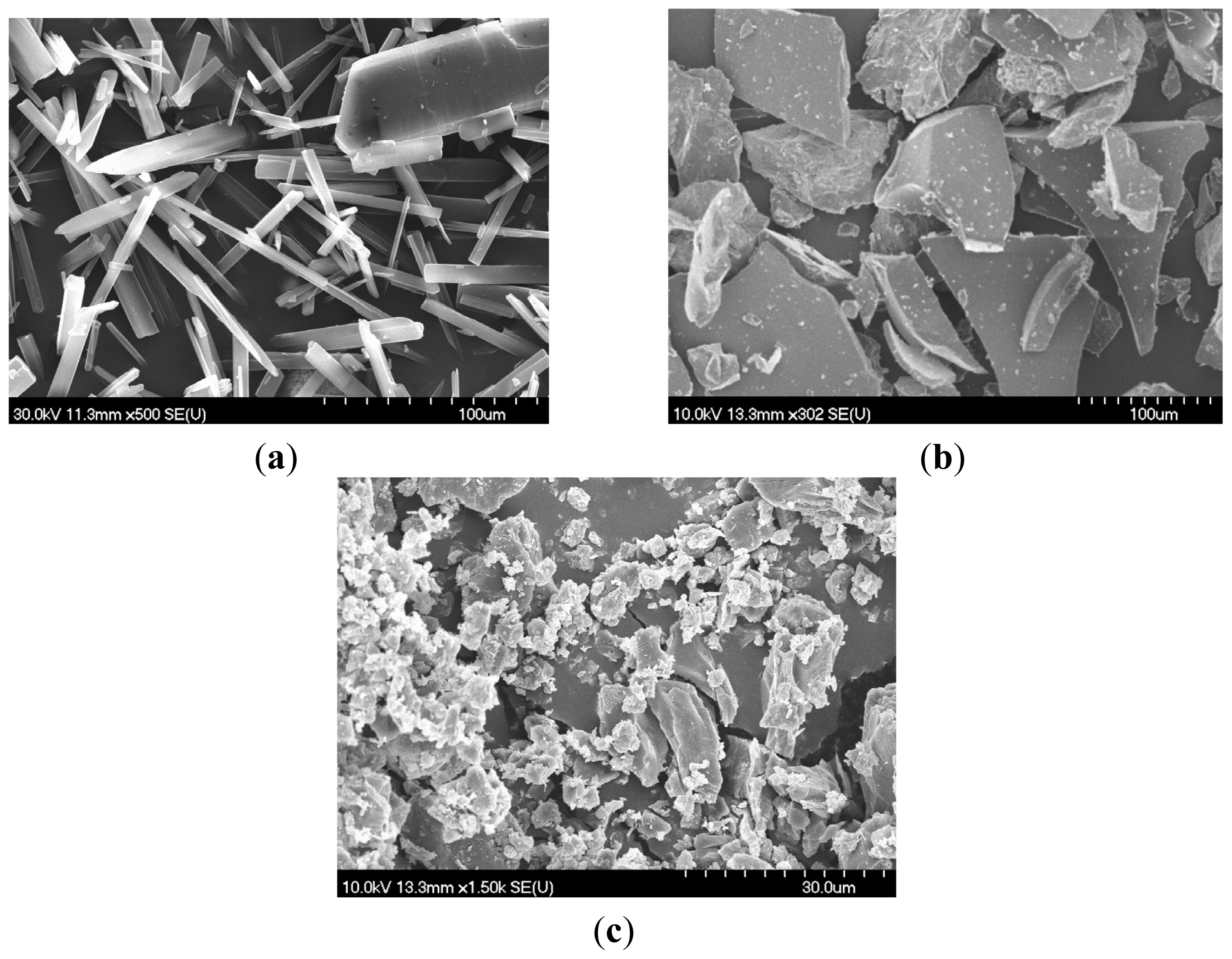
2.1. SEM
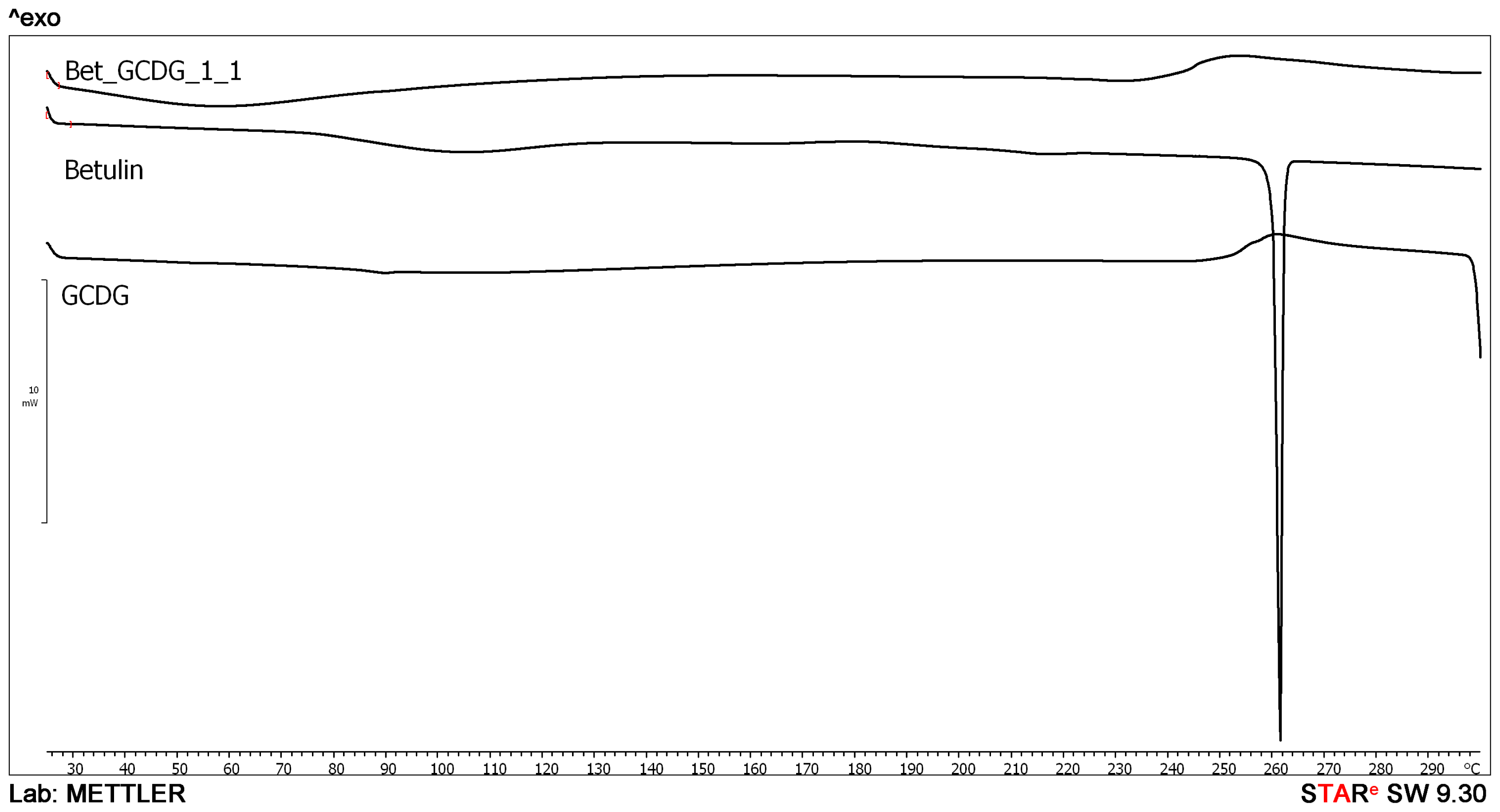
2.2. DSC
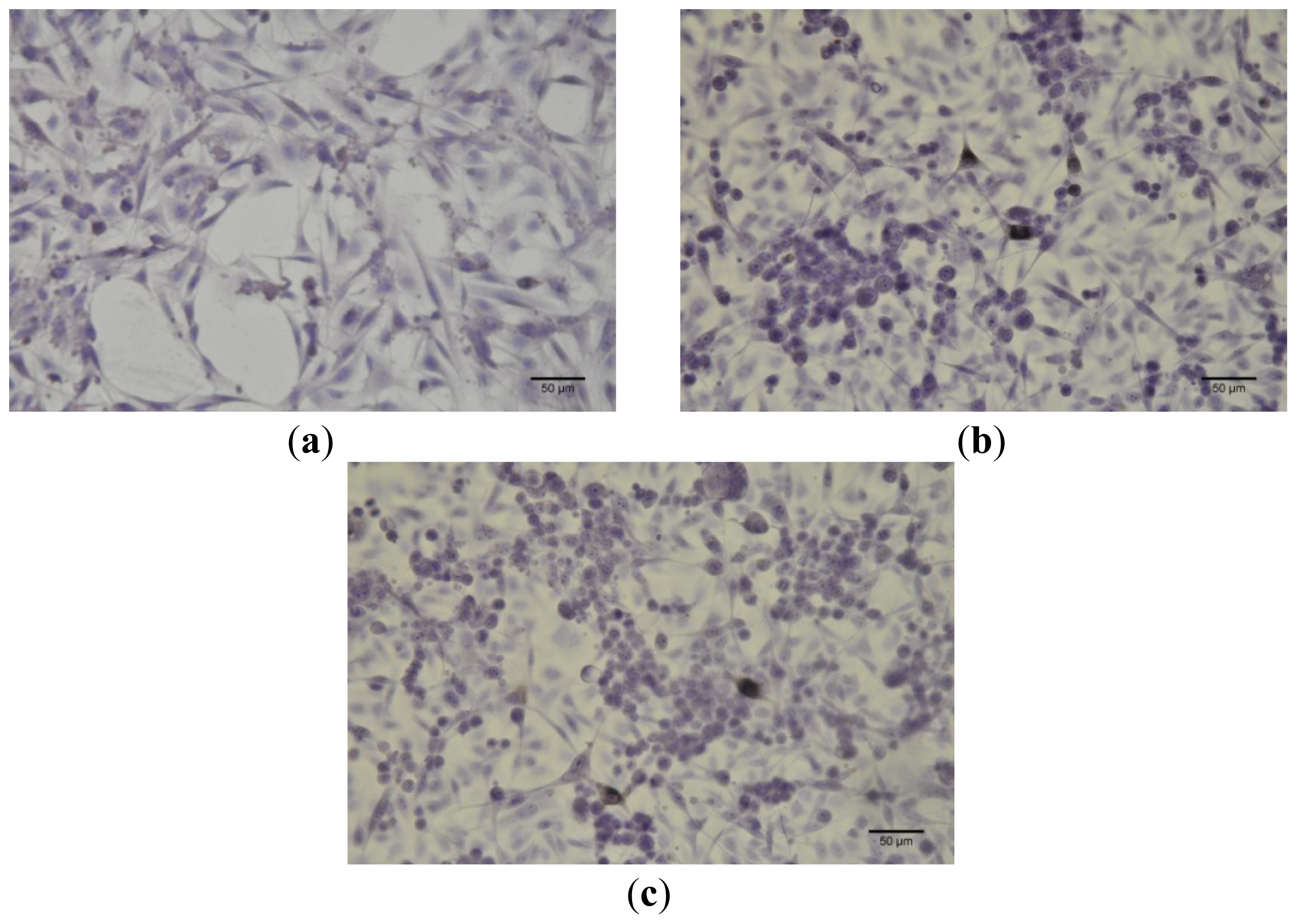
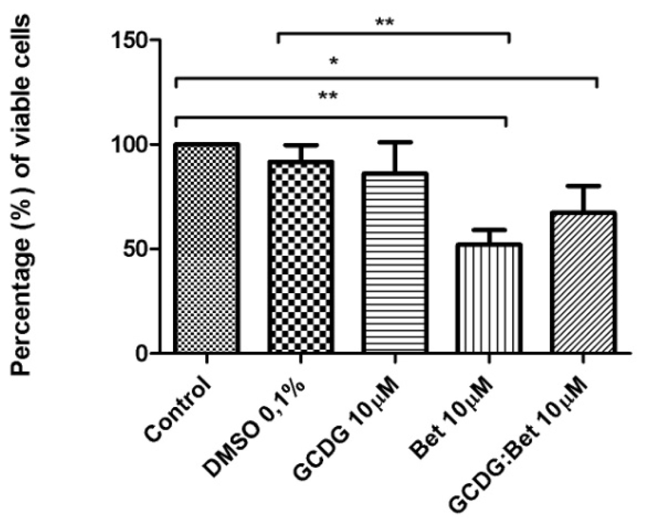
2.3. MTT in Vitro Analysis
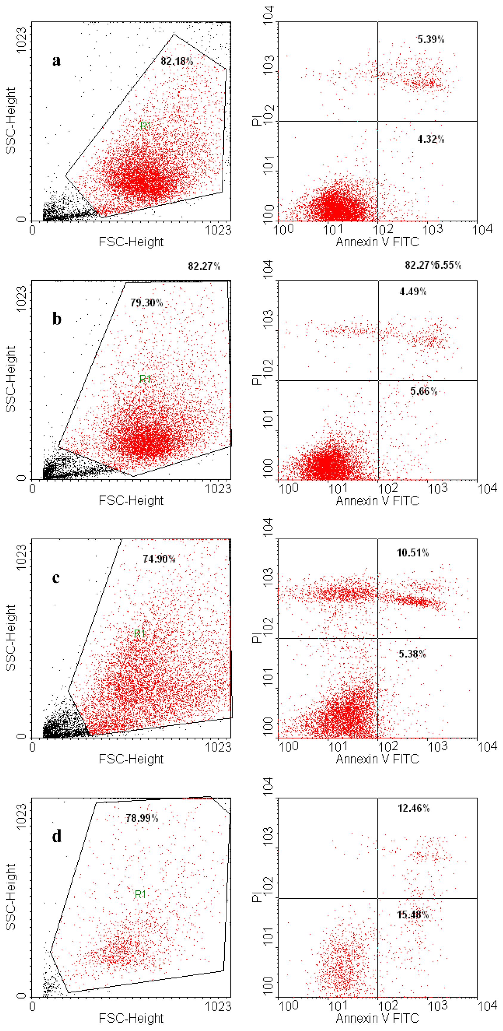
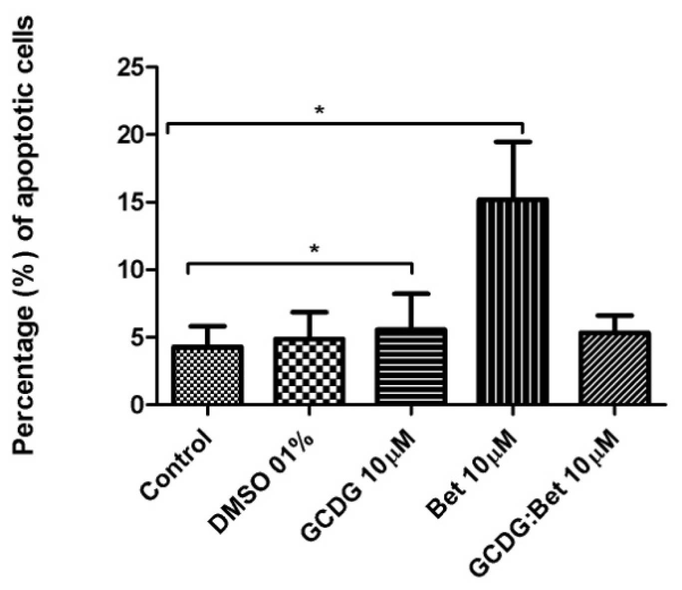
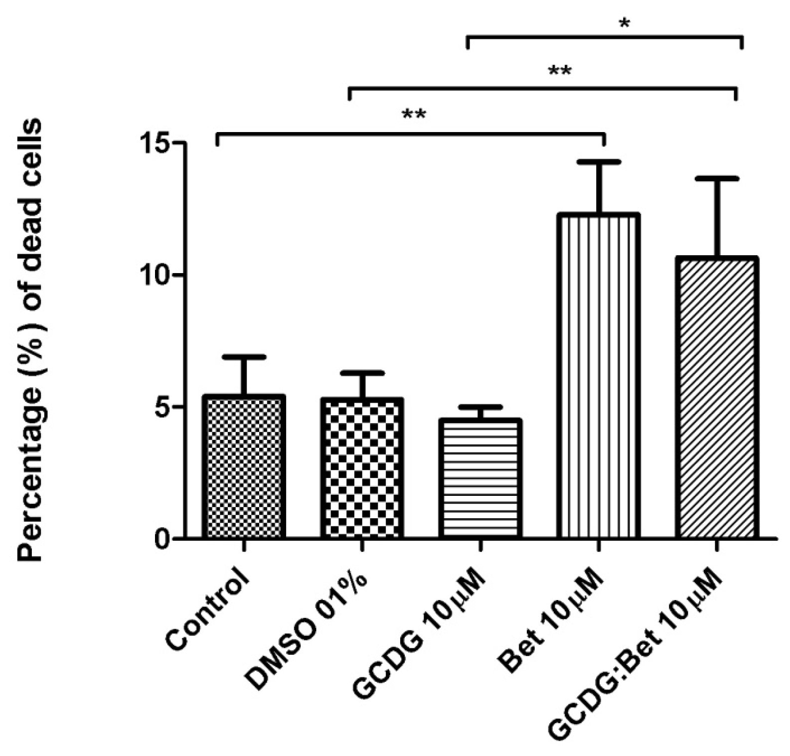
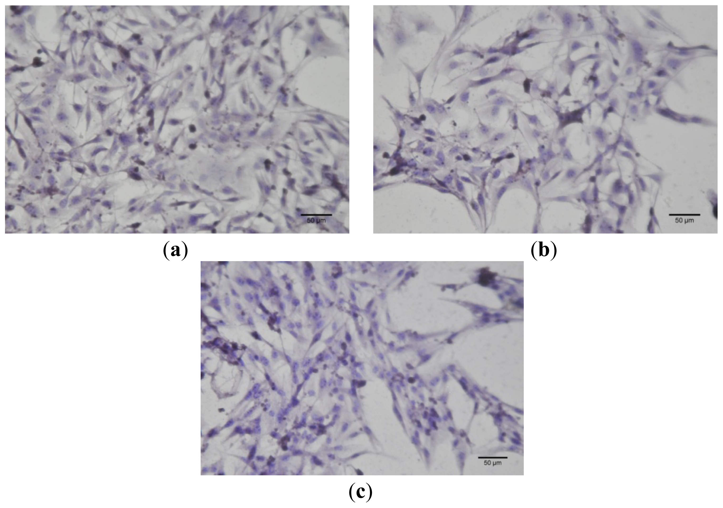
2.4. Defining Mechanism-Based Toxicity
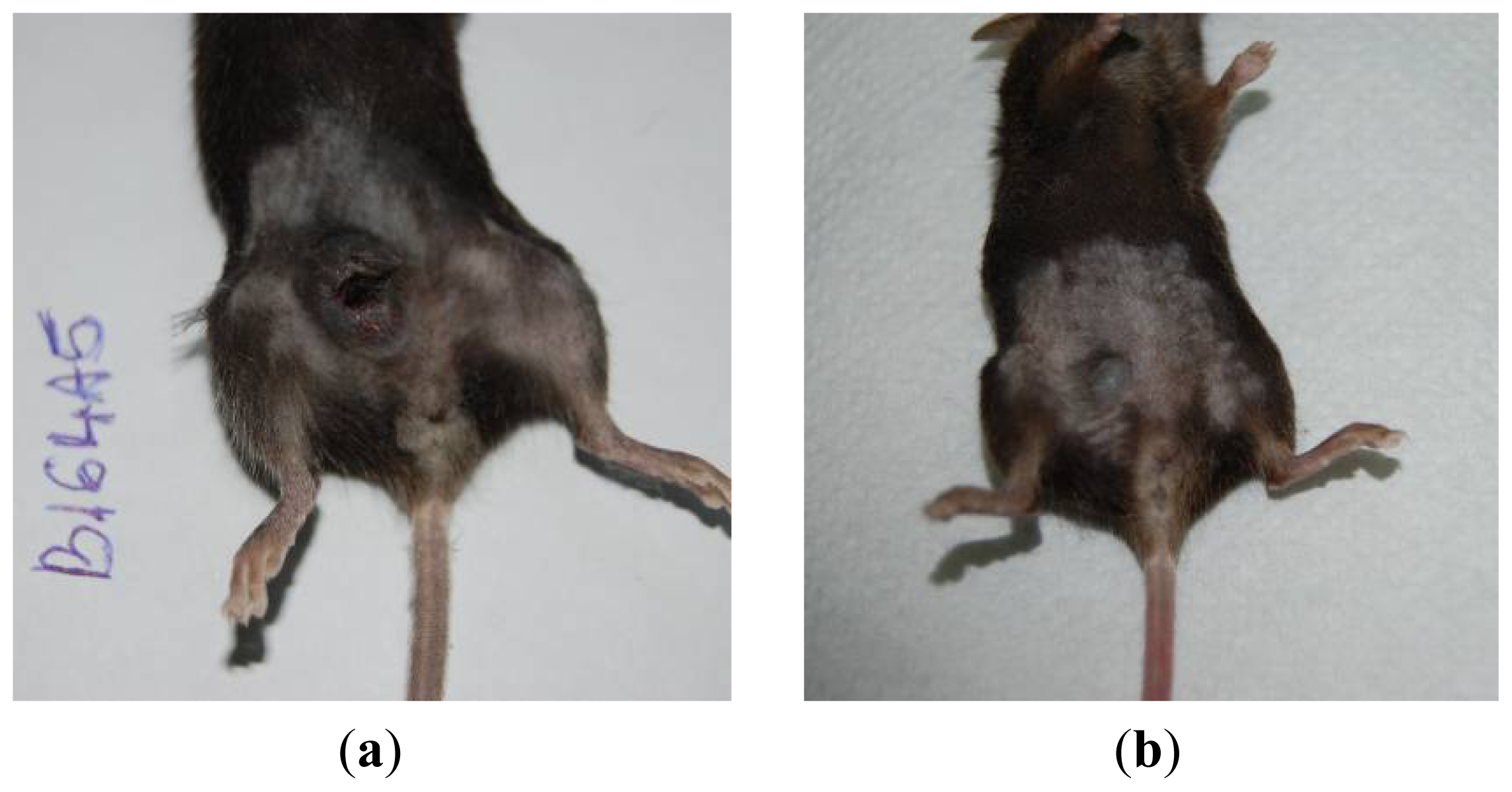
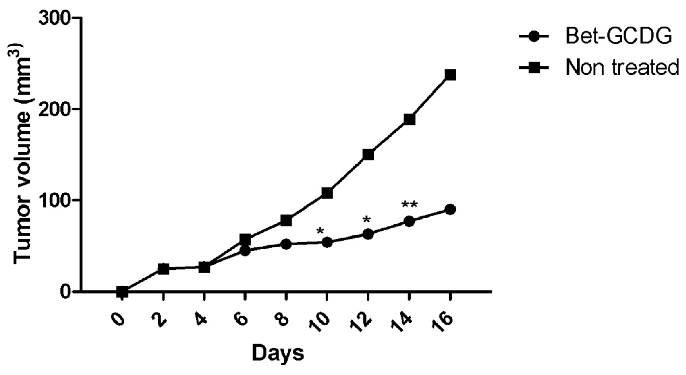
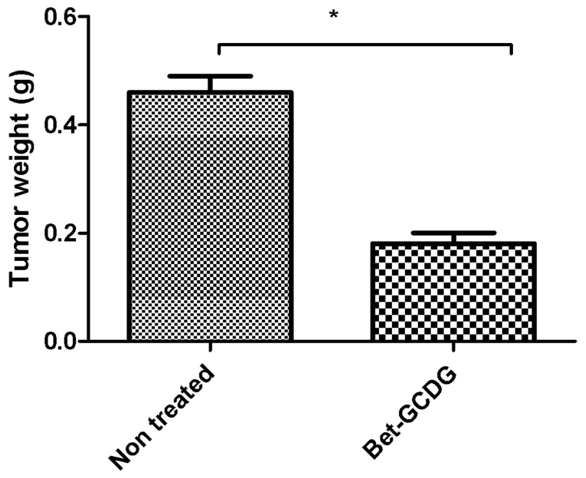
2.5. In Vivo Analysis
3. Discussion
4. Experimental Section
4.1. Preparation of Complexes
4.2. Scanning Electron Microscopy (SEM)
4.3. Differential Scanning Calorimetry (DSC)
4.4. MTT in Vitro Analysis
4.5. Cell Culture Immunocytochemistry
4.6. Annexin V/PI Assay
4.7. Cell Cycle Test
4.8. Syngeneic Tumor Model
Ethics Statement
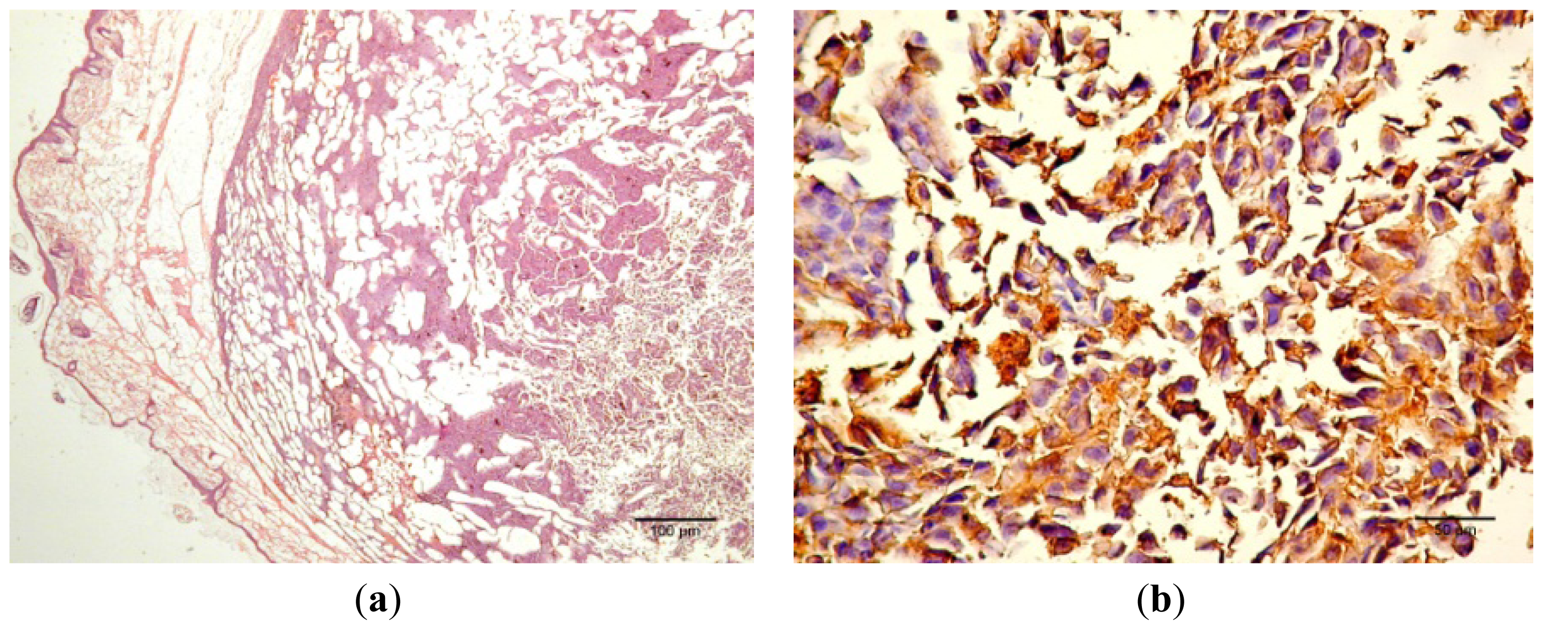
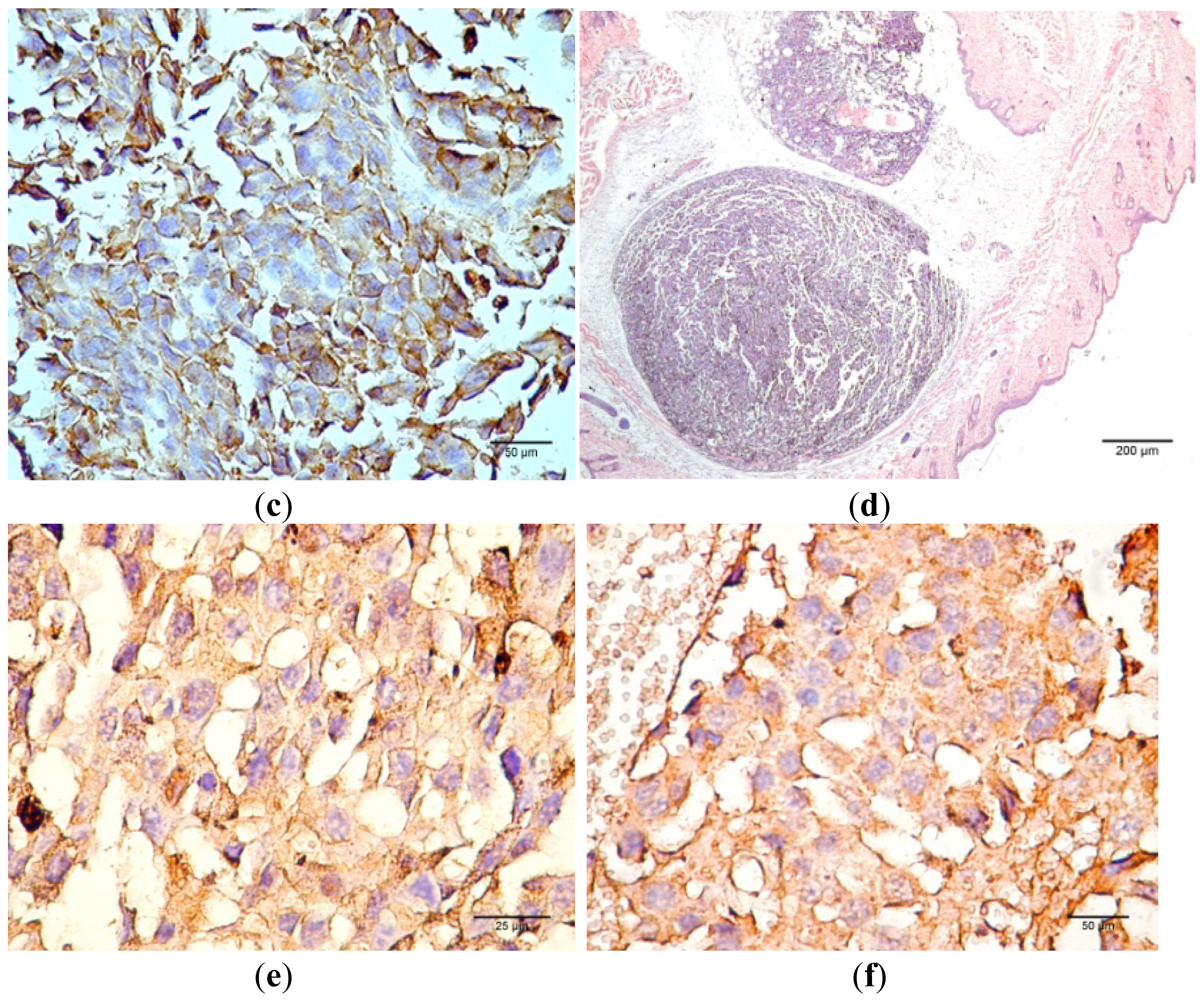
4.9. Histology, S100 and VEGF Expression
4.10. Statistical Analysis
5. Conclusions
Supplementary Information
ijms-13-14992-s001.pdf- Conflict of InterestThe authors declare no conflict of interest.
References
- Santos, R.C.; Salvador, J.A.R.; Marin, S.; Cascante, M. Novel semisynthetic derivatives of betulin and betulinic acid with cytotoxic activity. Bioorg. Med. Chem 2009, 17, 6241–6250. [Google Scholar]
- Yogeeswari, P.; Sriram, D. Betulinic acid and its derivatives: A review on their biological properties. Curr. Med. Chem 2005, 12, 657–666. [Google Scholar]
- Fujioka, T.; Kashiwada, Y.; Kilkuskie, R.E.; Consentino, L.M.; Ballas, L.M.; Jiang, J.B.; Janzen, W.P.; Chen, I.S.; Lee, K.H. Anti-AIDS agents, 11. Betulinic acid and platanic acid as anti-HIV principles from Syzigium claviflorum, and the anti-HIV activity of structurally related triterpenoids. J. Nat. Prod 1994, 57, 243–247. [Google Scholar]
- Pisha, E.; Chai, H.; Lee, I.S.; Chagwedera, T.E.; Farnsworth, N.R.; Cordell, G.A.; Beecher, C.W.; Fong, H.H.; Kinghorn, A.D.; Brown, D.M. Discovery of betulinic acid as a selective inhibitor of human melanoma that functions by induction of apoptosis. Nat. Med 1995, 1, 1046–1051. [Google Scholar]
- Cichewicz, R.H.; Kouzi, S.A. Chemistry, biological activity, and chemotherapeutic potential of betulinic acid for the prevention and treatment of cancer and HIV infection. Med. Res. Rev 2004, 24, 90–114. [Google Scholar]
- Patocka, J. Biologically active pentacyclic triterpenes and their current medicine signification. J. Appl. Biomed 2003, 1, 7–12. [Google Scholar]
- Kvasnica, M.; Sarek, J.; Klinotova, E.; Dzubak, P.; Hajduch, M. Synthesis of phthalates of betulinic acid and betulin with cytotoxic activity. Bioorg. Med. Chem 2005, 13, 3447–3454. [Google Scholar]
- Gauthier, C.; Legault, J.; Lavoie, S.; Rondeau, S.; Tremblay, S.; Pichette, A. Synthesis and cytotoxicity of bidesmosidic betulin and betulinic acid saponins. J. Nat. Prod 2009, 72, 72–81. [Google Scholar]
- Mullauer, F.B.; Kessler, J.H.; Medema, J.P. Betulin is a potent anti-tumor agent that is enhanced by cholesterol. PLoS One 2009, 4, e1. [Google Scholar]
- Dehelean, C.A.; Soica, C.; Peev, C.; Ciurlea, S.; Feflea, S.; Kasa, P., Jr. A pharmaco-toxicological evaluation for betulinic acid mixed with hydroxipropilgamma cyclodextrin on in vitro and in vivo models. Farmacia 2010, 59, 51–59. [Google Scholar]
- Steffen, A.; Thiele, C.; Tietze, S.; Strassnig, C.; Kämper, A.; Lengauer, T.; Wenz, G.; Apostolakis, J. Improved cyclodextrin-based receptors for camptothecin by inverse virtual screening. Chem. Eur. J 2007, 13, 6801–6809. [Google Scholar]
- Wang, H.M.; Wenz, G. Solubilization of polycyclic aromatics in water by γ-cyclodextrin derivatives. Chem. Asian J 2011, 6, 2390–2399. [Google Scholar]
- Wang, H.M.; Şoica, C.M.; Wenz, G. A comparison investigation on the solubilization of betulin and betulinic acid in cyclodextrin derivatives. Nat. Prod. Comm 2012, 7, 289–291. [Google Scholar]
- Eichenmuller, M.; Hemmerlein, B.; von Schweinitz, D.; Kappler, R. Betulinic acid induces apoptosis and inhibits hedgehog signalling in rhabdomyosarcoma. Br. J. Cancer 2010, 103, 43–51. [Google Scholar]
- Alakurtti, S.; Makela, T.; Koskimies, S.; Yli-Kauhaluoma, J. Pharmacological properties of the ubiquitous natural product betulin. Eur. J. Pharm. Sci 2006, 29, 1–13. [Google Scholar]
- Huyke, C.; Laszczyk, M.; Scheffler, A.; Ernst, R.; Schempp, C.M. Treatment of actinic keratoses with birch bark extract, a pilot study. J. Dtsch. Dermatol. Ges 2006, 4, 132–137. [Google Scholar]
- Huyke, C.; Reuter, J.; Rodig, M.; Kersten, A.; Laszczyk, M.; Scheffler, A.; Nashan, D.; Schempp, C. Treatment of actinic keratoses with anovel betulin-based oleogel. A prospective, randomized, comparative pilot study. J. Dtsch. Dermatol. Ges 2009, 7, 128–133. [Google Scholar]
- Dehelean, C.A.; Feflea, S.; Ganta, S.; Amiji, M. Anti-angiogenic effects of betulinic acid administered in nanoemulsion formulation using chorioallantoic membrane assay. J. Biomed. Nanotechnol 2011, 7, 317–324. [Google Scholar]
- Gauthier, C.; Legault, J.; Piochon-Gauthier, M.; Pichette, A. Advances in the synthesis and pharmacological activity of lupane-type triterpenoid saponins. Phytochem. Rev 2011, 10, 521–544. [Google Scholar]
- Şoica, C.; Dehelean, C.; Peev, C.; Coneac, G.; Gruia, A.T. Complexation with hydroxipropilgammacyclodextrin of some pentacyclic triterpenes. Characterisation of their binary products. Farmacia 2008, 56, 182–190. [Google Scholar]
- Wenz, G.; Strassnig, C.; Thiele, C.; Engelke, A.; Morgenstern, B.; Hegetschweiler, K. Recognition of ionic guests by ionic beta-cyclodextrin derivatives. Chem. Eur. J 2008, 14, 7202–7211. [Google Scholar]
- Hertrampf, A.; Gründemann, C.; Jäger, S.; Laszczyk, M.; Giesemann, T.; Huber, R. In vitro cytotoxicity of cyclodextrin-bonded birch bark extract. Planta Med 2012, 78, 881–889. [Google Scholar]
- Rekharsky, M.V.; Inoue, Y. Cyclodextrins and Their Complexes; Dodziuk, H., Ed.; Wiley-VCH Verlag GmbH &Co. KGaA: Weinheim, Germany, 2006; Volume Chapter 8, Microcalorimetry; pp. 200–203. [Google Scholar]
- Naidu, N.B.; Chowdary, K.P.R.; Murthy, K.V.R.; Satyanarayana, V.; Hayman, A.R.; Becket, G. Physicochemical characterization and dissolution properties of meloxicam–cyclodextrin binary system. J. Pharm. Biomed. Anal 2004, 35, 75–86. [Google Scholar]
- Mura, P.; Bettinetti, G.P.; Cirri, M.; Maestrelli, F.; Sorrenti, M.; Catenacci, L. Solid-state characterization and dissolution properties of naproxen-arginine-hydroxypropyl-beta-cyclodextrin ternary system. Eur. J. Pharm. Biopharm 2005, 59, 99–106. [Google Scholar]
- Liu, Y.; Chen, G.S.; Chen, Y.; Cao, D.X.; Ge, Z.Q.; Yuan, Y.J. Inclusion complexes of paclitaxel and oligo(ethylenediamino) bridged bis(beta-cyclodextrin)s: Solubilization and antitumor activity. Bioorg. Med. Chem 2004, 12, 5767–5775. [Google Scholar]
- Şoica, C.; Peev, C.; Ciurlea, S.; Ambrus, R.; Dehelean, C. Physico-chemical and toxicological evaluations of betulin and betulinic acid interactions with hydrophilic cyclodextrins. Farmacia 2010, 58, 611–619. [Google Scholar]
- Şoica, C.M.; Dehelean, C.A.; Peev, C.I.; Aluas, M.; Zupko, I.; Kása, P., Jr; Alexa, E. Physico-chemical comparison study of betulinic acid, betulin and birch bark extract and in vitro investigation of their cytotoxic effects towards skin epidermoid carcinoma (A431), breast carcinoma (MCF7) and cervix adenocarcinoma (HeLa) cell lines. Nat. Prod. Res. 2012, 26, 968–974. [Google Scholar]
- Muceniece, R.; Saleniece, K.; Riekstina, U.; Krigere, L.; Tirzitis, G.; Ancans, J. Betulin binds to melanocortin receptors and antagonizes alpha-melanocyte stimulating hormone induced cAMP generation in mouse melanoma cells. Cell. Biochem. Funct 2007, 25, 591–596. [Google Scholar]
- Rose, M.L.; Madren, J.; Bunzendahl, H.; Thurman, R.G. Dietary glycine inhibits the growth of B16 melanoma tumors in mice. Carcinogenesis 1999, 20, 793–798. [Google Scholar]
- Kommera, H.; Kaluderovid, G.N.; Kalbitz, J.; Paschke, R. Lupane triterpenoids—Betulin and betulinic acid derivatives induce apoptosis in tumor cells. Invest. New Drugs 2011, 29, 266–272. [Google Scholar]
- Li, Y.; He, K.; Huang, Y.; Zheng, D.; Gao, C.; Cui, L.; Jin, Y.H. Betulin induces mitochondrial cytochrome c release associated apoptosis in human cancer cells. Mol. Carcinog 2010, 49, 630–640. [Google Scholar]
- Wang, D.-Y.; Liu, J.; Yin, M.-Z.; Li, X.-T.; Lou, G.; Liu, Y.-D.; Chen, X.-W. Betulin induces apoptosis of HeLa cell lines in vitro and its possible mechanism. Tumor 2012, 32, 234–238. [Google Scholar]
- Oh, S.H.; Choi, J.E.; Lim, S.C. Protection of betulin against cadmium-induced apoptosis in hepatoma cells. Toxicology 2006, 220, 1–12. [Google Scholar]
- Gava, B.; Zorzet, S.; Spessotto, P.; Cocchietto, M.; Sava, G. Inhibition of B16 melanoma metastases with the ruthenium complex imidazolium trans-imidazoledimethylsulfoxide-tetrachlororuthenate and down-regulation of tumor cell invasion. J. Pharmacol. Exp. Ther 2006, 317, 284–291. [Google Scholar]
- Fulda, S. Betulinic acid for cancer treatment and prevention. Int. J. Mol. Sci 2008, 9, 1096–1107. [Google Scholar]
- Zhanataev, A.K.; Presnova, G.A.; Chistyakov, A.N.; Durnev, A.D. Effect of betula bark extract on spontaneous and induced mutagenesis in mice. Bull. Exp. Biol. Med 2004, 138, 475–478. [Google Scholar]
- Jager, S.; Laszczyk, M.N.; Scheffler, A. A preliminary pharmacokinetic study of betulin, the main pentacyclic triterpene from extract of outer bark of birch (Betulae alba cortex). Molecules 2008, 13, 3224–3235. [Google Scholar]
- Dehelean, C.A.; Soica, C.M.; Toma, C.-C.; Feflea, S.; Gruia, A.T.; Kasa, P., Jr. Antitumoral activity of betulin, a compound present in the birch tree, in formulation with cyclodextrin. Studia Univ. VG Seria St. Vietii 2010, 20, 55–58. [Google Scholar]
- Cheng, X.; Shin, Y.G.; Levine, B.S.; Smith, A.C.; Tomaszewski, J.E.; van Breemen, R.B. Quantitative analysis of betulinic acid in mouse, rat and dog plasma using electrospray liquid chromatography/mass spectrometry. Rapid Commun. Mass Spectrom 2003, 17, 2089–2092. [Google Scholar]


| Sample (10 μM) | Cell Cycle Phases | |||
|---|---|---|---|---|
| sub-G0 (%) | G0/G1 (%) | S (%) | G2/M (%) | |
| Control | 0.49 ± 0.02 | 79.65 ± 3.99 | 6.56 ± 0.16 | 10.01 ± 0.26 |
| DMSO | 0.73 ± 0.04 | 76.08 ± 2.21 | 11.54 ± 0.26 | 11.65 ± 0.18 |
| GCDG | 0.91 ± 0.04 | 79.42 ± 3.82 | 8.54 ± 0.18 | 8.54 ± 0.16 |
| Bet:GCDG 1:1 complex | 2.27 ± 0.05 | 82.76 ± 4.01 | 4.81 ± 0.15 | 8.41 ± 0.17 |
| Bet | 1.21 ± 0.03 | 67.83 ± 3.75 | 15.09 ± 0.31 | 10.39 ± 0.27 |
© 2012 by the authors; licensee Molecular Diversity Preservation International, Basel, Switzerland. This article is an open-access article distributed under the terms and conditions of the Creative Commons Attribution license (http://creativecommons.org/licenses/by/3.0/).
Share and Cite
Şoica, C.; Dehelean, C.; Danciu, C.; Wang, H.M.; Wenz, G.; Ambrus, R.; Bojin, F.; Anghel, M. Betulin Complex in γ-Cyclodextrin Derivatives: Properties and Antineoplasic Activities in In Vitro and In Vivo Tumor Models. Int. J. Mol. Sci. 2012, 13, 14992-15011. https://doi.org/10.3390/ijms131114992
Şoica C, Dehelean C, Danciu C, Wang HM, Wenz G, Ambrus R, Bojin F, Anghel M. Betulin Complex in γ-Cyclodextrin Derivatives: Properties and Antineoplasic Activities in In Vitro and In Vivo Tumor Models. International Journal of Molecular Sciences. 2012; 13(11):14992-15011. https://doi.org/10.3390/ijms131114992
Chicago/Turabian StyleŞoica, Codruta, Cristina Dehelean, Corina Danciu, Hai Ming Wang, Gerhard Wenz, Rita Ambrus, Florina Bojin, and Mariana Anghel. 2012. "Betulin Complex in γ-Cyclodextrin Derivatives: Properties and Antineoplasic Activities in In Vitro and In Vivo Tumor Models" International Journal of Molecular Sciences 13, no. 11: 14992-15011. https://doi.org/10.3390/ijms131114992
APA StyleŞoica, C., Dehelean, C., Danciu, C., Wang, H. M., Wenz, G., Ambrus, R., Bojin, F., & Anghel, M. (2012). Betulin Complex in γ-Cyclodextrin Derivatives: Properties and Antineoplasic Activities in In Vitro and In Vivo Tumor Models. International Journal of Molecular Sciences, 13(11), 14992-15011. https://doi.org/10.3390/ijms131114992