Article

Modulation of rosR Expression and Exopolysaccharide Production in Rhizobium leguminosarum bv. trifolii by Phosphate and Clover Root Exudates

Department of Genetics and Microbiology, Institute of Microbiology and Biotechnology, University of Maria Curie-Skłodowska, Akademicka 19 st., Lublin, 20–033, Poland
*
Author to whom correspondence should be addressed.
Int. J. Mol. Sci. 2011, 12(6), 4132-4155; https://doi.org/10.3390/ijms12064132
Submission received: 23 May 2011 / Revised: 14 June 2011 / Accepted: 15 June 2011 / Published: 22 June 2011
(This article belongs to the Section Biochemistry)

Abstract

:
The acidic exopolysaccharide (EPS) secreted in large amounts by the symbiotic nitrogen-fixing bacterium Rhizobium leguminosarum bv. trifolii is required for the establishment of an effective symbiosis with the host plant Trifolium spp. EPS biosynthesis in rhizobia is a very complex process regulated at both transcriptional and post-transcriptional levels and influenced by various nutritional and environmental conditions. The R. leguminosarum bv. trifolii rosR gene encodes a transcriptional regulator with a C2H2 type zinc-finger motif involved in positive regulation of EPS synthesis. In silico sequence analysis of the 450-bp long rosR upstream region revealed the presence of several inverted repeats (IR1 to IR6) and motifs with significant identity to consensus sequences recognized by PhoB and LysR-type proteins associated with phosphate- and flavonoid-dependent gene regulation in R. leguminosarum. Using a set of sequentially truncated rosR-lacZ transcriptional fusions, the role of the individual motifs and the effect of phosphate and clover root exudates on rosR expression were established. In addition, the significance of IR4 inverted repeats in the repression, and P2–10 hexamer in the activation of rosR transcription, respectively, was found. The expression of rosR increased in the presence of phosphate (0.1–20 mM) and clover root exudates (10 μM). PHO boxes and the LysR motif located upstream of the rosR translation start site were engaged in the regulation of rosR transcription. The synthesis of EPS and biofilm formation decreased at high phosphate concentrations, but increased in the presence of clover root exudates, indicating a complex regulation of these processes.

1. Introduction

Exopolysaccharide (EPS) production is a wide spread feature among bacteria, and several functions are ascribed to this polymer, including protection against environmental stress and host defense responses, nutrient gathering, attachment to surfaces, and biofilm formation [1,2]. In nitrogen-fixing symbiotic bacteria, commonly referred to as rhizobia, EPSs are required for successful nodulation of legumes, such as Trifolium, Pisum, Vicia and Medicago spp., which form indeterminate-type nodules [35]. EPS-deficient mutants of Rhizobium leguminosarum and Sinorhizobium (Ensifer) meliloti are impaired in the invasion of nodule cells and nitrogen fixation [69]. Acidic EPSs secreted in large amounts by rhizobia are species-specific heteropolymers consisting of common sugars substituted with non-carbohydrate residues [4,5,10,11]. Biosynthesis of R. leguminosarum EPS is a multi-step process requiring the activity of several enzymes, most of them encoded by pss genes located in the large chromosomal EPS cluster I [12,13]. Similarly to other bacterial EPSs, the EPS of R. leguminosarum plays a significant role in biofilm formation, being the major component of the matrix [1,14,15]. Also, the level of EPS polymerization is very important for normal biofilm formation, because prsD, plyB, and plyBplyA mutants, forming significantly longer molecules of this polymer than the wild type, are impaired in biofilm formation [16]. Similarly in S. meliloti, galactoglucan (EPS II) (especially its low-molecular-weight fraction) plays a crucial role in biofilm formation [17].
The regulation of EPS biosynthesis in rhizobia is controlled at both transcriptional and post-transcriptional levels and influenced by various nutrient sources and stress conditions. In S. meliloti, which produces two types of EPSs, several regulators of succinoglycan (EPS I) and galactoglucan (EPS II) synthesis have been identified on the chromosome (exoR, exoS, expR, syrM, mucR, and exoD) and megaplasmid pSymB (exsB, expG, and exoX) [3,5]. The majority of these regulators are engaged in negative regulation of EPS production; exoR, exoS, exsB, and exoX genes negatively influence EPS I synthesis, and expR and mucR negatively regulate EPS II synthesis [1820]. ExpG (WggR) is a positive regulator of EPS II [21]. MucR plays a key role in the regulation of both types of EPSs in S. meliloti as a positive regulator of EPS I and a negative regulator of EPS II synthesis [20,22]. The biosynthesis of EPS I is stimulated by hyperosmotic stress, very high phosphate concentrations (>150 mM), and limitation of some nutrient sources such as nitrogen and sulfur [2325]. On the other hand, phosphate limitation stimulates EPS II production [21,25,26], indicating that phosphate concentration is a signal affecting the type of EPS produced by S. meliloti [20]. Phosphate starvation stimulates the expression of exp genes involved in EPS II synthesis by the action of the phosphate-dependent WggR and PhoB regulatory proteins [20,21,27]. PhoB has been found to regulate the transcription of target genes by binding to PHO boxes, which are identified in the promoters of operons involved in EPS II synthesis [28]. The PHO box consensus has recently been found in a large-scale analysis of Pho-dependent promoters in S. meliloti and several other species of proteobacteria [28]. Regulation of EPS II production is further influenced by the Sin quorum sensing system in the presence of the ExpR regulator [29].
Under nitrogen limitation, two proteins, SyrM and NtrC, act as positive regulators of EPS I production in S. meliloti. SyrM increases the expression of some exo genes involved in the synthesis of this polymer via positive stimulation of syrA expression [30,31].
In contrast to S. meliloti, the regulatory mechanisms and external factors influencing EPS biosynthesis in R. leguminosarum have not been extensively studied. Two genes, psiA and psr, identified on the symbiotic plasmid pSym of R. leguminosarum bv. phaseoli, are involved in the regulation of EPS synthesis [32,33]. Multiple psiA copies inhibit EPS synthesis, but this effect is overcome in the presence of additional psr copies. EPS production is also negatively regulated by exoR, which shows extensive similarity to S. meliloti exoR [34]. In R. leguminosarum bv. trifolii, a negative regulatory effect of pssB, encoding inositol monophosphate phosphatase, on EPS synthesis has been described [35].
Recently, rosR encoding a positive transcriptional regulator of EPS synthesis has been identified in R. leguminosarum bv. trifolii [36]. It shares significant identity with mucR of S. meliloti [37], rosR of Rhizobium etli [38], rosAR of Agrobacterium radiobacter [39], and ros of Agrobacterium tumefaciens [40]. All these genes encode transcriptional regulators belonging to the family of Ros/MucR proteins, which have the Cys2His2 type zinc-finger motif and are required for positive or negative regulation of EPS biosynthesis. Mutation in R. leguminosarum bv. trifolii rosR results in substantially diminished amounts of EPS and ineffective symbiosis with clover, whereas multiple copies of this gene cause a nearly two-fold increase of EPS production [36,41]. RosR is a 15.7 kDa protein, which by binding to the 22-bp-long RosR-box sequence located in the rosR upstream region decreases rosR transcription, demonstrating autoregulation of its own gene [36]. Besides RosR-box, two P1 and P2 promoters, and three motifs with significant identity to E. coli consensus sequence binding the cAMP-CRP complex have been found in the promoter region [42]. Also, the significance of an AT-rich UP element located upstream of −35 hexamer and extended −10 TGG motif in an enhancement of the rosR transcription initiation from the P1 promoter was confirmed [42]. In the presence of glucose, rosR transcription has been significantly decreased showing the possibility of regulation of rosR expression by catabolic repression. It has been established that mutation in rosR resulted in several pleiotropic phenotypes, such as quantitative alterations in the polysaccharide constituent of lipopolysaccharide, changes in membrane and secreted protein profiles, and higher sensitivity to surface-active detergents and some osmolytes [43]. In addition, the rosR mutant has been shown to exhibit dramatically decreased attachment to and colonization of clover root hairs.
Here, we examine the significance of several sequence motifs identified in the R. leguminosarum bv. trifolii rosR upstream region in the expression of this gene via transcriptional fusions of the rosR upstream region with lacZ. The level of rosR expression and its correlation with EPS production and biofilm formation are assessed in the presence of phosphate and clover root exudates.

2. Results and Discussion

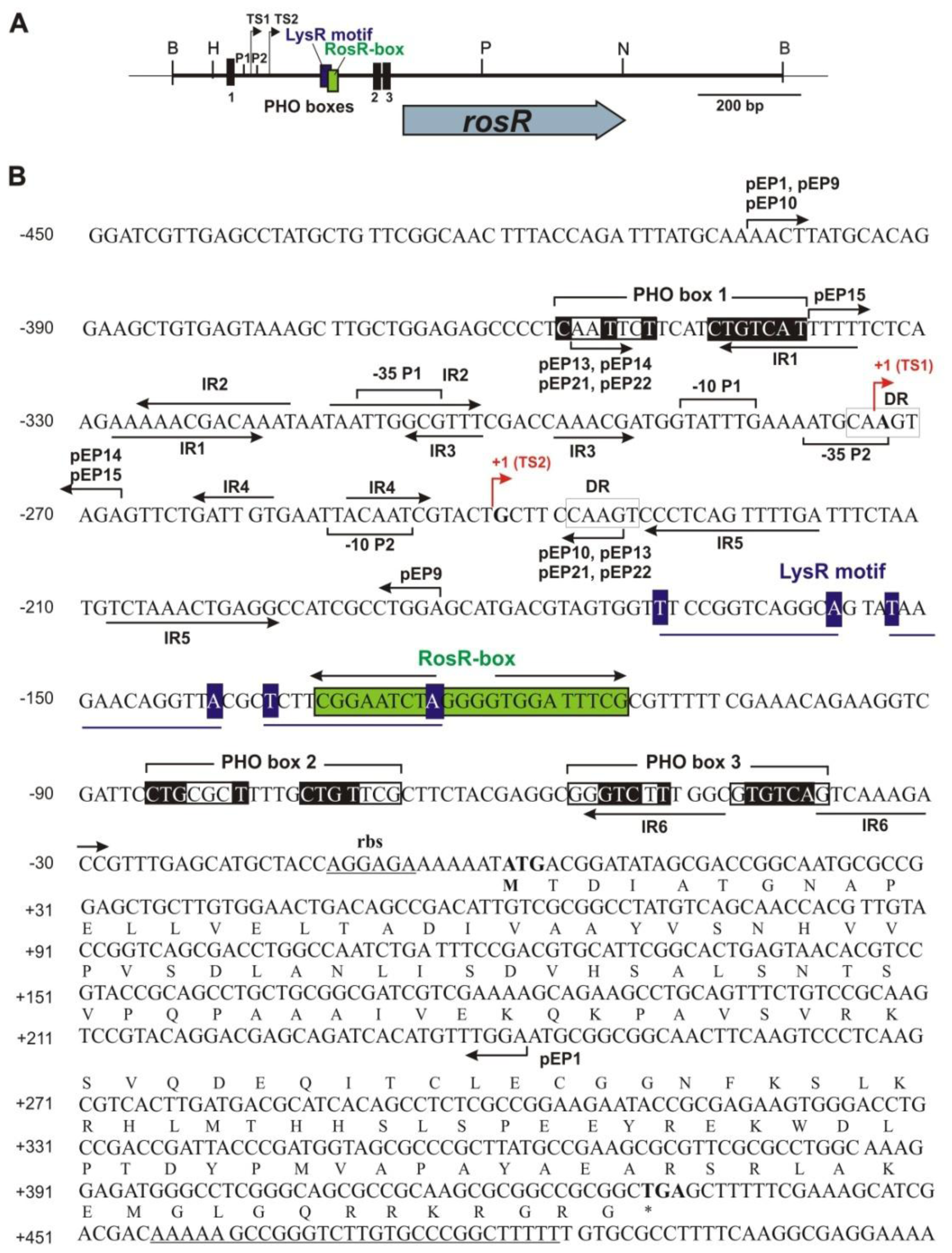
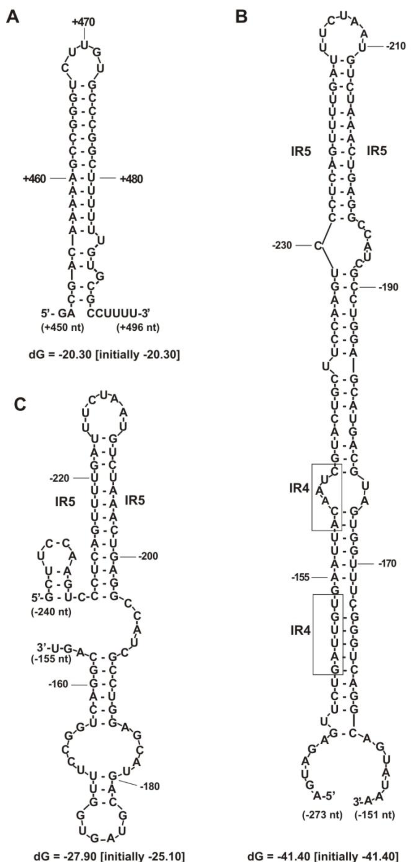
2.1. Identification of Putative Regulatory Motifs in the rosR Upstream Region

Previously, we established that rosR expression was driven from two separate promoters: the distal strong P1 promoter and the proximal, weaker, P2, and two rosR transcripts of different lengths were identified [36]. These transcripts contained 273-nt and 240-nt long 5′-untranslated regions (5′-UTR), respectively. Such long upstream regions have often been described as target sites for the regulation of gene expression [44,45]. In silico sequence analysis of the rosR upstream region revealed the presence of many inverted repeats of different lengths (named IR1 to IR6), and among these IR5 was the longest, with 12-bp inverted repeats (Figure 1). The IR2 motif had 11-bp long inverted repeats, and the IR1 and IR6 motifs had 10-bp long inverted repeats. The inverted repeats IR1, IR2, and IR3 were located upstream of the two transcriptional start sites TS1 and TS2. The IR4 motif was located between TS1 and TS2; the IR5 and the IR6 were located below TS2. Direct repeats (DR) and three motifs bearing similarity to the PHO box consensus sequence were also identified in the rosR upstream region (Figure 1). PHO boxes located in promoters of phosphate-regulated genes contain two 7-nt direct repeats of the 5′-CTGTCAT-3′ consensus sequence separated by a 4-nt A/T rich spacer, and these motifs are targets for the phosphorylated form of the PhoB regulatory protein [28,46].
Among three potential PHO box-like sequences found in the rosR promoter, PHO box 1 located just upstream of the distal P1 promoter comprised a weakly conserved 7-nt direct repeat separated by four nucleotides from a second direct repeat identical to the consensus sequence (Figure 1). Two other PHO boxes were located closer to the translation start codon of rosR and among them, PHO box 3 was more conserved than PHO box 2.
Moreover, a LysR motif, which is a putative target site for proteins belonging to the family of LysR transcriptional regulators, was identified just upstream of the RosR-box. Starting from the first transcription start site (position −273 bp), a 770-nt long transcript was generated. RNA synthesis from the TS2 (position −240 bp) resulted in a 737-nt long transcript. Downstream of the TGA stop codon of rosR, a palindrome sequence of a rho-independent transcription termination site was found (from +452 to +490 bp). This transcriptional terminator was a 39-base long sequence containing two 12-nucleotide inverted repeats separated by 5 nt, which formed a very stable stem structure with an energy of −20.3 kcal/mol (Figure 2A).
Secondary structure analysis of the rosR RNA transcripts initiated at TS1 and TS2 revealed the presence of several additional sequences, which stabilized their structures, especially in the upper part of both transcripts. Three stem structures were generated from the first 120 nucleotides of transcript 1 with a total energy of −41.4 kcal/mol, which were substantially stronger than the very stable structure of the transcriptional terminator (Figure 2B). These included inverted repeats IR5, which were located on the top of this structure. This sequence also played a significant role in stabilizing the upper part of the secondary structure of the shorter transcript 2. Within the first 90 nt of the transcript 2, a structure containing two stems was formed with a total energy of −27.9 kcal/mol (Figure 2C).

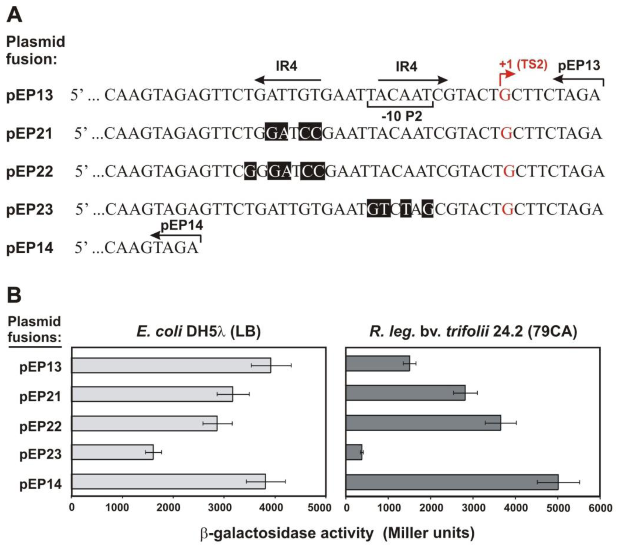
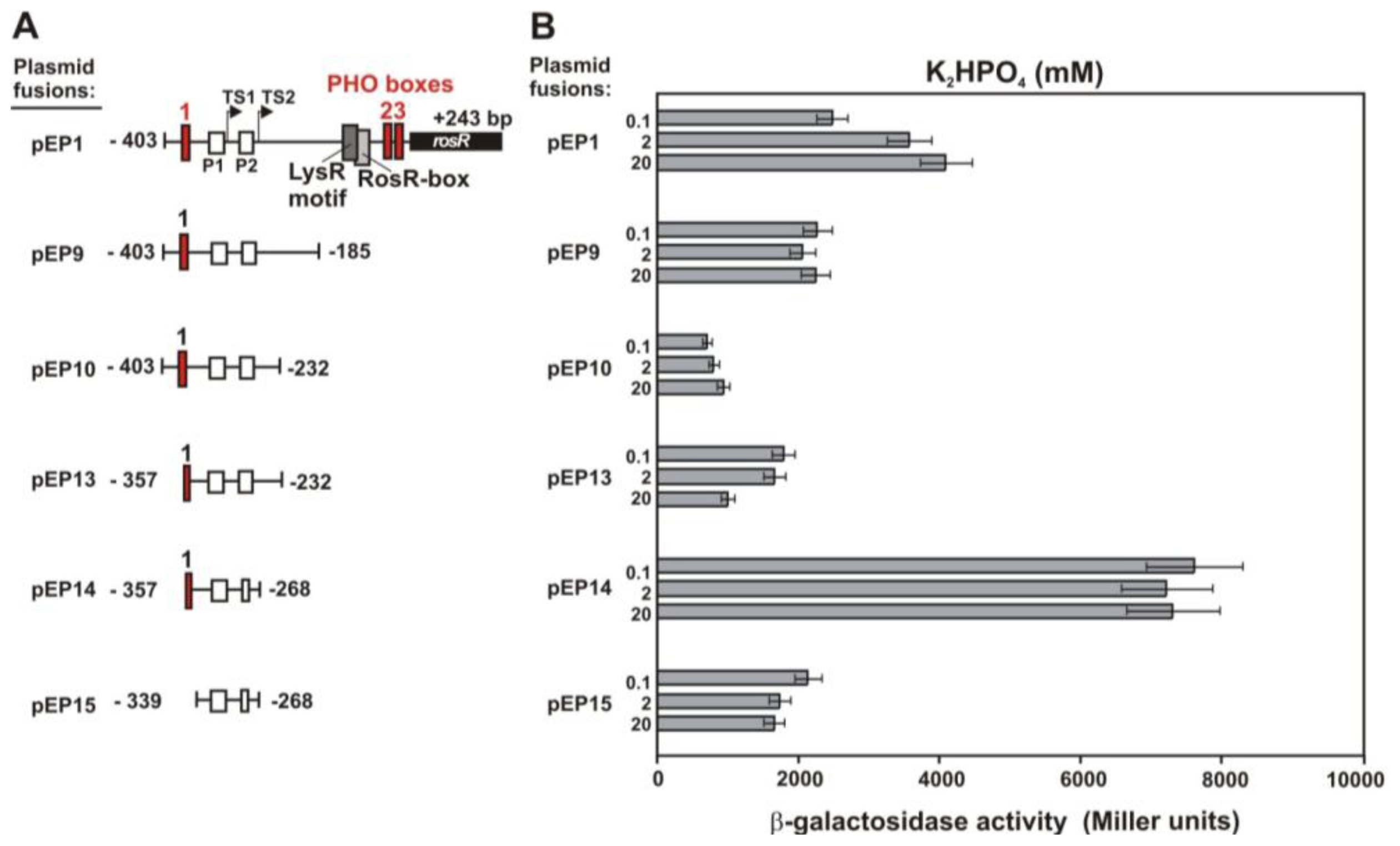
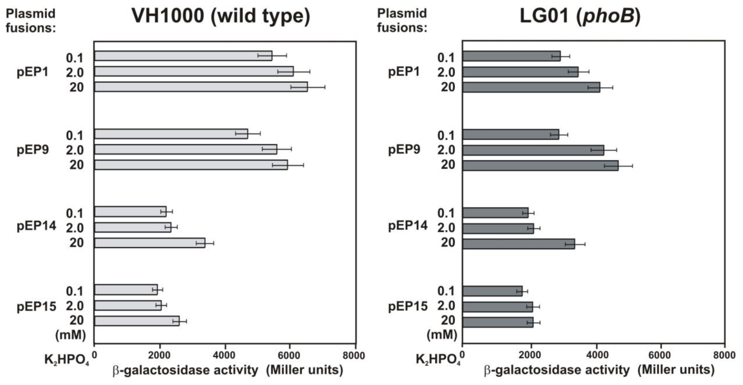
2.2. Functional Analysis of the Putative Regulatory Motif IR4 in the rosR Upstream Region

Previously, we established that deletion of a short DNA fragment in the rosR upstream region (from −232 to −268 bp), located just downstream of the P1 promoter, resulted in a very high increase of rosR transcription [42]. In this fragment, IR4 inverted repeats have been identified via in silico analyses.
To assess the significance of IR4 motif in rosR transcription, we used the previously described pEP13 and pEP14 rosR-lacZ fusions [42]. pEP14 contained a fragment of the rosR upstream region which was 36-bp shorter at the 3′ end than the insert of pEP13. β-galactosidase activity in pEP14 was ca. three-fold higher than in pEP13 when studied in Rt24.2. In contrast, rosR transcription for both fusions was at the same level in E. coli DH5α, suggesting that some element(s) specific for the rhizobial background could be engaged in the repression of rosR expression (Figure 3). In the 36-bp fragment, 6-bp-long inverted repeats, named IR4, separated by a 5-nt spacer were identified (Figures 1 and 3). The second part of IR4 partially overlapped the −10 hexamer of the proximal P2 promoter. Site-directed mutagenesis of both parts of IR4 present in pEP13 was performed, and a set of fusions was obtained (pEP21-pEP23) (Figure 3A). β-galactosidase activity was determined for these plasmids and compared with pEP13 and pEP14 control fusions in both E. coli and Rt24.2 backgrounds (Figure 3B). In the cases of pEP21 and pEP22, with substitutions in the first part of IR4, rosR-lacZ expression in Rt24.2 increased 1.87-fold and 2.44-fold, respectively, in comparison to pEP13, confirming that the IR4 motif was most likely engaged in the negative regulation of rosR transcription. In contrast, the mutation of the second part of IR4 overlapping the −10 hexamer of P2 (pEP23 fusion) resulted in a 4-fold decrease of rosR-lacZ expression in relation to pEP13 (Figure 3B). These data indicate that the P2–10 hexamer is indispensable for rosR expression and positively affects the transcription of this gene.
To elucidate the role of the IR4 motif in the stabilization of RNA, the secondary structure of the rosR transcript initiated at TS1 and containing mutated sequences in both parts of the IR4 was analyzed.
Three stem structures generated from the first 120 nucleotides of the wild type transcript 1 had a total energy of −41.4 kcal/mol, whereas the mutation of the first part of IR4 (corresponding to the pEP21 and pEP22 fusions) resulted in a moderate decrease of RNA secondary structure stability (dG = −35.4 and −35.2 kcal/mol, respectively). The mutation of the second part of IR4 slightly affected the rosR RNA secondary structure stability (dG = −39.6 kcal/mol). It is plausible that the first part of the IR4 motif functions as a target site for some unidentified repressor protein, and its role in the rosR RNA secondary structure stabilization is rather minor. The second part of IR4 that matches the P2–10 hexamer, primarily functions as a binding site of RNA polymerase. Taken together, the results described suggest that the mRNA transcript of this region forming an IR4 loop structure could modulate the level of rosR expression.

2.3. Functional Analysis of Other Putative Regulatory Motifs in the rosR Upstream Region

Phosphate is one of the most important nutrients for bacteria and is often limited in the environment [47]. S. meliloti mutants defective in phosphate transport did not fix nitrogen [48], suggesting a significant role of this nutrient for effective symbiosis. In this bacterium, phosphate regulates a considerable number of genes, among them genes involved in succinoglycan (EPS I) and galactoglucan (EPS II) biosynthesis, through PhoB response regulator forming a two-component regulatory system with PhoR as the sensor kinase [20,21,27,28,44,46]. Whereas phosphate-dependent exopolysaccharide genes regulated by PhoB have been well studied in S. meliloti [20,44], the phosphate regulation of genes involved in EPS synthesis in R. leguminosarum has not been investigated. Computational analysis revealed the presence of phoB (RL0547-position 591, 764–592, 447 nt) in R. leguminosarum bv. viciae 3841 genome [13], encoding a putative PhoB with 100% amino acid identity to R. leguminosarum bv. trifolii WSM1325 PhoB, 93% identity to S. meliloti PhoB and 54% identity to E. coli PhoB [49], indicating a high conservation of the structure and, plausibly, the function of this protein.
To establish whether the sequence of PHO boxes identified in silico played a role in the regulation of rosR expression, rosR-lacZ transcriptional fusions were investigated in vivo. R. leguminosarum bv. trifolii 24.2 wild type (Rt24.2) harboring the pEP1 carrying the longest promoter region (403-bp) or its derivatives containing the promoter region truncated at the 3′- or 5′-end were cultured in the presence of phosphate, and β-galactosidase activities were measured (Figure 4).
In general, rosR demonstrated a high level of expression when the longest fusion, pEP1, was examined in standard M1 medium, confirming the presence of strong promoter in its upstream region. Transcription of the pEP1 increased in the presence of increasing concentrations of phosphate, and the highest level (1.68-fold) was observed in the presence of 20 mM K2HPO4 (Figure 4B). Transcription of pEP1 decreased in the presence of 40 mM K2HPO4 (data not shown).
In order to establish which of the three PHO boxes identified could be involved in phosphate-dependent regulation of rosR transcription, strain Rt24.2 bearing fusions with different deletions was cultured in the individual concentrations of phosphate, and β-galactosidase activities were determined (Figure 4B). The deletion of the 3′-end of the rosR upstream region containing the RosR-box, the LysR motif, and two PHO boxes, resulted in a decrease of rosR-lacZ expression in the pEP9 fusion in relation to pEP1. This result confirmed the essential role of these regulatory elements in rosR expression. The transcription in the pEP10 fusion that had a progressively enlarged deletion of the 3′-end was further diminished. The deleted fragment contained IR5, indicating that this motif, predicted to play a dominant role in RNA secondary structure stabilization, is likely responsible for stimulation of rosR expression. Simultaneous deletions of 46 bp in the 5′-end of the rosR untranscribed region and 36 bp in the 3′-end in pEP14 resulted in a nearly 4-fold increase of the expression of this gene (Figure 4B). In contrast to pEP13, pEP14 lacked IR4 motif which played the substantial role in the negative regulation of rosR transcription.
Unlike the longest pEP1 fusion, the transcriptional activities of the remaining pEP9–pEP15 fusions seem to be phosphate independent (Figure 4B). pEP14, containing a putative PHO box 1 lacking the first nucleotide at the 5′-end, showed a very high transcriptional activity which was rather independent of (or only weakly dependent on) phosphate concentration. This finding excludes the role of PHO box 1 in rosR expression, despite its relatively high identity to the PHO box consensus sequence. The pEP15 fusion with a deletion of the full-length PHO box 1 demonstrated a very low level of rosR expression, which was totally independent of phosphate. These data show that PHO boxes 2 and 3, located close to the translation start site, may be engaged in phosphate-dependent regulation of rosR transcription.
To further assess the function of the PHO boxes identified in the rosR upstream region, the following plasmids were introduced into E. coli wild type and phoB mutant: pEP1 containing all three PHO boxes, pEP9 containing exclusively PHO box 1, pEP14 containing PHO box 1 without the first nucleotide, and pEP15 lacking the PHO box 1, and β-galactosidase activities were measured (Figure 5).
In general, rosR expression in the pEP1 was significantly higher in the wild type than in the phoB mutant. In a low-phosphate medium (0.1 mM), the transcription in pEP9 was lower in relation to pEP1 in the wild type strain (P < 0.05, Student’s test), but this difference disappeared in the phoB mutant. For pEP14 and pEP15, similar profiles of rosR expression were found in both genetic backgrounds. Moreover, a significant decrease of rosR-lacZ expression was observed for pEP14 in comparison to pEP1. The pEP15 fusion, lacking all three PHO box-like sequences, showed a very low rosR expression independent of phosphate in the wild type and the phoB mutant (Figure 5). These results indicate that PhoB from E. coli stimulates rosR transcription, most likely by binding to PHO boxes 2 and/or 3.
All above data suggest that PHO boxes 2 and/or 3, rather than PHO box 1, might play a role in phosphate-dependent regulation of rosR expression, although this effect is not considerable. In general, PhoB protein functions as a positive regulator, which induces the expression of genes belonging to Pho-regulon under phosphate limitation. However, Bardin et al. [50] demonstrated that the expression of orfA-pit encoding a phosphate transport membrane protein in S. meliloti is repressed upon Pi starvation. This finding is in contrast to E. coli, where pit genes are constitutively expressed [51]. In S. meliloti, the majority of PhoB-regulated genes possess PHO boxes located upstream of their promoters [28], whereas for orfA-pit and fixN3 this motif is located just upstream of their putative translation start [50], similarly to PHO boxes 2 and 3 in the rosR promoter.

2.4. Functional Analysis of a Putative LysR Motif

A well-conserved LysR motif (T-N11-A)3, possibly interacting with proteins of the LysR family, was identified in close vicinity of the RosR-box and two PHO boxes in the rosR upstream region (Figure 1). One of these LysR proteins is NodD involved in the transcription activation of nod genes responsible for Nod factor synthesis in the presence of flavonoids. To assess whether NodD influences rosR expression, fusions pEP1 containing the LysR motif and pEP9 lacking it, were introduced into R. leguminosarum bv. trifolii ANU843 wild type and ANU851 carrying a mutation in nodD, and β-galactosidase activity was examined in the presence and absence of clover root exudates (Table 1).
For ANU843(pEP1) growing in the presence of 10 μM exudates, a moderate increase of β-galactosidase activity (1.37-fold) was found. In the ANU851(pEP1) nodD mutant, the effect of exudates was negligible (1.04-fold increase). In the case of pEP9, lacking the LysR motif, rosR expression was similar in the wild type and the nodD mutant, and almost negligently responsive to exudates. These data suggest that the LysR motif present in the rosR upstream region might be functional, recognized by NodD and may influence rosR transcription.
The LysR motif was described as a binding site for several transcription factors containing a helix-turn-helix domain, among them NodD [52]. In S. meliloti, SyrM, also belonging to LysR-type transcription factors, is involved in the activation of the EPS I synthesis genes [30,31], but we did not find a homologous protein encoded by R. leguminosarum genome.

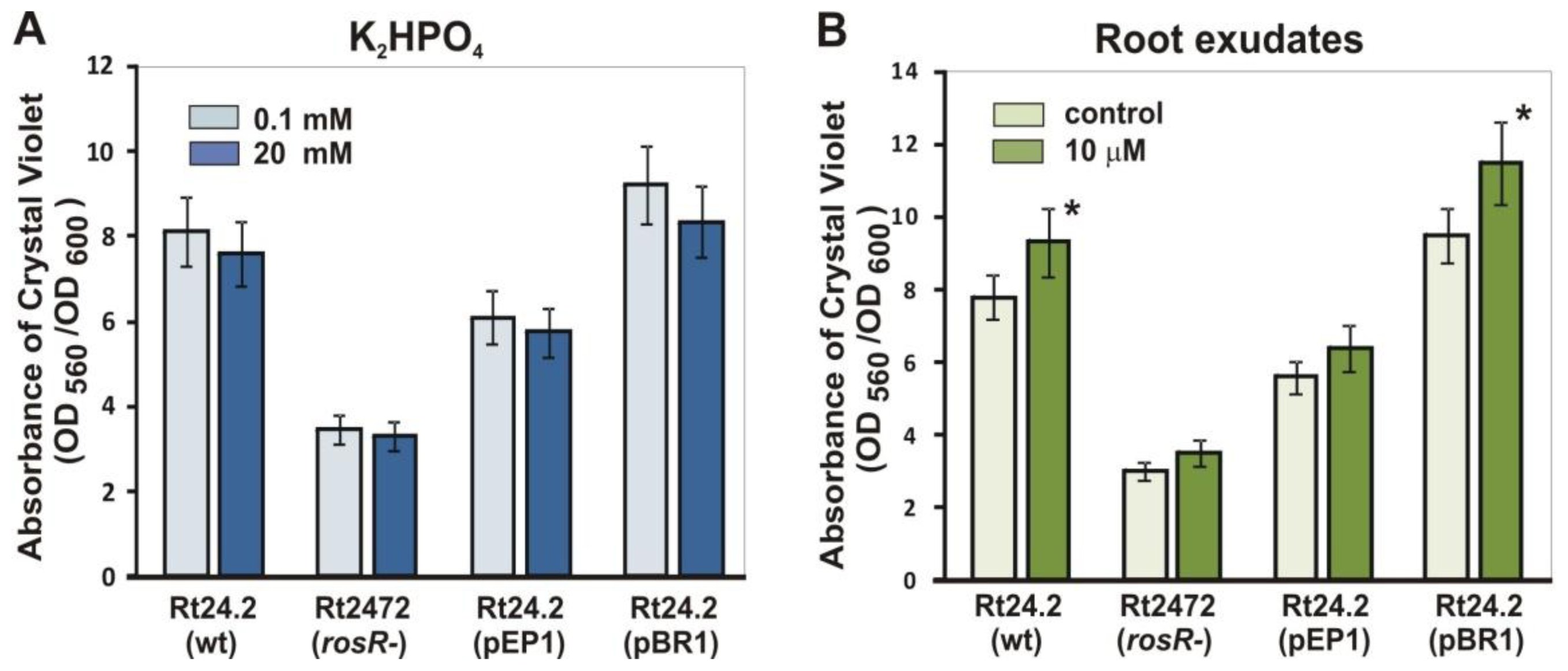
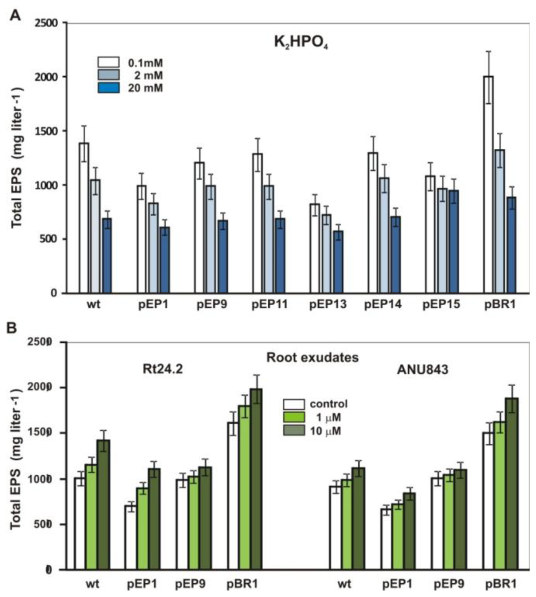
2.5. Effect of Phosphate and Clover Root Exudates on EPS Production

In our previous study, it was found that EPS synthesis in R. leguminosarum was positively controlled by RosR binding to the RosR-box in the promoter region of rosR and pssA encoding the first IP-glycosyl transferase [36]. Here, the effect of phosphate and root exudates on EPS production was studied in Rt24.2 wild type, Rt24.2(pEP1) carrying the full-length rosR upstream region on a low copy plasmid pMP220, Rt24.2 carrying the pEP9-pEP15 fusions, and Rt24.2(pBR1) harboring additional copies of rosR on the pBBR1MCS-2 plasmid (Figure 6).
Among tested growth conditions, the most effective production of this polysaccharide was observed in M1 with 0.1 mM phosphate, and the wild type strain produced 1.94-fold more EPS under phosphate limitation (0.1 mM) than in the presence of high phosphate concentration (20 mM). The stimulation of EPS synthesis in the phosphate starvation was even more visible for Rt24.2 (pBR1) bearing additional rosR copies, confirming positive regulation of EPS synthesis by RosR (1.42-fold more EPS than in the wild type) (Figure 6A). EPS production in all of the tested strains was reduced with increasing concentrations of phosphate, showing a negative effect of this compound even though the rosR transcription (pEP1 fusion) was stimulated by up to 20 mM phosphate. Introduction of multiple copies of the rosR upstream region containing the RosR-box, being the target site for RosR binding, caused a significant reduction of the amount of EPS (pEP1 fusion). Because in the case of pEP14, which almost totally lacked transcriptional responsiveness to phosphate, the strain carrying this fusion still produced more EPS under phosphate limitation than phosphate-rich condition, these results suggest that other chromosomal genes, besides rosR, involved in EPS production might also be regulated by phosphate. This demonstrates complexity of phosphate-dependent regulation of EPS synthesis in R. leguminosarum. The EPS synthesis in this bacterial species seems to be much more sensitive to phosphate concentration than EPS I production in S. meliloti, being efficiently synthesized under very high concentration of phosphate (above 150 mM). The expression of S. meliloti exoYFQ operon engaged in EPS I synthesis was affected by phosphate concentration and was PhoB-dependent, although PHO box-like sequence identified in its promoter had a relatively low sequence identity with the PHO box consensus [44]. In low-phosphate medium, galactoglucan was efficiently produced [25], and both WggR and PhoB regulatory proteins were required for a maximal induction of the transcription of the EPS II biosynthesis gene cluster under Pi starvation [20].
In the case of clover root exudates, a moderate stimulation of EPS synthesis was found for Rt24.2 (1.43-fold) and ANU843 (1.28-fold) wild type strains cultured in M1 supplemented with 10 μM exudates, and full congruence was observed between rosR transcription and EPS production (Table 1 and Figure 6B). This indicated that root exudates positively affected EPS production in R. leguminosarum via stimulation of rosR transcription in the presence of the LysR motif.

2.6. Biofilm Formation by R. leguminosarum bv. trifolii on Abiotic Surfaces in the Presence of Phosphate and Clover Root Exudates

Rhizobia, like many other bacteria, form surface-attached communities, known as biofilm, which most likely protect the bacterium against harmful environmental factors or nutrients deficiency [1,2,53]. Biofilm formation in the presence of phosphate and flavonoids was studied using Rt24.2 wild type, the Rt2472 rosR mutant, Rt24.2 harboring pEP1 with additional copies of the rosR upstream region, and Rt24.2 carrying pBR1 with multiple rosR copies (Figure 7).
Strain Rt24.2(pEP1) formed about 70% of control biofilm, and the Rt2472 rosR mutant formed about 3-fold less biofilm than Rt24.2 wild type in the presence of 20 mM phosphate, which most likely reflected the decrease of EPS synthesis under these conditions (Figure 7A). Phosphate limitation (0.1 mM) caused a slightly increase of the amounts of biofilm formed by Rt24.2 and its derivative Rt24.2(pBR1), but the differences observed between the low-phosphate and the high-phosphate conditions were not statistically significant.
On the other hand, the amounts of biofilm formed by Rt24.2 harboring extra copies of rosR were higher than those produced by the control strain, especially under low-phosphate. This was in accordance with the earlier observed increase of EPS production under these conditions, confirming the positive role of EPS in biofilm formation (Figure 6A and Figure 7A).
Root exudates exerted a positive effect on biofilm formation by the strains used (Figure 7B). This result was in agreement with the higher level of rosR expression and EPS synthesis in the presence of flavonoid extract (Table 1 and Figure 6B).
Recent studies of rhizobial species showed that they form biofilms either on abiotic surfaces and on host plant roots [1416,53]. From among several factors influencing biofilm formation in rhizobia, the ability to produce EPS, the degree of EPS polymerization, and functional quorum-sensing systems are the most important [1,14,16,17]. EPS non-producing pssA mutant of R. leguminosarum bv. viciae was found to be defective in attachment and biofilm formation both in vitro and on root hairs [15,16], confirming the crucial role of EPS in biofilm formation. Also, the R. leguminosarum bv. trifolii pssA mutant previously described by us [41], which does not produce EPS, formed significantly less (18.9%) biofilm than the wild type strain on abiotic surfaces. Moreover, both rosR and pssA mutants of the Rt24.2 strain demonstrate a decreased ability to attach to clover roots (the number of bacteria attached to host plant roots after 48-h incubation was about 7-fold [43] and 20-fold lower in the case of the rosR and the pssA mutants, respectively, in comparison to the wild type strain).
In S. meliloti, a key role of EPS II in mature biofilm formation for the correct interaction with the host plant has been shown [17]. Also, common nodD1ABC genes, whose products synthesize core Nod factor, were required for the establishment of a mature biofilm [54]. Moreover, some nutritional and environmental conditions, such as increasing concentrations of sucrose, phosphate and calcium enhance biofilm formation, whereas pH and extreme temperature negatively affect this process in S. meliloti [55].
In this work, we found that phosphate starvation and the presence of clover root exudates slightly increased biofilm development by R. leguminosarum bv. trifolii. Similarly, phosphate limitation only slightly enhanced biofilm formation by the plant pathogen Agrobacterium tumefaciens through the PhoR-PhoB regulatory system [56].

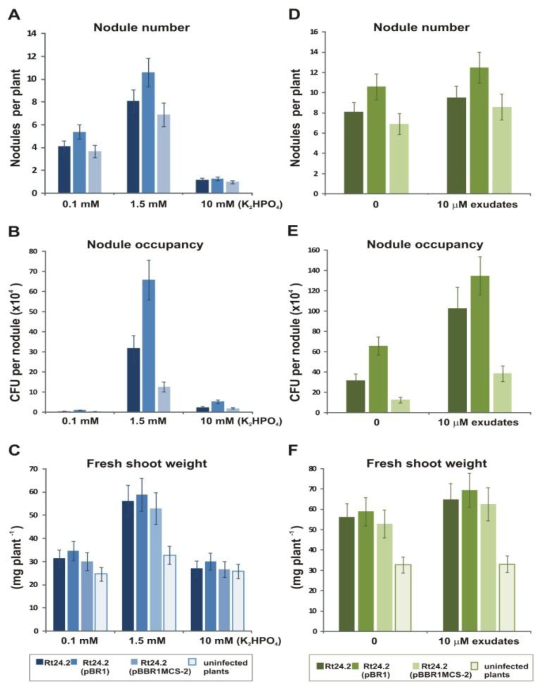
2.7. Effect of Phosphate and Flavonoids on Symbiosis of Rt24.2 and Its Derivatives with Clover

Previously, we established that the symbiotic efficiency of the rosR mutant was significantly decreased [41]. This mutant induced 2-fold fewer nodules on clover roots than the wild type strain, which were occupied by significantly lower numbers of bacteria and, as a consequence, a low shoot mass of the infected clover plants was observed. The pssA mutant, which did not produce EPS, induced a 3-fold lower number of nodules than the wild type, and those nodules were hardly occupied by bacteria. This resulted in a significant decrease in the wet shoot mass (60%) of clover plants in comparison to plants inoculated with the wild type strain, confirming the significant role of EPS in host plant infection and effective symbiosis. On the other hand, we observed that multiple copies of rosR and pssA genes significantly enhanced EPS production, competitiveness and clover nodulation in R. leguminosarum bv. trifolii [41].
In this work, the effect of phosphate and flavonoids on nodulation, nodule occupancy, and plant growth was examined using Rt24.2 wild type and its derivatives Rt24.2(pBR1) carrying additional rosR copies and Rt24.2 harboring the pBBR1MCS-2 vector (Figure 8). Nitrogen-free medium containing 1.5 mM phosphate was found to be optimal for symbiotic performance. Under phosphate-rich conditions (10 mM), a considerable reduction in nodule numbers, nodule occupancy, and fresh shoot weights was noticed (Figure 8A–C). This negative effect was even more drastic than the effect of phosphate limitation (0.1 mM), with the exception of nodule occupancy (Figure 8B).
To assess the effect of exudates on symbiosis of Rt24.2 and its derivatives, bacteria were treated for 3 h with flavonoids before plant inoculation (Figure 8D–F). This pretreatment of rhizobia resulted in a faster induction of a higher number of nodules, significantly improved nodule occupancy and higher masses of shoots. These results showed that root exudates from a compatible legume host helped the microsymbionts in the first steps of symbiosis, probably by enhancing the synthesis of Nod factors and, possibly, EPS.

3. Experimental Section

3.1. Bacterial Strains, Plasmids, and Growth Conditions

The bacterial strains, plasmids, and oligonucleotide primers used in this study are listed in Table 2.
R. leguminosarum bv. trifolii strains were grown in 79CA medium with 1% glycerol as a carbon source [62], in tryptone-yeast (TY) medium, and in M1 minimal medium [60] containing 1% glycerol and 2 mL L−1 vitamin stock solution [63] at 28 °C. E. coli strains were grown in Luria-Bertani (LB) medium at 37 °C [60]. To study the effect of phosphate on rosR expression, EPS production, and biofilm formation, the strains were cultured in M1 medium containing 20 mM morpholinopropane sulfonate (MOPS) supplemented with appropriate concentrations of K2HPO4. To establish the influence of clover root exudates on the expression of rosR-lacZ fusions, EPS and biofilm formation, the strains were grown in 5 mL M1 medium supplemented with vitamins and 10 μM clover root exudates. When required, antibiotics for E. coli and R. leguminosarum were used at the following final concentrations: kanamycin, 40 μg mL−1; ampicillin, 100 μg mL−1; tetracycline, 10 μg mL−1; and nalidixic acid, 40 μg mL−1.

3.2. DNA Methods and Sequence Analysis

Standard techniques were used for plasmid isolation, restriction enzyme digestion, agarose gel electrophoresis, cloning, and transformation [60]. For PCR amplification, plasmid or genomic DNAs isolated from R. leguminosarum bv. trifolii as templates and Pfu DNA polymerase (Promega, Madison, Wl, USA) or Taq DNA polymerase (Sigma-Aldrich, St. Louis, MO, USA) were used. The primers used in this study are listed in Table 2. Sequencing was performed using the BigDye terminator cycle sequencing kit (Applied Biosystems, Foster City, CA, USA) and the ABI Prism 310 DNA sequencer. Database searches were done with the BLAST and FASTA programs available at the National Center for Biotechnology Information (Bethesda, MD, USA) and the European Bioinformatic Institute (Hinxton, UK). The searches for PHO box motifs and inverted repeats (IR) in the promoter region of R. leguminosarum bv. trifolii rosR were performed with the “Malign” [64] and “Fuzznuc” [65] programs. RNA secondary structures were predicted using the RNA folding software mfold version 3.2 [66] using the default settings.

3.3. Construction of Transcriptional rosR-lacZ Fusions with Mutated IR4 Sites

To construct plasmid fusions containing specific rosR promoter fragments, the broad-host-range plasmid pMP220 carrying a promoterless lacZ gene was used. A set of promoter deletions was generated on the basis of the 1.2-kb insert of pB31 bearing the entire rosR promoter. For construction of the pEP21 fusion, two primer pairs, pROS2/pREW2 and pRBAM1/pREW3, were used (Table 2), giving amplicons of 116-bp and 67-bp, respectively. The 116-bp fragment was digested with EcoRI and BamHI enzymes, and the 67-bp amplicon with BamHI and XbaI enzymes. Both the EcoRI-BamHI and the BamHI-XbaI fragments were ligated and cloned into the EcoRI and XbaI sites of the pUC19 vector, giving construct pMJ221. For pEP22 fusion, the same approach was used as for pEP21, except that different primer pairs, pROS2/pREW4 and pRBAM1/pREW3, were used. The obtained amplicons of 116-bp and 67-bp were digested with EcoRI/BamHI and BamHI/XbaI, respectively, ligated, and cloned into the EcoRI and XbaI sites of the pUC19, giving plasmid pMJ222. The inserts of both pMJ221 and pMJ222 were verified by sequencing. For pEP23 fusion, the primers pROS2 and pREW1 were applied. The 139-bp PCR fragment was digested with EcoRI and XbaI and cloned into the respective sites of the pUC19 vector. The resulting plasmid was named pMJ223, and its insert was verified by sequencing. Then, the EcoRI-XbaI fragments of pUC19 derivatives were recloned into the respective sites of pMP220, resulting in plasmids pEP21, pEP22, and pEP23 (Table 2). The final constructs were introduced into E. coli S17-1 by transformation, and then transferred from E. coli S17-1 into R. leguminosarum bv. trifolii 24.2 (Rt24.2) via biparental conjugation.

3.4. β-Galactosidase Assay

Rt24.2 derivatives containing rosR-lacZ fusions were grown for 24 h in 79CA or M1 medium supplemented with tetracycline. E. coli VH1000 wild type and phoB mutant strains bearing pEP fusions were cultured for 24 h in M1 medium supplemented with tetracycline and different concentrations of phosphate. The assay for β-galactosidase activity was carried out as described previously [42].

3.5. EPS Isolation

For EPS isolation, 5-mL cultures of R. leguminosarum bv. trifolii strains were grown in M1 medium supplemented with 1% glycerol and Dilworth’s vitamins for 3 days at 28 °C in a rotary shaker. To study the effect of the different factors on the level of EPS produced by R. leguminosarum bv. trifolii, the strains were cultured in M1 medium supplemented with 10 μM root exudates, 1% glycerol as a carbon source, and appropriate concentrations of K2HPO4. EPS was precipitated from culture supernatants with 4 volumes of cold 96% ethanol, and then collected by centrifugation, lyophilized, resolved in water and analyzed for carbohydrates according to Loewus [67]. Total sugar content was calculated as glucose equivalents.

3.6. Biofilm Formation Assay

The biofilm formation assay was done in microtiter-plates as described by Rinaudi and Gonzalez [17]. Briefly, R. leguminosarum bv. trifolii strains were grown in M1 medium supplemented with Dilworth’s vitamins and different concentrations of the examined compounds at 28 °C for 48 h. The cultures were diluted to an OD600 of 0.3, introduced in 100 μL aliquots into microplate wells, and incubated at 28 °C for 48 h without shaking. Then, bacterial growth was quantified by measuring the OD600. The liquid was removed, and each well was washed three times with 150 μL of 0.85% NaCl, stained for 15 min with 150 μL of 0.1% crystal violet, and rinsed three times with 150 μL of water. After drying, biofilm formation was quantified by the addition of 150 μL of 95% ethanol and measurement of OD560 using a Benchmark Plus™ microplate reader (Bio-Rad Laboratories, Hercules, CA, USA).

3.7. Preparation of Exudates from Sprouted Seeds of Clover

Clover seeds after surface sterilization (0.1% HgCl2, 3 min, 70% ethanol, 3 min) were shaken in 1 L sterile water (proportion: 1 mL of water per 2 seeds) in darkness for 4 days at 28 °C [43]. Then, the supernatant was two times extracted with ethyl acetate in a 10:1 (volume/volume) ratio. Ethyl acetate was drained, and the pellet was solubilized in 10 mL 96% ethanol and stored at 4 °C. To determine the amount of substances present in the ethanol fraction of clover root exudates, 0.5 mL of this fraction was dried and weighed, and the obtained mass was calculated using the molecular mass of 7,4′-dihydroxyflavone as a standard flavonoid.

3.8. Plant Tests

Red clover (Trifolium pratense cv. Diana) seeds were surface sterilized, germinated, and grown on Fåhraeus medium slants [60]. 5-day-old seedlings were inoculated with bacterial suspensions of an OD600 of 0.2 (200 μL per plant) and grown under natural light supplemented with artificial light (14 h-day at 24 °C and 10 h-night at 18 °C) in a greenhouse. After 4 weeks, the number of nodules formed on clover roots was counted, and wet mass of clover shoots was estimated. For nodule occupancy, 4-week-old nodules were surface-sterilized, and each nodule was crushed in 100 μL of 79CA medium containing 0.3 M glucose; the obtained suspensions were plated in dilutions on plates containing 79CA agar.

4. Conclusions

Our previous studies indicated that RosR is an essential regulatory protein involved in EPS synthesis in R. leguminosarum bv. trifolii [36,42,43]. Mutation of rosR resulted in a 3-fold decrease of EPS production, whereas additional rosR copies caused a ca. 2-fold increase of EPS, confirming the significant role of RosR as a positive regulator of EPS production.
In this work, we found that the expression of rosR is very complex and is modulated by phosphate and clover root exudates. In the long rosR upstream region, besides the RosR-box that binds RosR and autoregulates rosR expression, several regulatory motifs were identified which showed a significant identity to consensus sequences recognized by PhoB and LysR-type proteins associated with phosphate- and flavonoid-dependent gene regulation. The complexity of rosR regulation is additionally enhanced by at least six inverted repeats (IR1–IR6) found in this region. The transcripts initiated from the distal P1 and proximal P2 promoters could form very stable stem-loop secondary structures, which could be involved in post-transcriptional regulation, affecting the stability of rosR transcripts. mRNA degradation is an important mechanism for controlling gene expression in bacteria and the lifetimes of the individual transcripts can differ significantly depending on various environmental conditions [68,69]. The rosR transcripts initiated at TS1 and TS2 contain a long 5′-untranslated region (5′-UTR) (273-nt and 240-nt, respectively) that might be potentially engaged in the regulation of transcription and translation initiation in response to specific proteins or signals [70,71].
In this study, we found that a deletion of a 36-bp fragment located downstream of TS1 and containing 6-nt inverted repeats IR4 caused a three-fold increase of rosR expression driven from the P1 promoter (pEP14 in relation to pEP13). Site-directed mutagenesis of the first part of the IR4 motif confirmed that this sequence negatively regulates rosR transcription in Rt24.2, and the second part of the IR4 overlapping the P2–10 sequence plays a significant role in the activation of this process, primarily by binding of RNA polymerase. Because introduced mutations did not significantly affect the stability of RNA secondary structures, we concluded that the IR4 motif could be a potential target for some as yet unidentified rhizobial trans-acting factor, which negatively regulates rosR expression.
Here, we studied the influence of phosphate and root exudates on rosR expression and its link with EPS production and biofilm formation. Our data indicate that EPS synthesis and biofilm formation in R. leguminosarum bv. trifolii was stimulated in the presence of clover root exudates and under phosphate limitation. Root exudates positively affected EPS production, which was in concordance with a moderate increase of rosR transcription in the presence of flavonoids and the LysR motif in pEP1. In the case of phosphate, a negative effect of this compound on EPS production was observed. The EPS synthesis in R. leguminosarum seems to be much more sensitive to phosphate concentration than EPS I production in S. meliloti, being efficiently synthesized under very high concentration of phosphate (above 150 mM).
Taken together, the presented data indicate that at least four proteins (RosR, PhoB, NodD and the repressor binding the IR4) influence rosR transcription by binding to specific DNA consensus sites and, consequently, modulate EPS production in the presence of suitable external nutritional signals. These results are in agreement with published data demonstrating that the activity of most bacterial promoters depends on multiple nutritional signals and is controlled by two or more transcriptional factors with each factor relaying one environmental signal [72].

Acknowledgments

The authors thank B. Spira (Departamento de Microbiologia, Universidade Săo Paulo, Brazil) for providing the E. coli LG01 strain. This research was supported by the Ministry of Science and Higher Education, grant No. N N303 092234. The authors thank Maria Małek for technical assistance.

References

  1. Downie, JA. The roles of extracellular proteins, polysaccharides and signals in the interactions of rhizobia with legume roots. FEMS Microbiol. Rev 2010, 34, 150–170. [Google Scholar]
  2. Flemming, HC; Wingender, J. The biofilm matrix. Nat. Rev. Microbiol 2010, 8, 623–633. [Google Scholar]
  3. Becker, A; Pühler, A. Spaink, HP, Kondorosi, A, Hooykaas, PJJ, Eds.; Production of exopolysaccharides. In Rhizobiaceae Molecular Biology of Plant-Associated Bacteria; Kluwer Academic Press: Dordrecht, The Netherlands, 1998; pp. 97–118. [Google Scholar]
  4. Fraysse, N; Couderc, F; Poinsot, V. Surface polysaccharide involvement in establishing the rhizobium-legume symbiosis. Eur. J. Biochem 2003, 270, 1365–1380. [Google Scholar]
  5. Skorupska, A; Janczarek, M; Marczak, M; Mazur, A; Król, J. Rhizobial exopolysaccharides: Genetic control and symbiotic functions. Microb Cell Fact 2006, 5. [Google Scholar]
  6. Borthakur, D; Barker, CE; Lamb, JW; Daniels, MJ; Downie, JA; Johnston, AWB. A mutation that blocks exopolysaccharide synthesis prevents nodulation of peas by Rhizobium leguminosarum but not of beans by R. phaseolii and is corrected by cloned DNA from Rhizobium or the phytopathogen Xanthomonas. Mol. Gen. Genet 1986, 203, 320–323. [Google Scholar]
  7. Niehaus, K; Kapp, D; Pühler, A. Plant defence and delayed infection of alfalfa pseudonodules induced by an exopolysaccharide (EPSI)-deficient Rhizobium meliloti mutant. Planta 1993, 190, 415–425. [Google Scholar]
  8. Rolfe, BG; Carlson, RW; Ridge, RW; Dazzo, RW; Mateos, FB; Pankhurst, CE. Defective infection and nodulation of clovers by exopolysaccharide mutants of Rhizobium leguminosarum bv. trifolii. Aust. J. Plant Physiol 1996, 23, 285–303. [Google Scholar]
  9. Cheng, HP; Walker, GC. Succinoglycan is required for initiation and elongation of infection threads during nodulation of alfalfa by Rhizobium meliloti. J. Bacteriol 1998, 180, 5183–5191. [Google Scholar]
  10. Canter-Cremers, HCJ; Stevens, K; Lugtenberg, BJJ; Wijffelman, CA; Batley, M; Redmond, JW; Breedveld, M; Zevenhuizen, LPTM. Unusual structure of the exopolysaccharide of Rhizobium leguminosarum biovar viciae strain 248. Carbohydr. Res 1991, 218, 185–200. [Google Scholar]
  11. O’Neill, MA; Darvill, AG; Albersheim, P. The degree of esterification and points of substitution by O-acetyl and O-(3-hydroxybutanoyl) groups in the acidic extracellular polysaccharides secreted by Rhizobium leguminosarum biovars viciae, trifolii, and phaseoli are not related to host range. J. Biol. Chem 1991, 266, 9549–9555. [Google Scholar]
  12. Król, JE; Mazur, A; Marczak, M; Skorupska, A. Syntenic arrangements of the surface polysaccharide biosynthesis genes in Rhizobium leguminosarum. Genomics 2007, 89, 237–247. [Google Scholar]
  13. Young, JPW; Crossman, LC; Johnston, AWB; Thomson, NR; Ghazoui, ZF; Hull, KH; Wexler, M; Curson, ARJ; Todd, JD; Poole, PS; et al. The genome of Rhizobium leguminosarum has recognizable core and accessory components. Genome Biol 2006, 7. [Google Scholar]
  14. Rinaudi, LV; Giordano, W. An integrated view of biofilm formation in rhizobia. FEMS Microiol. Lett 2010, 304, 1–11. [Google Scholar]
  15. Williams, A; Wilkinson, A; Krehenbrink, M; Russo, D; Zorreguieta, A; Downie, JA. Glucomannan-mediated attachment of Rhizobium leguminosarum to pea root hairs is required for competitive nodule infection. J. Bacteriol 2008, 190, 4706–4715. [Google Scholar]
  16. Russo, DM; Williams, A; Edwards, A; Posadas, DM; Finnie, C; Dankert, M; Downie, JA; Zorreguieta, A. Proteins exported via the PrsD-PrsE type I secretion system and the acidic exopolysaccharide are involved in biofilm formation by Rhizobium leguminosarum. J. Bacteriol 2006, 188, 4474–4486. [Google Scholar]
  17. Rinaudi, LV; González, JE. The low-molecular-weight fraction of the exopolysaccharide II from Sinorhizobium meliloti is a crucial determinant of biofilm formation. J. Bacteriol 2009, 191, 7216–7224. [Google Scholar]
  18. Becker, A; Küster, H; Niehaus, K; Pühler, A. Extension of the Rhizobium meliloti succinoglycan biosynthesis gene cluster: Identification of the exsA gene encoding an ABC transporter protein, and the exsB gene which probably codes for a regulator of succinoglycan biosynthesis. Mol. Gen. Genet 1995, 249, 487–497. [Google Scholar]
  19. Yao, SY; Luo, L; Har, KJ; Becker, A; Rüberg, S; Yu, GQ; Zhu, JB; Cheng, HP. Sinorhizobium meliloti ExoR and ExoS proteins regulate both succinoglycan and flagellum production. J. Bacteriol 2004, 186, 6042–6049. [Google Scholar]
  20. Bahlawane, C; McIntosh, M; Krol, E; Becker, A. Sinorhizobium meliloti regulator MucR couples exopolysaccharide synthesis and motility. Mol. Plant Microbe Interact 2008, 21, 1498–1509. [Google Scholar]
  21. Rüberg, S; Pühler, A; Becker, A. Biosynthesis of the exopolysaccharide galactoglucan in Sinorhizobium meliloti is subject to a complex control by the phosphate-dependent regulator PhoB and the proteins ExpG and MucR. Microbiology 1999, 145, 603–611. [Google Scholar]
  22. Bertram-Drogatz, PA; Quester, I; Becker, A; Pühler, A. The Sinorhizobium meliloti MucR protein, which is essential for the production of high-molecular-weight succinoglycan exopolysaccharide, binds to short DNA regions upstream of exoH and exoY. Mol. Gen. Genet 1998, 257, 433–441. [Google Scholar]
  23. Doherty, D; Leigh, JA; Glazebrook, J; Walker, GC. Rhizobium meliloti mutants that overproduce the R. meliloti acidic calcofluor-binding exopolysaccharide. J. Bacteriol 1988, 170, 4249–4256. [Google Scholar]
  24. Breedveld, MW; Zevenhuizen, LPTM; Zehnder, AJB. Osmotically induced oligo- and polysaccharide synthesis by Rhizobium meliloti SU-47. J. Gen. Microbiol 1990, 136, 2511–2519. [Google Scholar]
  25. Mendrygal, KE; González, JE. Environmental regulation of exopolysaccharide production in Sinorhizobium meliloti. J. Bacteriol 2000, 182, 599–606. [Google Scholar]
  26. Zhan, H; Lee, CC; Leigh, JA. Induction of the second exopolysaccharide (EPSb) in Rhizobium meliloti SU47 by low phosphate concentrations. J. Bacteriol 1991, 173, 7391–7394. [Google Scholar]
  27. Krol, E; Becker, A. Global transcriptional analysis of the phosphate starvation response in Sinorhizobium meliloti strains 1021 and 2011. Mol. Gen. Genomics 2004, 272, 1–17. [Google Scholar]
  28. Yuan, Z; Zaheer, R; Morton, R; Finan, TM. Genome prediction of PhoB regulated promoters in Sinorhizobium meliloti and twelve proteobacteria. Nucl. Acids Res 2006, 34, 2686–2697. [Google Scholar]
  29. Marketon, MM; Glenn, SA; Eberhard, A; González, JE. Quorum sensing controls exopolysaccharide production in Sinorhizobium meliloti. J. Bacteriol 2003, 185, 325–331. [Google Scholar]
  30. Barnett, M; Long, SR. Identification and characterization of a gene on Rhizobium meliloti pSymA, syrB, that negatively affects syrM expression. Mol. Plant Microbe Interact 1997, 10, 550–559. [Google Scholar]
  31. Dusha, I; Olah, B; Szegletes, Z; Erdei, L; Kondorosi, A. SyrM involved in the determination of the amount and ratio of the two forms of the acidic exopolysaccharide EPSI in Rhizobium meliloti. Mol. Plant Microbe Interact 1999, 12, 755–765. [Google Scholar]
  32. Borthakur, D; Johnston, AWB. Sequence of psi, a gene of the symbiotic plasmid of Rhizobium phaseoli which inhibits exopolysaccharide synthesis and nodulation and demonstration that its transcription is inhibited by psr, another gene on the symbiotic plasmid. Mol. Gen. Genet 1987, 207, 149–154. [Google Scholar]
  33. Latchford, JW; Borthakur, D; Johnston, AWB. The products of Rhizobium genes, psi and pss, which affect exopolysaccharide production, are associated with the bacterial cell surface. Mol. Microbiol 1991, 5, 2107–2114. [Google Scholar]
  34. Reeve, WG; Dilworth, MJ; Tiwari, RP; Glenn, AR. Regulation of exopolysaccharide production in Rhizobium leguminosarum biovar viciae WSM710 involves exoR. Microbiology 1997, 143, 1951–1958. [Google Scholar]
  35. Janczarek, M; Skorupska, A. The Rhizobium leguminosarum bv. trifolii pssB gene product is an inositol monophosphatase that influences exopolysaccharide synthesis. Arch. Microbiol 2001, 175, 143–151. [Google Scholar]
  36. Janczarek, M; Skorupska, A. The Rhizobium leguminosarum bv. trifolii RosR: Transcriptional regulator involved in exopolysaccharide production. Mol. Plant Microbe Interact 2007, 20, 867–881. [Google Scholar]
  37. Keller, M; Roxlau, A; Wenig, WM; Schmidt, M; Quandt, J; Niehaus, K; Jording, D; Arnold, W; Pühler, A. Molecular analysis of the Rhizobium meliloti mucR gene regulating the biosynthesis of the exopolysaccharides succinoglycan and galactoglucan. Mol. Plant Microbe Interact 1995, 8, 267–277. [Google Scholar]
  38. Bittinger, MA; Milner, JL; Saville, BJ; Handelsman, J. RosR, a determinant of nodulation competitiveness in Rhizobium etli. Mol. Plant Microbe Interact 1997, 10, 180–186. [Google Scholar]
  39. Hussain, H; Johnston, AW. Iron-dependent transcription of the regulatory gene ros of Agrobacterium radiobacter. Mol. Plant Microbe Interact 1997, 10, 1087–1093. [Google Scholar]
  40. Chou, AY; Archdeacon, J; Kado, CI. Agrobacterium transcriptional regulator Ros is a prokaryotic zinc finger protein that regulates the plant oncogene ipt. Proc. Natl. Acad. Sci. USA 1998, 95, 5293–5298. [Google Scholar]
  41. Janczarek, M; Jaroszuk-Œciseł, J; Skorupska, A. Multiple copies of rosR and pssA genes enhance exopolysaccharide production, symbiotic competitiveness and clover nodulation in Rhizobium leguminosarum bv. trifolii. Antonie van Leeuwenhoek 2009, 96, 471–486. [Google Scholar]
  42. Janczarek, M; Skorupska, A. Rhizobium leguminosarum bv. trifolii rosR gene expression is regulated by catabolic repression. FEMS Microbiol. Lett 2009, 291, 112–119. [Google Scholar]
  43. Janczarek, M; Kutkowska, J; Piersiak, T; Skorupska, A. Rhizobium leguminosarum bv. trifolii rosR is required for interaction with clover, biofilm formation and adaptation to the environment. BMC Microbiol 2010, 10. [Google Scholar]
  44. Quester, I; Becker, A. Four promoters subject to regulation by ExoR and PhoB direct transcription of the Sinorhizobium meliloti exoYFQ operon involved in the biosynthesis of succinoglycan. J. Mol. Microbiol. Biotech 2004, 7, 115–132. [Google Scholar]
  45. Spinelli, SV; Pontel, LB; Vescovi, EG; Soncini, FC. Regulation of magnesium homeostasis in Salmonella: Mg2+ targets the mgtA transcript for degradation by RNase E. FEMS Microbiol. Lett 2008, 280, 226–234. [Google Scholar]
  46. Summers, ML; Elkins, JG; Elliot, BA; McDermott, TR. Expression and regulation of phosphate stress inducible genes in Sinorhizobium meliloti. Mol. Plant Microbe Interact 1999, 11, 1094–1101. [Google Scholar]
  47. Bieleski, RL. Phosphate pools, phosphate transport and phosphate availability. Annu. Rev. Plant Physiol 1973, 24, 225–252. [Google Scholar]
  48. Bardin, S; Dan, S; Osteras, M; Finan, TM. A phosphate transport system is required for symbiotic nitrogen fixation by Rhizobium meliloti. J. Bacteriol 1996, 178, 4540–4547. [Google Scholar]
  49. Riley, M; Abe, T; Arnaud, MB; Berlyn, MK; Blattner, FR; Chaudhuri, RR; Glasner, JD; Horiuchi, T; Keseler, IM; Kosuge, T; et al. Escherichia coli K-12: A cooperatively developed annotation snapshot-2005. Nucl Acids Res 2006, 34, 1–9. [Google Scholar]
  50. Bardin, SD; Voegele, RT; Finan, TM. Phosphate assimilation in Rhizobium (Sinorhizobium) meliloti: Identification of a pit-like gene. J. Bacteriol 1998, 180, 4219–4226. [Google Scholar]
  51. Rosenberg, H; Gerdes, RG; Chegwidden, K. Two systems for the uptake of phosphate in Escherichia coli. J. Bacteriol 1977, 131, 505–511. [Google Scholar]
  52. Long, SR. Genes and signals in the Rhizobium—Legume symbiosis. Plant Physiol 2001, 125, 69–72. [Google Scholar]
  53. Fujishige, NA; Kapadia, NN; Hirsch, AM. A feeling for the micro-organism: Structure on a small scale. Biofilms on plant roots. Bot. J. Linn. Soc 2006, 150, 79–88. [Google Scholar]
  54. Fujishige, NA; Lum, MR; De Hoff, PL; Whitelegge, JP; Faull, KF; Hirsch, AM. Rhizobium common nod genes are required for biofilm formation. Mol. Microbiol 2008, 67, 504–515. [Google Scholar]
  55. Rinaudi, LV; Fujishige, NA; Hirsch, AM; Banchio, E; Zorreguieta, A; Giordano, W. Effects of nutritional and environmental conditions on Sinorhizobium meliloti biofilm formation. Res. Microbiol 2006, 157, 867–875. [Google Scholar]
  56. Danhorn, T; Hentzer, M; Givskov, M; Parsek, MR; Fuqua, C. Phosphorus limitation enhances biofilm formation of the plant pathogen Agrobacterium tumefaciens through the PhoR-PhoB regulatory system. J. Bacteriol 2004, 186, 4492–4501. [Google Scholar]
  57. Djordjevic, MA; Żurkowski, W; Shine, J; Rolfe, BG. Sym-plasmid transfer to various symbiotic mutants of Rhizobium trifolii, R. leguminosarum and R. meliloti. J. Bacteriol 1983, 156, 1035–1045. [Google Scholar]
  58. Gaal, T; Bartlett, MS; Ross, W; Turnbough, JCL; Gourse, RL. Transcription regulation by initiating NTP concentration: rRNA synthesis in bacteria. Science 1997, 278, 2092–2097. [Google Scholar]
  59. Spira, B. Departamento de Microbiologia, Instituto de Ciěncias Biomédicas, Universidade de Săo Paulo, Săo Paulo-SP: Brazil; Personal collection; 2009. [Google Scholar]
  60. Sambrook, J; Fitsch, EF; Maniatis, T. Molecular Cloning: A Laboratory Manual; Cold Spring Harbor Laboratory Press: Cold Spring Harbor, NY, USA, 1989. [Google Scholar]
  61. Spaink, HP; Okker, RJH; Wijffelman, CA; Pees, E; Lugtenberg, BJJ. Promoters in the nodulation region of the Rhizobium leguminosarum Sym plasmid pRL1JI. Plant Mol. Biol 1987, 9, 27–39. [Google Scholar]
  62. Vincent, JM. A manual for the practical study of root nodule bacteria. In International Biological Program Handbook No 15; Blackwell Scientific Publications Ltd: Oxford, UK, 1970. [Google Scholar]
  63. Brown, CM; Dilworth, MJ. Ammonia assimilation by Rhizobium cultures and bacteroids. J. Gen. Microbiol 1975, 86, 39–48. [Google Scholar]
  64. GeneBee, AN. Belozersky Institute of Physico-Chemical Biology, Moscow State University: Moscow, Russia, 1965. Available online: http://www.genebee.msu.su/ accessed on 16 June 2011.
  65. Rice, P; Longden, I; Bleasby, A. EMBOSS: The European Molecular Biology Open Software Suite. Trends in Genetics. 2000, pp. 276–277. Available online: http://emboss.ch.embnet.org/Pise accessed on 16 June 2011.
  66. Zuker, M. Mfold web server for nucleic acid folding and hybridization prediction. Nucl Acids Res. 2003, 31, pp. 3406–3415. Available online: http://mfold.rna.albany.edu accessed on 16 June 2011.
  67. Loewus, FA. Improvement in the anthrone method for determination of carbohydrates. Anal. Chem 1952, 24, 219. [Google Scholar]
  68. Newbury, SF; Smith, NH; Higgins, CF. Differential mRNA stability controls relative gene expression within a polycistronic operon. Cell 1987, 51, 1131–1143. [Google Scholar]
  69. Masse, E; Escorcia, FE; Gottesman, S. Coupled degradation of a small regulatory RNA and its mRNA targets in Escherichia coli. Genes Dev 2003, 17, 2374–2383. [Google Scholar]
  70. Narberhaus, F; Waldminghaus, T; Chowdhury, S. RNA thermometers. FEMS Microbiol. Rev 2006, 30, 3–16. [Google Scholar]
  71. Serganov, A. The long and the short of riboswitches. Curr. Opin. Struct. Biol 2009, 19, 251–259. [Google Scholar]
  72. Browning, DF; Busby, SJW. The regulation of bacterial transcription initiation. Nature Rev. Microbiol 2004, 2, 1–9. [Google Scholar]
Figure 1. (A) Physical and genetic map of plasmid pB31 carrying R. leguminosarum bv. trifolii 24.2 rosR gene. The blue arrow below the map shows the direction of rosR transcription. B, BamHI; H, HindIII; P, PstI; N, NotI. P1 and P2 are promoter sequences, and TS1 and TS2 transcription start sites. (B) Nucleotide sequence of 960-bp fragment containing rosR gene with the upstream region. The amino acid sequence of RosR is given in the single letter code. The −35 and −10 hexamers of P1 and P2 promoters, and PHO boxes are marked by square brackets. Nucleotides identical to the PHO box consensus sequence are shaded in black. TS1 and TS2 are marked by red arrows. RosR-box is shaded in green. LysR motif is underlined and conserved nucleotides are shaded in dark blue. Inverted repeats IR1 to IR6 are marked by inverted arrows, and direct repeats are marked by white boxes. Over lined short arrows indicate the upstream and downstream endpoints of PCR fragments of individual plasmid fusions, respectively. The sequence of ribosome-binding site (rbs) and rho-independent terminator are underlined.
Ijms 12 04132f1
Figure 2. (A) The secondary structure of rho-independent transcriptional terminator; (B) the 5′-upper part of transcript 1 initiating at TS1; and (C) transcript 2 initiating at TS2 of R. leguminosarum bv. trifolii 24.2 rosR. The structures and their ΔG were predicted and displayed using the mfold 2.3 program. The sequence of IR4 motif was marked by boxes.
Ijms 12 04132f2
Figure 3. (A) The 3′-end nucleotide sequence of selected pEP rosR-lacZ fusions. The −10 sequence of P2 promoter is marked by square brackets. IR4 motif is marked by inverted arrows. Transcription start site TS2 is marked by red arrow and the letter G. Over lined arrows indicate the 3′-end of the insert in the individual fusions. Nucleotides changed in the IR4 sequence are shaded in black; (B) Transcriptional activity of pEP fusions assayed in E. coli DH5α and R. leguminosarum bv. trifolii 24.2. Data shown are the mean ± SD (n = 4).
Ijms 12 04132f3
Figure 4. Effect of phosphate on the transcriptional activity of R. leguminosarum bv. trifolii 24.2 rosR. (A) Schematic map of pEP rosR-lacZ fusions containing different 5′- and 3′-end deletions of the rosR upstream region. Promoters P1 and P2 are marked by white boxes, and 5′ part of rosR open reading frame is marked by black box. Transcription start sites TS1 and TS2 are marked by angled arrows. The RosR-box, LysR motif and PHO boxes are marked by light gray, dark gray and red rectangles, respectively; (B) Effect of phosphate on the transcriptional activity of rosR assayed in the R. leguminosarum bv. trifolii 24.2 strain containing different pEP fusions. For each strain, β-galactosidase activity was assayed in triplicate. Data shown are the mean ± SD.
Ijms 12 04132f4
Figure 5. Effect of phosphate on the transcriptional activity of rosR as assayed in the E. coli VH1000 wild type strain and phoB mutant containing different rosR-lacZ fusions. For each strain, β-galactosidase activity was assayed in triplicate. Data shown are the mean ± SD.
Ijms 12 04132f5
Figure 6. Effect of phosphate and clover root exudates on EPS production in R. leguminosarum bv. trifolii. (A) EPS production in the presence of different concentrations of phosphate was assayed in the Rt24.2 wild type and its derivatives bearing different pEP fusions and pBR1 with additional rosR copies; (B) EPS production in the presence of clover root exudates assayed in Rt24.2 and ANU843 wild type strains and their derivatives. The given values are the mean ± SD of triplicate assays.
Ijms 12 04132f6
Figure 7. (A) Effect of phosphate and (B) clover root exudates on biofilm formation by R. leguminosarum bv. trifolii 24.2 wild type and its derivatives. For each strain, the assays were performed in triplicate and data shown are the means ± SD. * indicates statistically significant differences.
Ijms 12 04132f7
Figure 8. Effect of phosphate (A–C) and root exudates (D–F) on symbiosis of R. leguminosarum bv. trifolii 24.2 and its derivatives Rt24.2(pBR1) and Rt24.2(pBBR1MCS-2) with clover plants. The tested symbiotic parameters are: the nodule number (A and D), nodule occupancy (B and E) and fresh shoot weight (C and F). Plants were harvested 28 days after inoculation. Given values are averages of three independent experiments with 20 plants for each treatment. For nodule occupancy, values are means ± SD of 12 nodules.
Ijms 12 04132f8
Table 1. Effect of clover root exudates and nodD mutation on the rosR-lacZ transcription in R. leguminosarum bv. trifolii.
Plasmid fusionLysR motifANU843 (wt)ANU851 (nodD)

Clover root exudates (μM)
010010
pEP1+2630 ± 2883613 ± 3522682 ± 2532789 ± 245
pEP91778 ± 1851866 ± 1911709 ± 1671811 ± 177
Table 2. Bacterial strains, plasmids and oligonucleotide primers used in this study.
Strain or plasmidRelevant characteristicsReference
Rhizobium leguminosarum bv. trifolii
Rt24.2Wild type, Rifr, Nxr[36]
Rt2472Rt24.2 derivative carrying mini-Tn5 between 151–152 bp position of rosR, Rifr, Kmr[41]
ANU843Wild type, Rifr[57]
ANU851ANU843 derivative carrying Tn5::nodD, Kmr[57]
E. coli
VH1000MG1655 derivative, lacZ, lacI, pyrE+[58]
LG01MG1655 derivative, phoB519::Tn5, Kmr[59]
Plasmids
pUC19Cloning and sequencing vector, Apr[60]
pMP220IncP, mob, promoterless lacZ, Tcr[61]
pMJ221pUC19 containing 126-bp EcoRI-XbaI fragment of the rosR upstream region (based on primers: pROS2/pREW2 and pRBAM1/pREW3)This work
pMJ222pUC19 containing 126-bp EcoRI-XbaI fragment of the rosR upstream region (based on primers: pROS2/pREW4 and pRBAM1/pREW3)This work
pMJ223pUC19 containing 126-bp EcoRI-XbaI fragment of the rosR upstream region (based on primers: pROS2/pREW1)This work
pBR1pBBR1MCS-2 containing 1100-bp EcoRI-BamHI fragment with rosR of Rt24.2[41]
pEP1pMP220 carrying the −403 to +243 bp fragment of the rosR coding region[36]
pEP9pMP220 carrying the −403 to −185 bp fragment of the rosR upstream region[36]
pEP10pMP220 carrying the −403 to −232 bp fragment of the rosR upstream region[36]
pEP13pMP220 carrying the −357 to −232 bp fragment of the rosR upstream region[42]
pEP14pMP220 carrying the −357 to −268 bp fragment of the rosR upstream region[42]
pEP15pMP220 carrying the −339 to −268 bp fragment of the rosR upstream region[42]
pEP21pMP220 containing 126-bp EcoRI-XbaI fragment from pMJ221This work
pEP22pMP220 containing 126-bp EcoRI-XbaI fragment from pMJ222This work
pEP23pMP220 containing 126-bp EcoRI-XbaI fragment from pMJ223This work
Oligonucleotide primersSequence (5′→3) *
pROS2GAGCCCCTGAATTCTTCATCTGTCAThis work
pREW1AGGGATCTAGAAGCAGTACGCTAGACATTCACThis work
pREW2GTAATTCGGATCCAGAACTCTACTTGCAThis work
pREW3GAAATCAAAACTGAGGGATCTAGAAGCAThis work
pREW4GTAATTCGGATCCCGAACTCTACTTGCAThis work
pRBAM1AAATGCAAGTAGAGTTCTGGATCCGAATTAThis work
*Sequences for EcoRI, BamHI and XbaI restriction sites are underlined.

