Antimicrobial, Cytotoxic and Antioxidant Activities and Determination of the Total Tannin Content of Bark Extracts Endopleura uchi

 ,
,
Abstract
:1. Introduction
2. Results and Discussion
3. Experimental Section
3.1. Plant Material
3.2. Preparation of Plant Extracts
3.3. Total Tannin Content in Plant Extracts
3.4. Antimicrobial Assays
3.5. Broth Micro-Dilution Assay for Minimum Inhibitory Concentrations (MIC)
3.6. Cytotoxicity Assay
3.7. Scavenging Activity of DPPH Radical
3.8. Statistical Analysis
4. Conclusions
Acknowledgments
References
- Yunes, RA; Calixto, JB. Plantas Medicinais sob a ótica da Química Medicinal Moderna, 1st ed.; Argos: Chapecó (SC), Brazil, 2001; pp. 17–44. [Google Scholar]
- Soerjato, AD. Biodiversity prospecting and benefit haring: perspectives from the field. J. Ethnopharmacol 1996, 51, 1–15. [Google Scholar]
- WHO (World Health Organization). Traditional Medicine—Growing Needs and Potential. World Health Organization Policy Perspectives on Medicines; WHO (World Health Organization): Geneva, Switzerland, 2002. [Google Scholar]
- Gonçalves, AL; Alves Filho, A; Menezes, H. Estudo comparativo da atividade antimicrobiana de extratos de algumas árvores nativas. Arq. Inst. Biol 2005, 72, 353–358. [Google Scholar]
- Andremont, A. The future control of bacterial resistance to antimicrobial agents. Am. J. Infect Control 2001, 29, 256–258. [Google Scholar]
- Cechinel Filho, V. Principais avanços e perspectivas na área de produtos naturais ativos: estudos desenvolvidos no NIQFAR/Univali. Quim. Nova 2000, 23, 680. [Google Scholar]
- Souza, MM; Cruz, AB; Schumacher, MB; Kreuger, MRO; Freitas, RA; Cruz, RCB. Métodos de avaliação de atividade biológica de produtos naturais e sintéticos. In Ciências Farmacêuticas: Contribuição ao Desenvolvimento de Novos Fármacos e Medicamentos; Bresolin, TMB, Cechinel Filho, V, Eds.; Univali: Itajaí (SC), Brazil, 2003. [Google Scholar]
- Larcher, W. Ecofisiologia Vegetal; Rima: São Carlos (SP), Brazil, 2003. [Google Scholar]
- Hostettmann, K; Queiroz, EF; Vieira, PC. Princípios Ativos de Plantas Superiores; EdUFSCar: São Carlos (SP), Brazil, 2003. [Google Scholar]
- Simões, CMO; Schenkel, EP; Gosmann, G; Mello, JCP; Mentz, LA; Petrovick, PR. Farmacognosia: da Planta ao Medicamento, 5th ed.; UFRGS/UFSC: Porto Alegre (RS)/Florianópolis (SC), Brazil, 2004. [Google Scholar]
- Reschke, A; Marques, LM; Mayworm, MAS. Atividade antibacteriana de Fícus benjamina L. (Moraceae). Rev. Bras. Plant Med 2007, 9, 67–70. [Google Scholar]
- Cuatrecasas, JA. A taxonomic revision of Humiriaceae, contribuitions from the United States National Herbarium. US Nat. Mus. Bull 1961, 35, 25–214. [Google Scholar]
- Schultes, RE. De plantis toxicariis e mundo novo tropicale commentationes. XXI. Interesting native uses of the Humiriaceae in the northwest Amazon. J. Ethnopharmacol 1979, 1, 89–94. [Google Scholar]
- Corrêa, MP. Dicionário das Plantas úteis do Brasil e das Exóticas Cultivadas; Imprensa Nacional, R.J., Ed.; Imprensa Nacional: Rio de Janeiro (RJ), Brazil, 1984; Volume 6, p. 764. [Google Scholar]
- Politi, FAS. Estudos Farmacognósticos e Avaliação de Atividades Biológicas de Extratos Obtidos das Cascas Pulverizadas de Endopleura uchi (Huber) Cuatrec. (Humiriaceae). MS Thesis, Universidade Estadual Paulista, Araraquara, Brazil. 2009. [Google Scholar]
- Luna, JS. Estudo dos Constituintes Químicos de Endopleura uchi (Humiriaceae). MS Thesis, Universidade Federal de Alagoas, Maceió, Brazil. 2000. [Google Scholar]
- Scalbert, A. Antimicrobial properties of tannins. Phytochemistry 1991, 30, 3875–3883. [Google Scholar]
- Akiyama, H; Kazuyasu, F; Yamasaki, O; Oono, T; Iwatsuki, K. Antibacterial action of several tannins against Staphylococcus aureus. J. Antimicrob. Chemother 2001, 48, 487–491. [Google Scholar]
- Erasto, P; Bojase–Moleta, G; Majinda, RRT. Antimicrobial and antioxidant flavonoids from the roots wood of Bolusathus spesiosus. Phytochemistry 2004, 65, 875–880. [Google Scholar]
- Funatogawa, K; Hayashi, S; Shimomura, H; Yoshida, T; Hatano, T; Ito, H; Iría, Y. Antibacterial activity of hydrolysable tannins derived from medicinal plants against Helicobacter pylori. Microbiol. Immunol 2004, 48, 251–261. [Google Scholar]
- Banso, A; Adeyemo, SO. Evaluation of antibacterial properties of tannins isolated from Dichrostachys cinerea. Afr. J. Biotechnol 2007, 6, 1785–1787. [Google Scholar]
- Doss, A; Mubarack, HM; Dhanabalan, R. Antibacterial activity of tannins from the leaves of Solanum trilobatum Linn. Indian J. Sci. Technol 2009, 2, 41–43. [Google Scholar]
- Yamaguti-Sasaki, E; Ito, LA; Canteli, VCD; Ushirobira, TMA; Nakamura, TU; Filho, BPD; Nakamura, CV; Mello, JCP. Antioxidant capacity and in vitro prevention of dental plaque formation by extracts and condensed tannins of Paullinia cupana. Molecules 2007, 12, 1950–1963. [Google Scholar]
- Hagerman, AE; Butler, LG. Protein precipitation method for the quantitative determination of tannins. J. Agric. Food Chem 1978, 26, 809–812. [Google Scholar]
- Hagerman, AE; Butler, LG. Determination of protein in tannin-protein precipitates. J. Agric. Food Chem 1980, 28, 944–947. [Google Scholar]
- Haslam, E. Natural polyphenols (vegetable tannins) as drugs: possible modes of action. J. Nat. Prod 1996, 59, 205–215. [Google Scholar]
- Rates, SMK. Plants as source of drugs. Toxicon 2001, 39, 603–613. [Google Scholar]
- Banerjee, A; Dasgupta, N; De, B. In vitro study of antioxidant activity of Syzygium cumini fruit. Food Chem 2005, 90, 727–733. [Google Scholar]
- Politi, FAS; Moreira, RRD; Salgado, HRN; Pietro, RCLR. Preliminary tests on acute oral toxicity and intestinal motility with extract of pulverized bark of Endopleura uchi (Huber) Cuatrec. (Humiriaceae) in mice. Rev. Pan-Amaz. Saude 2010, 1, 187–189. [Google Scholar]
- Nunomura, CS; Oliveira, VG; Silva, SL; Nunomura, SM. Characterization of bergenin in Endopleura uchi bark and its anti-inflammatory activity. J. Braz. Chem. Soc 2009, 20, 187–192. [Google Scholar]
- Glasl, H. Zur photometrie in der drogenstandardisierung-3. Gehaltsbestimmung von Gerbstoffdrogen. Deutsche Apoth. Zeit 1983, 123, 1979–1987. [Google Scholar]
- Brazil Ministry of Health. Farmacopéia Brasileira, 4th ed.; Atheneu: São Paulo (SP), Brazil, 1996. [Google Scholar]
- NCCLS (National Committee for Clinical Laboratory Standards). Performance Standards for Antimicrobial Disk Susceptibility Tests; Approved Standard, 8th ed.; NCCLS: Wayne, PA, USA, 2003. [Google Scholar]
- NCCLS (National Committee for Clinical Laboratory Standards). Method for Antifungal Disk Diffusion Susceptibility Testing of Yeasts; Approved Standard, 2nd ed.; NCCLS: Wayne, PA, USA, 2004. [Google Scholar]
- NCCLS (National Committee for Clinical Laboratory Standards). Methods for Dilution Antimicrobial Susceptibility Tests for Bacteria That Grow Aerobically; Approved Standard, 6th ed.; NCCLS: Wayne, PA, USA, 2003. [Google Scholar]
- Lei, J; Yu, J; Yu, H; Liao, Z. Composition, cytotoxicity and antimicrobial activity of essential oil from Dictamnus dasycarpus. Food Chem 2008, 107, 1205–1209. [Google Scholar]
- Ohno, Y; Miyajima, A; Sunouchi, M. Alternative methods for mechanistic studies in toxicology. Screening of hepatotoxicity of pesticides using freshly isolated and primary cultured hepatocytes and non-liver-derived cells, SIRC cells. Toxicol Lett 1998, 102–103, 569–573. [Google Scholar]
- Takahashi, Y; Koike, M; Honda, H; Ito, Y; Sakaguchi, H; Suzuki, H; Nishiyama, N. Development of the short time exposure (STE) test: an in vitro eye irritation test using SIRC cells. Toxicol. in Vitro 2008, 22, 760–770. [Google Scholar]
- O’Brien, J; Wilson, I; Orton, T; Pognan, F. Investigation of the Alamar Blue (resazurin) fluorescent dye for the assessment of mammalian cell cytotoxicity. Eur. J. Biochem 2000, 267, 5421–5426. [Google Scholar]
- Brand-Williams, W; Cuvelieur, ME; Berset, C. Use of a free radical method to evaluate antioxidant activity. Lebensm. Wiss. Technol 1995, 28, 25–30. [Google Scholar]
- Machado, FA. Estudo Fitoquímico e Avaliação da Capacidade Antioxidante de Extratos das Cascas de Stryphnodendron Polyphyllum Mart., Leguminosae, Barbatimão. MS Thesis, Universidade Estadual Paulista, Araraquara, Brazil. 2005. [Google Scholar]
- Falcão, DQ; Costa, ER; Alviano, DS; Alviano, CS; Kuster, RM; Menezes, FS. Atividade antioxidante e antimicrobiana de Calceolaria chelidonioides Humb. Bonpl. & Kunth. Rev. Bras. Farmacogn 2006, 16, 73–76. [Google Scholar]

| Decoction | Infusion | Maceration | Percolation | T. Extraction | |
|---|---|---|---|---|---|
| EC5% | 12.53 ± 1.02 | 9.7 ± 0.05 | 13.73 ± 0.11 | 18.6 ± 0.26 | 12.83 ± 0.32 |
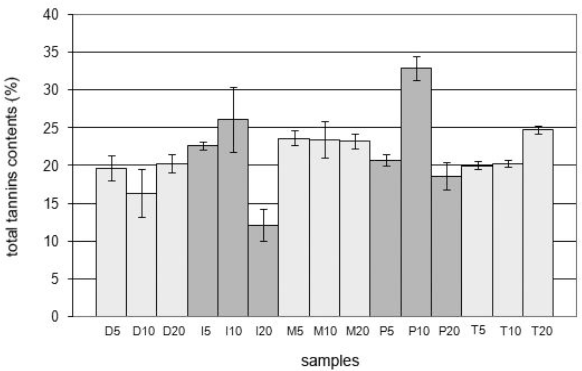
| TT5% | 19.65 ± 1.59 | 22.66 ± 0.53 | 23.62 ± 1.00 | 20.72 ± 0.79 | 19.99 ± 0.49 |
| EC10% | 11.41 ± 0.40 | 8.85 ± 0.05 | 13.1 ± 0.17 | 16.8 ± 0.39 | 12.95 ± 0.62 |
| TT10% | 16.34 ± 3.18 | 26.06 ± 4.27 | 23.41 ± 2.47 | 32.85 ± 1.62 | 20.26 ± 0.50 |
| EC20% | 9.65 ± 0.22 | 8.525 ± 0.25 | 13.04 ± 0.05 | 18.87 ± 0.27 | 14.85 ± 0.54 |
| TT20% | 20.27 ± 1.16 | 12.08 ± 2.11 | 23.18 ± 0.93 | 18.56 ± 1.79 | 24.77 ± 2.54 |
| Antimicrobial Assays | ||||||
|---|---|---|---|---|---|---|
| Template Technique | ||||||
| Samples | SA | EC | BS | SS | SE | CA |
| D20 | 15.0 | − | − | − | − | − |
| I10 | 15.0 | − | − | − | − | − |
| M5 | 13.0 | − | − | − | − | − |
| P10 | 13.0 | − | − | − | − | − |
| T20 | 14.0 | − | − | − | − | − |
| A | 23.0 | 18.0 | 15.5 | 13.0 | 17.5 | 23.5 |
| C | − | − | − | − | − | − |
| Paper Disc Technique | ||||||
| Samples | SA | EC | BS | SS | SE | CA |
| D20 | 7.0 | − | − | − | − | − |
| I10 | 7.0 | − | − | − | − | 8.0 |
| M5 | 7.0 | − | − | − | − | − |
| P10 | 8.1 | − | − | − | − | − |
| T20 | 7.9 | − | − | − | − | − |
| A | 19.3 | 13.7 | 19.7 | 17.5 | 12.7 | 17.5 |
| C | − | − | − | − | − | − |
| Samples (250 μg/mL) | Radical Scavenging Activity (%) |
|---|---|
| Gallic acid | 97.24 ± 0.05 |
| Rutin | 96.83 ± 0.06 |
| Vitamin C | 98.14 ± 0.06 |
| Gingko biloba extract | 96.97 ±0.09 |
| D20 | 87.46 ± 2.59 |
| I10 | 79.54 ± 2.63 |
| M5 | 89.22 ± 0.82 |
| P10 | 88.90 ± 2.52 |
| T20 | 90.58 ± 0.62 |
| Spearman Test (α = 0.05) | Kendall-Tau Test (α = 0.05) | Pearson Test (α = 0.05) | |
|---|---|---|---|
| TPC-TTC | r = −0.7; p-level = 0.18 | t = −0.6; p-level = 1.00 | ρ = −0.6636; p-level = 0.22 |
| TTC-AOA | r = −0.1; p-level = 0.87 | t = 0; p-level = 1.00 | ρ = −0.0065; p-level = 0.99 |
| TPC-AOA | r = −0.6; p-level = 0.28 | t = −0.4; p-level = 0.32 | ρ = −0.1937; p-level = 0.75 |
© 2011 by the authors; licensee MDPI, Basel, Switzerland. This article is an open-access article distributed under the terms and conditions of the Creative Commons Attribution license (http://creativecommons.org/licenses/by/3.0/).
Share and Cite
Politi, F.A.S.; Mello, J.C.P.d.; Migliato, K.F.; Nepomuceno, A.L.A.; Moreira, R.R.D.; Pietro, R.C.L.R. Antimicrobial, Cytotoxic and Antioxidant Activities and Determination of the Total Tannin Content of Bark Extracts Endopleura uchi. Int. J. Mol. Sci. 2011, 12, 2757-2768. https://doi.org/10.3390/ijms12042757
Politi FAS, Mello JCPd, Migliato KF, Nepomuceno ALA, Moreira RRD, Pietro RCLR. Antimicrobial, Cytotoxic and Antioxidant Activities and Determination of the Total Tannin Content of Bark Extracts Endopleura uchi. International Journal of Molecular Sciences. 2011; 12(4):2757-2768. https://doi.org/10.3390/ijms12042757
Chicago/Turabian StylePoliti, Flávio A. S., João C. P. de Mello, Ketylin F. Migliato, Andréa L. A. Nepomuceno, Raquel R. D. Moreira, and Rosemeire C. L. R. Pietro. 2011. "Antimicrobial, Cytotoxic and Antioxidant Activities and Determination of the Total Tannin Content of Bark Extracts Endopleura uchi" International Journal of Molecular Sciences 12, no. 4: 2757-2768. https://doi.org/10.3390/ijms12042757
APA StylePoliti, F. A. S., Mello, J. C. P. d., Migliato, K. F., Nepomuceno, A. L. A., Moreira, R. R. D., & Pietro, R. C. L. R. (2011). Antimicrobial, Cytotoxic and Antioxidant Activities and Determination of the Total Tannin Content of Bark Extracts Endopleura uchi. International Journal of Molecular Sciences, 12(4), 2757-2768. https://doi.org/10.3390/ijms12042757