Other

Supramolecular Bioorganic Chemistry: Nucleic Acids Recognition and Synthetic Vectors for Gene Transfer

by
Jean-Pierre Vigneron
Laboratoire de Chimie des Interactions Moléculaires (UPR 285), Collège de France, 11 Place Marcelin Berthelot, 75005 Paris, France
Molecules 1999, 4(7), 180-203; https://doi.org/10.3390/40700180
Submission received: 7 June 1999 / Accepted: 9 July 1999 / Published: 2 July 1999

Abstract

:
Our works on nucleic acids recognition and synthetic vectors for gene transfer have been reviewed.

1.Introduction

The results presented in this paper deal with two domains of bioorganic supramoleclar chemistry. Indeed, bioorganic chemistry is not only a source of inspiration but also a huge domain of applications for supramolecular chemistry. The first topic - nucleic acid recognition - illustrates the recognition and the transformation of a substrate by a receptor. The second one - design of synthetic vectors for gene transfer - concerns the transport of a substrate, a process which in this case involves very complex molecular assemblies. Thus, the two broad areas of supramolecular chemistry are illustrated: that of supramolecules and that of polymolecular assemblies.

2. Nucleic Acid Recognition

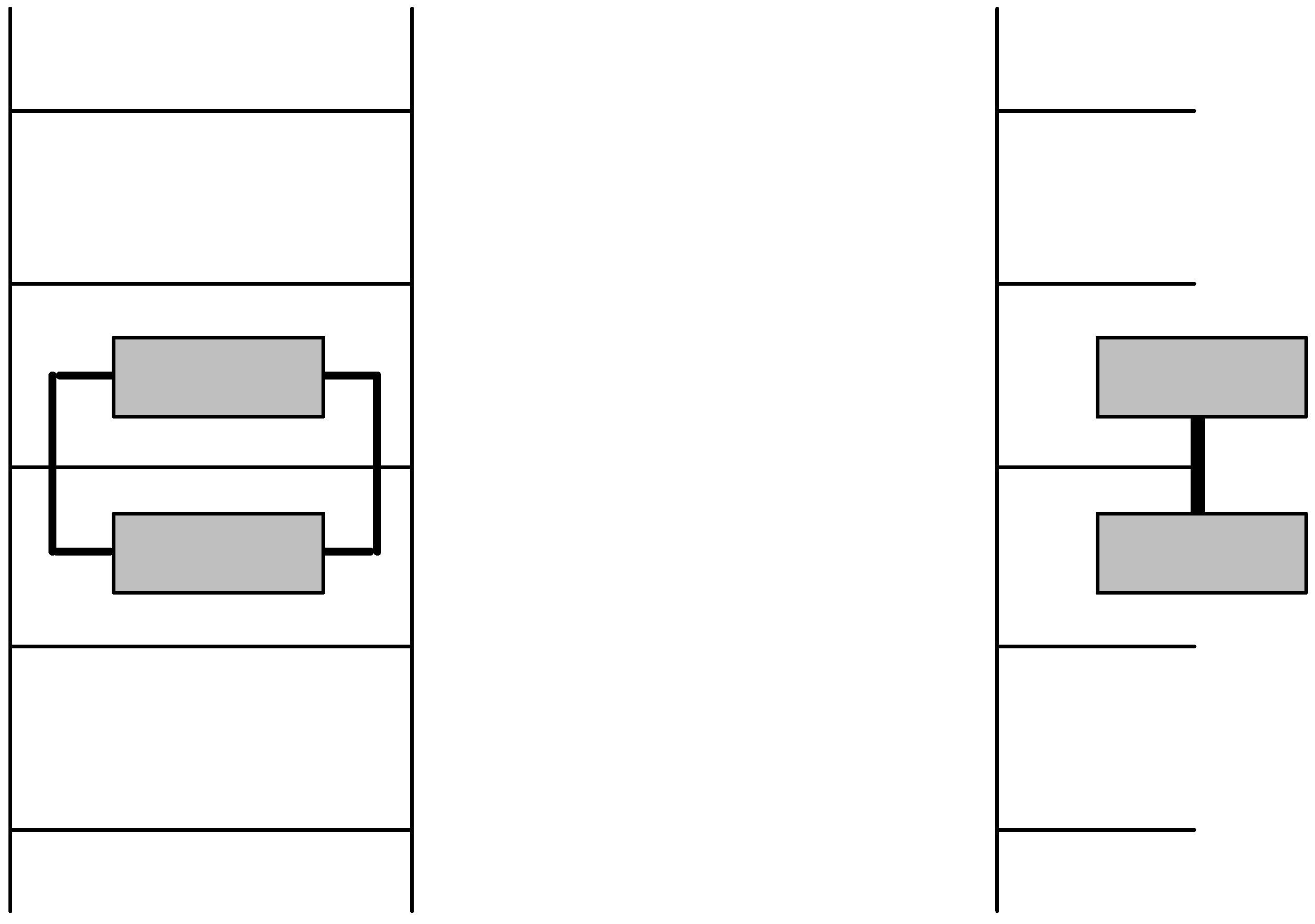
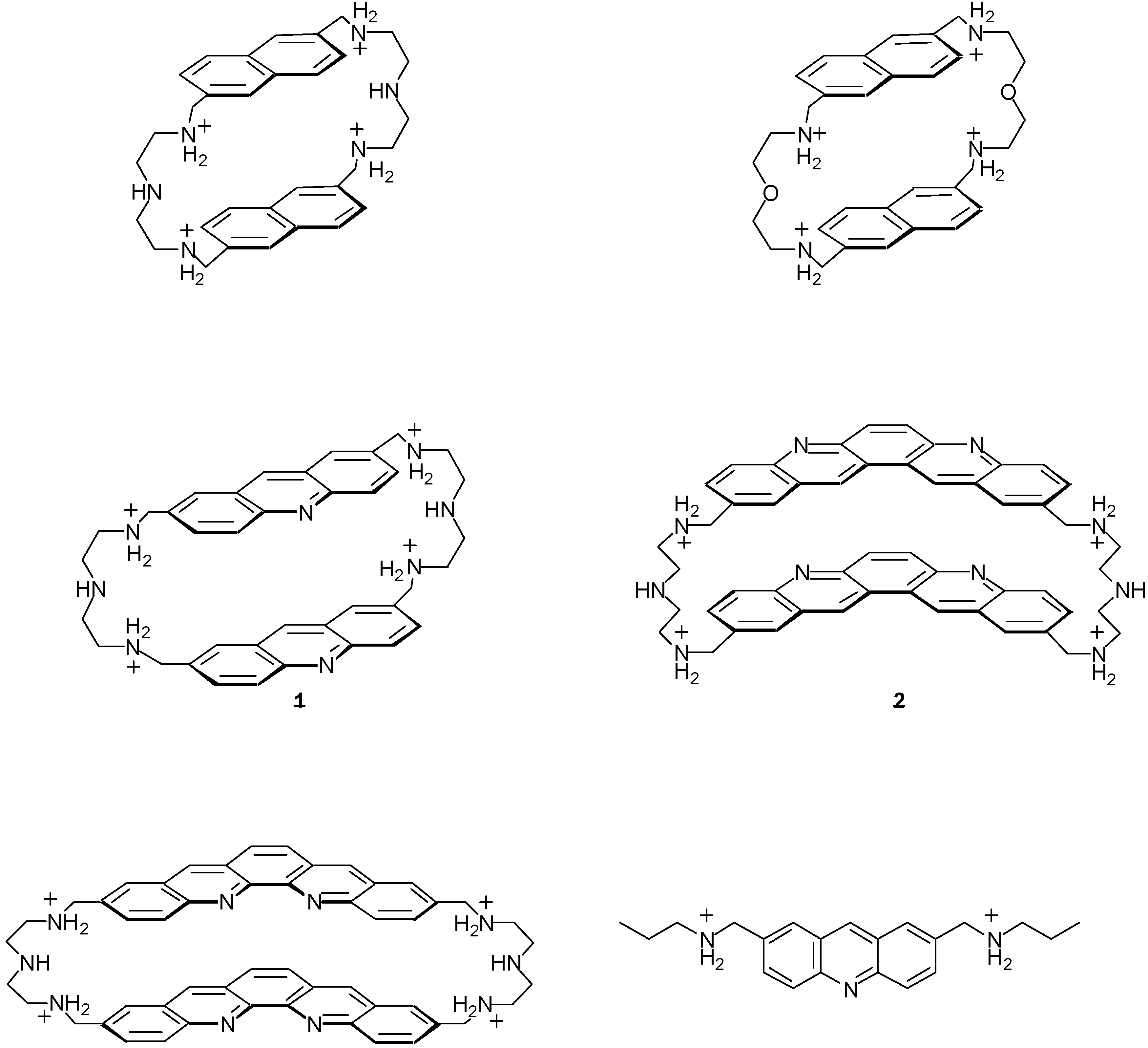
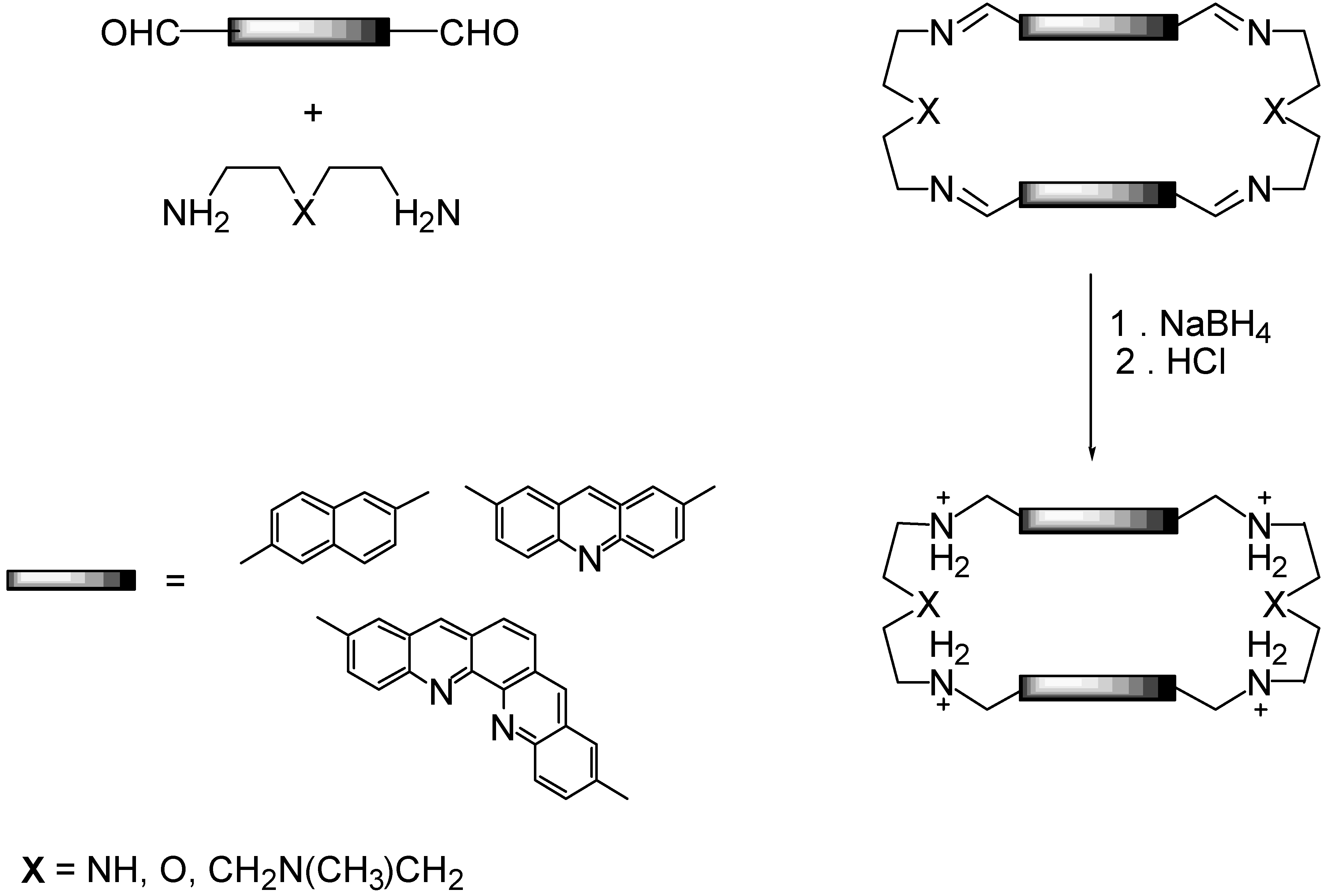
2.1. Design and Preparation of Cyclobisintercaland Receptors

In order to selectively bind anionic planar substrates such as nucleotides and polynucleotides, we have developed, for some years, a new family of compounds, the general structure of which is shown in Figure 1.
They contain two planar subunits, of large area, linked by more or less rigid bridges. This structure delineates a molecular cavity suitable for inclusion of planar organic substrates, in aqueous solutions, by a combination of stacking, electrostatic and hydrophobic effects. In fact, as planar subunits we have most often used planar heterocyclic dye molecules - well known for binding to double-stranded nucleic acids by intercalation between base pairs. It is why this type of compounds was named cyclobisintercalands.
By comparison to linear intercalands or polyintercalands some selectivity could be expected due to the macrocyclic structure of the receptor. Indeed we can suppose that the latter hinders the insertion of paired nucleic acid bases of a double-helix whereas it seems possible for a nucleic base of a single-stranded oligonucleotide to be inserted inside the macrocycle provided that the two parallel walls are situated at a correct distance (Figure 2).
Moreover dye molecules possess a variety of photochemical and electrochemical properties, so they may endow the cyclobisintercaland receptor with the ability to perform electro or photoinduced reactions on the bound substrate species. As a result, the combination of a selectivity due to the macrocyclic structure with the photochemical properties of the dye allows to consider the possibility of designing artificial nucleases specific of single-stranded polynucleotides.
Along these lines we have prepared a number of macrocycles containing two planar subunits linked by two or three bridges [1,2,3,4,5,6,7,8,9,10,11,12]. Only polyaminomacrocycles, especially the bisacridine derivative 1, will be studied here (Figure 3) [7,8].
These polyamino compounds were prepared via the very efficient 2+2 condensation between diethylenetriamine and dialdehydes (Figure 4).
For instance, the condensation between diethylenetriamine and 2,7-acridine-dicarboxaldehyde gave the tetraimine which was reduced by sodium borohydride. The macrocycle was isolated as its octachlorhydrate in 70% yield for the two consecutive reactions. It is also possible to isolate the tetraimine in 94% yield as a crude product.

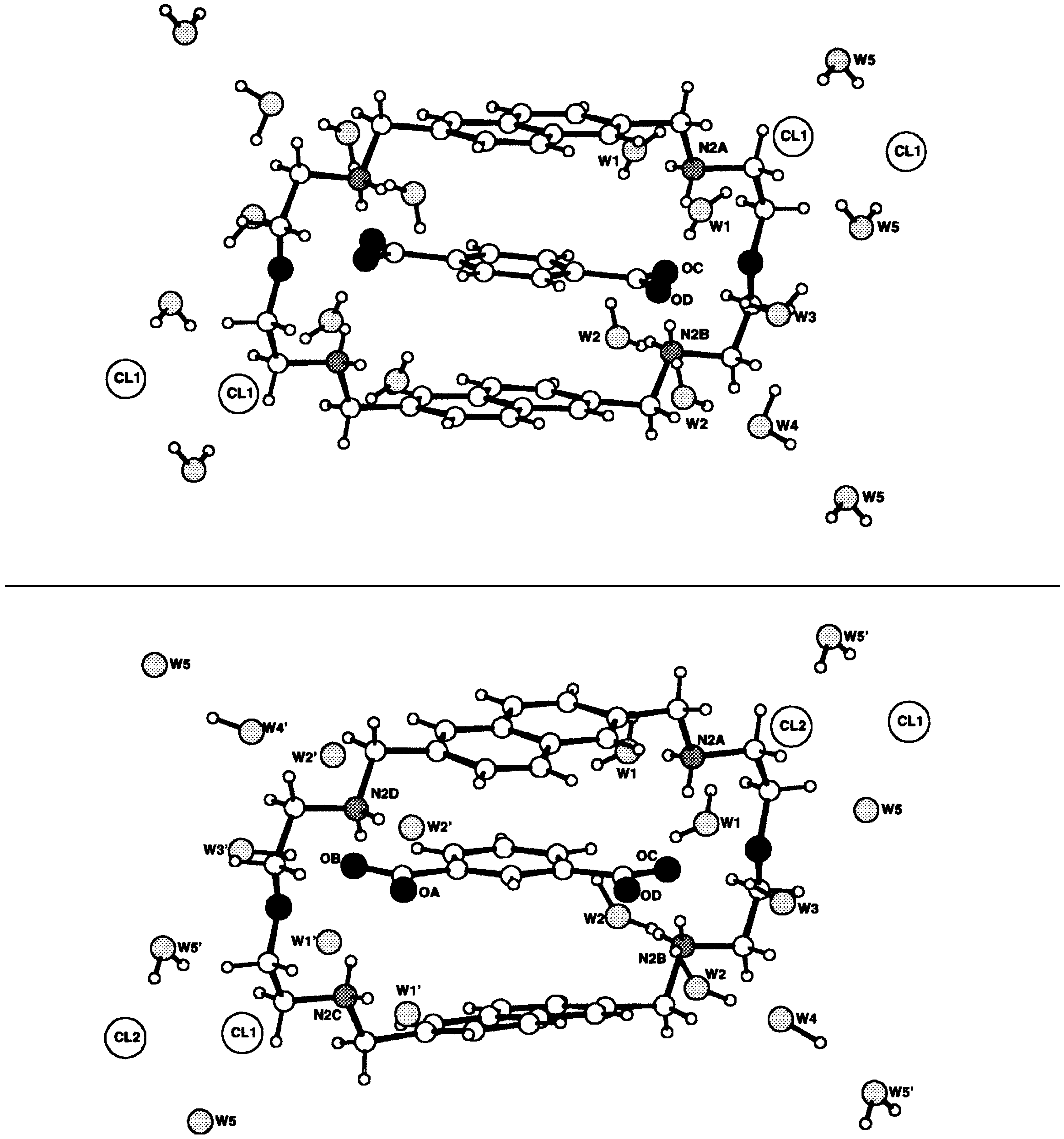
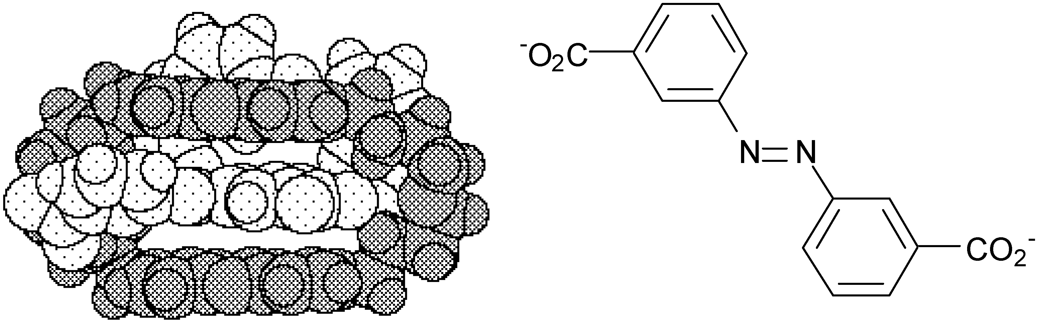
2.2. Binding Properties of Cyclobisintercaland Receptors

In aqueous solutions, all these positively charged compounds strongly bind neutral or anionic planar substrates - the nucleosides and the nucleotides in particular. Very high affinity constants were measured for the nucleotides, between 104 and 108 M-1 whereas with the monomer model they were under 100 [7,8].
The stability of the complexes increases with the number of negative charges in the substrate, as seen for the strong binding of AMP2-, ADP3- and ATP4-. The binding constant also increases markedly with the size of the surfaces - either with the substrate area for a given receptor (for instance purines are more strongly complexed than pyrimidines) or with the flat subunits area of the receptor for a given substrate (for instance, the bisacridine macrocycle binds flat substrates much more strongly than the parent bisnaphthalene compound).
The stoichiometry of the complexes formed was found to be 1 to 1 for all substrates, indicating that a well-defined species had been generated. In most cases a marked hypochromic effect was observed; it was found to be larger the more stable the complex formed by a given receptor. Taken together, the 1:1 stoichiometry, the hypochromism caracteristic of stacking of π-π systems and the influence of the aromatic surface areas suggest a sandwich-type structure for the complexes, the substrates being located between the two flat subunits of the receptors. This was confirmed by the crystal structures of two supramolecular species resulting from the binding of terephthalate and isophthalate dianions by the bisnaphthalene macrocyclic receptor [13]. The dicarboxylate substrate, terephthalate as well as isophthalate, is intercalated between the two naphthalene units of the macrocyclic receptor: the terephthalate dianion is planar and its plane is parallel to the macrocyclic aromatic units, at a van der Waals contact distance of 3.4 Å (Figure 5).
More recently the inclusion complex of the bisacridine receptor with trans 3,3’-azobenzenedicarboxylate has been isolated and its structure has also been determined by X-ray crystallography, confirming, here also, the intercalation of the substrate between the acridine residues in the species formed (Figure 6) [14].

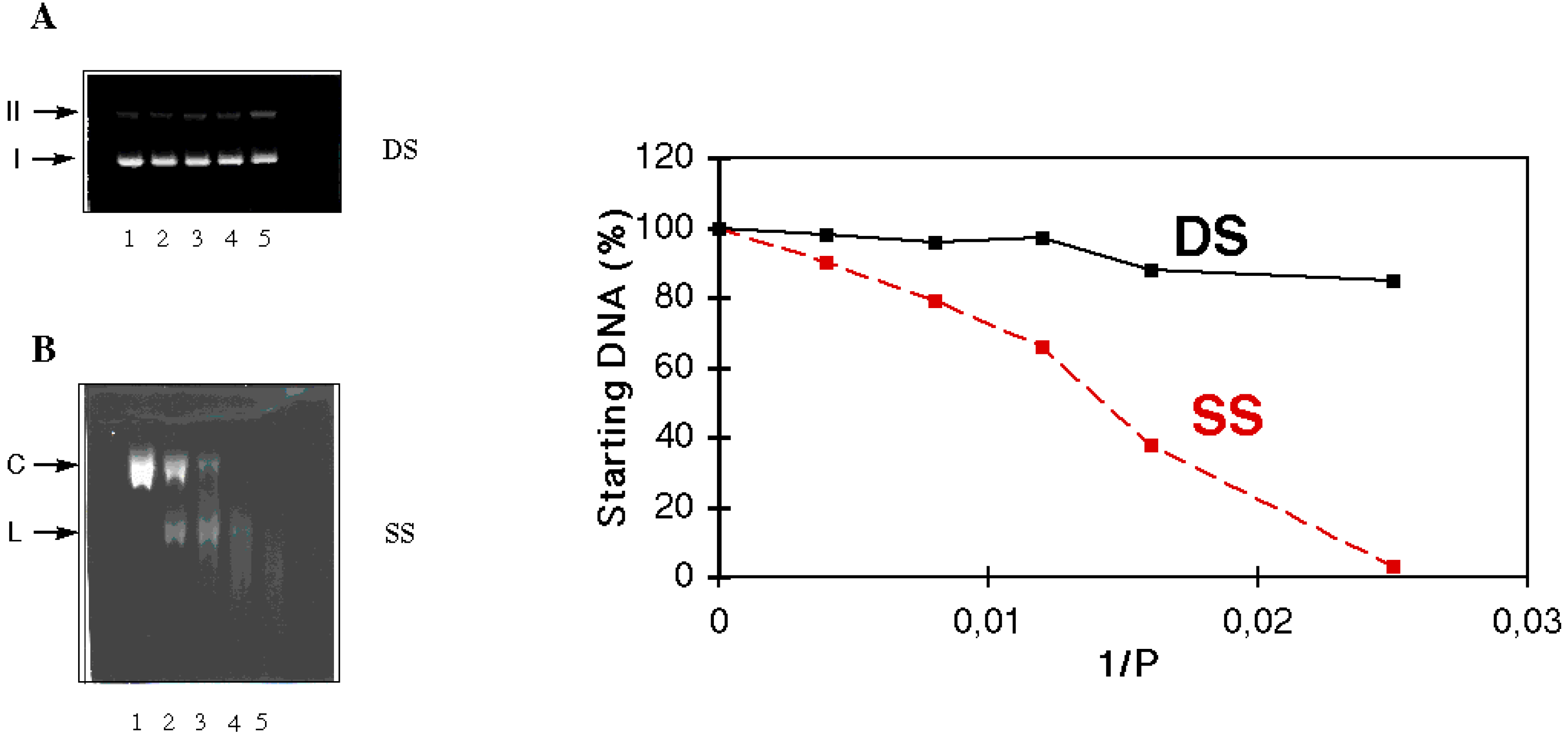
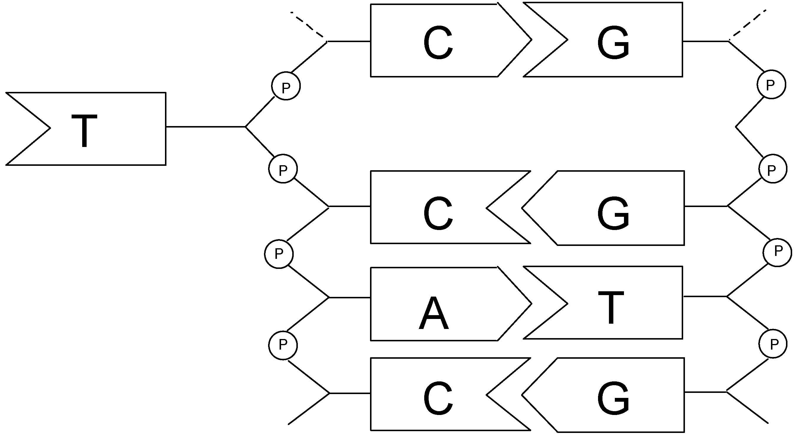
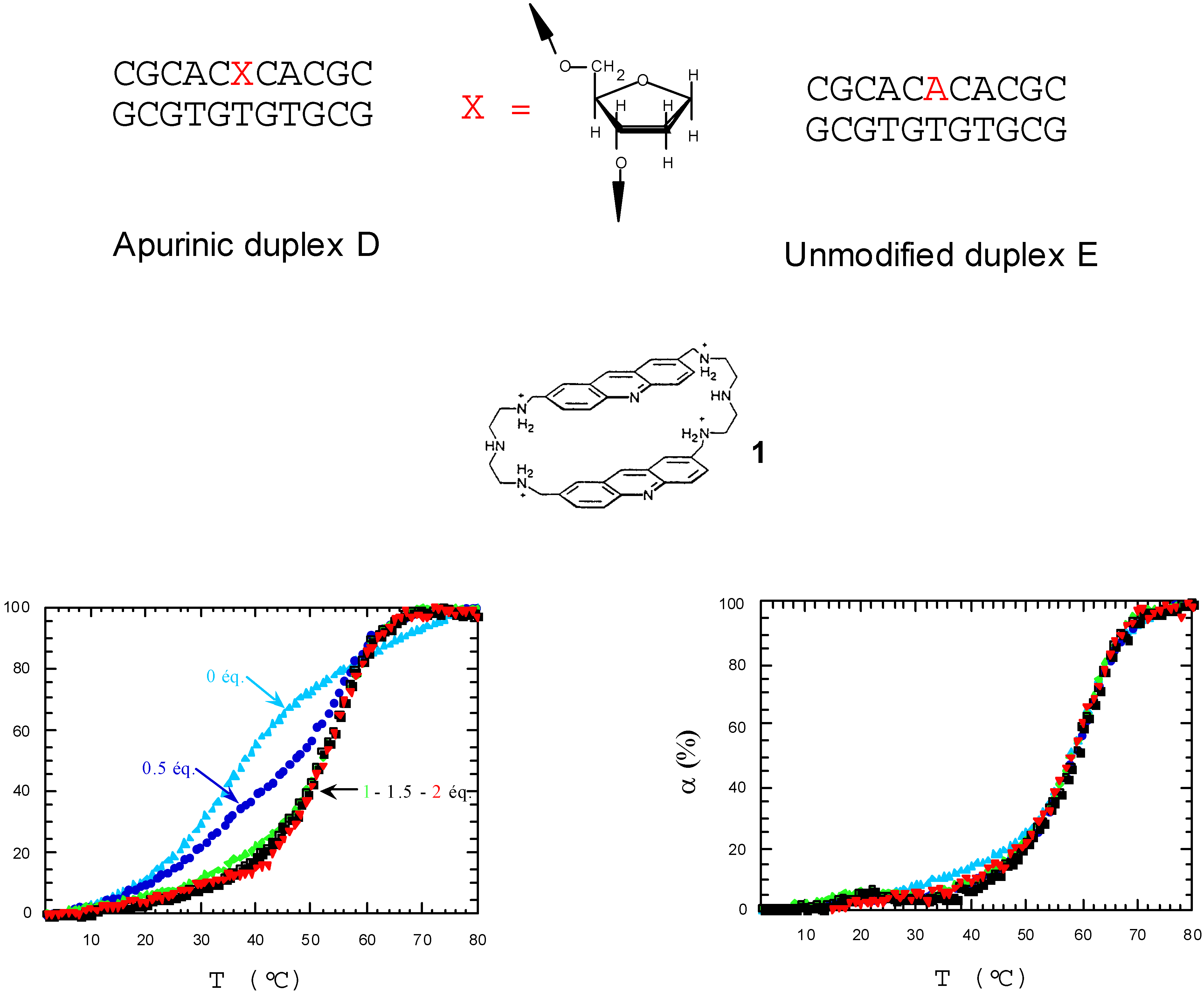
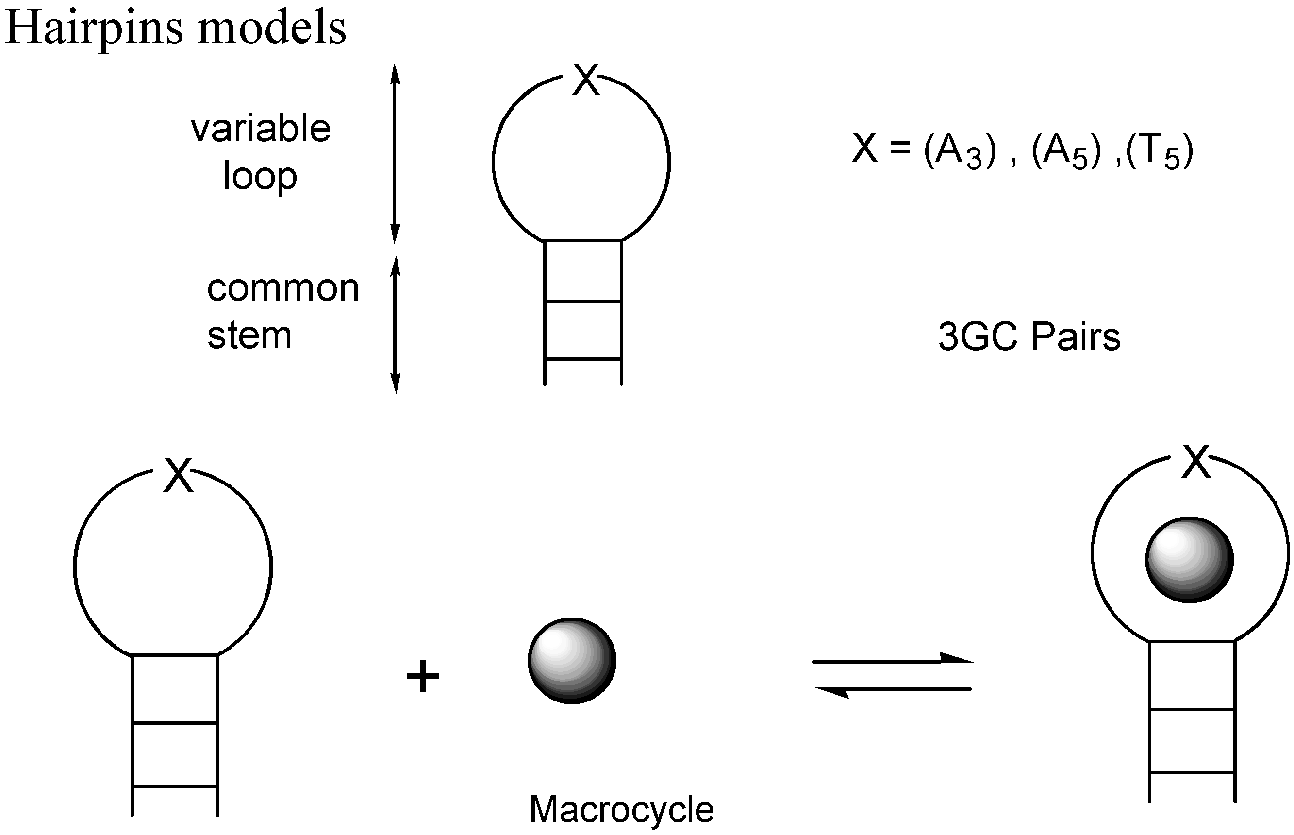
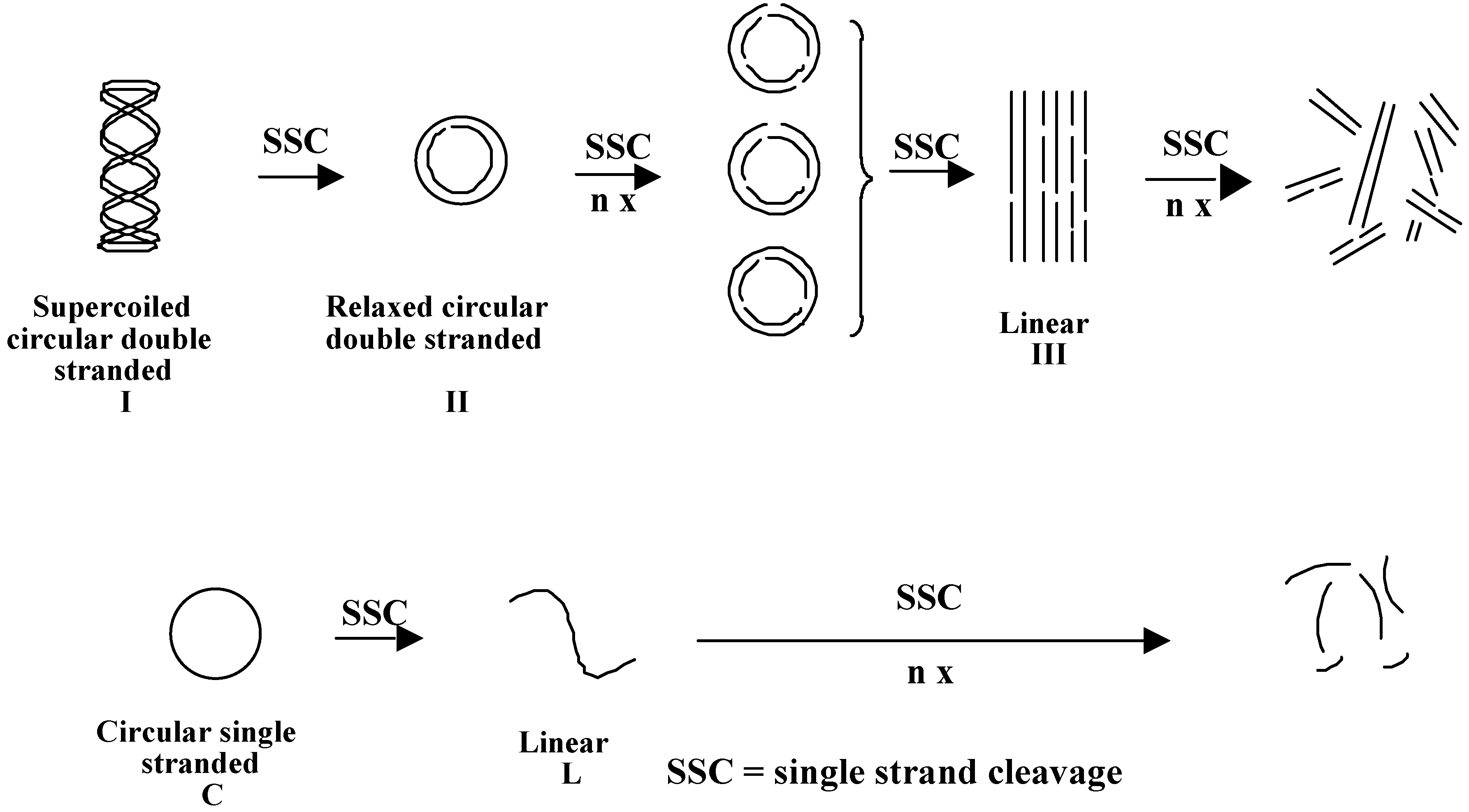
2.3. Discrimination between simple and double-stranded polynucleotides

The discrimination between single and double-stranded polynucleotides, mentioned above, is one of the most interesting features of this type of cyclobisintercaland receptors. To study it, we have used different approaches.
a) selective binding of the bisacridine cyclobisintercaland to DNA hairpins
First, we looked at the interaction of the cyclobisacridine compound with oligonucleotides designed to provide loop superstructures, as model hairpins (Figure 7) [15,16].
Using fluorescence and melting temperature experiments we have shown that the bisacridine compound binds preferentially to the loop area which contains unpaired nucleobases. Conversely, this selectivity is not observed with the acridine monomer model.
b) selective photocleavage of single-stranded nucleic acids by the bisacridine cyclobisintercaland
A confirmation of this selectivity for the unpaired areas was obtained from the selective photocleavage of single-stranded nucleic acids. Indeed, the bisacridine cyclobisintercaland, which contains the acridine subunit as photoactive group can cleave nucleic acids preferentially at single-stranded relative to double-stranded domains under light irradiation.
Photocleavage experiments were conducted on circular supercoiled double-stranded plasmid pUC18 and on circular single-stranded DNA M13mp18. These systems are very useful to show a photocleaving activity (Figure 8).
A single cut of either type of DNA results in the formation of a new species with very different electrophoretic migration properties. The double-stranded DNAs yield a relaxed circular form which may be followed by a linear double-stranded one after multiple cleavage events. On the other hand, the single-stranded circular DNAs give a linear single-stranded species after a single cut.
Figure 9 presents results obtained for the photocleavage of supercoiled ds pUC18 and of ss M13mp18 by the bisacridine 1 [17]. In the concentration range examined, the ds starting material (I) (Figure 9A) remains essentially intact except at the highest 1/phosphate ratio (Figure 9A, lane 5) where a slight transformation into the circular relaxed form (II) is observed. On the contrary, the circular ss DNA (C) is progressively converted into the linear form (L) (Figure 9B, lanes 2,3) and then completely degraded as indicated by the smear of the band (Figure 9B, lanes 4,5). This indicates that low concentrations of compound 1 are sufficient to extensively photocleave ss DNA in a selective manner. Much higher concentrations of 1 and longer irradiation times are required to induce a significant conversion of the ds supercoiled pUC18 (I) to its relaxed form (II) ; moreover the linear form is never observed. When mixtures of the two plasmids are treated in similar fashion, again selective cleavage of the ss DNA takes place whereas the ds DNA is little affected (Figure 9, right).
The same experiment (irradiation of mixtures of pUC18 and M13mp18) has been conducted in the presence of the monomeric reference compound [2,7(di-n-propylamino)acridine]. A similar degradation of both ds and ss DNA was observed, thus revealing the absence of selectivity of the monomeric compound by contrast with its dimeric analog.
As a conclusion, the present experiments establish that the bisacridine cyclobisintercaland is a selective reagent for the preferential photocleavage of single-stranded nucleic acids or single-stranded regions in complex nucleic acids such as transfer RNAs.
c) recognition of abasic sites in DNA by the cyclobisacridine molecule 1
As shown in Figure 10, abasic sites result from the release of a nucleic base from the double-helix framework, leaving a 2’-deoxyribose residue. In Figure 10 an apurinic site is represented as there is a thymine left. According to our starting hypothesis and according to the results just shown above, this unpaired base may be expected to be selectively bound by the cyclobisintercaland macrocycle.
Indeed, using oligonucleotides containing a stable tetrahydrofuranyl analog of the abasic site X, it was shown by Melting Temperature (MT) measurements, displacement of a nitroxide abasic sitespecific probe and photocleavage experiments that 1 binds specifically at the abasic lesion.
d) MT measurements
The melting temperature experiments were performed by using synthetic DNA fragments containing the stable tetrahydrofuranyl analog of the abasic site. This tetrahydrofuranyl moiety is considered to be a good model of the abasic site [18]. The interaction was first studied by examining the thermal denaturation of the apurinic undecamer D in the presence of increasing concentrations of 1. Regular increase of the melting temperature, (from 38°C to 52°C), was observed as the ratio [drug]/[duplex] was increased in the range 0-1. Further addition of the drug (r > 1) did not cause any further increase of the melting temperature. This is proof for formation of a 1:1 complex. By comparison the MT value for the parent unmodified duplex E remained unchanged in the presence of 1 (Mt=58°C) (Figure 11).
e) EPR study
The nitroxide probe ATac-NO has been shown previously to bind selectively to the model abasic duplex D. Figure 12 illustrates the sets of experiments that confirm binding of 1 at the abasic site [18]. The spectrum registered for the labeled probe in dilute solution (10-5 M) (Figure 12A) is characteristic of a nitroxide radical undergoing relatively rapid motion. The spectrum was not modified by addition of equimolar concentration of 1 (Figure 12A′) which indicates the absence of interaction between the two molecules at the weak concentrations of the experiments. Addition of a slight excess of the abasic undecamer duplex D (1.8 x 10⁻⁵M) to the nitroxide probe (1 x 10⁻⁵M) caused large modification in the observed spectrum (Figure 12B). It corresponds to the sum of the spectra of the two species, the radical ATAc-NO free in solution that accounts for ca. 10%, and the radical immobilized at the abasic site (ca. 90%). The spectrum characteristic of the latter species indicating slower movement of the radical was obtained by substraction of the spectrum corresponding to the radical free in solution from spectrum 12B (Figure 12C).
On addition to this probe-DNA solution of a double excess of 1 relative to nitroxide, a new spectrum was obtained (Figure 12B′) characterized by a considerable increase of the contribution of the radical free in solution (ca. 60%). 1 thus shows the ability to displace the labeled molecule ATAc-NO from the abasic duplex. By contrast the EPR spectrum of probe ATAc-NO in the presence of the parent natural duplex E was not modified on addition of bisacridine.
f) photocleavage of the abasic duplex C by bisacridine 1
As we have already seen, the acridine subunits of 1 are photoactive residues capable of cleaving DNA strands when illuminated. Additional information relative to the binding specificity of 1 could thus be obtained from photocleavage experiments using the 23-mer duplex C that contains the stable tetrahydrofuranyl analog of the abasic site in the middle of the sequence [18]. The two strands of the duplex were successively 32P labeled at the 5’-ends. Irradiation of the DNA duplex in the presence of four equivalents of 1 induced alkali labile modifications revealed by piperidine treatment (Figure 13).
One sample was treated by exonuclease III to localize on the gel the position of the abasic site in the duplex. The autoradiogram of the denaturating polyacrylamide gel (Figure 13) shows specific DNA cleavage on the two strands. The strand that carries the abasic site gives one single major labeled fragment corresponding to cleavage at the abasic position. Two photocleavage sites are observed in the complementary strand, a major one G34 and a minor G36, i.e., the two guanine residues that flank the unpaired thymine T35 opposite the lesion. These results provide strong indication for specific binding of the bisacridine drug at the abasic site prior to reaction. However the selectivity and the efficiency of cleavage suggest close contact between the photoactive subunits of 1 and the nucleobases at the abasic site. These observations are in full agreement with the results of a high field NMR study of the interaction between bisacridine and abasic duplex D (Figure 14) [19].
In the major complex formed one acridine subunit intercalates between base pairs C13 G34 and A14 T33 while the second subunit inserts inside the abasic pocket pushing aside the thymine T35 residue in the opposite strand. This scheme could account for major cleavage at guanine G34 that is « sandwiched » between the two acridine rings of the drug. The combination of thermal denaturation, EPR and photocleavage experiments clearly demonstrates the high specificity of the binding of the cyclobisacridine 1 at the abasic site examined, i.e. an apurinic site opposite a thymine residue flanked by two cytosines. Further studies are going on to determine the generality of the observations and evaluate the cyclobisacridine system as a potential inhibitor of the AP-endonucleases involved in repair of the abasic site lesions.

Conclusion of part I

The results obtained so far point to the potential of cyclobisintercaland reagents to function as structural probes for single-stranded domains such as loops, bulges, hairpins or for local defects in complex nucleic acids. Such domains could be possible targets for eliciting therapeutic effects,for instance antiviral activity.
Finally, cyclobisintercalands may be able to displace single strand binding proteins from their site of attachment and thus interfere with a variety of genomic processes.

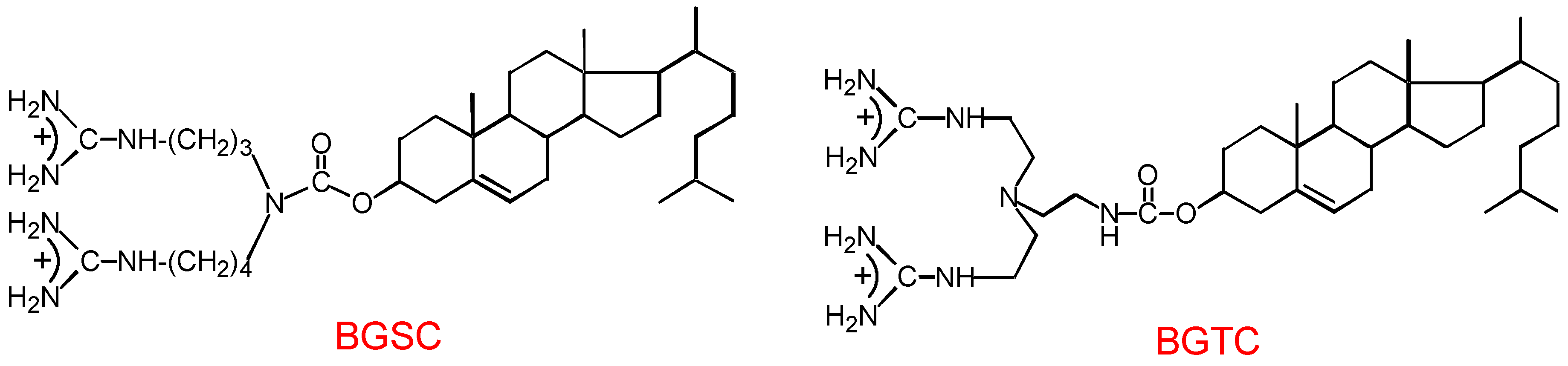
3. Gene Transfer by Guanidinium-Cholesterol Cationic Lipids

Synthetic nonviral vectors represent an attractive alternative approach to viral vectors for gene transfer studies and gene therapy applications. Particularly various cationic lipids have been shown to induce efficient transfection of a large number of eukaryotic cells. Some of them can be used directly as cationic amphiphilic reagents in solution, but most of the lipids are formulated as liposomes containing two lipid species. The spontaneous formation of DNA/lipid aggregates is, in any case, due to ionic interactions between the positively charged cationic lipid and the negatively charged phosphate groups of the DNA. Residual positive charges on the aggregates presumably mediate their binding to negatively charged residues on cell surfaces.This triggers their entry into the cell mainly by endocytosis.

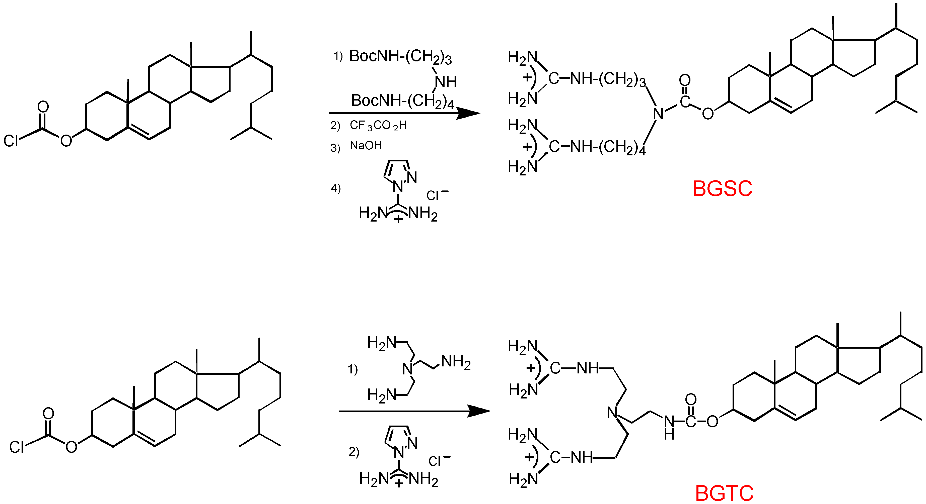
3.1. Design and preparation of new cationic lipids

In this context , we have prepared and tested two new cationic lipids, BGSC and BGTC, cholesterol derivatives bearing guanidinium groups (Figure 15) [20,21,22].
We have chosen the cholesterol unit as an hydrophobic part because it has been shown to facilitate the cellular uptake of various oligonucleotides and polar drugs.
As a polar head, the guanidinium group presents several interesting features (Figure 16):
1) it remains protonated over a much wider range of pH than the ammonium group due to its much higher pKa, 13.5 for the guanidinium itself.
2) it forms with phosphate anions caracteristic pairs of parallel zwitterionic hydrogen bonds which provide binding strength by their charge and structural organisation, as seen for instance in the crystal structure of methyl guanidinium dihydrogen phosphate;
3) the guanidinium group is also able to develop hydrogen bonding with nucleic bases, especially with guanine; such hydrogen bonds have been observed in a number of DNA/protein interactions.
4) finally the guanidinium group of arginyl residues has a major function in DNA-binding proteins, histones and protamines.
Due to the high pKa of the guanidinium group, the transfection efficiency of the lipids BGSC and BGTC should be relatively insensible to the variations of pH during the in vitro formation of the DNA/reagent aggregates and the trafficking in the cell toward the nucleus.
BGSC and BGTC were obtained with good yields by a straightforward synthesis using cheap reagents (Figure 17) [20].
To get BGSC, Di-boc spermidine was reacted with cholesteryl chloroformate and the resulting compound was deprotected by trifluoroacetic acid. Guanylation of the free diamine was effected by reaction with pyrazole carboxamidine hydrochloride.
To prepare BGTC a large excess of TREN was reacted with cholesteryl chloroformate in methylene chloride. The unreacted amine was removed by washing with water and the crude product was treated by pyrazole carboxamidine hydrochloride The global yield was about 60% in the two cases.

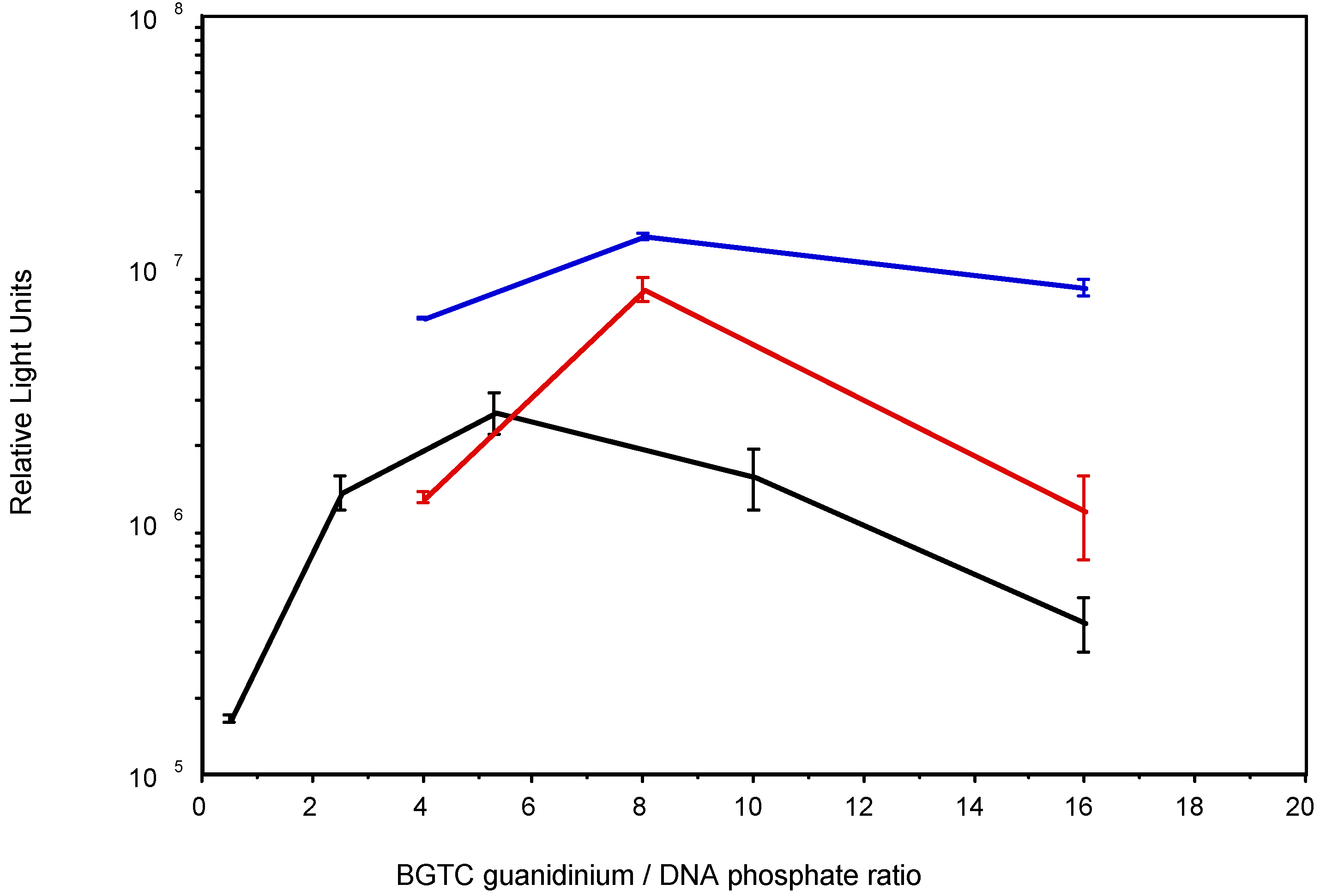
3.2. Transfection activity of BGTC in aqueous solution

As we have shown that compound BGTC gives true micellar solutions in the concentration range used in transfection experiments, we investigated its potential usefulness as a transfection reagent by direct mixing of its solution with the plasmid-containing solution. The luciferase reporter gene was chosen for these experiments. To optimize the BGTC lipid/DNA aggregates formation we first examined the influence of the ratio of BGTC lipid to DNA on transfection activity in 3 different types of mammalian cells known to be relatively easy to transfect with classical techniques [20].
Transfection results with the 3 cell lines are indicated in Figure 18 where luciferase reporter gene expression is shown as a function of the ratio of BGTC guanidinium to DNA phosphate . The ratio of BGTC lipid to DNA is critical to the reagent’s efficacy.
Luciferase expression was highest when working with aggregates containing ≈6 to ≈8 guanidinium groups per DNA phosphate, that's to say, with aggregates bearing a strong positive charge.
In these conditions, we performed transfections with a variety of cell lines. The results (Table 1) show that lipid BGTC is usually as efficient as Transfectam®, a commercially available cationic lipid very efficient for transferring genes into eukaryotic cells in vitro and which is also used as a solution without liposomal formulation. BGTC is also, in general, 1 to 2 orders of magnitude more efficient than the calcium phosphate technique. Thus, we could satisfactorily transfect all the cell lines screened using non toxic levels of BGTC.
Our data demonstrate that compound BGTC can be used for transient transfection of various cell types without liposomal formulation and that it is efficient despite the fact that it contains neither the fusogenic lipid DOPE nor two fatty acid chains, unlike many cationic lipids.

3.3. Transfection efficiency of BGSC and BGTC formulated as cationic liposomes with DOPE

The synthetic compound BGSC being less soluble in aqueous medium than BGTC, we have evaluated its potential for transfection using a liposome formulation with DOPE, with a molar ratio BGSC to DOPE of 3 to 2. We have also prepared BGTC/DOPE liposomes in the same molar ratio [20].
In these cases we have shown that the luciferase expression was highest when using aggregates containing only three guanidinium groups per DNA phosphate instead of six to eight in the case of BGTC/DNA complexes. This observation could support the role of a DOPE-facilitated step in cationic liposome-mediated transfection, involving in particular the well-known fusogenic properties of DOPE.
In these conditions we have transfected various adherent cell lines. Both BGSC and BGTC liposomes gave satisfactory transfection of all the cell lines tested (Table 2). Their efficacy is quite similar and appears also to be similar to that of the commercially available Lipofectin® reagent.
Thus these data demonstrate that bis-guanidinium cholesterol derivatives can also be used as liposome formulations with DOPE for efficient in vitro transient transfection.

3.4. In vivo transfection

We have shown the feasibility of gene transfer into primary human airway epithelial cells in culture using the lipid BGTC in aqueous solution. Most importantly, we also demonstrated the possibility and quantitavely assessed the efficiency of gene transfection by liposomes composed of BGTC and DOPE, into the mouse respiratory epithelium in vivo [21].
For these in vivo studies, the transfection mixture, containing BCTC/DOPE cationic liposomes and the E.Coli β galactosidase reporter gene, was delivered in the airways by intratracheal instillation. After staining with the chromogenic β-galactosidase substrate, X-Gal, X-Gal-positive cells could be detected in the airway epithelium of the treated mice at 48h after transfection of a galactosidase expressing plasmid. Mainly differentiated columnar cells were transfected in the surface epithelium but, interestingly, we could also detect transgene expression in the basal nuclei of submucosal glands.
These results may be of special interest for lung-directed gene therapy for cystic fibrosis as the predominant site of CFTR expression, the transmembrane conductance factor responsible for the desease, is the submucosal glands.
Transfection of the firefly luciferase gene into mouse airways allowed us to quantitatively assess the transfection efficiency of BGTC/DOPE liposomes in vivo. Luciferase activity was repeatedly detectable in the trachea homogenates but not in the lung homogenates. Luciferase expression was exclusively derived from the luciferase-expressing plasmid, since no luciferase activity could be detected in control mice receiving an irrelevant lacZ plasmid or a naked uncomplexed plasmid .
As a conclusion, the present results indicate that gene transfection mediated by guanidiniumcholesterol lipids is feasible within the mammalian airway epithelium. Positive data with primary human cells in vitro and in the mouse airways in vivo confirm the potential of cationic lipids for lungdirected gene therapy.

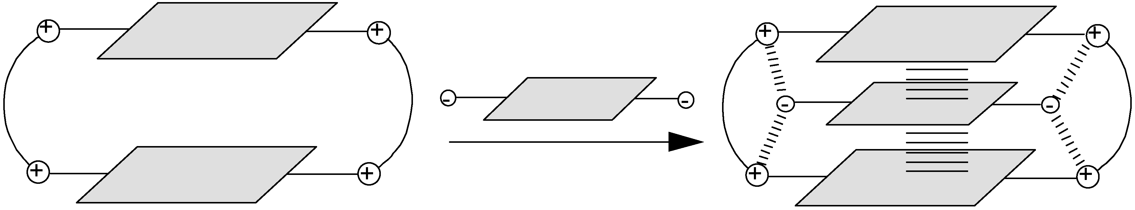
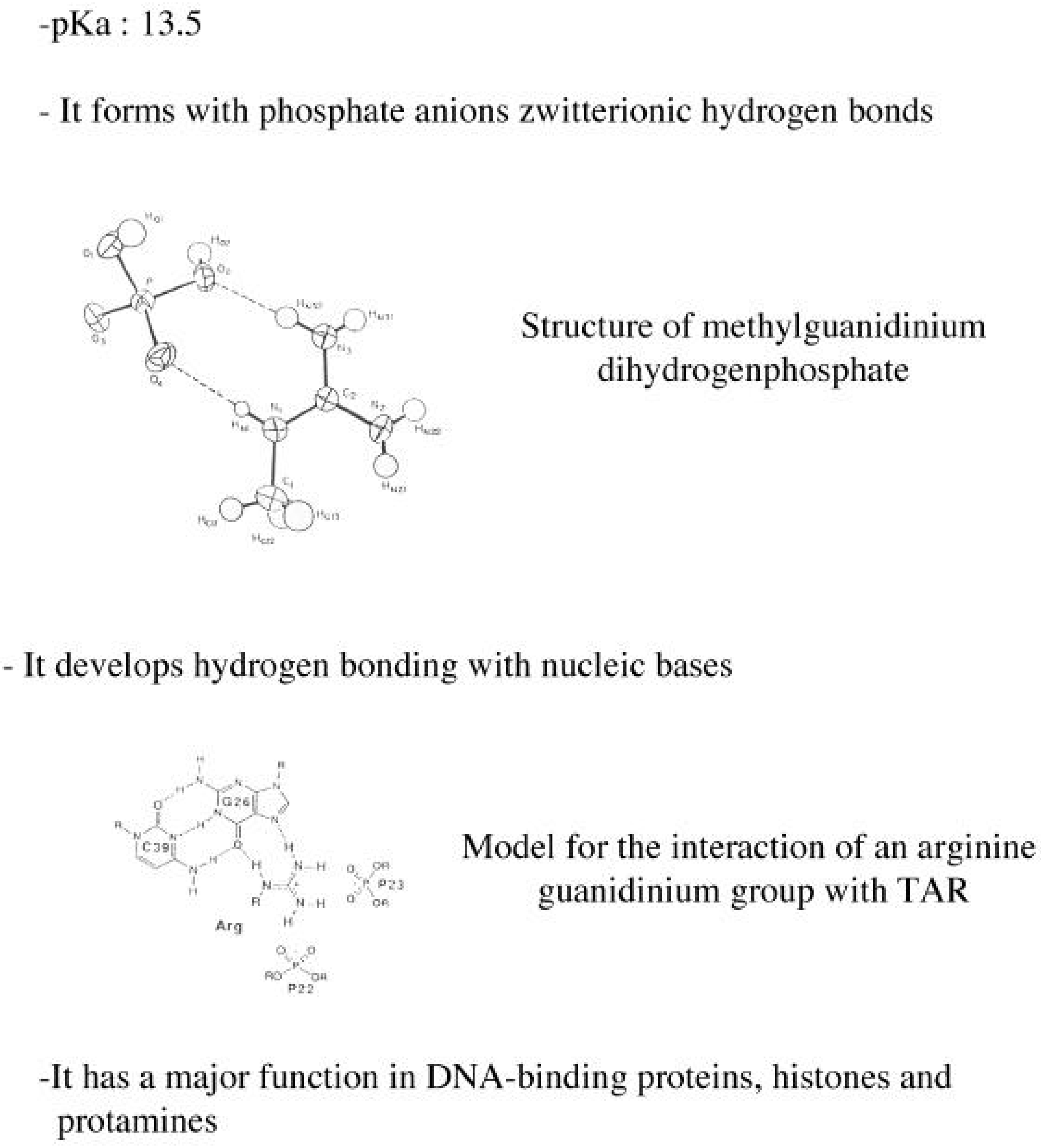
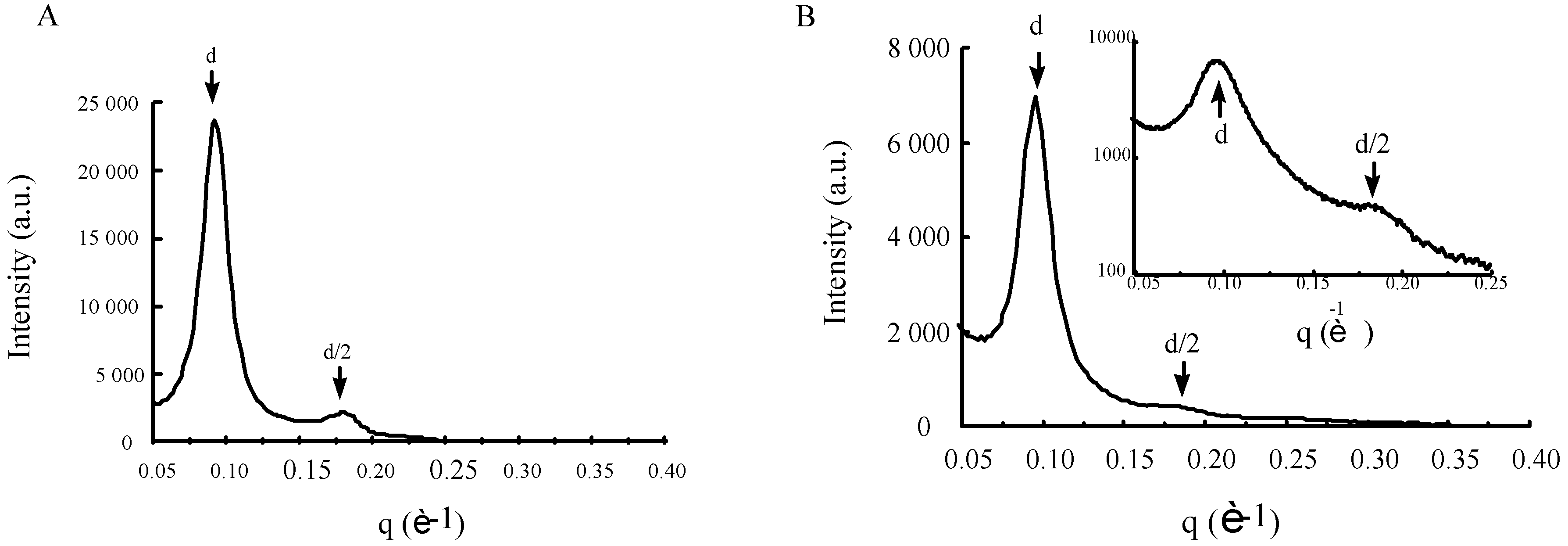
3.5. Structural characteristics of supramolecular assemblies formed by guanidinium-cholesterol reagents

Because gene transfection by cationic lipids involves the spontaneous in vitro formation of discrete lipid/DNA particles, the transfection efficacy of a given cationic lipid system is highly dependent on the structural and functional properties of the self-assembled supramolecular assemblies. Accordingly, characterization of the structure of the lipoplexes should help towards a better understanding of the mechanisms of BGTC-mediated transfection [23].
To visualize the structural features of the lipoplexes formed at lipid/DNA ratios that are used for transfection we used cryo-TEM because it allows imaging of bioassemblies close to their native state. Examination of unreacted BGTC/DOPE liposomes showed a fairly homogeneous population of unilamellar liposomes about 150 nm in diameter. BGTC/DOPE-DNA aggregates visualized discrete concentric multilamellar structures of approximatively 250 nm in diameter.
To characterize further the structural features of BGTC/DOPE-DNA lipoplexes, we also used synchrotron X-ray diffraction. In Figure 19A the scattering intensity of the lipoplexes is plotted as a function of the scattering vector. Importantly, in addition to the strong first-order reflexion at 70 Å, a second order peak at 35 Å could also be detected. This finding indicates lamellar organization with a regular spacing of 70 Å. The scattering pattern reflects the periodic spacing of lamellar BGTC/DOPE-DNA lipoplexes, the periodicity of 70 Å being consistent with stacks of alternating lipid bilayers and DNA.
Cryo-TEM observation of an aqueous solution of BGTC showed threadlike ribbons about 15nm in width and 5nm thick; these ribbons were long, i.e., 1 micron. The structural features of BGTC/DNA lipoplexes were obviously quite different from the concentric multilamellar vesicles of cationic BGTC/DOPE-DNA complexes. Indeed, it appears that BGTC-DNA lipoplexes rather form ordered domains characterized by a fingerprint-like repetition of flat lines; this suggests a lamellar organization.
X-ray diffraction showed a strong reflexion band at 68Å and a second order peak at 34 Å corresponding to a lamellar organization with a regular spacing of 68 Å (Figure 19B).
Thus our data strongly support the observations of previous studies recently reported in the literature. The structures of the lipoplexes formed by various lipid formulations were always found to consist of a multilamellar membrane in which DNA is intercalated.
Finally we have shown that these lamellar structures can also be detected inside cells transfected with DNA complexed with BGTC/DOPE liposomes or BGTC alone.
At this stage one can wonder: can these structures be detected in the nucleus? This is another story currently under investigation.

4. Conclusion

The studies reported in this presentation deal with and illustrate the two broad areas of supramolecular chemistry, that is to say that of supermolecules and that of molecular assemblies.
Simple starting hypothesis turned out to be very fruitful. On one hand, the bisacridine cyclobisintercaland 1 is a very interesting and promising compound. We recently showed that it is able to displace single strand binding proteins from their site of attachment and further studies are going on to evaluate it as potential inhibitor of the AP-endonucleases involved in repair of the abasic site lesions.
On the other hand, the new synthetic transfecting reagent BGTC holds also promise for gene therapy applications notably for lung-directed gene therapy of cystic fibrosis (CF). Interestingly, preliminary results indicate that lipid BGTC allows to transfer the human CFTR gene, the deficiency of which causes cystic fibrosis, into fully differentiated human airway epithelial cells (including primary cells isolated from nasal polyps of CF patients) that normally express CFTR; the transgene CFTR protein is indeed correctly located on the plasma membrane in the successfully transfected cells and also was found to be functionnal.
Finally studies in CF mice should allow better assessment of the utility of guanidinium-cholesterol lipids for lung gene therapy for CF.

Acknowledgments

I would like to thank all those who have taken part in these studies: M. Dhaenens, A. Slama-Schwok, M.-P.Teulade-Fichou (Collège de France-Paris), J. Lhomme and his group (Université Joseph Fourier-Grenoble) for the nucleic acids recognition ,- M. Fauquet (Collège de France-Paris), P. Lehn and his group ( Hôpital Robert Debré-Paris), B. Pitard and his collaborators (Rhône Poulenc Rorer-Vitry sur Seine) for the gene transfer studies, T. Bataille for preparation of the drawings and finally, last but not the least, J.-M. Lehn (Collège de France-Paris). The transfection studies were supported by the Association Française de Lutte contre la Mucoviscidose (AFLM), the Association Française contre les Myopathies (AFM) and Rhône-Poulenc-Rorer Recherche Developpement.

References and Notes

  1. Nucleic acids recognition
  2. Lehn, J.-M.; Schmidt, F.; Vigneron, J.P. Cyclointercalands. - Incorporation of the phenazine group and of metal binding subunits into macrocyclic receptor molecules. Tetrahedron Lett. 1988, 29, 5255. [Google Scholar] [CrossRef]
  3. Claude, S.; Lehn, J.-M.; Vigneron, J.P. Bicyclo-bis-Intercalands: Synthesis of triply bridged bisintercalands based on acridine subunits. Tetrahedron Lett. 1989, 30, 941. [Google Scholar] [CrossRef]
  4. Lehn, J.-M.; Schmidt, F.; Vigneron, J.-P. Preparation and reactivity of polyfunctional phenazine derivatives. J. Heterocyclic Chem. 1990, 27, 1633. [Google Scholar] [CrossRef]
  5. Claude, S.; Lehn, J.-M.; Schmidt, F.; Vigneron, J.-P. Binding of nucleosides, nucleotides and anionic planar substrates by bis-intercaland receptor molecules. J. Chem. Soc. Chem. Commun. 1991, 1182. [Google Scholar]
  6. Zinic, M.; Cudic, P.; Skaric, V.; Vigneron, J.-P.; Lehn, J.-M. Cyclo-bis-intercaland receptors with phenanthridine subunits. Tetrahedron Lett. 1992, 33, 7417. [Google Scholar] [CrossRef]
  7. Claude, S.; Lehn, J.-M.; Pérez de Vega, M.-J.; Vigneron, J.-P.; Baudoin, O. Synthèse de bicyclobis-intercalants dérivés de l’acridine. New J. Chem. 1992, 16, 21. [Google Scholar]
  8. Dhaenens, M.; Lehn, J.-M.; Vigneron, J.-P. Molecular recognition of nucleosides, nucleotides and anionic planar substrates by a water soluble bis-intercaland type receptor molecule. J. Chem. Soc., Perkin Trans 2 1993, 1379. [Google Scholar] [CrossRef]
  9. Teulade-Fichou, M.-P.; Vigneron, J.-P.; Lehn, J.-M. Molecular recognition of nucleosides and nucleotides by a water soluble cyclo-bis-intercaland type receptor molecule based on acridine subunits. Supramolecular Chemistry 1995, 5, 139. [Google Scholar] [CrossRef]
  10. Cudic, P.; Zinic, M.; Tomisic, V.; Simeon, V.; Vigneron, J.-P.; Lehn, J.-M. Binding of nucleotides in water by phenanthridinium bis-intercaland receptor molecules. J. Chem. Soc., Chem. Commun. 1995, 1073. [Google Scholar]
  11. Espinosa, J.F.; Jaime, C.; Lehn, J.-M.; Vigneron, J.-P. Cyclo-bis-intercalands with acridine subunits linked by rigid spacers. Tetrahedron Lett. 1995, 36, 5261. [Google Scholar]
  12. Lorente, A.; Fernández Saiz, M.; Lehn, J.-M.; Vigneron, J.-P. Cyclo-bis and cyclo-tris-intercalands based on acridine subunits. Tetrahedron Lett. 1995, 36, 8279. [Google Scholar] [CrossRef]
  13. Cudic, P.; Zinic, M.; Skaric, V.; Kiralj, R.; Kojic-Prodic, B.; Vigneron, J.-P.; Lehn, J.-M. Synthesis of cyclo-bis-intercaland receptor molecules with phenanthridinium units. Croatica Chem. Acta 1996, 69, 569. [Google Scholar]
  14. Paris, T.; Vigneron, J.-P.; Lehn, J.-M.; Cesario, M.; Guilhem, J.; Pascard, C. Molecular Recognition of Anionic Substrates. Crystal structures of the supramolecular inclusion complexes of terephthalate and isophthalate dianions with a bis-intercaland receptor molecule. J. Incl. Phenom. 1999, 33, 191. [Google Scholar] [CrossRef]
  15. Cudic, P.; Vigneron, J.-P.; Lehn, J.-M.; Cesario, M.; Prangé, T. Molecular Recognition of Azobenzene Dicarboxylates by Acridine-based Receptor Molecules. Crystal Structure of the Supramolecular Inclusion Complex of trans-3,3’-Azobenzene Dicarboxylate with a Cyclo-bisintercaland Receptor. Eur. J. Org. Chem. 1999, in press. [Google Scholar] [CrossRef]
  16. Slama-Schwok, A.; Teulade-Fichou, M.-P.; Vigneron, J.-P.; Taillandier, E.; Lehn, J.-M. Selective binding of macrocyclic bis-acridine to a DNA hairpin. J. Am. Chem. Soc. 1995, 117, 6822. [Google Scholar] [CrossRef]
  17. Slama-Schwok, A.; Peronnet, F.; Hantz-Brachete, E.; Taillandier, E.; Teulade-Fichou, M.-P.; Vigneron, J.-P.; Baudoin, O.; Best-Belpomme, M.; Lehn, J.-M. A macrocyclic bis-acridine shifts the equilibrium from duplexes towards DNA hairpins. Nucleic Acids Res. 1997, 25, 2574. [Google Scholar] [CrossRef]
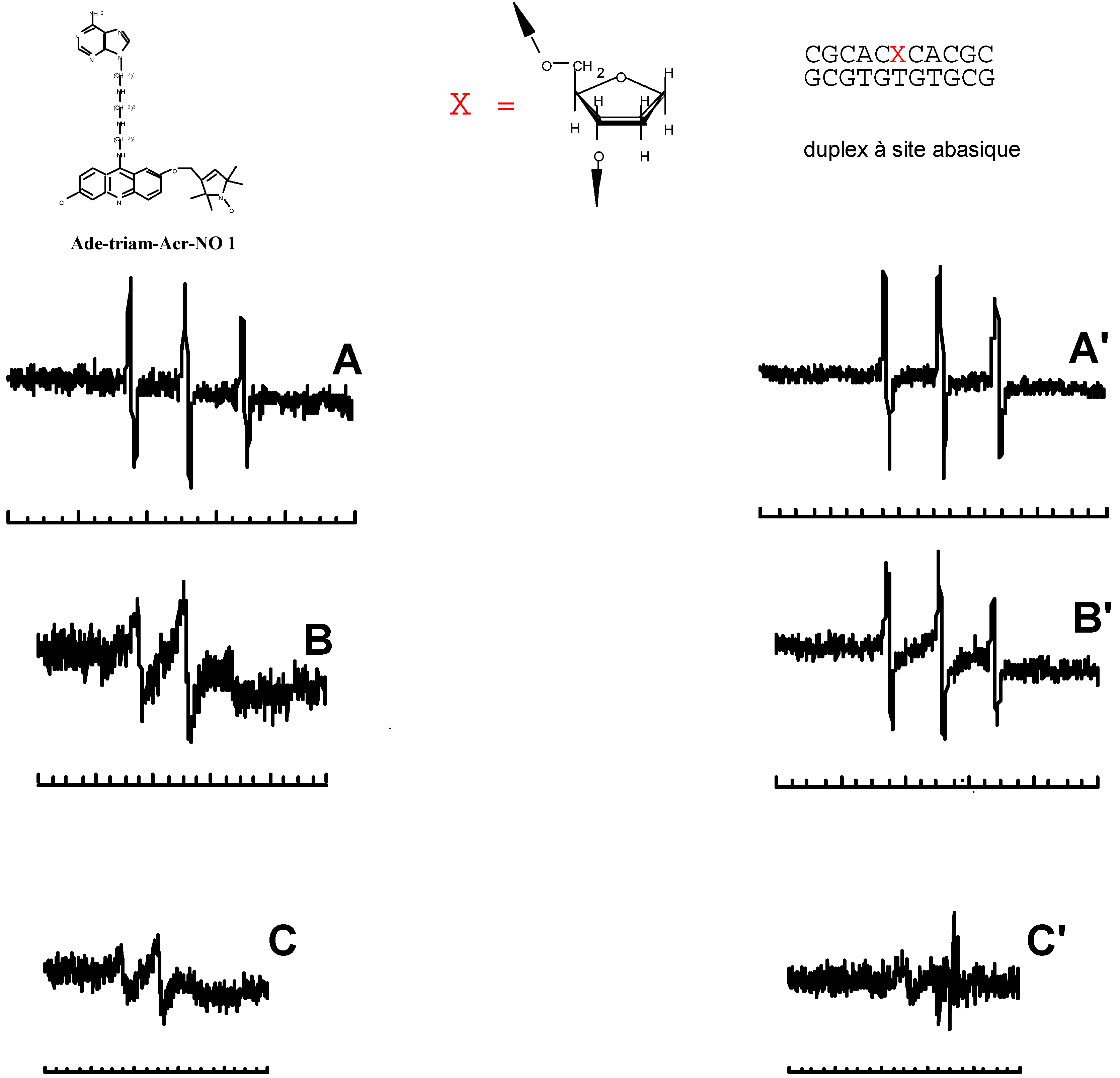
  18. Blacker, A.J.; Teulade-Fichou, M.-P.; Vigneron, J.-P.; Fauquet, M.; Lehn, J.-M. Selective photocleavage of single stranded DNA plasmids by cyclo-bisintercaland compounds. Bioorg. Med. Chem. Lett. 1998, 8, 601. [Google Scholar] [CrossRef]
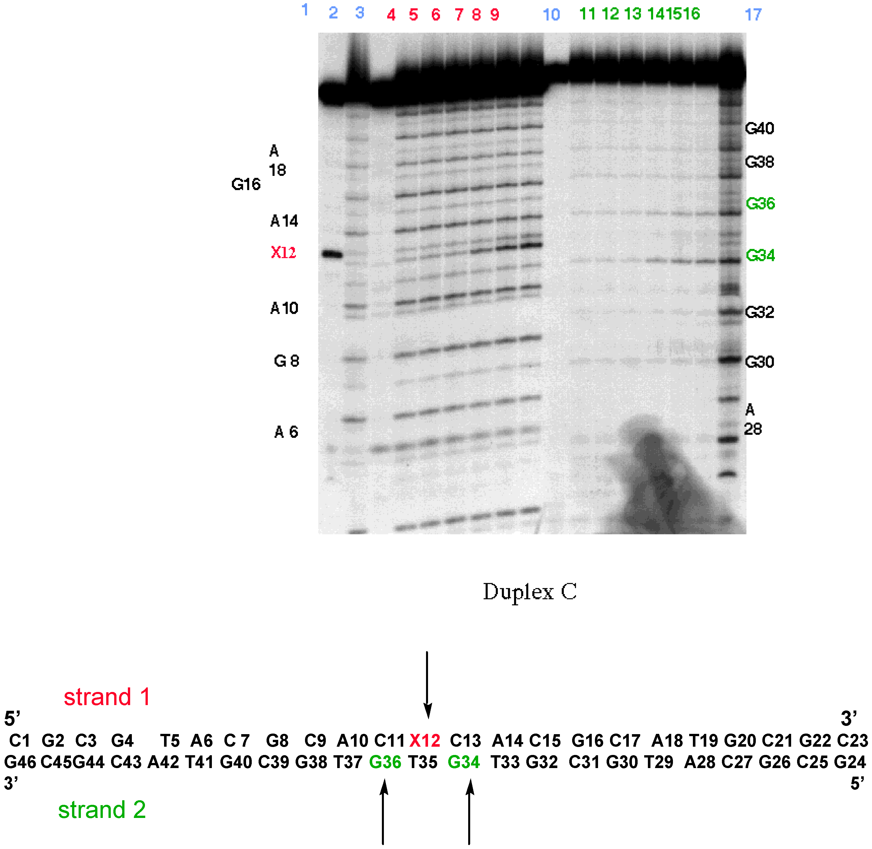
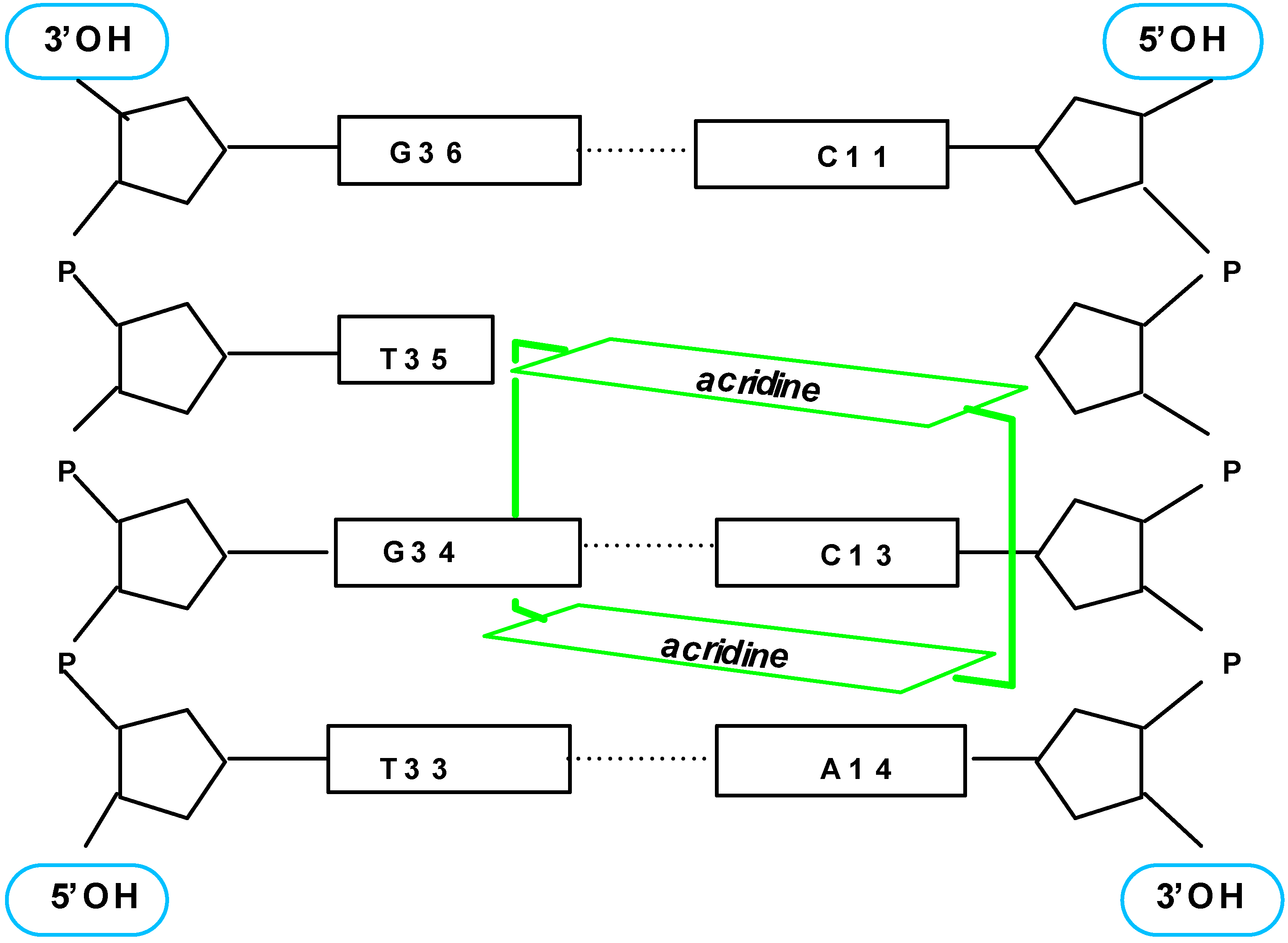
  19. Berthet, N.; Michon, J.; García, J.; Jourdan, M.; Lhomme, J.; Teulade-Fichou, M.-P.; Vigneron, J.P.; Lehn, J.-M. Specific Recognition and Stabilization of an Abasic Site-Containing DNA Duplex by a Macrocyclic Bis-acridine. Nucleic Acid Res. 1999, in press. [Google Scholar]
  20. Jourdan, M.; García, J.; Lhomme, J.; Teulade-Fichou, M.-P.; Vigneron, J.-P.; Lehn, J.-M. Threading Bis-intercalation of a Macrocyclic Bisacridine at Abasic Sites in DNA: 1H NMR and Molecular Modeling Study. Biochemistry. (submitted).
  21. Gene transfer studies
  22. Vigneron, J.-P.; Oudrhiri, N.; Fauquet, M.; Vergely, L.; Bradley, J.-C.; Basseville, M.; Lehn, P.; Lehn, J.-M. Guanidinium-cholesterol cationic lipids: Efficient vectors for the transfection of eukaryotic cells. Proc. Natl. Acad. Sci. USA 1996, 93, 9682. [Google Scholar] [CrossRef] [PubMed]
  23. Oudrhiri, N.; Vigneron, J.-P.; Peuchmaur, M.; Leclerc, T.; Lehn, J.-M.; Lehn, P. Gene transfer by guanidinium-cholesterol cationic lipids into airway epithelial cells in vitro and in vivo. Proc. Natl. Acad. Sci. USA 1997, 94, 1651. [Google Scholar] [CrossRef] [PubMed]
  24. Oudrhiri, N.; Vigneron, J.-P.; Hauchecorne, M.; Toury, R.; Lemoine, A.I.; Peuchmaur, M.; Navarro, J.; Lehn, J.-M.; Lehn, P. Guanidinium cholesterol cationic lipids: novel reagents for gene transfection and perspectives for gene therapy. Biogenic Amines 1998, 14, 537. [Google Scholar]
  25. Pitard, B.; Oudrhiri, N.; Vigneron, J.-P.; Hauchecorne, M.; Aguerre, O.; Toury, R.; Airiau, M.; Ramasawmy, R.; Scherman, D.; Grouzet, J.; Lehn, J.-M.; Lehn, P. Structural characteristics of supramolecular assemblies formed by guanidinium-cholesterol reagents for gene transfection. Proc. Natl. Acad. Sci. USA 1999, 96, 2621. [Google Scholar] [CrossRef] [PubMed]
  • Samples Availability: Available from MDPI.
Figure 1. Schematic representation of a cyclobisintercaland and of the intercalation process.
Molecules 04 00180 g001
Figure 2. Schematic representation of the modes of interaction of a cyclobisintercaland molecule with double-stranded (left) and single-stranded (right) nucleic acids.
Molecules 04 00180 g002
Figure 3. Representative polyamino cyclobisintercalands and the acridine monomer model.
Molecules 04 00180 g003
Figure 4. General synthesis of cyclobisintercalands.
Molecules 04 00180 g004
Figure 5. Crystal structures of supramolecular inclusion complexes of terephthalate and isophthalate dianions with a naphthalenic cyclobisintercaland molecule.
Molecules 04 00180 g005
Figure 6. Crystal structure of the inclusion complex of the bisacridine receptor with trans 3,3’-azobenzenedicarboxylate in space filling representation.
Molecules 04 00180 g006
Figure 7. Selective binding of the bisacridine receptor to DNA hairpins.
Molecules 04 00180 g007
Figure 8. Single-strand cleavage of supercoiled circular double-stranded and of circular single-stranded nucleic acids.
Molecules 04 00180 g008
Figure 9. (Left, A,B): agarose gel electrophoresis patterns for the separate photocleavage of ds pUC18 DNA (A) and ss M13mp18 (B) by 1; [DNA]: 0.3mM in nucleotide phosphate unit; [1]/[Phosphate]: lane 1,0; lane 2, 1/250; lane 3, 1/125; lane 4, 1/160; lane 5, 1/130. (Right): photocleavage of mixtures of pUC18 and M13mp18 by 1 : [ds DNA] = 0.3mM in nucleotide phosphate unit; variation of ds and ss DNA as a function of the concentration of 1.
Molecules 04 00180 g009
Figure 10. Schematic representation of an apurinic site.
Molecules 04 00180 g010
Figure 11. Recognition of abasic sites in DNA by cyclobisacridine 1: MT experiments.
Molecules 04 00180 g011
Figure 12. Recognition of abasic sites in DNA by cyclobisacridine 1: EPR experiments.
Molecules 04 00180 g012
Figure 13. Recognition of abasic sites in DNA by cyclobisacridine 1: Photocleavage experiments.
Molecules 04 00180 g013
Figure 14. Schematic representation of the major DNA-Bisacridine complex.
Molecules 04 00180 g014
Figure 15. Guanidinium-cholesterol cationic lipids.
Molecules 04 00180 g015
Figure 16. Caracteristics of the guanidinium group.
Molecules 04 00180 g016
Figure 17. Synthesis of BGSC and BGTC.
Molecules 04 00180 g017
Figure 18. Luciferase reporter gene expression as a function of the mean charge of the lipid/DNA aggregates.
Molecules 04 00180 g018
Figure 19. SAXS scans of BGTC/DOPE-DNA lipoplexes (A) and of BGTC-DNA lipoplexes (B).
Molecules 04 00180 g019
Table 1. Luciferase expression in various mammalian cell lines transfected with either lipid BGTC, calcium phosphate or Transfectam®.
SpeciesCell lineTissueRLU/mg of cell protein
BGTC lipidCalcium PhosphateTransfectam®
HumanA549Lung carcinoma4x1057x1033.1x105
MonkeyCOS-7SV-40 transformed kidney2.1x1073.7x1058.4x106
DogMDCK-1Kidney3x1064.5x1052.2x106
RatRIN-m5FPancreatic islet cell tumor2x1072x1053.3x105
ROSOsteosarcoma4.0x1064.5x1052.3x106
PC12Pheochromocytoma3x1071.7x1056x106
MouseAtT-20Pituitary tumor2x1078x1053x107
Data for luciferase activity are expressed as mean relative light units normalized to 1 mg of cell protein (RLU/mg protein).
Table 2. Luciferase expression in various eukaryotic cell lines transfected by BGSC/DOPE liposomes, by BGTC/DOPE liposomes and by Lipofectin® reagent.
Cell linesRLU/mg of cell protein
BGSC liposomesBGTC liposomesLipofectin ®
HeLa4.6 x 1067.7 x 1063.3 x 106
A 5496 x 1052 x 1054 x 106
COS-7ND1.4 x 1079.5 x 106
MDCK-11 x 1067 x 1061.9 x 106
ROSND9 x 1066 x 106
NB2 A a1.5 x 1071.4 x 107ND
NIH 3T3 a7 x 1061.5 x 106ND

