Article

Synthesis and Characterisation of Multivariate Metal–Organic Frameworks for Controlled Doxorubicin Absorption and Release

by
Ahmed Ahmed
1,2,
Andrey Bezrukov
3,
Debobroto Sensharma
1,3,
Ciaran O’Malley
2,
Michael J. Zaworotko
1,3,
Davide Tiana
1,4,* and
Constantina Papatriantafyllopoulou
1,2,*
1
SSPC Research Ireland Centre for Pharmaceuticals, Ireland
2
School of Biological and Chemical Sciences, College of Science and Engineering, University of Galway, H91 TK33 Galway, Ireland
3
Department of Chemical Sciences, Bernal Institute, University of Limerick, V94 T9PX Limerick, Ireland
4
School of Chemistry, University College Cork, College Road, T12 K8AF Cork, Ireland
*
Authors to whom correspondence should be addressed.
Molecules 2025, 30(9), 1968; https://doi.org/10.3390/molecules30091968
Submission received: 12 April 2025 / Revised: 25 April 2025 / Accepted: 26 April 2025 / Published: 29 April 2025

Abstract

The development of drug carriers with efficient absorption and controlled delivery properties is crucial for advancing medical treatments. Metal–organic frameworks (MOFs) with tunable porosity and a large surface area represent a promising class of materials for this application. Among them, NUIG4 stands out as a biocompatible MOF that exhibits exceptionally high doxorubicin (Dox) absorption (1995 mg dox/g NUIG4) and pH-controlled release properties. In this study, we report the synthesis and characterisation of multivariate MOFs (MV-NUIG4), which are analogues of NUIG4 that maintain the same topology while incorporating different functional groups within their framework. Eight new MV-NUIG4 MOFs have been synthesised through in situ reactions of the corresponding 4-aminobenzoic acid derivative with 4-formylbenzoic acid. The compounds were thoroughly characterised using a range of techniques, including powder X-ray diffraction, infrared spectroscopy, 1H-NMR, and single-crystal X-ray crystallography. The experimental ratio of the reagents and ligand precursors for the synthesis of MV-NUIG4 MOFs matched the ratio of the linkers in the final products. These structures incorporate additional functional groups, such as methyl and hydroxyl, in varying ratios. Computational modelling was used to provide further insight into the crystal structure of the MOFs, revealing a random distribution of the functional groups in the framework. The Dox absorption and release capacity of all analogues were studied, and the results revealed that all analogues displayed high drug absorption in the range of 1234–1995 mg Dox/g MOF. Furthermore, the absorption and release rates of the drug are modulated by the ratio of functional groups, providing a promising approach for controlling drug delivery properties in MOFs.

1. Introduction

Metal–organic frameworks (MOFs) [1,2,3,4,5,6,7,8] have attracted significant attention due to their unique structural features, as well as their potential to tackle challenges in the biomedical, environmental, and industrial fields [9,10,11,12,13,14,15,16,17]. The conceptual basis for MOFs traces back to the seminal work of Hoskins and Robson in 1989, who introduced the idea of infinite coordination polymers with permanent porosity, laying the foundation for this field [2]. Following this, the development of MOFs evolved into a vast field that has been shaped by many researchers globally. MOFs possess a large surface area and flexible and tunable structures, along with high porosity, which make them suitable for encapsulating a wide variety of guest molecules. As such, MOFs are promising candidates for applications in gas storage and separation [18,19,20,21,22,23], drug delivery [1,24,25,26,27,28], imaging [29,30], catalysis [31,32,33,34,35], sensing [36,37,38], and others]. Furthermore, they can protect large biomolecules from degradation and control their release, and they have advanced the fields of gene targeting, enzyme immobilization, and the crystallization and purification of proteins [39,40,41,42,43,44,45,46,47,48]. Recently, MOFs have also been explored as a potential solution to combat antibiotic resistance [24,25,49,50].
The intriguing properties and applications of MOFs have amplified the need to develop targeted synthesis approaches and deliberately integrate functional groups into their framework. These modifications aim to enhance MOF performance for specific uses, particularly in biomedical applications. In alignment with this, multivariate MOFs (MV-MOFs) [51,52,53,54,55,56,57] have gained significant attention due to their ability to introduce functionality to an MOF while maintaining the framework topology.
MV-MOFs are composed of various metal atoms and/or organic linkers, with the presence of these diverse components leading to a varied arrangement of functional groups while preserving the backbone structure [51,52,53,54,55,56,57]. Significantly, the properties of MV-MOFs are not simply a linear combination of the different functionalities, but rather it is the synergy between these functionalities that often leads to enhanced performance. One of the first examples of this is an MV-MOF5 analogue, which showcased a 400% increase in selectivity for CO2 over CO compared to analogues that consist of the link components separately [51,52].
Many families of MV-MOFs are now known, and most of them are being widely studied for their potential applications in gas absorption and separation, CO2 fixation, photocatalysis, and more [58,59,60,61,62,63,64]. Recently, they have also been explored in the field of drug delivery [63,64]. MV-MOFs are promising candidates in this context, as they enable fine-tuning of the drug release profile through the introduction of functional groups that regulate interactions between the framework and encapsulated drugs. A prime example of this is the family of MV-MIL101(Fe) MOFs [64], which are synthesised with three different linkers in varying ratios: benzenedicarboxylic acid (bdc), NH2-bdc, and 1,4-naphthalenedicarboxylic acid. Notably, increasing NH2 abundance resulted in slower Ibuprofen release but faster Dox and Rhodamine B release, with the control of the linker ratio enabling precise control of the release profile.
With the above in mind, we aimed to use the NUIG4 MOF (Figure 1) as a basis to create MOFs with similar structural features but with different functional groups (MV-NUIG4). NUIG4 has a Dox uptake capacity of 1995 mg Dox g−1, with a pH-dependent drug release rate that is greater under acidic conditions [1]. In particular, we aimed to synthesise MV-NUIG4 MOFs with various functional groups and investigate their potential for controlled drug absorption and release. The functional groups were carefully selected to investigate various aspects of MOF–drug interactions, including hydrogen bonding capabilities, effects on pore accessibility, steric parameters, and aromatic interactions.
Herein, we report on the synthesis and characterisation of eight new members of the MV-NUIG4 multivariate family of MOFs. The MOFs are isostructural to NUIG4 and contain the ligands CBABH2 and CBABH2-OH, and CBABH2 and CBABH2-Me (Scheme 1) in varying ratios. A computational analysis confirmed a random distribution of the ligands throughout the frameworks. All MV-NUIG4 analogues exhibited high Dox absorption, with their absorption and release profiles differing according to the type and proportion of functional groups within the framework, which provides a promising method for developing carriers with tailored drug delivery properties.

2. Results and Discussion

2.1. Synthetic Discussion

The possibility of isolating MV-NUIG4 frameworks was investigated by reacting 4-aminobenzoic acid derivatives with 4-formylbenzoic acid and Zn(CH3CO2)2∙2H2O in DMF. Although the initial approach was to follow the NUIG4 synthetic protocol, different reaction conditions and the use of modulators were explored.
Direct reactions between the linkers and Zn2+ sources produced amorphous materials or no solid products. By contrast, in situ reactions of the corresponding 4-aminobenzoic acid derivative with 4-formylbenzoic acid and Zn(CH3CO2)2∙2H2O resulted in crystalline materials.
A series of functional groups were investigated (CH3, OH, OCH3, naphthyl, NO2, pyridyl, and F) to create both individual frameworks and MV-MOFs. These groups were selected to study different MOF–drug interactions, including hydrogen bonding (hydroxy, fluoro, nitro, and pyridyl), pore accessibility (methyl), aromatic interactions (naphthyl), and steric effects (methoxy). Only CBABH2, CBABH2-OH, and CBABH2-Me successfully formed MV-MOFs, while bulkier linkers failed due to steric hindrance in the four-fold interpenetrated structure.
The experimental and product stoichiometry for the synthesis of the MV-NUIG4 MOFs showed excellent linear correlation for CBAB-OH and CBAB-Me (regression factors: 0.9986 and 0.9988, respectively; Figure 2), demonstrating precise control over functional group ratios. Multiple sample analyses, including individual single crystal examinations, confirmed the formation of pure compounds rather than physical mixtures, ruling out linker bias.

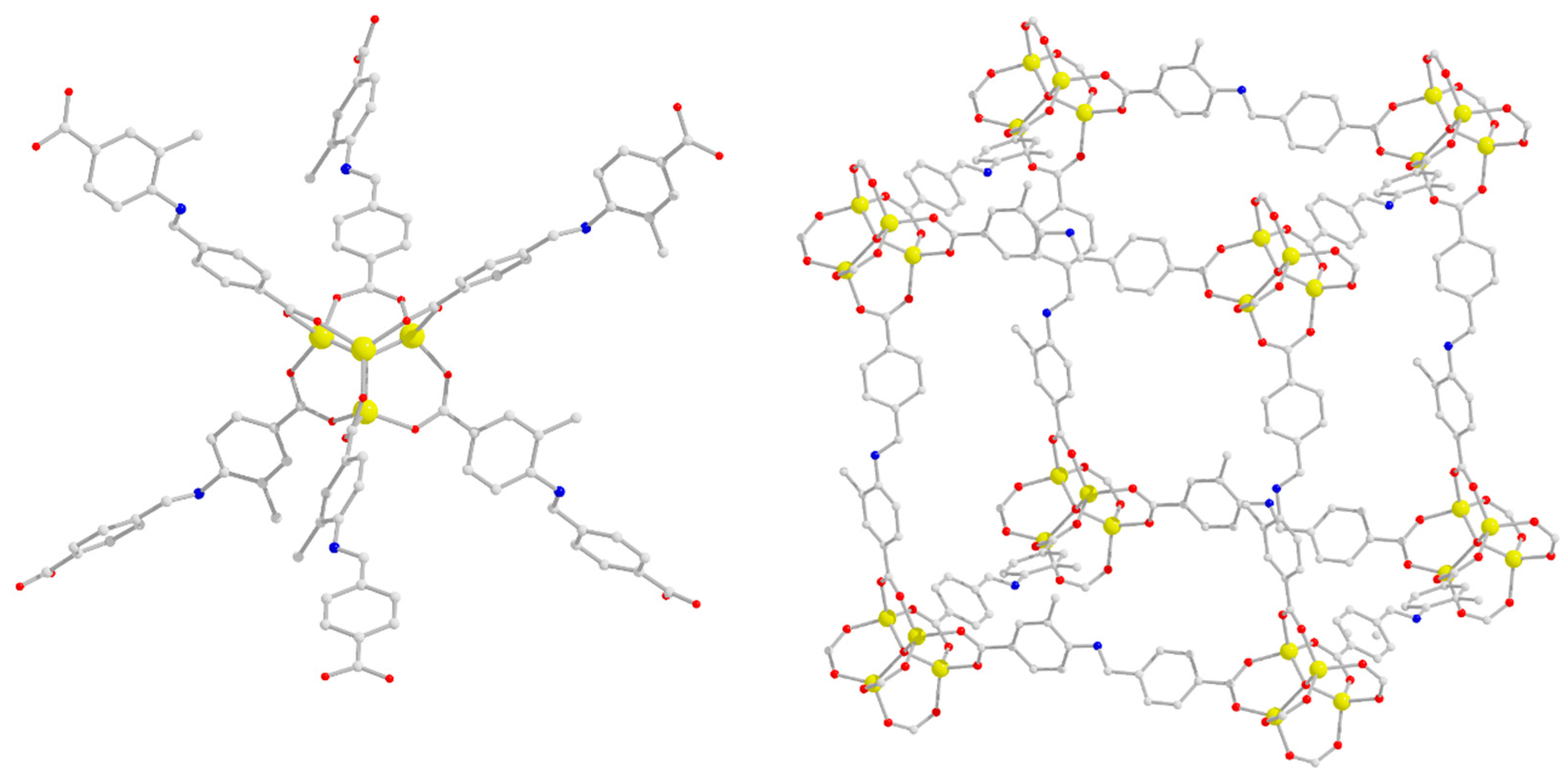
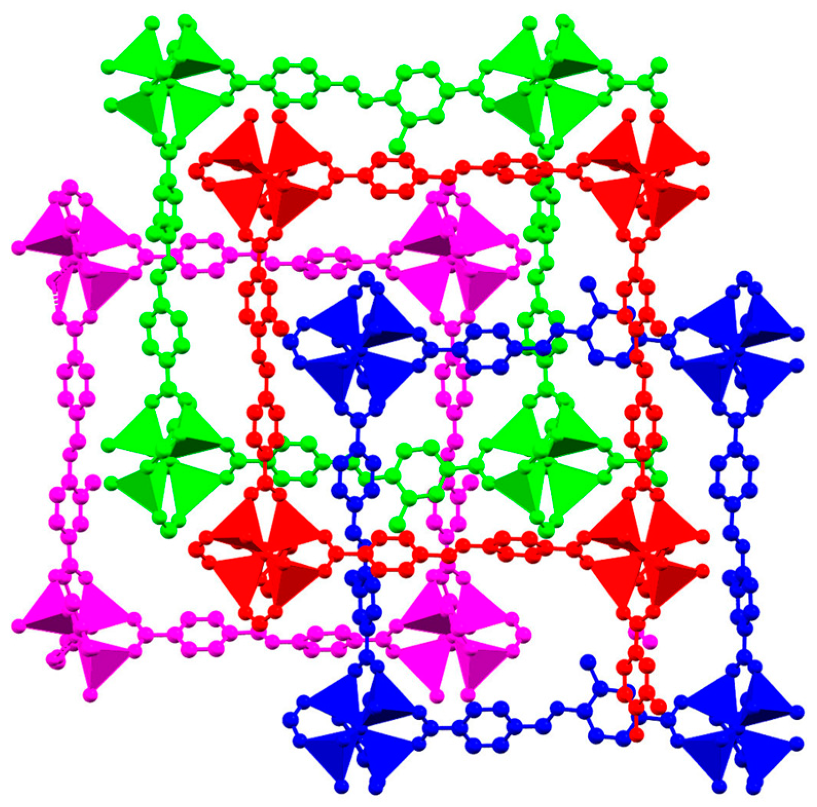
2.2. Description of Structures

The structures of the MV-NUIG4 MOFs were characterised with a variety of techniques, including 1H-NMR, single-crystal X-ray diffraction, powder X-ray diffraction (PXRD), and computational modelling.
The presence of two different types of ligands in the structure of MV-NUIG4 and the exact framework composition were initially investigated through 1H-NMR on activated and digested samples. Upon digestion, the CBAB-R linkers decompose into 4-formylbenzoic acid and 4-amino-3-R-benzoic acid. Linker stoichiometry was determined by integrating characteristic proton signals of 4-amino-3-R-benzoic acid derivatives. The relative integration of signals at 7.59 ppm (4-amino-3-hydroxybenzoic acid, doublet, 2H), 7.38 ppm (4-aminobenzoic acid, doublet 2H), and 2.36 ppm (4-amino-3-methylbenzoic acid, singlet, 3H) provided insights into the linker ratios within the MV-NUIG4 MOFs (Figures S1–S11 in the SI).
Single-crystal X-ray diffraction analysis revealed that the MV-NUIG4 MOFs are isostructural with NUIG4, as the unit cell parameters of NUIG4 (P41, a = b = 19.146, c = 19.117) were closely matched by the functionalised variants MV-NUIG4-H90-OH10 (P41, a = b = 19.15, c = 19.14) and MV-NUIG4-H75-OH25 (P41, a = b = 19.16, c = 19.12). PXRD analysis was employed to further confirm the crystallinity of the MV-NUIG4-Hx-OHy-Mez framework and its structural similarity to NUIG4 (Figure 3). The experimental PXRD patterns of the MV-NUIG4 MOFs were compared with that of NUIG4, confirming structural similarity. Notably, framework crystallinity showed an inverse relationship with increasing values of y and z, which was attributed to enhanced structural disorder resulting from the introduction of OH and Me groups [51,52].
Computational modelling was employed to further understand the crystal structure of MV-NUIG4 MOFs as well as the ligand distribution within the framework. NUIG4-Me (Figure 4) crystallises in the tetragonal space group P41, forming a cubic three-dimensional network constructed from tetrahedral [Zn4O(COO)6] secondary building units (SBUs). Like NUIG4, the structure exhibits four-fold interpenetration with interwoven networks (Figure 5). The solvent-accessible volume of NUIG4-Me, calculated using Mercury software, is 2843.6 Å3, which corresponds to 40.2% of the unit cell volume (7067.6 Å3). This value is lower than that of NUIG4 (48.9%), which is expected due to the presence of additional functional groups in the framework and pores of NUIG4-Me.
Computational modelling provided insight into the distribution of the different functional groups within the framework of MV-NUIG4. In particular, simulations were performed on systems with varying OH content, specifically examining configurations with one and two OH groups out of twelve ligands (CIF files in the SI). The results revealed minimal energy differences among various OH distributions, with the maximum energy difference being 1.12 kcal/mol. This suggests a random distribution of OH groups, supporting the concept of a homogeneous solid solution. This finding aligns with several previous studies on mixed-linker MOF systems, which have demonstrated random distributions of functional groups within their framework [65,66,67]. The solvent-accessible volume of the MV-NUIG4 MOFs bearing OH functional groups ranged from 43.6 to 44.0%, varying with the ratio of OH groups present in the framework. This value is larger than that of NUIG4-Me but smaller than that of NUIG4, as expected given the relative steric factors of the functional groups. The decrease in solvent-accessible volumes across MV-NUIG4 analogues is consistent with increasing functionalisation, with values ranging from 44.0% to 40.2%, compared to 48.9% in NUIG4. This trend reflects steric and electronic effects of the substituents on pore geometry.
The introduction of methyl and hydroxyl substituents was observed to affect the crystallinity of the resulting frameworks, as evidenced by the broadening and intensity reduction of the PXRD peaks. This reduction in crystallinity likely reflects increased framework disorder or a decrease in long-range order, particularly in NUIG4-OH. In parallel, the calculated solvent-accessible volumes decreased with functionalisation. These effects, combined with potential hydrogen bonding or steric hindrance from the substituents, contribute to reduced pore accessibility.

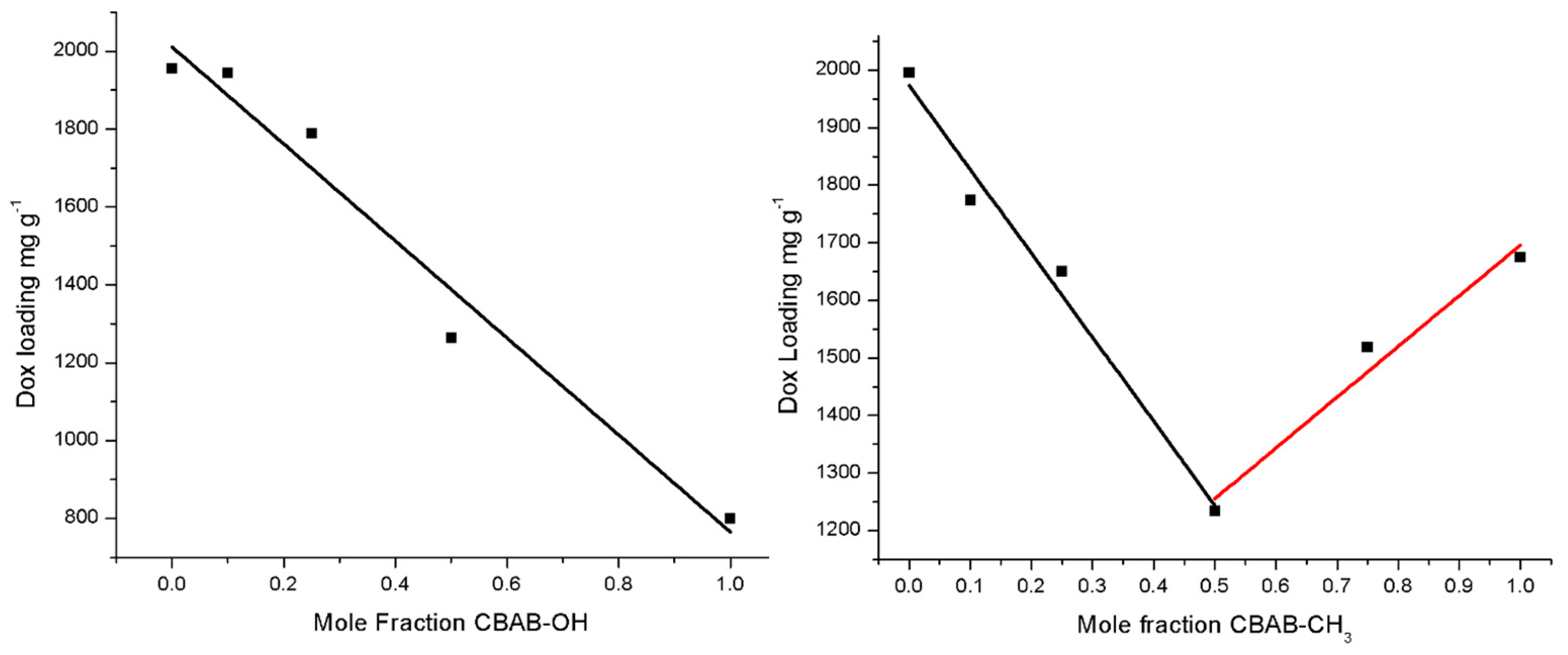
2.3. Dox Absorption and Release Studies

The Dox uptake studies of the MV-NUIG4 MOF family were conducted in MeOH:DMSO, 9:1, and the loadings of Dox were quantified using UV–vis spectroscopy (Figures S12–S19 in the SI). The quantified Dox loadings for MV-NUIG4 match those observed for NUIG4, as shown in Table 1.
The MOFs showed decreased Dox loadings as the mole fraction of CBAB-OH and CBAB-Me increased. For MV-NUIG4-Hx-OHy, there is an almost linear relationship between Dox loading and the CBAB-OH mole fraction (Figure 6, left), with a linear regression factor of 0.9596. The relationship between the CBAB-Me mole fraction and the Dox loading capacity in the MV-NUIG4-Hx-Mez family forms a V-shaped profile (Figure 6, right), with MV-NUIG4-H50-Me50 showing the lowest loading of 1234 mg Dox g−1. Frameworks that have CBAB-Me mole fractions closer to the parent MOFs NUIG4 and NUIG4-Me display greater loading capacities. The linear fitting of the decreasing and increasing loading capacity data yielded regression factors of 0.9748 and 0.9462, respectively. The linear relationship between Dox loading and functionalisation in both MV-NUIG4 families indicates that if the degree of functionalisation is known, it is possible to estimate the loading of functionalised MV-NUIG4 frameworks.
Drug release studies were conducted in both PBS solution (pH 7.4) and acetate buffer solution (pH 5.5), as shown in Figure 7. These frameworks exhibit a release behaviour akin to NUIG4, showcasing a slow release over a span of 10–20 days in water, and an accelerated release under acidic conditions. The quantity of Dox released in the MV-NUIG4-Hx-OHy family decreases as the CBAB-OH mole fraction increases. This is related to the increased water stability of the MOFs due to hydroxylation ortho to aromatic imines. Indeed, hydroxylation ortho to aromatic imines has been utilized for enhancing water stability in both MOFs and COFs [68,69,70]. 1,2-Hydroxyl imines can undergo oxidative cyclisation, forming a benzoxazole group, which remains stable in water and across a wide pH spectrum. It was possible to modulate the release from 49% to 32% after 40 days by increasing the CBAB-OH mole fraction.
The MV-NUIG4-Hx-Mez family exhibits a release profile similar to that of NUIG4, but with slower kinetics. As the CBAB-Me mole fraction increases, the initial release rate decreases. In particular, after 10 days, NUIG4 releases 17% of the drug load, while NUIG4-Me releases 12%. This can be attributed to the reduced pore accessibility, as well as increased hydrophobicity due to the growing presence of methyl groups, which may reduce pore accessibility for H2O molecules. Despite the decreased initial release rate, the MV-NUIG4-Hx-Mez frameworks eventually release the same quantity of Dox as NUIG4 after 55 days. Therefore, the MV-NUIG4-Hx-Mez MOF family offers a method to control the initial rate of Dox release by adjusting the proportion of Me groups in the framework.
To gain deeper insight into the release mechanism, the experimental data were analysed using the Higuchi and Korsmeyer–Peppas kinetic models (Figure S28) [71,72,73]. Both NUIG4-Me and MV-NUIG4-H50-Me50 exhibited a strong linear correlation in the Higuchi model (R2 = 0.99 and 0.95, respectively), indicating that diffusion is a dominant factor governing the drug release process. However, fitting the data to the Korsmeyer–Peppas model revealed release exponents (n) of 0.79 and 0.74 for NUIG4-Me and MV-NUIG4-H50-Me50, respectively, which fall within the range of anomalous (non-Fickian) transport. This suggests that, in addition to diffusion, other processes such as framework relaxation or partial structural breakdown may contribute to the observed drug release. Such behaviour is common in porous coordination materials and highlights the intricate interplay between framework composition and drug release kinetics in MOFs.

3. Materials and Methods

3.1. Materials, and Physical and Spectroscopic Measurements

All chemicals used were commercially available and used without further purification. All procedures were conducted under aerobic conditions.
Elemental analyses (C, H, N) were performed by the in-house facilities of University of Galway. FTIR spectra (4000–400 cm−1) were recorded using a Perkin Elmer 16PC FT-IR spectrometer, with samples prepared as KBr pellets. PXRD data were collected using an Inex Equinox 6000 diffractometer at room temperature and pressure.

3.2. Compound Synthesis

3.2.1. Synthesis of NUIG4-Me

4-Formylbenzoic acid (225 mg, 1.5 mmol) was dissolved in DMF (5 mL) within a glass scintillation vial. 4-Amino-3-methylbenzoic acid (226 mg, 1.5 mmol) was then added, resulting in a pale-yellow solution. Zn(CH3CO2)2·2H2O (330 mg, 1.5 mmol) was added, and the solution was sonicated for 2 min. The sealed vial was heated in an oven at 80 °C for 24 h, forming a white crystalline powder. The solvent was then decanted, and the powder was rinsed with DMF (2 × 5 mL). The yield was 41%, and the anal. calcd. (found) for NUIG4-Me was the following: C, 51.42 (51.87%); H, 2.97 (3.02%); N, 3.75 (3.28%).

3.2.2. Synthesis of MV-NUIG4 Analogues

MV-NUIG4 MOFs were synthesised similarly to NUIG4 [1], but with varying mole fractions of 4-aminobenzoic acids. The general synthesis process described below was followed: In a 20 mL glass scintillation vial, 4-formylbenzoic acid (225 mg, 1.5 mmol) was dissolved in DMF (5 mL). To this, 4-aminobenzoic acid was added, followed by either 4-amino-3-hydroxybenzoic acid or 4-amino-3-methylbenzoic acid, with exact reagent quantities detailed in Table S1 of the SI. The solution was sonicated for 2 min. Zn(CH3CO2)2·2H2O (330 mg, 1.5 mmol) was added, and the solution was sonicated for 2 further minutes. The vial was sealed and placed in the oven at 80 °C for 24 h, yielding crystalline powders or small cubic crystals in the case of MV-NUIG4-H90-OH10, MV-NUIG4-H75-OH25, MV-NUIG4-H90-Me10, and MV-NUIG4-H75-Me25. The solvent was decanted, and the powders were washed with DMF (3 × 10 mL), while the crystals were kept in the mother liquor for single-crystal X-ray diffraction studies.
MV-NUIG4-H90-OH10: Yield: 27%. Anal. Calcd. (found): C, 49.86 (50.13%); H, 2.51 (2.60%); N, 3.88 (4.01%).
MV-NUIG4-H75-OH25: Yield: 20%. Anal. Calcd. (found): C, 49.53 (49.99%); H, 2.49 (2.52%); N, 3.85 (3.75%).
MV-NUIG4-H50-OH50: Yield: 15%. Anal. Calcd. (found): C, 48.99 (49.23%); H, 2.47 (2.58%); N, 3.81 (3.72%).
MV-NUIG4-H90-Me10: Yield: 32%. Anal. Calcd. (found): C, 50.22 (50.61%); H, 2.57 (2.68%); N, 3.88 (3.91%).
MV-NUIG4-H75-Me25: Yield: 35%. Anal. Calcd. (found): C, 50.42 (50.77%); H, 2.64 (2.79%); N, 3.86 (3.74%).
MV-NUIG4-H50-Me50: Yield: 30%. Anal. Calcd. (found): C, 50.76 (51.00%); H, 2.75 (2.93%); N, 3.82 (3.83%).
MV-NUIG4-H25-Me75: Yield: 35%. Anal. Calcd. (found): C, 51.09 (51.28%); H, 2.86 (2.94%); N, 3.78 (3.62%).

3.3. X-Ray Crystallography

Crystallographic data for MV-NUIG4-H90-OH10, MV-NUIG4-H75-OH25, MV-NUIG4-H90-Me10, and MV-NUIG4-H75-Me25 were collected on an Oxford Diffraction Xcalibur CCD diffractometer using graphite-monochromatic Mo Kα radiation (λ = 0.71073 Å) at room temperature. Although the crystals diffracted poorly, reliable unit cell parameters were obtained for MV-NUIG4-H90-OH10 (a = b = 19.15 Å, c = 19.14 Å, P41) and MV-NUIG4-H75-OH25 (a = b = 19.16 Å, c = 19.12 Å, P41), confirming their structural similarity with NUIG4 (a = b = 19.146 Å, c = 19.117 Å, P41).

3.4. Crystal Structure Model Construction

Unit cell parameters of the frameworks were confirmed from powder X-ray diffraction pattern. Initial unit cell parameters for the frameworks were determined from positions of 10 observed peaks using indexing via DICVOL [74], which was implemented in DASH [75]. The unit cell parameters were refined for all frameworks via Pawley profile fit of the powder X-ray diffraction pattern using GSAS-II [76]. All calculations were performed using the open-source software Quantum Espresso v7.1 at the PBEsol level of theory [77]. Norm-conserving pseudopotentials were used, with an energy cutoff of 110 Ry. The unit cell parameters for the MV-NUIG4 MOFs display significant similarities to NUIG4. A full geometry optimization was then performed on the primitive cell of the MOF, relaxing both the atomic positions and the lattice parameters. The standard convergence parameters were adopted.

3.5. NMR Studies

A total of 15 mg of activated MOF material was suspended in DMSO-d6 (500 μL). Then, 25 μL DCl (35 wt% in D2O) was added to the suspension. The suspension was sonicated for 5 min, yielding a clear solution. 1H-NMR spectra were recorded at 500 MHz using a Bruker spectrometer and were processed using Bruker Topspin software, with calibration against solvent signals in the literature.

3.6. MOF Activation and Drug Loading

MV-NUIG4 MOFs were activated prior to drug uptake using the same procedure as for NUIG4 [1]. A sample of MOF (20 mg) was stirred in DMF (15 mL) for 24 h, with DMF replacement after 4 and 8 h. The solid material was then collected by centrifugation at 4000 rpm for 5 min and stirred in MeOH (15 mL) for another 24 h, again replacing the MeOH after 4 and 8 h. Finally, the solid was collected by centrifugation and heated in an oven at 80 °C overnight.
Dox (0.04 g) was dissolved in MeOH:DMSO (9:1, 10 mL) and added to a centrifuge vial that contained MV-NUIG4 (0.01 g). At specified time intervals, the solution was centrifuged, and 25 μL aliquots of the supernatant were removed and dissolved in MeOH (5 mL). The uptake of the drug was then monitored using UV–vis spectroscopy.
For the release studies, loaded MV-NUIG4 (5 mg) was suspended in distilled H2O or PBS/5.5 pH sodium acetate buffer solution (10 mL) and stirred at 37 °C. At specified time intervals, the solution was centrifuged, and 100 mL aliquots of the solution were removed and diluted in distilled H2O or PBS solution (5 mL), and 100 mL of fresh solution was added to the vial. The release of Dox was then monitored using UV–vis spectroscopy.

4. Conclusions

The introduction of different functional groups to the NUIG4 framework led to the synthesis and characterisation of eight new multivariate metal–organic frameworks (MV-NUIG4). The MOFs maintain the fundamental pcu topology of the parent NUIG4 framework while incorporating different ratios of methyl and hydroxyl functional groups. MV-NUIG4 MOFs were synthesised using an in situ approach, with reagent ratios accurately reflected in the final products. This methodology is particularly important because it enables the reliable production of frameworks with predictable structures and tunable functionalities.
The structural features of the MV-NUIG4 MOFs were investigated using a variety of techniques, including 1H-NMR, single-crystal X-ray diffraction, powder X-ray diffraction, and computational modelling, which confirmed structural similarity to the parent MOF as well as a random distribution of the functional groups within the framework.
The MV-NUIG4 analogues exhibited exceptional drug-loading capabilities, with doxorubicin absorption capacities ranging from 1234 to 1995 mg/g MOF, depending on the type and percentage of functional group present in the structure. This ability to modulate drug release rates through functional group ratios represents a particularly promising feature for pharmaceutical applications. Future studies will focus on investigating other functional groups and optimising their ratios to achieve targeted drug release profiles for Dox and other therapeutics.

Supplementary Materials

The following supporting information can be downloaded at: https://www.mdpi.com/article/10.3390/molecules30091968/s1, Figure S1: 1H-NMR of digested MV-NUIG4-H75-OH25; Figure S2: 1H-NMR of digested MV-NUIG4-H50-OH50; Figure S3: 1H-NMR of digested MV-NUIG4-H90-OH10; Figure S4: 1H-NMR of digested MV-NUIG4-H50-Me50; Figure S5: 1H-NMR of digested MV-NUIG4-H90-Me10; Figure S6: 1H-NMR of digested MV-NUIG4-H75-Me25; Figure S7: 1H-NMR of digested MV-NUIG4-H25-Me75; Figure S8: 1H-NMR of 4-aminobenzoic acid under digestion conditions; Figure S9: 1H-NMR of 4-amino-3-methylbenzoic acid under digestion conditions; Figure S10: 1H-NMR of 4-amino-3-hydroxybenzoic acid under digestion conditions; Figure S11: 1H-NMR of 4-formylbenzoic acid under digestion conditions; Figure S12: UV-Vis data for Dox uptake by MV-NUIG4-H50-OH50; Figure S13: UV-Vis data for Dox uptake by MV-NUIG4-H90-OH10; Figure S14: UV-Vis data for Dox uptake by MV-NUIG4-H75-OH25; Figure S15: UV-Vis data for Dox uptake by NUIG4-Me; Figure S16: UV-Vis data for Dox uptake by MV-NUIG4-H50-Me50; Figure S17: UV-Vis data for Dox uptake by MV-NUIG4-H90-Me10; Figure S18: UV-Vis data for Dox uptake by MV-NUIG4-H75-Me25; Figure S19: UV-Vis data for Dox uptake by MV-NUIG4-H75-Me25; Figure S20: FTIR spectrum of MV-NUIG4-H50-OH50; Figure S21: FTIR spectrum of MV-NUIG4-H90-OH10; Figure S22: FTIR spectrum of MV-NUIG4-H90-OH10; Figure S23: FTIR spectrum of MV-NUIG4-H25-OH75; Figure S24: FTIR spectrum of MV-NUIG4-H50-Me50; Figure S25: FTIR spectrum of MV-NUIG4-H75-Me25; Figure S26: FTIR spectrum of MV-NUIG4-H90-Me10; Figure S27: FTIR spectrum of NUIG4-Me; Figure S28: Higuchi (left) and Korsmeyer–Peppas model fitting (right) of Doxorubicin release from NUIG4-Me and MV-NUIG4-H50-Me50 (pH 5.5); Table S1: Quantities (mg) of 4-aminobenzoic acid and derivatives used in the synthesis of MV-NUIG4 MOFs.

Author Contributions

A.A. performed the synthesis, crystallisation, and preliminary characterisation of all compounds. C.O. conducted the single-crystal X-ray diffraction studies, while A.B., D.S. and M.J.Z. carried out the powder X-ray diffraction analyses. D.T. performed the computational modelling on the representative compounds. C.P. coordinated the research and wrote the manuscript based on input from all collaborators. All authors have read and agreed to the published version of the manuscript.

Funding

This work was funded by Research Ireland (Grant Number: 12/RC/2275_P2). We also acknowledge the support and computational facilities provided by the Irish Centre for High-End Computing (ICHEC).

Institutional Review Board Statement

Not applicable.

Informed Consent Statement

Not applicable.

Data Availability Statement

The data presented in this study are available on request from the corresponding author.

Conflicts of Interest

The authors declare no conflicts of interest.

References

  1. Ahmed, A.; Efthymiou, C.G.; Sanii, R.; Patyk-Kazmierczak, E.; Alsharabasy, A.M.; Winterlich, M.; Kumar, N.; Sensharma, D.; Tong, W.; Guerin, S.; et al. NUIG4: A biocompatible pcu metal–organic framework with an exceptional doxorubicin encapsulation capacity. J. Mater. Chem. B 2022, 10, 1378–1389. [Google Scholar] [CrossRef] [PubMed]
  2. Hoskins, B.F.; Robson, R. Infinite polymeric frameworks consisting of three dimensionally linked rod-like segments. J. Am. Chem. Soc. 1989, 111, 5962–5964. [Google Scholar] [CrossRef]
  3. Furukawa, H.; Cordova, K.E.; O’Keeffe, M.; Yaghi, O.M. The chemistry and applications of metal-organic frameworks. Science 2013, 341, 1230444. [Google Scholar] [CrossRef]
  4. Schneemann, A.; Bon, V.; Schwedler, I.; Senkovska, I.; Kaskel, S.; Fischer, R.A. Flexible metal–organic frameworks. Chem. Soc. Rev. 2014, 43, 6062–6096. [Google Scholar] [CrossRef] [PubMed]
  5. Zhou, H.-C.J.; Kitagawa, S. Metal–organic frameworks (MOFs). Chem. Soc. Rev. 2014, 43, 5415–5418. [Google Scholar] [CrossRef]
  6. Yusuf, V.F.; Malek, N.I.; Kumar, S.K. Recent Developments of Metal–Organic Framework-Based Materials for Drug Delivery Applications. ACS Omega 2022, 49, 44507–44519. [Google Scholar] [CrossRef]
  7. Yuan, S.; Feng, L.; Wang, K.; Pang, J.; Bosch, M.; Lollar, C.; Sun, Y.; Qin, J.; Yang, X.; Zhang, P.; et al. Stable Metal–Organic Frameworks: Design, Synthesis, and Applications. Adv. Mater. 2018, 37, 1704303. [Google Scholar] [CrossRef]
  8. Eddaoudi, M.; Moler, D.B.; Li, H.; Chen, B.; Reineke, T.M.; O’Keeffe, M.; Yaghi, O. Modular Chemistry: Secondary Building Units as the Basis of Design for Highly Porous and Robust Metal–Organic Carboxylate Frameworks. Acc. Chem. Res. 2001, 34, 319–330. [Google Scholar] [CrossRef]
  9. Kurmoo, M. Magnetic metal–organic frameworks. Chem. Soc. Rev. 2009, 38, 1353–1379. [Google Scholar] [CrossRef]
  10. Lazaro, I.A.; Chen, X.; Ding, M.; Eskandari, A.; Fairen-Jimenez, D.; Gimenez-Marques, M.; Gref, R.; Lin, W.; Luo, T.; Forgan, R.S. Metal–organic frameworks for biological applications. Nat. Rev. Methods Primers 2024, 4, 42. [Google Scholar] [CrossRef]
  11. Ahmed, A.; McHugh, D.; Papatriantafyllopoulou, C. Synthesis and Biomedical Applications of Highly Porous Metal–Organic Frameworks. Molecules 2022, 27, 6585. [Google Scholar] [CrossRef]
  12. Hefayathullah, M.; Singh, S.; Ganesan, V.; Maduraiveeran, G. Metal–organic frameworks as emerging smart nanoplatforms for electrochemical biosensors applications. Adv. Colloid Interface Sci. 2024, 331, 102321. [Google Scholar]
  13. Chao, Y.; Deng, N.; Zhou, Z. A review of recent advances in metal–organic frameworks materials for zero-energy passive adsorption of chemical pollutants in indoor environments. Sci. Total Environ. 2024, 953, 175926. [Google Scholar] [CrossRef] [PubMed]
  14. Wang, W.; Ibarlucea, B.; Huang, C.; Dong, R.; Al Aiti, M.; Huang, S.; Cuniberti, G. Multi-metallic MOF based composites for environmental applications: Synergizing metal centers and interactions. Nanoscale Horiz. 2024, 9, 1432–1474. [Google Scholar] [CrossRef]
  15. Zhang, Q.; Yang, H.; Zhou, T.; Chen, X.; Li, W.; Puang, H. Metal–Organic Frameworks and Their Composites for Environmental Applications. Adv. Sci. 2022, 9, e2204141. [Google Scholar] [CrossRef] [PubMed]
  16. Czaja, A.U.; Trukhan, N.; Muller, U. Industrial applications of metal–organic frameworks. Chem. Soc. Rev. 2009, 38, 1284–1293. [Google Scholar] [CrossRef]
  17. Mueller, U.; Schubert, M.; Teich, F.; Puetter, H.; Schierle-Arndt, K.; Pastre, J. Metal–organic frameworks—Prospective industrial applications. J. Mater. Chem. 2006, 16, 626–636. [Google Scholar] [CrossRef]
  18. Letwaba, J.; Uyor, U.O.; Mavhungu, M.L.; Achuka, N.O.; Popoola, P.A. A review on MOFs synthesis and effect of their structural characteristics for hydrogen adsorption. RSC Adv. 2024, 14, 14233–14253. [Google Scholar] [CrossRef]
  19. Wang, J.; Liu, J.; Wang, H.; Zhou, M.; Ke, G.; Zhang, L.; Wu, J.; Gao, Z.; Lu, D. A comprehensive transformer-based approach for high-accuracy gas adsorption predictions in metal–organic frameworks. Nat. Commun. 2024, 15, 1904. [Google Scholar] [CrossRef]
  20. Karth, N.; Wright, K.R.; Ahmed, A.; Siegel, D.J.; Matzger, A.J. High-throughput discovery of metal–organic frameworks for hydrogen storage. J. Am. Chem. Soc. 2024, 146, 10517–10526. [Google Scholar]
  21. Jiang, C.; Wang, X.; Ouyang, Y.; Lu, K.; Jiang, W.; Ku, H.; Wei, X.; Wang, Z.; Dai, F.; Sun, D. Recent advances in metal–organic frameworks for gas adsorption/separation. Nanoscale Adv. 2022, 4, 2077–2095. [Google Scholar] [CrossRef]
  22. Qian, Q.; Asinger, P.A.; Lee, M.J.; Han, G.; Rodriguez, K.M.; Lin, S.; Benedetti, F.M.; Wu, A.X.; Chi, W.S.; Smith, Z.P. MOF-based membranes for gas separations. Chem. Rev. 2020, 120, 8161–8266. [Google Scholar] [CrossRef]
  23. Hiraide, S.; Sakanaka, Y.; Kajiro, H.; Kawaguchi, S.; Miyahara, M.T.; Tanaka, H. High-throughput gas separation by flexible metal–organic frameworks with fast gating and thermal management capabilities. Nat. Commun. 2020, 11, 3867. [Google Scholar] [CrossRef] [PubMed]
  24. Winterlich, M.; Efthymiou, C.G.; Papawassiliou, W.; Carvalho, J.P.; Pell, A.J.; Mayans, J.; Escuer, A.; Carty, M.P.; McArdle, P.; Tylianakis, E.; et al. A biocompatible ZnNa₂-based metal–organic framework with high ibuprofen, nitric oxide and metal uptake capacity. Mater. Adv. 2020, 1, 2248–2260. [Google Scholar] [CrossRef]
  25. Ahmed, A.; Kelly, A.; Leonard, D.; Saleem, W.; Bezrukov, A.; Efthymiou, C.G.; Zaworotko, M.J.; Tiana, D.; Boyd, A.; Papatriantafyllopoulou, C. Synthesis and characterisation of antimicrobial metal–organic frameworks as multi-drug carriers. Dalton Trans. 2024, 53, 11867–11875. [Google Scholar] [CrossRef]
  26. Vikal, A.; Maurya, R.; Patel, P.; Paliwal, S.R.; Narang, R.K.; Das Gupta, G.; Das Kurmi, B. Exploring metal–organic frameworks (MOFs) in drug delivery: A concise overview of synthesis approaches, versatile applications, and current challenges. Appl. Mater. Today 2024, 41, 102443. [Google Scholar] [CrossRef]
  27. Saboorizadeh, B.; Zareh-Dorabei, R.; Safavi, M.; Safarifard, V. Applications of metal–organic frameworks (MOFs) in drug delivery, biosensing, and therapy: A comprehensive review. Langmuir 2024, 40, 22477–22503. [Google Scholar] [CrossRef] [PubMed]
  28. Khafaga, D.S.R.; El-Morsy, M.T.; Faried, H.; Diab, A.H.; Shehab, S.; Saleh, A.M.; Ali, G.A.M. Metal–organic frameworks in drug delivery: Engineering versatile platforms for therapeutic applications. RSC Adv. 2024, 14, 30201–30229. [Google Scholar] [CrossRef]
  29. Bunzen, H.; Jirak, D. Nanoscale Metal–Organic Frameworks as Contrast Agents for Magnetic Resonance Imaging. ACS Appl. Mater. Interfaces 2022, 14, 50445–50460. [Google Scholar] [CrossRef]
  30. Duman, F.D.; Forgan, R.S. Applications of Nanoscale Metal–Organic Frameworks as Imaging Agents in Biology and Medicine. J. Mater. Chem. B 2021, 9, 3423–3449. [Google Scholar] [CrossRef]
  31. Khan, M.S.; Li, Y.; Li, D.-S.; Qiu, J.; Xu, X.; Yang, H.Y. A review of metal–organic framework (MOF) materials as an effective photocatalyst for degradation of organic pollutants. Nanoscale Adv. 2023, 5, 6318–6348. [Google Scholar] [CrossRef]
  32. Sen, R.; Hazra, D.K.; Mukherjee, M.; Koner, S. Gd26 Cluster Consisting of Distorted Cubane Cores: Synthesis, Structure and Heterogeneous Catalytic Epoxidation of Olefins. Eur. J. Inorg. Chem. 2011, 18, 2826–2831. [Google Scholar] [CrossRef]
  33. Chen, H.; Liu, S.; Qin, Q.-P.; Zhang, X. A novel Zn(II)-based metal–organic framework as a luminescent sensor for the detection of nitroaromatic explosives. ACS Appl. Mater. Interfaces 2022, 14, 18589–18597. [Google Scholar] [CrossRef]
  34. Wang, Z.-F.; Li, C.; Qin, Q.-P.; Zhang, S.; Zhang, X. A new Zn(II)-based metal–organic framework for efficient sensing of Fe3+ ions in aqueous solution. ACS Appl. Nano Mater. 2023, 6, 23430–23438. [Google Scholar] [CrossRef]
  35. Zheng, Y.-T.; Li, S.; Huang, N.-Y.; Li, X.; Xu, Q. Recent advances in metal–organic framework-derived materials for electrocatalytic and photocatalytic CO₂ reduction. Coord. Chem. Rev. 2024, 510, 215858. [Google Scholar] [CrossRef]
  36. Sohrabi, H.; Ghasemzadeh, S.; Ghoreishi, Z.; Majidi, M.R.; Yoon, Y.; Dizge, N.; Khataee, A. Metal–organic frameworks (MOF)-based sensors for detection of toxic gases: A review of current status and future prospects. Mater. Chem. Phys. 2023, 299, 127512. [Google Scholar] [CrossRef]
  37. Wu, T.; Gao, X.; Ge, F.; Zheng, H. Metal–organic frameworks (MOFs) as fluorescence sensors: Principles, development and prospects. CrystEngComm 2022, 24, 7881–7901. [Google Scholar] [CrossRef]
  38. Shen, X.; Tissot, A.; Serre, C. Recent progress on MOF-based optical sensors for VOC sensing. Chem. Sci. 2022, 13, 13978–14007. [Google Scholar] [CrossRef] [PubMed]
  39. He, Y.; Li, D.; Wu, L.; Yin, X.; Zhang, X.; Patterson, L.H.; Zhang, J. Metal–Organic Frameworks for Gene Therapy and Detection. Adv. Funct. Mater. 2023, 12, 2212277. [Google Scholar] [CrossRef]
  40. Poddar, A.; Pyreddy, S.; Carraro, F.; Dhakal, S.; Russell, A.; Field, M.R.; Reddy, T.S.; Falcaro, P.; Doherty, C.M.; Shukla, R. ZIF-C for targeted RNA interference and CRISPR/Cas9 based gene editing in prostate cancer. Chem. Commun. 2020, 56, 15406–15409. [Google Scholar] [CrossRef]
  41. Ringaci, A.; Yaremenko, A.V.; Shevchenko, K.G.; Zvereva, S.D.; Nikitin, M.P. Metal–Organic Frameworks for Simultaneous Gene and Small Molecule Delivery In Vitro and In Vivo. Chem. Eng. J. 2021, 418, 129386. [Google Scholar] [CrossRef]
  42. Wang, X.; Lan, P.C.; Ma, S. Metal–Organic Frameworks for Enzyme Immobilization: Beyond Host Matrix Materials. ACS Cent. Sci. 2020, 9, 1497–1506. [Google Scholar] [CrossRef]
  43. Xia, H.; Li, N.; Zhong, X.; Jiang, Y. Metal–Organic Frameworks: A Potential Platform for Enzyme Immobilization and Related Applications. Front. Bioeng. Biotechnol. 2020, 8, 695. [Google Scholar] [CrossRef] [PubMed]
  44. Liu, X.; Qi, W.; Wang, Y.; Su, R.; He, Z. A Facile Strategy for Enzyme Immobilization with Highly Stable Hierarchically Porous Metal–Organic Frameworks. Nanoscale 2017, 9, 17561–17570. [Google Scholar] [CrossRef] [PubMed]
  45. Silva, A.R.M.; Alexandre, J.Y.N.H.; Souza, J.E.S.; Neto, J.G.L.; De Souza Junior, P.G.; Rocha, M.V.P.; Dos Santos, J.C.S. The Chemistry and Applications of Metal–Organic Frameworks (MOFs) as Industrial Enzyme Immobilization Systems. Molecules 2022, 27, 4529. [Google Scholar] [CrossRef]
  46. Zhang, X.; Xu, Z.; Zhou, J.; Xing, X.; Li, L. Enhancement of Protein Crystallization Using Nano-Sized Metal–Organic Framework. Crystals 2022, 12, 578. [Google Scholar] [CrossRef]
  47. Zhou, X.; Zhong, Z.; Xu, N.; Zhong, S. Nanoscale MOF–Protein Composites for Theranostics. Crystals 2023, 13, 1229. [Google Scholar] [CrossRef]
  48. Leite, J.P.; Rodrigues, D.; Ferreira, S.; Figueira, F.; Almeida Paz, F.A.; Gales, L. Mesoporous Metal–Organic Frameworks as Effective Nucleating Agents in Protein Crystallography. Cryst. Growth Des. 2019, 19, 1610–1615. [Google Scholar] [CrossRef]
  49. Andre, V.; Da Siva, A.R.F.; Fernandes, A.; Frade, R.; Garcia, C.; Rijo, P.; Antunes, A.M.M.; Rocha, J.; Duarte, M.T. Mg- and Mn-MOFs Boost the Antibiotic Activity of Nalidixic Acid. ACS Appl. Bio Mater. 2019, 2, 2347–2354. [Google Scholar] [CrossRef]
  50. Ahmed, S.A.; Hasan, M.N.; Bagchi, D.; Altass, H.M.; Morad, M.; Althagafi, I.I.; Hameed, A.M.; Sayqal, A.; Khder, A.R.S.; Asghar, B.H.; et al. Nano-metal–organic frameworks as targeted drug delivery agents to combat antibiotic-resistant bacterial infections. R. Soc. Open Sci. 2020, 7, 200959. [Google Scholar] [CrossRef]
  51. Helal, A.; Yamani, Z.H.; Cordova, K.E.; Yaghi, O.M. Multivariate metal-organic frameworks. Nat. Sci. Rev. 2017, 4, 296–298. [Google Scholar] [CrossRef]
  52. Deng, H.; Doonan, C.J.; Funukawa, H.; Ferreira, R.B.; Towne, J.; Knobler, C.B.; Wang, B.; Yaghi, O.M. Multiple functional groups of varying ratios in metal-organic frameworks. Science 2010, 327, 846–850. [Google Scholar] [CrossRef] [PubMed]
  53. Lee, S.J.; Telfer, S.G. Multicomponent Metal–Organic Frameworks. Angew. Chem. Int. Ed. 2023, 62, e202306341. [Google Scholar] [CrossRef]
  54. Dutta, A.; Pan, Y.; Liu, J.-Q.; Kumar, A. Multicomponent Isoreticular Metal–Organic Frameworks: Principles, Current Status and Challenges. Coord. Chem. Rev. 2021, 445, 214074. [Google Scholar] [CrossRef]
  55. Smith, K.T.; Stylianou, K.C. Multivariate metal–organic frameworks generated through post-synthetic modification: Impact and future directions. Dalton Trans. 2023, 52, 16578–16585. [Google Scholar] [CrossRef]
  56. Wang, Y.; Lv, H.; Grape, E.S.; Gaggioli, C.A.; Tayal, A.; Dharanipragada, A.; Willhammar, T.; Ken Inge, A.; Zou, X.; Liu, B.; et al. A Tunable Multivariate Metal–Organic Framework as a Platform for Designing Photocatalysts. J. Am. Chem. Soc. 2021, 143, 6333–6338. [Google Scholar] [CrossRef] [PubMed]
  57. Feng, L.; Yuan, S.; Li, J.-L.; Wang, K.-Y.; Day, G.S.; Zhang, P.; Wang, Y.; Zhou, H.-C. Uncovering two principles of multivariate hierarchical metal–organic framework synthesis via retrosynthetic design. ACS Cent. Sci. 2018, 4, 1719–1726. [Google Scholar] [CrossRef]
  58. Fan, W.; Yuan, S.; Wang, W.; Feng, L.; Liu, X.; Zhang, X.; Wang, X.; Kang, Z.; Dai, F.; Yuan, D.; et al. Optimizing Multivariate Metal–Organic Frameworks for Efficient C₂H₂/CO₂ Separation. J. Am. Chem. Soc. 2020, 142, 8728–8737. [Google Scholar] [CrossRef] [PubMed]
  59. Shang, S.; Yang, C.; Tian, Y.; Tao, Z.; Smith, M.; Zhang, H.; Zhang, L.; Li, L.; Gu, Q.; Zhou, H.-C.; et al. Designing multivariate porphyrin-based metal-organic frameworks with Ni/Co dual-metal atom sites for cooperative NO2 capture and NO retention. Sep. Purif. Technol. 2023, 320, 124080. [Google Scholar] [CrossRef]
  60. Lustig, W.P.; Shen, Z.; Teat, S.J.; Javed, N.; Velasco, E.; O’Carroll, D.M.; Li, J. Rational design of a high-efficiency, multivariate metal–organic framework phosphor for white LED bulbs. Chem. Sci. 2020, 11, 1814–1824. [Google Scholar] [CrossRef]
  61. Li, Y.-M.; Yuan, J.; Ren, H.; Ji, C.-Y.; Tao, Y.; Wu, Y.; Chou, L.-Y.; Zhang, Y.-B.; Cheng, L. Fine-Tuning the Micro-Environment to Optimize the Catalytic Activity of Enzymes Immobilized in Multivariate Metal–Organic Frameworks. J. Am. Chem. Soc. 2021, 143, 15378–15390. [Google Scholar] [CrossRef]
  62. Schrimpf, W.; Jiang, J.; Ji, Z.; Hirschle, P.; Lamb, D.C.; Yaghi, O.M.; Wuttke, S. Chemical diversity in a metal–organic framework revealed by fluorescence lifetime imaging. Nat. Commun. 2018, 9, 1647. [Google Scholar] [CrossRef] [PubMed]
  63. Abanades Lazaro, I.; Wells, C.J.R.; Forgan, R.S. Multivariate Modulation of the Zr MOF UiO-66 for Defect-Controlled Combination Anticancer Drug Delivery. Angew. Chem. 2020, 132, 5249–5255. [Google Scholar] [CrossRef]
  64. Dong, Z.; Sun, Y.; Chu, J.; Zhang, X.; Deng, H. Multivariate Metal–Organic Frameworks for Dialing-in the Binding and Programming the Release of Bioactive Molecules. J. Am. Chem. Soc. 2017, 139, 14209–14216. [Google Scholar] [CrossRef]
  65. Taddei, M.; Tiana, D.; Casati, N.; Van Bokhoven, J.A.; Smit, B.; Ranocchiari, M. Mixed-linker UiO-66: Structure–property relationships revealed by a combination of high-resolution powder X-ray diffraction and density functional theory calculations. Phys. Chem. Chem. Phys. 2017, 19, 1551–1559. [Google Scholar] [CrossRef]
  66. Karzenmeyer, A.M.; Canivet, J.; Holland, G.; Farrusseng, D. Assessing Chemical Heterogeneity at the Nanoscale in Mixed-Ligand Metal–Organic Frameworks with the PTIR Technique. Angew. Chem. Int. Ed. 2014, 53, 2852–2856. [Google Scholar] [CrossRef]
  67. Lescouet, T.; Kockrick, E.; Pera-Titus, M.; Aguado, S.; Farrusseng, D. Homogeneity of flexible metal–organic frameworks containing mixed linkers. J. Mater. Chem. 2012, 22, 10287–10293. [Google Scholar] [CrossRef]
  68. Rabbani, M.G.; Islamoglu, T.; El-Kaderi, H.M. Benzothiazole- and benzoxazole-linked porous polymers for carbon dioxide storage and separation. J. Mater. Chem. A 2017, 5, 258–265. [Google Scholar] [CrossRef]
  69. Wei, P.-F.; Qi, M.-Z.; Wang, Z.-P.; Ding, S.-Y.; Yu, W.; Liu, Q.; Wang, L.-K.; Wang, H.-Z.; An, W.-K.; Wang, W. Benzoxazole-Linked Ultrastable Covalent Organic Frameworks for Photocatalysis. J. Am. Chem. Soc. 2018, 140, 4623–4631. [Google Scholar] [CrossRef] [PubMed]
  70. Cusin, L.; Peng, H.; Ciesielski, A.; Samori, S. Chemical Conversion and Locking of the Imine Linkage: Enhancing the Functionality of Covalent Organic Frameworks. Angew. Chem. 2021, 133, 14356–14370. [Google Scholar] [CrossRef]
  71. Higuchi, T. Mechanism of Sustained-Action Medication. Theoretical Analysis of Rate of Release of Solid Drugs Dispersed in Solid Matrices. Pharm. Sci. 1963, 52, 1145–1149. [Google Scholar] [CrossRef]
  72. Korsmeyer, R.W.; Gurny, R.; Doelker, E.; Buri, P.; Peppas, N.A. Mechanisms of solute release from porous hydrophilic polymers. Int. J. Pharm. 1983, 15, 25–35. [Google Scholar] [CrossRef]
  73. Siepmann, J.; Peppas, N.A. Modeling of drug release from delivery systems based on hydroxypropyl methylcellulose (HPMC). Adv. Drug Deliv. Rev. 2001, 48, 139–157. [Google Scholar] [CrossRef]
  74. Boultif, A.; Loueer, D. Indexing of powder diffraction patterns for low-symmetry lattices by the successive dichotomy method. J. Appl. Crystallogr. 1991, 24 Pt 6, 987–993. [Google Scholar] [CrossRef]
  75. David, W.I.F.; Shankland, K.; Van De Streek, J.; Pidcock, E.; Motherwell, W.D.S.; Cole, J.C. DASH: A program for crystal structure determination from powder diffraction data. J. Appl. Crystallogr. 2006, 39, 910–915. [Google Scholar] [CrossRef]
  76. Toby, B.H.; Von Dreele, R.B. GSAS-II: The genesis of a modern open-source all purpose crystallography software package. J. Appl. Crystallogr. 2013, 46, 544–549. [Google Scholar] [CrossRef]
  77. Giannozzi, P.; Baroni, S.; Bonini, N.; Calandra, M.; Car, R.; Cavazzoni, C.; Ceresoli, D.; Chiarotti, G.L.; Cococcioni, M.; Dabo, I. QUANTUM ESPRESSO: A modular and open-source software project for quantum simulations of materials. J. Phys. Condens. Matter 2009, 21, 395502. [Google Scholar] [CrossRef]
Figure 1. Crystal structure of NUIG4: (left) secondary building unit (SBU) highlighting the tetrahedral [Zn4O(COO)6] cluster, and (right) extended 3D framework. Visualised from published crystallographic data in ref. [1].
Molecules 30 01968 g001
Scheme 1. Schematic representation of the target linkers used in the synthesis of MV-NUIG4 MOFs.
Molecules 30 01968 sch001
Figure 2. Plot of experimental vs. stoichiometric mole fraction of CBAB-OH (left) and CBAB-Me (right) in product. The line represents the best fit.
Molecules 30 01968 g002
Figure 3. (a) PXRD patterns of MV-NUIG4-H₉₀-OH₁₀, MV-NUIG4-H₇₅-OH₂₅, and MV-NUIG4-H₅₀-OH₅₀ compared to the simulated NUIG4; (b) PXRD patterns of MV-NUIG4-H₉₀-Me₁₀, MV-NUIG4-H₇₅-Me₂₅, MV-NUIG4-H₂₅-Me₇₅, and MV-NUIG4-H₅₀-Me₅₀ compared to the simulated NUIG4.
Molecules 30 01968 g003
Figure 4. Representations of the tetrahedral repeating unit (left) and the 3D structure (right) of NUIG4-Me. Colour code: N, dark blue; Zn, yellow; O, red; C, grey.
Molecules 30 01968 g004
Figure 5. The topology of the four interpenetrating networks in NUIG4-Me.
Molecules 30 01968 g005
Figure 6. Plots showing the correlation between Dox loading and functional group composition in MV-NUIG4 frameworks (MV-NUIG4-Hx-OHy, (left); MV-NUIG4-Hx-Mez, (right)).
Molecules 30 01968 g006
Figure 7. Dox release profiles of MV-NUIG4 in acetate buffer (pH 5.5).
Molecules 30 01968 g007
Table 1. Dox loading capacity of MV-NUIG4 MOFs.
MV-NUIG4 AnalogueDox Loading (mg g−1)
NUIG41995
NUIG4-Me1675
MV-NUIG4-H90-OH101944
MV-NUIG4-H75-OH251788
MV-NUIG4-H50-OH501264
MV-NUIG4-H90-Me101773
MV-NUIG4-H75-Me251650
MV-NUIG4-H50-Me501234
MV-NUIG4-H25-Me751518
Disclaimer/Publisher’s Note: The statements, opinions and data contained in all publications are solely those of the individual author(s) and contributor(s) and not of MDPI and/or the editor(s). MDPI and/or the editor(s) disclaim responsibility for any injury to people or property resulting from any ideas, methods, instructions or products referred to in the content.

