Review

Hyaluronan: Sources, Structure, Features and Applications

by
Katarína Valachová
1,*,
Mohamed E. Hassan
1,2 and
Ladislav Šoltés
1
1
Centre of Experimental Medicine, Institute of Experimental Pharmacology and Toxicology, Slovak Academy of Sciences, Dúbravská cesta 9, 84104 Bratislava, Slovakia
2
Centre of Excellence, Encapsulation & Nanobiotechnology Group, Chemistry of Natural and Microbial Products Department, National Research Centre, El Behouth Street, Cairo 12622, Egypt
*
Author to whom correspondence should be addressed.
Molecules 2024, 29(3), 739; https://doi.org/10.3390/molecules29030739
Submission received: 6 December 2023 / Revised: 28 January 2024 / Accepted: 1 February 2024 / Published: 5 February 2024
(This article belongs to the Special Issue Hyaluronan II)

Abstract

:
Hyaluronan (HA) is a non-sulfated glycosaminoglycan that is present in a variety of body tissues and organs. Hyaluronan has a wide range of biological activities that are frequently influenced by molar mass; however, they also depend greatly on the source, purity, and kind of impurities in hyaluronan. High-molar-mass HA has anti-inflammatory, immunosuppressive, and antiangiogenic properties, while low-molar-mass HA has opposite properties. A number of chemical modifications have been performed to enhance the stability of HA and its applications in medical practice. Hyaluronan is widely applied in medicine, such as viscosupplementation, ophthalmology, otolaryngology, wound healing, cosmetics, and drug delivery. In this review, we summarized several medical applications of polymers based on the hyaluronan backbone.

1. Introduction

Any substance that is produced by cells or living organisms is called a biological molecule or biomolecule. Biomolecules have many structures and sizes and perform a wide range of functions. They are divided into four major types: lipids, proteins, nucleic acids, and carbohydrates [1].
Carbohydrates are among the most prevalent macromolecules on Earth and are structural elements of life and energy sources. Carbohydrates are mainly composed of molecules with atoms of carbon, hydrogen, and oxygen. The four types of sugar units that make them up are monosaccharides, disaccharides, oligosaccharides, and polysaccharides [2]. Polysaccharide, or glycan, can possess either a straight chain (a linear polysaccharide) or a branched molecular structure (a branched polysaccharide). Unlike mono-, di-, or some oligosaccharides, polysaccharides are not sweet in taste and are hydrophobic in nature. They do not form crystals, they have a high molar mass, they form a white powder, and the oxygen-to-hydrogen ratio is 1:2 [3]. These intricate biomolecules are both a structural element of plant cells and an essential source of energy for animal cells. Depending on the kind of structural monosaccharide units, polysaccharides are divided into homopolysaccharides or heteropolysaccharides. Heteropolysaccharides are composed of different types of monosaccharides, such as heparin, chondroitin-4-sulfate, and hyaluronan (HA) [4].
This review is focused on hyaluronan, especially its features and several medical applications. Hyaluronan is involved in nearly every aspect of biology. Cell and organ development, the body’s reaction to inflammation and tissue damage, cell migration, the development of cancer, and resistance to cancer are all impacted by its interactions with cell receptors or other extracellular binding partners. Hyaluronan has differing and sometimes contradictory effects on many biological functions, e.g., pro- or anti-inflammatory, promoting and inhibiting either migration or cell proliferation [5]. The greatest way to comprehend the biology of HA is to ascertain how each of its individual effects fits into a dynamic mechanism that governs biology at the cellular, organ, and organismal levels [6].

2. Hyaluronan

2.1. Structure and Properties of Hyaluronan

Meyer and Palmer were the first who, in 1934, isolated a material from the bovine vitreous body and purified it, and it was subsequently recognized as hyaluronan [7]. The HA chemical structure was discovered twenty years later. Hyaluronan is one of the three main β-chain sugars on Earth besides chitin and cellulose [8].
Hyaluronan is a monomeric mucopolysaccharide that is linear, negatively charged, water-soluble, non-sulfated, biodegradable, and viscoelastic. It is composed of two repeating units of saccharides, such as N-acetylglucosamine and d-glucuronic acid [9,10], which are linked alternately by β-1,4- and β-1,3-glycosidic bonds (Figure 1). The great flexibility and solubility of HA polymers are just due to the interspersed β-1,3 links. The average adult human body has 15 g of HA, of which 33% are replaced every day. In addition to having the simplest structure and not being covalently linked to a core protein, HA is the only glycosaminoglycan (GAG) not synthesized in the Golgi apparatus [11]. Hyaluronan is the major component of the extracellular matrix (ECM) of soft connective tissues, e.g., the skin, heart valves, umbilical cord, and vitreous body. Moreover, the brain, lymphatic, and synovial fluid (SF) contain large quantities of HA [12,13]. In ECM, HA serves as a scaffold for the binding of additional large GAGs and proteoglycans, which are preserved by particular HA–protein interactions [14]. Although HA also exists inside cells, its purpose is still unknown [11].
The length of the linear uncoiled HA polymer can be in the range of 10 nm–25 µm, and the number of disaccharide units is in the range of 25–25,000 [15]. The molar mass of HA is in the range of 100 kDa in serum to 8 MDa in the vitreous body [16]. One of the most amazing characteristics of HA is its ability to envelop itself in a substantial amount of water, up to several thousand times its initial weight [17]. When HA is in a solution, its rheological characteristics are dependent on molar mass, pH, ionic strength, concentration, and shear rate. High-molar-mass HA aqueous solutions are cohesive, viscous, and lubricating. It is well known that the viscosities of solutions containing high-molar-mass polymers at moderate concentrations are variable and decrease as the shear rate increases [18]. Aqueous solutions of HA display primary, secondary, and tertiary structures due to a variety of interactions involving hydrogen bonds within and between molecules.
In addition to being involved in various physiological processes such as wound healing, embryological development, repair, and regeneration, HA is also involved in proliferation, cell migration, differentiation, and adhesion in rapid tissue growth and numerous signal transduction pathways [19]. Hyaluronan chains of a medium size are linked to ovulation, embryogenesis, and wound healing; oligosaccharides with 15–50 repeating disaccharide units are immunostimulatory, inflammatory, and angiogenic; and smaller HA oligomers are antiapoptotic and inducers of heat shock proteins. In contrast, high-molar-mass HA polymers have antiangiogenic, immunosuppressive, anti-inflammatory, and space-filling properties [20]. Water homeostasis, osmotic pressure preservation, physiological solution buffering, joint lubrication, chondroprotection, and a variety of other bodily functions are regulated by HA. Hyaluronan also plays a role in space and volume expansion [21,22], immunological modulation, malignant transformation, and inflammation [23,24]. Moreover, HA levels are frequently correlated with the aggressiveness of human malignancies [25].
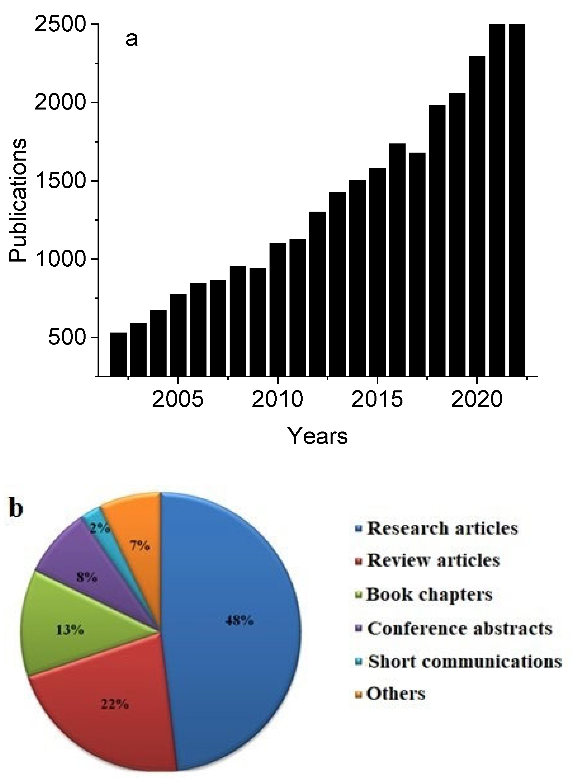
Hyaluronan is linked to significant phenomena that are essential to the quality of human life, such as increased longevity, cancer resistance, and newborn protection, which explains the long-term growth in interest in HA. Over the last 20 years (years 2002–2022), there has been an increase in the number of papers on hyaluronan/hyaluronic acid, which is summarized in Figure 2a. The types of publications on HA research within the last 20 years are denoted in Figure 2b, where almost 50% relate to research papers.

2.2. Sources of Hyaluronan

Hyaluronan has been isolated from a variety of sources, including rooster combs, umbilical cords, and microorganisms such as Streptococci (Table 1) [26].

2.3. Modifications of Hyaluronan

Hyaluronan can be modified to produce a biomaterial that is more mechanically and chemically resilient while still being biocompatible and biodegradable. However, such a biopolymer also has drawbacks, such as poor mechanical properties and a short in vivo residence time caused by hyaluronidase-catalyzed degradation [22,30,31,32,33,34]. In order to eliminate these problems, different chemical alterations of HA were used to create new HA derivatives (HADs) with improved mechanical qualities and decreased susceptibility to chemical and enzymatic hydrolysis [35]. Cross-linking and esterification are two modification techniques used to change the mechanical and chemical properties of HA, which has allowed materials to be specifically tailored for use in drug delivery, cell adhesion, joint lubrication, and signaling. The most widely used HA derivatization method is supposed to be mediated by carbodiimides and utilized for hydrogel preparation and drug molecule attachment to couple water-soluble hydrazides to HA carboxylic acid groups at pH 4.75. It has also been documented that hydrazides can be used to functionalize alkanes and enhance the rheological properties of HA. Furthermore, researchers have examined the use of alkanes, silylation, and acylation to produce HA that is more hydrophobic to be molded hot or dissolved in organic solvents [36].
It is important to consider that when modifying HA, changes made to -COOH group can affect the behavior of the HA molecule in the body since HA receptors and hyaluronidase recognize carboxylic groups [37]. Hyaluronan functional groups (hydroxyl, carboxyl, and acetyl) can be modified chemically in two different ways: either by conjugation or cross-linking [38]. Conjugation involves a variety of chemical modifications, such as the introduction of special functional units including ether, ester, or amide groups; the attachment of bioactive as well as prodrug moieties; and the incorporation of marker molecules, such as specific dyes. The approach to chemically modify HA includes both conjugation and cross-linking processes [39], while cross-linked HAs, also known as hylans, are recognized for their extended residence time (up to 9 days) compared to native HA, which is degraded by specific HA enzymes, hyaluronidases (HYALs), both in situ and systemically [40,41], and extremely high molar mass (up to 23 × 106 Da). Etherification (diglycidyl ether), bis-epoxide cross-linking, esterification, divynilsulfone cross-linking with epoxides, or dimethyl sulfone are common methods for changing HA hydroxyls. On the other hand, HA carboxyl can be changed by reactions mediated by carbodiimide, amidation, esterification, and dihydrazide, dialdehyde, or disulfide cross-linkers [42]. The preparation of new HA derivatives should take into account the multifunctionality of the HA molecule by selecting a mode of synthesis and applying mild reaction conditions. Special attention should also be paid to the properties of native HA, such as its excellent biocompatibility, adjustable biodegradability, and mucoadhesivity [39]. In order to achieve a delayed release and/or a prolonged effect, the majority of developments have concentrated on producing materials for implantation, such as films or sponges, and on combining HA with therapeutic agents to achieve a controlled release [43,44,45].
Cross-linking of HA can be performed by covalently joining tyramines (Corgel), methacrylates, and thiols (Extracel, HyStem). Formaldehyde, divinylsulfone, or formaldehyde can be used to cross-link HA to yield Hylan-A or Hylan-B [46]. Cross-linking with water-soluble carbodiimide, divinyl sulfone, polyvalent hydrazide, glutaraldehyde, disulfide, auto- and photo-cross-linking, and cross-linking with these hydrogels are in the process of being developed currently [47,48]. The cross-linking of HA increases its stability and prevents biodegradation of HA in vivo. Chemical modifications can be accomplished through the amidation of HA using homobifunctional reagents, which are new modifying reagents that have been synthesized and contain a new divalent disulfide-based protecting group. Amidation of HA with these reagents gives rise to either an one-end coupling product or intra- or intermolecular cross-linking of the HA chains [49], Ugi condensation (requiring four functional groups, such as primary amino, carboxyl, isocyanide, and aldehyde, for a typical Ugi condensation reaction) [50], ester formation (containing an irreversible ester bond that can be hydrolyzed by enzymes) [51], and ether formation (forming a thioethyl ether derivative of HA) [20].

3. Hyaluronan in Medical Practice

Hyaluronan solutions have amazing qualities such as hygroscopicity, non-thrombogenicity, non-immunogenicity, biodegradability, viscoelasticity, biocompatibility, and bioreactivity. These features enable their application in a variety of fields, such as the food, cosmetic, and biomedical industries [52].

3.1. Hyaluronan in Dermal Filler

In humans, about 50% of HA is found in the skin, i.e., 7–8 g per average adult human, with ~0.5 mg/g of HA found in the dermis and ~0.1 mg/g in the epidermis [53]. Recently, there has been a lot of interest in the field of cosmetic surgery for dermal rejuvenation due to the use of HA-based dermal fillers (DFs). Soft tissue augmentation with DF is now a standard procedure in aesthetics. Dermal fillers are used for facial rejuvenation and to temporarily eliminate the appearance of rhytides and skin folds [54]. Hyaluronan transports vital nutrients to these fibers and forms bonds with collagen and elastin. Due to the ability of HA to effectively augment soft tissue in a comprehensive nonsurgical facial rejuvenation approach, physicians utilize it to restore the durability of skin. A dermal filler exhibits clear antiaging benefits, such as a more youthful appearance, increased skin volume, elevated viscoelasticity, smoothness, improved lifting ability, and allow tighter skin [55].
Hyaluronan used to create DF is typically supplied as a dry powder. If this product is used as DF, drainage or degradation pathways would quickly remove it from the injection site. In order to overcome the inability of HA solutions to reside, a cross-linked HA can be used to create DFs. A weak gel is produced by cross-linking, which forms a network of polymers from a viscous solution. A chemical and physical barrier is added by the HA gel to prevent drainage or degradation. As a result, cross-linking HA lengthens residence time [56]. Since they have been in use for over 15 years, cross-linked HA fillers are thought to be well tolerated. They have good tissue integration, outstanding biocompatibility, and structural characteristics akin to native tissue. Cross-linking of HAD is necessary to prolong the duration of HA filler and stop it from being biodegraded by enzymes and free radicals. HA-based DFs differ in their physical and chemical characteristics, such as stress applied, particle size, cross-linking agent density, polymer concentration, extrusion force, and viscosity. HA-based DFs are made of bacterially fermented or rooster comb-derived HAs that have been cross-linked with butanedioldiglycidyl ether [57]. Dermal fillers must be elastic in a low-shear environment in order to be injected into the dermal connective tissue. It is suggested that greater persistence is caused by DF elasticity [58].

3.2. Hyaluronan in Tissue Engineering

The field of tissue engineering (TE) is broad and primarily concerned with the regeneration of lost or damaged tissues. By developing biological substitutes, TE allows for the replacement of damaged tissues both structurally and functionally while promoting tissue growth in vivo. These biological replacements are structures made by combining suitable cells and/or growth factors with scaffolds. The scaffold serves as a framework for developing tissues and cells and should function as a synthetic ECM to facilitate the attachment, migration, proliferation, and differentiation of cells until the creation of new tissue [59]. For TE applications such as soft tissue augmentation, wound healing, facial intradermal implants, and artificial skin, HA has demonstrated exceptional promise. Due to the high concentration of HA in the ECM, especially in nervous system tissue, HA has been utilized as the structural foundation for hydrogel scaffolds. Owing to its broad availability, nonimmunogenic properties, and ease of adjusting chain length, HA is a flexible macromolecule with enormous potential for use in TE applications. Hyaluronan hydrogels have the ability to exchange nutrients, oxygen, and metabolic waste while simulating the water content of individual tissues [60]. A variety of tissues, including skin, fibrocartilage, cartilage, intervertebral discs, bone, vascular tissues, adipose tissues, and heart valves, have been successfully regenerated using HADs and their blends as scaffolds. It has been established that the amides of HA are bioactive and appropriate carriers of cells for TE procedures [61].

3.3. Hyaluronan in Viscosupplementation

Degenerative joint disease, commonly referred to as osteoarthritis (OA), is the most prevalent type of arthritis. The financial toll that OA takes is substantial and multifaceted, encompassing expenses for medical care, hospital stays, missed work, and home care. With up to 41% involvement, the knee is the most commonly affected joint in OA, followed by the hands (30%) and hips (19%) [62]. In particular, the prevalence of knee OA has doubled since the middle of the 20th century. It has also been demonstrated that OA significantly lowers the quality of life. Viscosupplementation is the process of injecting HA into arthritic joints with the goal of restoring SF natural viscoelasticity and maintaining cartilage homeostasis and lubrication without inflammation [63,64]. One of the main functions of SF, a substance present in synovial joint cavities, is to reduce friction between the articular cartilages while the joint is in motion [65]. It has been observed that injecting HA preparations into patients with OA reduces their use of potentially hazardous nonsteroidal anti-inflammatory drugs and improves their symptoms [64].
Hyaluronan, coupled to a thermosensitive polymer, allowed for the spontaneous formation of nanoparticles at body temperature, thereby overcoming the drawbacks of repeatedly administered injections and the rapid breakdown of exogenous HA treatments. In addition to enabling the restoration of its mechanical characteristics, viscosupplementation also permits synoviocytes to be stimulated to produce high-molar-mass HA [66].

3.4. Hyaluronan in Ocular Treatment

A complicated and crippling inflammatory condition of the ocular surface is called dry eye disease (DED). Global epidemiological research indicates that the prevalence of DED varies between 5 and 50%. If it is extrapolated, there would be 400 million to 3.7 billion DED patients worldwide [67]. The prevalence of dry eye disease rises sharply after the age of fifty, with the condition becoming more common as people get older. DED creates significant financial burdens for both the individual patient and society at large by restricting participation in daily activities and the workforce and raising healthcare costs. Patients with DED experience a significant decline in their visual and overall quality of life. The healthy tear film coats and lubricates the ocular surface while serving as a chemical, physical, and immunological barrier against the outside world. An outer lipid layer and an inner mucoaqueous layer make up the tear film in a healthy eye, and they work together to maintain the stability of the ocular surface [68]. The initial line of treatment for DED is artificial tears. They shield the surface of the eyes and help stabilize and restore the tear film. This helps prevent further damage and reduce signs and symptoms by slowing or stopping the progression of DED.
On the market, there are varieties of artificial tears with different active ingredients. One often-used and clinically proven ingredient is hyaluronic acid [69]. Due to its abundance of hydroxyl groups, hyaluronic acid thickens and stabilizes the tear film, lubricates the ocular surface to reduce mechanical trauma, and aids in re-epithelialization. Additionally, hyaluronic acid lessens evaporation from the surface of the eyes, which is the primary cause of hyperosmolarity, one of the primary causes of inflammation and surface damage in DED [70]. In order to prolong the therapeutic effect of the eye-drop formulation, viscous HA solutions or hydrogels of higher concentrations are often used to slow down the drainage of the formulation. The primary benefit of HA solutions of high viscosity over polyvinyl alcohol or celluloses is their shear-thinning behavior. Hyaluronan solutions function as a general viscous mucoadhesive excipient, a lubricating and wetting agent, and a systemic drainage inhibitor for drug-containing eye drops, extending their ocular residence [71].

3.5. Hyaluronan in Wound Healing

Wound healing is an intrinsic field of study. In general, there are four main phases to normal wound healing: homeostasis, the inflammatory phase, the proliferative phase, and the remodeling phase. These phases share overlapping processes of coordinated cellular activity. These biological processes are mediated by cytokines, which enable the production of structural proteins and polymers by wound-healing cells [72,73]. Hyaluronan is involved in every phase of wound healing. There are different lengths of HA polymers, and each size of HA molecules has a specific function at a different phase of wound healing. In summary, large HA molecules regulate structure and occupy space; small HA fragments promote cell migration; medium-sized HA chains stimulate the expression of inflammatory cytokines; and very short, low-weight (four saccharides) HA fragments induce chemotaxis [74].
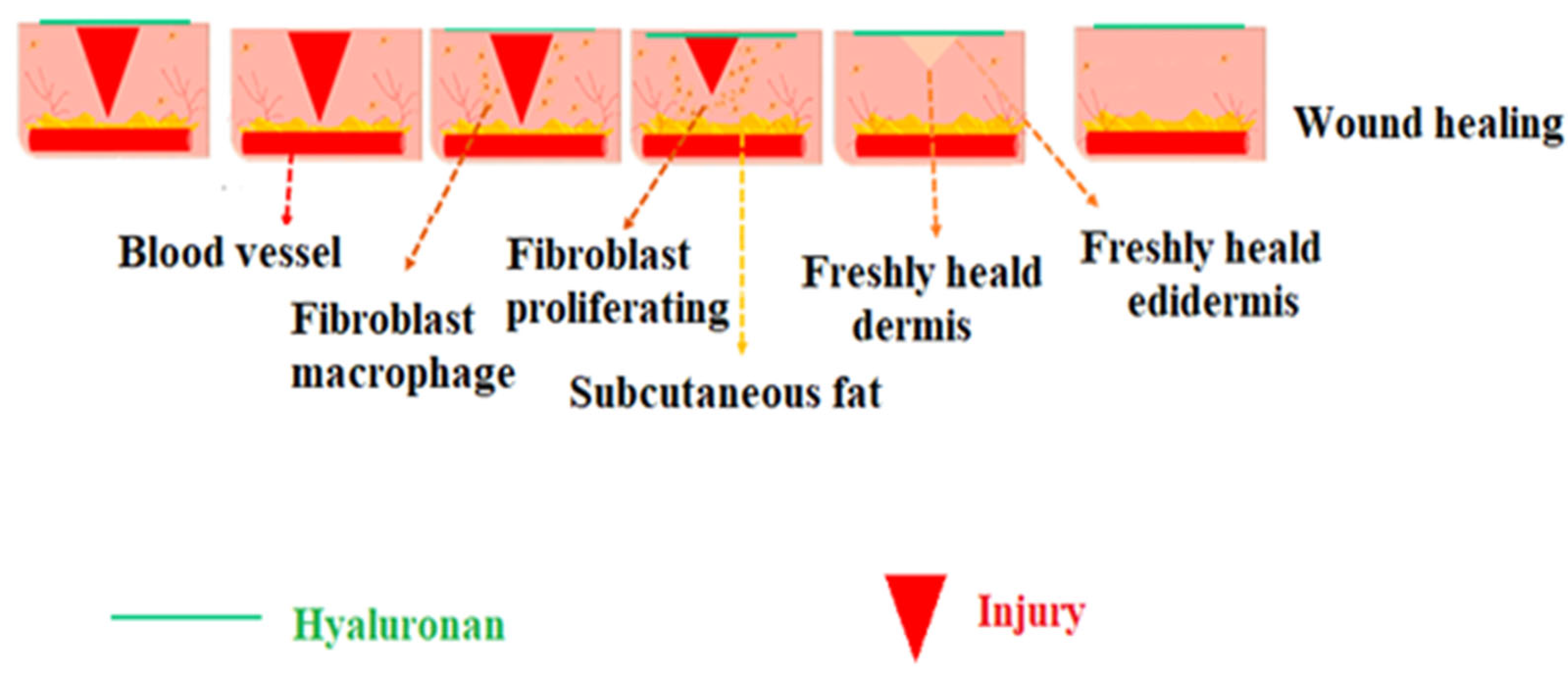
The inflammatory phase of wound healing is marked by a sharp rise in HA synthesis. Large, molecular-sized HA fragments are synthesized from platelets and are available in the bloodstream upon skin breach. By binding to fibrinogen, these HA fragments can start the extrinsic clotting process. Oedema develops at the wound site as a result of the significant volumes of HA released during wounding [75]. Due to the hydrophilic nature of HA, the tissue around the wound swells, allowing cells to move into the injured area and form a porous framework (Figure 3) [76].

3.6. Hyaluronan in Drug Delivery

The term “drug delivery” refers to technologies that transport medications into or through tissue. These technologies include delivery mechanisms such as an injectable vaccine or a pill that you swallow [77]. Drug delivery systems can also refer to the way that pharmaceuticals are enveloped, such as in a micelle or nanoparticle, to prevent degradation and enable the drug to go to the appropriate location within the body. The development of drug delivery has come a long way in the last few decades, and a more rapid advance is expected in the following years [78]. Biomedical engineering has made significant contributions to our knowledge of the physiological obstacles to effective drug delivery as well as to the creation of various novel drug delivery strategies that are now being used in clinical practice.
It has been discovered that the HA receptor CD44 is overexpressed in many tumor cells; however, it is expressed at a low level on the surface of epithelial, hematopoietic, and neuronal cells [79,80,81]. Consequently, the use of HA derivatives as drug carriers enhances drug targeting, transdermal absorption, sustained release, and thickening of the drug (Figure 4). When cytotoxic drugs are coupled with macromolecular substances, the drug’s pharmacokinetic profile is enhanced, its distribution is prolonged, and its elimination time is increased. Moreover, due to the drug’s delayed release from the carrier, the concentration of the medication in the plasma is reduced; however, it remains in the tumor tissue for a longer period of time. It has been shown that HA and drug conjugates have the dual benefits of receptor-mediated endocytosis and aggregation at the tumor site. Hyaluronic acid and its derivatives are applied in a variety of drug delivery systems, including cationic polymer gene carrier systems, drug delivery of gels [82,83], polyelectrolyte microcapsules, films [84], nanoparticles, etc. [85,86,87]. Since HA binds specifically to the receptors on the surface of cancer cells and is biodegradable and biocompatible, its use in targeted drug delivery of anticancer drugs has advanced significantly. It creates conjugates by reacting with other medications. The conjugates provide a targeted effect and controlled release to achieve the timing and directed release objectives. This makes it possible for them to deliver various medications to various pathological locations [88].

3.7. Hyaluronan in Transdermal Drug Delivery

Novel microneedle (MN) arrays made of HA were developed and used for transdermal insulin delivery [90]. It was proven that MNs could pierce the skin for at least an hour at 75% relative humidity. The fact that more than 90% of insulin in MNs remained in those reservoirs at all temperatures after they were stored for a month at a temperature in the range of −40–40 °C shows the high stability of insulin in MNs [91]. After being applied topically, these unique HA MNs have the ability to dissolve themselves, and insulin is released from them fast. Pharmacodynamic and pharmacokinetic parameters showed that insulin delivered via MNs was almost completely absorbed from the skin into the systemic circulation and that the hypoglycemic effect of insulin-loaded MNs was almost the same as that of insulin injected subcutaneously. Therefore, HA MNs could provide a secure and efficient transdermal insulin delivery system in clinical practice [92,93].
Bonet et al. (2023) [94] studied the therapeutic effect of HA when combined with each of three transdermal drug delivery enhancers (protamine, dimethyl sulfoxide (DMSO), or terpene) in preclinical models of inflammatory and neuropathic pain. The administration of a combination of HA with the transdermal enhancers (protamine and terpene) attenuated chemotherapy-induced painful peripheral neuropathy hyperalgesia rather than with the DMSO vehicle [95,96]. Also, Yuan et al. (2022) [97] prepared a hydrogel containing HA-modified transforms as a drug carrier for indomethacin (IND). Indomethacin was encapsulated in HA-modified transfersomes to enhance transdermal IND delivery and reduce adverse effects.
Son et al. (2017) [98] synthesized HA–dodecylamine and nanohydrogels with indocyanine green as a model agent and proved that nanohydrogels have a promising potential for applications as transdermal delivery systems in the pharmaceutical and cosmetic industries.

3.8. Hyaluronan in Nasal Drug Delivery

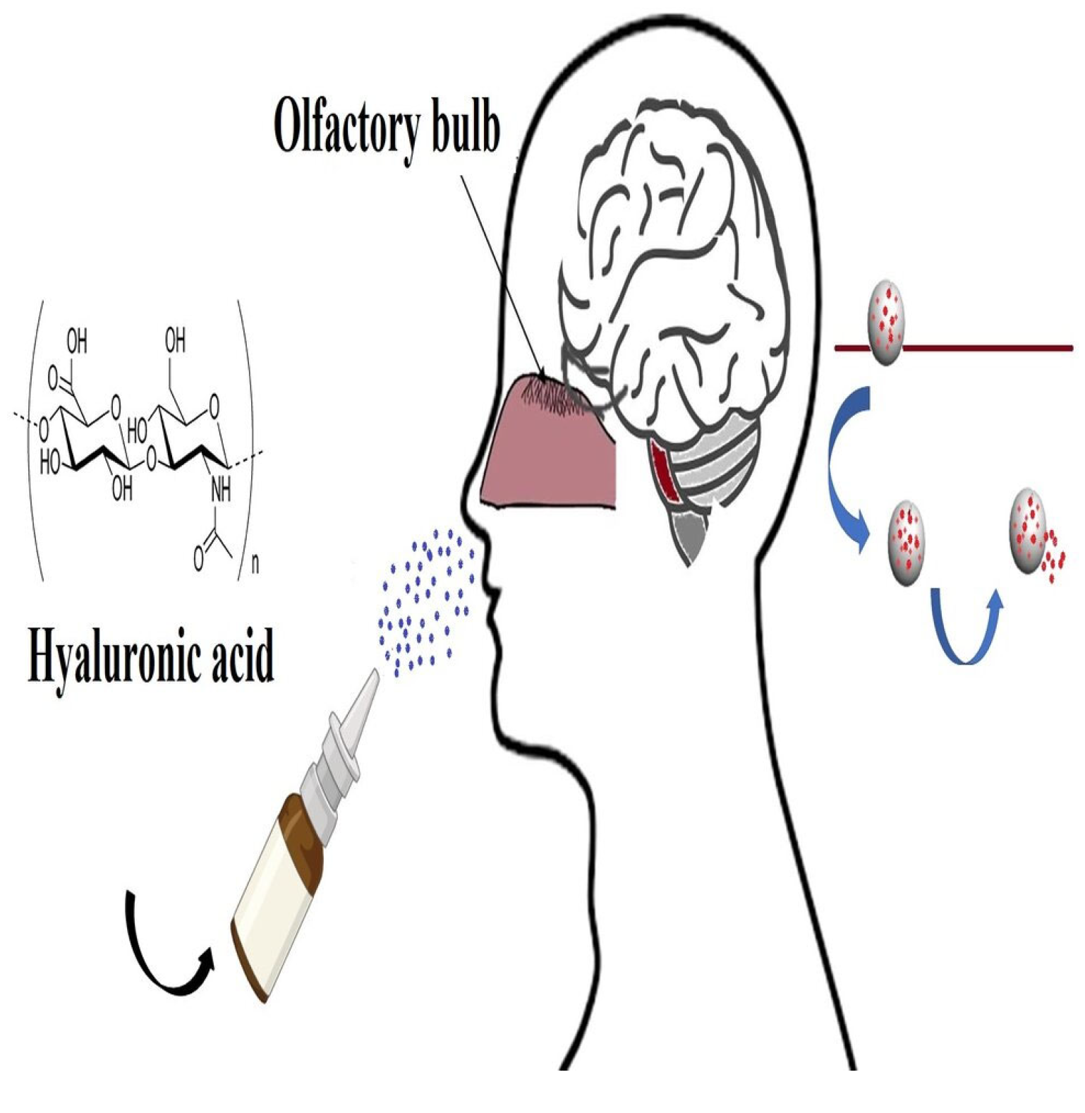
The intranasal administration of molecules has been investigated as a non-invasive way for the delivery of drugs to the brain in the last decade. Circumvention of both the blood–brain barrier and first-pass elimination by the liver and gastrointestinal tract are considered the main advantages of this method. Due to the rapid mucociliary clearance in the nasal cavity, bioadhesive formulations are needed for effective targeting. Hyaluronan is used as a mucoadhesive in a nasal formulation, increasing the brain penetration of a hydrophilic compound in the size of a peptide via the nasal route [99,100,101]. The blood-cerebrospinal fluid (BCSF) and blood–brain barriers (BBB) make it difficult to target medications to the central nervous system (CNS). Many techniques have been tried to improve blood–brain transport and deliver medications to the brain at effective concentrations [102]. Prodrugs, drug binding to transferrin or targeted vesicle systems, hyperosmotic shock, vasoactive substances, and inhibition of efflux transporters are some of the strategies used to manipulate the BBB (Figure 5). There are additional methods to improve drug transport to the central nervous system, though, aside from changing the drug molecule or the BBB’s functioning. Selecting an application site that stays aside from the BBB is one such strategy. The delivery of medicine to the brain through the olfactory region through the nasal route has garnered a lot of attention lately [101,102,103]. In a distal epithelium, olfactory sensory neurons are the only first-order neurons with cell bodies. For the distinct anatomical and physiological characteristics of the olfactory region, the direct exposure of dendritic processes to the external environment in the upper nasal passage permits both extracellular and intracellular pathways into the CNS that circumvent the BBB [104].
Intranasal administration is noninvasive, virtually painless, and finally simple for patients as well as physicians to perform in emergency situations. It also provides an immediate starting point for therapeutic effects and eliminates the first-pass effect or gastrointestinal drug degradation [105]. Moreover, it guarantees fast absorption, prevents first-pass metabolism in the liver and gut, and does not need to be sterile. Hyaluronan was shown to prevent chronic rhinosinusitis [106].
Horvát et al. (2009) [99] developed a formulation containing sodium hyaluronate in combination with a non-ionic surfactant to enhance the delivery of hydrophilic compounds to the brain via the olfactory route. They used fluorescence isothiocyanate-labeled 4 kDa dextran (FD-4) as a test molecule. Hyaluronan increased the viscosity of the vehicles and slowed down the in vitro release of FD-4. On the other hand, Laffleur et al. (2023) [107] modified HA with cysteine ethyl ester hydrochloride via amid bond formation, and a strongly increased bonding to the mucosa by the formation of covalent disulfide bridges could be observed in comparison to unmodified HA. The adhesion study with the model drug showed a more pronounced binding to the nasal mucosa in the case of modified HA in comparison to the unmodified one. Casula et al. (2021) [108] recently examined a natural and green nasal spray containing hyaluronic acid and the extract of Zingiber officinalis as a novel strategy for the treatment of rhinitis and rhinosinusitis.

3.9. Hyaluronan in Colon Drug Delivery

Colorectal cancer (CRC) is the fourth most common cause of cancer death around the world. Due to increased life expectancy and a diet rich in fat and low in fiber, the prevalence has been rapidly increasing. The primary method of treatment for CRC management is chemotherapy. The clinic uses a number of FDA-approved the olfactory chemotherapeutic medications, such as capecitabine, irinotecan hydrochloride, and fluorouracil [109]. There is a pressing need for advanced colon-specific local drug delivery systems that can provide major advantages in treating diseases associated with the colon, such as inflammatory bowel disease and colon cancer. A precise, colon-targeted drug delivery platform is expected to reduce drug side effects and increase the therapeutic response at the intended disease site locally [110]. Kotla et al. (2019) [111] reported a strategy that uses a HA-functionalized polymeric hybrid nanoconjugate system fabricated from processes that are generally recognized as safe reagents for enhanced drug absorption with controlled drug release and increasing local drug bioavailability in the colon lumen. They functionalized the HA polymeric hybrid nanoparticulate system, curcumin-HA NPs, whereas curcumin was used as a model fluorescent drug.
Liposomes are a type of nanocarrier for drug delivery that have been studied in detail and have many applications. Liposomes have the ability to control drug release synchronously, reduce drug accumulation in tumor sites, and lessen drug toxicity to cells other than cancerous ones. Moreover, since liposomes have a hydrophilic core surrounded by a lipid bilayer, they serve well as delivery systems for both lipophilic and hydrophilic drugs [112]. Active targeting of liposomal nanocarriers to the intended site of action can significantly increase their efficacy. Active targeting strategies for anti-cancer drugs investigate specific interactions between liposome-bound targeting groups and cell surface receptors. Moreover, the majority of cancerous cell types overexpress hyaluronan receptors to raise the concentration of HA in the surrounding tissue of tumors. Additionally, colorectal cancer has overexpressed hyaluronan receptor isoforms.
As a result, cancerous cells with the greatest potential for metastasis usually exhibit enhanced HA binding and internalization. Hence, the HA receptors overexpressed on colon cancer cells are more readily bonded to the HA-conjugated liposomes encasing both medications, cell-specific targeting is possible [113].

3.10. Hyaluronan in Chemoprevention and Cancer Therapy

It has been reported that HA significantly alters a number of signaling pathways linked to cancer. The majority of its effects are mediated by interactions with cell surface receptors (TLR-2, TLR-4, RHAMM, LYVE, and CD44), which bind and react to HA when they are activated. The main ligand for CD44 is HA. CD44 is overexpressed in a variety of malignancies, such as pancreatic, breast, lung, ovarian, prostate, and so forth. Transmembrane glycoproteins belonging to the CD44 family are important regulators of signal transduction [114]. As HA affects oncogenic signaling pathways, scientists have discovered effective ways to block hyaluronan-receptor interactions to impede the growth of tumors. Treating with hyaluronan oligomers that compete with polymeric hyaluronan for constitutive binding to its endogenous receptors appears to be one strategy. This approach is predicated on the idea that high-affinity, multivalent interactions with receptors are necessary for endogenous HA to transduce signals; small oligomers that bind monovalently will act as antagonists by acting as a substitute for cooperative, multivalent interactions with low-affinity and low-valency receptors [115]. Combining a drug with HA not only provides pharmacological benefits such as stabilization and solubilization, but it also confers selectivity and specificity for cancerous cells. Together, these data suggest that the use of HA, either itself or in conjunction with well-known medications (taxol, doxorubicin, methotrexate, vincristine, 5-fluorouracil, gemcitabine, imatinib, cisplatin, etc.), could be a promising option for the treatment of cancer [116,117].

4. Conclusions

Hyaluronan, an outstanding material, has established a strong foundation for biomedical applications due to its remarkable multifunctional capabilities. In order to build new platforms for various applications, hyaluronan has developed a new requirement through derivatives or grafting modification. Researchers have become more interested in using it in various biomedical applications, including drug delivery and regenerative medicine, due to its exceptional biodegradability, biocompatibility, ease of chemical functionalization, and distinctive physical, biological, and chemical properties.

Author Contributions

Conceptualization, K.V., L.Š. and M.E.H.; investigation, M.E.H.; writing—M.E.H.; writing—M.E.H., K.V. and L.Š.; visualization, M.E.H., K.V. and L.Š.; supervision, K.V. and L.Š.; project administration, K.V. and L.Š.; funding acquisition, K.V. and M.E.H. All authors have read and agreed to the published version of the manuscript.

Funding

This work was supported by the Slovak Academic Information Agency (SAIA) program and the VEGA grant 2/0008/23 and a collective of researchers of CEM.

Institutional Review Board Statement

Not applicable.

Informed Consent Statement

Not applicable.

Data Availability Statement

Not applicable.

Conflicts of Interest

The authors declare no conflicts of interest.

References

  1. Mohammed, A.S.A.; Naveed, M.; Jost, N. Polysaccharides; classification, chemical properties, and future perspective applications in fields of pharmacology and biological medicine (A review of current applications and upcoming potentialities). J. Polym. Environ. 2021, 29, 2359–2371. [Google Scholar] [CrossRef] [PubMed]
  2. Hassan, M.E.; Bai, J.; Dou, D.Q. Biopolymers; definition, classification and applications. Egypt. J. Chem. 2019, 62, 1725–1737. [Google Scholar] [CrossRef]
  3. Ghlissi, Z.; Kallel, R.; Krichen, F.; Hakim, A.; Zeghal, K.; Boudawara, T.; Bougatef, A.; Sahnoun, Z. Polysaccharide from Pimpinella anisum seeds: Structural characterization, anti-inflammatory and laser burn wound healing in mice. Int. J. Biol. Macromol. 2020, 156, 1530–1538. [Google Scholar] [CrossRef]
  4. Das, H. An Overview on heteropolysaccharides. J. Glycobiol. 2022, 11, 210. [Google Scholar]
  5. Garantziotis, S.; Savani, R.C. Hyaluronan biology: A complex balancing act of structure, function, location and context. Matrix Biol. 2019, 78–79, 1–10. [Google Scholar] [CrossRef] [PubMed]
  6. Johnson, P.; Arif, A.A.; Lee-Sayer, S.S.M.; Dong, Y. Hyaluronan and its interactions with immune cells in the healthy and inflamed lung. Front. Immunol. 2018, 9, 2787. [Google Scholar] [CrossRef] [PubMed]
  7. Meyer, K.; Palmer, J. The polysaccharide of the vitreous humor. J. Biol. Chem. 1935, 107, 629–634. [Google Scholar] [CrossRef]
  8. Csoka, A.B.; Stern, R. Hypotheses on the evolution of hyaluronan: A highly ironic acid. Glycobiology 2013, 23, 398–411. [Google Scholar] [CrossRef]
  9. Huang, G.; Huang, H. Application of hyaluronic acid as carriers in drug delivery. Drug Deliv. 2018, 25, 766–772. [Google Scholar] [CrossRef]
  10. Hrabárová, E.; Rychlý, J.; Sasinková, V.; Valachová, K.; Janigová, I.; Csomorová, K.; Juránek, I.; Šoltés, L. Structural characterisation of thiol-modified hyaluronans. Cellulose 2012, 19, 2093–2104. [Google Scholar] [CrossRef]
  11. Kogan, G.; Soltes, L.; Stern, R.; Gemeiner, P. Hyaluronic acid: A natural biopolymer with a broad range of biomedical and industrial applications. Biotechnol. Lett. 2007, 29, 17–25. [Google Scholar] [CrossRef] [PubMed]
  12. Jenkins, H.G.; Bachelard, H.S. Developmental and age-related changes in rat brain glycosaminoglycans. J. Neurochem. 1988, 51, 1634–1640. [Google Scholar] [CrossRef] [PubMed]
  13. Fraser, J.R.; Laurent, T.C.; Laurent, U.B. Hyaluronan: Its nature, distribution, functions and turnover. J. Intern. Med. 1997, 242, 27–33. [Google Scholar] [CrossRef] [PubMed]
  14. Valachová, K.; Volpi, N.; Stern, R.; Šoltés, L. Hyaluronan in medical practice. Curr. Med. Chem. 2016, 23, 1–11. [Google Scholar] [CrossRef] [PubMed]
  15. Menaa, T.; Menaa, A.; Menaa, B. Hyaluronic acid and derivatives for tissue engineering. J. Biotechnol. Biomater. 2011, S3, 001. [Google Scholar]
  16. Burdick, J.A.; Prestwich, G.D. Hyaluronic acid hydrogels for biomedical applications. Adv. Mater. 2011, 23, 41–56. [Google Scholar] [CrossRef] [PubMed]
  17. Almond, A.; Sheehan, J.K. Predicting the molecular shape of polysaccharides from dynamic interactions with water. Glycobiology 2003, 13, 255–264. [Google Scholar] [CrossRef] [PubMed]
  18. Csapo, E.; Szokolai, H.; Juhasz, A.; Varga, N.; Janovak, L.; Dekany, I. Cross-linked and hydrophobized hyaluronic acid-based controlled drug release systems. Carbohydr. Polym. 2018, 195, 99–106. [Google Scholar] [CrossRef]
  19. Liu, L.; Liu, Y.; Li, J.; Du, G.; Chen, J. Microbial production of hyaluronic acid: Current state, challenges, and perspectives. Microb. Cell Fact. 2011, 10, 99. [Google Scholar] [CrossRef]
  20. Serban, M.A.; Yang, G.; Prestwich, G.D. Synthesis, characterization and chondroprotective properties of a hyaluronan thioethyl ether derivative. Biomaterials 2008, 29, 1388–1399. [Google Scholar] [CrossRef]
  21. Girish, K.S.; Kemparaju, K. The magic glue hyaluronan and its eraser hyaluronidase: A biological overview. Life Sci. 2007, 80, 1921–1943. [Google Scholar] [CrossRef] [PubMed]
  22. Erickson, M.; Stern, R. Chain gangs: New aspects of hyaluronan metabolism. Int. Biochem. Res. 2012, 2012, 19–28. [Google Scholar] [CrossRef] [PubMed]
  23. Jiang, D.; Liang, J.; Noble, P.W. Hyaluronan in tissue injury and repair. Annu. Rev. Cell Dev. Biol. 2007, 23, 435–461. [Google Scholar] [CrossRef] [PubMed]
  24. Jiang, D.; Liang, J.; Noble, P.W. Hyaluronan as an immune regulator in human diseases. Physiol. Rev. 2011, 91, 221–264. [Google Scholar] [CrossRef] [PubMed]
  25. Zhang, L.; Underhill, C.B.; Chen, L. Hyaluronan on the surface of tumor cells is correlated with metastatic behavior. Cancer Res. 1995, 55, 428–433. [Google Scholar] [PubMed]
  26. Shiedlin, A.; Bigelow, R.; Christopher, W.; Arbabi, S.; Yang, L.; Maier, R.V.; Wainwright, N.; Childs, A.; Miller, R.J. Evaluation of hyaluronan from different sources: Streptococcus zooepidemicus, rooster comb, bovine vitreous, and human umbilical cord. Biomacromolecules 2004, 5, 2122–2127. [Google Scholar] [CrossRef]
  27. Fakhari, A.; Berkland, C. Applications and emerging trends of hyaluronic acid in tissue engineering, as a dermal filler and in osteoarthritis treatment. Acta Biomater. 2013, 9, 7081–7092. [Google Scholar] [CrossRef] [PubMed]
  28. Quinones, J.P.; Bruggemann, O.; Covas, C.P.; Ossipov, D.A. Self-assembled hyaluronic acid nanoparticles for controlled release of agrochemicals and diosgenin. Carbohydr. Polym. 2017, 173, 157–169. [Google Scholar] [CrossRef]
  29. Roughley, P.J.; Mason, R.M. Proteins and hyaluronic acid associated with proteoglycans extracted from bovine nasal cartilage with low-ionic-strength salt solution. Biochem. Soc. Trans. 1975, 3, 140–142. [Google Scholar] [CrossRef]
  30. Bulpitt, P.; Aeschlimann, D. New strategy for chemical modification of hyaluronic acid: Preparation of functionalized derivatives and their use in the formation of novel biocompatible hydrogels. J. Biomed. Mater. Res. 1999, 47, 152–169. [Google Scholar] [CrossRef]
  31. Liu, C.; Bae, K.H.; Yamashita, A.; Chung, J.E.; Kurisawa, M. Thiol-mediated synthesis of hyaluronic acid–epigallocatechin-3-O-gallate conjugates for the formation of injectable hydrogels with free radical scavenging property and degradation resistance. Biomacromolecules 2017, 18, 3143–3155. [Google Scholar] [CrossRef]
  32. Jia, X.; Burdick, J.A.; Kobler, J.; Clifton, R.J.; Rosowski, J.J.; Zeitels, S.M.; Langer, R. Synthesis and characterization of in situ cross-linkable hyaluronic acid-based hydrogels with potential application for vocal fold regeneration. Macromolecules 2004, 37, 3239–3248. [Google Scholar] [CrossRef]
  33. Oh, E.J.; Kang, S.; Kim, B.; Jiang, G.; Cho, I.H.; Hahn, S.K. Control of the molecular degradation of hyaluronic acid hydrogels for tissue augmentation. J. Biomed. Mater. Res. A 2008, 86, 685–693. [Google Scholar] [CrossRef] [PubMed]
  34. Prajapati, V.D.; Maheriya, M.P. Hyaluronic acid as potential carrier in biomedical and drug delivery applications. Funct. Polysacch. Biomed. Appl. 2019, 213–265. [Google Scholar] [CrossRef]
  35. Calles, J.A.; Ressia, J.A.; Llabot, J.M.; Valles, E.M.; Palma, S.D. Hyaluronan–itaconic acid–glutaraldehyde films for biomedical applications: Preliminary studies. Sci. Pharm. 2016, 84, 61–72. [Google Scholar] [CrossRef] [PubMed]
  36. Wang, X.; Messman, J.; Mays, J.W.; Baskaran, D. Polypeptide grafted hyaluronan: Synthesis and characterization. Biomacromolecules 2010, 11, 2313–2320. [Google Scholar] [CrossRef] [PubMed]
  37. Zhong, S.P.; Campoccia, D.; Williams, D.F.; Doherty, P.J.; Williams, R.L.; Benedetti, L. Biodegradation of hyaluronic acid derivatives by hyaluronidase. Biomaterials 1994, 15, 359–365. [Google Scholar] [CrossRef]
  38. Khunmanee, S.; Jeong, Y.; Park, H. Crosslinking method of hyaluronic-based hydrogel for biomedical applications. J. Tissue Eng. 2017, 8, 1–16. [Google Scholar] [CrossRef]
  39. Hintze, V.; Schnabelrauch, M.; Rother, S. Chemical modification of hyaluronan and their biomedical applications. Front. Chem. 2022, 10, 830671. [Google Scholar] [CrossRef]
  40. Schanté, C.E.; Zuber, G.; Herlin, C.; Vandamme, T.F. Chemical modifications of hyaluronic acid for the synthesis of derivatives for a broad range of biomedical applications. Carbohydr. Polym. 2011, 85, 469–489. [Google Scholar] [CrossRef]
  41. Williams, D. Essential Biomaterials Science; Cambridge University Press: Cambridge, UK, 2014; pp. 537–539. [Google Scholar]
  42. Luo, Y.; Kirker, K.R.; Prestwich, G.D. Cross-linked hyaluronic acid hydrogel films: New biomaterials for drug delivery. J. Control. Release 2000, 69, 169–184. [Google Scholar] [CrossRef] [PubMed]
  43. Carvalho, A.L.; Vale, A.C.; Sousa, M.P.; Barbosa, A.M.; Torrado, E.; Mano, J.F.; Alves, N.M. Antibacterial bioadhesive layer-by-layer coatings for orthopedic applications. J. Mater. Chem. B 2016, 4, 5385–5393. [Google Scholar] [CrossRef] [PubMed]
  44. Sgorla, D.; Almeida, A.; Azevedo, C.; Bunhak, E.J.; Sarmento, B.; Cavalcanti, O.A. Development and characterization of crosslinked hyaluronic acid polymeric films for use in coating processes. Int. J. Pharm. 2016, 511, 380–389. [Google Scholar] [CrossRef] [PubMed]
  45. Palumbo, F.S.; Fiorica, C.; Pitarresi, G.; Giorgi, M.; Abramo, F.; Gulino, A.; Di Stefano, M.; Giammona, G. Construction and evaluation of sponge scaffolds from hyaluronic acid derivatives for potential cartilage regeneration. J. Mater. Chem. B 2014, 2, 3243–3253. [Google Scholar] [CrossRef] [PubMed]
  46. Song, E.; Shang, J.; Ratner, D.M. Polysaccharides. In Polymer Science: A Comprehensive Reference; Matyjaszewski, K., Moller, M., Eds.; Elsevier: Oxford, UK, 2012; pp. 137–155. ISBN 978-0-08-087862-1. [Google Scholar]
  47. Allison, D.D.; Grande-Allen, K.J. Review. Hyaluronan: A powerful tissue engineering tool. Tissue Eng. 2006, 12, 2131–2140. [Google Scholar] [CrossRef] [PubMed]
  48. Prestwich, G.D.; Kuo, J.W. Chemically-modified HA for therapy and regenerative medicine. Curr. Pharm. Biotechnol. 2008, 9, 242–245. [Google Scholar] [CrossRef] [PubMed]
  49. Dmitri, A.O.; Sonya, P.; Oommen, P.V.; Jöns, H. Functionalization of hyaluronic acid with chemoselective groups via a disulfide-based protection strategy for in situ formation of mechanically stable hydrogels. Biomacromolecules 2010, 11, 2247–2254. [Google Scholar]
  50. Crescenzi, V.; Francescangeli, A.; Capitani, D.; Mannina, L.; Renier, D.; Bellini, D. Hyaluronan networking via Ugi’s condensation using lysine as cross-linker diamine. Carbohydr. Polym. 2003, 53, 311–316. [Google Scholar] [CrossRef]
  51. Bhatnagar, P.; Kumari, M.; Pahuja, R.; Pant, A.B.; Shukla, Y.; Kumar, P.; Gupta, K.C. Hyaluronic acid-grafted PLGA nanoparticles for the sustained delivery of berberine chloride for an efficient suppression of Ehrlich ascites tumors. Drug Deliv. Transl. Res. 2018, 8, 565–579. [Google Scholar] [CrossRef]
  52. Valachová, K.; Šoltés, L. Hyaluronan as a prominent biomolecule with numerous applications in medicine. Int. J. Mol. Sci. 2021, 22, 7077. [Google Scholar] [CrossRef]
  53. Becker, L.C.; Bergfeld, W.F.; Belsito, D.V.; Klaassen, C.D.; Marks, J.G.; Shank, R.C.; Slaga, T.J.; Snyder, P.W.; Cosmetic Ingredient Review Expert Panel; Andersen, F.A. Final report of the safety assessment of hyaluronic acid, potassium hyaluronate, and sodium hyaluronate. Int. J. Toxicol. 2009, 28, 5–67. [Google Scholar] [CrossRef] [PubMed]
  54. Kassir, R.; Kolluru, A.; Kassir, M. Extensive necrosis after injection of hyaluronic acid filler: Case report and review of the literature. J. Cosmet. Dermatol. 2011, 10, 224–231. [Google Scholar] [CrossRef] [PubMed]
  55. Jeong, S.H.; Fan, Y.; Cheon, K.H.; Baek, J.; Kim, S.; Kim, H.E. Hyaluronic acid-hydroxyapatite nanocomposite hydrogels for enhanced biophysical and biological performance in a dermal matrix. J. Biomed. Mater. Res. A 2017, 105, 3315–3325. [Google Scholar] [CrossRef] [PubMed]
  56. Li, H.; Xue, Y.; Jia, B.; Bai, Y.; Zuo, Y.; Wang, S.; Zhao, Y.; Yang, W.; Tang, H. The preparation of hyaluronic acid grafted pullulan polymers and their use in the formation of novel biocompatible wound healing film. Carbohydr. Polym. 2018, 188, 92–100. [Google Scholar] [CrossRef] [PubMed]
  57. Boulle, K.; Glogau, R.; Kono, T.; Nathan, M.; Tezel, A.; Roca-Martinez, J.X.; Paliwal, S.; Stroumpoulis, D. A review of the metabolism of 1,4-butanediol diglycidyl ether—Crosslinked hyaluronic acid dermal fillers. Dermatol. Surg. 2013, 39, 1758–1766. [Google Scholar] [CrossRef] [PubMed]
  58. Santoro, S.; Russo, L.; Argenzio, V.; Borzacchiello, A. Rheological properties of crosslinked hyaluronic acid dermal fillers. J. Appl. Biomater. Biomech. 2011, 9, 127–136. [Google Scholar] [PubMed]
  59. Chan, B.P.; Leong, K.W. Scaffolding in tissue engineering: General approaches and tissue-specific considerations. Eur Spine J. 2008, 17 (Suppl. S4), 467–479. [Google Scholar] [CrossRef]
  60. Rao, N.V. Hyaluronic acid–based hydrogels for tissue engineering. Biomater. Organ Tissue Regen 2020, 551–565. [Google Scholar] [CrossRef]
  61. Kim, H.; Jeong, H.; Han, S.; Beack, S.; Hwang, B.W.; Shin, M.; Oh, S.S.; Hahn, S.K. Hyaluronate and its derivatives for customized biomedical applications. Biomaterials 2017, 123, 155–171. [Google Scholar] [CrossRef]
  62. Migliore, A.; Gigliucci, G.; Tormenta, S.; Cata, A.D.; Gallelli, L.; Iolascon, G. High molecular weight hyaluronic acid (Hyalu- brix/HyalOne) for treating symptomatic hip osteoarthritis. J. Rare Dis. Res. Treat. 2019, 4, 28–42. [Google Scholar] [CrossRef]
  63. Barbour, K.E.; Helmick, C.G.; Boring, M.; Brady, T.J. Vital Signs: Prevalence of doctor-diagnosed arthritis and arthritis-attributable activity limitation—United States, 2013–2015. MMWR Morb. Mortal. Wkly Rep. 2017, 66, 246–253. [Google Scholar] [CrossRef]
  64. Mayol, L.; Biondi, M.; Russo, L.; Malle, B.M.; Schwach-Abdellaoui, K.; Borzacchiello, A. Amphiphilic hyaluronic acid derivatives toward the design of micelles for the sustained delivery of hydrophobic drugs. Carbohydr. Polym. 2014, 102, 110–116. [Google Scholar] [CrossRef] [PubMed]
  65. Peck, J.; Slovek, A.; Miro, P.; Vij, N.; Traube, B.; Lee, C.; Berger, A.A.; Kassem, H.; Kaye, A.D.; Sherman, W.F.; et al. A comprehensive review of viscosupplementation in osteoarthritis of the knee. Orthop. Rev. 2021, 13, 25549. [Google Scholar] [CrossRef] [PubMed]
  66. Ghosh, P.; Guidolin, D. Potential mechanism of action of intra-articular hyaluronan therapy in osteoarthritis: Are the effects molecular weight dependent? Semin. Arthritis. Rheum. 2002, 32, 10–37. [Google Scholar] [CrossRef] [PubMed]
  67. Vehof, J.; Snieder, H.; Jansonius, N.; Hammond, C.J. Prevalence and risk factors of dry eye in 79,866 participants of the population-based lifelines cohort study in the Netherlands. Ocul. Surf. 2021, 19, 83–93. [Google Scholar] [CrossRef] [PubMed]
  68. Aragona, P.; Papa, V.; Micali, A.; Santocono, M.; Milazzo, G. Long-term treatment with sodium hyaluronate–containing artificial tears reduces ocular surface damage in patients with dry eye. Br. J. Ophthalmol. 2002, 86, 181–184. [Google Scholar] [CrossRef] [PubMed]
  69. Hynnekleiv, L.; Magno, M.; Vernhardsdottir, R.R.; Moschowits, E.; Tønseth, K.A.; Dartt, D.A.; Vehof, J.; Utheim, T.P. Hyaluronic acid in the treatment of dry eye disease. Acta Ophthalmol. 2022, 100, 844–860. [Google Scholar] [CrossRef] [PubMed]
  70. Van Setten, G.B. Impact of attrition, intercellular shear in dry eye disease: When cells are challenged and neurons are triggered. Int. J. Mol. Sci. 2020, 21, 4333. [Google Scholar] [CrossRef]
  71. Paugh, J.R.; Nguyen, A.L.; Ketelson, H.A.; Christensen, M.T. Precorneal residence time of artificial tears measured in dry eye subjects. Optom. Vis. Sci. 2008, 85, 725–731. [Google Scholar] [CrossRef]
  72. Aravinthan, A.; Park, J.K.; Hossain, M.A.; Sharmila, J.; Kim, H.J.; Kang, C.W.; Kim, N.S.; Kim, J.H. Collagen-based sponge hastens wound healing via decrease of inflammatory cytokines. 3 Biotech 2018, 8, 487. [Google Scholar] [CrossRef]
  73. Broughton, G.; Janis, J.; Attinger, C. Wound healing: An overview. Plast. Reconstr. Surg. 2006, 117, 1e-S–32e-S. [Google Scholar] [CrossRef]
  74. Enoch, S.; Leaper, D. Basic science of wound healing. Surgery 2005, 23, 37–42. [Google Scholar]
  75. Stern, R.; Asari, A.A.; Sugahara, K.N. Hyaluronan fragments: An information-rich system. Eur. J. Cell Biol. 2006, 85, 699–715. [Google Scholar] [CrossRef] [PubMed]
  76. Sudhakar, K.; Ji, S.M.; Kummara, M.R.; Han, S.S. Recent progress on hyaluronan-based products for wound healing applications. Pharmaceutics 2022, 14, 2235. [Google Scholar] [CrossRef] [PubMed]
  77. Ling, Y.; Shengmao, L.; Shengnan, J.; Feng, X. Emerging frontiers in drug delivery with special focus on novel techniques for targeted therapies. Biomed. Pharmacother. 2023, 165, 115049. [Google Scholar]
  78. Ezike, T.C.; Okpala, U.S.; Onoja, U.L.; Nwike, C.P.; Ezeako, E.C.; Okpara, O.J.; Okoroafor, C.C.; Eze, S.C.; Kalu, O.L.; Odoh, E.C.; et al. Advances in drug delivery systems, challenges and future directions. Heliyon 2023, 9, e17488. [Google Scholar] [CrossRef] [PubMed]
  79. Zhijian, L.; Yan, D.; Huile, G. Development and application of hyaluronic acid in tumor targeting drug delivery. Acta Pharm. Sin. B 2019, 9, 1099–1112. [Google Scholar]
  80. Alaniz, L.; Cabrera, P.V.; Blanco, G.; Ernst, G.; Rimoldi, G.; Alvarez, E.; Hajos, S.E. Interaction of CD44 with different forms of hyaluronic acid. Its role in adhesion and migration of tumor cells. Cell Commun. Adhes. 2016, 9, 117–130. [Google Scholar] [CrossRef]
  81. Fu, C.P.; Cai, X.-Y.; Chen, S.-L.; Yu, H.-W.; Fang, Y.; Feng, X.-C.; Zhang, L.-M.; Li, C.-Y. Hyaluronic acid-based nanocarriers for anticancer drug delivery. Polymers 2023, 15, 2317. [Google Scholar] [CrossRef]
  82. Yang, C.; Wang, X.; Yao, X.; Zhang, Y.; Wu, W.; Jiang, X. Hyaluronic acid nanogels with enzyme-sensitive cross-linking group for drug delivery. J. Control. Release 2015, 205, 206–217. [Google Scholar] [CrossRef]
  83. Famili, A.; Rajagopal, K. Bio-orthogonal cross-linking chemistry enables in situ protein encapsulation and provides sustained release from hyaluronic acid based hydrogels. Mol. Pharm. 2017, 14, 1961–1968. [Google Scholar] [CrossRef]
  84. Kim, Y.K.; Jang, Y.S.; Kim, S.Y.; Lee, M.H. Functions achieved by the hyaluronic acid derivatives coating and hydroxide film on bio-absorbed Mg. Appl. Surf. Sci. 2019, 473, 31–39. [Google Scholar] [CrossRef]
  85. Alipoor, R.; Ayan, M.; Hamblin, M.R.; Ranjbar, R.; Rashki, S. Hyaluronic acid-based nanomaterials as a new approach to the treatment and prevention of bacterial infections. Front. Bioeng. Biotechnol. 2022, 10, 913912. [Google Scholar] [CrossRef] [PubMed]
  86. Bayer, I.S. Hyaluronic acid and controlled release: A review. Molecules 2020, 25, 2649. [Google Scholar] [CrossRef] [PubMed]
  87. Cho, H.J. Recent progresses in the development of hyaluronic acid-based nanosystems for tumor-targeted drug delivery and cancer imaging. J. Pharm. Investig. 2020, 50, 115–129. [Google Scholar] [CrossRef]
  88. Chen, B.; Miller, R.J.; Dhal, P.K. Hyaluronic acid-based drug conjugates: State-of-the-art and perspectives. J. Biomed. Nanotechnol. 2014, 10, 4–16. [Google Scholar] [CrossRef] [PubMed]
  89. Juhaščik, M.; Andrej, K.; Gloria, H. Recent advances of hyaluronan for skin delivery: From structure to fabrication strategies and applications. Polymers 2022, 14, 4833. [Google Scholar] [CrossRef] [PubMed]
  90. Alimardani, V.; Abolmaali, S.S.; Yousefi, G.; Rahiminezhad, Z.; Abedi, M.; Tamaddon, A.; Ahadian, S. Microneedle arrays combined with nanomedicine approaches for transdermal delivery of tTherapeutics. J. Clin. Med. 2021, 10, 181. [Google Scholar] [CrossRef] [PubMed]
  91. Liu, S.; Jin, M.; Quan, Y.; Kamiyama, F.; Katsumi, H. The development and characteristics of novel microneedle arrays fabricated from hyaluronic acid, and their application in the transdermal delivery of insulin. J. Control Release 2012, 161, 933–941. [Google Scholar] [CrossRef]
  92. Fakhraei, L.S.; Kim, Y.; Kang, G.; Suyong, K.; Seunghee, L.; Hyungil, J. Tissue interlocking dissolving microneedles for accurate and efficient transdermal delivery of biomolecules. Sci. Rep. 2019, 9, 7886. [Google Scholar] [CrossRef]
  93. Nur, F.N.A.; Nik, N.N.G.; Yew, H.W. Concept design of transdermal microneedles for diagnosis and drug delivery: A review. Adv. Eng. Mater. 2021, 23, 2100503. [Google Scholar]
  94. Bonet, I.J.M.; Araldi, D.; Green, P.G.; Levine, J.D. Topical coapplication of hyaluronan with transdermal drug delivery enhancers attenuates inflammatory and neuropathic pain. Pain 2023, 164, 2653–2664. [Google Scholar] [CrossRef] [PubMed]
  95. Araldi, D.; Ferrari, L.F.; Levine, J.D. Hyperalgesic priming (type II) induced by repeated opioid exposure: Maintenance mechanisms. Pain 2017, 158, 1204–1216. [Google Scholar] [CrossRef] [PubMed]
  96. Junqueira, L.A.; Polonini, H.; Loures, S.; Raposo, N.R.B.; Ferreira, A.O.; Brandao, M.A.F. Permeation efficacy of a transdermal vehicle with steroidal hormones and nonsteroidal anti-inflammatory agents as model drugs. Curr. Drug Deliv. 2019, 16, 136–141. [Google Scholar] [CrossRef] [PubMed]
  97. Yuan, M.; Niu, J.; Xiao, Q.; Ya, H.; Zhang, Y.; Fan, Y.; Li, L.; Li, X. Hyaluronan-modified transfersomes based hydrogel for enhanced transdermal delivery of indomethacin. Drug Deliv. 2022, 29, 1232–1242. [Google Scholar] [CrossRef] [PubMed]
  98. Son, S.U.; Lim, J.W.; Kang, T.; Jung, J.; Lim, E.-K. Hyaluronan-based nanohydrogels as effective carriers for transdermal delivery of lipophilic agents: Towards transdermal drug administration in neurological disorders. Nanomaterials 2017, 7, 427. [Google Scholar] [CrossRef] [PubMed]
  99. Horvát, S.; Fehér, A.; Wolburg, H.; Sipos, P.; Veszelka, S.; Tóth, A.; Kis, L.; Kurunczi, A.; Balogh, G.; Kürti, L.; et al. Sodium hyaluronate as a mucoadhesive component in nasal formulation enhances delivery of molecules to brain tissue. Eur. J. Pharm. Biopharm. 2009, 72, 252–259. [Google Scholar] [CrossRef]
  100. Garcia-Garcia, E.; Andrieux, K.; Gil, S.; Couvreur, P. Colloidal carriers and blood–brain barrier (BBB) translocation: A way to deliver drugs to the brain? Int. J. Pharm. 2005, 298, 274–292. [Google Scholar] [CrossRef]
  101. Wolburg, H.; Wolburg-Buchholz, K.; Sam, H.; Horvát, S.; Deli, M.A.; Mack, A.F. Epithelial and endothelial barriers in the olfactory region of the nasal cavity of the rat. Histochem. Cell Biol. 2008, 130, 127–140. [Google Scholar] [CrossRef]
  102. Lim, S.T.; Forbes, B.; Berry, D.J.; Martin, G.P.; Brown, M.B. In vivo evaluation of novel hyaluronan/chitosan microparticulate delivery systems for the nasal delivery of gentamicinin rabbits. Int. J. Pharm. 2002, 231, 73–82. [Google Scholar] [CrossRef]
  103. Huh, Y.; Cho, H.J.; Yoon, I.S.; Choi, M.K.; Kim, J.S.; Oh, E.; Chung, S.-J.; Shim, C.-K.; Kim, D.-D. Preparation and evaluation of spray-dried hyaluronic acid microspheres for intranasal delivery of fexofenadine hydrochloride. Eur. J. Pharm. Sci. 2010, 40, 9–15. [Google Scholar] [CrossRef] [PubMed]
  104. Hersh, D.S.; Wadajkar, A.S.; Roberts, N.; Perez, J.G.; Connolly, N.P.; Frenkel, V.; Winkles, J.A.; Woodworth, G.F.; Kim, A.J. Evolving drug delivery strategies to overcome the blood brain barrier. Curr. Pharm. Des. 2016, 22, 1177–1193. [Google Scholar] [CrossRef] [PubMed]
  105. Lisbeth, I. Nasal drug delivery: New developments and strategies. Drug Discov. Today 2002, 7, 1184–1189. [Google Scholar]
  106. Bitter, C.; Suter-Zimmermann, K.; Surbera, C. Nasal drug delivery in humans. Curr. Probl. Dermatol. 2011, 40, 20–35. [Google Scholar] [PubMed]
  107. Laffleur, F.; Hörmann, N.; Gust, R.; Ganner, A. Synthesis, characterization and evaluation of hyaluronic acid-based polymers for nasal delivery. Int. J. Pharm. 2023, 631, 122496. [Google Scholar] [CrossRef] [PubMed]
  108. Casula, E.; Manca, M.L.; Perra, M.; Pedraz, J.L.; Lopez-Mendez, T.B.; Lozano, A.; Calvo, E.; Zaru, M.; Manconi, M. Nasal spray formulations based on combined hyalurosomes and glycerosomes loading Zingiber officinalis extract as green and natural strategy for the treatment of rhinitis and rhinosinusitis. Antioxidants 2021, 10, 1109. [Google Scholar] [CrossRef]
  109. Feng, C.; Han, Y.X.; Guo, H.H.; Ma, X.L.; Wang, Z.Q.; Wang, L.L.; Zheng, W.-S.; Jiang, J.-D. Self-assembling HA/PEI/dsRNA-p21 ternary complexes for CD44 mediated small active RNA delivery to colorectal cancer. Drug Deliv. 2017, 24, 1537–1548. [Google Scholar] [CrossRef]
  110. Hua, S.; Marks, E.; Schneider, J.J.; Keely, S. Advances in oral nano-delivery systems for colon targeted drug delivery in inflammatory bowel disease: Selective targeting to diseased versus healthy tissue. Nanomed. Nanotechnol. Biol. Med. 2015, 11, 1117–1132. [Google Scholar] [CrossRef]
  111. Kotla, N.G.; Burke, O.; Pandit, A.; Rochev, Y. An orally administrated hyaluronan functionalized polymeric hybrid nanoparticle system for colon-specific drug delivery. Nanomaterials 2019, 9, 1246. [Google Scholar] [CrossRef]
  112. Batist, G.; Gelmon, K.A.; Chi, K.N.; Miller, W.H.; Chia, S.K.; Mayer, L.D.; Swenson, C.E.; Janoff, A.S.; Louie, A.C. Safety, Pharmacokinetics, and efficacy of CPX-1 liposome injection in patients with advanced solid tumors. Clin. Cancer Res. 2009, 15, 692–700. [Google Scholar] [CrossRef]
  113. Allen, T.M.; Cullis, P.R. Liposomal drug delivery systems: From concept to clinical applications. Adv. Drug Deliv. Rev. 2013, 65, 36–48. [Google Scholar] [CrossRef]
  114. Fallacara, A.; Baldini, E.; Manfredini, S.; Vertuani, S. Hyaluronic acid in the third millennium. Polymers 2018, 10, 701. [Google Scholar] [CrossRef] [PubMed]
  115. Montagner, I.M.; Merlo, A.; Carpanese, D.; Zuccolotto, G.; Renier, D.; Campisi, M.; Pasut, G.; Zanovello, P.; Rosato, A. Drug conjugation to hyaluronan widens therapeutic indications for ovarian cancer. Oncoscience 2015, 2, 373–381. [Google Scholar] [CrossRef] [PubMed]
  116. Han, N.K.; Shin, D.H.; Kim, J.S.; Weon, K.Y.; Jang, C.Y.; Kim, J.S. Hyaluronan-conjugated liposomes encapsulating gemcitabine for breast cancer stem cells. Int. J. Nanomed. 2016, 11, 1413–1425. [Google Scholar] [CrossRef]
  117. Gupta, R.C.; Lall, R.; Srivastava, A.; Sinha, A. Hyaluronic acid: Molecular mechanisms and therapeutic trajectory. Front. Vet. Sci. 2019, 6, 192. [Google Scholar] [CrossRef] [PubMed]
Figure 1. Hyaluronic acid chemical structure.
Molecules 29 00739 g001
Figure 2. (a) Annual number of HA articles indexed in Pubmed database over the last 20 years. (b) Types of HA articles indexed in Science Direct database during the last 20 years.
Molecules 29 00739 g002
Figure 3. The impact of HA at wound sites throughout the stages of wound healing includes hemostasis, inflammation, proliferation, and remodeling (adapted from Sudhakar et al. [76]).
Molecules 29 00739 g003
Figure 4. Drug delivery formulations using HA and its derivatives (adapted from Juhaščik et al. [89]).
Molecules 29 00739 g004
Figure 5. Application of hyaluronan in nasal drug delivery.
Molecules 29 00739 g005
Table 1. Occurrence of hyaluronan in living organisms.
SourceOccurrenceReference
HumansUrine, epidermis, dermis, serum, vitreous body, umbilical cord, synovial fluid[27,28]
BacteriaCertain bacteria capsules (such as those of streptococci)[26]
RoostersRooster comb[19]
SheepSynovial fluid, medulla cortex, lungs[13]
RabbitsRenal papillae, kidney, vitreous body, muscle, liver[14]
RatsLung, kidney, brain, liver[14]
CattleBovine nasal cartilage[29]
Disclaimer/Publisher’s Note: The statements, opinions and data contained in all publications are solely those of the individual author(s) and contributor(s) and not of MDPI and/or the editor(s). MDPI and/or the editor(s) disclaim responsibility for any injury to people or property resulting from any ideas, methods, instructions or products referred to in the content.