Article

Synthesis and Evaluation of Phenyltriazole-Deoxynojirimycin Hybrids as Potent α-Glucosidase Inhibitors

1
School of Pharmacy, Xinyang Agriculture and Forestry University, Xinyang 464000, China
2
Nanjing Institute for Food and Drug Control, Nanjing 211198, China
*
Author to whom correspondence should be addressed.
Molecules 2024, 29(21), 5062; https://doi.org/10.3390/molecules29215062
Submission received: 26 September 2024 / Revised: 19 October 2024 / Accepted: 23 October 2024 / Published: 26 October 2024

Abstract

:
1-deoxynojirimycin (DNJ) is a well-known α-glucosidase inhibitor. A series of phenyltriazole-deoxynojirimycin hybrids containing C4 and C6 (4 and 6 methylenes, respectively) linkers were synthesized. These novel compounds were assessed for preliminary glucosidase inhibition and cytotoxicity tests in vitro. Among them, compounds 1214 and 1620 (IC50: 105 ± 9–11 ± 1 μM) were more active than deoxynojirimycin (DNJ, IC50 = 155 ± 15 μM). The kinetics of enzyme inhibition measured by using Lineweaver–Burk plots indicated that compounds 18 and 19 were competitive inhibitors. In addition, a molecular docking study of α-glucosidase revealed that the interaction modes and the orientations of compound 18 and DNJ were clearly different. Furthermore, in tissue culture, HL60 cell compounds showed no cytotoxicity at low concentrations. When the concentration reached 50 µM, only compound 20 exhibited cytotoxicity. The structure–activity relationships exhibit that the length of the linker and the nature of 4-position substituents on the phenyl have a significant effect on the inhibitory potency of glucosidases and cytotoxicity.

1. Introduction

Glucosidases are important enzymes which catalyze the hydrolysis of glycosidic bonds. They are found in all organisms and are involved in many biological processes, from the catabolism of carbohydrates to the biosynthesis of O- and N-glycoproteins and glycolipids [1,2,3]. Inhibitors of α-glucosidase can be used as oral antidiabetic drugs which suppress postprandial blood glucose levels by inhibiting the hydrolysis of disaccharides in the brush border membrane [4,5]. Among these drugs, voglibose [6], acarbose [7], and miglitol 2 [8] (Figure 1a) have been used for the treatment of Type 2 diabetes in the clinical setting. Miglitol was designed from the natural product DNJ 1 (Figure 1a). Resembling D-glucose in structure as well as mimicking an oxocarbeniumion-like transition state, DNJ exhibits good α-glucosidase inhibitory activity in a competitive manner, whereas its efficacy in vivo is only moderate [9,10].
The Cu-catalyzed azide-alkyne cycloaddition (CuAAC) is one of the powerful tools for the generation of glycosidase inhibitors based on DNJ. Gouin et al. [11] developed a click procedure to tether hydrophobic substituents to the N-propyl-DNJ core. All compounds were evaluated for inhibitory potency of commercially available glucosidases. Several studies have reported the synthesis of multivalent inhibitors using this method, presented on various scaffolds, such as fullerene [12], cyclodextrin [13], calixarene [14], peptoid [15] and self-assembled scaffolds [16]. Multivalent iminosugars allowed a significant enhancement in inhibitory activity and a remarkable selectivity against glycosidases [17]. The multivalent interactions between inhibitors and glycosidases showed a feature called the “multivalent effect” or “glycoside cluster effect” [18].
Modifying a known α-glucosidase inhibitor, especially a natural product, is a viable strategy to obtain more selective and stronger inhibitors. Generally, there are two main strategies for modification of DNJ: (a) introduction of different alkyl groups on the nitrogen atom or (b) alterations of the ring hydroxyl residues [19]. An important structural feature shared by DNJ with therapeutic significance is N-alkylation. Two N-alkylated DNJ derivatives, N-hydroxyethyl-DNJ (miglitol) and N-butyl-DNJ (miglustat) 3 [20], have been approved for the treatment of type II diabetes and Gaucher disease, respectively. Furthermore, it has been demonstrated that a lengthening of the alkyl chain linking DNJ provides better selectivity and potency towards α-glucosidase (e.g., 4) [21].
The synthetic modification of DNJ is expected to tune and optimize its properties. In our previous work, we found compounds 6a and 6b (Figure 1a), which possessed a longer alkyl chain (C4), exhibited better inhibition potency against α-glucosidase compared to 5a and 5b carrying a C2 chain. Nevertheless, they were weaker α-glucosidase inhibitors than DNJ, being inactive against β-glucosidase [22]. In the subsequent structural modification of 6a and 6b, compound 9 (IC50 = 0.063 ± 0.006 mM) bearing amyl on the 4-position of the phenyl showed increased inhibitory potency against α-glucosidase compared to compounds 7 (IC50 = 0.551 ± 0.023 mM), 8 (IC50 = 0.324 ± 0.065 mM) and DNJ (IC50 = 0.155 ± 0.015 mM), which suggested that the length of alkyl chain on the 4-position of the phenyl had a great effect on inhibition against α-glucosidase [23].
Previous studies have indicated that DNJ with chain lengths above C8 exhibited a chain-length-dependent increase in cytotoxicity [24]. With this rationale in mind, and considering that three leads 6a, 6b and 9 showed potent α-glucosidase inhibitory activities. We here describe a series of new compounds characterized by the presence of the DNJ moiety linked to the phenyl with linkers of variable length (4 and 6 methylenes, respectively) via a triazole and bearing straight linear alkyl chain of various lengths on the 4-position of the phenyl (Figure 1b). The activity of these new hybrids was evaluated towards commercial α- and β-glucosidases. Kinetic analyses and the molecular docking study were further performed to evaluate their potency and mechanism of action. Finally, the cell toxicity of these compounds was analysed by the CCK-8 assay.

2. Results and Discussion

2.1. Synthesis of Phenyltriazole-Deoxynojirimycin Hybrids

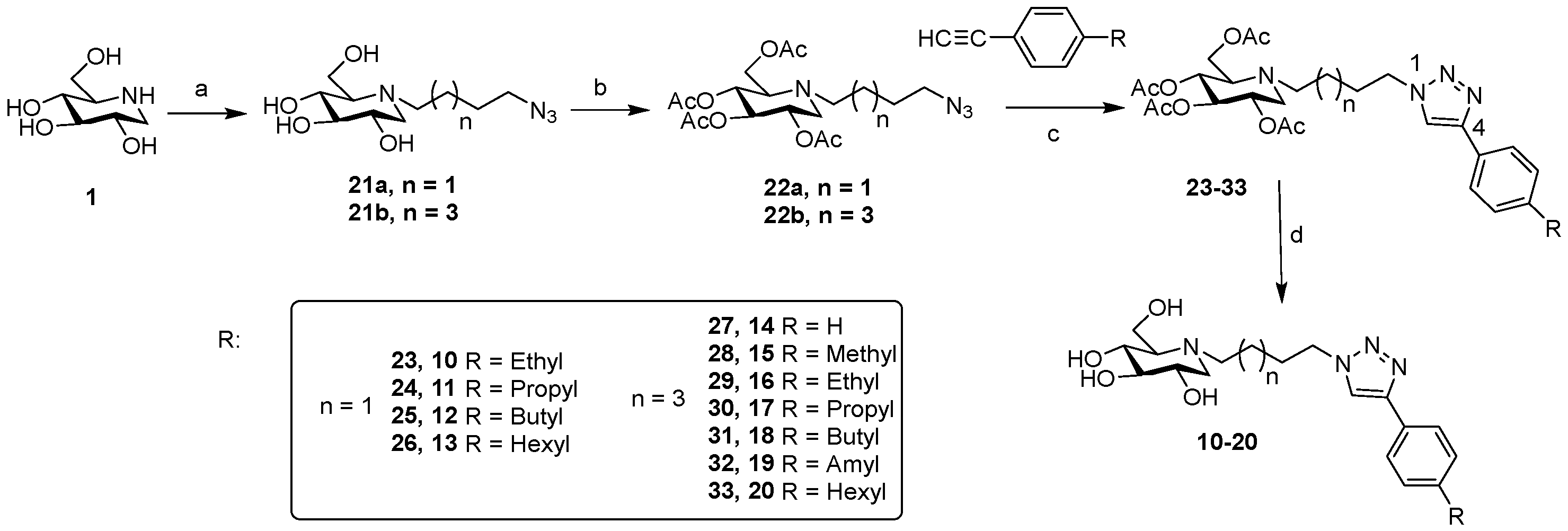
We synthesized the targeted molecules 10–20 (Scheme 1) using a procedure that we implemented for the synthesis of 7, 8 and 9. N-alkylation of unprotected DNJ 1 through the nucleophilic displacement of 1-azido-4-iodobutane or 1-azido-6-iodohexyl in DMF in the presence of K2CO3 at 80 °C afforded 21, which were followed by peracetylation to provide the click precursors 22 in a mixture of pyridine and acetic anhydride at room temperature.
Compounds 23–33 were obtained between 22 and substituted phenylacetylenes through the copper-catalyzed azide-alkyne cycloaddition (CuAAC). 23–33 were obtained in good (81%) to excellent (92%) yields independently of the chain length of the coupling partners 22a and 22b. The structures of 23–33 were confirmed by 1H and 13C NMR spectroscopy, which showed the presence of a single 1,4-disubstituted regioisomer. Hydrogen chemical shifts of the triazole hydrogen (δH5) was ∼7.76 ppm. The large and positive Δ (δC4–δC5) values (∼28 ppm) were observed by 13C NMR spectroscopy in agreement with earlier findings [25,26,27]. Finally, O-deacetylation with NaOMe in methanol at room temperature afforded the deprotected 10–20 in near quantitative yield. All compounds (except 15) were soluble in water. Their structures were confirmed by 1H NMR spectroscopy, which displayed the absence of singlets for the COCH3 groups, which were further substantiated by HR-MS analysis ([M + H]+ for 10–20).

2.2. Glycosidase Inhibitory Activity

Compounds 10–20 (except 15 for solubility reasons) were evaluated by in vitro α-glucosidase (yeast), β-glucosidase (almonds) inhibition studies. The corresponding IC50 values of compounds 10–20 were summarized in Table 1. Inhibition data for 7–9 and DNJ had also been included for comparative purposes.
It was first observed for a remarkable correlation between the length of the linker (C4 and C6) and the inhibition values, with a trend correlating higher inhibition with increased linker. In particular, compounds exhibited higher and more selective inhibitory effects on α-glucosidase. With the exception of analogues 7, 8, 10 and 11, all compounds showed increased inhibitory activities against α-glucosidase compared to the parent DNJ. The inhibition profile was notably different with respect to β-glucosidase. Only compounds equipped with a C6 linker displayed more potent inhibition of β-glucosidase than DNJ. These results were in accordance with previous reports [28,29]. The affinity of N-alkyl-1-deoxynojirimycin derivatives for α- and β-glucosidase may be explained in terms of the enzyme active site having a lipophilic pocket [30,31].
On the other hand, the straight linear length on the 4-position of the phenyl also had a great effect on inhibition against α- and β-glucosidase. Amongst 7–13, the simple extension of the alkyl chain by only one CH2 (from butyl to amyl) was sufficient to promote a 1.7-fold increase in α-glucosidase inhibitory activity. It was, however, interesting to note that increasing the alkyl chain length had little effect on β-glucosidase activity. For compounds 14–20, no dramatic effect on α- and β-glucosidase was seen on a further increase in chain length from propyl to hexyl or butyl to hexyl, respectively. These results indicated that straight linear length on the 4-position of the phenyl only partially determined the accessibility of the compounds to the active site of the enzymes [29]. It was noteworthy to mention that 17–20 displayed more potent inhibition of α-glucosidase than β-glucosidase.

2.3. Inhibition Kinetics of α-Glucosidase

In order to gain further insight into how these hybrids interact with α-glucosidase, the mode of inhibition for compounds 18 and 19 as representative examples was analyzed using the Lineweaver–Burk plot (Figure 2). The double reciprocal plots of 18 and 19 showed straight lines with the same Vmax, suggesting that both were competitive α-glucosidase inhibitors. The inhibition constants (Ki) were 3.8 µM for compound 18 and 3.7 µM for compound 19.

2.4. Molecular Docking Studies

Molecular docking studies were performed to gain further insights into the binding mode of these DNJ analogues. Compound 18, as a representative example, was chosen for the docking simulations. The parent compound DNJ was also studied for comparison. Since the X-ray crystal structure of a-glucosidase from Saccharomyces cerevisiae (MAL12, P53341) is currently unavailable, the homology modeling provided by SWISS-MODEL Repository, with the model quality estimation performed, was used as the receptor. From the docking studies, the binding affinity of compound 18 was predicted to be −8.6 kcal/mol, greater than the parent DNJ (−5.8 Kcal/mol). This indicated that compound 18 had stronger binding with α-glucosidase, which was consistent with the results observed in the enzyme inhibitory essays (Table 2).
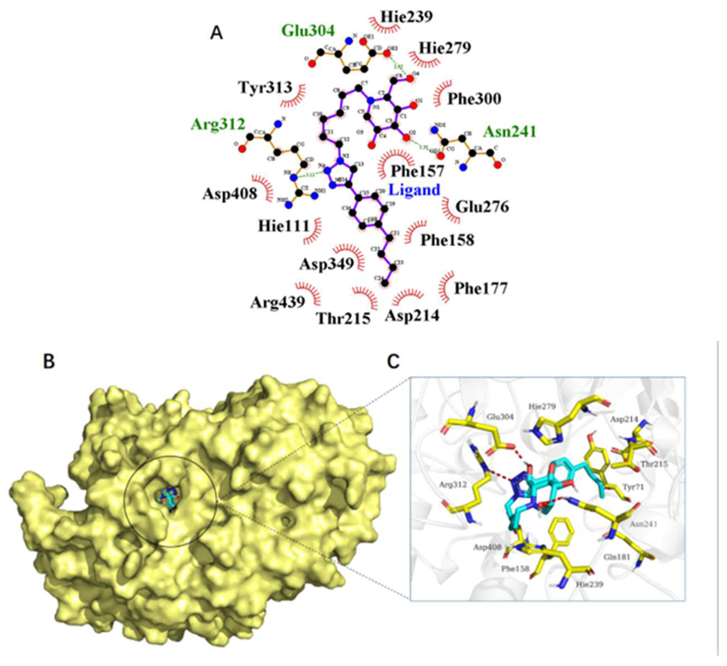
Molecular docking analysis indicated that several types of interactions were formed between compound 18, DNJ and enzyme upon binding, as seen in Figure 3 and Figure 4. DNJ was positioned to form hydrogen bonds with Hie348, Arg439, Asp68, Hie111 and Gln181. The DNJ moiety of compound 18 could form hydrogen bonds with Asn241 and Glu304. An additional hydrogen bond was established between the triazole moiety and Arg312. However, it was suggested that the alkyl chain containing a C6 spacer had additional hydrophobic and van der Waals interactions with hydrophilic residues (Tyr313, Phe300, et al.). Moreover, both interactions also exist between the butyl group on the 4-position of the phenyl hydrophilic residues (Phe158, Phe177, Thr215, et al.). Thus, the strengthening of the hydrophobic effect and van der Waals interactions seemed to compensate for the weakening of the hydrogen bond interactions, which could be an explanation for why compound 18 bonds to α-glucosidase more tightly than DNJ and resulted in stronger inhibitory activity. This was also consistent with the observations from the enzyme kinetic assay that compound 18 was a competitive inhibitor of α-glucosidase.

2.5. Cell Toxicity of Deoxynojirimycin-Triazole Hybrid Iminosugars

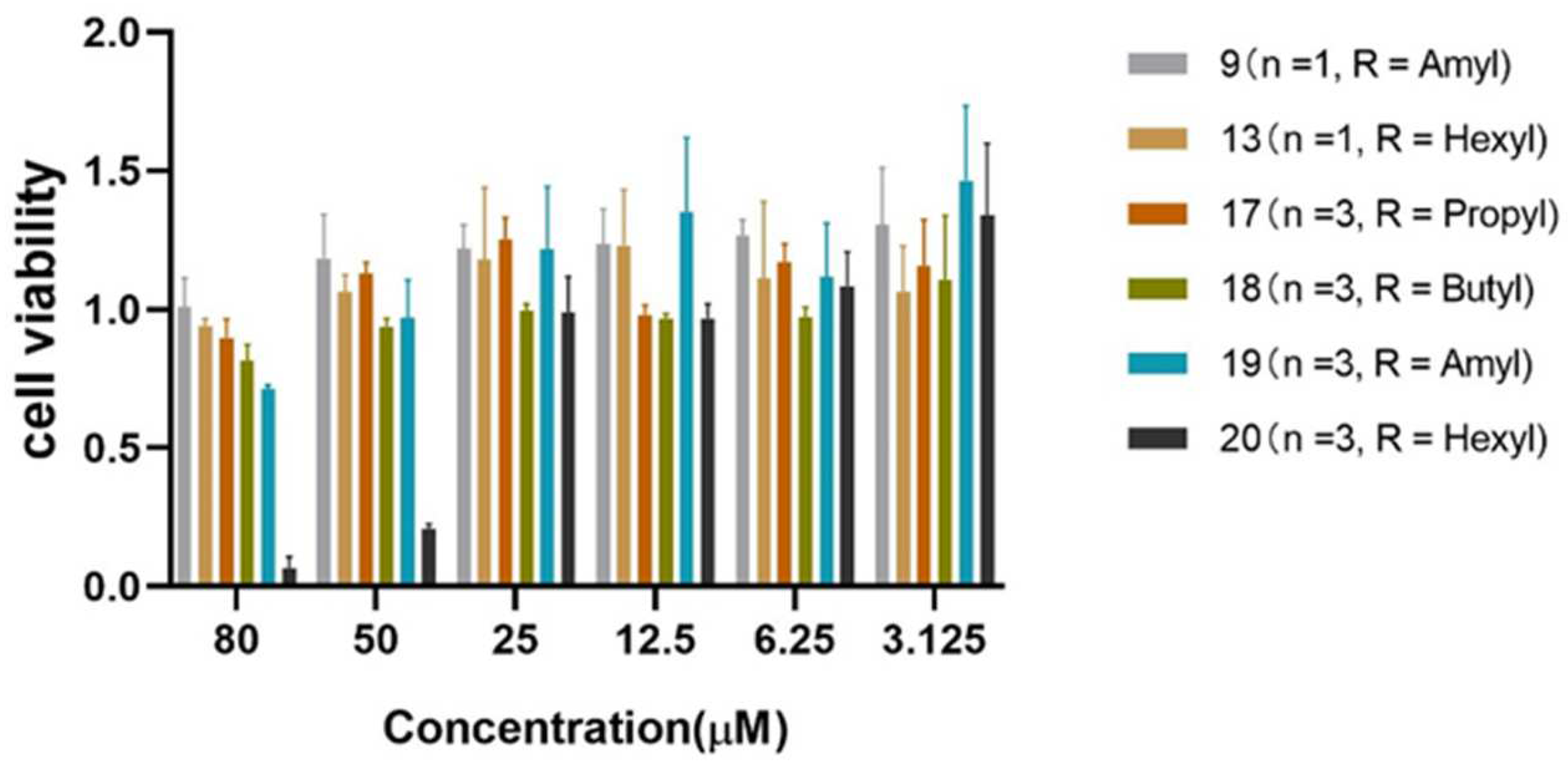
Compounds 9, 13 and 17–20 with better inhibitory activity on α-glucosidase were selected to study the effect on cell viability and proliferation by CCK-8 assay (Figure 5). The cell survival rate gradually decreased with the increase in compound concentration. When viability was measured at 50 µM for these compounds, the viability of compound 19 was 96.97% compared with 20.89% for compound 20. When the concentration was 80 µM, the cell survival rate was still above 80% except for compounds 19 (71.28%) and 20 (6.66%). With the increase in compound concentration, a lot of dead cells and more cell debris were observed, and the culture medium became muddy for 20, which was not obviously observed for 9, 13 and 17–19.

3. Experimental Section

3.1. General Information

Two glycosidases, α-glucosidase (yeast) and β-glucosidase (almonds), and their corresponding substrates, p-nitrophenyl glycopyranosides, were purchased from Sigma Chemical Co., St. Louis, MO, USA. Other commercial reagents and solvents were used as received. Reactions were monitored by Thin Layer Chromatography (TLC) analyses using silica gel GF254 plates bearing a 0.25 mm layer. Visualization of compounds was detected by short-wave UV fluorescence (λ = 254 nm) and/or by dipping into 5% phosphomolybdic acid in EtOH. Column chromatography was carried out by silica gel (200–300 mesh). NMR spectra (1H and 13C) were recorded with Bruker AV III 500 MHz spectrometer using CDCl3 (22–33), D2O (10–13, 16–20) or CD3OD (14, 15) as solvents. High-resolution mass spectra (HRMS) were obtained by direct injection on a mass spectrometer (Thermo Scientific LTQ Orbitrap XL, Waltham, MA, USA) equipped with an electrospray ion source in positive mode (see Supplementary Materials).

3.2. General Procedure A—Peracetylation with 21a or 21b

To a solution of DNJ (1.0 eq.), 1-azido-4-iodobutane (1.5 eq.) or 1-azido-6-iodohexyl (1.5 eq.) in DMF was added potassium carbonate (2.0 eq.). The reaction mixture was heated to 80 °C for 4 h. Most of the DMF was evaporated under reduced pressure. The crude product was dissolved in pyridine-acetic anhydride (1:1) and stirred for 12 h. The mixture was poured into saturated aqueous NaHCO3, and the aqueous phase was extracted with EtOAc. The combined organic layers were dried (Na2SO4), filtered and concentrated. The residue was purified by silica gel column chromatography (EtOAc/petroleum ether 1:6 to 1:2).
(2R,3R,4R,5S)-2-(acetoxymethyl)-1-(4-azidobutyl)piperidine-3,4,5-triyl triacetate 22a
Prepared according to procedure A. DNJ (1.0 g, 6.1 mmol), 1-azido-4-iodobutane (2.1 g, 9.3 mmol), potassium carbonate (2.5 g, 18 mmol), pyridine-acetic anhydride (1:1, 30 mL). Yield: 2.3 g, 5.4 mmol, 86%, a pale-yellow oil. 1H NMR (500 MHz, CDCl3) δ 5.15–5.02 (m, 2H, H-6), 4.96 (td, J = 9.9, 5.2 Hz, 1H, H-2), 4.17 (qd, J = 12.9, 2.7 Hz, 2H, H-3, H-4), 3.31 (m, 2H, H-10), 3.21 (dd, J = 11.5, 5.1 Hz, 1H, H-1a), 2.88–2.73 (m, 1H, H-7a), 2.64 (dd, J = 6.3, 2.7 Hz, 1H, H-5), 2.56 (m, 1H, H-7b), 2.36–2.25 (m, 1H, H-1b), 2.12–1.92 (4s, 12H, COCH3), 1.56 (m, 4H, H-8, H-9); 13C NMR (125 MHz, CDCl3) δ 170.82, 170.32, 170.04, 169.74 (4 × C=O), 74.57 (C-3), 69.43 (C-4), 69.29 (C-2), 61.74 (C-6), 59.46 (C-5), 52.68 (C-1), 51.14 (C-7), 50.94 (C-10), 26.44 (C-9), 22.45 (C-8), 20.84, 20.79, 20.72, 20.66 (4 × COCH3).
(2R,3R,4R,5S)-2-(acetoxymethyl)-1-(6-azidohexyl)piperidine-3,4,5-triyl triacetate 22b
Prepared according to procedure A. DNJ (1 g, 6.1 mmol), 1-azido-6-iodohexyl (2.2 g, 9.3 mmol), potassium carbonate (2.5 g, 18 mmol), pyridineyridine-acetic anhydride (1:1, 30 mL). Yield: 2.3 g, 5.1 mmol, 83%, a pale-yellow oil. 1H NMR (500 MHz, CDCl3) δ 5.06 (dt, J = 18.5, 9.3 Hz, 2H, H-6), 4.98–4.89 (m, 1H, H-2), 4.25–4.08 (m, 2H, H-3, H-4), 3.27 (t, J = 6.9 Hz, 2H, H-12), 3.20 (dd, J = 11.4, 5.1 Hz, 1H, H-1a), 2.75 (ddd, J = 13.8, 9.8, 6.1 Hz, 1H, H-7a), 2.64 (d, J = 9.0 Hz, 1H, H-5), 2.60–2.50 (m, 1H, H-7b), 2.32 (t, J = 10.9 Hz, 1H, H-1b), 2.06 (4s, 12H, 4 × COCH3), 1.65–1.53 (m, 2H, H-11), 1.53–1.22 (m, 6H, H-8, H-9. H-10). 13C NMR (125 MHz, CDCl3) δ 170.95, 170.41, 170.09, 169.79 (4 × C=O), 74.68 (C-3), 69.51 (C-4), 69.41 (C-2), 61.57 (C-6), 59.52 (C-5), 52.89 (C-1), 51.60 (C-7), 51.35 (C-12), 28.81 (C-11), 26.75 (C-8), 26.60 (C-9), 24.77 (C-10), 20.89, 20.85, 20.77, 20.71 (4 × COCH3).

3.3. General Procedure B—CuAAC Reaction with 22a or 22b

To a solution of 22a or 22b (1.0 eq.) and substituted phenylacetylenes (2 eq.) in DMF/H2O (2:1) was added Copper (II) sulfate (0.3 eq.) and sodium ascorbate (0.6 eq.). The mixture was stirred at room temperature for 6 h. Saturated NaHCO3 solution and EtOAc were added. The organic layer was dried over Na2SO4 and concentrated. The residue was purified by flash column chromatography using EtOAc:PE (2:1 → 1:4).
(2R,3R,4R,5S)-2-(acetoxymethyl)-1-(4-(4-(4-ethylphenyl)-1H-1,2,3-triazol-1-yl)butyl)piperidine-3,4,5-triyl triacetate 23
Prepared according to procedure B. Compound 22a (200 mg, 0.47 mmol), 1-ethyl-4-ethynylbenzene (122 mg, 0.94 mmol), CuSO4·5H2O (34.96 mg, 0.14 mmol), sodium ascorbate (55.47 mg, 0.28 mmol), DMF/H2O (3 mL, 2:1). Yield: 210 mg, 0.38 mmol, 81%, white solid, Rf = 0.49 (1:2; PE:EtOAc). 1H NMR (500 MHz, CDCl3) δ 7.86–7.65 (m, 3H, ArH, CH- triazole), 7.39–7.10 (m, 2H, ArH), 5.14–5.00 (m, 2H, H-6), 4.94 (td, J = 9.9, 5.1 Hz, 1H, H-2), 4.42 (dq, J = 13.9, 6.8 Hz, 2H, H-10), 4.16 (qd, J = 12.9, 2.7 Hz, 2H, H-3, H-4), 3.17 (dd, J = 11.5, 5.0 Hz, 1H, H-1a), 2.84 (dt, J = 13.5, 7.9 Hz, 1H, H-7a), 2.68 (q, J = 7.6 Hz, 2H, H-13), 2.61 (dd, J = 6.5, 2.6 Hz, 1H, H-5), 2.53 (ddd, J = 13.2, 8.3, 4.8 Hz, 1H, H-7b), 2.23 (t, J = 10.9 Hz, 1H, H-1b), 2.09–1.97 (m, 12H, 4 × COCH3), 1.97–1.85 (m, 2H, H-9), 1.60–1.43 (m, 2H, H-8), 1.26 (t, 3H, H-14). 13C NMR (125 MHz, CDCl3) δ 170.83, 170.32, 170.13, 169.79 (4 × C=O), 147.99 (C-12), 144.40 (Car), 128.36 (2 × CHar), 128.04 (Car), 125.73 (2 × CHar), 119.15 (C-11), 74.52 (C-3), 69.42 (C-4), 69.25 (C-2), 62.00 (C-6), 59.39 (C-5), 52.52 (C-1), 50.58 (C-7), 49.95 (C-10), 28.69 (C-13), 27.82 (C-9), 22.50 (C-8), 20.87, 20.84, 20.76, 20.70 (4 × COCH3), 15.56 (C-14).
(2R,3R,4R,5S)-2-(acetoxymethyl)-1-(4-(4-(4-propylphenyl)-1H-1,2,3-triazol-1-yl)butyl)piperidine-3,4,5-triyl triacetate 24
Prepared according to procedure B. Compound 22a (200 mg, 0.47 mmol), 1-eth-1-ynyl-4-propylbenzene (135 mg, 0.94 mmol), CuSO4·5H2O (34.96 mg, 0.14 mmol), sodium ascorbate (55.47 mg, 0.28 mmol), DMF/H2O (3 mL, 2:1). Yield: 220 mg, 0.39 mmol, 82%, white solid, Rf = 0.51 (1:2; PE:EtOAc). 1H NMR (500 MHz, CDCl3) δ 7.89–7.68 (m, 3H, ArH, CH- triazole), 7.25 (dd, J = 8.1, 4.0 Hz, 2H, ArH), 5.16–5.02 (m, 2H, H-6), 4.96 (dd, J = 9.2, 4.8 Hz, 1H, H-2), 4.43 (td, J = 11.7, 7.0 Hz, 2H, H-10), 4.26–4.10 (m, 2H, H-3, H-4), 3.18 (dt, J = 11.2, 4.5 Hz, 1H, H-1a), 2.90–2.80 (m, 1H, H-7a), 2.68–2.58 (m, 3H, H-5, H-13), 2.59–2.48 (m, 1H, H-7b), 2.25 (td, J = 11.4, 3.3 Hz, 1H, H-1b), 2.11–1.86 (m, 14H, 4 × COCH3, H-9), 1.68 (ddd, J = 15.1, 7.5, 4.1 Hz, 2H, H-14), 1.58–1.43 (m, 2H, H-8), 0.97 (td, J = 7.3, 4.0 Hz, 3H, H-15). 13C NMR (125 MHz, CDCl3) δ 170.83, 170.32, 170.13, 169.80 (4 × C=O), 147.97 (C-12), 142.83 (Car), 128.96 (2 × CHar), 128.01 (Car), 125.61 (2 × CHar), 119.19 (C-11), 74.51 (C-3), 69.41 (C-4), 69.25 (C-2), 61.95 (C-6), 59.37 (C-5), 52.49 (C-1), 50.57 (C-7), 49.93 (C-10), 37.81 (C-13), 27.80 (C-9), 24.50 (C-14), 22.44 (C-8), 20.86, 20.82, 20.74, 20.69 (4 × COCH3), 13.81 (C-15).
(2R,3R,4R,5S)-2-(acetoxymethyl)-1-(4-(4-(4-butylphenyl)-1H-1,2,3-triazol-1-yl)butyl)piperidine-3,4,5-triyl triacetate 25
Prepared according to procedure B. Compound 22a (200 mg, 0.47 mmol), 1-butyl-4-eth-1-ynylbenzene (148 mg, 0.94 mmol), CuSO4·5H2O (34.96 mg, 0.14 mmol), sodium ascorbate (55.47 mg, 0.28 mmol), DMF/H2O (3 mL, 2:1). Yield: 220 mg, 0.38 mmol, 80%, white solid, Rf = 0.53 (1:2; PE:EtOAc). 1H NMR (500 MHz, CDCl3) δ 7.85–7.67 (m, 3H, ArH, CH- triazole), 7.23 (d, J = 8.0 Hz, 2, ArH), 5.04 (dt, J = 15.5, 6.1 Hz, 2H, H-6), 4.94 (d, J = 4.4 Hz, 1H, H-2), 4.53–4.35 (m, 2H, H-10), 4.16 (qd, J = 12.9, 2.4 Hz, 2H, H-3, H-4), 3.16 (dd, J = 11.3, 4.7 Hz, 1H, H-1a), 2.92–2.77 (m, 1H, H-7a), 2.62 (m, 3H, H-5, H-13), 2.57–2.46 (m, 1H, H-7b), 2.23 (t, J = 10.9 Hz, 1H, H-1b), 2.09–1.82 (m, 14H, 4 × COCH3, H-9), 1.71–1.57 (m, 2H, H-14), 1.54–1.41 (m, 2H, H-8), 1.41–1.31 (m, 2H, H-15), 0.93 (t, J = 7.3 Hz, 3H, H-16). 13C NMR (125 MHz, CDCl3) δ 170.80, 170.29, 170.10, 169.77 (4 × C=O), 147.94 (C-12), 143.04 (Car), 128.89 (2 × CHar), 127.96 (Car), 125.61 (2 × CHar), 119.20 (C-11), 74.50 (C-3), 69.41 (C-4), 69.24 (C-2), 61.95 (C-6), 59.37 (C-5), 52.48 (C-1), 50.57 (C-7), 49.92 (C-10), 35.42 (C-13), 33.55 (C-14), 27.78 (C-9), 22.43 (C-15), 22.31 (C-8), 20.83, 20.80, 20.72, 20.66 (4 × COCH3), 13.96 (C-16).
(2R,3R,4R,5S)-2-(acetoxymethyl)-1-(4-(4-(4-hexylphenyl)-1H-1,2,3-triazol-1-yl)butyl)piperidine-3,4,5-triyl triacetate 26
Prepared according to procedure B. Compound 22a (200 mg, 0.47 mmol), 1-ethynyl-4-hexylbenzene (174 mg, 0.94 mmol), CuSO4·5H2O (34.96 mg, 0.14 mmol), sodium ascorbate (55.47 mg, 0.28 mmol), DMF/H2O (3 mL, 2:1). Yield: 260 mg, 0.42 mmol, 90%, white solid, Rf = 0.55 (1:2; PE:EtOAc). 1H NMR (500 MHz, CDCl3) δ 7.82–7.69 (m, 3H, ArH, CH- triazole), 7.24 (d, J = 8.1 Hz, 2H, ArH), 5.11–5.00 (m, 2H, H-6), 4.94 (td, J = 9.9, 5.1 Hz, 1H, H-2), 4.54–4.33 (m, 2H, H-10), 4.16 (qd, J = 12.9, 2.7 Hz, 2H, H-3, H-4), 3.16 (dd, J = 11.5, 5.0 Hz, 1H, H-1a), 2.83 (m, 1H, H-7a), 2.71–2.56 (m, 3H, H-5, H-13), 2.52 (m, 1H, H-7b), 2.34–2.16 (m, 1H, H-1b), 2.07–1.87 (m, 14H, 4 × COCH3, H-9), 1.62 (dt, J = 15.3, 7.6 Hz, 2H, H-14), 1.49 (m, 2H, H-8), 1.42–1.27 (m,6H, H-15, H-16, H-17), 0.88 (t, J = 6.9 Hz, 3H, H-18). 13C NMR (125 MHz, CDCl3) δ 170.83, 170.32, 170.12, 169.79 (4 × C=O), 147.99 (C-12), 143.11 (Car), 128.90 (2 × CHar), 127.97 (Car), 125.63 (2 × CHar), 119.16 (C-11), 74.51 (C-3), 69.42 (C-4), 69.25 (C-2), 61.97 (C-6), 59.37 (C-5), 52.50 (C-1), 50.57 (C-7), 49.93 (C-10), 35.76 (C-13), 31.73 (C-14), 31.39 (C-15), 28.96 (C-16), 27.80 (C-9), 22.62 (C-17), 22.45 (C-8), 20.86, 20.83, 20.75, 20.69 (4 × COCH3), 14.12 (C-18).
(2R,3R,4R,5S)-2-(acetoxymethyl)-1-(6-(4-phenyl-1H-1,2,3-triazol-1-yl)hexyl)piperidine-3,4,5-triyl triacetate 27
Prepared according to procedure B. Compound 22b (200 mg, 0.44 mmol), phenylacetylene (89 mg, 0.88 mmol), CuSO4·5H2O (33 mg, 0.13 mmol), sodium ascorbate (52 mg, 0.26 mmol), DMF/H2O (3 mL, 2:1). Yield: 190 mg, 0.34 mmol, 78%, white solid, Rf = 0.17 (1:1; PE:EtOAc). 1H NMR (500 MHz, CDCl3) δ 7.86–7.81 (m, 2H, ArH), 7.80 (s, 1H, CH- triazole), 7.42 (t, J = 7.6 Hz, 2H, ArH), 7.32 (dd, J = 10.3, 4.3 Hz, 1H, ArH), 5.16–5.00 (m, 2H, H-6), 4.95 (td, J = 9.8, 5.1 Hz, 1H, H-2), 4.38 (t, J = 7.1 Hz, 2H, H-12), 4.14 (qd, J = 12.9, 2.5 Hz, 2H, H-3, H-4), 3.18 (dd, J = 11.4, 5.0 Hz, 1H, H-1a), 2.77–2.66 (m, 1H, H-7a), 2.61 (d, J = 8.8 Hz, 1H, H-5), 2.57–2.46 (m, 1H, H-7b), 2.28 (t, J = 10.9 Hz, 1H, H-1b), 2.09–1.89 (m, 14H, 4×COCH3, H-11), 1.53–1.31 (m, 6H, H-8, H-9, H-10). 13C NMR (125 MHz, CDCl3) δ 170.86, 170.33, 170.07, 169.77 (4 × C=O), 147.70 (C-14), 130.70 (Car), 128.83 (2 × CHar), 128.09 (Car), 125.67 (2 × CHar), 119.59 (C-13), 74.63 (C-3), 69.51 (C-4), 69.38 (C-2), 61.62 (C-6), 59.49 (C-5), 52.79 (C-1), 51.49 (C-7), 50.23 (C-12), 30.25 (C-11), 26.56 (C-8), 26.32 (C-9), 24.78 (C-10), 20.86, 20.83, 20.74, 20.68 (4 × COCH3).
(2R,3R,4R,5S)-2-(acetoxymethyl)-1-(6-(4-(p-tolyl)-1H-1,2,3-triazol-1-yl)hexyl)piperidine-3,4,5-triyl triacetate 28
Prepared according to procedure B. Compound 22b (200 mg, 0.44 mmol), 4-ethynyltoluene (102 mg, 0.88 mmol), CuSO4·5H2O (33 mg, 0.13 mmol), sodium ascorbate (52 mg, 0.26 mmol), DMF/H2O (3 mL, 2:1). Yield: 180 mg, 0.32 mmol, 72%, white solid, Rf = 0.21 (1:1; PE:EtOAc). 1H NMR (500 MHz, CDCl3) δ 7.86–7.66 (m, 3H, ArH, CH- triazole), 7.24 (d, J = 7.9 Hz, 2H, ArH), 5.17–5.00 (m, 2H, H-6), 5.00–4.91 (m, 1H, H-2), 4.39 (t, J = 7.1 Hz, 2H, H-12), 4.16 (qd, J = 12.9, 2.4 Hz, 2H, H-3, H-4), 3.20 (dd, J = 11.4, 5.0 Hz, 1H, H-1a), 2.81–2.69 (m, 1H, H-7a), 2.63 (d, J = 8.8 Hz, 1H, H-5), 2.55–2.48 (m, 1H, H-7b), 2.39 (s, 3H, H-15), 2.30 (t, J = 10.9 Hz, 1H, H-1b), 2.12–1.88 (m, 14H, 4 × COCH3, H-11), 1.55–1.32 (m, 6H, H-8, H-9, H-10). 13C NMR (125 MHz, CDCl3) δ 170.89, 170.36, 170.09, 169.78 (4 × C=O), 147.81 (C-14), 137.92 (Car), 129.52 (2 × CHar), 127.87 (Car), 125.60 (2 × CHar), 119.21 (C-13), 74.65 (C-3), 69.52 (C-4), 69.39 (C-2), 61.63 (C-6), 59.51 (C-5), 52.81 (C-1), 51.51 (C-7), 50.22 (C-12), 30.27 (C-11), 26.58 (C-8), 26.34 (C-9), 24.78 (C-10), 21.29 (C-15), 20.87, 20.84, 20.75, 20.69 (4 × COCH3).
(2R,3R,4R,5S)-2-(acetoxymethyl)-1-(6-(4-(4-ethylphenyl)-1H-1,2,3-triazol-1-yl)hexyl)piperidine-3,4,5-triyl triacetate 29
Prepared according to procedure B. Compound 22b (200 mg, 0.44 mmol), 1-ethyl-4-ethynylbenzene (114 mg, 0.88 mmol), CuSO4·5H2O (33 mg, 0.13 mmol), sodium ascorbate (52 mg, 0.26 mmol), DMF/H2O (3 mL, 2:1). Yield: 220 mg, 0.38 mmol, 86%, white solid, Rf =0.25 (1:1; PE:EtOAc). 1H NMR (500 MHz, CDCl3) δ 7.89–7.67 (m, 3H, ArH, CH- triazole), 7.24 (d, J = 7.4 Hz, 2H, ArH), 5.13–4.99 (m, 2H, H-6), 4.95 (dd, J = 9.2, 4.9 Hz, 1H, H-2), 4.36 (t, J = 6.7 Hz, 2H, H-12), 4.21–4.06 (m, 2H, H-3, H-4), 3.17 (dd, J = 11.3, 4.4 Hz, 1H, H-1a), 2.72–2.50 (m, 5H, H-5, H-7, H-15), 2.28 (t, J = 10.8 Hz, 1H, H-1b), 2.16–1.87 (m, 14H, 4×COCH3, H-11), 1.55–1.23 (m, 8H, H-8, H-9, H-10, H-16). 13C NMR (125 MHz, CDCl3) δ 170.79, 170.26, 170.00, 169.72 (4 × C=O), 147.69 (C-14), 144.21 (Car), 128.28 (2 × CHar), 128.11 (Car), 125.63 (2 × CHar), 119.32 (C-13), 74.61 (C-3), 69.48 (C-4), 69.34 (C-2), 61.57 (C-6), 59.45 (C-5), 52.75 (C-1), 51.47 (C-7), 50.14 (C-12), 30.20 (C-11), 28.62 (C-15), 26.52 (C-8), 26.27 (C-9), 24.71 (C-10), 20.81, 20.77, 20.69, 20.63 (4 × COCH3), 15.52 (C-16).
(2R,3R,4R,5S)-2-(acetoxymethyl)-1-(6-(4-(4-propylphenyl)-1H-1,2,3-triazol-1-yl)hexyl)piperidine-3,4,5-triyl triacetate 30
Prepared according to procedure B. Compound 22b (200 mg, 0.44 mmol), 1-eth-1-ynyl-4-propylbenzene (126.9 mg, 0.88 mmol), CuSO4·5H2O (33 mg, 0.13 mmol), sodium ascorbate (52 mg, 0.26 mmol), DMF/H2O (3 mL, 2:1). Yield: 230 mg, 0.38 mmol, 87%, white solid, Rf = 0.31 (1:1; PE:EtOAc). 1H NMR (500 MHz, CDCl3) δ 7.82 (s, 1H, CH- triazole), 7.75 (d, J = 7.9 Hz, 2H, ArH), 7.22 (d, J = 7.9 Hz, 2H, ArH), 5.16–4.99 (m, 2H, H-6), 4.96 (dd, J = 9.4, 4.9 Hz, 1H, H-2), 4.36 (t, J = 6.9 Hz, 2H, H-12), 4.16 (d, J = 12.6 Hz, 2H, H-3, H-4), 3.18 (dd, J = 11.3, 4.9 Hz, 1H, H-1a), 2.72 (dt, J = 14.4, 7.6 Hz, 1H, H-7a), 2.60 (t, J = 7.5 Hz, 3H, H-5, H-15), 2.56–2.45 (m, 1H, H-7b), 2.29 (t, J = 10.8 Hz, 1H, H-1b), 2.13–1.81 (m, 14H, 4×COCH3, H-11), 1.66 (dd, J = 14.9, 7.4 Hz, 2H, H-16), 1.51–1.21 (m, 6H, H-8, H-9, H-10), 0.95 (t, J = 7.3 Hz, 3H, H-17). 13C NMR (125 MHz, CDCl3) δ 170.67, 170.16, 169.91, 169.65 (4 × C=O), 147.59 (C-14), 142.54 (Car), 128.84 (2 × CHar), 128.13 (Car), 125.48 (2 × CHar), 119.38 (C-13), 74.56 (C-3), 69.44 (C-4), 69.30 (C-2), 61.51 (C-6), 59.40 (C-5), 52.70 (C-1), 51.43 (C-7), 50.06 (C-12), 37.69 (C-15), 30.14 (C-11), 26.46 (C-8), 26.20 (C-9), 24.64 (C-10), 24.40 (C-16), 20.74, 20.71, 20.62, 20.57 (4 × COCH3), 13.72 (C-17).
(2R,3R,4R,5S)-2-(acetoxymethyl)-1-(6-(4-(4-butylphenyl)-1H-1,2,3-triazol-1-yl)hexyl)piperidine-3,4,5-triyl triacetate 31
Prepared according to procedure B. Compound 22b (200 mg, 0.44 mmol), 1-butyl-4-eth-1-ynylbenzene (138 mg, 0.88 mmol), CuSO4·5H2O (33 mg, 0.13 mmol), sodium ascorbate (52 mg, 0.26 mmol), DMF/H2O (3 mL, 2:1). Yield: 230 mg, 0.37 mmol, 85%, white solid, Rf =0.35 (1:1; PE:EtOAc). 1H NMR (500 MHz, CDCl3) δ 7.82–7.69 (m, 3H, ArH, CH- triazole), 7.22 (d, J = 6.3 Hz, 2H, ArH), 5.03 (dd, J = 15.8, 7.9 Hz, 2H, H-6), 4.95 (m, 1H, H-2), 4.37 (d, J = 5.0 Hz, 2H, H-12), 4.14 (d, J = 3.6 Hz, 2H, H-3, H-4), 3.17 (d, J = 6.7 Hz, 1H, H-1a), 2.72 (m, 1H, H-7a), 2.62 (m, 3H, H-5, H-15), 2.52 (m, 1H, H-7b), 2.28 (t, J = 10.7 Hz, 1H, H-1b), 2.14–1.87 (m, 14H, 4 × COCH3, H-11), 1.68–1.55 (m, 2H, H-16), 1.50–1.20 (m, 8H, H-8, H-9, H-10, H-17), 0.92 (t, J = 5.9 Hz, 3H, H-18). 13C NMR (125 MHz, CDCl3) δ 170.82, 170.30, 170.04, 169.74 (4 × C=O), 147.77 (C-14), 142.94 (Car), 128.86 (2 × CHar), 128.06 (Car), 125.57(2 × CHar), 119.27 (C-13), 74.63 (C-3), 69.50 (C-4), 69.36 (C-2), 61.60 (C-6), 59.47 (C-5), 52.78 (C-1), 51.49 (C-7), 50.17 (C-12), 35.40 (C-15), 33.54 (C-16), 30.23 (C-11), 26.55 (C-8), 26.30 (C-9), 24.74 (C-10), 22.30 (C-17), 20.84, 20.80, 20.72, 20.66 (4 × COCH3), 13.95 (C-18).
(2R,3R,4R,5S)-2-(acetoxymethyl)-1-(6-(4-(4-pentylphenyl)-1H-1,2,3-triazol-1-yl)hexyl)piperidine-3,4,5-triyl triacetate 32
Prepared according to procedure B. Compound 22b (200 mg, 0.44 mmol), 1-ethynyl-4-pentylbenzene (151 mg, 0.88 mmol), CuSO4·5H2O (33 mg, 0.13 mmol), sodium ascorbate (52 mg, 0.26 mmol), DMF/H2O (3 mL, 2:1). Yield: 250 mg, 0.4 mmol, 91%, white solid, Rf = 0.38 (1:1; PE:EtOAc). 1H NMR (500 MHz, CDCl3) δ 7.74 (m, 3H, ArH, CH-triazole), 7.23 (d, J = 8.0 Hz, 2H, ArH), 5.13–5.00 (m, 2H, H-6), 4.95 (dd, J = 14.0, 9.4 Hz, 1H, H-2), 4.38 (t, J = 6.9 Hz, 2H, H-12), 4.15 (qd, J = 13.0, 2.5 Hz, 2H, H-3, H-4), 3.18 (dd, J = 11.3, 4.9 Hz, 1H, H-1a), 2.72 (dd, J = 11.8, 7.8 Hz, 1H, H-7a), 2.62 (m, 3H, H-5, H-15), 2.51 (dd, J = 15.9, 6.9 Hz, 1H, H-7b), 2.28 (t, J = 10.8 Hz, 1H, H-1b), 2.12–1.85 (m, 14H, 4 × COCH3, H-11), 1.70–1.57 (m, 2H, H-16), 1.52–1.19 (m, 10H, H-8, H-9, H-10, H-17, H-18), 0.89 (d, J = 7.0 Hz, 3H, H-19). 13C NMR (125 MHz, CDCl3) δ 170.87, 170.34, 170.07, 169.77 (4 × C=O), 147.83 (C-14), 143.01 (Car), 128.87 (2 × CHar), 128.07 (Car), 125.60 (2 × CHar), 119.22 (C-13), 74.65 (C-3), 69.51 (C-4), 69.38 (C-2), 61.62 (C-6), 59.49 (C-5), 52.81 (C-1), 51.51 (C-7), 50.19 (C-12), 35.70 (C-15), 31.46 (C-16), 31.09 (C-17), 30.26 (C-11), 26.57 (C-8), 26.33 (C-9), 24.77 (C-10), 22.54 (C-18), 20.86, 20.83, 20.74, 20.68 (4 × COCH3), 14.05 (C-19).
(2R,3R,4R,5S)-2-(acetoxymethyl)-1-(6-(4-(4-hexylphenyl)-1H-1,2,3-triazol-1-yl)hexyl)piperidine-3,4,5-triyl triacetate 33
Prepared according to procedure B. Compound 22b (200 mg, 0.44 mmol), 1-ethynyl-4-hexylbenzene (163 mg, 0.88 mmol), CuSO4·5H2O (33 mg, 0.13 mmol), sodium ascorbate (52 mg, 0.26 mmol), DMF/H2O (3 mL, 2:1). Yield: 220 mg, 0.34 mmol, 78%, white solid, Rf =0.41 (1:1; PE:EtOAc). 1H NMR (500 MHz, CDCl3) δ 7.84–7.69 (m, 3H, ArH, CH-triazole), 7.31–7.17 (m, 2H, ArH), 5.15–5.01 (m, 2H, H-6), 4.97 (dd, J = 9.4, 4.6 Hz, 1H, H-2), 4.38 (q, J = 6.7 Hz, 2H, H-12), 4.24–4.08 (m, 2H, H-3, H-4), 3.26–3.10 (m, 1H, H-1a), 2.74 (m, 1H, H-7a), 2.63 (t, J = 10.0 Hz, 3H, H-5, H-15), 2.53 (td, J = 9.1, 4.6 Hz, 1H, H-7b), 2.30 (td, J = 11.2, 4.9 Hz, 1H, H-1b), 2.13–1.85 (m, 14H, 4×COCH3, H-11), 1.71–1.53 (m, 2H, H-16), 1.50–1.22 (m, 12H, H-8, H-9, H-10, H-17, H-18, H-19), 0.98–0.74 (m, 3H, H-20). 13C NMR (125 MHz, CDCl3) δ 170.80, 170.27, 170.01, 169.73 (4 × C=O), 147.76 (C-14), 142.94 (Car), 128.84 (2 × CHar), 128.08 (Car), 125.56 (2 × CHar), 119.25 (C-13), 74.63 (C-3), 69.50 (C-4), 69.36 (C-2), 61.60 (C-6), 59.47 (C-5), 52.78 (C-1), 51.49 (C-7), 50.15 (C-12), 35.71 (C-15), 31.69 (C-16), 31.35 (C-17), 30.23 (C-11), 28.91 (C-18), 26.54 (C-8), 26.29 (C-9), 24.75 (C-10), 22.58 (C-19), 20.82, 20.80, 20.70, 20.65 (4 × COCH3), 14.09 (C-20).

3.4. General Procedure C—Deacetylation Reaction of Compounds 2333

Compounds 23–33 were dissolved in methanol, and NaOMe (1.0 M in methanol) was added until pH 9.0 was achieved. After stirring overnight at room temperature, the mixture was neutralized with ion exchange resin DOWEX® 50WX4-50 (H+), filtered and concentrated under reduced pressure. The resulting product (compounds 10–20) was obtained in quantitative yield.
(2R,3R,4R,5S)-1-(4-(4-(4-ethylphenyl)-1H-1,2,3-triazol-1-yl)butyl)-2-(hydroxymethyl)piperidine-3,4,5-triol 10
Prepared according to procedure C. Compound 23 (190 mg, 0.34 mmol). Yield: 130 mg, 0.33 mmol, 96%, white solid, Rf = 0.22 (3:1; CH2Cl2: MeOH + 1% NH4OH), [α]D25 22.6 (c 0.5, H2O). 1H NMR (500 MHz, D2O) δ 7.78 (s, 1H, CH- triazole), 7.42 (d, J = 7.4 Hz, 2H, ArH), 6.73 (d, J = 7.8 Hz, 2H, ArH), 4.09 (m, 2H, H-10), 3.69 (dd, J = 20.9, 12.1 Hz, 2H, H-6), 3.49 (d, J = 4.6 Hz, 1H, H-2), 3.33 (t, J = 9.4 Hz, 1H, H-3), 3.16 (t, J = 9.1 Hz, 1H, H-4), 2.86 (d, J = 6.2 Hz, 1H, H-1a), 2.58 (m, 1H, H-7a), 2.37 (m, 1H, H-7b), 2.17–1.88 (m, 4H, H-5, H-1b, H-13), 1.52 (m, 2H, H-9), 1.26 (m, 2H. H-8), 0.77 (t, J = 7.4 Hz, 3H, H-14). 13C NMR (125 MHz, D2O) δ 147.02 (C-12), 143.61 (Car), 128.09 (2 × CHar), 127.62 (Car), 125.40 (2 × CHar), 120.74 (C-13), 78.35 (C-3), 69.80 (C-4), 68.75 (C-2), 65.25 (C-6), 57.26 (C-5), 55.46 (C-1), 51.30 (C-7), 49.93 (C-10), 28.15 (C-13), 27.88 (C-9), 20.35 (C-8), 14.95 (C-14). HRMS (ESI) m/z calcd for C20H31N4O4+ [M+H]+ 391.23398, found 391.23346.
(2R,3R,4R,5S)-2-(hydroxymethyl)-1-(4-(4-(4-propylphenyl)-1H-1,2,3-triazol-1-yl)butyl)piperidine-3,4,5-triol 11
Prepared according to procedure C. Compound 24 (150 mg, 0.26 mmol). Yield:100 mg, 0.25 mmol, 95%, white solid, Rf = 0.25 (3:1; CH2Cl2: MeOH + 1% NH4OH), [α]D25 −10.7 (c 0.35, H2O). 1H NMR (500 MHz, D2O) δ 7.84 (s, 1H, CH-triazole), 7.45 (d, J = 6.6 Hz, 2H, ArH), 6.72 (d, J = 7.1 Hz, 2H, ArH), 4.11 (m, 2H, H-10), 3.70 (dd, J = 21.6, 12.4 Hz, 2H, H-6), 3.50 (dd, J = 13.8, 9.3 Hz, 1H, H-2), 3.34 (t, J = 9.3 Hz, 1H, H-3), 3.17 (t, J = 9.0 Hz, 1H, H-4), 2.87 (d, J = 5.5 Hz, 1H, H-1a), 2.59 (m, 1H, H-7a), 2.38 (m, 1H, H-7b), 2.16–1.93 (m, 4H, H-5, H-1b, H-13), 1.53 (m, 2H, H-9), 1.38–1.07 (m, 4H, H-8, H-14), 0.59 (t, J = 6.6 Hz, 3H, H-15). 13C NMR (125 MHz, D2O) δ 147.01 (C-12), 141.95 (Car), 128.63 (2 × CHar), 127.81(Car), 125.35(2 × CHar), 120.74 (C-13), 78.34 (C-3), 69.76 (C-4), 68.71 (C-2), 65.34 (C-6), 57.22 (C-5), 55.47 (C-1), 51.35 (C-7), 49.96 (C-10), 37.45 (C-13), 27.91 (C-9), 24.06 (C-14), 20.47 (C-8), 13.60 (C-15). HRMS (ESI) m/z calcd for C21H33N4O4+ [M+H]+ 405.24963, found 405.24997.
(2R,3R,4R,5S)-1-(4-(4-(4-butylphenyl)-1H-1,2,3-triazol-1-yl)butyl)-2-(hydroxymethyl)piperidine-3,4,5-triol 12
Prepared according to procedure C. Compound 25 (200 mg, 0.34 mmol). Yield: 130 mg, 0.31 mmol, 93%, white solid, Rf = 0.31 (3:1; CH2Cl2: MeOH + 1% NH4OH), [α]D25 −12.1 (c 0.39, H2O). 1H NMR (500 MHz, D2O) δ 7.83 (s, 1H, CH-triazole), 7.46 (s, 2H, ArH), 6.72 (s, 2H, ArH), 4.10 (m, 2H, H-10), 3.68 (d, J = 19.6 Hz, 2H, H-6), 3.49 (m, 1H, H-2), 3.33 (t, J = 8.8 Hz, 1H, H-3), 3.15 (t, J = 8.4 Hz, 1H, H-4), 2.86 (m, 1H, H-1a), 2.58 (m, 1H, H-7a), 2.36 (m, 1H, H-7b), 2.18–1.94 (m, 4H, H-5, H-1b, H-13), 1.51 (m, 2H, H-9), 1.39–0.91 (m, 6H, H-8, H-14, H-15), 0.65 (m, 3H, H-16). 13C NMR (125 MHz, D2O) δ 146.96 (C-12), 142.05 (Car), 128.52 (2 × CHar), 127.82(Car), 125.35(2 × CHar), 120.74 (C-13), 78.35 (C-3), 69.74 (C-4), 68.70 (C-2), 65.34 (C-6), 57.20 (C-5), 55.48 (C-1), 51.35 (C-7), 49.96 (C-10), 35.15 (C-13), 33.19 (C-14), 27.94 (C-9), 22.37 (C-15), 20.50 (C-8), 13.72 (C-16). HRMS (ESI) m/z calcd for C22H35N4O4+ [M+H]+ 419.26528, found 419.26492.
(2R,3R,4R,5S)-1-(4-(4-(4-hexylphenyl)-1H-1,2,3-triazol-1-yl)butyl)-2-(hydroxymethyl)piperidine-3,4,5-triol 13
Prepared according to procedure C. Compound 26 (200 mg, 0.33 mmol). Yield: 140 mg, 0.31 mmol, 95%, white solid, Rf = 0.34 (3:1; CH2Cl2: MeOH + 1% NH4OH), [α]D25 7 (c 0.2, H2O). 1H NMR (500 MHz, D2O) δ 7.84 (s, 1H, CH-triazole), 7.49 (s, 2H, ArH), 6.78 (s, 2H, ArH), 4.09 (m, 2H, H-10), 3.71 (m, 2H, H-6), 3.50 (m, 1H, H-2), 3.34 (m, 1H, H-3), 3.16 (m, 1H, H-4), 2.87 (m, 1H, H-1a), 2.59 (m, 1H, H-7a), 2.36 (m, 1H, H-7b), 2.05 (m, 4H, H-5, H-1b, H-13), 1.25 (m, 12H, H-8, H-9, H-14, H-15, H-16, H-17), 0.80 (m, 3H, H-18). 13C NMR (125 MHz, D2O) δ 146.94(C-12), 142.05(Car), 128.49 (2 × CHar), 128.01(Car), 125.40(2 × CHar), 120.70 (C-13), 78.48 (C-3), 69.85 (C-4), 68.79 (C-2), 65.43 (C-6), 57.34 (C-5), 55.62 (C-1), 51.43 (C-7), 49.99 (C-10), 35.64 (C-13), 31.91 (C-14), 31.20 (C-15), 29.60 (C-16), 28.02 (C-9), 22.83 (C-17), 20.69 (C-8), 14.01 (C-18). HRMS (ESI) m/z calcd for C24H39N4O4+ [M+H]+ 447.29658, found 447.29654.
(2R,3R,4R,5S)-2-(hydroxymethyl)-1-(6-(4-phenyl-1H-1,2,3-triazol-1-yl)hexyl)piperidine-3,4,5-triol 14
Prepared according to procedure C. Compound 27 (122 mg, 0.22 mmol). Yield: 80 mg, 0.2 mmol, 93%, white solid, Rf = 0.21 (3:1; CH2Cl2: MeOH + 1% NH4OH), [α]D25 −8.2 (c 0.28, CH3OH). 1H NMR (500 MHz, CD3OD) δ 8.31 (s, 1H, CH- triazole), 7.81 (d, J = 7.4 Hz, 2H, ArH), 7.42 (t, J = 7.6 Hz, 2H, ArH), 7.33 (t, J = 7.4 Hz, 1H, ArH), 4.43 (t, J = 7.1 Hz, 2H, H-12), 3.85 (d, J = 2.1 Hz, 2H, H-6), 3.50 (td, J = 10.1, 4.8 Hz, 1H, H-2), 3.37 (t, J = 9.3 Hz, 1H, H-3), 3.16 (t, J = 9.1 Hz, 1H, H-4), 3.02 (dd, J = 11.2, 4.8 Hz, 1H, H-1a), 2.90–2.75 (m, 1H, H-7a), 2.65–2.56 (m, 1H, H-7b), 2.22 (m, 2H, H-5, H-1b), 1.94 (dd, J = 14.1, 7.0 Hz, 2H, H-11), 1.50 (m, 2H, H-8), 1.34 (m, 4H, H-9, H-10). 13C NMR (125 MHz, CD3OD) δ 147.42 (C-14), 130.35 (Car), 128.63 (2 × CHar), 127.97 (CHar), 125.27 (2 × CHar), 120.84 (C-13), 78.93 (C-3), 70.32 (C-4), 69.07 (C-2), 66.00 (C-6), 57.69 (C-5), 56.02 (C-1), 52.24 (C-7), 50.01 (C-12), 29.81 (C-11), 26.45 (C-8), 25.95 (C-9), 23.59 (C-10). HRMS (ESI) m/z calcd for C20H31N4O4+ [M+H]+ 391.23398, found 391.23373.
(2R,3R,4R,5S)-2-(hydroxymethyl)-1-(6-(4-(p-tolyl)-1H-1,2,3-triazol-1-yl)hexyl)piperidine-3,4,5-triol 15
Prepared according to procedure C. Compound 28 (152 mg, 0.27 mmol). Yield: 100 mg, 0.25 mmol, 94%, white solid, Rf = 0.25 (3:1; CH2Cl2: MeOH + 1% NH4OH), [α]D25 −7.1 (c 0.35, CH3OH). 1H NMR (500 MHz, CD3OD) δ 8.23 (s, 1H, CH- triazole), 7.68 (d, J = 7.9 Hz, 2H, ArH), 7.22 (d, J = 7.9 Hz, 2H, ArH), 4.39 (t, J = 7.0 Hz, 2H, H-12), 3.85 (m, 2H, H-6), 3.50 (td, J = 9.9, 4.8 Hz, 1H, H-2), 3.38 (t, J = 9.3 Hz, 1H, H-3), 3.17 (t, J = 9.1 Hz, 1H, H-4), 3.00 (dd, J = 11.1, 4.6 Hz, 1H, H-1a), 2.88–2.70 (m, 1H, H-7a), 2.58 (d, J = 6.6 Hz, 1H, H-7b), 2.33 (s, 3H, PhCH3, H-15), 2.25–2.09 (m, 2H, H-5, H-1b), 1.92 (dd, J = 14.2, 7.8 Hz, 2H, H-11), 1.47 (m, 2H, H-8), 1.28 (m, 4H, H-9, H-10). 13C NMR (125 MHz, CD3OD) δ 147.48 (C-14), 137.99 (Car), 129.26 (2 × CHar), 127.51 (Car), 125.26 (2 × CHar), 120.50 (C-13), 79.04 (C-3), 70.45 (C-4), 69.20 (C-2), 65.97 (C-6), 57.86 (C-5), 56.14 (C-1), 52.24 (C-7), 50.00 (C-12), 29.84 (C-11), 26.51 (C-8), 25.99 (C-9), 23.61 (C-10), 20.04 (C-15). HRMS (ESI) m/z calcd for C21H33N4O4+ [M+H]+ 405.24963, found 405.24973.
(2R,3R,4R,5S)-1-(6-(4-(4-ethylphenyl)-1H-1,2,3-triazol-1-yl)hexyl)-2-(hydroxymethyl)piperidine-3,4,5-triol 16
Prepared according to procedure C. Compound 29 (205 mg, 0.35 mmol). Yield: 140 mg, 0.33 mmol, 93%, white solid, Rf = 0.26 (3:1; CH2Cl2: MeOH + 1% NH4OH), [α]D25 −9.1 (c 0.5, CH3OH). 1H NMR (500 MHz, D2O) δ 7.88 (s, 1H, CH-triazole), 7.55 (s, 2H, ArH), 6.84 (s, 2H, ArH), 4.11 (m, 2H, H-12), 3.79 (m, 2H, H-6), 3.58 (m, 1H, H-2), 3.43 (m, 1H, H-3), 3.25 (d, J = 14.5 Hz, 1H, H-4), 2.95 (m, 1H, H-1a), 2.52 (m, 2H, H-7), 2.19 (m, 4H, H-5, H-1b, H-15), 1.58 (m, 2H, H-11), 1.23 (m, 2H, H-8), 0.92 (m, 7H, H-9, H-10, H-16). 13C NMR (125 MHz, D2O) δ 147.04 (C-14), 143.66 (Car), 128.13 (2 × CHar), 127.75 (Car), 125.42 (2 × CHar), 120.62 (C-13), 78.44 (C-3), 69.89 (C-4), 68.82 (C-2), 65.26 (C-6), 57.39 (C-5), 55.65 (C-1), 52.10 (C-7), 50.19 (C-12), 30.03 (C-11), 28.23 (C-15), 26.51 (C-8), 25.92 (C-9), 22.96 (C-10), 15.02 (C-16). HRMS (ESI) m/z calcd for C22H35N4O4+ [M+H]+ 419.26528, found 419.26465.
(2R,3R,4R,5S)-2-(hydroxymethyl)-1-(6-(4-(4-propylphenyl)-1H-1,2,3-triazol-1-yl)hexyl)piperidine-3,4,5-triol 17
Prepared according to procedure C. Compound 30 (228 mg, 0.38 mmol). Yield: 150 mg, 0.35 mmol, 92%, white solid, Rf = 0.28 (3:1; CH2Cl2: MeOH + 1% NH4OH), [α]D25 −8 (c 0.5, CH3OH). 1H NMR (500 MHz, D2O) δ 7.84 (s, 1H, CH- triazole), 7.49 (s, 2H, ArH), 6.73 (s, 2H, ArH), 4.06 (m, 2H, H-12), 3.74 (dd, J = 27.0, 10.8 Hz, 2H, H-6), 3.51 (m, 1H, H-2), 3.36 (t, J = 8.8 Hz, 1H, H-3), 3.19 (t, J = 8.5 Hz, 1H, H-4), 2.89 (m, 1H, H-1a), 2.46 (m, 2H, H-7), 2.11 (m, 4H, H-5, H-1b, H-15), 1.51 (m, 2H, H-11), 1.28–0.81 (m, 8H, H-8, H-9, H-10, H-16), 0.56 (m, 3H, H-17). 13C NMR (125 MHz, D2O) δ 147.00 (C-14), 141.88 (Car), 128.62 (2 × CHar), 127.94 (Car), 125.31 (2 × CHar), 120.61 (C-13), 78.46 (C-3), 69.89 (C-4), 68.82 (C-2), 65.30 (C-6), 57.42 (C-5), 55.71 (C-1), 52.09 (C-7), 50.20 (C-12), 37.44 (C-15), 30.09 (C-11), 26.54 (C-8), 25.97 (C-9), 24.07 (C-10), 22.99 (C-16), 13.56 (C-17). HRMS (ESI) m/z calcd for C23H37N4O4+ [M+H]+ 433.28093, found 433.28061.
(2R,3R,4R,5S)-1-(6-(4-(4-butylphenyl)-1H-1,2,3-triazol-1-yl)hexyl)-2-(hydroxymethyl)piperidine-3,4,5-triol 18
Prepared according to procedure C. Compound 31 (184 mg, 0.3 mmol). Yield: 120 mg, 0.27 mmol, 90%, white solid, Rf = 0.32 (3:1; CH2Cl2: MeOH + 1% NH4OH), [α]D25 −6.9 (c 0.35, CH3OH). 1H NMR (500 MHz, D2O) δ 7.86 (s, 1H, CH- triazole), 7.50 (s, 2H, ArH), 6.74 (s, 2H, ArH), 4.07 (m, 2H, H-12), 3.74 (dd, J = 15.4, 10.0 Hz, 2H, H-6), 3.52 (m, 1H, H-2), 3.36 (t, J = 8.6 Hz, 1H, H-3), 3.20 (t, J = 8.2 Hz, 1H, H-4), 2.90 (m, 1H, H-1a), 2.48 (m, 2H, H-7), 2.09 (m, 4H, H-5, H-1b, H-15), 1.53 (m, 2H, H-11), 1.09 (m, 10H, H-8, H-9, H-10, H-16, H-17), 0.63 (m, 3H, H-18). 13C NMR (125 MHz, D2O) δ 146.98 (C-14), 141.99 (Car), 128.54 (2 × CHar), 127.98 (Car), 125.35 (2 × CHar), 120.51 (C-13), 78.47 (C-3), 69.89 (C-4), 68.81 (C-2), 65.35 (C-6), 57.44 (C-5), 55.73 (C-1), 52.11(C-7), 50.23 (C-12), 35.16 (C-15), 33.18 (C-16), 30.15 (C-11), 26.59 (C-8), 26.02 (C-9), 23.08 (C-10), 22.34 (C-17), 13.71 (C-18). HRMS (ESI) m/z calcd for C24H39N4O4+ [M+H]+ 447.29658, found 447.29678.
(2R,3R,4R,5S)-2-(hydroxymethyl)-1-(6-(4-(4-pentylphenyl)-1H-1,2,3-triazol-1-yl)hexyl)piperidine-3,4,5-triol 19
Prepared according to procedure C. Compound 32 (214 mg, 0.34 mmol). Yield: 150 mg, 0.33 mmol, 96%, white solid, Rf = 0.36 (3:1; CH2Cl2: MeOH + 1% NH4OH), [α]D25 −7.5 (c 0.2, CH3OH). 1H NMR (500 MHz, D2O) δ 7.86 (s, 1H, CH- triazole), 7.51 (m, 2H, ArH), 6.75 (m, 2H, ArH), 4.04 (m, 2H, H-12), 3.73 (m, 2H, H-6), 3.51 (m, 1H, H-2), 3.34 (d, J = 7.6 Hz, 1H, H-3), 3.19 (m, 1H, H-4), 2.89 (m, 1H, H-1a), 2.47 (m, 2H, H-7), 2.10 (m, 4H, H-5, H-1b, H-15), 1.52 (m, 2H, H-11), 1.12 (m, 12H, H-8, H-9, H-10, H-16, H-17, H-18), 0.68 (m, 3H, H-19). 13C NMR (125 MHz, D2O) δ 149.40 (C-14), 144.41 (Car), 130.96 (2 × CHar), 130.52 (Car), 127.83 (2 × CHar), 123.01 (C-13), 80.97 (C-3), 72.37 (C-4), 71.29 (C-2), 67.85 (C-6), 59.93 (C-5), 58.23 (C-1), 54.59 (C-7), 52.69 (C-12), 37.95 (C-15), 34.29 (C-16), 33.32 (C-17), 32.66 (C-11), 29.08 (C-8), 28.55 (C-9), 25.60 (C-10), 24.97 (C-18), 16.41 (C-19). HRMS (ESI) m/z calcd for C25H41N4O4+ [M+H]+ 461.31223, found 461.31213.
(2R,3R,4R,5S)-1-(6-(4-(4-hexylphenyl)-1H-1,2,3-triazol-1-yl)hexyl)-2-(hydroxymethyl)piperidine-3,4,5-triol 20
Prepared according to procedure C. Compound 33 (160 mg, 0.25 mmol). Yield: 110 mg, 0.23 mmol, 92%, white solid, Rf = 0.4 (3:1; CH2Cl2: MeOH + 1% NH4OH), [α]D25 7 (c 0.2, CH3OH). 1H NMR (500 MHz, D2O) δ 7.86 (s, 1H, CH- triazole), 7.52 (s, 2H, ArH), 6.77 (s, 2H, ArH), 4.04 (m, 2H, H-12), 3.76 (m, 2H, H-6), 3.52 (m, 1H, H-2), 3.36 (m, 1H, H-3), 3.20 (m, 1H, H-4), 2.91 (m, 1H, H-1a), 2.48 (m, 2H, H-7), 2.11 (m, 4H, H-5, H-1b, H-15), 1.53 (m, 2H, H-11), 1.35–0.63 (m, 17H, H-8, H-9, H-10, H-16, H-17, H-18, H-19, H-20). 13C NMR (125 MHz, D2O) δ 146.95 (C-14), 141.78 (Car), 128.44 (2 × CHar), 127.99 (Car), 125.40 (2 × CHar), 120.50 (C-13), 78.56 (C-3), 69.95 (C-4), 68.87 (C-2), 65.42 (C-6), 57.52 (C-5), 55.85 (C-1), 52.13 (C-7), 50.21 (C-12), 35.66 (C-15), 31.88 (C-16), 31.18 (C-17), 30.23 (C-11), 29.59 (C-18), 26.67 (C-8), 26.13 (C-9), 23.20 (C-10), 22.81 (C-19), 13.98 (C-20). HRMS (ESI) m/z calcd for C26H43N4O4+ [M+H]+ 475.32788, found 475.32794.

3.5. Glucosidase Inhibitory Assays

The α-glucosidase [32] and β-glucosidase assays [33] were performed in 50 mM phosphate buffer (pH 6.8) at 37 °C. The inhibitors were pre-incubated with the enzyme solutions at 37 °C for 15 min, and then the corresponding substrates were added. After the enzymatic reaction was incubated at 37 °C for 15 min., it was quenched by adding Na2CO3. Absorbance readings were taken on a BioTek µQuant Microplate Spectrophotometer at 405 nm. Experiments were performed in triplicate. The IC50 values were determined graphically using GraphPad Prism (version 8.0).

3.6. Kinetics of Enzyme Inhibition

The inhibition types of the selected compounds were determined from Lineweavere–Burk plots [34,35]. Inhibition constant (Ki) was performed in 50 mM phosphate buffer (pH 6.8) at 37 °C, using para-nitrophenyl α-D-glucopyranoside as the substrate. The assay was initiated by adding α-glucosidase (Km = 0.35 mM, yeast) to a solution of the substrate (concentrations used: 0.0625 mM, 0.125 mM, 0.25 mM, 0.5 mM, 1 mM) in the presence of inhibitors (concentrations used: 0 M, 3.125 µM, 12.5 µM). The enzyme reaction was performed according to the conditions described above.

3.7. Cell Proliferation Assay [36,37]

HL60 cells in DMEM supplemented with 10% fetal bovine serum were seeded at a density of 5 × 10³ cells/well in a 96-well plate and incubated at 37 °C for 24 h. The medium was substituted with fresh medium containing a range of concentrations of inhibitors in DMSO and cultured for another 24 h. CCK-8 solution (10 µL) was added to each well incubated at 37 °C for 1 h. The absorbance was read on a BioTek µQuant Microplate Spectrophotometer at 450 nm. The cell vitality curve was depicted with GraphPad Prism (version 8.0).

3.8. Molecular Docking Studies

The three-dimensional model of yeast α-glucosidase (MAL12, P53341) for docking studies, obtained from the SWISS-MODEL [38], was constructed by homology modeling using oligo-1,6-glucosidase from S. cerevisiae (PDB code: 3AXI, P53051) as the template (sequence identity: 72.12%). MOE Dock was used for the molecular docking of MAL12 with two ligands (DNJ and compound 20). The 2D structure of ligands was converted to 3D in MOE through energy minimization. Then, LigX optimized the protonation state of the target and the orientation of the hydrogens. All docked poses of molecules were ranked by London dG scoring first; then, a force field refinement was carried out on the top 30 poses, followed by a rescoring of GBVI/WSA dG.

4. Conclusions

A range of DNJ derivatives were synthesized, and the preliminary glucosidase inhibition activities were evaluated in vitro. The structure–activity relationship studies had shown that the potency and selectivity were significantly dependent on the length of the linker and the alkyl chain at the 4-position of the phenyl group. These data confirmed our previous findings on the importance of alkyl chain length for enzyme inhibition. Compounds 17, 18, 19 and 20 were particularly potent α-glucosidase inhibitors compared to DNJ. Compound 20 was cytotoxic when viability was measured at 50 µM. Molecular docking studies showed that compound 18 was bound to the active site pocket of α-glucosidase through the hydrogen bond, hydrophobic and van der Waals interactions. Overall, these novel compounds represent new chemical scaffolds for developing potent and selecting glucosidase inhibitors.

Supplementary Materials

The following supporting information can be downloaded at: https://www.mdpi.com/article/10.3390/molecules29215062/s1, Compounds characterization–Pages 2–31: 1H NMR, 13C NMR, and HRMS of all the target compounds. Homology modeling of Protein–Page 32.

Author Contributions

Conceptualization, L.W.; methodology, W.L. and X.B.; software, X.G. and L.G.; data curation, L.W. and Y.Z.; writing—original draft preparation, L.W. and N.Z.; writing—review and editing, L.W. All authors have read and agreed to the published version of the manuscript.

Funding

This research was funded by the Natural Science Foundation of Henan (No. 232300420065), the Henan Science and Technology Program (No. 242102320266 and No. 232102320294), Innovative Research Team of the Active Ingredients and Efficacy Correlation Evaluation System with Dabie Mountain Traditional Chinese Medicine in Xinyang Agriculture and Forestry University (XNKJTD-008), Xinyang Agriculture and Forestry University Youth Fund Project (No. QN2022021 and No. QN2022023), and Xinyang Agriculture and Forestry University National Research Project Cultivation Fund Project (No. pyjj20230101).

Institutional Review Board Statement

Not applicable.

Informed Consent Statement

Not applicable.

Data Availability Statement

Data is contained within the article.

Conflicts of Interest

The authors declare no conflicts of interest.

References

  1. Asano, N. Glycosidase inhibitors: Update and perspectives on practical use. Glycobiology 2003, 13, 93R–104R. [Google Scholar] [CrossRef] [PubMed]
  2. Wong, C.H.; Dumas, D.P.; Ichikawa, Y.; Koseki, K.; Danishefsky, S.J.; Weston, B.W.; Lowe, J.B. Specificity, inhibition, and synthetic utility of a recombinant human. alpha.-1,3-fucosyltransferase. J. Am. Chem. Soc. 1992, 114, 7321–7322. [Google Scholar] [CrossRef]
  3. Bols, M.; López, Ó.; Ortega, C.F. 1.21—Glycosidase Inhibitors: Structure, Activity, Synthesis, and Medical Relevance. In Comprehensive Glycoscience; Kamerling, H., Ed.; Elsevier: Oxford, UK, 2007; Volume 1, pp. 815–884. [Google Scholar] [CrossRef]
  4. Borges de Melo, E.; da Silveira Gomes, A.; Carvalho, I. α- and β-Glucosidase inhibitors: Chemical structure and biological activity. Tetrahedron 2006, 62, 10277–10302. [Google Scholar] [CrossRef]
  5. Yoshikuni, Y. Inhibition of Intestinal α-Glucosidase Activity and Postprandial Hyperglycemia by Moranoline and Its N-Alkyl Derivatives. Agric. Biol. Chem. 1988, 52, 121–128. [Google Scholar] [CrossRef]
  6. Kawamori, R.; Tajima, N.; Iwamoto, Y.; Kashiwagi, A.; Shimamoto, K.; Kaku, K. Voglibose for prevention of type 2 diabetes mellitus: A randomised, double-blind trial in Japanese individuals with impaired glucose tolerance. Lancet 2009, 373, 1607–1614. [Google Scholar] [CrossRef]
  7. Meneilly, G.S.; Ryan, E.A.; Radziuk, J.; Lau, D.C.; Yale, J.F.; Morais, J.; Chiasson, J.L.; Rabasa-Lhoret, R.; Maheux, P.; Tessier, D.; et al. Effect of acarbose on insulin sensitivity in elderly patients with diabetes. Diabetes Care 2000, 23, 1162–1167. [Google Scholar] [CrossRef]
  8. Segal, P.; Feig, P.U.; Schernthaner, G.; Ratzmann, K.P.; Rybka, J.; Petzinna, D.; Berlin, C. The Efficacy and Safety of Miglitol Therapy Compared With Glibenclamide in Patients With NIDDM Inadequately Controlled by Diet Alone. Diabetes Care 1997, 20, 687–691. [Google Scholar] [CrossRef]
  9. Gao, K.; Zheng, C.; Wang, T.; Zhao, H.; Wang, J.; Wang, Z.; Zhai, X.; Jia, Z.; Chen, J.; Zhou, Y.; et al. 1-Deoxynojirimycin: Occurrence, Extraction, Chemistry, Oral Pharmacokinetics, Biological Activities and In Silico Target Fishing. Molecules 2016, 21, 1600. [Google Scholar] [CrossRef]
  10. Kimura, T.; Nakagawa, K.; Kubota, H.; Kojima, Y.; Goto, Y.; Yamagishi, K.; Oita, S.; Oikawa, S.; Miyazawa, T. Food-Grade Mulberry Powder Enriched with 1-Deoxynojirimycin Suppresses the Elevation of Postprandial Blood Glucose in Humans. J. Agric. Food. Chem. 2007, 55, 5869–5874. [Google Scholar] [CrossRef]
  11. Diot, J.D.; Moreno, I.G.; Twigg, G.; Mellet, C.O.; Haupt, K.; Butters, T.D.; Kovensky, J.; Gouin, S.G. Amphiphilic 1-Deoxynojirimycin Derivatives through Click Strategies for Chemical Chaperoning in N370S Gaucher Cells. J. Org. Chem. 2011, 76, 7757–7768. [Google Scholar] [CrossRef]
  12. Compain, P.; Decroocq, C.; Iehl, J.; Holler, M.; Hazelard, D.; Barragan, T.M.; Mellet, C.O.; Nierengarten, J.F. Glycosidase inhibition with fullerene iminosugar balls: A dramatic multivalent effect. Angew. Chem. Int. Ed. 2010, 49, 5753–5756. [Google Scholar] [CrossRef] [PubMed]
  13. Joosten, A.; Schneider, J.P.; Lepage, M.L.; Tarnus, C.; Bodlenner, A.; Compain, P. A Convergent Strategy for the Synthesis of Second-Generation Iminosugar Clusters Using “Clickable” Trivalent Dendrons. Eur. J. Org. Chem. 2014, 2014, 1866–1872. [Google Scholar] [CrossRef]
  14. Marra, A.; Zelli, R.; D’Orazio, G.; La Ferla, B.; Dondoni, A. Synthesis and glycosidase inhibition properties of triazole-linked calixarene–iminosugar clusters. Tetrahedron 2014, 70, 9387–9393. [Google Scholar] [CrossRef]
  15. Wang, G.Y.; Yan, D.X.; Rong, R.X.; Shi, B.Y.; Lin, G.J.; Yin, F.Q.; Wei, W.T.; Li, X.L.; Wang, K.R. Amphiphilic α-Peptoid-deoxynojirimycin Conjugate-based Multivalent Glycosidase Inhibitor for Hypoglycemic Effect and Fluorescence Imaging. J. Med. Chem. 2024, 67, 5945–5956. [Google Scholar] [CrossRef]
  16. Li, R.F.; Yang, J.X.; Liu, J.; Ai, G.M.; Zhang, H.Y.; Xu, L.Y.; Chen, S.B.; Zhang, H.X.; Li, X.L.; Cao, Z.R.; et al. Positional Isomeric Effects on the Optical Properties, Multivalent Glycosidase Inhibition Effect, and Hypoglycemic Effect of Perylene Bisimide–deoxynojirimycin Conjugates. J. Med. Chem. 2021, 64, 5863–5873. [Google Scholar] [CrossRef]
  17. Howard, E.; Cousido-Siah, A.; Lepage, M.L.; Schneider, J.P.; Bodlenner, A.; Mitschler, A.; Meli, A.; Izzo, I.; Alvarez, A.; Podjarny, A.; et al. Structural Basis of Outstanding Multivalent Effects in Jack Bean α-Mannosidase Inhibition. Angew. Chem. Int. Ed. 2018, 57, 8002–8006. [Google Scholar] [CrossRef]
  18. Alvarez-Dorta, D.; King, D.T.; Legigan, T.; Ide, D.; Adachi, I.; Deniaud, D.; Désiré, J.; Kato, A.; Vocadlo, D.; Gouin, S.G.; et al. Multivalency To Inhibit and Discriminate Hexosaminidases. Chem.-Eur. J. 2017, 23, 9022–9025. [Google Scholar] [CrossRef]
  19. Wang, R.W.; Qiu, X.L.; Bols, M.; Ortega-Caballero, F.; Qing, F.L. Synthesis and Biological Evaluation of Glycosidase Inhibitors:  gem-Difluoromethylenated Nojirimycin Analogues. J. Med. Chem. 2006, 49, 2989–2997. [Google Scholar] [CrossRef]
  20. Platt, F.M.; Neises, G.R.; Dwek, R.A.; Butters, T.D. N-butyldeoxynojirimycin is a novel inhibitor of glycolipid biosynthesis. J. Biol. Chem. 1994, 269, 8362–8365. [Google Scholar] [CrossRef]
  21. Mehta, A.; Zitzmann, N.; Rudd, P.M.; Block, T.M.; Dwek, R.A. α-Glucosidase inhibitors as potential broad based anti-viral agents. FEBS Lett. 1998, 430, 17–22. [Google Scholar] [CrossRef]
  22. Wang, L.; Fang, Z. Study on the Synthesis and Biological Activities of N-Alkylated Deoxynojirimycin Derivatives with a Terminal Tertiary Amine. Acta Chim. Slov. 2020, 67, 812–821. [Google Scholar] [CrossRef] [PubMed]
  23. Wang, L.; Fang, Z. Exploring substituent diversity of deoxynojirimycin–triazole hybrid iminosugars: Discovery of potent glucosidase inhibitors. J. Carbohydr. Chem. 2020, 39, 415–436. [Google Scholar] [CrossRef]
  24. Mellor, H.R.; Nolan, J.; Pickering, L.; Wormald, M.R.; Platt, F.M.; Dwek, R.A.; Fleet, G.W.; Butters, T.D. Preparation, biochemical characterization and biological properties of radiolabelled N-alkylated deoxynojirimycins. Biochem. J. 2002, 366, 225–233. [Google Scholar] [CrossRef] [PubMed]
  25. Bock, V.D.; Hiemstra, H.; van Maarseveen, J.H. CuI-Catalyzed Alkyne-Azide “Click” Cycloadditions from a Mechanistic and Synthetic Perspective. Eur. J. Org. Chem. 2006, 2006, 51–68. [Google Scholar] [CrossRef]
  26. Marra, A.; Vecchi, A.; Chiappe, C.; Melai, B.; Dondoni, A. Validation of the Copper(I)-Catalyzed Azide-Alkyne Coupling in Ionic Liquids. Synthesis of a Triazole-Linked C-Disaccharide as a Case Study. J. Org. Chem. 2008, 73, 2458–2461. [Google Scholar] [CrossRef]
  27. Tornoe, C.W.; Christensen, C.; Meldal, M. Peptidotriazoles on Solid Phase: [1,2,3]-Triazoles by Regiospecific Copper(I)-Catalyzed 1,3-Dipolar Cycloadditions of Terminal Alkynes to Azides. J. Org. Chem. 2002, 67, 3057–3064. [Google Scholar] [CrossRef]
  28. Ardes-Guisot, N.; Alonzi, D.S.; Reinkensmeier, G.; Butters, T.D.; Norez, C.; Becq, F.; Shimada, Y.; Nakagawa, S.; Kato, A.; Blériot, Y.; et al. Selection of the biological activity of DNJ neoglycoconjugates through click length variation of the side chain. Org. Biomol. Chem. 2011, 9, 5373–5388. [Google Scholar] [CrossRef]
  29. Godin, G.; Compain, P.; Martin, O.R.; Ikeda, K.; Yu, L.; Asano, N. Alpha-1-C-alkyl-1-deoxynojirimycin derivatives as potent and selective inhibitors of intestinal isomaltase: Remarkable effect of the alkyl chain length on glycosidase inhibitory profile. Bioorg. Med. Chem. Lett. 2004, 14, 5991–5995. [Google Scholar] [CrossRef]
  30. Lesur, B.; Ducep, J.B.; Lalloz, M.N.; Ehrhard, A.; Danzin, C. New deoxynojirimycin derivatives as potent inhibitors of intestinal α-glucohydrolases. Bioorg. Med. Chem. Lett. 1997, 7, 355–360. [Google Scholar] [CrossRef]
  31. Martinez, B.M.; Jimenez, O.E.; Carmona, A.T.; Robina, I.; Sanz-Aparicio, J.; Talens, P.D.; Polaina, J.; Matassini, C.; Cardona, F.; Moreno Vargas, A.J. Structural basis of the inhibition of GH1 beta-glucosidases by multivalent pyrrolidine iminosugars. Bioorg. Chem. 2019, 89, 103026–103036. [Google Scholar] [CrossRef]
  32. Dong, W.; Jespersen, T.; Bols, M.; Skrydstrup, T.; Sierks, M.R. Evaluation of Isofagomine and Its Derivatives As Potent Glycosidase Inhibitors. Biochemistry 1996, 35, 2788–2795. [Google Scholar] [CrossRef] [PubMed]
  33. Panday, N.; Canac, Y.; Vasella, A. Very Strong Inhibition of Glucosidases by C(2)-Substituted Tetrahydroimidazopyridines. Helv. Chim. Acta 2000, 83, 58–79. [Google Scholar] [CrossRef]
  34. Wang, Y.; Ma, L.; Pang, C.; Huang, M.; Huang, Z.; Gu, L. Synergetic inhibition of genistein and d-glucose on α-glucosidase. Bioorg. Med. Chem. Lett. 2004, 14, 2947–2950. [Google Scholar] [CrossRef] [PubMed]
  35. Ferhati, X.; Matassini, C.; Fabbrini, M.G.; Goti, A.; Morrone, A.; Cardona, F.; Moreno-Vargas, A.J.; Paoli, P. Dual targeting of PTP1B and glucosidases with new bifunctional iminosugar inhibitors to address type 2 diabetes. Bioorg. Chem. 2019, 87, 534–549. [Google Scholar] [CrossRef] [PubMed]
  36. Xing, Y.X.; Li, M.H.; Tao, L.; Ruan, L.Y.; Hong, W.; Chen, C.; Zhao, W.L.; Xu, H.; Chen, J.F.; Wang, J.S. Anti-Cancer Effects of Emodin on HepG2 Cells as Revealed by 1H NMR Based Metabolic Profiling. J. Proteome Res. 2018, 17, 1943–1952. [Google Scholar] [CrossRef]
  37. Wang, K.; Sun, J.; Teng, J.; Yu, Y.; Zhong, D.; Fan, Y. Overexpression of spindle and kinetochore-associated protein 1 contributes to the progression of prostate cancer. Tumour. Biol. 2017, 39, 1–8. [Google Scholar] [CrossRef]
  38. Waterhouse, A.; Bertoni, M.; Bienert, S.; Studer, G.; Tauriello, G.; Gumienny, R.; Heer, F.T.; de Beer, T.A.P.; Rempfer, C.; Bordoli, L.; et al. SWISS-MODEL: Homology modelling of protein structures and complexes. Nucleic. Acids. Res. 2018, 46, W296–W303. [Google Scholar] [CrossRef]
Figure 1. (a) Known potent glycosidase inhibitors derived from DNJ and (b) general structure of the library of phenyltriazole-deoxynojirimycin hybrids in this work.
Molecules 29 05062 g001
Scheme 1. Synthesis of the target 1,4-disubstituted cycloadducts 10–20. Reaction conditions: (a) K2CO3, DMF, 80 °C, 4 h; (b) Ac2O, Py, r.t., 12 h (over two steps, 86% (22a), 83% (22b); (c) CuSO4·5H2O, Na ascorbate, DMF/H2O (2:1), r.t., 6 h, 81–92% (23–33); (d) MeOH, NaOMe, r.t., 91–96% (10–20).
Molecules 29 05062 sch001
Figure 2. Double-reciprocal plots of the inhibition kinetics of yeast α-glucosidase by compounds 18 (a), 19 (b). Substrate concentration: 0.0625, 0.125, 0.25, 0.5, 1 mM; inhibitor concentration: 0 µM (control, ●), 3.125 µM (), 12.5 µM ().
Molecules 29 05062 g002
Figure 3. Molecular docking simulation of MAL12 and DNJ. (A) The 2D binding mode of MAL12 and DNJ. (B) The binding model of DNJ on the molecular surface of MAL12. DNJ is colored in cyan, and the molecular surface of MAL12 is colored in pale yellow. (C) The 3D binding mode of MAL12 and DNJ. DNJ is colored in cyan, the surrounding residues in the binding pockets are colored in yellow, and the backbone of the receptor is depicted as white cartoons with transparency.
Molecules 29 05062 g003
Figure 4. Molecular docking simulation of MAL12 and compound 18: (A) The 2D binding mode of MAL12 and compound 18. (B) The binding model of compound 18 on the molecular surface of MAL12. Compound 18 is colored in cyan, and the molecular surface of MAL12 is colored in pale yellow. (C) The 3D binding mode of MAL12 and compound 18. Compound 18 is colored in cyan, the surrounding residues in the binding pockets are colored in yellow, and the backbone of the receptor is depicted as white cartoons with transparency.
Molecules 29 05062 g004
Figure 5. Effect of DNJ analogues on HL60 cell growth.
Molecules 29 05062 g005
Table 1. Glycosidase inhibitory activity values of IC50 a (μM).
Molecules 29 05062 i001
n = 1
α-Glucosidaseβ-GlucosidaseMolecules 29 05062 i002
n = 3
α-Glucosidaseβ-Glucosidase
7551 ± 25NI b14130 ± 9130 ± 5
8324 ± 65NI1668 ± 465 ± 2
10244 ± 32NI1714 ± 162 ± 4
11171 ± 25739 ± 781811 ± 169 ± 5
12105 ± 9770 ± 921911 ± 173 ± 7
963 ± 6665 ± 572016 ± 277 ± 4
1382 ± 5529 ± 41DNJ155 ± 15648 ± 36
a IC50 is defined as the compound concentration at which 50% of the activities of glucosidases. The values are mean ± SD from three independent experiments. b NI = no inhibition (less than 50% inhibition at 1 mM).
Table 2. Docking scores for DNJ and compound 18 against α-glucosidase.
ReceptorLigandDocking Scores (Kcal/mol)
MAL12DNJ−5.8
MAL1218−8.7
Disclaimer/Publisher’s Note: The statements, opinions and data contained in all publications are solely those of the individual author(s) and contributor(s) and not of MDPI and/or the editor(s). MDPI and/or the editor(s) disclaim responsibility for any injury to people or property resulting from any ideas, methods, instructions or products referred to in the content.