Article

Heteronuclear Complexes of Hg(II) and Zn(II) with Sodium Monensinate as a Ligand

1
Faculty of Chemistry and Pharmacy, Sofia University “St. Kliment Ohridski”, 1164 Sofia, Bulgaria
2
Institute of Physical Chemistry, Bulgarian Academy of Sciences, 1113 Sofia, Bulgaria
3
Institute of Organic Chemistry with Centre of Phytochemistry, Bulgarian Academy of Sciences, 1113 Sofia, Bulgaria
4
Institute of General and Inorganic Chemistry, Bulgarian Academy of Sciences, 1113 Sofia, Bulgaria
5
Research and Development Department, Biovet Ltd., 4550 Peshtera, Bulgaria
6
Department of Chemistry and Biochemistry, North Dakota State University, Fargo, ND 58108-6050, USA
*
Authors to whom correspondence should be addressed.
Molecules 2024, 29(13), 3106; https://doi.org/10.3390/molecules29133106
Submission received: 18 May 2024 / Revised: 27 June 2024 / Accepted: 27 June 2024 / Published: 29 June 2024
(This article belongs to the Section Inorganic Chemistry)

Abstract

:
The commercial veterinary antibiotic sodium monensinate (MonNa) binds mercury(II) or zinc(II) cations as thiocyanate [Hg(MonNa)2(SCN)2] (1) or isothiocyanate [Zn(MonNa)2(NCS)2] (2) neutral coordination compounds. The structure and physicochemical properties of 1 and 2 were evaluated by the methods of single crystal and/or powder X-ray diffraction, infrared, nuclear magnetic resonance, X-ray photoelectron spectroscopies, and electrospray-mass spectrometry. The primary cores of the two complexes comprise HgS2O2 (1) and ZnN2O2 (2) coordination motifs, respectively, due to the ambidentate binding modes of the SCN–ligands. The directly bound oxygen atoms originate from the carboxylate function of the parent antibiotic. Sodium cations remain in the hydrophilic cavity of monensin and cannot be replaced by the competing divalent metal ions. Zinc(II) binding does not influence the monensin efficacy in the case of Bacillus cereus and Staphylococcus aureus whereas the antimicrobial assay reveals the potential of complex 2 as a therapeutic candidate for the treatment of infections caused by Bacillus subtilis, Kocuria rhizophila, and Staphylococcus saprophyticus.

Graphical Abstract

1. Introduction

The early history of polyether antibiotics dates back to 1951, when nigericin and X-537A (lasalocid A) were isolated from Streptomyces spp. [1,2]. These were found to exhibit activity against Gram-positive microorganisms and mycobacteria but were ineffective against Gram-negative bacteria and were not classified as polyether compounds at the time. Years later, in 1967, the structure of monensic acid (MonH, Figure 1) has been proved to be the first representative of polyether antibiotics, and its isolation, fermentation, chemical properties, anticoccidial activity, and mode of action have become known [3,4,5,6,7,8,9]. Today, more than 120 natural polyether ionophores have been reported [10], and the main use of some of them is for the control of coccidiosis in agriculture. Statistics show that the most used antibiotics in veterinary medicine are lasalocid, monensin, salinomycin, narasin, and maduramycin [11].
The striking feature of the ionophores is the selective binding to certain metal ions, which is largely determined by the length of their polyether chain. These antibiotics adopt a cyclic conformation where the O-donor atoms are internally oriented and form a hydrophilic cavity capable of accommodating a water molecule or a positively charged cation. Some of the ionophores form neutral coordination compounds with monovalent metals and are therefore known as monovalent polyether antibiotics [12,13,14,15,16,17,18,19,20]. On the other hand, the biological action of monensin, in particular, is sensitive to environmental conditions: it depends on pH in Streptomyces bovis [21] and is influenced by the presence of closely located Mg(II) cations [22]. The latter can be at least partially explained by the potential interaction of this monovalent polyether antibiotic with divalent metal ions to form new metal(II)-containing complex species [23,24]. To prove this hypothesis, we began an extensive study of monensin’s ability to bind metal cations with different oxidation states [25,26,27,28,29,30].
In this paper, we discuss the coordination behaviour of the commercially available sodium monensinate (MonNa) in the presence of bio (Zn(II)) and toxic (Hg(II)) ions. As will be seen, the reaction of MonNa with metal(II) thiocyanates leads to the isolation of new heteronuclear monensinates, the structures of which have been solved by X-ray diffraction (XRD) methods. The binding mode of the ligands has also been evaluated by infrared (IR) and nuclear magnetic resonance (NMR) spectroscopies. The formation of new mixed-metal species of the compositions [Hg(MonNa)2(SCN)2] and [Zn(MonNa)2(NCS)2] confirms the potential of sodium monensin to participate in additional complexation reactions and the data obtained enrich its coordination chemistry.

2. Results

Reactions of sodium monensinate with metal(II) thiocyanates lead to the isolation of crystalline solids with the composition [Hg(MonNa)2(SCN)2] (1) and [Zn(MonNa)2(NCS)2] (2). The structure of 1 is solved by a single crystal X-ray diffraction method, and the coordination mode of the ligands in 1 and 2 is additionally assessed by IR and NMR spectroscopies. The structure of 2 is also confirmed with powder XRD. The mixed-metal complexes exhibit similar structures, with hosted sodium cations that cannot be removed from the antibiotic cavity. The overall neutral character of the reported coordination species is secured by two monodentate antibiotic monoanions and two thiocyanates enveloping the primary coordination core of the divalent metal ions. The SCN–ligands possess different binding patterns in the two complexes—they are S-bonded to Hg(II) and N-bonded to Zn(II) cations. The complex species reported herein are the first examples of diamagnetic heteronuclear complexes of monensin.

2.1. Description of the Crystal Structure of Complex 1

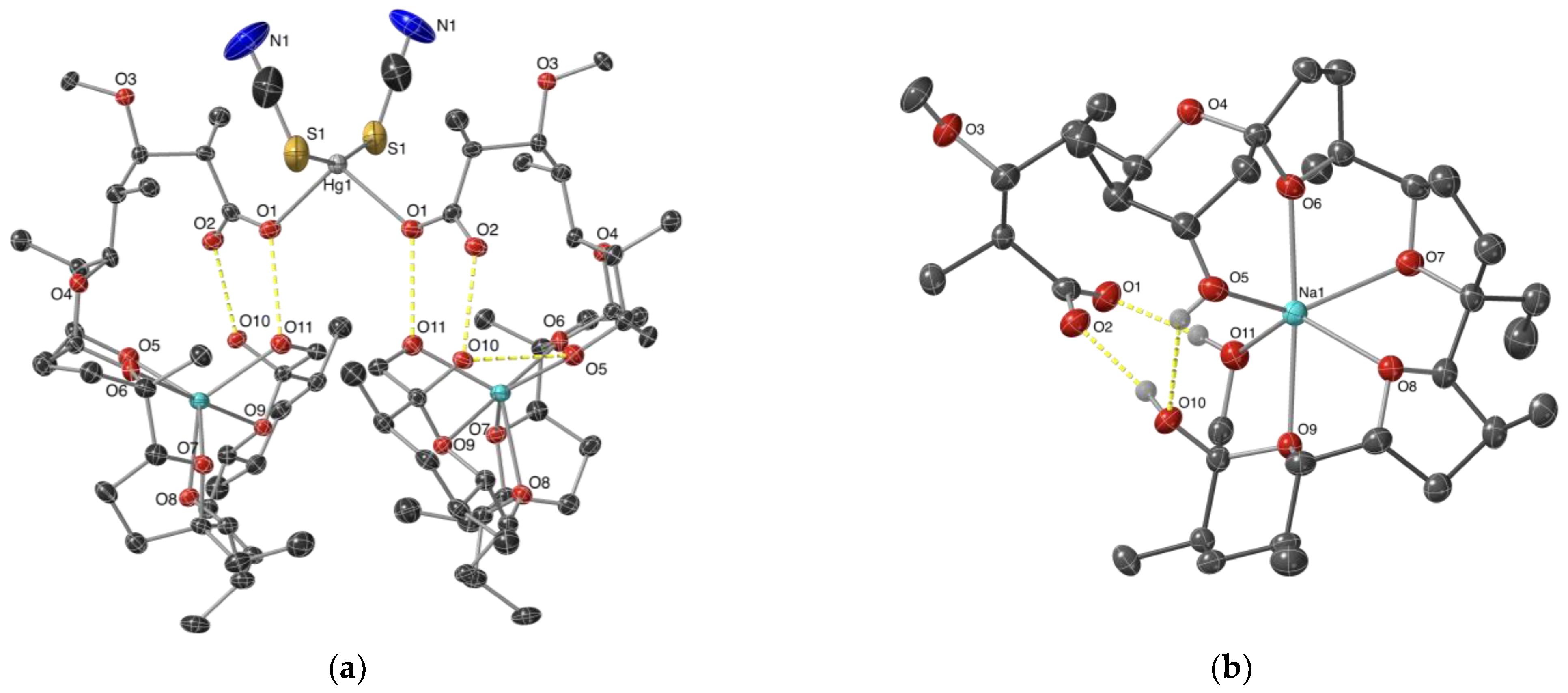
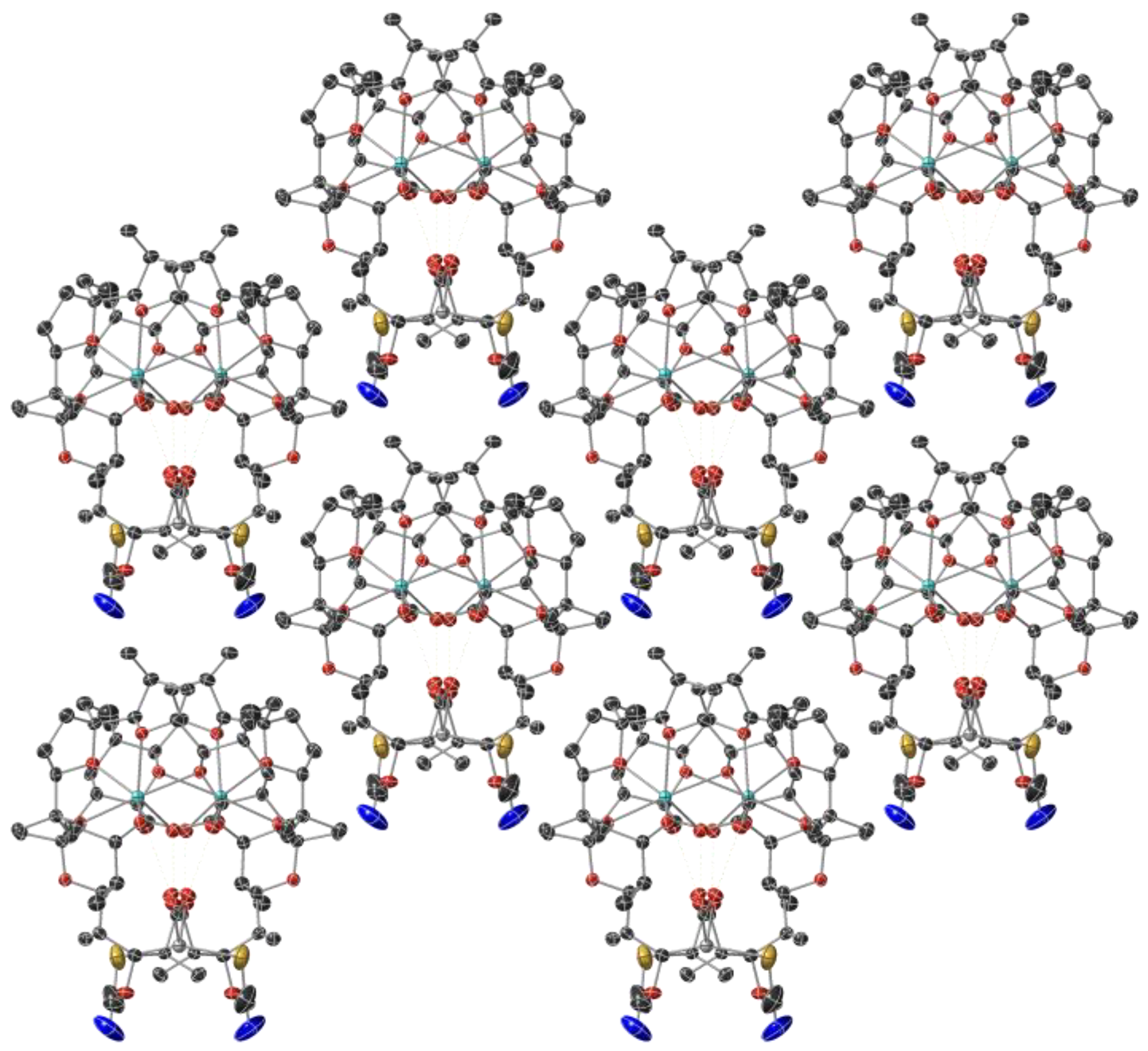
The ORTEP diagram and crystal packing of complex 1 are plotted in Figure 2a and Figure 3, respectively. Each Hg(II) cation is four-coordinated by two sodium monensinates and two thiocyanates in a rather distorted tetrahedral geometry with bond angles around the metal centre varying from 91.4 to 159.1° (Table 1). The Hg-O bonds arise from the interaction between the metal cation and the monodentate carboxylate function of the antibiotic anions, with a distance typical of Hg(II)-carboxylates [31,32]. The length of the Hg–S bonds is similar to that in the HgS2O2 coordination motif [33,34], but is significantly shorter compared with Hg(II)-thiocyanate complexes bearing nitrogen- or sulphur-comprising co-ligands [35,36,37,38,39]. Representative cases of Hg-O and Hg-S distances in various mercury(II) coordination species are shown in Table 2. To the best of our knowledge, the structure of [Hg(MonNa)2(SCN)2] is the first example of a mercury(II) dicarboxylate–dithiocyanate complex. As commonly observed in Hg(II)–thiocyanate compounds, the SCN-groups are linear, the Hg-S-C angles are close to 100°, and the C-S and C-N bond lengths are 1.65 and 1.16 Å, respectively [40,41].
Sodium cations cannot be replaced by Hg(II) ions and remain accommodated into the hydrophilic space of monensinate ligands. The metal(I) centre is coordinated to four ether (O6, O7, O8, O9) and two hydroxyl (O5, O10) donor atoms in a highly distorted octahedral geometry (Table 1, Figure 2b). Na-O bonds range within 2.33–2.46 Å; the observed distances are in close agreement with previously reported data on sodium monensinate [42,43] and its heteronuclear complexes that contain Co(II) or Mn(II) cations and chloride co-ligands [44]. The carboxylate and hydroxyl ends of monensinate are interconnected with each other by O11-H ··· O1 (2.700 Å) and O10-H ··· O2 (2.631 Å) intramolecular hydrogen bonds; in addition, the antibiotic anions are stabilized by a third H-bond O5-H ··· O10 (2.921 Å). Neither additional and obvious intermolecular interactions such as H-bonds between the ligands, nor the inclusion of solvent molecules that may serve as bridge(s) for H-bonds are observed in the crystal packing of complex 1 (Figure 3).
Table 1. Selected bond lengths (Å) and angles (°) for [Hg(MonNa)2(SCN)2], 1.
Hg-O12.429(4)Na-O52.328(5)
Hg-S12.375(2)Na-O62.350(5)
O1-Hg-O1 11102.60(20)Na-O72.447(5)
S11-Hg-S1159.13(11)Na-O82.400(8)
S1-Hg-O191.42(12)Na-O2.457(5)
S11-Hg-O1101.64(11)Na-O112.339(5)
Table 2. Mercury-donor atom bond distances in Hg(II) complexes bearing variable coordination motifs.
Ligand/ComplexCore UnitBond Length [Å]Refs.
Hg-OHg-NHg-S
quinoline-2-carboxylic acid/[HgL2X],
X = H2O, EtOH
HgN2O32.31–2.522.19–2.26[39]
picolinic acid/[HgL2Cl]HgN2O2Cl2.49–2.542.12–2.35[40]
pyridine-2-thione/[HgL2]HgS42.56–2.68[43]
biquinoline/[HgL2(SCN)2]HgN2S22.30–2.502.44–2.47[44]
bipyridyl-based carbazoles/[HgL2(SCN)2]HgN2S22.30–2.452.44–2.45[45]
nicotinamide/[HgL2(SCN)2]HgN2S22.33–2.392.42–2.46[46]
4,5-diazafluoren-9-one/[HgL2(SCN)2]HgN2S22.482.4[47]
3-(2-chlorophenyl)-2-sulfanylpropenoic acid [41]
[HgL2]2−HgO2S22.55–2.612.34
[HgL(HL)]HgO2S22.54–2.672.34
[Hg(HL)2]HgO2S22.652.36
thiol (L′) & carboxylic acid (L″)/[HgL′2L″]HgO2S2 *2.46–27 2.33–2.41[42]
* bidentate carboxylate function.

2.2. Spectral Characterization of 1 and 2

The IR spectrum of MonNa (Figure 4a,b) consists of two bands at 1560 cm−1 and 1408 cm−1 (Δ = 152 cm−1) attributed respectively to the νCOOasym and νCOOsym of the carboxylate function that is not involved in direct interaction with sodium cations. The stretching vibrations of the hydroxyl groups of the antibiotic appear as a broad signal in the range of 3550−3300 cm−1, accompanied by a low-intensity band at 1640 cm−1 assigned to δOH [45,46,47,48]. The parent mercury(II) salt exists in a pure thiocyanate form, as evidenced by its strong absorption band assigned to the C≡N bond (2100 cm−1), complemented by a weak C-S stretch appearing at 718 cm−1 (Figure 4a). In Zn(SCN)2, most of the zinc(II) ions are involved in the formation of metal-nitrogen bonds, apparent from the intense broad band at 2092 cm−1CN) and the weak absorption at 893 cm−1CS), while the rest exist in a bridge-like M-SCN-M structure with characteristic stretches at 2156 cm−1 and 784 cm−1, related to νCN and νCS, respectively [49,50,51] (Figure 4b).
The IR spectra of 1 and 2 (Figure 4a,b) show several characteristic bands assigned to the main donor groups participating in complex formation. The asymmetric and symmetric stretches of the carboxylate function are observed at 1574/1405 cm−1 (1, Δ = 169 cm−1) and at 1640/1430 cm−1 (2, Δ = 210 cm−1), respectively, assuming its monodentate coordination mode to the heavy metal(II) centre. The presence of thiocyanate ligands is evident from the absorption bands detected in the range of 2200−2000 cm−1. The C≡N frequency in 1 appears as a strong sharp signal at 2135 cm−1, indicative of the formation of a Hg-thiocyanate bond (Hg-SCN), while the spectrum of 2 consists of an intense but much broader band at 2070 cm−1 revealing the presence of Zn-isothiocyanate (Zn-NCS) interaction [52]. The bands at 3555 (1) and 3562 cm−1 (2) are assigned to O11-H groups participating only in the formation of a hydrogen bond with the carboxylate O2-oxygens, which are not involved in the formation of the first coordination sphere. The rest of the signals, namely those at 3158 cm−1 (Hg(II)complex) and 3293/3110 cm−1 (Zn(II)species), are typical of monensinate OH-functions involved in donor–acceptor interactions with sodium cations and/or H bond(s).
The 1H- and 13C-NMR spectra of the diamagnetic Hg(II) (1) and Zn(II) (2) mixed-metal monensinates were also studied using NMR spectroscopy. The chemical shifts of the 1H- and 13C-resonances in MonNa and 12 are presented in Section 2.2. and Table 3, respectively, following the numbering shown in Figure 1. The 1H- and 13C-NMR spectra of the studied compounds 12 are shown in Figures S1 and S2.
The NMR spectra of complex 1 in solution do not change significantly compared with those of sodium monensinate. Most of the carbon signals in the 13C-NMR spectrum of 1 retain their positions, with minor exceptions such as the upfield shifts for 2C and 3C atoms by 0.2 ppm. The observed spectra of 1 show that this complex is not sufficiently stable in chloroform solution and does not hold its molecular form in an identical manner to that in the solid state. On the contrary, the 13C-NMR spectrum of complex 2 is sufficiently different from that of MonNa. For carbon atom 1C, a very weak broad peak is observed, shifted downfield by 0.6 ppm compared with MonNa. Additionally, another low-intensity signal at 135.2 ppm indicates the presence of the isothiocyanate ligand. 2C and 3C atoms that are located close to the metal cation differ by 0.9 ppm upfield and the signals of all carbon atoms directly bonded to the oxygens of the polyether chain are broadened, most likely due to O-participation in numerous intramolecular bonds. The observed difference in several NMR resonances between complex 2 and MonNa indicates that the Zn(II) heteronuclear complex does not dissociate and retains its structure both in the solid state and in solution.
The ESI-MS+ spectra of complexes 1 and 2 (Figures S3 and S4, respectively) consist mainly of peaks assigned to [MonNa]H+ (693.42 m/z), [MonNa]Na+ (715.40 m/z) and [MonNa]2Na+ (1407.81 m/z), revealing the structure’s breakdown of the two neutral coordination species into their key compartments—MonNa (observable) and Hg(SCN)2/Zn(NCS)2 (not observable). The very low-intensity peak in the spectrum of 1 at 1725.74 m/z is assigned to the molecular ion of the parent complex [Hg(MonNa)2(SCN)2]Na+, thus confirming its existence and its following dissociation under ESI conditions.

2.3. Structure Elucidation of Complex 2

Based on the spectral properties of Zn(II) complex 2 we suggest that its structure resembles that of [Hg(MonNa)2(SCN)2] (1), with one major difference, which is due to the coordination mode of the thiocyanate as ambidentate ligand. The main evidence for the formation of zinc–isothiocyanate (Zn-NCS) bonds in 2 is apparent from its IR spectral characteristics. Unfortunately, we failed to grow single crystals suitable for X-ray diffraction; however, we obtained polycrystalline material of reasonable quality in order to conduct powder XRD analysis (Figure 5, blue). The powder pattern of 2 was compared with that calculated for complex 1 (green), and the significant similarities found suggest identical space groups between complexes 1 and 2.
Following the last observation and spectral properties of 2, we assumed that 1 and 2 are isostructural, where Hg(SCN)2 is replaced by a Zn(NCS)2 fragment. It is well known from XRD theory that the position and numbers of observed peaks in powder diffraction experiments are based on identical cells parameters, while their intensity is a function of the cell contents. We tried to go further. We simulated the powder pattern of the assumed Zn structure (Zn connected to N in thiocyanate anion) and compared it with the experimentally observed powder patter of the Zn–compound. Thus, we replaced the mercury(II) cations into the crystal structure of 1 with zinc(II) ions, preserving the S-binding of thiocyanate ligands, and simulated the corresponding pattern before (2A, yellow) and after (2B, red) optimization. In the final step, the sulphur and nitrogen atoms of the SCN groups were exchanged to model the Zn-NCS interaction (2C, purple, after optimization). The description of the protocol for powder diffraction patterns and crystal structure simulations of Zn(II) complex is provided in the Supplementary Materials (Figure S5 and Tables S1 and S2).
In all five diffraction patterns, a complete conservation of the number of reflections can be seen, which is due to the similar unit cell of the complexes. The peaks of the studied species 1, 2AC and 2 retain their positions up to 10°, maintaining the same intensity ratio. The observed splitting at 11.1° and 11.4° in 1 and 2A gradually shifts from 2B to 2C towards lower 2θ values (11.0° and 11.3°) to better fit the broad and clearly composite reflection of 2 in this position. An interesting result is evident for the reflection at 13.2°—in 1 it appears as an asymmetric peak of relatively low intensity, while in 2A it is observed as a symmetric and more intense reflection. Optimization of the S-bound construct (2B) raises an additional peak at 12.9°. Replacement of sulphur with nitrogen as a donor atom in the modelled Zn(II) structure 2C further diminishes the original reflection at the expense of the new one. Although the intensity of the two peaks is reversed in 2C compared with 2, their position corroborates well the broad asymmetric reflection observed in the diffractogram of the isolated complex 2. The remaining patterns in the range 14−17° deviate to some extent between all species within 0.1−0.3°; however, the overall peaks comparison reveals the close similarity between the structures of [Hg(MonNa)2(SCN)2] (1) and [Zn(MonNa)2(NCS)2] (2). In conclusion, the negligible differences, mostly in the peak intensities and shoulders in the XRD patterns of 1 and 2, can be explained by the described chemical changes in the contents of the unit cell. In addition, the temperature dependence of the unit cell parameters must be taken into account, since the single-crystal X-ray diffraction was measured at 107 K and powder XRD at 298 K. Shrinkage of the unit cell will lead to a slight shift in the position of peaks.
To further examine the ambidentate properties of SCN-anions in Hg(II) and Zn(II) mixed-ligand complexes, we also studied the behaviour of 12 employing XPS. The full scan of both XPS spectra presented in Figure S6a shows typical peaks at 284.9 eV, 531.9 eV and 1070.7 eV, attributable to carbon (C1s), oxygen (O1s), and sodium (Na1s), respectively. In addition, the spectrum of 1 consists of signals characteristic of mercury(II) (Hg4f, 101.0 eV), sulphur (S2p, 163.0 eV) and nitrogen (N1s, 398.5 eV), and that of 2 comprises peaks at 162.4 eV, 398.4 eV and 1021.9 eV assigned respectively to sulphur (S2p), nitrogen (N1s) and zinc(II) (Zn2p) [53].
The high-resolution spectra (HRS) of C1s (Figure S6b) for Hg(II) and Zn(II) complexes were deconvoluted into three peaks at 284.7 eV, 286.1 eV and 288.5 eV, belonging to carbon electrons respectively involved in the formation of C-C, C-O, and COO-bonds. The binding energy of O1s appears as a single symmetric peak attributable to C-OH and C-O-C. The equivalent shape, position and intensity of carbon and oxygen BE confirm the identity of the antibiotic ligand (both qualitatively and quantitatively) involved in the structure of coordination species 1 and 2. HRS of Hg4f (Figure S6c) and Zn2p (Figure S6d) encompass peaks at 101.0 eV and 1021.6 eV, respectively, which confirm the expected presence of Hg(II) and Zn(II) cations in the studied mixed-metal monensinates [54,55,56,57].
The binding energy of S2p electrons (Figure S6e) is observed as a single peak with FWHM = 3.0 eV (1) and FWHM = 2.5 eV (2), respectively, while the signal of N1s in 1 is narrower (FWHM = 2.6 eV) compared with that of complex 2 (FWHM = 2.9 eV) (Figure S6f). The observed broadening of the corresponding S2p (1) and N1s (2) BE can be explained by the presence of Hg-S/S-C bonds in complex 1 and Zn-N/N-C bonding in complex 2. Vice versa, the narrow peaks for N1s in 1 and for S2p in 2 can be referred to the formation of only N-C bonds in Hg(II)-monensinate and S-C in Zn(II)-monensinate, respectively. The results obtained demonstrate that the total difference in binding energy [N1s—S2p] for the target coordination species is 235.5 eV for 1 and 236.0 eV for 2. This observation corroborates well with some of the available data for metal thiocyanates, where the [N1s—S2p] value is larger for M-NCS than for M-SCN and is used as a discriminator of different SCN-ligand bonding modes [58]. On the other hand, XPS data should be treated with considerable caution as there are cases where the energy difference is the same for related series of complexes containing N- and S-bound thiocyanate [59]. In the present study, despite the questionable reliability of XPS when seeking to usefully study complex coordination systems, we infer that it can be applied as a complementary technique for the structural evaluation of SCN-containing metal complexes.

2.4. Antibacterial Properties of MonNa and Complex 2

To evaluate the impact of Zn(II) as a biologically relevant metal ion on the activity of sodium monensinate, we conducted an antimicrobial assay using five Gram-positive microorganisms, representative of Bacillus (B. subtilis, B. cereus), Kocuria (K. rhizophila) and Staphylococcus (S. aureus, S. saprophyticus). Gram-negative bacteria are inherently resistant to polyether ionophorous antibiotics and their complexes, probably due to the size of the molecules with molecular weights above 600 Da [44] and were excluded from the present study.
The effect of Zn(SCN)2 on bacterial growth is negligible as the metal salt is not effective at the highest concentration studied (1 mg/mL, 5.6 mM). K. rhizophila and B. cereus appear to be the most resistant and sensitive strains, respectively, among the microorganisms studied for the effect of MonNa or complex 2 on their growth ability (Table 4). The incorporation of Zn(II) cations into the structure of sodium monensinate preserves or enhances the activity of the parent antibiotic. With the exceptions of B. cereus and S. aureus, the remaining target bacteria are 2 to 4 times more susceptible to the effect of Zn(II) complex compared with MonNa. In the case of K. rhizophila, the two-fold increase in the MIC (µM) of 2 can be explained by the introduction of two moles of antibiotic per one mole of the complex. In contrast, the higher potency of Zn(II) species against the strains of B. subtilis and S. saprophyticus cannot be attributed to a simple synergic effect of MonNa and Zn(II), while complex 2 retains the inhibitory ability of MonNa towards B. cereus and S. aureus. In our study, the target bacteria can be arranged in the following hierarchy according to their decreasing susceptibility: BC < SASS < BS < KR (MonNa) and BC < SS < SA < BS < KR (2).
The outcome of the conducted assay reveals that each therapeutic candidate should be investigated as thoroughly as possible, and its effect on a specific bacterial strain cannot be unambiguously transferred to other similar microorganisms. On the other hand, mercury(II) is a toxic ion with no known positive biological functions, although some of its complexes would be potent antimicrobial agents [60,61,62]. At the same time, reducing concentration levels of Hg(II) in animal and human organisms is challenging, so the electroneutral mercury(II)-containing complex 1 can be treated as a possible species formed by coordination with sodium monensinate, especially at heavy metal intoxication in stock farming (animal husbandry).

3. Materials and Methods

3.1. Materials and Reagents

Sodium monensinate (MonNa, p.a.) was provided by Biovet Ltd. (Peshtera, Bulgaria). The metal(II) thiocyanates (Hg(SCN)2, Zn(SCN)2)), acetonitrile (MeCN), and methanol (MeOH) (p.a. grade) were purchased from local suppliers, and CDCl3 was acquired from Deutero GmbH (Kastellaun, Germany).

3.2. Synthesis of 1 and 2

Metal(II) thiocyanate (0.1 mmol in 2 mL MeOH, 31.7 mg Hg(SCN)2 or 18.2 mg Zn(SCN)2) was added dropwise to a solution of MonNa (0.1 mmol in 3 mL MeCN:MeOH = 2:1, 69.3 mg). The resulting mixtures were stirred for 30 min. at r. t. and the slow evaporation of the solvent mixtures led to the formation of colourless crystals (1) or polycrystalline solids (2). These were washed with MeCN, filtered, and dried in an exicator.
MonNa [63], composition C36H61O11Na: 1H-NMR (600 MHz, δ (ppm, assignment), CDCl3): 4.40 (20CH), 4.02 (5CH), 3.97/3.29 (26CH2), 3.93 (17CH), 3.89 (7CH), 3.82 (21CH), 3.53 (13CH), 3.37 (28OCH3), 3.18 (3CH), 2.52 (2CH), 2.30/1.46 (15CH2), 2.25 (18CH), 2.21 (6CH), 2.18/1.54 (19CH2), 2.06 (4CH), 2.00/1.70 (10CH2), 1.97/1.71 (11CH2), 1.90/1.68 (8CH2), 1.77/1.53 (14CH2), 1.59/1.50 (32CH2), 1.50 (31CH3), 1.45 (24CH), 1.40/1.31 (23CH2), 1.35 (22CH), 1.23 (27CH3), 1.17 (29CH3), 0.94 (33CH3), 0.93 (30CH3), 0.90 (34CH3), 0.84 (36CH3), 0.80 (35CH3).
Complex 1, [Hg(MonNa)2(SCN)2] composition: C74H122O22S2N2Na2Hg, MW 1702.4 g/mol, yield 63.0 mg (74%). Calc. C, 52.21; H, 7.22; N, 1.65; S, 3.77; Na, 2.70; Hg, 11.78%. Found: C, 51.78; H, 6.82; N, 1.82; S, 4.14; Na, 2.50; Hg, 10.96%. 1H-NMR (600 MHz, δ (ppm, assignment, multiplicity, J [Hz]), CDCl3): 4.37 (20CH, m), 4.00 (5CH, dd, 2.1; 11.1), 3.95/3.27 (26CH2, d, 11.8), 3.91 (17CH, d, 3.5), 3.86 (7CH, m), 3.80 (21CH, dd, 3.9; 10.0), 3.51 (13CH, dd, 4.9; 10.9), 3.35 (28OCH3, s), 3.17 (3CH, dd, 1.8; 11.9), 2.51 (2CH, m), 2.27/1.44 (15CH2, m/m), 2.23 (18CH, m), 2.18 (6CH, m), 2.16/1.51 (19CH2, m/m), 2.04 (4CH, m), 1.98/1.67 (10CH2, m/m), 1.96/1.69 (11CH2, m/m), 1.88/1.67 (8CH2, m/m), 1.75/1.53 (14CH2, m/m), 1.56/1.48 (32CH2, qd, 7.7; 14.4, qd, 7.0; 14.6), 1.47 (31CH3, s), 1.45 (24CH, m), 1.41/1.30 (23CH2, m/m), 1.30 (22CH, m), 1.22 (27CH3, d, 6.7), 1.15 (29CH3, d, 6.8), 0.91 (33CH3, t, 7.4), 0.91 (30CH3, d, 7.1), 0.87 (34CH3, d, 6.9), 0.82 (36CH3, d, 6.2), 0.78 (35CH3, d, 6.2).
Complex 2, [Zn(MonNa)2(NCS)2] composition: C74H122O22S2N2Na2Zn, MW 1567.3 g/mol, yield 61.9 mg (79%). Calc. C, 56.71; H, 7.85; N, 1.79; S, 4.09; Na, 2.93; Zn, 4.17%. Found C, 56.95; H, 7.54; N, 1.77; S, 3.85; Na, 2.40; Zn, 4.65%. 1H-NMR (600 MHz, δ (ppm, assignment, multiplicity, J [Hz]), CDCl3): 4.41 (20CH, m), 3.88 (5CH, dd, 2.0; 11.5), 3.96/3.40 (26CH2, d, 12.0), 3.95 (7CH, m), 3.92 (17CH, d, 3.6), 3.85 (21CH, dd, 3.7; 10.0), 3.37 (28OCH3, s), 3.52 (3CH, dd, 5.1; 10.9), 3.27 (13CH, d, 9.6), 2.58 (2CH, dd, 7.1; 9.3), 2.28/1.44 (15CH2, m/m), 2.26 (18CH, m), 2.17/1.54 (19CH2, m/m), 2.09 (6CH, m), 2.06 (4CH, m), 1.98/1.70 (10CH2, m/m), 1.92/1.70 (11CH2, m/m), 1.92/1.68 (8CH2, m/m), 1.77/1.53 (14CH2, m/m), 1.61/1.48 (32CH2, qd, 7.9; 14.7, qd, 7.3; 14.6), 1.51 (31CH3, s), 1.51 (24CH, m), 1.51/1.36 (23CH2, m/m), 1.40 (22CH, m), 1.25 (27CH3, d, 6.6), 1.11 (29CH3, d, 6.7), 0.94 (33CH3, t, 7.6), 0.93 (30CH3, d, 7.1), 0.90 (36CH3, br d), 0.89 (34CH3, d, 7.1), 0.83 (35CH3, d, 6.2).

3.3. Methods

3.3.1. X-ray Crystallography

Data collection and structure solution were conducted at the X-ray Crystallographic Facility, Department of Chemistry and Biochemistry, NDSU, Fargo, ND, USA.
A crystal (approximate dimensions 0.17 × 0.16 × 0.07 mm3) was mounted on a Bruker APEX-II CCD diffractometer (Billerica, MA, USA) for data collection at 107 K. A preliminary set of cell constants was calculated from the reflections collected from 4 sets of 30 frames. This produced initial orientation matrices determined from 401 reflections. Data collection was carried out using IµS Cu Kα radiation with a frame time of 10 s at higher angles of data collection (detector’s position 2θ = 100.855°) or 5 s for lower angles of data collection (2θ = −34.239°), and with a detector distance of 4.0 cm. A randomly oriented region of reciprocal space was surveyed to the extent of one sphere and a resolution of 0.84 Å. Nineteen ω-scan sections of frames and φ-scan sections with 1.8° width were collected to achieve the desired completeness of 99.8%. Intensity data were corrected for absorption and decay [64]. The final cell constants were calculated from the xyz centroids of 9823 strong reflections from the actual data collection after integration [65].
The structure was solved and refined using the SHELX 2018 [66] set of programs with Olex 2 v.1.5 software package [67]. The structure was solved with SHELXT 2015 [68] program using intrinsic phasing, which provided most of the non-hydrogen atoms, while full-matrix least squares/difference Fourier cycles were performed in order to locate the remaining non-hydrogen atoms. All non-hydrogen atoms were refined with anisotropic displacement parameters. All hydrogen atoms were placed in ideal positions and refined as riding atoms with relative isotropic displacement parameters. The final full matrix least squares refinement converged to R1 = 3.51% and wR2 = 8.53% (F2, all data). The data collection parameters and refinement information for the single-crystal X-ray diffraction experiment are summarized in Table 5. Details on the geometrical data can be found in the Supplementary Materials (Tables S3–S6). Images were generated using CrystalMaker Software Ltd., Oxford, England (www.crystalmaker.com, accessed on 14 June 2024).

3.3.2. Physical Measurements

IR spectra were recorded on a Nicolet 6700 FT-IR, Thermo Scientific (Madison, WI, USA) in KBr pellets. 1H- (600.18 MHz) and 13C- (150.93 MHz) NMR spectra were acquired on a Bruker NEO 600 spectrometer (Ettlingen, Germany). All spectra were recorded in CDCl3 at 298.0 ± 0.1K. The residual solvent peaks (1H—7.26 ppm and 13C—77.16 ppm) were used as internal standards for the 1H- and 13C-NMR spectra, respectively. The unambiguous assignment of signals was made based on the gradient-enhanced versions of COSY, HSQC, and HMBC experiments. The chemical shift values of the overlapped protons in the complexes have been determined from the HSQC spectra.
The C, H, N, S analysis was performed on an organic elemental analyser vario MACRO cube (Elementar analysensysteme GmbH, Stuttgart, Germany). Sodium content was calculated by AAS on a Perkin Elmer 1100 B (Walthman, MA, USA) using standard stock solution (1000 µg/mL, Merck, Darmstadt, Germany). Working reference solutions were prepared after suitable dilution in appropriate solvent mixture. Mercury(II) and zinc(II) were determined by complexometric titrations at pH 5.5 (acetate buffer) using xylenol orange as an indicator. Hg(II) content was evaluated by back titration with standard solution of zinc acetate, while Zn(II) was quantified by direct titration with standard solution of ethylenediaminetetracetic acid (EDTA).
Electrospray-mass spectrometry (ESI-MS) measurements were performed on a Waters SYNAPT G2-Si ToF high resolution mass spectrometer (HRMS, Milford, MA, USA). The sample was dissolved in methanol and directly injected for analysis in an electro spray ionization positive mode source (ESI+). ESI+ conditions were as follows: capillary potential 3.0 kV, sample cone potential 40 V, temperature source 90 °C, desolvation temperature 250 °C, desolvation gas flow 350 L/h. The observed range was set from 50 to 2000 m/z. The powder diffraction pattern was obtained on a PANalytical Empyrean X-ray powder diffractometer (Malvern Panalytical, Malvern, UK) with Cu Kα radiation (λ = 1.5418 Å) operating at 40 kV and 30 mA. The X-ray photoelectron spectroscopy (XP, XPS) studies were performed on a VG Escalab II system (Thermo Fisher Scientific, Waltham, MA, USA), using Al Kα radiation with an energy of 1486.6 eV. The chamber pressure was 1.10−9 Torr. The C1s line of adventitious carbon at 284.9 eV was used as an internal standard by which to calibrate the binding energies (BE). Photoelectron spectra were corrected by Shirley-type background subtraction and quantified using peak area and Scofield’s photo-ionization cross-section. The accuracy of the measured BE was ±0.2 eV. Spectra were evaluated by CasaXPS software v. 2.3.25PR1.0 and fitted using a mixed Gaussian/Lorentzian product formula with 30% Lorentzian.

3.3.3. Antibacterial Assay

The ability of MonNa and complexes 12 to inhibit the visible growth of microorganisms was evaluated as their minimum inhibitory concentration (MIC, mg/mL, µM). A series of methanol solutions down to 0.25 µg/mL was prepared by double-dilution method starting from 1 mg/mL. The assay included a set of five Gram-positive non-pathogenic bacteria, supplied by the National Bank for Industrial Microorganisms and Cell Cultures (NBIMCC, Sofia, Bulgaria): Bacillus subtilis (BS, NBIMCC 1050, ATCC 11774) (ATCC, Manassas, VA, USA), Bacillus cereus (BC, NBIMCC 1085, ATCC 11778), Kocuria rhizophila (KR, NBIMCC 159, ATCC 9341), Staphylococcus aureus (SA, NBIMCC 509, ATCC 6538) and Staphylococcus saprophyticus (SS, NBIMCC 3348). Nutrient agar (pH 7.2–7.4) containing meat extract (1%), peptone (1%) and NaCl (0.5%) was used as culture media.
The double-layer agar diffusion method was carried out on Petri dishes (90 mm) containing sterile (10 mL) and inoculated (10 mL, 1.5% inoculum, McFarland 4, A650 = 0.8–1) agar layers. After media solidification, the holes (punched in 6 mm diameters) were filled with 20 µL of the tested solutions. The diameter of the inhibited zones was read 24 h after incubation at 30 °C. Three separate experiments were performed in triplicate (a total of nine measurements). All equipment and culture media (delivered from local suppliers) were sterile. Methanol served as a negative control.

4. Conclusions

The reaction of sodium monensinate (MonNa) with Hg(SCN)2 or Zn(SCN)2 in acetonitrile–methanol solution leads to the respective formation of new heteronuclear complexes, [Hg(MonNa)2(SCN)2] and [Zn(MonNa)2(NCS)2]. The coordination species are isostructural, with the metal(II) ions placed in a distorted tetrahedral environment where two of the positions are occupied by oxygen atoms originating from the parent antibiotic. The other two binding sites involve S-donor atoms in the case of Hg(II) complex and N-donors in its Zn(II) counterpart. The inclusion of biometal ions positively affects the activity of MonNa, while the binding of Hg(II) cations reveals the antidote potential of the antibiotic against heavy metal intoxications that may occur in animal husbandry.

Supplementary Materials

The following supporting information can be downloaded at: https://www.mdpi.com/article/10.3390/molecules29133106/s1, Table S1: Orthogonal coordinates [Å] of Zn, O1 and SCN groups in the cell after relaxation of structures 2B and 2C; Table S2: Selected bond lengths and angles that undergo major changes during relaxation processes; Table S3: Fractional atomic coordinates (×104) and equivalent isotropic displacement parameters (Å2 × 10³) for complex 1. Ueq is defined as 1/3 of the trace of the orthogonalized UIJ tensor; Table S4: Bond lengths for complex 1; Table S5: Bond angles for complex 1; Table S6: Torsion angles for complex 1; Figure S1: 1H-NMR of complexes 12 in CDCl3; Figure S2: 13C-NMR spectra of complexes 12 in CDCl3; Figure S3: ESI-MS+ of Hg(II) complex 1; Figure S4: ESI-MS+ of Zn(II) complex 2; Figure S5: Structure of simulated Zn(II) species (a) 2B and (b) 2C. H-atoms are omitted for clarity; Figure S6: (a) XPS survey spectra of complexes 1 and 2; (b−f) high-resolution spectra of C1s, Hg4f, Zn2p, S2p, and N1s. CCDC 2355767 contains the supplementary crystallographic data for this paper. These data can be obtained free of charge via http://www.ccdc.cam.ac.uk (or by an application from the Cambridge Crystallographic Data Centre, 12 Union Road, Cambridge CB2 1EZ, UK; Fax: +44-01223-336033; or e-mail: [email protected].

Author Contributions

Conceptualization, N.P. and I.P.; methodology and analysis, N.P., S.S., E.E., S.K., A.T. and A.U.; resources, P.D.; writing—original draft preparation, I.P.; writing—review and editing, I.P., N.P. and A.U. All authors have read and agreed to the published version of the manuscript.

Funding

This research was funded by the European Union—NextGenerationEU, through the National Recovery and Resilience Plan of the Republic of Bulgaria, project SUMMIT BG-RRP-2.004-0008-C01 (No 70-123-186).

Institutional Review Board Statement

Not applicable.

Informed Consent Statement

Not applicable.

Data Availability Statement

Data are available from the authors upon request.

Acknowledgments

The INFRAMAT (Distributed Research Infrastructure) equipment, part of the National Roadmap for Research Infrastructures in Bulgaria, supported by the Ministry of Education and Science, was used in this study.

Conflicts of Interest

Author Petar Dorkov was employed by the company Biovet Ltd. The remaining authors declare that the research was conducted in the absence of any commercial or financial relationships that could be construed as a potential conflict of interest.

References

  1. Berger, J.; Rachlin, A.I.; Scott, W.E.; Sternbach, L.H.; Goldberg, M. W 1951. The isolation of three new crystalline antibiotics from Streptomyces. J. Am. Chem. Soc. 1951, 73, 5295–5298. [Google Scholar] [CrossRef]
  2. Harned, R.L.; Hidy, P.H.; Corum, C.J.; Jones, K.L. Nigericin, a new crystalline antibiotic from an unidentified Streptomyces. Antibiot. Chemother. 1951, 1, 594–596. Available online: https://pubmed.ncbi.nlm.nih.gov/24541690 (accessed on 8 May 2024).
  3. Agtarap, A.; Chamberlin, J.W.; Pinkerton, M.; Steinrauf, L.K. The structure of monensic acid, a new biologically active compound. J. Am. Chem. Soc. 1967, 89, 5737–5739. [Google Scholar] [CrossRef] [PubMed]
  4. Haney, M.E.; Hoehn, M.M. Monensin, a new biologically active compound. I. Discovery and isolation. Antimicrob. Agents Chemother. 1967, 7, 349–352. [Google Scholar] [CrossRef]
  5. Stark, W.M.; Knox, N.G.; Westhead, J.E. Monensin, a new biologically active compound. II. Fermentation studies. Antimicrob. Agents Chemother. 1967, 7, 353–358. Available online: https://pubmed.ncbi.nlm.nih.gov/5596159/ (accessed on 8 May 2024).
  6. Agtarap, A.; Chamberlin, J.W. Monensin, a new biologically active compound. IV. Chemistry. Antimicrob. Agents Chemother. 1967, 7, 359–362. Available online: https://pubmed.ncbi.nlm.nih.gov/5596160/ (accessed on 8 May 2024).
  7. Shumard, R.F.; Callender, M.E. Monensin, a new biologically active compound. VI. Anticoccidial activity. Antimicrob. Agents Chemother. 1967, 7, 369–377. Available online: https://pubmed.ncbi.nlm.nih.gov/5596162/ (accessed on 8 May 2024).
  8. Donoho, A.L.; Kline, R.M. Monensin, a new biologically active compound. VII. Thin-layer bioautographic assay for monensin in chick tissues. Antimicrob. Agents Chemother. 1967, 7, 763–766. Available online: https://pubmed.ncbi.nlm.nih.gov/4970208/ (accessed on 8 May 2024).
  9. Estrada, S.; Rightmire, B.; Lardy, H.A. Antibiotics as tools for metabolic studies. X1. Specific inhibition of ion transport in mitochondria by the monensins. Antimicrob. Agents Chemother. 1967, 7, 279–288. Available online: https://pubmed.ncbi.nlm.nih.gov/5596149/ (accessed on 8 May 2024).
  10. Dutton, C.J.; Banks, B.J.; Cooper, C.B. Polyether ionophores. Nat. Prod. Rep. 1995, 12, 165–181. [Google Scholar] [CrossRef]
  11. Callaway, T.R.; Edrington, T.S.; Rychlik, J.L.; Genovese, K.J.; Poole, T.L.; Jung, Y.S.; Bischoff, K.M.; Anderson, R.C.; Nisbet, D.J. Ionophores: Their use as ruminant growth promotants and impact on food safety. Curr. Issues Intest. Microbiol. 2003, 4, 43–51. Available online: https://pubmed.ncbi.nlm.nih.gov/14503688/ (accessed on 8 May 2024). [PubMed]
  12. Allèaume, M.; Busetta, B.; Farges, C.; Gachon, P.; Kergomard, A.; Staron, T. X-Ray structure of alborixin, a new antibiotic ionophore. J. Chem. Soc. Chem. Comm. 1975, 411–412. [Google Scholar] [CrossRef]
  13. Otake, N.; Ogita, T.; Nakayama, H.; Miyamae, H.; Sato, S.; Saito, Y. X-Ray crystal structure of the thallium salt of antibiotic-6016, a new polyether ionophore. J. Chem. Soc. Chem. Comm. 1978, 875–876. [Google Scholar] [CrossRef]
  14. Pangborn, W.; Duax, W.L.; Langs, D. The hydrated potassium complex of the ionophore monensin A. J. Am. Chem. Soc. 1987, 109, 2163–2165. [Google Scholar] [CrossRef]
  15. Oscarson, J.R.; Bordner, J.; Celmer, W.D.; Cullen, W.P.; Huang, L.H.; Maeda, H.; Moshier, P.M.; Nishiyama, S.; Presseau, L.; Shibakawa, R.; et al. Endusamycin, a novel polycyclic ether antibiotic produced by a strain of Streptomyces endus subsp. aureus. J. Antib. 1989, 42, 37–48. [Google Scholar] [CrossRef] [PubMed]
  16. Takahashi, Y.; Nakamura, H.; Ogata, R.; Matsuda, M.; Hamada, M.; Naganawa, H.; Takita, T.; Iitaka, Y.; Sato, K.; Takeuchi, T. Kijimicin, a polyether antibiotic. J. Antib. 1990, 43, 441–443. [Google Scholar] [CrossRef]
  17. Paulus, E.F.; Kurz, M.; Matter, H.; Vertesy, L. Solid-state and solution structure of the salinomycin-sodium complex: Stabilization of different conformers for an ionophore in different environments. J. Am. Chem. Soc. 1998, 120, 8209–8221. [Google Scholar] [CrossRef]
  18. Huczynski, A.; Ratajczak-Sitarz, M.; Katrusiak, A.; Brzezinski, B. Molecular structure of the 1:1 inclusion complex of monensin A sodium salt with acetonitrile. J. Mol. Struct. 2007, 832, 84–89. [Google Scholar] [CrossRef]
  19. Huczynski, A.; Ratajczak-Sitarz, M.; Katrusiak, A.; Brzezinski, B. Molecular structure of the 1:1 inclusion complex of monensin A lithium salt with acetonitrile. J. Mol. Struct. 2007, 871, 92–97. [Google Scholar] [CrossRef]
  20. Yildirim, S.O.; McKee, V.; Khardli, F.Z.; Mimouni, M.; Hadda, T.B. Rubidium (I) monensinate dihydrate. Acta Cryst. 2008, E64, m154–m155. [Google Scholar] [CrossRef]
  21. Chow, J.M.; Russell, J.B. Effect of ionophores and pH on growth of Streptococcus bovis in batch and continuous culture. Appl. Environ. Microbiol. 1990, 56, 1588–1593. [Google Scholar] [CrossRef] [PubMed]
  22. Chirase, N.K.; Greene, L.W.; Schelling, G.T.; Byers, F.M. Effect of magnesium and potassium on microbial fermentation in a continuous culture fermentation system with different levels of monensin or lasalocid. J. Anim. Sci. 1987, 65, 1633–1638. [Google Scholar] [CrossRef] [PubMed]
  23. Hamidinia, S.A.; Shimelis, O.I.; Tan, B.; Erdahl, W.L.; Chapman, C.J.; Renkes, G.D.; Taylor, R.W.; Pfeiffer, D.R. Monensin mediates a rapid and selective transport of Pb2+: Possible application of monensin for the treatment of Pb2+ intoxication. J. Biol. Chem 2002, 277, 38111–38120. [Google Scholar] [CrossRef]
  24. Hamidinia, S.A.; Erdahl, W.L.; Chapman, C.J.; Steinbaugh, G.E.; Taylor, R.W.; Pfeiffer, D.R. Monensin improves the effectiveness of meso-dimercaptosuccinate when used to treat lead intoxication in rats. Environm. Health Persp. 2006, 114, 484–493. Available online: http://www.jstor.org/stable/3650926 (accessed on 10 May 2024). [CrossRef] [PubMed]
  25. Pantcheva, I.N.; Ivanova, J.; Zhorova, R.; Mitewa, M.; Simova, S.; Mayer-Figge, H.; Sheldrick, W.S. Nickel(II) and zinc(II) dimonensinates: Crystal structure, spectral properties and bactericidal activity. Inorg. Chim. Acta 2010, 363, 1879–1886. [Google Scholar] [CrossRef]
  26. Ivanova, J.; Pantcheva, I.N.; Mitewa, M.; Simova, S.; Mayer-Figge, H.; Sheldrick, W.S. Crystal structures and spectral properties of new Cd(II) and Hg(II) complexes of monensic acid with different coordination modes of the ligand. Centr. Eur. J. Chem. 2010, 8, 852–860. [Google Scholar] [CrossRef]
  27. Pantcheva, I.; Dimitrova, R.; Ivanova, V.; Nedzhib, A.; Dorkov, P.; Dinev, D.; Spasov, R.; Alexandrova, R. Spectral properties and biological activity of La(III) and Nd(III) monensinates. Open Chem. 2019, 17, 1423–1434. [Google Scholar] [CrossRef]
  28. Petkov, N.; Pantcheva, I.; Ivanova, A.; Stoyanova, R.; Kukeva, R.; Alexandrova, R.; Abudalleh, A.; Dorkov, P. Novel cerium(IV) coordination compounds of monensin and salinomycin. Molecules 2023, 28, 4676. [Google Scholar] [CrossRef] [PubMed]
  29. Petkov, N.; Tadjer, A.; Encheva, E.; Cherkezova-Zheleva, Z.; Paneva, D.; Stoyanova, R.; Kukeva, R.; Dorkov, P.; Pantcheva, I. Experimental and DFT study of monensinate and salinomycinate complexes containing {Fe33–O)}7+ core. Molecules 2024, 26, 364. [Google Scholar] [CrossRef]
  30. Petkov, N.; Tadjer, A.; Simova, S.; Cherkezova-Zheleva, Z.; Paneva, D.; Stoyanova, R.; Kukeva, R.; Dorkov, P.; Pantcheva, I. Synthesis, spectral characterization and structural modelling of di- and trinuclear iron(III) monensinates with different bridging patterns. Inorganics 2024, 12, 114. [Google Scholar] [CrossRef]
  31. Soldin, Z.; Matković-Čalogović, D.; Pavlović, G.; Popović, J.; Vinković, M.; Vikić-Topić, D.; Popović, Z. Various coordination modes in mercury(II) complexes with quinoline-2-carboxylic acid: Preparation and structural characterization. Polyhedron 2009, 28, 2735–2743. [Google Scholar] [CrossRef]
  32. Soldin, Z.; Kukovec, B.-M.; Matković-Čalogović, D.; Popović, Z. The solvent effect on composition and dimensionality of mercury(II) complexes with picolinic acid. Molecules 2021, 26, 5002. [Google Scholar] [CrossRef] [PubMed]
  33. Casas, J.S.; Collazo, R.; Couce, M.D.; García-Vega, M.; Sánchez, A.; Sordo, J.; Vázquez-López, E.M. Mercury(II) complexes containing the [Hg(L)2]2−, [Hg(L)(HL)], and [Hg(HL)2] units [H2L = 3-(2-chlorophenyl)-2-sulfanylpropenoic acid]. Structural and spectroscopic effects of the different degrees of ligand protonation. Cryst. Growth Des. 2011, 11, 5370–5377. [Google Scholar] [CrossRef]
  34. Tang, X.-Y.; Zheng, A.-X.; Shang, H.; Yuan, R.-X.; Li, H.-X.; Ren, Z.-G.; Lang, J.-P. Binding of a coordinatively unsaturated mercury(II) thiolate compound by carboxylate anions. Inorg. Chem. 2011, 50, 503–516. [Google Scholar] [CrossRef]
  35. Popović, Z.; Matković-Čalogović, D.; Hasić, J.; Vikić-Topić, D. Preparation and spectroscopic properties of the complexes of mercuric thiocyanate with pyridine-2-thione and pyridine-2-carboxylic acid.: Crystal and molecular structure of two polymorphs of Hg(SCN)2(C5H5NS)2. Inorg. Chim. Acta 1999, 285, 208–216. [Google Scholar] [CrossRef]
  36. Morsali, A.; Mahjoub, A.R.; Ramazani, A. Zn(II), Cd(II) and Hg(II) complexes with 2,2′-biquinoline, syntheses and X-ray crystal structures of [Hg(bq)(SCN)2]. J. Coord. Chem. 2004, 57, 347–352. [Google Scholar] [CrossRef]
  37. Zhou, H.-P.; Tian, Y.-P.; Wu, J.-Y.; Zhang, J.-Z.; Li, D.-M.; Zhu, Y.-M.; Hu, Z.-J.; Tao, X.-T.; Jiang, M.-H.; Xie, Y. Synthesis, crystal structures and photoluminescence of mercury(II) complexes with two homologous novel functional rigid ligands. Eur. J. Inorg. Chem. 2005, 2005, 4976–4984. [Google Scholar] [CrossRef]
  38. Đaković, M.; Popović, Z.; Giester, G.; Rajić-Linarić, M. Synthesis, spectroscopic and structural investigation of Zn(NCS)2(nicotinamide)2 and [Hg(SCN)2(nicotinamide)]n. Polyhedron 2008, 27, 465–472. [Google Scholar] [CrossRef]
  39. Machura, B.; Wolff, M.; Palion, J.; Świtlicka, A.; Nawrot, I.; Michalik, K. Cu(II), Ni(II), and Hg(II) thiocyanate complexes incorporating 4,5-diazafluoren-9-one: Synthesis, spectroscopic characterization, X-ray studies, and magnetic properties. Struct. Chem. 2011, 22, 1053–1064. [Google Scholar] [CrossRef]
  40. Hubert, J.; Beauchamp, A.L.; Rivets, R. Crystal and molecular structure of dithiocyanato(triphenylarsine)mercury(II). Can. J. Chem. 1975, 53, 3383–3387. [Google Scholar] [CrossRef]
  41. Baskaran, A.; Rajarajan, K.; Nizam-Mohideen, M.; Sagayaraj, P. Crystal structure of bis-(thio-cyanato-κS)bis-(thio-urea-κS)mercury(II). Acta Cryst. E Cryst. Commun. 2015, 71, m28–29. [Google Scholar] [CrossRef] [PubMed]
  42. Duax, W.L.; Smith, G.D.; Strong, P.D. Complexation of metal ions by monensin. Crystal and molecular structure of hydrated and anhydrous crystal forms of sodium monensin. J. Am. Chem. Soc. 1980, 102, 6725–6729. [Google Scholar] [CrossRef]
  43. Paz, F.A.A.; Gates, P.J.; Fowler, S.; Gallimore, A.; Harvey, B.; Lopes, N.P.; Stark, C.B.W.; Staunton, J.; Klinowski, J.; Spencer, J.B. Sodium monensin dihydrate. Acta Cryst. E 2003, 59, m1050–m1052. [Google Scholar] [CrossRef]
  44. Dorkov, P.; Pantcheva, I.N.; Sheldrick, W.S.; Mayer-Figge, H.; Petrova, R.; Mitewa, M. Synthesis, structure and antimicrobial activity of manganese(II) and cobalt(II) complexes of the polyether ionophore antibiotic sodium monensin A. J. Inorg. Biochem. 2008, 102, 26–32. [Google Scholar] [CrossRef] [PubMed]
  45. Gertenbach, G.; Popov, A.I. Solution chemistry of monensin and its alkali metal ion complexes. Potentiometric and spectroscopic studies. J. Am. Chem. Soc. 1975, 97, 4738–4744. [Google Scholar] [CrossRef]
  46. Nakamoto, K. Infrared and Raman Spectroscopy of Inorganic and Coordination Compounds, 5th ed.; Wiley: Toronto, ON, Canada, 1997. [Google Scholar]
  47. Carvalho, C.T.; Siqueira, A.B.; Rodrigues, E.C.; Ionashiro, M. Synthesis, characterization and thermal behaviour of solid-state compounds of 2-methoxybenzoate with some bivalent transition metal ions. Ecl. Quim. 2005, 30, 19–26. [Google Scholar] [CrossRef]
  48. Vučković, G.; Stanić, V.; Sovilj, S.P.; Antonijević-Nikolić, M.; Mrozinski, J. Cobalt(II) complexes with aromatic carboxylates and N-functionalized cyclam bearing 2-pyridylmethyl pendant arms. J. Serb. Chem. Soc. 2005, 70, 1121–1129. [Google Scholar] [CrossRef]
  49. Tramer, A. Spectra and structure of thiocyanate complexes. In Theory and Structure of Complex Compounds; Jezowska-Trzebiatowska, B., Ed.; Pergamon: Oxford, UK, 1964; pp. 225–228. [Google Scholar] [CrossRef]
  50. Bailey, R.A.; Kozak, S.L.; Michelsen, T.W.; Mills, W.N. Infrared spectra of complexes of the thiocyanate and related ions. Coord. Chem. Rev. 1971, 6, 407–445. [Google Scholar] [CrossRef]
  51. Baer, C.; Pike, J. Infrared spectroscopic analysis of linkage isomerism in metal-thiocyanate complexes. J. Chem. Edu. 2010, 87, 724–726. [Google Scholar] [CrossRef]
  52. Bertini, I.; Sabatini, A. Infrared spectra of substituted thiocyanate complexes. The effect of the substituent on bond type. II. Inorg. Chem. 1966, 5, 1025–1028. [Google Scholar] [CrossRef]
  53. Escard, J.; Mavel, G.; Guerchais, J.E.; Kergoat, R. X-ray photoelectron spectroscopy study of some metal(II) halide and pseudohalide complexes. Inorg. Chem. 1974, 13, 695–701. [Google Scholar] [CrossRef]
  54. Yao, W.; Hua, Y.; Yan, Z.; Wu, C.; Zhou, F.; Liu, Y. Sulfhydryl functionalized carbon quantum dots as a turn-off fluorescent probe for sensitive detection of Hg2+. RSC Adv. 2021, 11, 36310–36318. [Google Scholar] [CrossRef] [PubMed]
  55. Awan, S.U.; Hasanain, S.K.; Bertino, M.F.; Jaffari, G.H. Ferromagnetism in Li doped ZnO nanoparticles: The role of interstitial Li. J. Appl. Phys. 2012, 112, 103924. [Google Scholar] [CrossRef]
  56. Dengo, N.; Vittadini, A.; Natile, M.M.; Gross, S. In-depth study of ZnS nanoparticle surface properties with a combined experimental and theoretical approach. J. Phys. Chem. C 2020, 124, 7777–7789. [Google Scholar] [CrossRef]
  57. Wang, P.; Zhang, Y.-L.; Fu, K.-L.; Liu, Z.; Zhang, L.; Liu, C.; Deng, Y.; Xie, R.; Ju, X.-J.; Wang, W.; et al. Zinc-coordinated polydopamine surface with a nanostructure and superhydrophilicity for antibiofouling and antibacterial applications. Mater. Adv. 2022, 3, 5476–5487. [Google Scholar] [CrossRef]
  58. Walton, R.A. X-ray photoelectron spectra of inorganic molecules [1]. XXIII. On the question of the usefulness of XPS in studying the ambidentate nature of the thiocyanate ligand. Inorg. Chim. Acta 1979, 37, 237–240. [Google Scholar] [CrossRef]
  59. Best, S.A.; Walton, R.A. X-ray photoelectron spectra of inorganic molecules. XIX. Thiocyanato and isothiocyanato complexes of the transition metals: Carbon 1s, nitrogen ls and sulfur 2p binding energies and their possible correlation with the mode of thiocyanate bonding. Isr. J. Chem. 1976, 15, 160–164. [Google Scholar] [CrossRef]
  60. Dwyer, J.; Levason, W.; McAuliffe, C.A. The preparation of some mercury(II) complexes of group VB donors and preliminary toxicity studies towards lysozyme. J. Inorg. Nucl. Chem. 1976, 38, 1919–1921. [Google Scholar] [CrossRef]
  61. Farrell, P.E.; Germida, J.J.; Huang, P.M. Effects of chemical speciation in growth media on the toxicity of mercury(II). Appl. Environm. Microbiol. 1993, 59, 1507–1514. [Google Scholar] [CrossRef]
  62. Lam, P.L.; Lu, G.L.; Choi, K.H.; Lin, Z.; Kok, S.H.L.; Lee, K.K.H.; Lam, K.H.; Li, H.; Gambari, R.; Bian, Z.X.; et al. Antimicrobial and toxicological evaluations of binuclear mercury(II) bis(alkynyl) complexes containing oligothiophenes and bithiazoles. RSC Adv. 2016, 6, 16736–16744. [Google Scholar] [CrossRef]
  63. Ajaz, A.A.; Robinson, J.A. Biosynthesis of the polyether ionophore antibiotic monensin A: Assignment of the carbon-I3 and proton N.M.R. spectra of monensin A by two-dimensional spectroscopy. Incorporation of oxygen-18 labelled molecular oxygen. J. Chem. Soc. Perkin Trans. 1 1987, 27–36. [Google Scholar] [CrossRef]
  64. Bruker Analytical X-ray Systems, SADABS version 2016/2; Bruker: Madison, WI, USA, 2016.
  65. Bruker Analytical X-ray Systems; SAINTV8.38A; Bruker: Madison, WI, USA, 2018.
  66. Bruker Analytical X-ray Systems; SHELXTL 2018; Bruker: Madison, WI, USA, 2018.
  67. Dolomanov, O.V.; Bourhis, L.J.; Gildea, R.J.; Howard, J.A.; Puschmann, H.J. OLEX2: A complete structure solution, refinement and analysis program. J. Appl. Crystallogr. 2009, 42, 339–341. [Google Scholar] [CrossRef]
  68. Sheldrick, G.M. SHELXT—Integrated space-group and crystal-structure determination. Acta Cryst. A 2015, 71, 3–8. [Google Scholar] [CrossRef] [PubMed]
  69. Sheldrick, G.M. A short history of SHELX. Acta Cryst. A 2008, 64, 112–122. [Google Scholar] [CrossRef]
  70. Parsons, S.; Flack, H.D.; Wagner, T. Use of intensity quotients and differences in absolute structure refinement. Acta Cryst. B 2013, 69, 249–259. [Google Scholar] [CrossRef]
Figure 1. Chemical structure and numbering scheme of monensic acid.
Molecules 29 03106 g001
Figure 2. ORTEP diagram (50% probability level) of (a) complex 1 and (b) sodium monensinate core in 1. Protons are omitted for clarity. Colour code: C—dark grey, O—red, N—dark blue, S—yellow, Na—light blue, Hg—grey, H bonds—yellow dash.
Molecules 29 03106 g002
Figure 3. Crystal packing of complex 1 along the c-axis. Protons are omitted for clarity. Colour code: C—dark grey, O—red, N—dark blue, S—yellow, Na—light blue, Hg—grey.
Molecules 29 03106 g003
Figure 4. IR spectra of (a) MonNa, complex 1 and Hg(II) thiocyanate and of (b) MonNa, complex 2 and Zn(II) isothiocyanate.
Molecules 29 03106 g004
Figure 5. Powder XRD of complex 1, modelled species 2AC and complex 2 within 6–20°.
Molecules 29 03106 g005
Table 3. 13C-NMR data of MonNa and complexes 12 in CDCl3.
C AtomMonNa Hg(II) Complex, 1Zn(II) Complex, 2
δ, ppmδ1, ppmΔ1, ppmδ2, ppmΔ2, ppm
1C181.3181.30.0181.9−0.6
9C107.2107.10.1107.10.1
25C98.498.40.098.6−0.2
16C86.085.90.185.90.1
12C85.485.30.185.5−0.1
17C85.185.10.084.90.2
3C83.283.00.282.30.9
13C82.782.60.182.20.5
20C76.676.50.176.40.2
21C74.674.60.074.8−0.2
7C70.670.50.170.40.2
5C68.568.40.168.30.2
26C65.165.00.165.6−0.5
28C58.058.00.058.1−0.1
2C45.245.00.244.30.9
10C39.439.30.139.30.1
4C37.637.50.137.50.1
24C36.736.60.136.20.5
23C35.935.70.235.40.5
6C35.034.90.135.2−0.2
18C34.534.40.134.30.2
8C33.733.60.133.50.2
19C33.533.40.133.50.0
11C33.433.30.133.30.1
22C32.031.90.131.90.1
32C30.730.70.030.60.1
15C30.029.90.130.2−0.2
31C27.627.50.127.60.0
14C27.427.40.027.30.1
35C16.916.90.016.80.1
27C16.916.80.116.40.5
36C16.216.20.016.3−0.1
34C14.714.70.014.70.0
29C11.111.2−0.111.4−0.3
30C10.610.60.010.7−0.1
33C8.38.30.08.30.0
Δ1 = δMonNa − δ1; Δ2 = δMonNa − δ2.
Table 4. Effect of tested compounds on bacterial growth.
CompoundConcentrationBacterial Strain
BSBCKRSASS
MonNaµg/mL62.501.95125.031.2531.25
µM902.81804545
2µg/mL31.253.91125.061.2515.63
µM202.5804010
Table 5. Experimental details [65,67,69].
Crystal Data
Chemical formulaC74H122HgN2Na2O22S2
Mr1702.42
Crystal system, space groupMonoclinic, C2
Temperature (K)107.01
a, b, c (Å)19.3234(7), 15.4753(5), 13.5120(5)
V3)4039.2(2)
Z2
Radiation type, λ [Å]Cu Kα, 1.54178
µ (mm−1)4.594
Crystal size (mm3)0.169 × 0.163 × 0.073
Data Collection
DiffractometerBruker APEX-II CCD
Absorption correctionMulti-scan
Tmin, Tmax0.549, 0.753
No. of measured, independent andobserved [I > 2σ(I)] reflections27485, 7140, 7136
Rint0.0441
Resolution (Å−1)0.84
Refinement
R[F2 > 2σ(F2)], wR(F2), S0.0351, 0.0883, 1.072
No. of reflections7140
No. of parameters477
No. of restraints1
Δρmax, Δρmin (e Å−3)1.86, −0.51
Absolute structureFlack x determined using quotients [(I+)−(I−)]/[(I+)+(I−)] [70]
Absolute structure parameter−0.026(5)
Disclaimer/Publisher’s Note: The statements, opinions and data contained in all publications are solely those of the individual author(s) and contributor(s) and not of MDPI and/or the editor(s). MDPI and/or the editor(s) disclaim responsibility for any injury to people or property resulting from any ideas, methods, instructions or products referred to in the content.