Review

Photocatalytic Bacterial Destruction and Mineralization by TiO2-Based Photocatalysts: A Mini Review

by
Paulina Rokicka-Konieczna
* and
Antoni W. Morawski
Department of Inorganic Chemical Technology and Environment Engineering, Faculty of Chemical Technology and Engineering, West Pomeranian University of Technology in Szczecin, Pułaskiego 10, 70-322 Szczecin, Poland
*
Author to whom correspondence should be addressed.
Molecules 2024, 29(10), 2221; https://doi.org/10.3390/molecules29102221
Submission received: 18 April 2024 / Revised: 7 May 2024 / Accepted: 7 May 2024 / Published: 9 May 2024

Abstract

:
This work presents an overview of the reports on the bacterial cell photocatalytic destruction and mineralization process in the presence of TiO2-based photocatalysts. The presented research included experiments conducted in air and water. Numerous works confirmed that a photocatalytic process with TiO2 led to bacteria and their organic residues’ mineralization. Additionally, based on the obtained results, a possible two-stage mechanism of photocatalytic mineralization in the presence of TiO2-based materials was proposed. To help future studies, challenges of photocatalytic microorganism mineralization are also proposed. There are some aspects that need to be addressed, such as the lack of standardization of conducted research or relatively small amount of research on photocatalytic microorganism mineralization. According to our best knowledge, in the available literature, no work regarding a summary of previous research on photocatalytic bacterial mineralization process was found.

Graphical Abstract

1. Introduction

Increased awareness of environmental protection results in increased interest about new sustainable methods of dealing with harmful pollutants. New technologies of water and air purification should face a number of requirements, such as efficiency, low cost, and minimum environmental impact. It seems that the method of photocatalytic oxidation with titanium dioxide (TiO2) fulfills the above-mentioned conditions.
Photocatalysis has come to global attention as a “green” advanced oxidation process that can be used in various applications, such as water treatment, air purification, or self-cleaning glass coating [1,2]. Among many metal oxide semiconductors, titanium dioxide is considered as one of the most popular and significant photocatalyst. Its great popularity is due to such properties as chemical stability, non-toxicity, and relatively inexpensive [3,4].
TiO2-based photocatalysts, due to their properties, have been widely used in a variety of applications and products, including environmental and energy-related fields. Among the most important are self-cleaning surfaces [5], carbon dioxide (CO2) conversion to chemical fuels [6], water reduction to produce hydrogen [7] for the decomposition of organic pollutants [8], and chemical degradation, e.g., for environmental remediation [9,10].
TiO2-based materials have also been extensively examined because of their ability to inactivate pathogenic microorganisms (such as bacteria, viruses, fungi, algae). Therefore, TiO2 photocatalysis has been recognized as an effective disinfection technique in wastewater treatment, food protection applications, biomedical applications, or disinfection. In the available literature, there are many summary papers concerning the application of TiO2 materials in antibacterial and environmental fields. Jafari and co-workers [11] reviewed the biomedical application of TiO2 nanomaterials. The review provides information about TiO2-based nanomaterial applications in drug delivery, medical implants, and antibacterial fields. Kodithuwakku et al. [12] have reviewed recent developments in the structural modification of TiO2, which are applied in food packaging applications. The use of TiO2 photocatalysts in microorganism inactivation and disinfection was discussed by Reddy et al. [13], Demirel at al. [14], Amiri et al. [15], Bono et al. [16], and Pasquale et al. [17]. Bono and co-workers [16] have made a literature review concerning the effect of TiO2-photocatalysis on airborne bacteria and viruses. Pasquale et al. [17] have additionally analyzed the use of TiO2-based photocatalytic nanomaterials in surface disinfection. The mentioned works are just some of the available literature on the subject.
One of the most important advantages of photocatalysis is the degradation of impurities into less toxic end compounds or even their total mineralization to CO2 and H2O [18]. The great majority of studies refers to the photocatalytic mineralization of organic pollutants and antibiotics [19,20,21]. The effectiveness of TiO2 photocatalysis on the bacterial mineralization process in air and aquatic environments has also been examined by several researchers. What is more, it has been shown that the total mineralization of bacterial cells is possible [18,22]. However, the number of papers on photocatalytic bacterial mineralization is relatively small compared to the total number of papers on photocatalytic bacterial inactivation or photocatalytic water disinfection. It should be mentioned that the first reference to photocatalytic microorganism mineralization dates back to 1998 [18]. Since then, this topic has appeared from time to time in the literature, but there are still many questions and issues to be explored. Additionally, no work regarding a summary of previous research in the literature was found. The mechanism of photocatalytic bacterial mineralization has also not been well studied and summarized.
This paper aims to provide a brief overview of articles concerning photocatalytic bacterial mineralization. First, the mechanisms of photocatalytic bacterial inactivation were briefly described because there is no bacterial mineralization without microorganism inactivation. Second, the research concerning photocatalytic bacterial mineralization are presented. This review also includes reports concerning photocatalytic bacterial destruction, photocatalytic bacterial cell decomposition, or photocatalytic bacterial oxidation, in which the authors suggested the possible mineralization of bacteria or their organic material on the TiO2 surface during the photocatalytic process. Additionally, emphasis was placed on such information as the type of photocatalyst, test methods used to determine the mineralization process, used microorganism, and time of experiment. Based on previous research in this field, an attempt to describe the mechanism of the photocatalytic mineralization of bacteria will also be undertaken. Finally, challenges and perspectives in future research on photocatalytic microorganism mineralization are presented.

2. The Mechanisms of Photocatalytic Bacterial Inactivation—Fundamentals

In the literature concerning the subject of the antimicrobial activity of TiO2-based photocatalysts and TiO2-based nanomaterials, different terms and research are applied. The most common research involves the photocatalytic disinfection of microorganisms—a process during which they are inactivated. According to the literature, the term “bacterial inactivation” refers to the reduction or complete termination of bacterial growth, mainly due to damage in DNA or protein synthesis. This process is often accompanied by impairment of cellular integrity [23]. However, in most cases dead bacterial cells are presented and identifiable. The term “bacterial inactivation” is often used interchangeably with “bacterial destruction”. In this case, beyond a loss of viability, obvious damage of the bacterial cell (damage to the peptidoglycan layer, distortion of treated cells) can be observed [24]. Sometimes, only residuals or parts of bacterial cells are observed. In turn, “photocatalytic mineralization” means the complete decomposition of organic substances into simple solid inorganic compounds (such as CO2 and H2O).
Considering the process of photocatalytic bacterial mineralization, first, the mechanism of photocatalytic bacterial inactivation should be briefly mentioned. The discovery that gave rise to research on the antimicrobial properties of titanium dioxide was the removal of Escherichia coli and Lactobacillus acidophilus bacteria and Saccharomyces cerevisiae yeast from water using a photoelectrochemical process using a Pt/TiO2 electrode. The experiment was performed by a research team under the direction of Professor Matsunaga [25]. Additionally, authors observed that the photocatalytic process caused the photooxidation of Coenzyme A (CoA), which in turn led to the inhibition of cell respiration and finally cell death. Since then, research on photocatalytic microorganism inactivation has become increasingly popular, and year by year, the number of works on this topic increased. It has been shown that the photocatalytic process is effective against a wide range of microorganisms, such as bacteria, viruses, fungi, algae, and even cancer cells. In the available literature, there are many very good and interesting review papers on TiO2-based materials for antimicrobial application [12,13,14,15,16,17,24].
Although the antimicrobial properties of TiO2 have been confirmed many times, the biocidal mechanism has not been fully elucidated. It is believed that the antimicrobial activity of titanium dioxide is multifactorial and involves many mechanisms [26]. When analysing the mechanisms and models of photocatalytic bacterial inactivation, at the beginning, attention should be paid to understanding the interactions between the photocatalyst and microorganism cells. Many studies have found that the degree of bacterial cell damage is strongly related to the photocatalyst form—namely whether it is immobilized on a carrier or suspended in a reaction suspension. Numerous studies confirmed that titanium dioxide suspended in a solution presents higher biocidal activity than a photocatalyst in an immobilized form [24]. This may be the result of facilitated contact between bacterial cells and TiO2 particles, which can lead to physical damage of bacterial cells. Based on the photocatalyst crystallite size and the way it interacts with bacterial cells, Dalrymple et al. [26] proposed three types of photocatalyst–bacteria interactions. In the first one, the photocatalyst particles are bigger than the bacterial cells, e.g., as a result of the agglomeration of the photocatalyst particles or when the photocatalysts takes the form of a continuous immobilized surface (such as in the case of thin film). Bacterial cells can adhere to a single particle of the photocatalyst or to its entire surface. In the second one, the photocatalyst particles present a much smaller size than the bacterial cells. This is the case when the photocatalyst (usually suspended in a solution) is characterized by very small crystallite sizes (about 10 nm—20 nm). In this case, the photocatalyst particles may enter the bacterial cell and initiate internal photocatalytic reactions inside them—which can significantly increase its biocidal activity. In the third mechanism, the photocatalyst particles and microbial cells are of a similar size. They remain in a heterogeneous system and interact with each other. In each of the above-mentioned models, bacterial cells may be physically damaged by particles of photocatalysts.
In view of that, very close or even direct contact of photocatalysts with the surface of the bacterial cell could be a significant factor in its biocidal activity; it is also necessary to mention electrostatic interactions between TiO2 and microorganisms in aquatic environments [27]. The surface charge can be evaluated based on zeta potential—which is the electrical potential between the bacterial surface and the surrounding aquatic environment. Bacterial cells are characterized by a negative electrostatic surface. In turn, the zeta potential value of TiO2 nanoparticles may be different depending on different titanium precursors, the method of TiO2 synthesis, the chemical composition of the particle surfaces, TiO2 modification, etc. [28,29]. The opposite surface charges of the bacteria (negative zeta potential) and TiO2-based photocatalysts (positive zeta potential) promotes their interactions and, therefore, may cause their faster inactivation.
Regardless of the differences in size or electrostatic charge between the photocatalyst and the bacterial cells, the occurrence of reactive oxygen species (ROS) is considered the main factor leading to bacterial cell death. hydroxyl radicals, •OH (despite their short lifetimes), are characterized by the highest oxidation potential. Superoxide anion radicals (O2•−) are long-lived, but due to the negative charge, they cannot penetrate inside the bacterial cells (they are being repelled from them). Reactive oxygen species (generated during the photocatalytic process) lead to gradual damages of the peptidoglycan, which is part of the cell wall. Then, the oxidation of cell membrane components is observed. Various research teams assumed different criteria for assessing the damage that has occurred. Saito et al. [30], Hu et al. [31], and Gogniat et al. [32] studied the leakage of potassium ions (K+), which indicated the increase in the cell membrane permeability of Streptococcus sobrinus, Staphylococcus aureus, and E. coli, respectively. The leakage of K+ ions disrupted the integrity of the cell membrane and caused the disappearance of the functional membrane potential, which finally led to bacterial cell death. The products formed during the oxidation of compounds that constitute the membrane structures of bacteria were also examined. It was found, for example, that the most susceptible to oxidation are fatty acids (especially polyunsaturated), which are a part of phospholipids. Peroxidation (the process of fatty acid oxidation that can occur during the photocatalytic process) has been confirmed by Maness et al. [33] and by Kiwi and Nadtochenko [34]. The oxidation of unsaturated fatty acids could also lead to the formation of superoxide radicals, which further react with neighbouring lipid molecules, generating the formation of lipid radicals (peroxyl and alkoxyl). Figure 1 schematically illustrates the process of photocatalytic bacterial inactivation.
The generation of reactive oxygen species also induce the oxidative interference of proteins, metabolic activities, or damage in bacterial DNA and enzyme structures. Carré et al. [35] studied the damage of various proteins in E. coli during the photocatalytic process. The authors observed that the main targets of the free radical attack were proteins in the cell membrane and enzymes involved in protective responses to oxidative stress. Koizumi et al. [36], Kumar et al. [37], and Rokicka et al. [38] observed that TiO2 caused a decrease in the intracellular superoxide dismutase, glutathione, and catalase activity. This has led to a decrease in cellular antioxidant defense and ROS accumulation, which finally caused bacterial death. Zhang et al. [39] also observed that ROS produced during the photocatalytic process as a result of binding to catalase could cause the disorders in enzyme second and tertiary structure. Gogniat and Dukan [40] proposed that DNA damage may be caused by the process of hydroxyl radical formation in the Fenton reaction. Fe2+ ions, which are part of the enzymes active centers (e.g., superoxide dismutase SOD), react with hydrogen peroxide, which is formed on the TiO2 surface. Finally, they could be able to generate a certain amount of “extra” hydroxyl radicals in vivo or ex vivo reactions.
In turn, Liu et al. [41] categorized the disinfection mechanisms of different photocatalysts into three aspects. In addition to the two mentioned above (physical damage to the cells by TiO2 particles and the chemical oxidation of ROS), the toxicity of metal ions that may be released from metal-modified photocatalysts are also mentioned.

3. Photocatalytic Bacterial Mineralization

3.1. Photocatalytic Bacterial Mineralization in Air

According to some sources [42,43], the Goswami et al. reports [44,45] can be considered the pioneering work on complete bacterial destruction in air via the photocatalytic process. The authors examined the effectiveness of the photocatalytic process in the inactivation of Serratia marcescens in air in closed spaces. Research was conducted in UV-transparent air recirculation systems, covered by a thin nano-TiO2 layer. Total destruction of the bacterial cells was observed after 5 h of the photocatalytic process [44]. In the following work, the time needed for total bacterial destruction was reduced to less than 3 min [45]. Admittedly, the authors themselves did not literally use the term “mineralization”, but they did not observe dead bacterial cell masses and their residues on TiO2-coated air filters after photocatalytic process. This could indicate that the mineralization of the microorganism occurred.
One of the first works in which the term “mineralization” was literally used is a paper published by Jacoby et al. [18]. The authors examined E. coli mineralization in the air in the presence of P25-coated glass activated by UV light. In order to confirm bacterial mineralization, three methods (scanning electron microscopy (SEM), 14C radioisotope labelling, and measurements of evolved CO2) were applied. The SEM analysis showed that after 75 h of the photocatalytic process, no E. coli bacterial cell on TiO2-coated glass was observed. Products of complete bacterial cell mass mineralization were confirmed with 14C radioisotope labeling. A total of 51% of the radioactivity from the added E. coli cell mass was recovered as a product of complete bacterial cell mass mineralization. The CO2 measurements using gas chromatography also indicate a 54% mineralization of the bacterial cell by photocatalytic oxidation. As the authors themselves pointed out, the above-mentioned work provides the first evidence that the organic matter derived from a bacterial cell can be completely oxidized. In the following works, Wolfrum and Jacoby [46] examined the photocatalytic mineralization of E. coli, Micrococcus luteus, Bacillus cereus (both cells and spores), and Aspergillus niger spores on P25-coated quartz disks. This time, the authors also analysed the amount of evolved CO2, but the obtained results have been additionally based on kinetic data and carbon mass balance. In addition, the analysis of the results over the photocatalytic destruction of various microorganisms showed no significant difference in the oxidation rates between Gram-positive and Gram-negative bacteria and between bacteria and their spores. In turn, A. niger spores have proven more resistant to photocatalytic oxidation; therefore, the time needed for the total oxidation of their spores must be significantly longer.
Kiwi and Nadtochenko [47] and Nadtochenko et al. [48] examined the photocatalytic peroxidation of E. coli membrane wall components (lipopolysaccharide, phosphatidyl-ethanolcholine, and peptidoglycan) on TiO2 porous films using attenuated total reflectance–Fourier transform infrared (ATR-FTIR) spectroscopy. The authors have taken into account kinetics of the ATR-FTIR spectra of E. coli and membrane wall components (the integral absorbance of ATR-FTIR spectra as a function of time) during photocatalytic peroxidation. It was noted that changes in spectral bands suggest the mineralization of organic material on the TiO2 surface during the photocatalysis process. According to the observation, lipopolysaccharide was the easiest wall component to mineralize. In turn, the time needed for peptidoglycan mineralization was more than 10 times longer [47].
The photocatalytic mineralization of aerosol-deposited microorganisms was also examined by Kozlova et al. [22]. The mineralization of Bacillus thuringiensis on slides with TiO2 and Pt/TiO2 was performed. In this case, similarly as in previous works, the photocatalytic mineralization of microorganisms was studied based on the CO2 evolution measurements. Additionally, the impact of different photocatalyst and microorganism loadings on the course of the mineralization process was examined. The research has shown that the completeness of the mineralization process depended on the photocatalyst-to-bacteria mass ratio. The rate of the photocatalytic CO2 production (from decomposed bacterial cells) increased with both the cell mass increase and the photocatalyst mass increase.
The summary of the studies concerning the photocatalytic bacterial mineralization in air was presented in Table 1.

3.2. Photocatalytic Bacterial Mineralization in Aqueous Solutions

Sökmen et al. [50] was one of the first who undertook research on photocatalytic bacterial mineralization in water suspension. The authors have shown a complete mineralization of E. coli in aqueous solutions containing TiO2 and silver-loaded TiO2. In the presented work, the formation of malondialdehyde (MDA), which is a lipidperoxidation product formed during the oxidation of bacterial membrane phospholipids (e.g., phosphatidylethanolamine), was monitored. First, the MDA formation during the photocatalytic degradation of E. coli on TiO2 and Ag-TiO2 neat was measured. Then, the photocatalytic degradation of MDA was examined. It was observed that all E. coli bacteria was degraded to MDA via the lipidperoxidation process. MDA in turn was further degraded to simpler compounds and finally to harmless products, such as CO2 (which was confirmed by the gas chromatography analysis).
The photocatalytic mineralization of E. coli in the membrane photocatalytic oxidation (MPCO) reactor was presented by Sun et al. [51]. The authors observed bacterial mineralization in the presence of the TiO2–Fe2O3 composite. The impact of such parameters as dissolved oxygen, the time of hydraulic retention, and bacterial concentration on removal process efficiency were examined. Experimental results revealed that the reaction rate of the photomineralization process could be described by pseudo-first-order kinetic behaviour by the role of dissolved molecular oxygen.
Photocatalytic bacterial mineralization was also confirmed in the presence of a composite of TiO2 nanotubes/Ti plates modified by g-C3N4 and SnO2 [52]. In this case, complete E. coli mineralization was observed after 32 h of the photocatalytic process. In turn, Cheng et al. [53], who examined the photocatalytic oxidation of Legionella pneumophila, did not observe in a transmission electron microscopy (TEM) analysis any bacterial cellular residues after only 2 h of photocatalytic treatment. The authors noted that the cellular organic substances have most likely been mineralized. Additionally, for a proven cell mineralization and quantitative analysis of the level of mineralization, the total organic carbon analysis was performed. After 75 min of photocatalytic oxidation, all total organic carbon (TOC) was removed, which indicated that the organic matter from bacterial cells can be totally oxidized by photocatalysis.
An almost complete degradation of E. coli bacterial cells after an approx. 4 h treatment on an activated carbon-supported TiO2 was confirmed by Youji et al. [54]. Bacterial cell decomposition was evaluated based on SEM photographs of bacteria on TiO2/AC before and after the photocatalytic process.
A thorough analysis of the literature has shown that in most cases, the presented results were only an initial stage of the bacterial cell photocatalytic mineralization process [55,56,57]. This is due, among other things, to the relatively short time of the experiments—less than 6 h (Table 2). The authors themselves also emphasized that they only studied the initial stages of the possible mineralization process. Rokicka et al. [57], for example, studied an initial stage of the photocatalytic mineralization of E. coli and S. epidermidis bacteria in the presence of carbon-modified TiO2 activated by UV-A and artificial solar light. Research has already shown that the 3 h photocatalytic process led to CO2 evolution from reaction suspension-contained bacteria. The amount of evolved CO2 increased gradually during the photocatalytic process. This might indicate that the initial stage of the photocatalytic bacterial mineralization process has been observed. The amount of evolved CO2 was relatively small, which might indicate that only the simple sugars, such as pentoses, hexoses, heptoses or amino sugars, and uronic acids composing the outer membrane (O-antigen in Gram-negative bacteria) or cell wall (Gram-positive bacteria), were decomposed.
An interesting approach to the examination of photocatalytic bacterial destruction was presented by Shi and co-workers [58]. The authors examined the inactivation of E. coli in the presence of Ag/AgX-CNTs (X = Cl, Br, I) plasmonic photocatalysts. In the presented work, a thorough analysis of the surface elemental composition of the photocatalysts, before and after photocatalytic treatment (in suspension containing E. coli) by an FT-IR analysis, was performed. It was observed that after the photocatalytic process, some additional absorption peaks on the surface of photocatalysts are observed. The observed bands can be attributed, among others, to the vibration of the sugar rings of the lipopolysaccharide (1096 cm−1), the –COO−group of the fatty acid (1395 cm−1), the glutamate carboxylate stretching (1577 cm−1), and oligosaccharide (1087 cm−1) and thus bands that can be attributed to bacterial cell residues. Admittedly, the authors did not directly use the mineralization term, but based on the obtained results, they noted that bacterial cells could be destructed to large biomolecules.

4. The Proposed Mechanism of Photocatalytic Bacterial Mineralization

One of the first possible mechanisms of photocatalytic bacterial mineralization was proposed by Sun et al. [51]. The authors suggested that E. coli photomineralization proceeds with a combination of UV photolysis breakdown upon mass cell and twofold mechanism, which involved the surface-controlled O2 adsorption and fission of organic cells on electron-rich and positive vacant sites. Inactivated bacterial cells have been successively attacked by highly reactive peroxy or hydroxyl radicals, which led to progressive destruction and finally mineralization to CO2 and H2O.
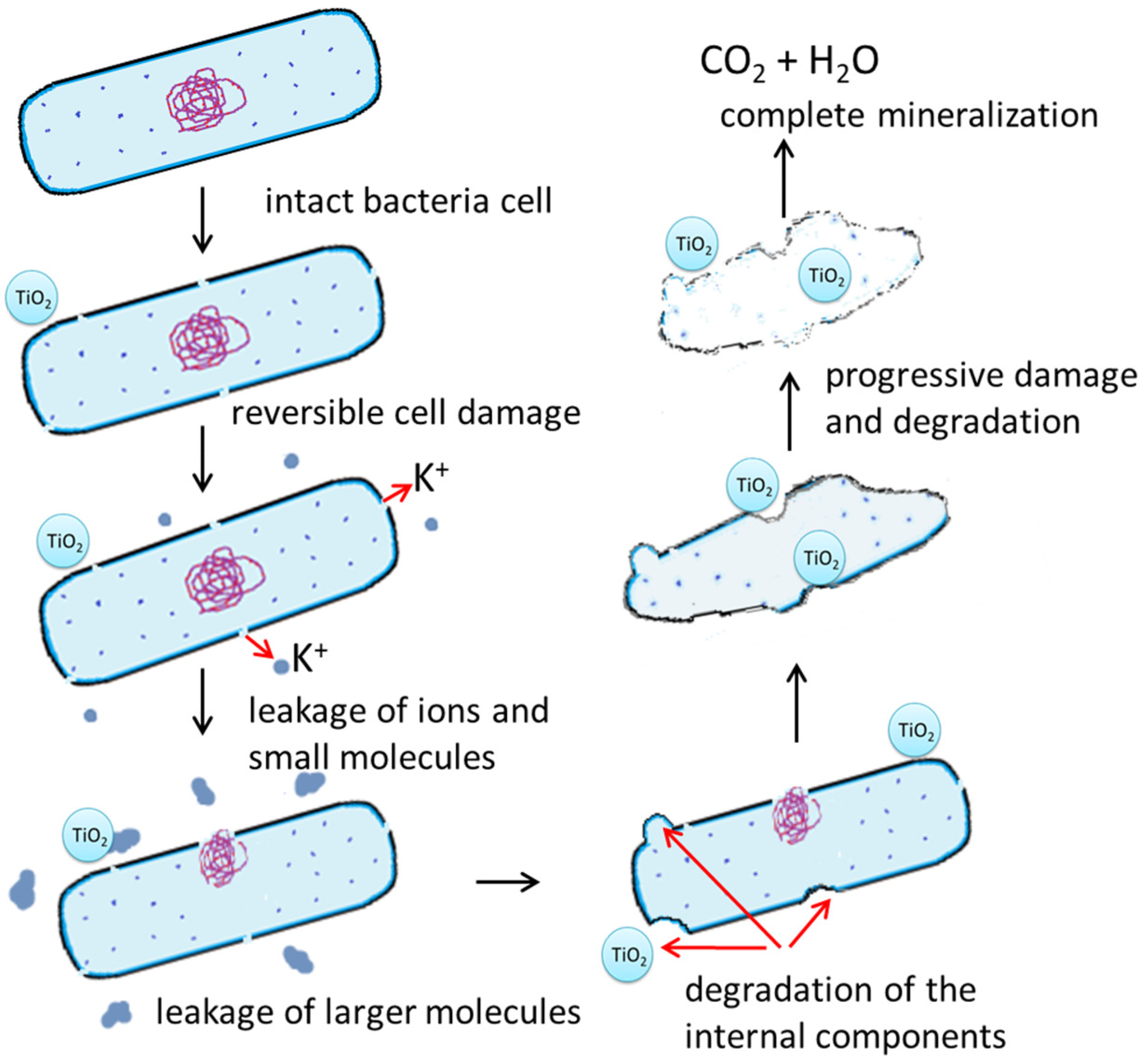
It is well known that in the first stage of bacterial destruction, bacterial cell inactivation must occurred. All presented in Section 2 mechanisms work together, ultimately leading to bacterial cell death. It is worth mentioning that already at this stage, the bacterial cell may present clear and significant damages (shape-shifting, membrane depressions, disruption of the cell membrane). Bacterial inactivation via membrane and cell wall damages, cytoplasmic membrane, or the leakage of intercellular components has been, for example, observed [24]. Research has shown that bacterial cell destruction could be initiated at any point on the cells. However, a statistical analysis showed that the bacterial cell damages most frequently appear on poles of rod-shaped E. coli cells [61,62]. The prolonged generation of free radicals leads to further and higher damage in bacterial cells. Membrane damage caused the leakage first of small molecules (such as ions) and further higher molecules, such as protein. Additionally, the protrusion of the cytoplasmic membrane and cytoplasmic components from degraded areas of the cell wall may have occurred. Simultaneous peroxidation processes of membrane wall components (e.g., lipopolysaccharide, phosphatidylethanolcholine, and peptidoglycan) are taking place. First comes the oxidation and mineralization of relatively small molecular compounds, such as simple sugars (e.g., pentoses, hexose forming part of lipo-polysaccharide) and uronic acids composing the outer membrane [57]. Next, phosphatidylethanolcholine and finally peptidoglycan are oxidized and mineralized [47]. If the photocatalytic process takes place in a suspension containing a suspended photocatalyst, the organic residual of decomposed bacteria, or even a part of a destructed cell, it may be absorbed on the surface of nanomaterials and be destructed to large biomolecules [58]. Bacterial cell could also be degraded to MDA (via the lipid peroxidation process), which in turn can be degraded to ring cleavage products (such as monoaldehydes, monoketones, carboxylic acids) and finally mineralized to CO2 and H2O. A simple scheme for the degradation and mineralization mechanism of TiO2 on bacteria is presented in Figure 2.
Kozlova et al. [22] observed that the mineralization of B. thuringiensis deposited on the surface of TiO2-covered glass slides proceeded in two stages. The division into stages was made on the basis of the rate of CO2 evolution. First comes the transformation part of bacteria and the large molecules of organic compounds constituting bacteria into the products of partial oxidation. This stage last approx. 30 h and is characterized by a gradual slow evolution of CO2. In the next stage, the products of partial oxidation are transformed into carbon dioxide; therefore, the rate of CO2 evolution increases. This stage can last up to 150 h.

5. Conclusions and Future Perspectives

According to the literature, TiO2 could photomineralize the bacterial cell, leaving no organic materials in purified water and on the surface of air purification filters. The results obtained so far indicate that the process of photocatalytic bacterial mineralization most probably proceeds in two stages. In the first stage, gradually, cell wall and membrane oxidation occurs. In the next stage, it comes to the mineralization of internal components that have been released from the injured cell. Unfortunately, due to large discrepancies in the timing of the various stages in studies conducted by different teams and researchers, it is difficult to determine the duration of the various stages of mineralization.
In reviewing the literature concerning photocatalytic microorganism mineralization, it was observed that this topic has not been thoroughly investigated. Therefore, an important aspect is further research on photocatalytic bacterial mineralization.
A significant aspect to be addressed in the future is the lack of standardization of conducted research in the field of photocatalytic microorganism inactivation and mineralization. The standardization of conducted studies will facilitate a comparison of future results. Additionally, the range of tested microorganisms should be expanded (such as fungi or algae).
Interestingly enough, as early as 2003, Sun et al. [51] mentioned that photocatalytic bacterial mineralization could improve the microbiology safety level of treated water by reducing the microorganism regrowth and elimination of biomass from the membrane. Gupta and Modak [63], in their critical review about the mechanistic aspects of photocatalysis microbial disinfection, noted that one of the aspects that reduces the activity of the photocatalyst and its reuse in subsequent cycles can be a deposition of dead bacterial cell mass on its surface. Considering that the deposition of a dead bacterial cell mass on the TiO2 surface can cause a decrease in photocatalytic activity, the examination of the bacterial cell mineralization and determination of optimal process parameters is useful from an economic point of view.
It is also worth expanding the range of research methods used to examine photocatalytic bacterial mineralization. Molecular techniques based on the quantification of DNA could be, for example, considered. These methods would allow for the detection of individual DNA or protein fragments after the photocatalytic process. It is also important to investigate the toxicity of the bacterial degradation intermediates during the photocatalytic mineralization process, e.g., by using methods based on bioluminescence technology to monitor contaminations.

Author Contributions

Conceptualization, P.R.-K.; validation, A.W.M.; formal analysis, P.R.-K. and A.W.M.; resources, P.R.-K.; data curation, P.R.-K.; writing—original draft preparation, P.R.-K.; writing—review and editing, A.W.M.; visualization, P.R.-K., supervision, A.W.M.; All authors have read and agreed to the published version of the manuscript.

Funding

This study received no external funding.

Institutional Review Board Statement

Not applicable.

Informed Consent Statement

Not applicable.

Data Availability Statement

The data presented in this study are available upon request from the corresponding author.

Conflicts of Interest

The authors declare no conflicts of interest.

References

  1. Liu, H.; Wang, C.; Wang, G. Photocatalytic advanced oxidation processes for water treatment: Recent advances and perspective. Chem. Asian J. 2020, 15, 3239–3253. [Google Scholar] [CrossRef]
  2. Verma, V.; Al-Dossari, M.; Singh, J.; Rawat, M.; Kordy, M.G.M.; Shaban, M. A Review on green synthesis of TiO2 NPs: Photocatalysis and antimicrobial applications. Polymers 2022, 14, 1444. [Google Scholar] [CrossRef]
  3. Eidsvåg, H.; Bentouba, S.; Vajeeston, P.; Yohi, S.; Velauthapillai, D. TiO2 as a photocatalyst for water splitting—An experimental and theoretical review. Molecules 2021, 26, 1687. [Google Scholar] [CrossRef]
  4. Li, Z.; Wang, S.; Wu, J.; Zhou, W. Recent progress in defective TiO2 photocatalysts for energy and environmental applications. Renew. Sustain. Energy Rev. 2022, 156, 111980. [Google Scholar] [CrossRef]
  5. Banerjee, S.; Dionysiou, D.D.; Pillai, S.C. Self-cleaning applications of TiO2 by photo-induced hydrophilicity and photocatalysis. Appl. Catal. B Environ. 2015, 176, 396–428. [Google Scholar] [CrossRef]
  6. Al Jitan, S.; Palmisano, G.; Garlisi, C. Synthesis and surface modification of TiO2-based photocatalysts for the conversion of CO2. Catalysts 2020, 10, 227. [Google Scholar] [CrossRef]
  7. Rafique, M.; Hajra, S.; Irshad, M.; Usman, M.; Imran, M.; Assiri, M.A.; Ashraf, W.M. Hydrogen production using TiO2-based photocatalysts: A comprehensive review. ACS Omega 2023, 8, 25640–25648. [Google Scholar] [CrossRef]
  8. Chen, D.; Cheng, Y.; Zhou, N.; Chen, P.; Wang, Y.; Li, K.; Huo, S.; Cheng, P.; Peng, P.; Zhang, R.; et al. Photocatalytic degradation of organic pollutants using TiO2-based photocatalysts: A review. J. Clean. Prod. 2020, 268, 121725. [Google Scholar] [CrossRef]
  9. Zeshan, M.; Bhatti, I.A.; Mohsin, M.; Iqbal, M.; Amjed, N.; Nisar, J.; Al Masoud, L.; Alomar, T.S. Remediation of pesticides using TiO2 based photocatalytic strategies: A review. Chemosphere 2022, 300, 134525. [Google Scholar] [CrossRef]
  10. Zhang, B.; Sun, B.; Liu, F.; Gao, T.; Zhou, G. TiO2-based S-scheme photocatalysts for solar energy conversion and environmental remediation. Sci. China Mater. 2024, 67, 424–443. [Google Scholar] [CrossRef]
  11. Jafari, S.; Mahyad, B.; Hashemzadeh, H.; Janfaza, S.; Gholikhani, T.; Tayebi, L. Biomedical applications of TiO2 nanostructures: Recent advances. Int. J. Nanomed. 2020, 15, 3447–3470. [Google Scholar] [CrossRef]
  12. Kodithuwakku, P.; Jayasundara, D.R.; Munaweera, I.; Jayasinghe, R.; Thoradeniya, T.; Weerasekera, M.; Ayajan, P.A.; Kottegoda, N. A review on recent developments in structural modification of TiO2 for food packaging applications. Prog. Solid State Chem. 2022, 67, 100369. [Google Scholar] [CrossRef]
  13. Reddy, P.V.L.; Kavitha, B.; Reddy, P.A.K.; Kim, K.H. TiO2-based photocatalytic disinfection of microbes in aqueous media: A review. Environ. Res. 2017, 154, 296–303. [Google Scholar] [CrossRef]
  14. Demirel, C.S.U.; Birben, N.C.; Bekbolet, M. A comprehensive review on the use of second generation TiO2 photocatalysts: Microorganism inactivation. Chemosphere 2018, 211, 420–448. [Google Scholar] [CrossRef]
  15. Amiri, M.R.; Alavi, M.; Taran, M.; Kahrizi, D. Antibacterial, antifungal, antiviral, and photocatalytic activities of TiO2 nanoparticles, nanocomposites, and bio-nanocomposites: Recent advances and challenges. J. Public Health Res. 2022, 11, 22799036221104151. [Google Scholar] [CrossRef]
  16. Bono, N.; Ponti, F.; Punta, C.; Candiani, G. Effect of UV Irradiation and TiO2-photocatalysis on airborne bacteria and viruses: An overview. Materials 2021, 14, 1075. [Google Scholar] [CrossRef]
  17. De Pasquale, I.; Porto, C.L.; Dell’Edera, M.; Curri, M.L.; Comparelli, R. TiO2-based nanomaterials assisted photocatalytic treatment for virus inactivation: Perspectives and applications. Curr. Opin. Chem. Eng. 2021, 34, 100716. [Google Scholar] [CrossRef] [PubMed]
  18. Jacoby, W.A.; Maness, P.C.; Wolfrum, E.J.; Blake, D.M.; Fennell, J.A. Mineralization of bacterial cell mass on a photocatalytic surface in air. Environ. Sci. Technol. 1998, 32, 2650–2653. [Google Scholar] [CrossRef]
  19. Mudhoo, A.; Paliya, S.; Goswami, P.; Singh, M.; Lofrano, G.; Carotenuto, M.; Libralato, G.; Guida, M.; Usman, M.; Kumar, S. Fabrication, functionalization and performance of doped photocatalysts for dye degradation and mineralization: A review. Environ. Chem. Lett. 2020, 18, 1825–1903. [Google Scholar] [CrossRef]
  20. Adeyemi, J.O.; Ajiboye, T.; Onwudiwe, D.C. Mineralization of antibiotics in wastewater via photocatalysis. Water Air Soil Pollut. 2021, 232, 219. [Google Scholar] [CrossRef]
  21. Kumar, S.; Sharma, R.; Gupta, A.; Dubey, K.K.; Khan, A.M.; Singhal, R.; Kumar, R.; Bharti, A.; Singh, P.; Kant, R.; et al. TiO2 based Photocatalysis membranes: An efficient strategy for pharmaceutical mineralization. Sci. Total Environ. 2022, 845, 157221. [Google Scholar] [CrossRef]
  22. Kozlova, E.A.; Safatov, A.S.; Kiselev, S.A.; Marchenko, V.Y.; Sergeev, A.A.; Skarnovich, M.O.; Emelyanova, E.K.; Smetannikova, M.A.; Buryak, G.A.; Vorontsov, A.V. Inactivation and mineralization of aerosol deposited model pathogenic microorganisms over TiO2 and Pt/TiO2. Environ. Sci. Technol. 2010, 44, 5121–5126. [Google Scholar] [CrossRef] [PubMed]
  23. Taddese, R.; Belzer, C.; Aalvink, S.; de Jonge, M.I.; Nagtegaal, I.D.; Dutilh, B.E.; Boleij, A. Production of inactivated gram-positive and gram-negative species with preserved cellular morphology and integrity. J. Microbiol. Methods 2021, 184, 106208. [Google Scholar] [CrossRef]
  24. Foster, H.A.; Ditta, I.B.; Varghese, S.; Steele, A. Photocatalytic disinfection using titanium dioxide: Spectrum and mechanism of antimicrobial activity. Appl. Microbiol. Biotechnol. 2011, 90, 1847–1868. [Google Scholar] [CrossRef]
  25. Matsunaga, T.; Tomoda, R.; Nakajima, T.; Wake, H. Photochemical sterilization of microbial cells by semiconductor powder. FEMS Microbiol. Lett. 1985, 1, 211–214. [Google Scholar]
  26. Dalrymple, O.K.; Stefanakos, E.; Trotz, M.A.; Goswami, D.Y. A review of the mechanisms and modeling of photocatalytic disinfection. Appl. Catal. B Environ. 2010, 98, 27–38. [Google Scholar] [CrossRef]
  27. Pagnout, C.; Jomini, S.; Dadhwal, M.; Caillet, C.; Thomas, F.; Bauda, P. Role of electrostatic interactions in the toxicity of titanium dioxide nanoparticles toward Escherichia coli. Colloids Surf. B 2012, 92, 315–321. [Google Scholar] [CrossRef]
  28. Liao, D.L.; Wu, G.S.; Liao, B.Q. Zeta potential of shape-controlled TiO2 nanoparticles with surfactants. Colloids Surf. A Physicochem. Eng. Asp. 2009, 348, 270–275. [Google Scholar] [CrossRef]
  29. Shah, A.H.; Rather, M.A. Effect of calcination temperature on the crystallite size, particle size and zeta potential of TiO2 nanoparticles synthesized via polyol-mediated method. Mater. Today Proc. 2021, 44, 482–488. [Google Scholar] [CrossRef]
  30. Saito, T.; Iwase, T.; Horie, J.; Morioka, T. Mode of photocatalytic bactericidal action of powdered semiconductor TiO2 on mutans streptococci. J. Photochem. Photobiol. B Biol. 1992, 14, 369–379. [Google Scholar] [CrossRef]
  31. Hu, C.; Guo, J.; Qu, J.; Hu, X. Photocatalytic degradation of pathogenic bacteria with AgI/TiO2 under visible light irradiation. Langmuir 2007, 23, 4982–4987. [Google Scholar] [CrossRef] [PubMed]
  32. Gogniat, G.; Thyssen, M.; Denis, M.; Pulgarin, C.; Dukan, S. The bactericidal effect of TiO2 photocatalysis involves adsorption onto catalyst and the loss of membrane integrity. FEMS Microbiol. Lett. 2006, 258, 18–24. [Google Scholar] [CrossRef] [PubMed]
  33. Maness, P.C.; Smolinski, S.; Blake, D.M.; Huang, Z.; Wolfrum, E.J.; Jacoby, W.A. Bactericidal activity of photocatalytic TiO2 reaction: Toward an understanding of its killing mechanism. Appl. Environ. Microbiol. 1999, 65, 4094–4098. [Google Scholar] [CrossRef] [PubMed]
  34. Kiwi, J.; Nadtochenko, V. New evidence for TiO2 photocatalysis during bilayer lipid peroxidation. J. Phys. Chem. B 2004, 108, 17675–17684. [Google Scholar] [CrossRef]
  35. Carré, G.; Hamon, E.; Ennahar, S.; Estner, M.; Lett, M.C.; Horvatovich, P.; Gies, J.P.; Andre, P. TiO2 photocatalysis damages lipids and proteins in Escherichia coli. Appl. Environ. Microbiol. 2014, 80, 2573–2581. [Google Scholar] [CrossRef] [PubMed]
  36. Koizumi, Y.; Yamada, R.; Nishioka, M.; Matsumura, Y.; Tsuchido, T.; Taya, M. Deactivation kinetics of Escherichia coli cells correlated with intracellular superoxide dismutase activity in photoreaction with titanium dioxide particles. J. Chem. Technol. Biotechnol. 2002, 77, 671–677. [Google Scholar] [CrossRef]
  37. Kumar, A.; Pandey, A.K.; Singh, S.S.; Shanker, R.; Dhawan, A. Engineered ZnO and TiO2 nanoparticles induce oxidative stress and DNA damage leading to reduced viability of Escherichia coli. Free. Radic. Biol. Med. 2011, 51, 1872–1881. [Google Scholar] [CrossRef]
  38. Rokicka-Konieczna, P.; Wanag, A.; Sienkiewicz, A.; Kusiak-Nejman, E.; Morawski, A.W. Effect of APTES modified TiO2 on antioxidant enzymes activity secreted by Escherichia coli and Staphylococcus epidermidis. Biochem. Biophys. Res. Commun. 2021, 534, 1064–1068. [Google Scholar] [CrossRef]
  39. Zhang, H.M.; Cao, J.; Tang, B.P.; Wang, T.G. Effect of TiO2 nanoparticles on the structure and activity of catalase. Chem. Biol. Interact. 2014, 19, 168–174. [Google Scholar] [CrossRef]
  40. Gogniat, G.; Dukan, S. TiO2 photocatalysis causes DNA damage via Fenton reaction-generated hydroxyl radicals during the recovery period. Appl. Environ. Microbiol. 2007, 73, 7740–7743. [Google Scholar] [CrossRef]
  41. Liu, Y.; Huang, J.; Feng, X.; Li, H. Thermal-sprayed photocatalytic coatings for biocidal applications: A review. J. Therm. Spray Technol. 2021, 30, 1–24. [Google Scholar] [CrossRef] [PubMed]
  42. Vohra, A.; Goswami, D.Y.; Deshpande, D.A.; Block, S.S. Enhanced photocatalytic disinfection of indoor air. Appl. Catal. B Environ. 2006, 64, 57–65. [Google Scholar] [CrossRef]
  43. Rao, N.G.; Kumar, A.; Wong, J.S.; Shridhar, R.; Goswami, D.Y. Effect of a novel photoelectrochemical oxidation air purifier on nasal and ocular allergy symptoms. Ther. Adv. Allergy Rhinol. 2018, 9, 2152656718781609. [Google Scholar] [CrossRef]
  44. Goswami, D.Y. A review of engineering developments of aqueous phase solar photocatalytic detoxification and disinfection processes. J. Sol. Energy Eng. 1997, 119, 101–107. [Google Scholar] [CrossRef]
  45. Goswami, T.K.; Hingorani, S.K.; Greist, H.; Goswami, D.Y.; Block, S.S. Photocatalytic system to destroy bioaerosols in air. J. Adv. Oxid. Technol. 1999, 4, 185–188. [Google Scholar]
  46. Wolfrum, E.J.; Huang, J.; Blake, D.M.; Maness, P.C.; Huang, Z.; Fiest, J.; Jacoby, W.A. Photocatalytic oxidation of bacteria, bacterial and fungal spores, and model biofilm components to carbon dioxide on titanium dioxide-coated surfaces. Environ. Sci. Technol. 2002, 36, 3412–3419. [Google Scholar] [CrossRef] [PubMed]
  47. Kiwi, J.; Nadtochenko, V. Evidence for the mechanism of photocatalytic degradation of the bacterial wall membrane at the TiO2 interface by ATR-FTIR and laser kinetic spectroscopy. Langmuir 2005, 21, 4631–4641. [Google Scholar] [CrossRef]
  48. Nadtochenko, V.A.; Sarkisov, O.M.; Nikandrov, V.V.; Chubukov, P.A.; Denisov, N.N. Inactivation of pathogenic microorganisms in the photocatalytic process on nanosized TiO2 crystals. Russ. J. Phys. Chem. B 2008, 2, 105–114. [Google Scholar] [CrossRef]
  49. López, J.E.O.; Jacoby, W.A. Microfibrous mesh coated with titanium dioxide: A self-sterilizing, self-cleaning filter. J. Air Waste Manag. Assoc. 2002, 52, 1206–1213. [Google Scholar] [CrossRef]
  50. Sökmen, M.; Candan, F.; Sümer, Z. Disinfection of E. coli by the Ag-TiO2/UV system: Lipidperoxidation. J. Photochem. Photobiol. A Chem. 2001, 143, 241–244. [Google Scholar] [CrossRef]
  51. Sun, D.D.; Tay, J.H.; Tan, K.M. Photocatalytic degradation of E. coliform in water. Water Res. 2003, 37, 3452–3462. [Google Scholar] [CrossRef] [PubMed]
  52. Faraji, M.; Mohaghegh, N.; Abedini, A. Ternary composite of TiO2 nanotubes/Ti plates modified by g-C3N4 and SnO2 with enhanced photocatalytic activity for enhancing antibacterial and photocatalytic activity. J. Photochem. Photobiol. B 2018, 178, 124–132. [Google Scholar] [CrossRef]
  53. Cheng, Y.W.; Chan, R.C.; Wong, P.K. Disinfection of Legionella pneumophila by photocatalytic oxidation. Water Res. 2007, 41, 842–852. [Google Scholar] [CrossRef] [PubMed]
  54. Youji, L.I.; Mingyuan, M.A.; Xiaohu, W.A.N.G.; Xiaohua, W.A.N.G. Inactivated properties of activated carbon-supported TiO2 nanoparticles for bacteria and kinetic study. J. Environ. Sci. 2008, 20, 1527–1533. [Google Scholar] [CrossRef] [PubMed]
  55. Kowalska, E.; Wei, Z.; Karabiyik, B.; Herissan, A.; Janczarek, M.; Endo, M.; Markowska-Szczupak, A.; Remita, H.; Ohtani, B. Silver-modified titania with enhanced photocatalytic and antimicrobial properties under UV and visible light irradiation. Catal. Today 2015, 252, 136–142. [Google Scholar] [CrossRef]
  56. Endo, M.; Wei, Z.; Wang, K.; Karabiyik, B.; Yoshiiri, K.; Rokicka, P.; Ohtani, B.; Markowska-Szczupak, A.; Kowalska, E. Noble metal-modified titania with visible-light activity for the decomposition of microorganisms. Beilstein J. Nanotechnol. 2018, 9, 829–841. [Google Scholar] [CrossRef]
  57. Rokicka-Konieczna, P.; Markowska-Szczupak, A.; Kusiak-Nejman, E.; Morawski, A.W. Photocatalytic water disinfection under the artificial solar light by fructose-modified TiO2. Chem. Eng. J. 2019, 372, 203–215. [Google Scholar] [CrossRef]
  58. Shi, H.; Li, G.; Sun, H.; An, T.; Zhao, H.; Wong, P.K. Visible-light-driven photocatalytic inactivation of E. coli by Ag/AgX-CNTs (X = Cl, Br, I) plasmonic photocatalysts: Bacterial performance and deactivation mechanism. Appl. Catal. B Environ. 2014, 158, 301–307. [Google Scholar] [CrossRef]
  59. Yang, G.; Yin, H.; Liu, W.; Yang, Y.; Zou, Q.; Luo, L.; Li, H.; Huo, Y.; Li, H. Synergistic Ag/TiO2-N photocatalytic system and its enhanced antibacterial activity towards Acinetobacter baumannii. Appl. Catal. B Environ. 2018, 224, 175–182. [Google Scholar] [CrossRef]
  60. Kozak, M.; Mazierski, P.; Żebrowska, J.; Klimczuk, T.; Lisowski, W.; Żak, A.M.; Skowron, P.M.; Zaleska-Medynska, A. Detailed insight into photocatalytic inactivation of pathogenic bacteria in the presence of visible-light-active multicomponent photocatalysts. Nanomaterials 2024, 14, 409. [Google Scholar] [CrossRef]
  61. Liou, J.W.; Chang, H.H. Bactericidal effects and mechanisms of visible light-responsive titanium dioxide photocatalysts on pathogenic bacteria. Arch. Immunol. Ther. Exp. 2012, 60, 267–275. [Google Scholar] [CrossRef] [PubMed]
  62. Liou, J.W.; Gu, M.H.; Chen, Y.K.; Chen, W.Y.; Chen, Y.C.; Tseng, Y.H.; Hung, J.L.; Chang, H.H. Visible light responsive photocatalyst induces progressive and apical-terminus preferential damages on Escherichia coli surfaces. PLoS ONE 2011, 6, e19982. [Google Scholar] [CrossRef] [PubMed]
  63. Gupta, R.; Modak, J. Bacterial lysis via photocatalysis-A critical mechanistic review. ChemCatChem 2020, 12, 2148–2170. [Google Scholar] [CrossRef]
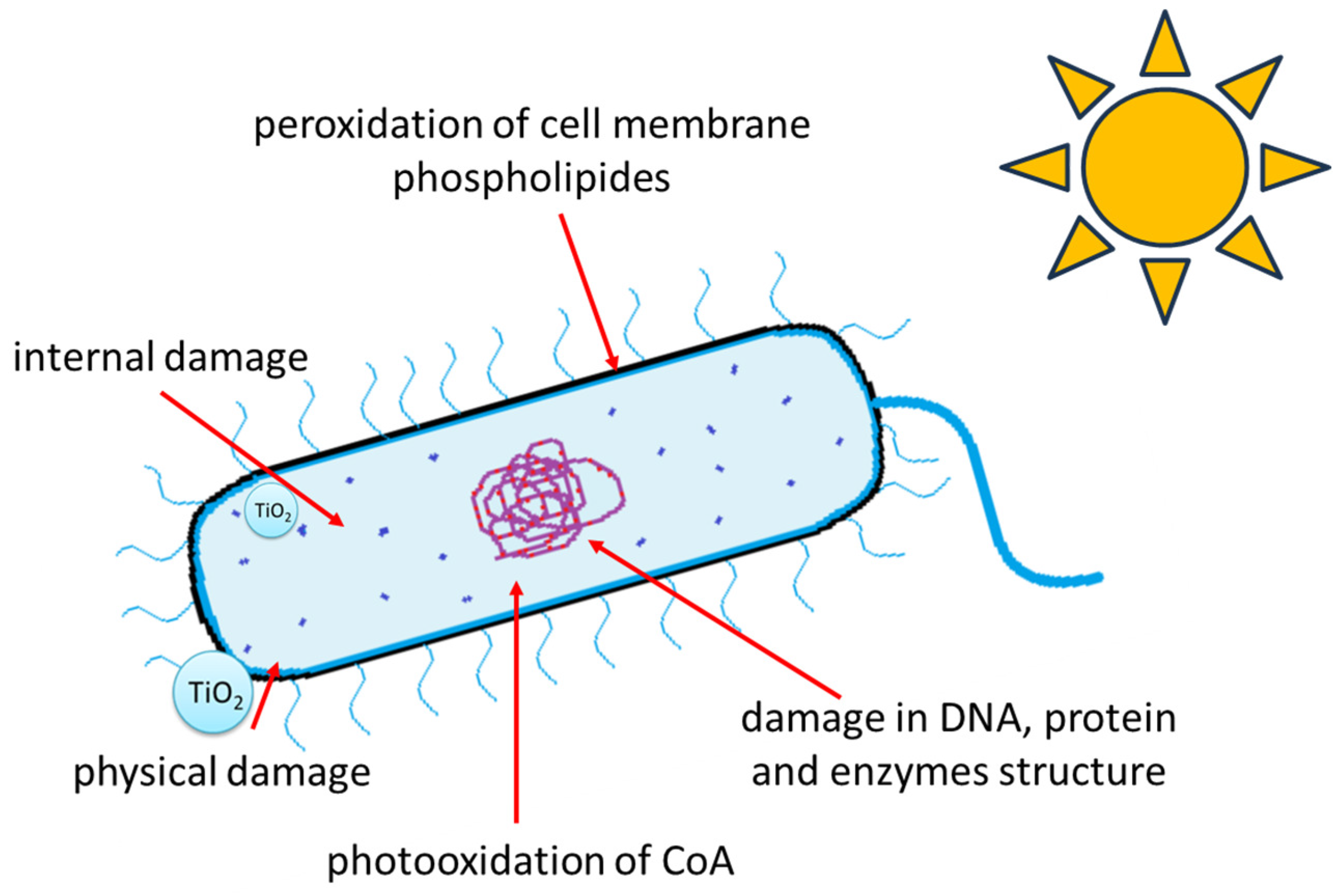
Figure 1. Summary of mechanisms leading to photocatalytic bacterial inactivation.
Molecules 29 02221 g001
Figure 2. Scheme for proposed photocatalytic destruction and mineralization of bacteria in the presence of TiO2.
Molecules 29 02221 g002
Table 1. Research conducted on photocatalytic microorganism destruction/mineralization in air.
Research MethodsPhotocatalystRadiation TypeUsed
Microorganisms
Experiment TimeResults/ObservationsRef.
GC—CO2 measurements,
14C radioisotope labelling
SEM analysis
P25 coated on glassUVE. coli75 hmineralization of bacteria[18]
GC—CO2 measurements + kinetic data and carbon mass balanceP25 coated on quartz disksUVE. coli,
M. luteus,
B. cereus,
A. niger
72–144 hmineralization of microorganisms[46]
FTIR spectrometer fitted with a gas cell—CO2 measurements,
SEM analysis
metal microfibrous
mesh coated with TiO2 (P25)
UVE. coli168 htotal remove bacteria from mesh[49]
ATR-FTIR spectroscopyP25 porous filmUV-AE. coli44 hchanges in spectral bands suggesting the mineralization[47]
ATR-FTIR spectroscopyP25 porous filmUV-AE. coli16 htotal oxidation of cell organic matter[48]
GC—CO2 measurementsTiO2 (Hombifine N) and Pt/TiO2 on glass platesUVB. thuringiensis50–150 hfull photocatalytic bacterial mineralization[22]
GC—gas chromatography, PM—photocatalytic mineralization.
Table 2. Research conducted on photocatalytic microorganism destruction/mineralization in aqueous solutions.
Research MethodsPhotocatalystRadiation TypeUsed
Microorganisms
Experiment TimeResults/ObservationsRef.
MDA formation
GC—CO2 measurements
TiO2
or Ag-TiO2
UVE. coli40 mincomplete mineralization of E. coli[50]
GC—CO2 measurements,
SEM analysis
TiO2–Fe2O3 powderUVE. coli75 hE. coli mineralization[51]
TEM analysis,
TOC analysis
P25UVL. pneumophila2 hbacterial mineralization[53]
SEM analysisTiO2/AC compositesUVE. coli250 minnearly total bacterial decomposition[54]
FT-IR analysis Ag/AgX-CNTs
(X = Cl, Br, I)
VLE. coli2 hdestruction of E. coli to large
biomolecules
[58]
GC—CO2 measurementsAg@TiO2UVE. coli6 hinitial stage of bacterial mineralization[55]
TOC analysisAg/TiO2-NVLA. baumannii0.5 hinitial stage of bacterial mineralization[59]
GC—CO2 measurements,
SEM analysis
g-C3N4-SnO2/TiO2 nanotubes/Ti platesVLE. coli32 hcomplete mineralization of E. coli[52]
FID-GC—CO2 measurementsAg/TiO2VLE. coli3 hinitial stage of bacterial mineralization[56]
GC—CO2 measurementsTiO2 modified by carbonUVA
ASL
E. coli
S. epidermidis
3 hinitial stage of bacterial mineralization[57]
GC—CO2 measurementsTiO2/Ag2O/AuO NTsVLS. aureus4 hinitial stage of bacterial mineralization[60]
GC—gas chromatography, VL—visible light, ASL—artificial solar light, FID—GC—flame ionization detector for gas chromatography.
Disclaimer/Publisher’s Note: The statements, opinions and data contained in all publications are solely those of the individual author(s) and contributor(s) and not of MDPI and/or the editor(s). MDPI and/or the editor(s) disclaim responsibility for any injury to people or property resulting from any ideas, methods, instructions or products referred to in the content.