Article

Discovery of Anti-Inflammatory Triterpenoid Glucosides from the Heritiera littoralis Dryand

1
Guangxi Key Laboratory of Green Chemical Materials and Safety Technology, College of Petroleum and Chemical Engineering, Beibu Gulf University, Qinzhou 535000, China
2
State Key Laboratory for the Chemistry and Molecular Engineering of Medicinal Resources, School of Chemical & Pharmaceutical Sciences, Guangxi Normal University, Guilin 541004, China
3
Peoples’ Hospital of Pubei, Qinzhou 535300, China
*
Authors to whom correspondence should be addressed.
Molecules 2023, 28(4), 1658; https://doi.org/10.3390/molecules28041658
Submission received: 19 December 2022 / Revised: 3 February 2023 / Accepted: 6 February 2023 / Published: 9 February 2023
(This article belongs to the Section Natural Products Chemistry)

Abstract

:
Two new triterpenoid glucosides, Heritiera A (1) and Heritiera B (2), and six known triterpenoid analogs (38) were isolated from Heritiera littoralis Dryand. Their structures were identified by comprehensive spectroscopic analyses and comparisons with the literature. The anti-inflammatory activity of the isolates from H. littoralis was evaluated using a lipopolysaccharide (LPS) stimulated RAW 264.7 cells model. The result showed that four triterpenoids exhibited potent anti-inflammatory activity. Among these compounds, compound 2 substantially inhibits the release of nitric oxide (NO) with an IC50 value of 10.33 μM. The triterpenoids from H. littoralis could be used as potential candidates for the development of new anti-inflammatory agents.

Graphical Abstract

1. Introduction

Inflammation is a host defense response to danger signals from pathogens, damaged cells, irradiation, etc. [1]. The characteristic signs of acute inflammation include redness, swelling, warmth, and pain, which occur during various acute infections or tissue damage [2]. In the inflammatory response, there is an increase in the permeability of endothelial lining cells and influxes of blood leukocytes into the interstitium, oxidative burst, and release of cytokines (interleukins and tumor necrosis factor-α). At the same time, there is also induction of the activity of several enzymes (oxygenases, NO synthases, and peroxidases) as well as the arachidonic acid metabolism [3]. NO is a gaseous short-lived free radical that is considered to be a mediator of inflammation. Regulating the biosynthesis or activity of NO results in the amelioration of the acute inflammation model [4]. Thus, inhibitors of some pro-inflammatory mediators from folk herbs have potential anti-inflammatory activities, which are of great interest to scientific researchers.
At present, the main drugs used in the treatment of inflammation are non-steroidal anti-inflammatory drugs, steroidal anti-inflammatory drugs, and traditional Chinese medicine. As synthetic anti-inflammatory drugs used in clinical currently have obvious adverse reactions, however traditional Chinese medicines with less toxicity and side effects have attracted more and more attention [5,6]. Natural plant components, such as triterpenoids and their glycosides, have various structural types, strong biochemical specificity, good curative effect, and low drug resistance, which are good resources for the development of potentially effective anti-inflammatory drug candidates [7,8]. These compounds have a good inhibitory effect on acute and chronic inflammation, which show an anti-inflammatory activity by inhibiting the production of inflammatory factors, and oleanane and ursane triterpenoids exhibit the most significant hypoglycemic activity, especially among triterpenoids [9,10,11].
The genus Heritiera belongs to the family Malvaceae growing in tropical and subtropical regions of Asia [12]. It consists of 35 species, of which 3 species can be found in the provinces of Guangdong, Guangxi, Hainan, and Yunnan in China. The genus Heritiera has a long history of medicinal use in China, especially for folk use [13,14]. Heritiera littoralis Dryand., a semi-mangrove plant, has been used as a traditional medicine for the Jing nationality [15]. This plant has been reported for its wide range of activities, including the treatment of hematuria, diarrhea, dysentery, oral infection, and toothache [16,17,18]. So far, Heritiera littoralis has been reported to have significant anti-inflammatory activity in the folk [14]. The isolation and characterization of the chemical constituents from H. littoralis have yielded a range of classes of natural products, such as triterpenoid, flavonoid, phenylpropanoid-glycerol, etc. [19,20,21,22,23]. However, there have been few reports on the biologically active constituents of H. littoralis [24].
To explore the anti-inflammatory lead compounds from the genus Heritiera, the active ingredients were separated from the leaves of H. littoralis. The structures of all isolated constituents were characterized by comprehensive spectroscopic analyses and comparisons with the literature. These findings have led to the isolation of two new triterpenoid glucosides, Heritiera A (1) and Heritiera B (2), and six known triterpenoid analogs (38) Figure 1. Furthermore, the isolates were evaluated for their anti-inflammatory activities against the production of the NO in LPS-induced inflammation in RAW264.7 macrophage cells. Herein, the isolation, purification, and determination of these isolates and the assays used to determine the production of NO in RAW264.7 macrophage cells of the constituents are described. The research provided the basis for expanding the utilization and development of this medicinal plant.

2. Results and Discussion

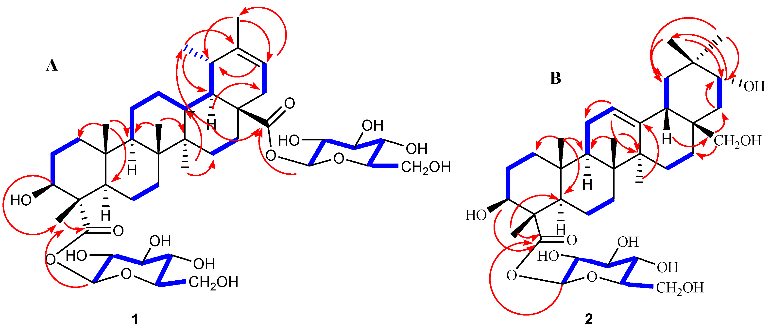
2.1. Elucidation of the Chemical Structure of Heritiera A (1) and Heritiera B (2)

Heritiera A (1) was obtained as a colorless gum whose molecular formula was determined to be C42H66O15 from the HRESIMS data at m/z 834.4338 [M + Na]+ with ten indices of hydrogen deficiency. The 1H NMR data Table 1 displayed six methyl singlets (δH 0.86, s, 6H; δH 1.05, s, 3H; δH 1.11, s, 3H; δH 1.59, s, 3H; δH 1.67, s, 3H), one olefinic proton (δH 5.35, 1H, d, J = 7.2 Hz), two anomeric protons δH 6.48 (1H, dd, J = 8.0, 2.8 Hz) and δH 6.35 (1H, dd, J = 8.0, 2.8 Hz), as well as overlapping signals belonging to sugar moieties around δH 3.88–4.39 (12 H). The 13C NMR and DEPT data (Table 1) revealed the existence of 42 carbon resonances, including six methyls, eleven methylenes, seventeen methines (one olefinic at δC 117.8), and eight quaternary carbons (two carbonyls at δc 174.8, 177.8; one olefinic at δC 143.6). The 1H and 13C NMR spectra also showed the signals of two anomeric carbons at δH 6.48/δC 96.9 (C-1′) and δH 6.35/δC 95.7 (C-1″) indicating the presence of two sugar moieties in 1. Furthermore, D-glucose was identified by the acid hydrolysis solution of 1 by comparison with authentic sugar samples in an HPLC assay [25]. Moreover, five of the ten indices of hydrogen deficiency were ascribed to two carboxyl groups, one double bond, and two hexacylic rings, with the remaining five indices indicating the presence of a pentacyclic system. With the aid of 2D NMR experiments, the 1H−1H COSY (Figure 2A, bold lines) and HSQC data revealed the partial structures –CH2 (1)−CH2 (2)−CH2 (3)−, –CH2 (6)−CH2 (7), −CH (9)−CH2 (11)−CH2 (12)−CH (13)−CH (18)−CH (19)−CH3 (29), –CH2 (15)−CH2 (16) and –CH (21)−CH2 (22)−. Detailed analyses of the abovementioned NMR data showed that the data of 1 were highly similar data to those of 3β-hydroxy-18α,19α-urs-20-en-28-oic acid [26,27], except for the presence of a carboxyl carbon at C-23 (δC 177.8 ppm) and two glycosyl units in 1. Furthermore, the HMBC correlations from the anomeric protons H-1′ (δH 6.48, dd, J = 8.0, 2.8 Hz) to C-23 (δC 177.8) and H-1″ (δH 6.35, dd, J = 8.0, 2.8 Hz) to C-28 (δC 174.8) suggested that the two D-glucoses are situated at C-23 and C-28, respectively. The configuration of the sugar was assigned as β on the basis of coupling constant values (J = 8.0 Hz) of anomeric protons [28]. In addition, the relative stereochemistry was deduced from ROESY spectrum (double arrows in Figure 3A). In the ROESY spectrum, the correlations of H-3α/H-5α and H-9α/H-27α/H-18α indicated that H-3, H-5, H-9, H-18, and H-27 are α-oriented. The correlations of H-24β/H-25β, and H-25β/H-26β/H-13β suggested that H-13, Me-24, Me-25, Me-26 are β-oriented. Consequently, the structure of 1 was established as 3β-hydroxy-18α,19α-urs-20(21)-en-23,28-dioic acid-23,28-O-β-D-lucopyranosyl diester, and named Heritiera A (Figure 1).
Heritiera B (2) was obtained as a colorless gum whose molecular formula was determined to be C36H58O10 from the HRESIMS data at m/z 673.3920 [M + Na]+ with eight indices of hydrogen deficiency. The 1H NMR data (Table 1) displayed six methyl singlets (δH 0.92, s, 3H; δH 0.96, s, 3H; δH 1.00, s, 3H; δH 1.20, s, 3H; δH 1.34, s, 3H; δH 1.60, s, 3H) respectively, and one olefinic proton (δH 5.45, 1H, d, J = 8.0 Hz), one anomeric proton δH 6.40 (1H, d, J = 8.0 Hz), as well as overlapping hydrogen signals in oxygenated carbons perhaps belonging to sugar moieties. The 13C NMR and DEPT data (Table 1) revealed the existence of thirty-six carbon resonances, including six methyls, eleven methylenes (one olefinic at δC 117.6), eleven methines (one olefinic at δC 158.5), and eight quaternary carbons (one carbonyl at δC 178.1). The 1H and 13C NMR spectra also showed the signals of one anomeric carbon at δH 6.40 (d, J = 8.0 Hz)/δC 96.9 (C-1′), indicating the presence of one sugar moiety in the molecule, after acid hydrolysis of 2, the D-glucose existed in 2 by analysis of HPLC [25]. In addition, the configuration of the sugar was assigned as β on the basis of the coupling constant value (J = 8.0 Hz) of the anomeric proton [28]. Three of the eight indices of hydrogen deficiency were ascribed to one carboxyl group, one double bond, and one hexacylic ring, with the remaining five indices indicating the presence of a pentacyclic system. Above mentioned facts suggested that the aglycone part of 2 was similar to that of erythrodiol [29]; the differences existed mainly in the C (23), C (21), and one more glycosyl unit in 2. The C (23) was a carboxyl carbon with δC 178.1 ppm, and the C (21) was an oxygenated carbon with δC 73.1 ppm. Furthermore, in the HMBC spectrum (arrows in Figure 2B), the anomeric proton at δH 6.40 (H-1′) showed a correlation with δC 178.1 (C-23), which indicated that the sugar moiety is situated at C-23. In addition, the relative stereochemistry was deduced from the ROESY spectrum (double arrows in Figure 3B). In the ROESY spectrum, the correlations of H-3α/H-5α and H-9α/H-27α indicated that H-3, H-5, H-9, and H-27 are α-oriented. The correlations of H-13β/H-20β, H-24β/H-25β, and H-25β/H-26β suggested that H-13, H-20, Me-24, Me-25, Me-26 are β-oriented. Thus, the structure of 2 was established as 3β,21α,28β-trihydroxy-oleana-12(13)-en-23-acid-O-β-D-glucopyranosyl ester, named Heritiera B.
By comparing the measured NMR (1H and 13C) and MS data to those reported in the literature, the known triterpenoids were identified as juglansin A (3) [30], quadranoside IV (4) [31], 2α,3α,23-trihydroxy-12, 20(30)-dien-28-ursolic acid 28-O-β-D-glucopyranoside (5) [32], arjunglucoside II (6) [33], 1-oxo-3β,23-dihydroxy olean-12-en-28-oic acid 28-O-β-D-glucopyranoside (7) [34], and 3β,28-dihydroxy-oleana-11, 13(18) diene (8) [35] (Figure 1).

2.2. Anti-Inflammatory Assay of the Isolates

The RAW264.7 cell viability assays showed that the survival rate was greater than 90% after treatment with all isolates at different concentrations from 0 to 50 μM. The effects of all compounds on the production of NO by LPS-induced RAW 264.7 cells are shown in Table 2. Compound 2 substantially inhibited the release of NO, with an IC50 value of 10.33 μM. The value is slightly lower than that of the positive control, dexamethasone, with an IC50 value of 6.39 μM. Compounds 1, 3, and 4 showed moderate effects with IC50 values of 32.11, 39.32, and 29.98 μM, respectively. Compounds 5, 6, 7, and 8 showed no significant effects against LPS-induced nitric oxide production in RAW264.7 macrophages. However, the anti-inflammatory mechanism of compounds needs to be further discussed. We will conduct research in the next step.

2.3. Similarities and Differences of Some Triterpenoids and Their Anti-Inflammatory Activity from Malvaceae

It was reported that some triterpenoids isolated from Malvaceae exhibited good anti-inflammatory activity, such as taraxerol (oleanane triterpenoid) and lupeol (lupane triterpenoid) from Grewia flava roots yielded promising IC50 values of 21.88 ± 0.02 and 14.2 ± 0.01 μg/mL against NO production in RAW264.7 cells respectively [36]. Betulinic acid (lupane triterpenoid) and taraxasterol (ursane triterpenoid) were isolated from Luehea ochrophylla Mart [37]. The literature reported that betulinic acid was justified by inhibiting the release of pro-inflammatory mediators, mainly NO, IL-1β, TNF-α, and reduction of COX-2 levels [38,39]. Taraxasterol had efficacy comparable to prednisolone in a paw edema model induced by formalin [40]. Compound 2, as an oleanane triterpenoid, showed anti-inflammatory activity and inhibited the release of NO, with an IC50 value of 10.33 μM too. These results support our initial investigation to obtain more anti-inflammatory active triterpenoids from this experiment.

3. Experimental Section

3.1. General Experimental Procedures

Optical rotations were measured on an Anton Paar MCP500 polarimeter (λ 589 nm, path length 1.0 cm). UV spectra were acquired using a TU-1901 spectrophotometer (λ 190~800 nm, slit width 2.0 nm). NMR experiments were conducted on a Bruker Advance 600 MHz or 400 MHz spectrometer with the residual solvent as an internal standard. HRESIMS were recorded by a BRUKER MAT 95XP mass spectrometer, respectively. Semi-preparative HPLC was conducted on a Waters 2545 instrument equipped with a PAD detecter and a Waters C18 column (5 μm, 9.3 × 250 mm) at a flow rate of 2.5 mL/min. Analytical HPLC was conducted on a Waters 2695 instrument equipped with a PAD detecter, ELSD detecter, and a Waters C18 column (5 μm, 4.6 × 250 mm) at a flow rate of 1.0 mL/min. OD values of 96-well were measured with an imark Bio-Rad plate microplate reader.

3.2. Plant Material

Dried leave (5.0 kg) of H. littoralis was obtained from Beilun Estuary in Guangxi Province, China, in September 2018 and identified by professor Qiuping Zhong (College of marine science, Beibu Gulf University). A voucher specimen (No. ID-20180910) is deposited at the Guangxi key laboratory of green chemical materials and safety technology, Beibu Gulf University, China.

3.3. Extraction and Isolation

Dried leaves power (5.0 kg) of H. littoralis was extracted with EtOH-H2O (20 L each, 75:25) under the extraction tank (solid-liquid ratio 1:4 g/mL, 80 °C, 4 h each, repeated 3 times), The filtrates were combined and dried under reduced pressure to yield a residue (0.52 kg, yield extract 10.4%). The concentrates were suspended in H2O (1 L) and partitioned with EtOAc (5 × 1 L) and n-BuOH fraction (5 × 1 L). The n-BuOH fraction (125.3 g) was subjected to silica gel CC (200~300 mesh) and eluted sequentially with CHCl3-MeOH (100:0, 90:10, 80:20, 50:50, and 0:100, v/v) to yield five fractions (A–E). Fraction B (13.2 g) was using RP-C18 silica gel CC and eluted with H2O-MeOH (40:60, 30:70, 20:80, 0:100, v/v) to afford four subfractions; further separation of subfraction B-2 using Sephadex LH-20 CC (eluted with 100% MeOH) yielded compounds 8 (8.9 mg). Fraction C (35.2 g) was purified using RP-C18 silica gel CC and eluted with H2O-MeOH (70:30, 60:40, 50:50, 40:60, 30:70, 20:80, 10:90, 0:100, v/v) to afford eight subfractions (C-1~C-8). Fraction C-1 (1.1 g) was fractionated by silica gel CC with a CH2Cl2-EA gradient elution (100:0 → 0:100, v/v) to afford five fractions C-1-(1–5). Fraction C-1-2 was subjected to Sephadex LH-20 CC (eluted with 100% MeOH) and recrystallization to yield compounds 5 (5.6 mg) and 7 (4.9 mg). Fraction C-1-3 was subjected to Sephadex LH-20 CC (eluted with H2O-MeOH (50:50 → 0:100), Subfraction C-1-3-2 was separated using semi-preparative HPLC (9.3 × 250 mm, 5 μm, SB-C18, 2.5 mL/min, λ = 230 nm) eluted with H2O-MeOH (40:60) to obtain compounds 4 (tR 21.4 min, 7.0 mg) and 6 (tR 35.9 min, 6.2 mg). Subfraction C-2-3 (231.3 mg) was separated using semi-preparative HPLC (9.3 × 250 mm, 5 μm, SB-C18, 2.5 mL/min), eluted with H2O-MeOH (55:45 → 25:75) to afford six subfractions C-2-3-1 to C-2-3-6. Compounds 1 (tR 23.4 min, 3.0 mg), 3 (tR 35.4 min, 2.5 mg), and 2 (tR 43.2 min, 3.6 mg) were obtained from subfractions C-2-3-3 using semi-preparative HPLC (9.3 × 250 mm, 5 μm, SB-C18, 2.5 mL/min, λ = 210 nm) eluted with H2O-CH3CN, 35:65). (Figure S20 Supplementary Materials). The yield was calculated as follows:
Yield extract % = weight of dry extract/weight of dried plant material × 100%

3.4. Characterization of the Isolates

Heritiera A (1): Colorless gum; [α] 25D +15.7 (c 0.2, MeOH); HRESIMS m/z 833.4304 [M + Na]+, calcd for C42H66O15Na). For 1H (pyridine-d5, 600 MHz) and 13C NMR (pyridine-d5, 150 MHz) spectroscopic data, see Table 1; The spectral data identified Heritiera A (1) as a new compound. All significant data are given in the electronic supporting information materials (Figures S1−S8).
Heritiera B (2): Colorless gum; [α] 25D + 13.5 (c 0.20, MeOH); HRESIMS m/z 673.3920 ([M + Na]+, calcd for C36H58O10Na). For 1H (pyridine-d5, 600 MHz) and 13C NMR (pyridine-d5, 150 MHz) spectroscopic data, see Table 1; All significant data are given in the online Supplementary Material (Figures S9−S16).
Juglansin A (3): Colorless amorphous power. 1H NMR (pyridine-d5, 600 MHz) δH 4.51 (1H, brs, H-3), 4.41–4.36 (3H, m, H-11, 28, 30a), 4.24 (1H, d, J = 10.7 Hz, H-24a), 4.12 (1H, d, J = 11.1 Hz, H-30b), 3.97 (1H, d, J = 10.8 Hz, H-24b), 3.41 (3H, s, -OCH3), 1.85 (3H, s, H-29), 1.69 (3H, s, H-23), 1.41 (3H, s, H-25), 1.17 (3H, s, H-26), 0.96 (3H, s, H-27). 13C NMR (pyridine-d5, 150 MHz) δC 37.1 (C-1), 27.4 (C-2), 70.4 (C-3), 44.9 (C-4), 50.7 (C-5), 19.3 (C-6), 36.2 (C-7), 43.6 (C-8), 57.7 (C-9), 40.3 (C-10), 70.1 (C-11), 42.2 (C-12), 41.6 (C-14), 28.5 (C-15), 31.7 (C-16), 34.7 (C-17), 50.1 (C-18), 76.6 (C-19), 78.1 (C-20), 23.6 (C-21), 35.7 (C-22), 24.5 (C-23), 66.3 (C-24), 18.2 (C-25), 17.6 (C-26), 15.9 (C-27), 109.8 (C-28), 19.2 (C-29), 65.9 (C-30), 54.9 (C-OCH3).
Quadranoside IV (4): Colorless amorphous power. 1H NMR (pyridine-d5, 600 MHz) δH 5.45 (1H, br s, H-12), 1.22 (3H, s, H-26), 1.12 (3H, s, H-25), 1.10 (3H, s, H-27), 1.08 (3H, s, H-24), 0.92 (3H, d, J = 6.1 Hz, H-29), 0.88 (3H. br s, H-30); sugar signals: δH 6.31 (1H, d, J = 8.0 Hz, H-1′), 4.35–4.05 (4H, m, H-2′, 3′, 4′ 5′), 4.44 (2H, m, H-6′). 13C NMR (pyridine-d5, 100 MHz) δC 48.5 (C-1), 69.3 (C-2), 78.5 (C-3), 44.1 (C-4), 48.5 (C-5), 18.8 (C-6), 33.5 (C-7), 40.6 (C-8), 48.2 (C-9), 38.7 (C-10), 24.1 (C-11), 126.4 (C-12), 138.8 (C-13), 42.9 (C-14), 29.0 (C-15), 25.0 (C-16), 48.7 (C-17), 53.6 (C-18), 39.6 (C-19), 39.5 (C-20), 31.1 (C-21), 37.1 (C-22), 66.7 (C-23), 14.8 (C-24), 18.0 (C-25), 18.1 (C-26), 24.2 (C-27),176.6 (C-28), 17.7 (C-29), 21.6 (C-30), 96.1 (C-1′), 74.4 (C-2′), 79.3 (C-3′), 71.5 (C-4′), 79.6 (C-5′), 62.6 (C-6′).
2α,3α,23-trihydroxy-12, 20(30)-dien-28-ursolic acid 28-O-β-glucopyranoside (5): Colorless amorphous power. 1H NMR (MeOD, 400 MHz) δH 5.31 (1H, t, J = 3.7 Hz, H-12), 4.70 (1H, brs, H-30a), 4.66 (1H, brs, H-30b), 3.81 (1H, dd, J = 11.9, 1.8 Hz, H-23a), 3.69 (1H, dd, J = 11.9, 4.4 Hz, H-23b), 1.21 (s, 3H, H-26), 1.05 (3H, s, H-27), 1.03 (3H, s, H-25), 0.86 (3H, s, H-29), 0.80 (3H, s, H-24); sugar signals: δH 5.36 (1H, d, J = 8.0 Hz, H-1′), 3.55–3.35 (4H, m, H-2′, 3′, 4′, 5′), 3.89 (1H, m, H-6′a), 3.63 (1H, m, H-6′b). 13C NMR (MeOD, 100 MHz) δH 43.5 (C-1), 67.2 (C-2), 78.3 (C-3), 42.4 (C-4), 44.2 (C-5), 19.0 (C-6), 33.7 (C-7), 41.0 (C-8), 49.3 (C-9), 39.1 (C-10), 24.5 (C-11), 127.4 (C-12), 139.0 (C-13), 42.5 (C-14), 29.2 (C-15), 25.2 (C-16), 49.6 (C-17), 56.4 (C-18), 38.5 (C-19), 154.3 (C-20), 33.2 (C-21), 39.7 (C-22), 71.1 (C-23), 17.9 (C-24), 17.5 (C-25), 17.6 (C-26), 24.0 (C-27), 177.2 (C-28), 16.7 (C-29), 105.4 (C-30), 95.7 (C-1′), 73.9 (C-2′), 78.6 (C-3′), 71.3 (C-4′), 78.7 (C-5′), 62.4 (C-6′).
Arjunglucoside II (6): Colorless amorphous power. 1H NMR (pyridine-d5, 400 MHz) δH 5.43 (1H, brs, H-12), 4.31 (1H, m, H-3), 4.24 (1H, m, H-2), 1.17 (3H, s, H-26), 1.16 (3H, s, H-27), 1.11 (3H, s, H-25), 1.08 (3H, s, H-24), 0.87 (6H, s, H-29, 30); sugar signals: δH 6.37 (1H, d, J = 8.0 Hz, H-1′), 4.24–4.46 (4H, m, H-2′, H-3′, H-4′, H-5′), 3.73 (1H, d, J = 10.2 Hz, H-6′a), 3.20 (1H, d, J = 10.2 Hz, H-6′b). 13C NMR (pyridine-d5, 100 MHz) δH 48.2 (C-1), 69.2 (C-2), 78.5 (C-3), 44.0 (C-4), 48.6 (C-5), 18.9 (C-6), 33.1 (C-7), 40.4 (C-8), 48.2 (C-9), 38.8 (C-10), 24.3 (C-11), 123.3 (C-12), 144.5 (C-13), 42.0 (C-14), 28.6 (C-15), 23.7 (C-16), 47.3 (C-17), 42.5 (C-18), 46.4 (C-19), 31.1 (C-20), 34.3 (C-21), 32.8 (C-22), 66.7 (C-23), 14.8 (C-24), 17.8 (C-25), 18.0 (C-26), 26.4 (C-27), 176.8 (C-28), 33.4 (C-29), 24.0 (C-30), 96.1 (C-1′), 74.5 (C-2′), 79.3 (C-3′), 71.4 (C-4′), 79.8 (C-5′), 62.5 (C-6′).
1-oxo-3β,23-dihydroxy olean-12-en-28-oic acid 28-O-β-D-glucopyranoside (7): Colorless amorphous power. 1H NMR (MeOD, 600 MHz) δH 5.25 (1H, t, J = 3.5 Hz, H-12), 3.78 (1H, m, H-3), 1.18 (3H, s, H-27), 0.94 (3H, s, H-30), 0.92 (3H, s, H-29), 0.87 (6H, s, H-24, 26); sugar signals: δH 5.38 (1H, d, J = 8.2 Hz, H-1′), 3.38–3.64 (6H, m, H-2′, H-3′, H-4′, H-5′, H-6′). 13C NMR (MeOD, 150 MHz) δC 215.2 (C-1), 44.7 (C-2), 73.4 (C-3), 44.1 (C-4), 47.7 (C-5), 18.5 (C-6), 33.3 (C-7), 40.5 (C-8), 40.3 (C-9), 53.3 (C-10), 26.3 (C-11), 124.2 (C-12), 144.2 (C-13), 43.3 (C-14), 28.9 (C-15), 24.0 (C-16), 48.2 (C-17), 42.8 (C-18), 47.0 (C-19), 31.5 (C-20), 34.9 (C-21), 33.5 (C-22), 65.8 (C-23), 13.3 (C-24), 16.0 (C-25), 18.4 (C-26), 26.2 (C-27), 178.1 (C-28), 33.1 (C-29), 23.9 (C-30), 95.7 (C-1′), 78.7 (C-5′), 78.3 (C-3′), 73.9 (C-2′), 71.1 (C-4′), 62.4 (C-6′).
3β,28-dihydroxyoleana-11, 13(18) diene (8): 3β,28-dihydroxy-oleana-11, 13(18) diene (8): Colorless amorphous power. 1H NMR (CDCl3, 400 MHz) δH 6.46 (1H, dd, J = 10.6, 1.8 Hz, H-12), 5.61 (1H, dd, J = 10.8, 1.2 Hz, H-11), 3.20 (1H, dd, J = 9.4, 6.8 Hz, H-3), 1.00 (3H, s, H-27), 0.98 (3H, s, H-23), 0.97 (3H, s, H-30), 0.90 (3H, s, H-25), 0.80 (3H, s, H-29), 0.78 (3H, s, H-24), 0.73 (3H, s, H-26). 13C NMR (CDCl3, 100 MHz) δC 38.2 (C-1), 32.8 (C-2), 79.1 (C-3), 39.0 (C-4), 54.9 (C-5), 18.5 (C-6), 32.5 (C-7), 36.8 (C-8), 54.3 (C-9), 40.4 (C-10), 126.6 (C-11), 125.4 (C-12), 137.4 (C-13), 42.2 (C-14), 24.6 (C-15), 30.5 (C-16), 39.8 (C-17), 133.8 (C-18), 38.1 (C-19), 33.1 (C-20), 35.2 (C-21), 27.2 (C-22), 28.0 (C-23), 15.2 (C-24), 18.2 (C-25), 16.6 (C-26), 20.5 (C-27), 65.4 (C-28), 24.4 (C-29), 32.4 (C-30).

3.5. Enzymatic Hydrolysis of Compounds 12

Acidic hydrolysis of compounds 12 was carried out according to the method described previously [25]. The configuration of the sugar moiety was determined by comparing the Rf values with the derivatives of authentic samples. The Rf values were 0.36 (D-glucose). D-glucose was confirmed by comparison of their retention times and optical rotations with those of authentic samples tR (CH3CN:H2O, 78:22, v/v, 1 mL/min): 12.3 min (D-glucose, positive optical rotation).

3.6. Cell Viability and Anti-Inflammatory Activity Test [41,42]

All terpenoids isolated from the leaves of H. littoralis were subjected to MTT assays to assess the cell viability of LPS-stimulated RAW264.7 cell models.
Cell viability: The cell culture was maintained on RPMI-1640 medium supplemented with 10% fetal bovine serum, 100 U/mL penicillin, and 100 μg/mL streptomycin in 25 cm2 culture flasks at 37 °C humidified atmospheres with 5% CO2. All the cells to be tested in the following assays have a passage number of 3–6. The RAW264.7 macrophage cells (5.0×10³) per well were seeded in triplicate in 96-well plates and incubated for 24 h at 37 °C and 5% CO2/95% air. Then the cells were incubated for 12 h before treatment to reach 70% confluency, and the cells were stimulated with 10 μL 2 μg/mL LPS; after 2 h, 20 µL of tested various concentrations of compounds or dexamethasone (concentration of compounds ranges from 0 to 50 μM) were added to each well. The final concentration of Heritiera A (1), Heritiera B (2), juglansin A (3), quadranoside IV (4), 2α,3α,23-trihydroxy-12, 20(30)-dien-28-ursolic acid 28-O-β-D-gluco pyranoside (5), arjunglucoside II (6), 1-oxo-3β,23-dihydroxy olean-12-en-28-oic acid 28-O-β-D-glucopyranoside (7), and 3β,28-dihydroxy-oleana-11, 13(18) diene (8) were kept at 6.25, 12.5, 2.5.0, 50.0 µM, respectively. After 24 h of culture, 0.1 mg of MTT (in 20 μL of PBS) was added to each well, and cells were incubated at 37 °C for 6 h. The formed formazan°Crystals were then dissolved in 100 μL of DMSO, and the absorbance was read by enzyme labeling instrument with 570 nm wavelength measurement. Untreated PBMC was used as the unstimulated cell culture control.
Anti-inflammatory activity test: The anti-inflammatory activity of the isolates was evaluated by production of NO in RAW 264.7 cells by the Griess assay. Incubated procedure of cells was same as cell viability assay, a series of compounds treated with cell after 24 h, then production of NO in cell supernatant was determined based on the Griess reaction, and the absorbance was measured at 550 nm in a microplate reader. The final IC50 values were calculated (n = 5). The IC50 values are presented as the mean ± SD (standard deviation of the average value) from five independent experiments.

4. Conclusions

To explore the anti-inflammatory lead compounds of H. littoralis (Malvaceae), a phytochemical investigation of n-BuOH extract from the leaves of H. littoralis was carried out. Two new compounds, Heritiera A (1) and Heritiera B (2), and six known triterpenoids (3–8) were isolated from the leaves of H. littoralis. Four triterpenoids decreased the production of NO on LPS-stimulated RAW 264.7 cells. Among these compounds, compound 2 substantially inhibits the release of NO, with an IC50 value of 10.33 μM. Compounds 1, 3, and 4 showed moderate effects with IC50 values of 32.11, 39.32, and 29.98 μM, respectively. Collectively, H. littoralis leaves contain abundant triterpenoids that affect the production of NO in RAW 264.7 cells, which could be meaningful for the development of new anti-inflammatory agents.

Supplementary Materials

The following supporting information can be downloaded at: https://www.mdpi.com/article/10.3390/molecules28041658/s1, Figure S1: HRESIMS spectrum of compound 1; Figure S2: 1H NMR (600 MHz, Pyridine-d5) spectrum of compound 1; Figure S3: 13C NMR (125 MHz, Pyridine-d5) spectrum of compound 1; Figure S4: DEPT (125 MHz, Pyridine-d5) spectrum of compound 1; Figure S5: 1H-1H COSY (600 MHz, Pyridine-d5) spectrum of compound 1; Figure S6: HSQC spectrum of compound 1; Figure S7: HMBC spectrum of compound 1; Figure S8: ROESY spectrum of compound 1; Figure S9: HRESIMS spectrum of compound 2; Figure S10: 1H NMR (600 MHz, Pyridine-d5) spectrum of compound 2; Figure S11: 13C NMR (125 MHz, Pyridine-d5) spectrum of compound 2; Figure S12: DEPT (125 MHz, Pyridine-d5) spectrum of compound 2; Figure S13: 1H-1H COSY (600 MHz, Pyridine-d5) spectrum of compound 2; Figure S14: HSQC spectrum of compound 2; Figure S15: HMBC spectrum of compound 2; Figure S16: ROESY spectrum of compound 2; Figure S17: The structures of compounds 18 from H. littoralis; Figure S18: 1H-1H COSY (blue bold bonds) and key HMBC (red arrows) correlations of 1(A) and 2(B); Figure S19: ROESY (double dashed arrows) correlations of 1(A) and 2(B); Figure S20: The flowchart for the isolation procedure of H. littoralis.

Author Contributions

Y.Z. conceived, designed and wrote the article; J.L. (Jun Li) analyzed the data; P.N. contributed the samples/reagents/materials/analysis tools; X.L., X.G. and J.L. (Jian Li) performed the experiments. All authors have read and agreed to the published version of the manuscript.

Funding

This work was funded by the Natural Science Foundation of Guangxi Province (2021GXNSFBA220100); Research project of Beibu Gulf University (2021KYQD05); Marine Science First-Class Subject, Beibu Gulf University (TRA002).

Institutional Review Board Statement

Not applicable.

Informed Consent Statement

Not applicable.

Data Availability Statement

The data of the article can be obtained from the authors.

Acknowledgments

The author (Y. J. Zhang) acknowledges the following grants for funding this project: The Natural Science Foundation of Guangxi Province (2021GXNSFBA220100); Research project of Beibu Gulf University (2021KYQD05);Marine Science First-Class Subject, Beibu Gulf University (TRA002).

Conflicts of Interest

The authors declare no conflict of interest.

References

  1. Medzhitov, R. Origin and physiological roles of inflammation. Nature 2008, 454, 428–435. [Google Scholar]
  2. Antonelli, M.; Kushner, I. It’s time to redefine inflammation. FASEB J. 2017, 31, 1787–1791. [Google Scholar]
  3. Gomes, A.; Fernandes, E.; Lima, J.L.; Mira, L.; Corvo, M.L. Molecular mechanisms of anti-inflammatory activity mediated by flavonoids. Curr. Med. Chem. 2008, 15, 1586–1605. [Google Scholar]
  4. Zamora, R.; Vodovotz, Y.; Billiar, T.R. Inducible nitric oxide synthase and inflammatory diseases. Mol. Med. 2000, 6, 347–373. [Google Scholar]
  5. Pereira-Leite, C.; Nunes, C.; Jamal, S.K.; Cuccovia, I.M.; Reis, S. Nonsteroidal anti-inflammatory therapy: A journey toward safety. Med. Res. Rev. 2017, 37, 802–859. [Google Scholar] [CrossRef]
  6. Prakash, V. Terpenoids as source of anti-inflammatory compounds. Asian J. Pharm. Clin. Res. 2017, 10, 68–76. [Google Scholar] [CrossRef]
  7. Kuo, P.C.; Tai, S.H.; Hung, C.C.; Hwang, T.L.; Kuo, L.M.; Lam, S.H.; Cheng, K.C.; Kuo, D.H.; Hung, H.Y.; Wu, T.S. Antiinflammatory triterpenoids from the fruiting bodies of Fomitopsis pinicola. Bioorg. Chem. 2021, 108, 104562. [Google Scholar]
  8. Kuang, Y.; Li, B.; Wang, Z.L.; Qiao, X.; Ye, M. Terpenoids from the medicinal mushroom Antrodia camphorata: Chemistry and medicinal potential. Nat. Prod. Rep. 2021, 38, 83–102. [Google Scholar]
  9. Lim, H.J.; Jang, H.J.; Kim, M.H.; Lee, S.; Lee, S.W.; Rho, M.C. Oleanolic acid acetate exerts anti-inflammatory activity via IKKα/β suppression in TLR3-mediated NF-κB activation. Molecules 2019, 24, 4002. [Google Scholar] [CrossRef]
  10. Honda, T.; Finlay, H.J.; Gribble, G.W.; Suh, N.; Sporn, M.B. New enone derivatives of oleanolic acid and ursolic acid as inhibitors of nitric oxide production in mouse macrophages. Bioorg. Med. Chem. Lett. 1997, 7, 1623–1628. [Google Scholar] [CrossRef]
  11. Silva, M.L.; David, J.P.; Silva, L.C.R.C.; Santos, R.A.F.; David, J.M.; Lima, L.; Reis, P.S.; Fontana, R. Bioactive oleanane, lupane and ursane triterpene acid derivatives. Molecules 2012, 17, 12197–12205. [Google Scholar] [CrossRef]
  12. Editorial Committee of Flora of China. Flora of China; Science Press: Beijing, China, 1984; Volume 49, pp. 39–40. [Google Scholar]
  13. Lin, P. Medicinal plants of mangrove in China. J. Mar. Drugs 1984, 12, 45–51. [Google Scholar]
  14. Shao, C.L.; Fu, X.M.; Wang, C.Y.; Han, L.; Fang, Y.C.; Li, G.Q.; Zeng, X.Q.; Liu, G.X.; Guan, H.S. Investigation on the status of mangrove resources and medicinal research in China III;. Status of folk medicinal usage and medicinal research. J. Ocean Univ. Chin. 2009, 39, 712–718. [Google Scholar]
  15. Du, Q.; Wei, W.M.; Mi, D.Q. Knowledge and existing status of medicinal ethnobotany of mangrove among Jing People in Guangxi. Guihaia 2016, 36, 405–412. [Google Scholar]
  16. Ning, X.Q.; Li, J.F.; Huang, Y.; Tan, Y.F.; Deng, J.G. Study on the species of medicinal mangroves and their folk medicinal efficacy in Guangxi. Guide Chin. Med. 2013, 11, 73–75. [Google Scholar]
  17. Fan, H.Q.; Liang, S.C. (Eds.) Research and Management on China Mangroves; Science Press: Beijing, China, 1995; pp. 164–172. [Google Scholar]
  18. Cui, J.G.; Lu, Y.; Huang, Y.M. Review on bioactive substances from mangrove. Nat. Prod. Res. Dev. 2017, 29, 1626–1633. [Google Scholar]
  19. Ge, L.; Li, Y.J.; Yang, K.D. Chemical constituents of the leaves of Heritiera littoralis. Chem. Nat. Compd. 2016, 52, 603–604. [Google Scholar] [CrossRef]
  20. Miles, D.H.; Vallapa, C.W. Toxicants from mangrove plants, VII. Vallapin and vallapianin, novel sesquiterpene lactones from the mangrove plant Heritiera littoralis. J. Nat. Prod. 1991, 54, 286–289. [Google Scholar]
  21. Takeda, Y.; Miyazaki, K.; Shimizu, H.; Masuda, T.; Otsuka, H. A new phenylpropanoid-glycerol conjugate from Heritiera littoralis Dryand. Nat. Med. 2000, 54, 22–25. [Google Scholar]
  22. Tian, Y.; Wu, J.; Xi, S.H.; Zhang, D.J.; Xu, L.R.; Zhang, S. Studies on the triterpenoid components of Heritiera littoralis. Chin. Trad. Herb. Drugs 2007, 3, 35–36. [Google Scholar]
  23. Tian, Y.; Wu, J.; Zhang, S. Flavonoids from leaves of Heritiera littoralis. J. Chin. Pharm. Sci. 2004, 13, 214–216. [Google Scholar]
  24. Christopher, R.; Nyandoro, S.S.; Chacha, M.; De Koning, C.B. A new cinnamoylglycoflavonid antimycobacterial and antioxidant constituents from Heritiera littoralis leaf extracts. Nat. Prod. Res. 2014, 28, 351–358. [Google Scholar] [CrossRef] [PubMed]
  25. Zhang, Y.J.; Wang, K.; Chen, H.C.; He, R.J.; Cai, R.L.; Li, J.; Zhou, D.X.; Liu, W.; Huang, X.S.; Yang, R.Y.; et al. Anti-inflammatory lignans and phenylethanoid glycosides from the root of Isodon ternifolius (D.Don) Kudô. Phytochemistry 2018, 153, 36–47. [Google Scholar] [CrossRef] [PubMed]
  26. Ansari, A.A.; Kenne, L.; Atta-ur-Rahman. Isolation and characterization of two saponins from Fagonia indica. Phytochemistry 1987, 26, 1487–1490. [Google Scholar] [CrossRef]
  27. Kanwal, N.; Adhikari, A.; Hameed, A.; Hafizur, R.M.; Musharraf, S.G. Isolation and characterization of non-sulfated and sulfated triterpenoid saponins from Fagonia indica. Phytochemistry 2017, 143, 151–159. [Google Scholar] [CrossRef] [PubMed]
  28. Khalik, S.A.; Miyase, T.; El-Ashaal, H.A.; Melek, F. Triterpenoid saponins from Fagonia cretica. Phytochemistry 2000, 54, 853–859. [Google Scholar] [PubMed]
  29. Xue, H.Z.; Lu, Z.Z.; Konno, C.; Soejarto, D.D.; Cordell, G.A.; Fong, H.H.G.; Hodgson, W. β-(3,4-dihydroxycinnamoyl)-erythrodiol and 3β-(4-hydroxycinnamoyl)-erythrodiol from Larrea tridentate. Phytochemistry 1988, 27, 233–235. [Google Scholar]
  30. Tan, J.Y.; Cheng, Y.G.; Li, J.L.; Ren, H.Q.; Huang, Y.R.; Qiao, Y.B.; Li, Q.S.; Wang, Y.L. New taraxasterane-type triterpenes from Diaphragma juglandis Fructus. Tetrahedron Lett. 2022, 100, 153868. [Google Scholar]
  31. Adnyana, I.K.; Tezuka, Y.; Banskota, A.H.; Xiong, Q.B.; Tran, K.Q.; Kadota, S. Quadranosides I-V, new triterpenoid glucosides from the seeds of Combretum quadrangulare. J. Nat. Prod. 2000, 63, 496–500. [Google Scholar]
  32. Yang, H.M.; Yin, Z.Q.; Zha, M.G.; Jiang, C.H.; Pan, K. Pentacyclic triterpenoids from Cyclocarya paliurus and their antioxidant activities in FFA-induced HepG2 steatosis cells. Phytochemistry 2018, 151, 119–127. [Google Scholar] [CrossRef]
  33. Bisoli, E.; Garcez, W.S.; Hamerski, L.; Tieppo, G.; Garcez, F.R. Bioactive pentacyclic triterpenoids from the stems of Combretum laxum. Molecules 2008, 13, 2717–2728. [Google Scholar] [CrossRef] [PubMed]
  34. Yang, H.J.; Jeong, E.J.; Kim, J.W.; Sung, S.H.; Kim, Y.C. Antiproliferative triterpenes from the leaves and twigs of Juglans sinensis on HSC-T6 cells. J. Nat. Prod. 2011, 74, 751–756. [Google Scholar] [CrossRef] [PubMed]
  35. Shi, S.; Kong, L.Y.; Zhang, H.Q.; He, S.A. Triterpenes from Angelica cartilagino–marginata var. foliata Yuan et Shan and their significance as chemosystematic characters. Biochem. Syst. Ecol. 2005, 33, 1121–1129. [Google Scholar]
  36. Mongalo, N.I.; McGaw, L.J.; Finnie, J.F.; Van Staden, J. Isolation and characterization of antimicrobial and anti-inflflammatory triterpenoids from the acetone extract of Grewia flflava DC. (Malvaceae) Roots. S. Afr. J. Bot. 2022, 149, 87–95. [Google Scholar] [CrossRef]
  37. Araújo, C.R.R.; Silva, T.M.; Santos, M.G.; Ottoni, M.H.F.; Fagundes, E.M.S.; Fontoura, H.S.G.; Melo, E.B.A.; Alcântara, A.F.C. Anti-inflammatory and cytotoxic activities of the extracts, fractions, and chemical constituents isolated from Luehea ochrophylla Mart. BMC Complem. Altern. M. 2019, 19, 284. [Google Scholar]
  38. Benincá, J.P.; Dalmarco, J.B.; Pizzolatti, M.G.; Frode, T.S. Analysis of the antiinflammatory properties of Rosmarinus officinalis L. in mice. Food Chem. 2011, 124, 468–475. [Google Scholar] [CrossRef]
  39. Tsai, J.C.; Peng, W.H.; Chiu, T.H.; Lai, S.C.; Lee, C.Y. Anti-inflammatory effects of Scoparia dulcis L. and betulinic acid. Am. J. Chin. Med. 2011, 39, 943–956. [Google Scholar]
  40. Shakurova, É.R.; Parfenova, T.I.; Sufiyarova, R.S.; Khalilova, A.Z.; Akhmetova, V.R.; Bashkatov, A.S. Synthesis and anti-inflammatory activity of acyl derivatives of taraxasterol. Pharm. Chem. J. 2008, 42, 319–321. [Google Scholar] [CrossRef]
  41. Zhang, R.; Rupa, E.J.; Zheng, S.; Nahar, J.; Yang, D.C.; Kang, S.C.; Wang, Y. Panos-fermented extract-mediated nanoemulsion: Preparation, characterization, and in vitro anti-inflammatory effects on RAW 264.7 cells. Molecules 2022, 27, 218. [Google Scholar] [CrossRef]
  42. Zhang, Y.J.; Huang, X.S.; Chen, H.C.; Zhou, D.X.; Yang, Z.M.; Wang, K.; Liu, W.; Deng, S.P.; Yang, R.Y.; Li, J.; et al. Discovery of anti-inflammatory terpenoids from Mallotus conspurcatus Croizat. J. Ethnopharmacol. 2019, 231, 170–178. [Google Scholar]
Figure 1. The structures of compounds (1)–(8) from H. littoralis.
Molecules 28 01658 g001
Figure 2. 1H-1H COSY (blue bold bonds) and key HMBC (red arrows) correlations of 1 (A) and 2 (B).
Molecules 28 01658 g002
Figure 3. ROESY (double dashed arrows) correlations of 1 (A) and 2 (B).
Molecules 28 01658 g003
Table 1. 1H NMR (600 MHz), 13C NMR (150 MHz) data for Heritiera A (1) and Heritiera B (2). (in Pyridine-d5).
12
PosititionδH (J = Hz)δC (DEPT)δH (J = Hz)δC (DEPT)
11.70, m; 1.20, m39.7, CH21.54, m38.5, CH2
21.96, m; 1.72, m28.2, CH21.89, m27.8, CH2
34.71, m75.7, CH4.64, dd, (10.6, 5.9)75.5, CH
4 55.3, C 55.5, C
51.88, m52.8, CH1.90, m52.4, CH
61.48, m21.9, CH21.48, m; 1.65, m18.3, CH2
71.52, m; 1.26 m22.3, CH21.72 m; 1.64, m22.1, CH2
8 42.6, C 40.1, C
91.46, m51.4, CH1.49, m49.8, CH
10 37.3, C 38.0, C
111.74, m34.7, CH22.61, dd, (14.8, 8.1); 1.88, m31.6, CH2
121.95, m; 1.16, m28.2, CH25.45, d, (8.0)117.6, CH
132.67, m39.8, CH 158.5, C
14 42.1, C 38.1, C
152.00, m; 1.04, m29.8, CH21.47, m; 1.58, m34.5, CH2
16
17
18
19
20
21
22
23
24
25
26
27
28
29
30
1′
2′
3′
4′
5′
6′
1″
2″
3″
4″
5″
6′
2.44 m

1.29, m
2.55, d, (12.5)

5.35, d, (7.2)
2.40, m; 2.03, m

1.59, s
0.86, s
1.11, s
0.86, s

1.05, s
1.67, s
6.50, d, (8.2)
4.25, m
4.25, m
4.38, m
3.88, dd, (6.6, 3.2)
4.39, m
6.40, d, (8.2)
4.14, m
4.32, m
4.38, m
4.05, m
4.39, m
33.7, CH2
49.8, C
49.7, CH
38.1, CH
143.6, C
117.8, CH
38.0, CH2
177.8, C
12.2, CH3
17.5, CH3
16.6, CH3
15.5, CH3
174.8, C
24.0, CH3
22.7, CH3
96.9, CH
74.9, CH
79.4, CH
71.5, CH
79.7, CH
62.6, CH2
95.7, CH
74.9, CH
79.3, CH
71.4, CH
80.0, CH
62.6, CH2
1.43, m; 1.80, m

0.75, dd, (13.4, 3.7)
1.81, m; 1.25, m

4.15, dd, (12.5, 3.5)
1.65, m; 2.51, m

1.60, s
0.92, s
1.00, s
0.96, s
3.63, m; 3.49, m
1.20, s
1.34, s
6.40, d, (8.0)
4.23, m
4.28, m
4.29, m
4.04, br s
4.40, d, (6.5); 4.32, dd, (11.8, 4.6)


41.6, CH2
42.8, C
45.3, CH
38.7, CH2
35.1, C
73.1, CH
38.5, CH2
178.1, C
12.5, CH3
16.5, CH3
26.8, CH3
22.7, CH3
65.5, CH2
29.2, CH3
26.3, CH3
96.9, CH
74.7, CH
78.9, CH
71.5, CH
79.8, CH
62.5, CH2


Table 2. Anti-inflammatory effects of compounds 1–8 on the production of the NO in LPS-stimulated RAW264.7 Cells a.
CompoundsIC50 (μM)
132.11 ± 0.62
210.33 ± 0.43
339.32 ± 0.75
429.98 ± 0.42
5>50
6>50
7>50
8>50
Dexamethasone b6.39 ± 0.64
a All values are means of three independent experiments. Values present mean ± SD of triplicate experiments. b Dexamethasone as positive control.
Disclaimer/Publisher’s Note: The statements, opinions and data contained in all publications are solely those of the individual author(s) and contributor(s) and not of MDPI and/or the editor(s). MDPI and/or the editor(s) disclaim responsibility for any injury to people or property resulting from any ideas, methods, instructions or products referred to in the content.

