Article

Experimental, Spectroscopic, and Computational Insights into the Reactivity of “Methanal” with 2-Naphthylamines

by
Martin Havlík
1,
Tereza Navrátilová
1,
Michaela Drozdová
1,
Ameneh Tatar
1,
Priscila A. Lanza
2,
Diego Dusso
2,3,
Elizabeth Laura Moyano
3,
Carlos A. Chesta
4,
Domingo Mariano A. Vera
2,* and
Bohumil Dolenský
1,*
1
Department of Analytical Chemistry, University of Chemistry and Technology Prague, Technická 5, 166 28 Praha, Czech Republic
2
QUIAMM-INBIOTEC, Department of Chemistry, Facultad de Ciencias Exactas y Naturales, Universidad Nacional de Mar del Plata, Mar del Plata B7602AYL, Argentina
3
INFIQC, Department of Organic Chemistry, Facultad de Ciencias Químicas, Universidad Nacional de Córdoba, Córdoba 5000, Argentina
4
Instituto de Investigaciones en Tecnologías Energéticas y Materiales Avanzados (IITEMA), Universidad Nacional de Río Cuarto (UNRC), Consejo Nacional de Investigaciones Científicas y Tecnológicas (CONICET), Campus Universitario, Río Cuarto 5800, Argentina
*
Authors to whom correspondence should be addressed.
Molecules 2023, 28(4), 1549; https://doi.org/10.3390/molecules28041549
Submission received: 31 December 2022 / Revised: 31 January 2023 / Accepted: 3 February 2023 / Published: 6 February 2023
(This article belongs to the Special Issue Advances in Computational Spectroscopy)

Abstract

:
The reactions of 2-naphthylamine and methyl 6-amino-2-naphthoate with formalin and paraformaldehyde were studied experimentally, spectrally, and by quantum chemical calculations. It was found that neither the corresponding aminals nor imines were formed under the described conditions but could be prepared and spectrally characterized at least in situ under modified conditions. Several of the previously undescribed intermediates and by-products were isolated or at least spectrally characterized. First principle density functional theory (DFT) calculations were performed to shed light on the key aspects of the thermochemistry of decomposition and further condensation of the corresponding aminals and imines. The calculations also revealed that the electrophilicity of methanal was significantly greater than that of ordinary oxo-compounds, except for perfluorinated ones. In summary, methanal was not behaving as the simplest aldehyde but as a very electron-deficient oxo-compound.

1. Introduction

Although it is generally believed that the chemical reactions of methanal (1) with arylamines are already well studied, the opposite is true. The reactivity of methanal has been studied since the days of alchemy, and many of its reactions were described when the theory of bonds was born with the discovery of molecular structures. In those days, scientific methods for determining molecular structure were limited to elemental analyses (EA), which even nowadays have a rather limited accuracy (approx. ±0.3%). Indeed, these analyses are, therefore, incapable of distinguishing compounds with similar elemental compositions and are useless for recognizing isomers. Although the art of the early pioneers of chemistry was enthralling, contemporary scientific knowledge was insufficient to design the molecular structure of isolated products correctly.
Unfortunately, the inaccuracies that arose during these times have only occasionally been corrected, even at a time when nuclear magnetic resonance (NMR) spectroscopy was already a widely available technique for proving the molecular structure of organic compounds. For example, in our previous study [1] on the reactivity of 2-naphthylamine (2a) with a methanal equivalent under acidic conditions (Scheme 1), we isolated not only the known Tröger’s base (TB) [2,3,4,5,6] derivative 3a and its methylated side products, but we also discovered its constitutional isomer 4a named spiro-Tröger’s base (spiroTB). Through a literature survey, we found that TB 3a was prepared by Reed in 1886 [7,8,9], i.e., a year before Tröger published his base; both studies lacked a correct estimation of the molecular structure of the base. Indeed, SpiroTB 4a was not discovered by Farrar in 1964 [10] or Margitfalvi in 1998 [11]. In addition to the above-mentioned TB isomers, other described reaction products have included acridine 5a, quinazoline 6a, and dihydroquinazoline 7a, based on the reaction conditions used.
Further ambiguities arise from the fact that the molecular structures of the reaction intermediates have not been accurately identified. Thus, if the true intermediate has a reactivity that is analogous to the expected one, the expected molecular structure of the intermediate may be mistaken for the correct one. This is the case of methanal itself.
Methanal is a very reactive gas with a boiling point of 19.3 °C. It is usually considered the simplest organic aldehyde, but its reactivity differs significantly from ordinary aldehydes. Methanal is more similar to electron-deficient aldehydes such as trichloro- or trifluoroacetaldehyde, which form rather stable hydrates, hemiacetals, and hemiaminals in the presence of water, alcohols, and amines, respectively. The reactivity of methanal is even more unique since it contains no bulky groups attached to a carbonyl group; its low steric hindrance is probably the main cause of oligomerization, and thus, hydrates or hemiacetal of methanal dimer, trimer, etc., are well known. In addition, methanal can undergo a Cannizzaro reaction, i.e., it can act both as a reducing agent as well as an oxidizing one. The ability of methanal to act as a dehydrogenation agent could lead to the formation of dihydroquinazoline 7a from quinazoline 6a; such ability was observed for trifluoropyruvate [12].
Formalin is an aqueous solution of methanal, where methanal spontaneously forms the rather stable hydrate 10 (methanediol; equilibrium constant 2300) [13], followed by higher diols and O-methyl diols when methanol is used as a stabilizer. Since the concentration of free methanal is low [14,15] and the reaction products of both methanal and diols can be identical, there is no compelling reason to describe the formalin reaction as the reaction of methanal.
Similarly, paraformaldehyde is a mixture of higher diols with the general formula HO-(CH2O)n-H. Contrary to sources in the literature, boiling paraformaldehyde in methanol or ethanol does not produce methanal but mainly O-methyl or O-ethyl diols [16]. Pure methanal can only be generated through the dry decomposition of paraformaldehyde [14,15].
In this article, we focus on the conversion of naphthylamine 2a and methyl 6-amino-2-naphthoate (2b) into the corresponding aminals 8a and 8b, and imines 9a and 9b, respectively (Scheme 2). Both aminal 8a and imine 9a have been described as the products of the treatment of naphthylamine 2a with formalin or paraformaldehyde, however, without any spectral evidence of their molecular structures. In light of the current knowledge, their formation under described conditions seems questionable. Since these compounds and their analogs are key intermediates for various syntheses, we have decided to re-examine their preparation.
The calculations presented here form part of our ongoing efforts to identify the best pathways for obtaining Tröger and spiro-Tröger species. A study of the mechanisms leading to the synthesis of these compounds, starting from anilines, has recently revealed important aspects of the mechanisms taking place under strong acid catalysis conditions [17]. The calculations here focus on neutral conditions with acetone as the model solvent.

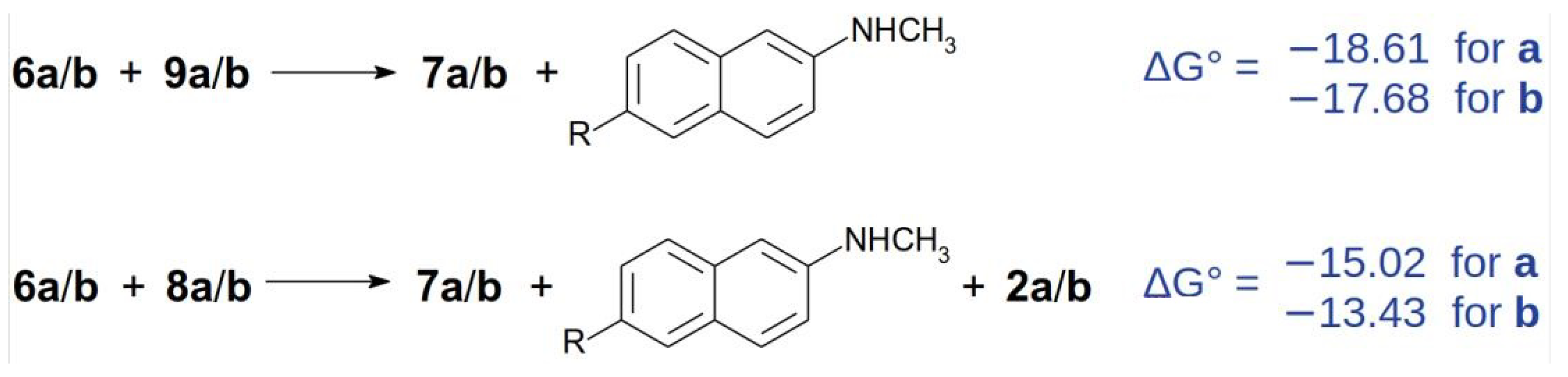
2. Results and Discussion

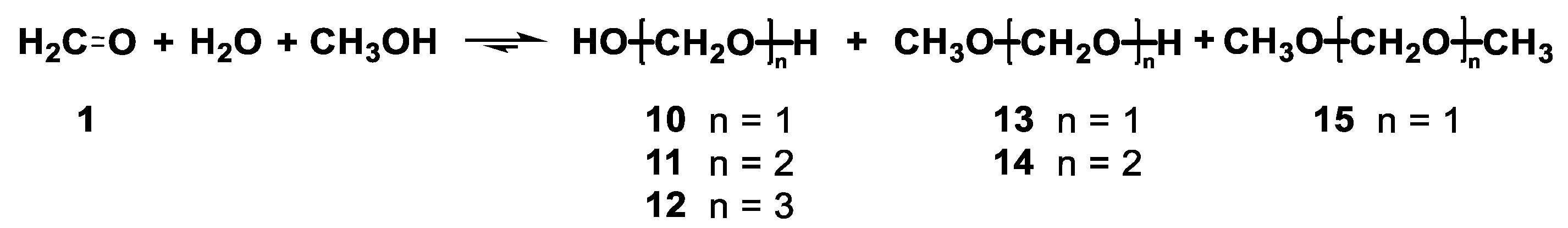
2.1. Studies of Formalin and Paraformaldehyde

First, we studied the ability of formalin and paraformaldehyde to act as a methanal source in a solution using 1H, 13C, DQF-COSY, HSQC, and HMBC NMR spectra. It is worth noting that the true methanal content was greater than observed because part of it was probably in the gas phase above the solution in the NMR tube. The 1H NMR spectra were not recorded under quantitative conditions so that determined contents may have varied within +/−10%.
In the solution of 5 μL of formalin in 0.5 mL of DMSO-d6 (dimethyl sulfoxide), we unambiguously identified (Scheme 3) methanal, methanol, water, diols 10, 11, and 12, hemiacetals 13 and 14, and acetal 15 in a molar ratio of 4:14:783:100:35:10:40:12:3, in addition to traces of formic acid and higher diols and hemiacetals. This corresponded to approximately 12% m/m of methanol in formalin (10–15% was declared by the supplier) and more than 33% m/m of methanal forms (37% was declared by the supplier). In accordance with a similar study in D2O [14,15], we did not observe any 1,3,5-trioxane (metaformaldehyde). On the other hand, small amounts of methanal and acetal 15 were observed, which were not mentioned in the study [14,15]. The relatively low concentrations of free methanal (>2%) increased dramatically at higher temperatures, as summarized in Table 1.
The dissolution of 1 mg of paraformaldehyde in 0.5 mL DMSO-d6 with 10 μL of water produced the equilibrated solution, which contained methanal, water, and diols 10, 11, and 12 in a molar ratio of 5:9494:441:51:9, alongside traces of higher diols. The methanal contents increased with increasing temperatures while the diols decomposed, as summarized in Table 2. Analogous results were obtained when methanol or ethanol was added to DMSO-d6 instead of water.
Similarly, high relative concentrations of methanal were identified when paraformaldehyde was treated with DMSO-d6 without any added water, so the majority of the paraformaldehyde remained undissolved (both the solvent and the paraformaldehyde contained moisture). The equilibrated mixture in the solution contained methanal, water, and diols 10, 11, and 12 in a molar ratio of 61:9038:727:159:15 (the concentrations of higher diols were less than 1‰). As expected, the methanal contents increased (>40%) upon raising the temperature as the diols decomposed; however, a new methanal form appeared and became the major one at 115 °C. This form was characterized by the sole singlet at 4.81 ppm in the 1H NMR spectrum (1JHC = 165.5 Hz, from 13C satellites), which was correlated in both the HSQC and the HMBC spectra to a 13C singlet at 89.07 ppm. Unfortunately, the molecular structure was not determined; 1,3,5-trioxane has an identical HSQC/HMBC pattern; however, different chemical shifts as was confirmed by the 1,3,5-trioxane standard addition.
It is worth emphasizing that the composition of those samples strongly depended on the water contents, total concentrations, presence of formic acid (common impurity of aged paraformaldehyde), and the time required to reach equilibrium, and thus, reproducing of these experiments would be difficult.

2.2. Calculations on the Reactivity of Methanal

As a first task, it was worth quantitatively addressing the power of methanal as a nucleophile, which was also related to its strong tendency to form the hydrate 10. The reactivity of a species is traditionally related to its HOMO/LUMO (highest energy occupied molecular orbital/lowest energy unoccupied molecular orbital) gap. Thus, methanal was compared with ethanal, acetone, and the perfluorinated aldehyde and ketone parents. As shown in Table 3, methanal had a smaller HOMO/LUMO gap than the heavier parents, and it was close to the gaps found for the perfluorinated oxo-species.
A more specific and well-established indicator of electrophilicity is available through the density functional theory (DFT) since it could be derived from the total density (i.e., the all-electron wavefunction) instead of the orbital energies (i.e., one-electron wavefunctions). Thus, the electrophilicities were calculated at the most accurate level, using the total energies of the neutral, radical anion, and radical cation [18]:
ω = μ 2 2 η = V I P + V E A 2 2 V I P V E A
where μ and η stand for the chemical potential and the chemical hardness: VIP and VEA are the vertical ionization potential and electron affinities, respectively. Considering that negative electron affinities were involved, the VEA and VIP were obtained using the methodology proposed by Puiatti et al. [19,20]. According to the results summarized in Table 4, the calculated ω values suggest that methanal was a very strong electrophile.
The higher electrophilicity of methanal also accounted for its ease of hydration, which is well-known in water. Table 5 reports the ΔG°hyd of methanal in a moderately polar solvent, using the ε of acetone as the IEFPCM model solvent [21]. Once again, methanal was found to be more similar to the electron poorer partner than to ethanal.

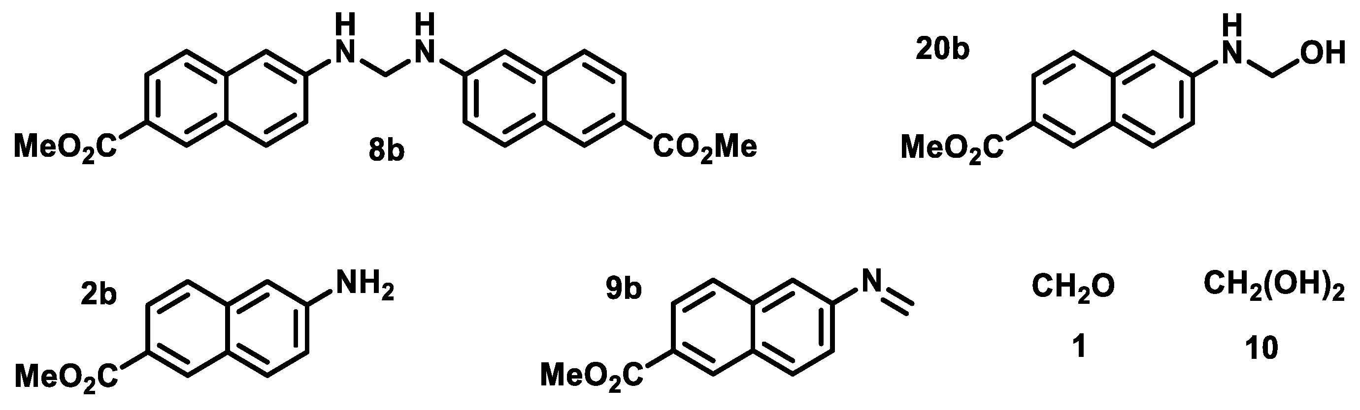
2.3. Attempts to Prepare Aminal 8a

Next, we revisited the preparation of aminal 8a, which was reported in 1902, as the product of the treatment of naphthylamine 2a with formalin in a molar ratio of 2:1 in acetone under reflux for five hours [22]. The obtained product (a yield was not given) was identified through elemental analyses (84.73 %C, 6.04 %H, and 9.25 %N) as aminal 8a (calcd. EA: 84.53 %C, 6.08 %H, 9.39 %N) and characterized as having a melting point of 104 °C (from ethanol).
The described procedure was then repeated; however, according to the 1D and 2D NMR spectra, the crude product mainly contained the starting naphthylamine 2a, quinazoline 6a, dinaphthylamine 17a, and bisquinazoline 16a in a molar ratio of 48:48:3:1, in addition to numerous trace products (Scheme 4). The presence of the expected aminal 8a could be deduced based on the 1H NMR signal at 4.70 ppm, which was correlated in the HSQC spectrum with the 13C signal at 53.16 ppm, and in the HMBC spectrum with the 13C signal at 146.03 ppm. No clear signals of TB 3a, acridine 5a, or bisnapthylamine 18a were observed. The crystallization of the crude product from ethanol allowed us to obtain an insoluble part that contained quinazoline 6a (2% yield) and bisquinazoline 16a (3% yield) in addition to crystals of pure quinazoline 6a (35% yield). The calculated EA for both quinazoline 6a (85.13 %C, 5.85 %H, 9.03 %N) and bisquinazoline 16a (85.41 %C, 5.73 %H, 8.85 %N) was not far from the published values. Note that the elemental compositions of dinaphthylamine 17a and aminal 8a are identical. Following the subsequent purification and recrystallization of quinazoline 6a from ethanol, its melting point of 100–102 °C (from ethanol) was measured. Therefore, we conclude that the product published in 1902 [22] consisted of quinazoline 6a and not aminal 8a.
The reaction was then repeated under the same condition; however, the crude product contained naphthylamine 2a, quinazoline 6a, and dinaphthylamine 17a in a slightly different molar ratio of 41:45:14, and surprisingly, no NMR signals of either bisquinazoline 16a or aminal 8a were identified. Owing to greater concentrations of dinaphthylamine 17a, we were able to assign all its 1H and 13C NMR signals and determine its molecular structure. Then, unlike in the original processing [22], we used column chromatography on silica instead of crystallization. Surprisingly, three fractions of various compositions were obtained. The 1H NMR spectra were then used to calculate the total yields of the expected starting naphthylamine 2a (30% recovery) and quinazoline 6a (51% yield), and surprisingly only 1% yield of dinaphthylamine 17a. Moreover, acridine 5a (9% yield) and bisnapthylamine 18a (8% yield), which were present in all fractions, were also isolated alongside TB 3a (1% yield), which occurred in a single fraction.
Since neither bisnapthylamine 18a nor acridine 5a were present in the crude product, and dinaphthylamine 17a was isolated in a very low yield, a rearrangement of 17a into 18a during the chromatography could be considered, followed by the conversion of 18a into acridine 5a, since 18a was the expected intermediate product of 5a [1,23]. A similar rearrangement and formation of 18b when exposed to silica or to air and ambient light for prolonged periods have previously been described [24].
It should be emphasized that the formation of quinazoline 6a and dinaphthylamine 17a was surprising since it requires an attack of an R-CH2 moiety on the naphthalene core, which generally requires a process of acid catalysis. However, no acid was added to the reaction, and the presence of acid would have led to the formation of TB 3a and/or spiroTB 4a. However, these compounds were not observed in the crude product, but after the chromatography procedure, i.e., silica was acidic enough to catalyze the rearrangement of 17a into 18a and to TB 3a formation.
An inspection of the molecular structure of quinazoline 6a revealed that it might have formed through the cycloaddition of two molecules of imine 9a, which could have formed during the reaction. However, the proposed mechanism was improbable, as suggested by our experiments on aminal 8b and imine 9b (vide infra).

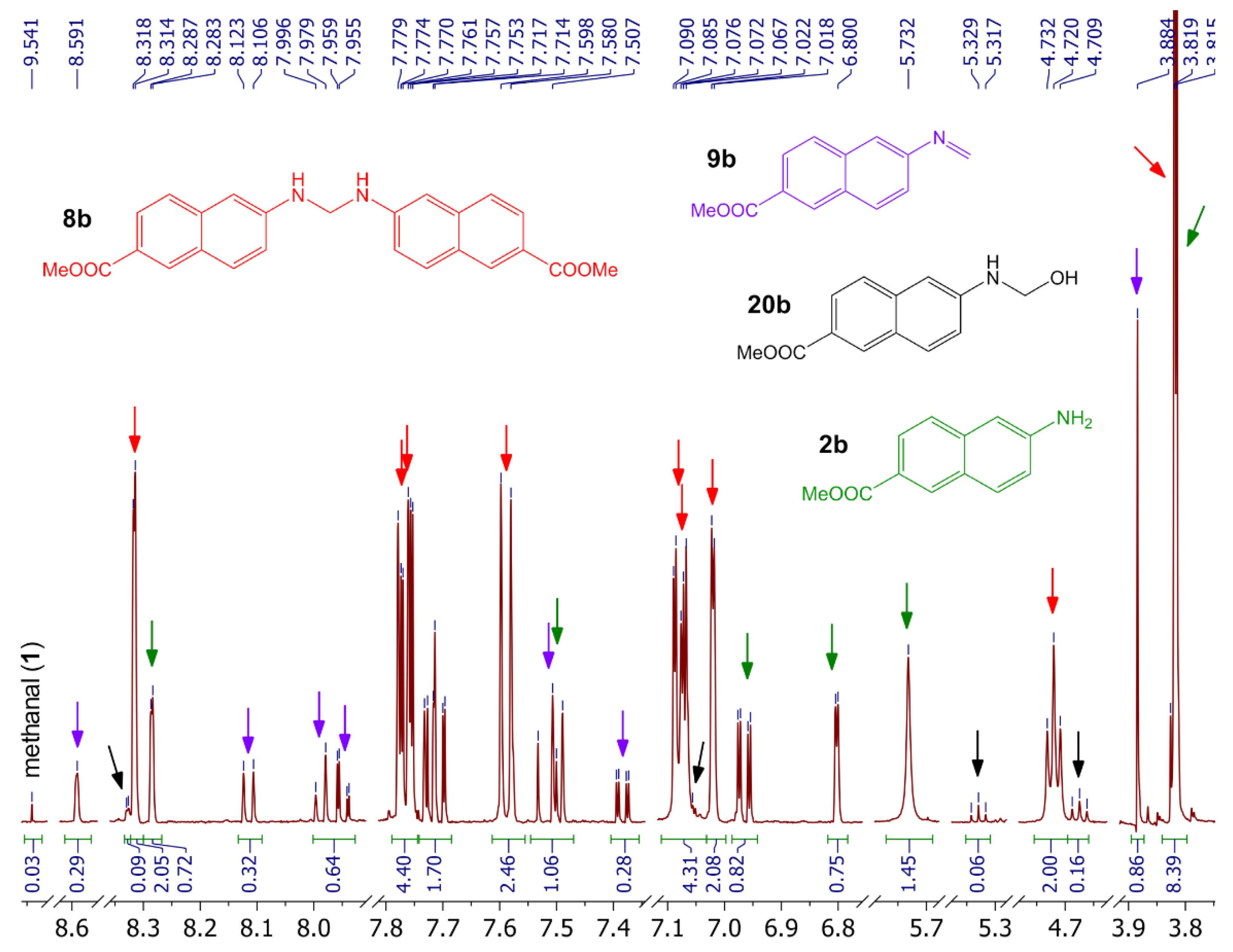
2.4. Attempts to Prepare Aminal 8b

When the reaction of [22] was performed with methyl 6-amino-2-naphthoate (2b) instead of naphthylamine 2a, we observed a different behavior. While the reaction mixture was a homogenous solution for 2a, a white solid precipitated shortly after the addition of formalin to the solution of naphthylamine 2b, a white solid precipitated before slowly dissolving.
The reaction was repeated again, and the white solid intermediate was isolated by filtration (84% yield of crude aminal 8 after 2 h reflux; only 46% after 8 h reflux). The 1D and 2D NMR techniques allowed us to observe that the DMSO-d6 solution of the white solid contained mainly aminal 8b, which was in equilibrium with hemiaminal 20b and starting naphthylamine 2b. When the sample was heated above 85 °C, the decomposition of both aminal 8b and hemiaminal 20b was observed, along with the formation of naphthylamine 2b, imine 9b, and traces of methanal (Figure 1). Imine 9b was clearly recognized by the two doublets with 2JHH = 16.2 Hz in the 1H NMR spectra, which are typical for terminal N = CH2 groups. However, the low contents (5% n/n) and low concentrations of imine 9b prevented us from identifying all of its NMR signals. When the heating was performed with the addition of water, only negligible amounts of imine 9b were formed, but a significant decomposition of aminal 8b and formation of methandiol (10) occurred (Scheme 5). When the sample was cooled back to 25 °C, the imine 9b was slowly converted back to aminal 8b and hemiaminal 20b, over a few hours. No formation of quinazoline 6b was observed.

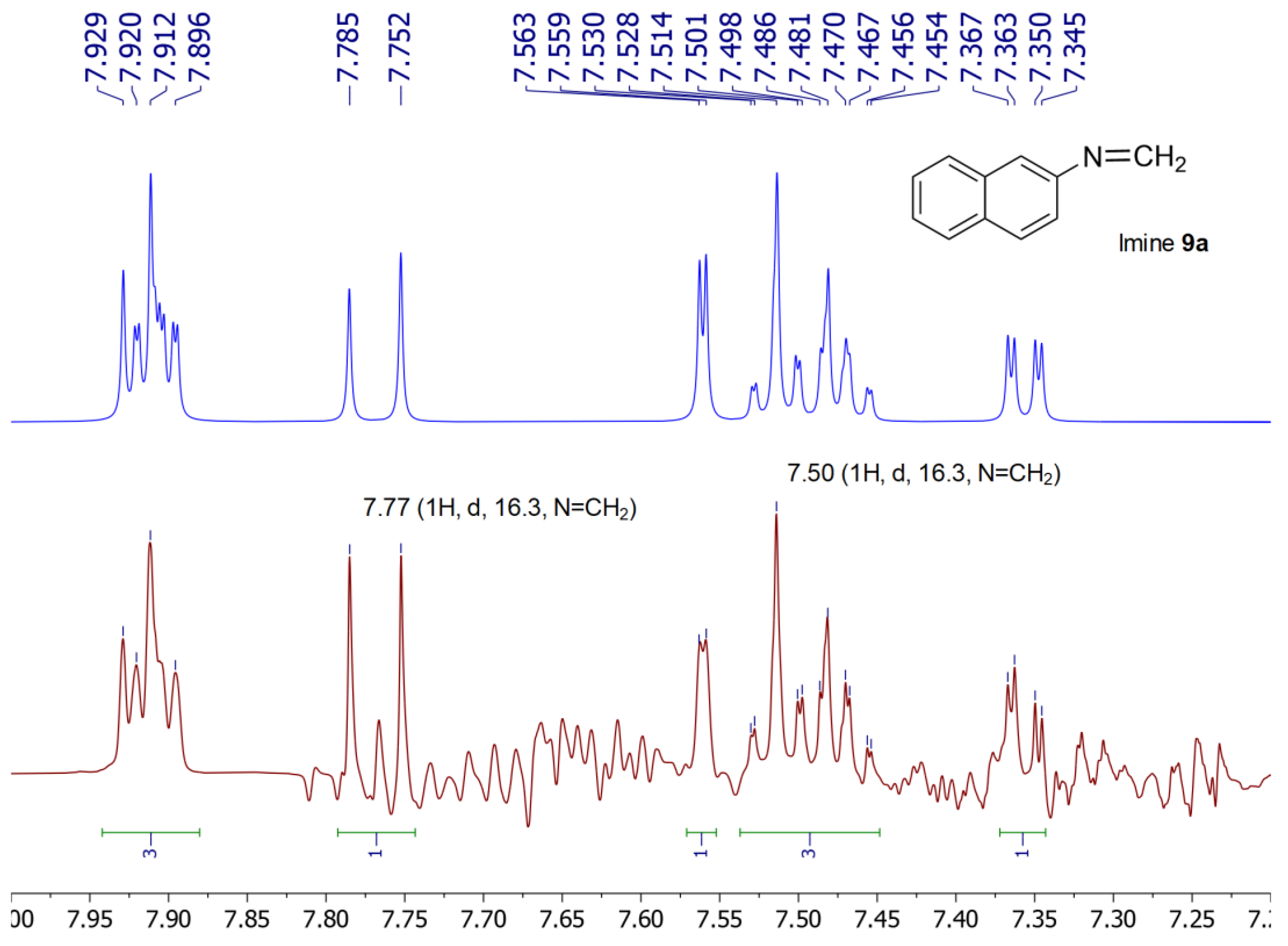
2.5. Preparation of Imine 9a under Acidic Conditions

The identification of imine 9b as an unstable compound in the presence of nucleophiles such as water or naphthylamine 2b (vide supra) prompted us to prepare imine 9a for comparison via known procedures.
The attempt to prepare imine 9a was likely first described in 1902 by Möhlau, who produced it as a product of the treatment of naphthylamine 2b with formalin in a molar ratio of 1:1 in ice acetic acid (a yield was not given) [22]. The molecular structure was suggested based on its EA (found: 85.36 %C, 6.07 %H, 9.17 %N; calcd. in 1902: 85.16 %C, 5.81 %H, 9.03 %N) and characterized by a melting point of 62–64 °C; no attempts were made to purify the compound.
However, when the procedure was reproduced, the obtained white solid contained quinazoline 6a, TB 3a, bisquinazoline 16a, and acridine 5a in a molar ratio of 71:23:3:3, contaminated with a few unidentified minor products. However, no 1H NMR signals of imine 9a were observed.
In addition, after the aqueous acid filtrate had been left to stand overnight, a few milligrams of a greenish solid precipitated. The 1D and 2D NMR spectra of the product showed that the solid contained acridine 5a, bisnaphthylamine 18a (probably as salt 19a), TB 3a, dihydrogenquinazoline 7a, and quinazoline 6a in a molar ratio of 39:29:23:6:3; due to the high contents of acetic acid, all these compounds were at least partially protonated. The composition of the NMR sample (solution in DMSO-d6) changed over one day. Bisnaphthylamine 19a disappeared while the acridine 5a contents increased, and the 1:1:1 triplet of a 14N-ammonium cation appeared (7.12 ppm, 51.1 Hz); this confirmed the pathway for acridine 5a formation suggested by [9]. On the other hand, at least two unidentified compounds were formed.
The reaction was then repeated; however, immediately after quenching the reaction by adding water, the ammonium solution was added until basic pH. After the extraction and repeating the column chromatography, we isolated quinazoline 6a (50% yield), TB 3a (10% yield), acridine 5a (3% yield), and the starting naphthylamine 2a (21% yield), and dihydrogenquinazoline 7a (3% yield), and surprisingly, also oxo-TB 21a (2% yield) and oxo-quinazoline 22a in yield of less than 1% (Scheme 6).
The formation of the oxo-compounds 21a and 22a as side products of a TB derivative preparation has never been reported. The only known oxo-TB analogs were previously prepared via formylation (via sBuLi and DMF) followed by aerial oxidation or through the direct oxidation of TB via KMnO4 (9 h reflux in CH2Cl2) [25]. Hence, the formation of the oxo-compounds 21a and 22a under our mild conditions over a two-minute reaction time was surprising. The low yields of the oxo-compounds 21a and 22a were relatively high compared to the yields of their possible precursors 3a and 7a, respectively. In addition, the most polar fraction resulting from the chromatographic separation was a complex mixture; however, the 1D and 2D NMR spectra revealed a set of signals which could be attributed to an exo-diastereomer of hydroxy-TB 23a formed as a possible intermediate product of oxo-TB 21a.

2.6. Preparation of Imine 9a under Basic Conditions

Imine 9a formation was previously mentioned by Kadutskii in 2002, 2006, and 2012 through the treatment of naphthylamine 2a with paraformaldehyde (1:1) in ethanol in the presence of a catalytic amount of NaOH [26,27,28]. However, no spectroscopic data confirming the presence of imine 9a were reported. The formation of imine 9a in situ was only assumed based on the molecular structure of the isolated products, which could be considered the result of imine 9a reactivity.
Thus, the reaction was repeated and followed by NMR spectroscopy. Our analysis showed that the naphthylamine 2a was slowly consumed to reach an equilibrium with the majority of the imine-ethanol adduct 24a, followed by traces of the starting naphthylamine 2a and probably methanediol (10) and minor products (Scheme 7). Methanediol (10) was identified based on the singlet at 4.61 ppm in the 1H NMR spectrum, which was correlated in the HSQC spectrum with a 13C signal at 88.83 ppm, and exhibited no correlation in the HMBC spectrum (the chemical shifts may have been strongly affected by the presence of ethanol and sodium hydroxide).
When the sample was heated from 25 to 115 °C, no formation of imine 9a was observed, possibly due to the large excess of ethanol in the solution. This observation was similar to the heating of aminal 8b after the addition of water (vide supra).
Thus, a part of the reaction mixture was evaporated to dryness, and the residue was dissolved in 500 μL of DMSO-d6. A combination of 1D and 2D NMR spectra and mass spectrometry (MS) spectra revealed that the mixture contained not only adducts 24a and 25a but also its higher analog 26a. Newly, the mixture also contained aminal 8a and its higher analog 27a (Scheme 7). These compounds were not unambiguously proved through NMR spectra due to overlaid signals. Neither methanal nor imine 9a were observed. However, when the sample was heated to 115 °C, the formation of imine 9a was observed, and the 1H NMR spectrum of imine 9a was obtained by subtracting the spectra obtained at 40 °C before and after heating (Figure 2). The formation of imine 9a at high temperatures was in accord with the formation of imine 9b and methanal (1) by heating aminal 8b and methanal equivalents, respectively (vide supra).
It should also be emphasized that we observed a 1H-13C HMBC correlation of CH2 hydrogen atoms occurring exclusively with nitrogen-bearing carbon (well-separated signals around 145 ppm). This implies that there was no C-alkylation of the naphthalene core under these basic conditions, as is typical for reactions in the presence of an acid (the formation of TB derivatives) or in its absence (the formation of quinazoline). This means that the C-alkylation requires, at least not a too basic condition. In addition, since imine 9a was formed under these conditions in at least trace amounts, and quinazoline 6a was not observed as a product, the possible formation of quinazoline 6a by cycloaddition of imine 9a is unlikely.

2.7. Density Functional Theory Calculations

The processes studied here are summarized in Scheme 8, where the relative free energies of the main species refer to the energies of aminals 8a/b and two methanal molecules as ΔG° = 0. Views of the 3D structures of the main stationary points in Scheme 8 are available in the Supporting Information.
The thermal decomposition of aminals 8a/b yielding imines 9a/b and naphthylamines 2a/b was found to be slightly endergodic. The entrance of the first methanal to position 1 of the naphtylamine to yield i1a/b was found to be slightly exergodic, passing through transition states (TS) at 32.0 and 34.2 kcal/mol of the relative free energies for obtaining i1a and i1b, respectively. Then, the i1a/b species could react with the imine 9a/b to yield the adduct i2a/b, a process that was exergodic by about 1 kcal/mol and with a relatively small activation barrier. The intermediate products i2a/b could, in principle, close the quinazoline ring through an intramolecular Diels-Alder-like TS for yielding 6a/b. However, this process seems unlikely due to the high activation barrier of the TS-i2-6 compound (Scheme 8). A more likely pathway would have been the direct recombination of two imines 9a/b condensing to a tautomer of 6a/b through TS-9-6tau, which had a relative free energy of about 20 kcal/mol lower than the former TS-i2-6. The intermediate product could easily tautomerize to 6a/b, which had remarkable stability of −14.1 and −14.7 kcal/mol for 6a and 6b, respectively.
Considering the stabilities of 6a/b, it is interesting to consider the different fates of this quinazoline. The thermochemistries of the formal dehydrogenation of 6a/b to yield 7a/b were found to be 2.3 and 4.7 kcal/mol endergodic in the cases of 6a and 6b, respectively. While the current calculations were performed considering a neutral acetone medium, the process has previously been described under different conditions, i.e., in an acid media [23]. Under those conditions, the overall thermochemistry of the process mediated by the reduction of imines 9a/b or reductive cleavage of 8a/b is shown separately in Scheme 9. Both processes had overall spontaneous thermochemistry by 18–19 and 13–15 kcal/mol. Both reactions leading to the formation of imines 7a/b were more favored for 6b than for 6a.
Another expected process for 6a/b (Scheme 8) was the entrance of another methanal molecule. At this stage, a partner of this quinazoline has already been proven as a key intermediate product formed in the last steps of the formation of TBs in the case of anilines as starting amines [17]. The attachment of the methylene unit involves the formation of an unstable i3tau intermediate that is rapidly tautomerized to i3. The activation barrier was found to be at 32.8 kcal/mol for 6a and was 2 kcal/mol higher for 6b. The intramolecular electrophilic attack of the alcohol carbon concerted with the water release (TS-i3-3) finally led to the formation of TB 3a/b. Once again, the process was easier for i3a than for i3b. The overall thermochemistry leading to the TB 3a/b starting from aminals 8a/b was found clearly exergodic by 30–31 kcal/mol.

3. Materials and Methods

3.1. Measurements and Materials

All chemicals and solvents were purchased from commercial suppliers and used without further purification. The NMR spectra were recorded on a 500 MHz instrument. The chemical shifts (δ) are indicated in ppm followed by their multiplicity, integral intensity, corresponding coupling constants (J) in Hz, and by the signal assignment, which is based on an analysis of ordinary 1H-1H COSY, 1H-1H NOESY, 1H-13C HSQC, and 1H-13C HMBC correlation spectra. The 2D spectra were recorded with high-resolution conditions utilizing the nonuniform sampling and processing with a linear prediction; hence the reproducibility of the chemical shifts was a few tens of ppb. The “cov.” in the spectra description means that the signal is seriously covered by others, so a full description was impossible. The 1H and 13C APT chemical shifts are referenced to TMS (using the solvent signals CHCl3 7.26 ppm, CDCl3 77.0 ppm, CHD2SOCD3 2.50 ppm, CD3SOCD3 39.52 ppm). The MNOVA software was used for the processing, prediction, and simulation of NMR spectra. The interpretation of most spectra required a simultaneous analysis of various 1D and 2D spectra of two or more samples with different compositions. See also Supplementary Material. The mass spectra were obtained using electrospray ionization (ESI) or atmospheric pressure chemical ionization (APCI) with a linear quadrupole trap (LTQ) Orbitrap spectrometer. Silica (40–63 D, 60 Å) was used to separate the compounds by column chromatography.

3.2. Computational Procedure

The electronic structure calculations were all performed in the Gaussian 16. Rev. A03 package [21]. The structures of the reagents, transition states (TS), intermediaries, and products were all optimized at the level of theory CAM-B3LYP/6-311+G(d,p), with the solvent model SCRF-IEFPCM [29] (acetone). The CAM-B3LYP energies were corrected to consider the dispersion effects using the D3 version of the Grimme’s dispersion with the Becke–Johnson damping, i.e., CAM-B3LYP-GD3BJ [30,31] (uncorrected energies available as Supplementary Material). The stationary points were characterized by their Hessian matrix, which was diagonalized to obtain the harmonic frequencies and then the zero point corrections to energy, enthalpy, and free energy. In relevant cases, an in vacuo intrinsic reaction coordinate (IRC) was determined using a mass-weighted step of 0.02 atomic units and by recalculating the Hessian every ten or 20 steps. The procedure was previously described and tested elsewhere [17,32]. For the relative free energies of Scheme 8, the ΔG° = 0 was taken as the energies of aminals 8a/b plus two methanal molecules to close the mass balance.
The level of theory and general procedure were previously described for the synthesis of symmetric and asymmetric TBs from anilines as starting reagents [17]. The procedure for obtaining the VEAs and VIPs to compute the electrophilicity using CAM-B3LYP was described in detail in [20].

3.3. Studies of Formalin and Paraformaldehyde

(a)
An NMR tube was charged with 500 μL of DMSO-d6 (standard quality) and 5 μL of formalin (ASC reagent, formaldehyde solution, 37% m/m in H2O, containing 10–15% methanol as a stabilizer) and closed with a gas-tight cup. The solution was monitored by 1H NMR spectra at 25 °C. Equilibrium was reached within a few hours. The sample composition was determined by 1D and 2D NMR (Table 1). The sample was heated to 50 °C, left to equilibrate (1–2 h), and analyzed by 1H NMR. The same was done at temperatures of 75, 100, and 115 °C. After cooling back to 25 °C, the compositions were slowly returned to equilibrium (two days).
(b)
An NMR tube was charged with paraformaldehyde (1.0 mg, 33 μmol), DMSO-d6 (0.5 mL), and water (10 μL, 555 μmol) and closed with a gas-tight cup. The mixture was shaken until the paraformaldehyde dissolved and then monitored by 1H NMR at 25 °C. Equilibrium was reached within several hours. The sample composition was determined by 1D and 2D NMR (Table 2); the higher diols contents did not exceed 0.5‰ (n/n). The sample was heated following the procedure described in (a).
(c)
An NMR tube was charged with paraformaldehyde (1.0 mg, 33 μmol) and DMSO-d6 (0.5 mL) and closed with a gas-tight cup. The mixture was shaken, but part of the paraformaldehyde remained undissolved. The sample was heated following the procedure described in (a).
Methanal (1): 1H NMR (500 MHz, DMSO-d6, 25 °C): 9.57 (2H, s, 1JHC = 178.7). 13C{1H} NMR (126 MHz, DMSO-d6, 25 °C): 197.58.
Methandiol (10): 1H NMR (500 MHz, DMSO-d6, 25 °C): 5.78 (2H, t, 7.4, OH), 4.59 (2H, t, 7.4, CH2). 13C{1H} NMR (126 MHz, DMSO-d6, 25 °C): 81.95.
Diol (11): 1H NMR (500 MHz, DMSO-d6, 25 °C): 6.11 (2H, t, 7.8, OH), 4.68 (4H, d, 7.8, CH2). 13C{1H} NMR (126 MHz, DMSO-d6, 25 °C): 84.05.
Diol (12): 1H NMR (500 MHz, DMSO-d6, 25 °C): 6.33 (2H, t, 7.8, OH), 4.67 (4H, d, 7.8), 4.78 (2H, s). 13C{1H} NMR (126 MHz, DMSO-d6, 25 °C): 86.19 (CH2), 84.95 (CH2OH).
Hemiacetal (13): 1H NMR (500 MHz, DMSO-d6, 25 °C): 6.15 (1H, t, 7.7, OH), 4.53 (2H, d, 7.7, CH2), 3.22 (3H, s, CH3). 13C{1H} NMR (126 MHz, DMSO-d6, 25 °C): 89.46 (CH2), 53.92 (CH3).
Hemiacetal (14): 1H NMR (500 MHz, DMSO-d6, 25 °C): 6.35 (1H, t, 8.0, OH), 4.66 (2H, d, 8.0, CH2OH), 4.64 (2H, s, CH2), 3.25 (3H, s, CH3). 13C{1H} NMR (126 MHz, DMSO-d6, 25 °C): 91.71 (CH2), 84.81 (CH2OH), 54.90 (CH3).
Acetal (15): 1H NMR (500 MHz, DMSO-d6, 25 °C): 3.23 (6H, s, CH3), 4.49 (2H, s, CH2). 13C{1H} NMR (126 MHz, DMSO-d6, 25 °C): 96.79 (CH2), 54.53 (CH3).
Water: 1H NMR (500 MHz, DMSO-d6, 25 °C): 3.34 (2H, s).
Methanol: 1H NMR (500 MHz, DMSO-d6, 25 °C): 4.10 (1H, q, 5.1), 3.17 (3H, d, 5.1). 13C{1H} NMR (126 MHz, DMSO-d6, 25 °C): 48.60.
Formic acid: 1H NMR (500 MHz, DMSO-d6, 25 °C): 8.14 (1H, s, CH), the signal of OH was not observed, likely due to the signal broadness. 13C{1H} NMR (126 MHz, DMSO-d6, 25 °C): 163.09.
The unknown form: 1H NMR (500 MHz, DMSO-d6, 25 °C): 4.10. 13C{1H} NMR (126 MHz, DMSO-d6, 25 °C): 48.60.
1,3,5-Trioxane (a standard): 1H NMR (500 MHz, DMSO-d6, 25 °C): 5.12 (2H, s, 1JHC = 166.3). 13C{1H} NMR (126 MHz, DMSO-d6, 25 °C): 92.88.

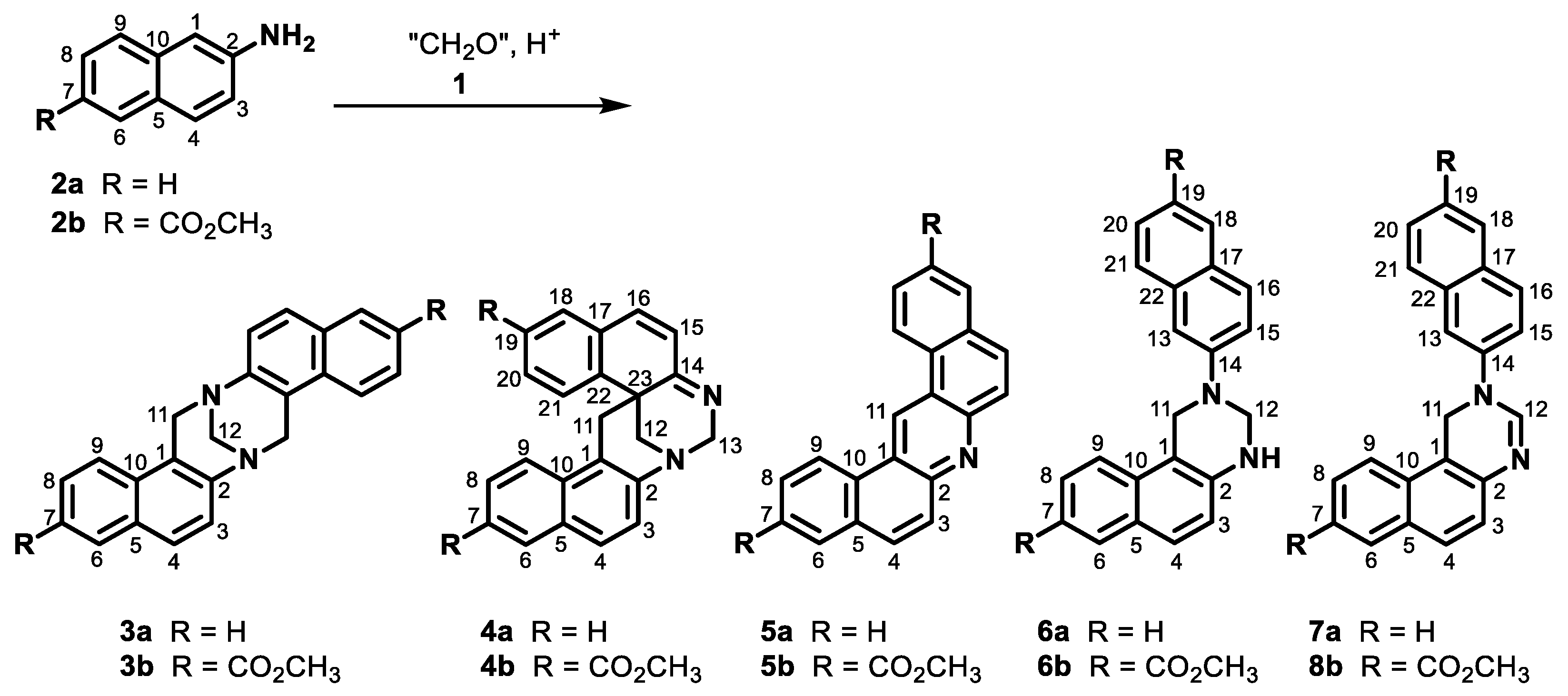
3.4. Reaction of Naphthylamine 2a with Formalin under Neutral Condition

(a)
Aqueous formaldehyde (37%, 0.1 mL, 1.23 mmol) was added to a solution of naphthylamine 2a (352 mg, 2.46 mmol) in acetone (20 mL). The mixture was refluxed for five hours. The reaction mixture was evaporated to dryness in vacuo, and the residue was analyzed using 1D and 2D NMR experiments. The residue was purified by crystallization from ethanol. An insoluble fraction (18 mg) was obtained, which contained quinazoline 6a (7 mg, 2% yield) and bisquinazoline 16a (11 mg, 3% yield), as well as crystals of pure quinazoline 6a (133 mg, 35% yield).
(b)
Aqueous formaldehyde (37%, 0.1 mL, 1.23 mmol) was added to the solution of naphthylamine 2a (352 mg, 2.46 mmol) in acetone (20 mL). The mixture was refluxed for five hours and then evaporated to dryness in vacuo. The obtained solid (387 mg) contained mostly naphthylamine 2a, quinazoline 6a, and dinaphthylamine 17a in a molar ratio of 41:45:14, according to NMR. The solid was purified by column chromatography on silica (dichloromethane/methanol from 1:0 to 4:1) to produce four fractions of various compositions. The yields were calculated based on the 1H NMR spectra: 105 mg (30% recovered) of naphthylamine 2a, 196 mg (51% yield) of quinazoline 6a, 3 mg (1% yield) of dinaphthylamine 17a, 31 mg (9% yield) of acridine 5a, 31 mg (8% yield) of bisnaphthylamine 18a, and 5 mg (1% yield) of TB 3a.
Quinazoline 6a: 1H NMR (500 MHz, DMSO-d6, 25 °C): 7.85 (1H, ddt, 8.4, 1.1, 0.8, H9), 7.77 (1H, ddt, 9.1, 0.8, 0.5, H16), 7.73 (1H, dddd, 8.1, 1.3, 0.8, 0.5, H18), 7.71 (1H, ddt, 8.0, 1.4, 0.7, H6), 7.66 (1H, ddt, 8.2, 1.2, 0.8, H21), 7.55 (1H, dd, 8.7, 2.5, H4), 7.55 (1H, dd, 9.1, 2.5, H15), 7.46 (1H, ddd, 8.3, 6.8, 1.4, H8), 7.39 (1H, dd, 2.5, 0.5, H13), 7.36 (1H, ddd, 8.2, 6.8, 1.3, H20), 7.24 (1H, ddd, 8.1, 6.8, 1.2, H19), 7.23 (1H, ddd, 8.0, 6.8, 1.1, H7), 6.90 (1H, d, 8.7, H3), 6.46 (1H, br t, 3.6, NH), 4.89 (2H, br s, H11), 4.83 (2H, d, 3.6, H12). 13C{1H} NMR (126 MHz, DMSO-d6, 25 °C): 146.89 (C14), 141.49 (C2), 134.31 (C22), 131.65 (C10), 128.52 (C16), 128.34 (C6), 127.79 (C17), 127.24 (C4), 127.23 (C18), 127.12 (C5), 126.43 (C8), 126.39 (C21), 126.11 (C20), 122.96 (C19), 121.58 (C7), 120.47 (C9), 119.35 (C3), 118.40 (C15), 110.30 (C13), 109.17 (C1), 59.32 (C12), 47.53 (C11). HRMS (APCI+, MeOH): for C22H18N2 calcd. [M + H]+ 311.1543, found 311.1543. M.p. 100–102 °C (from ethanol).
Bisquinazoline 16a: 1H NMR (500 MHz, DMSO-d6, 20.5 °C): 8.05 (2H, br d, 8.5, H9), 7.84 (2H, dd, 8.0, 1.4, H6), 7.74 (2H, d, 8.8, H4), 7.67 (2H, br d, 8.1, H18), 7.58 (2H, d, 9.0, H16), 7.57 (2H, ddd, 8.5, 6.8, 1.4, H8), 7.57 (2H, d, 8.8, H3), 7.53 (2H, br d, 8.2, H21), 7.42 (2H, dd, 9.0, 2.5, H15), 7.40 (2H, ddd, 8.1, 6.8, 1.1, H7), 7.39 (2H, cov., H13), 7.31 (2H, ddd, 8.2, 6.8, 13, H20), 7.22 (2H, ddd, 8.1, 6.8, 1.2, H19), 5.13 (2H, br s, H23), 4.91 (4H, br s, H12), 4.90 (4H, br s, H11). 13C{1H} NMR (126 MHz, DMSO-d6, 25 °C): 146.42 (C14), 141.61 (C2), 134.28 (C22), 131.11 (C10), 128.46 (C16), 128.32 (C6), 128.24 (C5), 127.77 (C17), 127.64 (C4), 127.17 (C18), 126.75 (C8), 126.41 (C21), 126.10 (C20), 123.42 (C7), 123.00 (C19), 121.48 (C9), 119.25 (C3), 118.62 (C15), 115.55 (C1), 109.76 (C13), 68.71 (C23), 63.85 (C12), 48.02 (C11).
Naphthylamine 2a: 1H NMR (500 MHz, DMSO-d6, 25 °C): 7.61 (1H, dtd, 8.1, 1.3, 0.7, H6), 7.57 (1H, br d, 8.7, H4), 7.49 (1H, dtd, 8.3, 1.2, 0.7, H9), 7.26 (1H, ddd, 8.3, 6.8, 1.3, H8), 7.08 (1H, ddd, 8.1, 6.8, 1.2, H7), 6.93 (1H, dd, 8.7, 2.3, H3), 6.81 (1H, ddd, 2.3, 0.8, 0.5, H1), 5.34 (2H, br s, 15N satellites 1JHN = 83.5, NH2). 13C{1H} NMR (126 MHz, DMSO-d6, 25 °C): 146.64 (C2), 135.00 (C10), 128.46 (C4), 127.45 (C6), 126.32 (C5), 125.83 (C8), 125.01 (C9), 120.83 (C7), 118.39 (C3), 105.79 (C1). HRMS (APCI+, MeOH): for C10H9N calcd. [M + H]+ 144.0808, found 144.0809.
Dinaphthylamine 17a: 1H NMR (500 MHz, DMSO-d6, 25 °C): 7.76 (1H, d, cov., H9), 7.69 (1H, dd, 8.1, 1.4, H6), 7.64 (1H, d, cov., H17), 7.62 (1H, d, cov., H4), 7.62 (1H, d, cov., H20), 7.57 (1H, d, cov., H15), 7.35 1H, ddd, cov., H8), 7.32 (1H, ddd, 8.2, 6.8, 1.3, H19), 7.14 (1H, ddd, 8.1, 6.8, 1.1, H7), 7.11 (1H, ddd, 8.0, 6.8, 1.3, H18), 7.08 (1H, dd, 8.8, 2.3, H14), 7.07 (1H, d, cov., H3), 6.99 (1H, d, 2.3, H12), 5.85 (1H, br t, 4.5, NH), 4.50 (2H, d, 4.5, H11). 13C{1H} NMR (126 MHz, DMSO-d6, 25 °C): 147.25 (C13), 145.01 (C2), 135.24 (C21), 133.91 (C10), 128.48 (C4), 128.12 (C6), 127.97 (C15), 127.41 (C17), 127.01 (C5), 126.44 (C16), 126.30 (C8), 125.92 (C19), 125.43 (C20), 121.87 (C9), 120.86 (C18), 120.84 (C7), 119.10 (C3), 118.69 (C14), 110.40 (C1), 102.38 (C12), 38.79 (C11). HRMS (APCI+, MeOH): for C21H18N2 calcd. [M + H]+ 299.1543, found 299.1544.
Acridine 5a: 1H NMR (500 MHz, DMSO-d6, 25 °C): 10.64 (1H, br s, H11), 9.44 (2H, ddt, 8.2, 1.2, 0.7, H9), 8.21 (2H, ddd, 9.1, 0.8, 0.5, H4), 8.12 (2H, br d, 7.8, H6), 8.06 (2H, dd, 9.1, 0.8, H3), 7.87 (2H, dddd 8.2, 7.1, 1.4, 0.3, H8), 7.79 (2H, ddd 7.8, 7.1, 1.2, H7). 13C{1H} NMR (126 MHz, DMSO-d6, 25 °C): 147.80 (C2), 132.16 (C4), 131.03 (C5), 129.86 (C10), 128.76 (C6), 127.84 (C7), 127.65 (C3), 127.63 (C8), 126.25 (C11), 124.27 (C9), 123.71 (C1). HRMS (APCI+, MeOH): for C21H13N calcd. [M + H]+ 280.1121, found 280.1120.
Bisnaphthylamine 18a: 1H NMR (500 MHz, DMSO-d6, 25 °C): 7.95 (2H, ddt, 8.6, 1.2, 0.7, H9), 7.60 (2H, ddt, 8.0, 1.5, 0.5, H6), 7.45 (2H, br d, 8.7, H4), 7.15 (2H, ddd, 8.6, 6.8, 1.5, H8), 7.07 (2H, ddd, 8.0, 6.8, 1.1, H7), 6.99 (2H, d, 8.7, H3), 5.47 (2H, br s, NH2), 4.36 (2H, s, H11). 13C{1H} NMR (126 MHz, DMSO-d6, 25 °C): 143.81 (C2), 133.69 (C10), 131.79 (C1), 128.27 (C6), 127.46 (C5), 127.07 (C4), 125.62 (C8), 122.48 (C9), 120.62 (C7), 119.18 (C3), 23.33 (C11).
TB 3a: 1H NMR (500 MHz, DMSO-d6, 25 °C): 7.76 (2H, ddt 8.0, 1.4, 0.6, H6), 7.74 (2H, ddt 8.5, 1.2, 0.8, H9), 7.69 (2H, br d, 8.8, H4), 7.47 (2H, ddd, 8.5, 6.9, 1.4, H8), 7.38 (2H, d, 8.8, H3), 7.37 (2H, ddd, 8.0, 6.9, 1.2, H7), 4.96 (2H, dd, 16.8, 0.8, H11a), 4.72 (2H, br d, 16.8, H11b), 4.44 (2H, br s, H12). 13C{1H} NMR (126 MHz, DMSO-d6, 25 °C): 145.41 (C2), 130.93 (C10), 130.19 (C5), 128.26 (C6), 127.24 (C4), 126.44 (C8), 124.67 (C3), 124.57 (C7), 121.44 (C9), 121.23 (C1), 66.07 (C12), 55.16 (C11). HRMS (APCI+, MeOH): for C23H18N2 calcd. [M + H]+ 323.1543, found 323.1544. M.p. 208–210 °C (from ethanol).

3.5. Reaction of Naphthylamine 2b with Formalin under Neutral Conditions

Aqueous formaldehyde (37%, 0.1 mL, 1.23 mmol) was added to a solution of naphthylamine 2b (496 mg, 2.46 mmol) in acetone (20 mL). The mixture was refluxed for two hours (a white solid precipitated after 20 min). After cooling to room temperature, the solid was filtered off, washed with acetone, methanol, and diethyl ether, and dried to obtain 428 mg (84% yield) of crude aminal 8b. The filtrate was evaporated to dryness. According to the 1H NMR and HRMS spectra, the obtained solid contained naphthylamine 2b, quinazoline 6b, and aminal 8b in a molar ratio of 81:15:4. When eight hours of reflux was, only a 46% yield of crude aminal 8b was obtained.
The NMR tube was charged with 1 mg of the crude aminal 8b and 500 μL of dry DMSO-d6 and shaken until a solution was produced. The 1D and 2D spectra revealed the presence of aminal 8b, naphthylamine 2b, and hemiaminal 20b in a molar ratio of 71:23:6 at 25 °C. The sample was heated to 100 °C for an hour and monitored by 1H NMR until it reached equilibrium. The 1D and 2D spectra revealed the presence of aminal 8b, naphthylamine 2b, hemiaminal 20b, and imine 9b in a molar ratio of 67:25:3:5, alongside traces of methanal.
Another NMR tube was charged with 0.64 mg of crude aminal 8b, 10 μL water, and 500 μL of dry DMSO-d6 and shaken to get a clumsy solution. The 1D and 2D spectra revealed the presence of aminal 8b, naphthylamine 2b, and hemiaminal 20b in a molar ratio of 70:25:5 at 25 °C. The sample was heated to 100 °C for an hour and monitored by 1H NMR until an equilibrium was reached before being rapidly cooled back to 25 °C. The 1D and 2D spectra evidenced the presence of aminal 8b, naphthylamine 2b, hemiaminal 20b, and methanediol (10) in a molar ratio of 37:43:16:4, as well as traces of methanal and imine 9b.
Aminal 8b: 1H NMR (500 MHz, DMSO-d6, 25 °C): 8.36 (2H, d, 1.9, H6), 7.81 (2H, d, 8.9, H4), 7.80 (2H, dd, 8.7, 1.8, H8), 7.63 (2H, d, 8.7, H9), 7.20 (2H, t, 6.7, NH), 7.11 (2H, dd, 8.9, 2.3, H3), 7.06 (2H, d, 2.3, H1), 4.75 (2H, t, 6.7, H11), 3.85 (6H, s, H13). 1H NMR (500 MHz, DMSO-d6, 100 °C): 8.35 (2H, br d, H6), 7.81 (2H, dd, 8.7, 1.8, H8), 7.79 (2H, d, 8.9, H4), 7.63 (2H, d, 8.7, H9), 7.16 (2H, dd, 8.9, 2.3, H3), 7.08 (2H, d, 2.3, H1), 6.78 (2H, br t, 5.8, NH), 4.80 (2H, br t, 5.8, H11), 3.88 (6H, s, H13). 13C{1H} NMR (126 MHz, DMSO-d6, 100 °C): 166.16 (C12), 147.29 (C2), 137.08 (C10), 129.87 (C6), 129.59 (C4), 125.10 (C5), 125.07 (C9), 124.65 (C8), 121.91 (C7), 118.28 (C3), 103.37 (C1), 52.20 (C11), 50.98 (C13). HRMS (APCI+, MeOH): for C25H22N2O4 calcd. [M + H]+ 415.1652, found 415.1656.
Naphthylamine 2b: 1H NMR (500 MHz, DMSO-d6, 25 °C): 8.33 (1H, d, 1.8, H6), 7.77 (1H, d, 8.8, H4), 7.74 (1H, dd, 8.7, 1.8, H8), 7.54 (1H, d, 8.7, H9), 6.99 (1H, dd, 8.8, 2.2, H3), 6.83 (1H, d, 2.2, H1), 5.84 (2H, br s, NH2), 3.85 (3H, s, H12). 1H NMR (500 MHz, DMSO-d6, 100 °C): 8.32 (1H, br d, H6), 7.76 (1H, dd, 8.7, 1.8, H8), 7.74 (1H, d, cov., H4), 7.54 (1H, d, 8.7, H9), 7.03 (1H, dd, 8.8, 2.3, H3), 6.89 (1H, d, 2.3, H1), 5.51 (2H, br s, NH2), 3.87 (3H, s, H12). 13C{1H} NMR (126 MHz, DMSO-d6, 100 °C, from 1D and HSQC): 166.21 (C11), 148.71 (C2), 137.16 (C10), 129.97 (C6), 129.77 (C4), 124.74 (C5), 124.55 (C9), 124.50 (C8), 121.45 (C7), 118.66 (C3), 105.13 (C1), 50.94 (C12). HRMS (APCI+, MeOH): for C12H11NO2 calcd. [M + H]+ 202.0863, found 202.0863.
Hemiaminal 20b: 1H NMR (500 MHz, DMSO-d6, 40 °C): 8.37 (1H, br d, 1.8, H6), 7.83 (1H, cov., H4), 7.79 (1H, dd, 8.6, 1.8, H8), 7.63 (1H, d, 8.6, H9), 7.10 (1H, dd, 8.8, 2.3, H3), 7.08 (1H, br t, 6.8, OH), 7.02 (1H, cov., H1), 5.37 (1H, t, 6.3, NH), 4.71 (1H, t, 6.5, CH2), a signal for OCH3 was not unambiguously recognized. 13C NMR (126 MHz, DMSO-d6, 40 °C): only 66.46 (CH2) from HSQC and 148.35 (C2) from HMBC were identified. HRMS (APCI+, MeOH): for C13H13NO3 calcd. [M + H]+ 232.0968, found 232.0967.
Imine 9b: 1H NMR (500 MHz, DMSO-d6, 40 °C): 8.63 (1H, br s), 8.15 (1H, d, 8.6), 8.03 (1H, d, 8.6), 7.99 (1H, dd, 8.6, 1.7), 7.78 (1H, d, 16.2), 7.55 (1H, d, 16.2), 7.42 (1H, dd, 8.6, 2.1), ca. 7.1 (1H, cov.), 3.92 (3H, s). 13C NMR (126 MHz, DMSO-d6, 40 °C, from HSQC): 130.93, 131.06, 128.84, 125.80, 122.12, 104.55; other signals were not observed due to low concentrations. HRMS (APCI+, MeOH): for C13H11NO2 calcd. [M + H]+ 214.0863, found 214.0861.
Quinazoline 6b: 1H NMR (500 MHz, DMSO-d6, 25 °C): characteristic signals: 4.98 (2H, s), 4.94 (2H, d, 3.3). HRMS (APCI+, MeOH): for C26H22N2O4 calcd. [M + H]+ 427.1652, found 427.1656.

3.6. Reaction of Naphthylamine 2a with Formalin under Acidic Conditions

(a)
Aqueous formaldehyde (37%, 3.8 mL, 50 mmol) in acetic acid (99%, 7 mL) was added dropwise (1 min) to the solution of naphthylamine 2a (7.2 g, 50 mmol) in acetic acid (99%, 110 mL) at room temperature. The mixture was stirred for the next 2 min and then poured into a 1% brine solution (20 mL) and stirred for 30 min. The solid was filtered off, washed with water, and dried in vacuo to obtain 9.1 g of the crude product exhibiting no signals for imine 9a in the 1H NMR spectrum. The crystallization of the crude product from ethanol produced white crystals consisting of a mixture of quinazoline 6a and bisquinazoline 16a in a molar ratio of 94:6. Repeating the crystallization procedure produced pure quinazoline 6a (4.70 g, 60% yield) and pure bisquinazoline 16a (0.24 g, 3% yield).
(b)
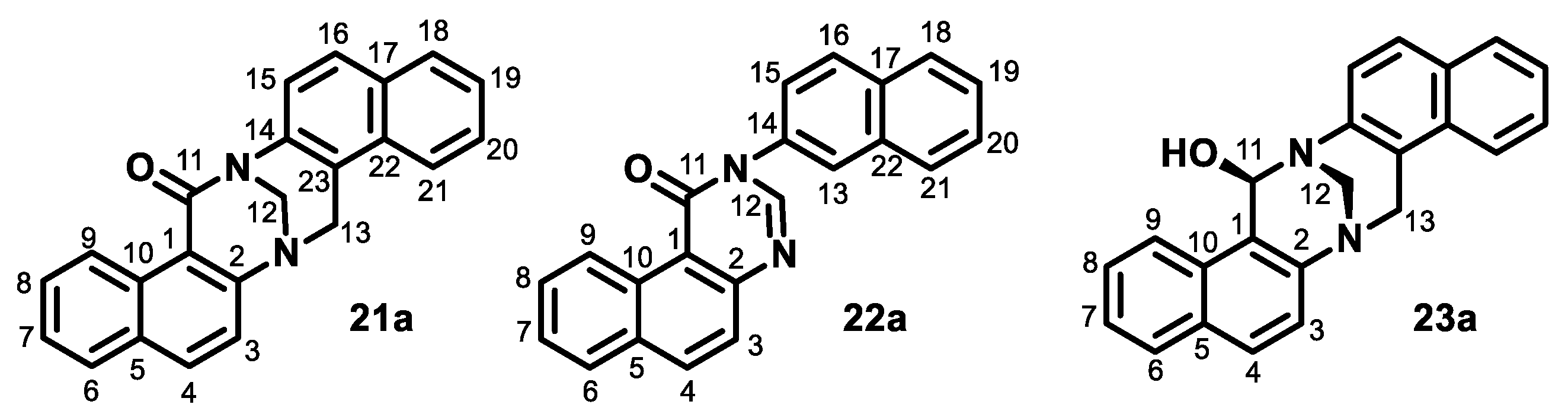
Aqueous formaldehyde (37%, 0.38 mL, 5 mmol) in acetic acid (99%, 1 mL) was added dropwise (1 min) to the solution of naphthylamine 2a (0.72 g, 5 mmol) in acetic acid (99%, 11 mL) at room temperature. The mixture was stirred for the next 2 min. The mixture was then diluted with water (20 mL) and alkalized with aqueous NH3, and the product was extracted with dichloromethane. The organic solution was washed sequentially with water and brine, dried over anhydrous sodium sulfate, and evaporated to dryness in vacuo. The residue was purified by column chromatography on silica (dichloromethane/methanol from 1:0 to 8:2) to produce quinazoline 6a (390 mg, 50%), TB 3a (81 mg, 10%), acridine 5a (23 mg, 3%), starting naphthylamine 2a (150 mg, 21%), oxo-TB 21a (17 mg, 2%), dihydroquinazoline 7a (25 mg, 3%), and oxo-quinazoline 22a (5 mg, under 1%). Those compounds were followed by a polar fraction that contained spiroTB 4a and a diastereomer of unidentified hydroxy-TB 23a.
Oxo-TB 21a: 1H NMR (500 MHz, DMSO-d6, 25 °C): 8.78 (1H, ddt, 8.6, 1.3, 0.7, H9), 8.13 (1H, d, 8.9, H4), 7.91 (1H, br d, 8.2, H6), 7.87 (1H, br d, cov., H18), 7.86 (1H, br d, cov., H21), 7.81 (1H, d, 8.8, H16), 7.64 (1H, d, 8.9, H3), 7.64 (1H, ddd, 8.6, 7.0, 1.4, H8), 7.56 (1H, ddd, 8.5, 6.8, 1.3, H20), 7.52 (1H, d, 8.8, H15), 7.49 (1H, ddd, 8.1, 6.9, 1.2, H7), 7.48 (1H, ddd, 8.1, 6.9, 1.2, H19), 5.28 (1H, d, 17.3, H13 exo), 5.06 (1H, dd, 12.6, 1.5, H12a), 5.01 (1H, br d, 17.3, H13endo), 4.98 (1H, d, 12.6, H12b). 13C{1H} NMR (126 MHz, DMSO-d6, 25 °C, from HSQC and HMBC): 173.83 (C11), 154.16 (C2), 140.41 (C14), 135.44 (C4), 131.87 (C10), 131.02 (C17), 130.88 (C5), 130.58 (C22), 128.51 (C6), 128.49 (C18), 128.27 (C8), 127.36 (C16), 126.75 (C20), 126.32 (C9), 125.58 (C19), 125.39 (C7), 124.86 (C15), 124.74 (C23), 122.89 (C3), 121.92 (C21), 115.07 (C1), 64.34 (C12), 51.36 (C13). HRMS (APCI+, MeOH): for C23H16N2O calcd. [M + H]+ 337.1335 found 337.1339. HRMS (ESI+): for C23H16N2O calcd. [M + H]+ 337.1335, too low intensity (<5%); calcd. for [M + Na]+ 359.1155 found 359.1157 (100%); calcd. [2M + Na]+ 695.2418, found 695.2417 (5%); calcd. [3M + Na]+ 1031.3680, found 1031.3695.
Dihydroquinazoline 7a: identified according to the characteristic singlet at 5.46 ppm (2H, s) having an HSQC correlation to the 13C signal at 45.28 ppm from the CH2 group (an HMBC correlation to the 13C signal at 146.66 ppm), and the singlet at 8.09 ppm (1H, br s) having an HSQC correlation to the 13C signal at 146.66 ppm from the N=CH-N group. HRMS (APCI+, MeOH): for C22H16N2 calcd. [M + H]+ 309.1386, found 309.1388.
Oxo-quinazoline 22a: 1H NMR (500 MHz, DMSO-d6, 25 °C): 9.83 (1H, ddt, 8.6, 1.3, 0.7, H9), 8.71 (1H, s, H12), 8.41 (1H, br d, 8.8, H4), 8.20 (1H, br d, 2.2, H13), 8.14 (1H, br d, 8.0, H6), 8.13 (1H, dq, 8.7, 0.7, H16), ~8.08 (1H, m, H18), ~8.05 (1H, m, H21), 7.83 (1H, d, 8.8, H3), 7.78 (1H, ddd, 8.6, 6.9, 1.6, H8), 7.74 (1H, dd, 8.7, 2.2, H15), 7.72 (1H, ddd, 8.0, 6.9, 1.3, H7), 7.68–7.63 (2H, m, H19 and H20). 13C{1H} NMR (126 MHz, DMSO-d6, 25 °C): 160.71 (C11), 150.26 (C2), 148.22 (C12), 136.05 (C4), 135.57 (C14), 132.94 (C22), 132.57 (C17), 131.79 (C5), 130.49 (C10), 128.75 (C6 or C8), 128.73 (C8 or C6), 128.70 (C16), 128.12 (C21), 127.76 (C18), 127.16 (C19), 126.91 (C20), 126.83 (C7), 126.43 (C9), 126.18 (C13), 126.15 (C3), 125.80 (C15), 115.12 (C1). HRMS (APCI+, MeOH): for C22H14N2O calcd. [M + H]+ 323.1179, found 323.1172. HRMS (ESI+): for C22H14N2O calcd. [M + H]+ 323.1179, found 323.1182 (70%); calcd. for [M + Na]+ 345.0998 found 345.1001 (93%); calcd. [2M + Na]+ 667.2105, found 667.2107 (100%); calcd. [3M + Na]+ 989.3211, found 989.3223 (23%).
SpiroTB 4a: 1H NMR (500 MHz, DMSO-d6, 25 °C): 7.85 (1H, m, H6), 7.77 (1H, d, 8.8, H4), 7.73 (1H, m, H9), 7.42 (1H, m, cov., H8), 7.41 (1H, m, cov., H7), 7.39 (1H, m, cov., H21), 7.36 (1H, m, cov., H20), 7.35 (1H, m, cov., H18), 7.34 (1H, m, cov., H3), 7.34 (1H, m, cov., H19), 7.02 (1H, d, 9.8, H16), 6.01 (1H, d, 9.8, H15), 5.09 (1H, d, 17.8, H13a), 4.89 (1H, d, 17.8, H13b), 3.85 (1H, br d, 12.6, H12a), 3.67 (1H, dd, 12.6, 1.8, H12b), 3.44 (1H, br d, 17.2, H11a), 2.93 (1H, d, 17.2, H11b). 13C{1H} NMR (126 MHz, DMSO-d6, 25 °C): 166.99 (C14), 144.54 (C2), 143.04 (C22), 133.89 (C16), 131.84 (C10), 131.65 (C17), 130.26 (C5), 129.00 (C20), 128.31 (C6), 128.12 (C18), 127.40 (C4), 127.31 (C19), 126.58 (C15), 126.55 (C8), 124.97 (C3), 124.73 (C21), 124.63 (C7), 122.74 (C1), 122.16 (C9), 73.33 (C13), 48.67 (C12), 38.76 (C11), 35.59 (C23). HRMS (APCI+, MeOH): for C23H18N2 calcd. [M + H]+ 323.1543, found 323.1547. M.p. 84–86 °C decomp. (from methanol) did not match any of the bases isolated by Farrar [10].
Unidentified hydroxy-TB 23a: 1H NMR (500 MHz, DMSO-d6, 25 °C): 5.84 (1H, br d, 5.2, H11), 6.90 (d, 5.2, OH), 4.77 (1H, d, 12.4, 1.6, H12a), 4.40 (1H, d, 12.4, H12b), 4.96 (1H, d, 16.9, H13a), 4.70 (1H, br d, 16.9, H13b). 13C{1H} NMR (126 MHz, DMSO-d6, 25 °C): 82.83 (C11), 60.08 (C12), 54.25 (C13). HRMS (APCI+, MeOH): for C23H18N2O calcd. [M + H]+ 339.1492, found 339.1494.

3.7. Reaction of Naphthylamine 2a with Formalin under Basic Conditions

Sodium hydroxide (0.02 g, 0.5 mmol) was added to a mixture of paraformaldehyde (0.30 mg, 10.0 mmol) and naphthylamine 2a (1.43 g, 10.0 mmol) in ethanol (20 mL). The reaction mixture was heated in a water bath (50 °C) until paraformaldehyde completely dissolved after ca. 5 min. The reaction mixture was monitored by NMR by sampling 30 μL of the reaction mixture into 500 μL of DMSO-d6. After one day, 2 mL of the reaction mixture was evaporated to dryness and analyzed by NMR and MS. The solid mainly consisted of the imine-ethanol adduct 24a, aminal 8a, adduct 25a, and diamine 27a in a molar ratio of 46:28:18:8, and several minor products.
Imine 9a: 1H NMR (500 MHz, DMSO-d6, 40 °C): the characteristic 1H NMR signals were obtained through the subtraction of 1D spectra (Figure 2). 7.92 (1H, d, 8.7, H4), 7.91 (1H, br d, 8.1, H6 or H9), 7.90 (1H, br d, 7.2, H6 or H9), 7.77 (1H, d, 16.3, H11a), 7.56 (1H, d, 2.1, H1), 7.51 (1H, ddd, cov., H7 or H8), 7.50 (1H, d, 16.3, H11b), 7.47 (1H, ddd, 8.1, 6.9, 1.2, H7 or H8), 7.36 (1H, dd, 8.7, 2.1, H3). HRMS (APCI+, MeOH): for C11H9N calcd. [M + H]+ 156.0808, found 156.0807.
Adduct 24a: 1H NMR (500 MHz, DMSO-d6, 25 °C, presence of excess ethanol and NaOH traces): 7.65 (1H, br d, 8.2, H6), 7.64 (1H, br d, 8.7, H4), 7.59 (1H, br d, 8.2, H9), 7.31 (1H, ddd, 8.2, 6.8, 1.3, H8), 7.14 (1H, ddd, 8.2, 6.8, 1.2, H7), 7.03 (1H, dd, 8.7, 2.3, H3), 6.97 (1H, d, 2.3, H1), 4.67 (1H, s, H11), 3.47 (2H, q, 7.0, CH2CH3), 1.10 (3H, t, 7.0, CH2CH3), the NH signal underwent a fast chemical exchange with the OH signal of ethanol. 13C{1H} NMR (126 MHz, DMSO-d6, 25 °C, from HSQC and HMBC, presence of ethanol excess and NaOH traces): 145.19 (C2), 135.05 (C10), 128.48 (C4), 127.47 (C6), 127.35 (C5), 126.02 (C8), 125.75 (C9), 121.64 (C7), 117.76 (C3), 104.92 (C1), 73.98 (C11), 61.15 (CH2CH3), 15.11 (CH2CH3).
Adduct 25a: identified by the correlation patterns in HSQC and HMBC corresponding to N-CH2-OCH2-OCH2CH3, i.e., 1H/13C: 4.98/59.81, 5.02/80.42, 3.52/62.05, 1.14/15.14, resp. HRMS (APCI+, MeOH): for C14H17NO2 calcd. [M + H]+ 232.1332, found 232.1327.
Adduct 26a: identified by the correlation patterns in HSQC and HMBC corresponding to N-CH2-OCH2-OCH2-OCH2CH3, i.e., 1H/13C: 4.96/58.08, 5.27/65.08, 4.92/79.67, 3.42/62.10, 1.11/15.07, resp. HRMS (APCI+, MeOH): the peak of a molecular ion was not observed.
Aminal 8a: identified by the correlation patterns in HSQC and HMBC corresponding to N-CH2-N, i.e., 1H/13C: 4.72/52.57. HRMS (APCI+, MeOH): for C21H18N2 calcd. [M + H]+ 299.1543, found 299.1546.
Diamine 27a: identified by the correlation patterns in HSQC and HMBC corresponding to N-CH2-O-CH2-N, i.e., 1H/13C: 5.03/58.64. HRMS (APCI+, MeOH): the peak of a molecular ion was not observed.

4. Conclusions

According to the careful observations and tedious analysis of the 1D and 2D NMR spectra of mixtures at various temperatures, we corrected the molecular structures of several products reported previously [9,22]. Moreover, we suggested an alternative explanation for the formation of products reported in the recent literature [26,27,28], namely aminal 8a and imine 9a. We discovered how to prepare or generate these compounds in situ and obtained their spectral characterizations for the first time.
We have shown that free methanal should be generated through the known dry decomposition of paraformaldehyde [14,15] and used in a nucleophile-free environment. Alternatively, methanal or its imine could be obtained by heating to above 85 °C when nucleophiles are not present in great excess. Once the methanal or imine is generated, it will be stable but will slowly react back to hydrates, hemiaminals, or aminals in the presence of nucleophiles.
Since methanal (1) concentrations during a reaction of formalin, paraformaldehyde, trioxane, dimethoxymethane, or others could be very low or even negligible in the presence of a high excess of nucleophiles such as water, alcohols, or amines, we recommend calling them methanal equivalents instead of the usual methanal sources.
Finally, we have found that the formation of TB 3a and spiroTB 4a occurs rapidly even in acetic acid and that even silica can enable the formation of acridine 5a, dinaphthylamine 18a, TB 3a, and spiroTB 4a.
According to our experiments and DFT calculations, methanal is not behaving as the simplest aldehyde as is commonly assumed, but instead, its behavior resembled that of a very electron-deficient oxo-compound such as trifluoroethanal.
Computational calculations in the absence of acid catalysis showed plausible pathways for the formation of a stable intermediate product 6, which could evolve into imine 9 and even into TB 3. Under the conditions of the in silico study, both the imines and TBs are thermodynamically more stable than the aminals. Under these conditions, TB 3a was preferentially formed from 6a with respect to TB 3b.

Supplementary Materials

The following supporting information can be downloaded at: https://www.mdpi.com/article/10.3390/molecules28041549/s1, (I) Total free energies in atomic units of all stationary points calculated. (II) Comparison of the relative free energies in Scheme 8 and Scheme 9 obtained with CAM-B3LYP-GD3BJ and standard CAM-B3LYP functional. (III) 3D structures of the key compounds shown in Scheme 8 and Scheme 9. (IV) The NMR spectra of pure quinazoline 6a and bisquinazoline 16a, and a few examples of the 2D NMR spectra of the studied mixtures.

Author Contributions

Conceptualization, methodology, writing original draft preparation, M.H. and B.D.; methodology, writing review and editing, M.H., B.D., T.N., M.D., A.T. and P.A.L.; computational methodology writing review P.A.L., D.D., C.A.C., E.L.M. and D.M.A.V.; project administration B.D., funding acquisition, B.D. and D.M.A.V. All authors have read and agreed to the published version of the manuscript.

Funding

This work was supported by the Ministry of Education, Youth and Sports of the Czech Republic (program INTER-EXCELLENCE, project no. LTAUSA19065) and CONICET (National Council for Research and Technology, Argentina-grant PIP #11220200103033CO). D.D. and P.A.L. thank CONICET for postdoctoral fellowships.

Institutional Review Board Statement

Not applicable.

Informed Consent Statement

Not applicable.

Data Availability Statement

All data used to support the findings of this study are included within the article and Supplementary Materials. A few examples of simple NMR spectra are provided. The other spectra are available upon request as raw data files since their readability requires computer assistance due to their complexity.

Conflicts of Interest

The authors declare no conflict of interest.

Sample Availability

Not applicable.

References

  1. Tatar, A.; Čejka, J.; Král, V.; Dolenský, B. Spiro Tröger’s Base Derivatives: Another Structural Phoenix? Org. Lett. 2010, 12, 1872–1875. [Google Scholar] [CrossRef] [PubMed]
  2. Tröger, J. Ueber einige mittelst nascirenden Formaldehydes entstehende Basen. J. Prakt. Chem. 1887, 36, 225–245. [Google Scholar] [CrossRef]
  3. Spielman, M.A. The Structure of Troeger’s Base. J. Am. Chem. Soc. 1935, 57, 583–585. [Google Scholar] [CrossRef]
  4. Vögtle, F. Heterocycles and Biologically Active Compounds. In Fascinating Molecules in Organic Chemistry, 1st ed.; John Wiley & Sons: Chichester, UK, 1992; pp. 237–290. [Google Scholar]
  5. Dolenský, B.; Elguero, J.; Král, V.; Pardo, C.; Valík, M. Current Tröger’s Base Chemistry. Adv. Heterocycl. Chem. 2007, 93, 1–56. [Google Scholar]
  6. Sergeyev, S. Recent Developments in Synthetic Chemistry, Chiral Separations, and Applications of Tröger’s Base Analogues. Helv. Chim. Acta 2009, 92, 415–444. [Google Scholar] [CrossRef]
  7. Reed, J.H. Ueber β-Naphtoacridin. Vorläufige Mittheilung. J. Prakt. Chem. 1886, 34, 160–161. [Google Scholar] [CrossRef]
  8. Reed, J.H., VIII. Ueber Methylderivate der Naphtochinoline und über β-Naphtoacridin. J. Prakt. Chem. 1887, 35, 298–322. [Google Scholar] [CrossRef]
  9. Morgan, G.T. LVI—Action of formaldehyde on amines of the naphthalene series. Part I. J. Chem. Soc., Trans. 1898, 73, 536–554. [Google Scholar] [CrossRef]
  10. Farrar, W.V. Reactions of formaldehyde with aromatic amines. J. Appl. Chem. 1964, 14, 389–399. [Google Scholar] [CrossRef]
  11. Tálas, E.; Margitfalvi, J.; Machytka, D.; Czugler, M. Synthesis and resolution of naphthyl-Tröger’s base. Tetrahedron Asymmetry 1998, 9, 4151–4156. [Google Scholar] [CrossRef]
  12. Paleta, O.; Dolenský, B.; Paleček, J.; Kvíčala, J. Three-Component (Domino) Reaction Affording Substituted Pyrroloquinazolines: Cyclization Regioselectivity and Stereoselectivity. Eur. J. Org. Chem. 2013, 2013, 1262–1270. [Google Scholar] [CrossRef]
  13. Valenta, P. Oszillographische strom-spannungs-kurven III. Untersuchung des formaldehyds in gepuffertem milieu. Collect. Czech. Chem. Commun. 1960, 25, 853–861. [Google Scholar] [CrossRef]
  14. Albert, K.; Peters, B.; Bayer, E.; Treiber, U.; Zwilling, M. Crosslinking of Gelatin with Formaldehyde; a 13C NMR Study. Z. Naturforsch. B J. Chem. Sci. 1986, 41b, 351–358. [Google Scholar] [CrossRef]
  15. Cluşaru, A.; Crişan, I.; Kůta, J. Equilibrium constants and rates of dehydration of formaldehyde in buffered solutions of light and heavy water studied at DME. J. Electroanal. Chem. Interfacial Electrochem. 1973, 46, 51–62. [Google Scholar] [CrossRef]
  16. Walker, F. Some Properties of Anhydrous Formaldehyde. J. Am. Chem. Soc. 1933, 55, 2821–2826. [Google Scholar] [CrossRef]
  17. Lanza, P.A.; Dusso, D.; Ramírez, C.L.; Parise, A.R.; Chesta, C.A.; Moyano, E.L.; Vera, D.M.A. Uncovering the mechanism leading to the synthesis of symmetric and asymmetric Tröger’s bases. Eur. J. Org. Chem. 2019, 2019, 7644–7655. [Google Scholar] [CrossRef]
  18. Parr, R.G.; Szentpály, L.V.; Shubin, L. Electrophilicity Index. J. Am. Chem. Soc. 1999, 121, 1922–1924. [Google Scholar] [CrossRef]
  19. Puiatti, M.; Vera, D.M.A.; Pierini, A.B. In search for an optimal methodology to calculate the valence electron affinities of temporary anions. Phys. Chem. Chem. Phys. 2009, 11, 9013–9024. [Google Scholar] [CrossRef]
  20. Borioni, L.; Puiatti, M.; Vera, D.M.A.; Pierini, A.B. In search of the best DFT functional for dealing with organic anionic species. Phys. Chem. Chem. Phys. 2017, 19, 9189–9198. [Google Scholar] [CrossRef]
  21. Frisch, M.J.; Trucks, G.W.; Schlegel, H.B.; Scuseria, G.E.; Robb, M.A.; Cheeseman, J.R.; Scalmani, G.; Barone, V.; Petersson, G.A.; Nakatsuji, H.; et al. Gaussian 16 Revision C.01; Gaussian Inc.: Wallingford, CT, USA, 2019. [Google Scholar]
  22. Möhlau, R.; Haase, O. Ueber Naphtacrihydridin. Ber. Dtsch. Chem. Ges. 1902, 35, 4164–4172. [Google Scholar] [CrossRef]
  23. Wagner, E.C. A rationalization of acid-induced reactions of methylene-bis-amines, methylene-amines, and of formaldehyde and amines. J. Org. Chem. 1954, 19, 1863–1881. [Google Scholar] [CrossRef]
  24. Mahon, A.B.; Craig, D.C.; Try, A.C. Synthesis of 5,6,11,12-tetrahydrodibenzo[b,f][1,5]diazocines and a demonstration of their reactivity to afford methano strap-modified Tröger’s base analogues. ARKIVOC 2008, 13, 148–163. [Google Scholar] [CrossRef]
  25. Artacho, J.; Ascic, E.; Rantanen, T.; Karlsson, J.; Wallentin, C.-J.; Wang, R.; Wendt, O.F.; Harmata, M.; Snieckus, V.; Wärnmark, K. Twisted Amide Analogues of Tröger’s Base. Chem. Eur. J. 2012, 18, 1038–1042. [Google Scholar] [CrossRef] [PubMed]
  26. Kozlov, N.G.; Kadutskii, A.P. Synthesis of Azaphenanthrene Derivatives by Condensation of 2-Methyleneaminonaphthalene with Cyclic β-Diketones. Russ. J. Org. Chem. 2002, 38, 129–133. [Google Scholar] [CrossRef]
  27. Kadutskii, A.P.; Kozlov, N.G. One-stage synthesis of benzoacridine and benzophenanthroline functionalized derivatives. Russ. J. Org. Chem. 2006, 42, 748–751. [Google Scholar] [CrossRef]
  28. Kozlov, N.G.; Kadutskii, A.P.; Baranovskii, A.V. Condensation of 2-naphthylamine or N-benzyl-2-naphthylamines with formaldehyde and methyl 2,2-dimethyl-4,6-dioxocyclohexane-3-carboxylate. Russ. J. Org. Chem. 2012, 48, 1456–1463. [Google Scholar] [CrossRef]
  29. Scalmani, G.; Frisch, M.J.J. Continuous surface charge polarizable continuum models of solvation. I. General formalism. Chem. Phys. 2010, 132, 114110–114115. [Google Scholar] [CrossRef] [PubMed]
  30. Grimme, S.; Ehrlich, S.; Goerigk, L. Effect of the damping function in dispersion corrected density functional theory. J. Comp. Chem. 2011, 32, 1456–1465. [Google Scholar] [CrossRef]
  31. Grimme, S.; Antony, J.; Ehrlich, S.; Krieg, G. A consistent and accurate ab initio parameterization of density functional dispersion correction (DFT-D) for the 94 elements H-Pu. J. Chem. Phys. 2010, 132, 154104. [Google Scholar] [CrossRef]
  32. Colomer, J.P.; Sciú, M.-L.; Ramirez, C.L.; Vera, D.M.A.; Moyano, E.L. Thermal ring opening of pyrazolo[3,4-d][1,2,3]triazin-4-ones: An experimental and theoretical study. Eur. J. Org. Chem. 2018, 13, 1514–1524. [Google Scholar] [CrossRef]
Scheme 1. Previously described products of the reaction of formaldehyde with 2-naphthylamines 2 under acidic conditions [1].
Molecules 28 01549 sch001
Scheme 2. The molecular structures of aminals 8a and 8b, and imines 9a and 9b.
Molecules 28 01549 sch002
Scheme 3. Major compounds identified in methanal solutions.
Molecules 28 01549 sch003
Scheme 4. Molecular structures of identified products with arbitrary numbering for NMR assignments.
Molecules 28 01549 sch004
Scheme 5. The compounds in equilibrium at 85–115 °C in the DMSO-d6 solution.
Molecules 28 01549 sch005
Figure 1. The 1H NMR spectrum of the sample containing imine 9b (violet), aminal 8b (red), hemiaminal 20b (black), naphthylamine 2b (green), and methanal (1).
Molecules 28 01549 g001
Scheme 6. Molecular structures of oxo-TB 21a, oxo-quinazoline 22a, and an unidentified exo-diastereomer of hydroxy-TB 23a (racemic).
Molecules 28 01549 sch006
Scheme 7. The products formed from naphthylamine 2a and paraformaldehyde under basic conditions.
Molecules 28 01549 sch007
Figure 2. Experimental 1H NMR spectrum of imine 9a constructed from the subtraction of spectra (lower), and a simulated spectrum (upper).
Molecules 28 01549 g002
Scheme 8. Summary of the main stationary points calculated. All relative free energies are expressed in kcal/mol.
Molecules 28 01549 sch008
Scheme 9. Thermochemistry of the dehydrogenation of 6a/b to yield 7a/b.
Molecules 28 01549 sch009
Table 1. The molar ratios of selected compounds at various temperatures for formalin in DMSO-d6.
TemperatureMethanal (1)Methandiol (10)Hemiacetal 13
25 °C36928
50 °C65935
75 °C134740
100 °C403129
115 °C582319
Table 2. The molar ratios of selected compounds at various temperatures for paraformaldehyde and water in DMSO-d6.
TemperatureMethanal (1)Methandiol (10)Diol 11Diol 12
25 °C187102
50 °C178192
75 °C578161
100 °C177481
115 °C365950
Table 3. Comparison of the HOMO/LUMO gaps for the solvated oxo-species.
SpeciesGap HOMO/LUMO (eV)
methanal9.24
ethanal9.57
acetone9.59
trifluoroethanal9.39
hexafluoroacetone9.29
Table 4. Electrophilicities were calculated for different carbonyls.
SpeciesVIP (eV)VEA (eV)ω (eV)
methanal11.10−1.014.20
ethanal10.46−1.603.25
acetone9.95−1.472.87
trifluoroethanal11.93−0.165.72
Table 5. ΔG°hyd (oxo-compound + H2O → hydrate) was calculated using acetone as a solvent.
ΔG°hyd (kcal/mol)
methanal−19.27
ethanal−13.93
trifluoroethanal−20.54
Disclaimer/Publisher’s Note: The statements, opinions and data contained in all publications are solely those of the individual author(s) and contributor(s) and not of MDPI and/or the editor(s). MDPI and/or the editor(s) disclaim responsibility for any injury to people or property resulting from any ideas, methods, instructions or products referred to in the content.

