Review

A Review on Bioactive Anthraquinone and Derivatives as the Regulators for ROS

1
Tianjin Renai College, Tianjin 301636, China
2
College of Pharmaceutical Engineering of Traditional Chinese Medicine, Tianjin University of Traditional Chinese Medicine, Tianjin 301617, China
*
Author to whom correspondence should be addressed.
Molecules 2023, 28(24), 8139; https://doi.org/10.3390/molecules28248139
Submission received: 5 November 2023 / Revised: 9 December 2023 / Accepted: 11 December 2023 / Published: 17 December 2023

Abstract

:
Anthraquinones are bioactive natural products, which are often found in medicinal herbs. These compounds exert antioxidant-related pharmacological actions including neuroprotective effects, anti-inflammation, anticancer, hepatoprotective effects and anti-aging, etc. Considering the benefits from their pharmacological use, recently, there was an upsurge in the development and utilization of anthraquinones as reactive oxygen species (ROS) regulators. In this review, a deep discussion was carried out on their antioxidant activities and the structure-activity relationships. The antioxidant mechanisms and the chemistry behind the antioxidant activities of both natural and synthesized compounds were furtherly explored and demonstrated. Due to the specific chemical activity of ROS, antioxidants are essential for human health. Therefore, the development of reagents that regulate the imbalance between ROS formation and elimination should be more extensive and rational, and the exploration of antioxidant mechanisms of anthraquinones may provide new therapeutic tools and ideas for various diseases mediated by ROS.

Graphical Abstract

1. Introduction

According to the biological theory of free radicals, the generation and scavenging of free radicals should be in a dynamic balance under normal circumstances. The broken balance of free radicals will harm the body and cause diseases. Reactive oxygen species (ROS), which include several kinds of oxygen related free radicals, are of Janus-faced nature in cells. On one hand, ROS signaling is of vital importance in maintaining crucial cellular functions. They are involved in the signaling cascades and the regulation of cellular processes. On the other hand, they provide detrimental damages to macromolecules. The overproduction of ROS can damage cells and induce a variety of diseases, such as neurodegenerative diseases, cancer, hepatopathic diseases, and cardiovascular diseases, etc. [1]. Mechanisms of ROS mediated diseases have also been studied. For example, the brains often show oxidative damage in neurodegenerative diseases. Alzheimer’s disease (AD) is a classic example. Long-lasting ROS generation via amyloid beta (Aβ)-Cu(II) peptides and mitochondrial dysfunction may lead to neuronal damage and ultimately dementia. Elevated ROS can damage DNA, transform normal cells into cancer cells, trigger enhanced epidermal growth factor receptor signaling and promote tumor progression [2,3,4]. Excessive ROS can lead to fibrosis, liver inflammation and hepatocellular carcinom [5]. Endothelial cells and vascular smooth muscle cells, the most important cells for maintaining the integrity and homeostasis of the vascular system, both represent targets of ROS and ROS signaling. Excessive ROS may damage vascular cells, induce the proliferation and migration of vascular smooth muscle cells, and then cause vascular remodeling [6,7]. Therefore, therapeutic strategy targeting ROS and ROS-generating system is to maintain the dynamic balance of ROS.
Antioxidants are a well-studied class of reagents that mainly include enzymes and non-enzymes. Enzymatic antioxidants, including superoxide dismutase (SOD), catalase (CAT), glutathione peroxidase (GPX), glutathione reductase (GR), and glutathione S-transferases (GST), are involved in ROS production and peroxides reduction, thus playing a major role in maintaining the redox balance. Nonenzymatic antioxidants, such as polyphenols, anthocyanins, polyenes, etc., can directly increase rates of ROS degradation and halt excessive ROS accumulation [8,9]. Another class of reagents can interact indirectly with signaling pathways or key molecules involved in the production and elimination of ROS to reduce oxidative damage [10].
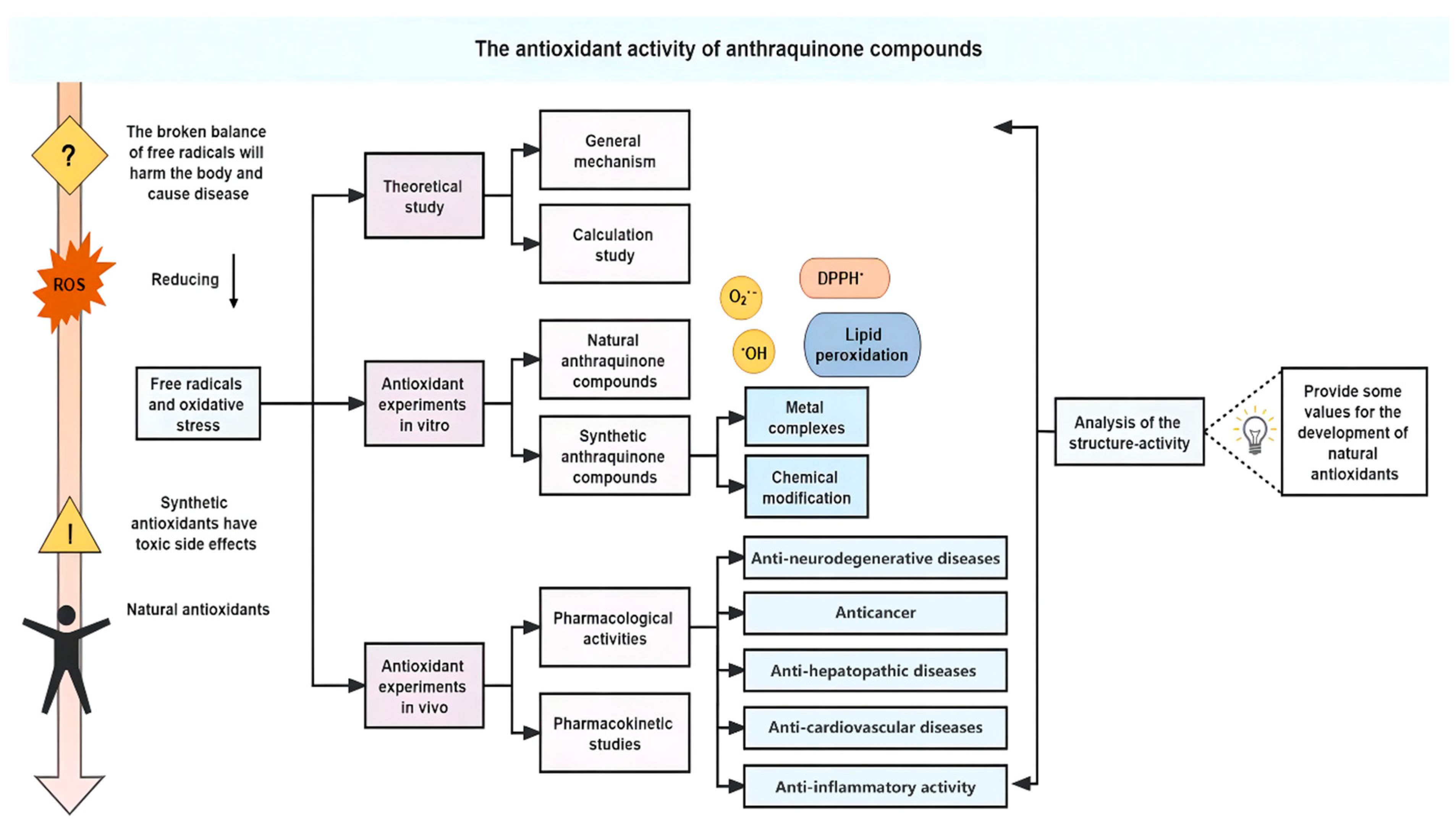
Natural nonenzymatic antioxidants have attracted the attention of researchers because of their wide distribution and easy availability [11,12]. The largest group of natural quinones are phenol-substituted anthraquinones [13], which are abundant in many plants, such as Rheum rhabarbarum L. (Polygonaceae), Polygonum cuspidatum Sieb. et Zucc. (Polygonaceae), Polygonum multiflorum Thunb. (Polygonaceae), Rubia cordifolia Thunb. (Rubiaceae), Cassia sieberiana DC. (Caesalpiniaceae), Rhamnus dahurica Pall. (Rhamnaceae), Salvia mitiorrhiza Bge. (Labiatae), etc. [14,15,16,17,18,19]. The anthraquinone parent structure has a three fused-ringed structure with two carbonyl groups (Figure 1). In the structure of anthraquinone derivatives, the anthraquinone ring is the basic scaffold, with the substituent groups connected to the position of C1–C8 [13,20]. Naturally occurring anthraquinones are often substituted with hydroxyl, hydroxymethyl, methyl, methoxy, and carboxyl groups on the parent structure and are present in the plant either in free form or in combination with sugars to form glycosides. The structures of some natural anthraquinone compounds are shown in Figure 1. Recently, a large number of studies have shown that natural anthraquinone compounds have not only strong antioxidant activity but also little toxicity or few side effects [21,22,23,24]. Therefore, the natural anthraquinone compounds are particularly attractive and have great potential as antioxidant agents. In this work, the antioxidant potential of bioactive anthraquinone compounds has been reviewed. The main line of this work is showed in Figure 2.

2. Free Radicals and Oxidative Stress

Free radicals are present in any chemical entity with one or more unpaired electrons in the outer orbital [25]. Humans are aerobic organisms, so the body inevitably produces free radicals. The sources of free radicals can be endogenous or exogenous [26]. The most important endogenous sources are mitochondrial respiratory chain enzymes, reduced nicotinamide adenine dinucleotide phosphate (NADPH) oxidase, nitric oxide synthase and xanthine oxidase. At the same time, external factors, such as environmental pollutants, radiation, smoking and drugs, may also contribute to the generation of free radicals [27,28,29,30].
ROS or reactive nitrogen species (RNS) are products of cell metabolism [31]. ROS can be in radical or nonradical forms, and they may interchange with each other in the body. Common free radical ROS include hydroxyl radicals (OH), hydroperoxyl radicals (HO2), superoxide anions (O2•−), nitric oxide (NO) and peroxy radicals (ROO) [32]. In addition, the common nonradical ROS are hydrogen peroxide (H2O2), singlet oxygen (1O2), hypochlorous acid (HOCl), and peroxynitrite (ONOO) [33,34].
ROS have both beneficial and harmful effects on biological systems [31]. The delicate balance between these two effects is achieved through the mechanism of “redox regulation” [35]. Oxidative stress is caused by the imbalance between the antioxidant defense system and the intracellular accumulation of ROS [36]. The excessive production of ROS and oxidative stress play vital roles in several kinds of life-threatening diseases, such as Alzheimer’s disease, liver injury, and cancer. Consequently, it is significant to counteract the harmful effects generated by ROS, which is possible through the use of antioxidants [37].
Metals play an important role in the processes of ROS formation and elimination [38,39]. Metal ions with redox activity, such as iron and copper, are cofactors of various enzymes. However, the iron and copper ions may also produce ROS and further damage the organism in the process of reactions [40]. For example, Fe2+ is necessary for DNA synthesis, hemoglobin synthesis and electron transport [41]. However, Fe2+ could catalyze the Haber–Weiss reaction to generate more harmful OH from O2•−. When H2O2 reacts with Fe2+, the Fenton reaction can occur to form OH [42].
Fe2+ + H2O2OH + OH + Fe3+
Fe3+ + O2•− → Fe2+ + O2
Or merged into: O2•− + H2O2 → O2 + OH + OH
Cu2+ catalyzes the oxidation of low-density lipoprotein, which has a similar Fenton reaction, directly causing ROS production and significantly reducing glutathione (GSH) levels with exposure to high concentrations of Cu2+ [43]. Zinc is also an essential nutrient for the maintenance of all life forms. Zn2+ is the most abundant metal in the brain, mainly protecting the mercapto group of proteins and enzymes from oxidation or inhibiting the formation of OH by H2O2 through the Fenton reaction [44]. OH is the most destructive species in free radical pathology and can induce oxidative damage in almost all cell molecules [45].
A detailed illustration of ROS formation is shown in Figure S1.

3. Theoretical Study on the Antioxidant Activity of Anthraquinone Compounds

3.1. General Mechanism of Antioxidant Activity of Anthraquinone Compounds with Phenolic Substituents

Living cells contain both low molecular weight antioxidants and high molecular weight antioxidant enzymes, both of which prevent and repair damage caused by free radicals. Antioxidants, according to their properties, can be divided into two types: preventive antioxidants [46] and chain-breaking antioxidants. Preventive antioxidants can scavenge free radicals in the chain initiation stage, such as SOD, CAT and metal (iron and copper) chelators. Chain-breaking antioxidants, often referred to as free radical scavengers, can capture peroxy radicals in the free radical reaction chain and prevent or slow down the progress of the free radical chain reaction. A large number of studies have shown that phenolic hydroxyl groups, such as ortho-phenolic hydroxyl groups and para-phenolic hydroxyl groups, could react with free radicals to produce stable semiquinone free radicals.
Phenolic compounds have three main antioxidant mechanisms that are widely accepted [19,47,48,49]. The first is the hydrogen atom transfer (HAT) mechanism, in which R free radicals attract a hydrogen atom from the antioxidant ArOH (Equation (1)) [50,51,52]. In this mechanism, the bond dissociation enthalpy (BDE) of the O-H bond is a significant parameter for scavenging activity evaluation. Although BDE is a thermodynamic parameter, the BDE of the same type of antioxidant correlates well with the logarithm of radical-scavenging rate constants (logKs) [53].
R + ArOH → RH + ArO
The second kind of mechanism is a single-electron transfer followed by proton transfer (SET-PT), namely, the rate determining step of ArOH losing one electron to generate ArOH+• followed by a proton transfer step (Equation (2)) [54].
R + ArOH → R + ArOH+• → RH +ArO
In the SET-PT mechanism, the ionization potential (IP) is the most important parameter for evaluating antioxidant activity. The lower the IP value is, the easier the formation of phenoxy radicals [50]. Nevertheless, the extremely low IP may convert the antioxidant to a prooxidant, because prooxidants can generate O2•− by directly transferring an electron to the surrounding O2 [55].
The third antioxidant mechanism is sequential proton loss and electron transfer (SPLET). ArOH loses protons to form ArO, and then electron transfer occurs [50,52,56,57]. According to Equation (3), the reaction enthalpy change is related mainly to the proton affinity potential of the anion ArO, and the subsequent reaction enthalpy change is related mainly to the electron transfer enthalpy.
ArOH → ArO + H+
ArO + R → ArO + R
R + H+ → RH
The mechanism adopted is also related to the solvent. HAT is clarified to be the most favorable mechanism to describe the antioxidant activity of hydroxyanthraquinone in the gas phase and non-aqueous solvent (e.g., benzene). While in a aqueous solution, SPLET represents the most reasonable reaction pathway in thermodynamics. Because of the intermolecular hydrogen bond between water and phenoxyl groups in an aqueous solution, protons are more likely to be lost. Whereas, in a non-aqueous solution, intramolecular hydrogen bonds are more likely formed, hydrogen atoms are more likely to be lost.

3.2. Calculation Study of Antioxidant Activity of Anthraquinone Compounds with Phenolic Substituents

A density functional theory approach has been successfully used to calculate the biochemical parameters of polyphenol extracts and to elucidate the structure-activity relationship of phenolic antioxidants. Reza Nazifi et al. [58] predicted the antioxidant activity of three aloe compounds (aloe-emodin (2), aloesone (2a) and isoeleutheol (2b)) by using the density functional theory calculation of the B3LYP hybrid functional and the 6-311++G** basis set. HOMO-LUMO energy gaps showed that compound 2 had the lowest Egap value (Table S1 in Supplementary Materials), so it was more prone to give electrons and showed the best antioxidant activity among the compounds 2, 2a, and 2b (Figure 3). From the structures of them, it clearly showed that compound 2 had more electron-donating phenoxy groups.
Jeremic et al. [59] used the M06-2X/6-311++G(d,p) theory to study the antioxidant capacity of compounds 1, 2, 5, 6, 10 and 11. The results showed that compounds 10 and 11, with the hydroxy groups on the ortho position, had the highest reactivity. Compounds 1 and 6 with three hydroxy groups had moderate antioxidant capacity. Compounds 2 and 5 with two hydroxy groups had the lowest antioxidant capacity. Isin [60] used B3LYP/6-3111++G(2d,2p) and the conductor-like polarizable continuum model to study the free radical scavenging activities of four hydroxyanthraquinones (compounds 11, 12, 13 and 14). The order of hydrogen supply capacity is compounds 11 > 12 > 14 > 13, and the BDE values of compounds 11 and 12 (ortho-hydroxy groups) are lower than the BDE values of compounds 13 and 14 (carboxyl group).
It was found that 1-OH and 8-OH are the most active sites of compounds 2 and 5, while 3-OH is the most active site for compounds 1 and 6 [61]. Based on the theoretical level of B3LYP/6-311++G**, Markovic et al. [62] analyzed the BDE values of all hydroxyl positions of compound 1 and clarified the role of 3-OH in antioxidant properties. The BDE values of 1-OH and 8-OH are higher because H removal also means the fracture of hydrogen bonds (Table S2 in Supplementary Materials). These results can be explained by the intramolecular hydrogen bonds formed between 1-OH/8-OH and 9-carbonyl group, which make the hydrogen atom difficult to be lost.
Jeremic et al. [63] evaluated the antioxidant activity of compound 10 by its BDE and IP theoretically (Table S3 in Supplementary Materials). The free radical scavenging characteristics of 10 in the gas phase were well explained by HAT. The lower IP value for aqueous solution indicated that the SET-PT mechanism was reasonable.

4. Antioxidant Experiments of Anthraquinone Compounds In Vitro

In terms of antioxidant properties, anthraquinone compounds can scavenge OH, 1,1-diphenyl-2-picrylhydrazyl (DPPH) and O2•− free radicals and inhibit lipid peroxide to exert their antioxidant effects, which is of great significance for the development of high efficiency antioxidants. In this section, the antioxidant properties of natural and synthetic anthraquinone compounds are discussed in detail according to scavenging activities of different radicals.

4.1. Antioxidant Experiments of Natural Anthraquinone Compounds

4.1.1. Study on Scavenging Activities of OH Radical

Jung et al. [64] found that compound 9 had a stronger inhibitory effect on OH than compound 1, with IC50 values of 3.05 ± 0.26 µM and 13.29 ± 3.20 μM, respectively. In addition, compounds 1 and 9 exhibited hepatoprotective effects against tacrine-induced cytotoxicity. Vargas et al. [65] investigated the ability of compounds 1, 2 and 3 to inhibit ROS (OH, 1O2, H2O2) in cell-free systems. The results showed that the scavenging ability was in the following order: compounds 1 > 3 > 2. Kumar et al. [66] found that compound 11 was an extraordinarily fine scavenger of OH, which in turn protected plasmid DNA from damage. Lin et al. [67] found that the reason why Folium Sennae could protect against DNA and mesenchymal stem cell damage induced by OH. It might be because plant phenols (especially compounds 1, 2, 3) played a protective role through HAT and/or SET-PT mechanisms. Their phenolic hydroxyl groups were partially oxidized into stable semiquinone forms. The stability of the semiquinone form ultimately determined the protective or antioxidant effect of plant phenols.

4.1.2. Study on Scavenging Activity of DPPH Radical

It was reported that compound 1 and 15 could significantly eliminate DPPH radicals and that the scavenging ability was dose-dependent at concentrations of 0.5~100 µM [23,68]. Both Nam et al. [69] and Baghiani et al. [70] found that compound 10 had a high radical scavenging effect with an IC50 = 3.491 μg/mL. The experimental data demonstrated that the number and site of the OH groups seemed to be the primary factors affecting the abilities of antioxidants. The ortho-hydroxyl groups in compound 10 can react with free radicals to form a more stable conjugated semiquinone free radical and thus interrupt the free radical chain reaction. Therefore, compound 10 had stronger antioxidant properties. Shi and Huang [71] found that compound 5 had a scavenging effect on DPPH radicals (IC50 = 26.56 µg/mL). Zengin et al. [22] estimated the free radical scavenging effects of compounds 10 and 11 through ABTS and DPPH assays. The results showed that compound 10 had the greatest scavenging activity, followed by compound 11.
In general, scavenging of OH radicals is often proceeded via a radical adduct formation mechanism. First, a radical adduct with phenol is formed. Then, water molecules are eliminated [51]. Scavenging of DPPH is often based on the abstraction of hydrogen atoms from antioxidants or the transfer of electrons from phenoxide anions to DPPH [72].

4.1.3. Study on Scavenging Activity of O2•− Radical

Our group found that compound 1 showed weaker O2•− scavenging capacity (IC50 = 235.73 μM) than Vitamin C (IC50 = 12.68 μM) using pyrogallol autoxidation. The mechanism for scavenging of O2•− might be by electron donation from the reductants, so Vitamin C had better ability [73].

4.1.4. Determination of Anti-Lipid Peroxidation

The lipid peroxidation reaction is shown in Figure S1. Lipid peroxidation is one of the results of free radical formation in cells and tissues. O2•−, H2O2 and OH actively participate in the initiation of lipid peroxidation.
Yen et al. [74] reported that anthraquinone compounds inhibited the peroxidation of linoleic acid, and the order of the activity was anthrone compound > 2 > 3 > 1 > anthraquinone. Although the inhibitory activities of anthraquinone compounds were weaker than anthrone, which may be attributed to the keto carbonyl group of C-10, other reports showed that the above anthraquinone compounds were good antioxidants against lipid peroxidation [75,76,77]. For example, the inhibitory effect of compound 1 was even stronger than that of α-tocopherol. The activities could be tested with three kinds of systems: (1) in linoleic acid; (2) 2′,7′-dichlorodihydrofluorescein diacetate (DCHF-DA); (3) 3-morpholinosydnonimine (SIN-1) system. The inhibitory effect of anthraquinone compounds might be related to radical scavenging, iron chelation, and enzyme affinity.

4.1.5. Natural Anthraquinone Compounds in Cell Culture-Based Experiments

Compared with other in vitro experiments, cellular experiments are performed in an environment that more closely resembles the human physiological environment, and some of the results are listed in Table 1. The results from cellular experiments showed that anthraquinone compounds may have a series of beneficial bioactivities, such as oxidative stress reducing, cytotoxicity prevention, and inflammatory inhibition. Some of the results are listed as followings:
Compound 1 could fight cisplatin-induced oxidative stress in human embryonic kidney 293 (HEK 293) cells by restoring the GSH and total antioxidant capacity (TAC) depletion as well as augmenting the cisplatin-inhibited antioxidant enzymes, such as SOD, CAT, GPX, GR, and GST [68].Compound 2 could prevent H2O2-induced cytotoxicity of PC12 cells by significantly reducing both the extracellular release of NO and lactate dehydrogenase and intracellular accumulation of ROS [78]. Compound 3 could obviously increase the viability of H2O2-injured human umbilical vein endothelial cells (HUVECs) by downregulating malondialdehyde (MDA), lactate dehydrogenase (LDH) and the expression of caspase-3, caspase-8, caspase-9 mRNA, while upregulating the NO content, nitric oxide synthase (NOS), SOD, and glutathione peroxidase (GSH-PX) [79]. Compound 5 could inhibit lipopolysaccharide (LPS)-induced inflammatory reaction of BV-2 mucin microglia by downregulating dynamin-related protein 1 (Drp1) (S637), mitogen-activated protein kinase (MAPK), nuclear factor-kappa B (NF-κB) and ROS generation [80]. Compound 10 could ameliorate alcohol-induced hepatotoxicity by reducing excess ROS generation, promoting nuclear factor erythroid-2 related factor 2 (Nrf2) expression and enhancing hepatic antioxidant defense systems [81]. Kim et al. also determined that compound 10 could reduce neuronal damage and inflammatory responses after oxidative stress in HT22 cells or ischemic damage in gerbils via MAPKs, Bax, and oxidative stress cascades [82].

4.2. Antioxidant Experiments of Synthetic Anthraquinone Compounds

4.2.1. Anthraquinone Metal Complexes and Antioxidation Activities

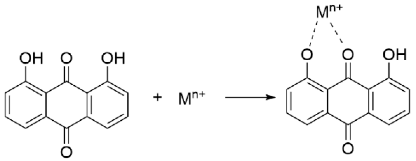
Metal complexes often have excellent antioxidant activity and are useful for diseases related to oxidative stress. Many studies have reported that the combination of the active ingredients of traditional Chinese medicine and metal ions may play a synergistic role. Compared with ligands, metal complexes often have enhanced pharmacological activities or produce some new pharmacological effects and may also reduce toxicity and side effects. Our group found that metal complexes had higher antioxidant activity, which may be due to the synergistic effect of metals and ligands [73,83,84,85,86,87]. Several reports studied the antioxidative activities of the metal complexes of 1, such as Cu(II), Fe(II), Zn(II), Mg(II) and Mn(II). The results showed that all of them have higher antioxidant activities than the ligand. Compound 1 formed the metal complexes through the coordination of the 9-carbonyl group and 1-phenolic hydroxyl group and in the structures of the complexes, the O atoms of 1-hydroxyl and 9-carbonyl coordinated with metal ions to form a stable hexacoordinated ring structure [88,89,90].
In the case of metal complexes, the active center of the potential reaction is transferred from the hydroxyl group to the metal ion. The C=O and phenolic hydroxyl groups in the anthraquinone structure are the active sites with the electron donor before coordination. For metals, the merged orbitals can be split into different energy levels when coordinating with ligands. After absorbing the electromagnetic wave energy, the electrons can jump from the low-level d orbital to the high-level d* orbital and generate absorption bands in the visible region. As shown in Figure 4, the 1-hydroxyl group and 9-carbony1 group of anthraquinone compounds can provide metal chelation sites. They have a completely large π-conjugated system, strong coordination of oxygen atoms and an appropriate spatial configuration, so they can be used as good chelating ligands for metal ions to coordinate with metal ions and form complexes [50,73].

4.2.2. Chemical Modification of Anthraquinone Compounds

Taking anthraquinone as the lead compound, it is of great practical significance to study its structural modification to enhance its antioxidant activity. The structural characteristics of the substituents have an important effect on the scavenging activity of a single molecule. This effect is reflected mainly in the possibility of stabilizing the free radicals [91].
As shown in Figure 5, compounds 17a and 18 have different parent structures with the same substituent group. It could be found that the antioxidant capacity of compound 18 was higher than that of compound 17a. The possible reason was that the electronegativity of S atom was lower than that of O atom. The reducing capacity was stronger, the antioxidant activity was higher. As for the compounds 17a17e, they had the same parent structure and different R groups. The antioxidant assay showed that the chain length was related to the antioxidant activity when comparing compounds 17a and 17b. When phenyl substitution (compounds 17d and 17e) was introduced, the antioxidant activities were increased due to the conjugation system formed between phenyl group and the parent structure. However, compound 17c had a relatively lower inhibition which may be due to the breaking of conjugation structure caused by the steric hindrance effect of the ortho substitution [92].

5. In Vivo Antioxidant Experiments with Anthraquinone Compounds

Anthraquinones have a wide range of biological activities and can be used to treat many kinds of diseases, such as Alzheimer’s disease, inflammation, cancer, liver injury, diabetes, gastrointestinal disorders (e.g., diarrhea, constipation and dysentery), ulcers, radiation injury, and burns [93,94,95]. Especially, anthraquinones have remarkable abilities in scavenge free radicals and prevent oxidative damage to tissues [96]. Although the specific relationship between the antioxidant mechanism and the antioxidant activity in vivo has not been clarified, some information can be obtained from the bioactivity and pharmacokinetic studies. In general, the mechanism for enhancing the activity of antioxidant enzymes may relate to the redox activity of anthraquinones or their ability to bind specific proteins or both. Some of the in vivo antioxidant activities of anthraquinones are listed in Table 2.

5.1. Pharmacological Activities of Anthraquinone Compounds

5.1.1. Anti-Neurodegenerative Diseases

Oxidative stress is an early event in the development and progression of Alzheimer’s disease [97]. As seen in Alzheimer’s disease patients, oxidative stress contributes significantly to the perturbation of calcium homeostasis and subsequent apoptosis [98]. Many clinical studies have reported strong evidence of the involvement of oxidative stress in the pathogenesis of Alzheimer’s disease [99]. The Aβ aggregation inhibition assay of compound 1 showed that compound 1 could block Aβ42 fibrillogenesis and Aβ-induced cytotoxicity [100]. Chen et al. [101] found that compound 1 could protect nerve cells, normalize synaptic damage by reducing the phosphorylation of the extracellular signal-regulated kinase 1/2 (ERK1/2), decrease ROS and protect mitochondrial function. Tao et al. [78] found that compound 2 could show an important role in cognitive deficits in a scopolamine-induced amnesia animal model. In addition, compound 2 increased SOD, GPX and the content of acetylcholine but decreased the level of MDA and acetylcholinesterase activity. Therefore, the results indicated that compound 2 might have a neuroprotective effect on Alzheimer’s disease by inhibiting the activity of acetylcholinesterase and regulating oxidative stress. In a mouse middle cerebral artery occlusion (MCAO) model, Zhao et al. [102] found that compound 5 reduced neuronal damage related to nitric oxide production by reducing the expression of cleaved caspase-3 and enhancing the activity of SOD and manganese-dependent SOD. Zhang et al. [103] found that compound 5 could alleviate hippocampal neuronal injury in lead-exposed neonatal mice, while significantly alleviate the level of MDA in the brain, kidney, and liver and increase the activity of SOD and GPX.

5.1.2. Anticancer

Previous reports showed that the oxidative stress level in cancer cells was higher than that in healthy cells. This stress is accompanied mainly by the production of higher levels of free radicals, causing DNA degradation leading to the carcinogenesis of normal cells and their transformation into cancer cells [104]. In addition, reducing nuclear factor erythroid 2-related factor 2 signaling and strengthening the inflammatory pathway through ROS production are related to carcinogenesis [105,106]. Compound 1/cisplatin combination therapy inhibited the growth of human ovarian carcinoma cells and gallbladder carcinoma cells in vivo. The mechanism may involve the downregulation of multidrug resistance-related protein (MRP1) expression [107] and ATP-binding cassette superfamily G member (ABCG2) expression [108]. Wang et al. [109] found that compound 1 could induce necroptosis through ROS-mediated activation of the c-Jun N-terminal kinases (JNK) signaling pathway and also inhibit glycolysis by downregulating GLUT1 through ROS-mediated inactivation of the phosphoinositide 3-kinases (P13K)/AKT signaling pathway.

5.1.3. Anti-Hepatopathic Diseases

Oxidative stress is also a pathogen in liver inflammation that can lead to fibrosis and hepatocellular carcinoma [5]. Emodin and Rubiadin (compounds 1 and 16) are well-known anthraquinone compounds whose hepatoprotective activity has been widely studied [24,110,111,112]. The results showed that compounds 1 and 16 could inhibit ROS generation and mitochondrial damage through the AMP-activated protein kinase (AMPK)/Yes-associated protein (YAP)-mediated pathway in a hepatocyte cell line and decrease acetaminophen (APAP)-induced oxidative damage in mice. Increases in the levels of GSH, glutathione reductase (GRD), GPX and GST by treatment with compound 1 were associated with a decrease in the level of MDA, indicating a decrease in oxidative stress.

5.1.4. Anti-Cardiovascular Diseases

Excessive ROS may damage vascular cells, induce the proliferation and migration of vascular smooth muscle cells, and then cause vascular remodeling [6,7]. Emodin (compound 1) showed a potential protective effect on cardiovascular system and the mechanism was connected with its antioxidation activities. The antioxidation activities might be related to the inhibition of the contractile effect of 5-hydroxytryptamine and synergizing the diastolic effect of acetylcholine related to up-regulation of free radicals, hydrogen peroxidation and cGMP [7,113].

5.1.5. Anti-Inflammatory Activity

The inflammatory response produces a large amount of ROS and is related to many kinds of diseases. The NF-κB transcription factor family has been considered the central mediator of the inflammatory process and participates in adaptive and innate immune responses [114]. A large number of studies have shown that ROS or metals can affect nuclear NF-κB transcription factors [115]. Lipid peroxidation has been reported to occur in acute pancreatitis [116,117]. ROS can activate NF-κB, which can in turn adjust the expression of inflammatory cytokines [118].
Rat models of severe acute pancreatitis (SAP) was used to analyze the molecular mechanisms and effect of compound 1 [119,120]. As a natural antioxidant, compound 1 could inhibit NF-κB activation and regulate the production of cytokines, such as tumor necrosis factor (TNF)-α, interleukin (IL)-1β, and IL-6, and sequentially regulate the oxidative stress response. Song et al. [121] used proteomics to investigate the antioxidant mechanism of compound 1 on the liver of large herring. Compound 1 was found to significantly increase the expression of antioxidant-related mRNAs, such as GPX-1, GST and heat shock 70 kDa protein (HSP70), compared with the control group. mRNA expression of sorbitol dehydrogenase (SORD) and glyceraldehyde-3-phosphate dehydrogenase (GAPDH) decreased, which was consistent with protein expression. There were also reports about the protection activity of compound 1 from cigarette smoke-induced lung inflammation and oxidative damage. Xue et al. [122] found that compound 1 significantly attenuated the expression of TNF-α, IL-6 and IL-1β and oxidative stress by enhancing the activities of SOD, CAT and GPX in mice with acute pulmonary inflammation induced by cigarette smoke. The results of anti-neuroinflammatory activities showed that compound 2 protected against brain impairment, mainly attributed to the antioxidant activities of compound 2 via the P13K/AKT/mTOR and NF-κB activation [123]. Compound 5 was also found to have good hepatoprotective activity, which was expressed by significantly reducing of inducible nitric oxide synthase (iNOS), TNF-α, IL-6 and IL-10 through inhibiting the expression of NF-κB and receptor-interacting protein 140 (RIP140) in LPS-induced acute liver injury in mice [124].
Table 2. In vivo antioxidant activities of anthraquinone compounds.
Comp.Model aMajor Discoveries and Proposed Mechanisms bDosage of AdministrationRefs.
Emodin (1)C57B/6 mice orally injected with APAP↓ Oxidative damage10, 30 mg/kg[110]
CCl4-intoxicated mice↑ GSH, GRD, GPX, GST
↓ MDA
↓ Oxidative stress
2.6 mg/g[112]
SAP-induced mice↑ SOD
↓ NF-κB, TNF-α, IL-6, IL-1β
↓ MDA
↓ Oxidative stress
1 mg/kg[120]
SAP-induced mice↑ VDAC1
↓ ROS
↓ Serum amylase, lipase, TNF-α, IL-18, caspase-1, NLRP3
6 mg/mL[119]
Female C57B1/6 mice↑ SOD, CAT, GPX
↓ TNF-α, IL-6, IL-1β
40 mg/kg[122]
Healthy M. amblycephala fingerlings↑ GPX1, GSTm, HSP70
↓ GAPDH, Sord
30 mg/kg[78]
6-week-old BALB/c-nu/nu mice↓ ABCG2 expression20, 40, 60 mM[108]
Aloe-emodin (2)Scopolamine-induced amnesia animal model↑ SOD, GPX, ACh
↓ MDA, AChE
IC50 = 18.37 μg/mL[78]
Male C57BL mice↑ SOD, MnSOD
↓ Cleaved caspase-3
0.1–10 mg/kg[102]
Chrysophanol (5)Male BALB/c mice↑ SOD, GPX, GSH, CAT
↓ MDA
↓ Oxidative stress
↓ TNF-α, IL-6, IL-10, iNOS, NF-κB, RIP140
1, 10 mg/kg[124]
Lead poisoned Kunming mice↑ SOD, GPX
↓ MDA
10.0 mg/kg[103]
a Cell lines: APAP (acetaminophen), CCl4 (carbon tetrachloride), SAP (severe acute pancreatitis) b “↑” upregulating or activation. “↓” downregulating or inhibition.

5.2. Pharmacokinetic Studies of Anthraquinone Compounds In Vivo

Pharmacokinetic studies, as an important part of preclinical and clinical studies during the development of innovative drugs, provide a theoretical basis for guiding the clinical and rational use of drugs, predicting drug interactions, understanding drug action mechanisms, exploring new modification methods for natural products, and designing drug delivery systems, etc. [125]. Since traditional Chinese medicine is mostly administered orally, the metabolism of anthraquinones contained in traditional Chinese medicine will first enter the absorption phase. The absorption of anthraquinone compounds depends on their physicochemical properties. The main site of absorption of anthraquinones is the intestine, not the stomach. The absorption of anthraquinone is related to the weak acidity of anthraquinones and the pH conditions in the intestine. When placed in a weakly acidic environment, since most anthraquinones are weakly acidic, resulting in lower ionization. In contrast, at higher pH values, the degree of ionization of anthraquinones increases, resulting in little absorption of anthraquinones [126].
In general, the pharmacokinetic data of anthraquinones are in a wide range in vivo. This may be due to differences in drug dosages, detection instruments, and protocols. As described in Table S4 within the Supplementary Materials, compound 3 had the minimum peak time (Tmax), the highest peak concentration (Cmax) and area under the curve (AUC) in dogs. A large number of pharmacokinetic studies have shown that compound 3 is the main component absorbed into the blood after gastrointestinal administration of rhubarb and its compound preparation containing rhubarb in both rats and humans. The peak concentration of oral absorption and bioavailability of compound 3 are superior to other free anthraquinones contained in rhubarb [127,128,129,130]. The water solubility of compound 3 may be improved due to its carboxyl group. A better lipid water distribution coefficient is favorable for compound 3 penetrate the amphiphilic biofilm, leading to the best bioavailability.
Biotransformation is a significant process during which anthraquinones are converted into inactive or more active metabolites and removed from the body. The transformation occurs mainly in the liver. Phase II conjugates, in particular glucuronides of rhubarb anthraquinones, were considered to be the predominant form in vivo [131,132]. The metabolic pathways and metabolites of anthraquinones are listed in Table 3.
Xu et al. [133] found that in a rat liver microsome incubation system, both compounds 1 and 5 were hydroxylated. Compounds 2 and 5 could be converted to compound 3 by oxidation. Compound 5 was transformed to demethylated forms and acetylated forms as dihydroxy-5, while compound 4 was transformed to demethylation forms as compound 1 isomer. Notably, the demethylation of compound 4 may be the reason for the low bioavailability of 4 [134]. Song et al. [132] separated and identified the metabolites in rat urine, bile and plasma after oral administration of rhubarb decoction and found that compounds 15 might be metabolized to sulfonated forms. Compound 2 was transformed to 2-1-O-glucoside-8-O-glucuronide or 2-8-O-glucoside-1-O-glucuronide, 2-hydroxyaloe-1-ω-O-glucuronide through hydroxylation, hydrogenation, glucuronidation and oxidized to compound 3 by oxidation. The oxidation reaction increases the bioavailability of anthraquinones. The order of bioavailability of anthraquinones was compounds 3 > 1 > 5 > 2, which may result from compounds 2 and 5 being oxidized to compound 3 [135]. In addition, compounds 15 could be metabolized to sulfonated forms [132], which may result in a decrease in the oral bioavailability of anthraquinones [131].
Note that anthraquinones are not only phenols but also p-quinones, and the oxidation process of phenols also involves different forms of quinones. As such, quinones undergo enzyme-catalyzed one- or two-electron reduction reactions generating the corresponding semiquinone or hydroquinone, respectively. As shown in Figure S2, semiquinone, generated through the one-electron reduction pathway, undergoes rapid autoxidation under atmospheric conditions, transferring excess electrons to molecular oxygen, thereby generating O2•− and regenerating the parent quinone. The redox cycling of enzyme-catalyzed reduction of quinone and the aerobic oxidation cycle of the semiquinone producing O2•− continue until the system becomes anaerobic. At this time, the production of O2•− is reduced, and the accumulation of semiquinone occurs. The result of this redox cycle is oxidative stress. Under anaerobic conditions, semiquinone is disproportionate to quinone and hydroquinone. However, hydroquinone from the two-electron reduction pathway, depending on its stability, may be discharged through four different pathways: (1) the detoxification pathway; (2) oxidized by O2 one electron at a time to produce O2•− and the semiquinone; (3) forming reactive alkylating agents by a rearrangement reaction; and (4) undergoing a comproportionation reaction with quinone to produce semiquinone [136].
After oral administration, natural anthraquinones are absorbed into the blood by the intestinal mucosa, combined with glucuronic acid or sulfuric acid in the liver and intestinal tract, and then transported by blood circulation to various tissues and organs throughout the body to exert a variety of pharmacological effects. Anthraquinones undergo oxidation, methylation, esterification and glycosidation reactions when being metabolized. Anthraquinones have good biological activity in vitro, but due to their low solubility and rapid elimination rate, the oral absorption utilization ratio is low. In addition, side effects of anthraquinones have also been reported, including genotoxic [137], nephrotoxic [138], and hepatotoxic effects [139]. Therefore, it is necessary to modify the structure, add absorption promoters, or change drug dosage forms to improve the bioavailability of anthraquinones. Further systematic studies on the absorption and metabolism of anthraquinone compounds in vivo on the basis of pharmacology are needed to ensure their safety and effectiveness in clinical applications.

6. Analysis of the Structure-Activity Relationship

The following three structural characteristics of anthraquinone compounds are closely related to their antioxidant activity: (1) the benzene ring and carbonyl group in the parent structure; (2) the number and position of the hydroxyl groups; (3) the position and number of other substituents, such as carboxyl group, methyl group and methoxyl group. The benzene ring in anthraquinone compounds is hydrophobic, while the phenolic hydroxyl group is hydrophilic. As shown in Figure 1, anthraquinone compounds can be roughly divided into two types according to their structure: emodin type and alizarin type. The hydroxyl groups of the emodin type, such as compounds 19, are distributed on both sides of the benzene ring. Hydroxyl groups of the alizarin type are distributed on the benzene ring on one side, such as compounds 1012.
The antioxidant activity of anthraquinones is mainly related to phenolic hydroxyl groups. There are three main types of phenolic hydroxy-related antioxidant mechanisms. The first is the HAT mechanism, in which the hydrogen atom is extracted from ArOH. So, BDE of the O-H bond is a significant parameter for scavenging activity evaluation. The second mechanism is SET-PT, namely, the rate determining step is losing one electron from ArOH to generate ArOH+• and then followed by a proton transfer step. Therefore, IP is the most important parameter for evaluating antioxidant activity. The third mechanism is SPLET, which means a proton is first lost to form ArO, and then electron transfer occurs. Therefore, pKa of ArO-H bond is main determining factor in this mechanism.
The experimental results also supported these mechanisms and the results will be discussed according to the following structural characteristics: (1) The position and the number of the phenoxyl groups (Table S5 in the supplementary materials) [54,59,60]: the distribution of hydroxyl groups on the ipsilateral benzene ring improved the free radical scavenging activity, which may be related to the o-diphenol structure (e.g., compounds 9, 10, 11). In this kind of structure, the formation of intramolecular hydrogen bonds leads to lower BDE values of OH bonds and lower pKa of ArO-H, and thus higher antioxidative activity, which accord to both HAT and SPLET mechanism; (2) The position of other substitutions: When the carboxyl group is on the ortho position to the hydroxyl group (compounds 12 and 13) or the methoxyl group is on the meta position (compound 4), they are the electron withdrawing groups. The electron density on the hydroxyl group decreases and the electron is difficult to lose with a higher IP value. The antioxidant activity decreases, which is consistent with the mechanism of SET-PT; (3) The carbonyl group on the parent structure will also affect the free radical scavenging activity. It worth to mention that 1-OH/8-OH group and carbonyl group can work as the chelating sites to coordinate with Fe(II) or Cu(I) ions, which may terminate the Fenton reaction. Furthermore, natural compounds with similar structure have antioxidative activities following this path, which has been proved by the research results from our group [73,83,84,85,86,87,88].

7. The Neutraceutical Properties of Natural Anthraquinone Compounds for Clinical Application

Anthraquinones are widely distributed in various botanicals, such as rhubarb, aloe and Fo-Ti [140,141], which are commonly clinically used in traditional Chinese medicines and dietary supplements. As shown in Table 4, various levels of compounds 1, 2 and 3 have been reported in three botanicals, rhubarb, Fo-Ti and aloe. The daily intake levels of the three botanicals in traditional Chinese medicines and dietary supplements were also identified. In addition, the diseases that could be treated by these botanicals were listed in Table 4.

8. Conclusions and Future Perspectives

Natural anthraquinones are a class of natural products with important biological activities, commonly found in many plants and microorganisms. These compounds have been extensively studied and proven to be promising for a wide range of applications in many fields, one of which is as antioxidants. In recent years, numerous studies have shown that natural anthraquinones possess strong antioxidant activity. As shown in this review, anthraquinones (1) have a significant free radical scavenging ability, (2) can modulate complex antioxidant enzymes and non-enzymatic systems to reduce oxidative stress, and (3) inhibit the formation of ROS or inhibit enzymes involved in free radical generation by chelating trace elements. Therefore, natural anthraquinones have a broad application prospect in the field of antioxidants. However, more in-depth research is needed in the following two aspects to make anthraquinones more reliably and widely used in medicine in the future.
First, although drugs based on anthraquinone compounds, such as doxorubicin, mitoxantrone, and idarubicin, etc., have been used successfully to treat diseases and the anthraquinone core may continue to be a potential scaffold for developing novel therapeutic candidates, side effects and low absorption of anthraquinones have also been reported. Therefore, further systematic studies in relation to its SAR as well as the absorption and metabolism in vivo are needed to accelerate the development and utilization of anthraquinone compounds as promising drug candidates in the future. And in this regard, a brief summary of SAR studies on anthraquinone compounds was provided in Section 6 of this review. At the same time, it is also necessary to optimize the structure of anthraquinone compounds by chemical modification. In this review, the summary of the properties of some synthetic anthraquinone compounds was reported in the Section 4.1 and recently our group also reported a series of anthraquinone compounds with tertiary amine substituents with promising anti-AD activities [73].
Second, although antioxidants are essential for human health, due to the specific complicated activities of ROS, drugs that regulate the imbalance between the formation and elimination of ROS should be developed more rationally to provide new treatments for various diseases mediated by ROS. After in-depth literature research, we believe that more detailed studies on the mechanism are needed in the future, especially in the following two kinds of relationship. One is about the relationship between the concentration of the anthraquinone compounds clinically applied and their ROS mediation properties. As shown in the review, most of literature reported anthraquinone compounds’ inhibition of the expression of ROS via SOD, H2O2, superoxide and NF-κB pathway. However, some groups reported that anthraquinone of high concentration may alter the subcellular redox equilibrium and produce cytotoxicity induced by ROS. For example, emodin (1–25 μg·mL−1) induced mitochondria-induced cell apoptosis associated with generation of ROS [7,121]. So, in Section 7 of this review, botanical daily intake (g/day) values were given for the neutraceutical application of natural anthraquinone compounds. The other one is about the relationship between scavenging of ROS and the treatment of diseases. Most time, decreasing of ROS is needed for the treatment of diseases with anthraquinone compounds. However, there were reports about elevation of ROS in the process of disease treatment. For example, Anthraquinones [149], were reported to increase ROS and reduce the activities of antioxidant enzymes in cells, and thus induce autophagy and apoptosis in cancer cells.
The results of this work can be applied to further understand the antioxidant mechanism of anthraquinone compounds and it also has guiding significance for the study of antioxidant ability of other natural products with similar structure.

Supplementary Materials

The following supporting information can be downloaded at: https://www.mdpi.com/article/10.3390/molecules28248139/s1. Refs. [150,151] are cited in Supplementary Materials.

Author Contributions

Conceptualization, L.Z. (Lihua Zhao); writing—original draft preparation, L.Z. (Lihua Zhao); writing—review and editing, L.Z. (Lin Zheng); supervision, L.Z. (Lin Zheng); project administration, L.Z. (Lin Zheng). All authors have read and agreed to the published version of the manuscript.

Funding

This research received no external funding.

Institutional Review Board Statement

Not applicable.

Informed Consent Statement

Not applicable.

Data Availability Statement

Not applicable.

Conflicts of Interest

The authors declare no conflict of interest.

Abbreviations

ABCG2: ATP-binding cassette superfamily G member; AD: Alzheimer’s disease; AMPK: AMP-activated protein kinase; APAP: acetaminophen; AUC: area under the curve; Bax: B-cell lymphoma-2 associated X protein; BDE: bond dissociation enthalpy; CAT: catalase; DPPH: 1,1-diphenyl-2-picrylhydrazyl; Drp1: dynamin-related protein 1; GAPDH: Glyceraldehyde-3-phosphate dehydrogenase; GPX: glutathione peroxidase; GR: Glutathione reductase; GSH: glutathione; GSH-PX: glutathione peroxidase; GST: Glutathione S-transferase; HAT: hydrogen atom transfer; HSP70: heat shock 70 kDa protein; HUVECs: human umbilical vein endothelial cells; IL: interleukin; iNOS: inducible nitric oxide synthase; IP: ionization potential; JNK: c-Jun N-terminal kinases; LDH: lactate dehydrogenase; LPS: lipopolysaccharide; MAPK: mitogen-activated protein kinase; MCAO: middle cerebral artery occlusion; MDA: malondialdehyde; MRP1: multidrug resistance-related protein; NADPH: nicotinamide adenine dinucleotide phosphate; NF-κB: nuclear factor-kappa B; NOS: nitric oxide synthase; NLRP3: NOD-like receptor thermal protein domain associated protein 3; Nrf2: nuclear factor erythroid-2 related factor 2; P13K: phosphoinositide 3-kinases; RNS: reactive nitrogen species; ROS: reactive oxygen species; SAP: severe acute pancreatitis; SAR: structure and activity relationship; SET-PT: single electron transfer followed by proton transfer: SOD: superoxide dismutase; SORD: sorbitol dehydrogenase; SPLET: sequential proton loss and electron transfer; TNF: tumour necrosis factor; VDAC1: voltage-dependent anion channel 1; YAP: yes-associated protein.

References

  1. Yang, B.; Chen, Y.; Shi, J. Reactive Oxygen Species (ROS)-Based Nanomedicine. Chem. Rev. 2019, 119, 4881–4985. [Google Scholar] [CrossRef] [PubMed]
  2. Singh, A.; Kukreti, R.; Saso, L.; Kukreti, S. Oxidative Stress: A Key Modulator in Neurodegenerative Diseases. Molecules 2019, 24, 1583. [Google Scholar] [CrossRef] [PubMed]
  3. Perluigi, M.; Di Domenico, F.; Butterfield, D.A. Oxidative damage in neurodegeneration: Roles in the pathogenesis and progression of Alzheimer disease. Physiol. Rev. 2024, 104, 103–197. [Google Scholar] [CrossRef] [PubMed]
  4. Yang, S.; Lian, G. ROS and diseases: Role in metabolism and energy supply. Mol. Cell Biochem. 2020, 467, 1–12. [Google Scholar] [CrossRef]
  5. Li, S.; Tan, H.; Wang, N.; Zhang, Z.; Lao, L.; Wong, C.; Feng, Y. The Role of Oxidative Stress and Antioxidants in Liver Diseases. Int. J. Mol. Sci. 2015, 16, 26087–26124. [Google Scholar] [CrossRef] [PubMed]
  6. Stark, J. Oxidative stress and atherosclerosis. Orv. Hetil. 2015, 156, 1115–1119. [Google Scholar] [CrossRef] [PubMed]
  7. Cui, Y.; Chen, L.J.; Huang, T.; Ying, J.Q.; Li, J. The pharmacology, toxicology and therapeutic potential of anthraquinone derivative emodin. Chin. J. Nat. Med. 2020, 18, 425–435. [Google Scholar] [CrossRef]
  8. Hecht, F.; Zocchi, M.; Alimohammadi, F.; Harris, I.S. Regulation of antioxidants in cancer. Mol. Cell 2023. Online ahead of print. [Google Scholar] [CrossRef]
  9. Rudenko, N.N.; Vetoshkina, D.V.; Marenkova, T.V.; Borisova-Mubarakshina, M.M. Antioxidants of Non-Enzymatic Nature: Their Function in Higher Plant Cells and the Ways of Boosting Their Biosynthesis. Antioxidants 2023, 12, 2014. [Google Scholar] [CrossRef]
  10. Chen, Q.; Wang, Q.; Zhu, J.; Xiao, Q.; Zhang, L. Reactive oxygen species: Key regulators in vascular health and diseases. Br. J. Pharmacol. 2018, 175, 1279–1292. [Google Scholar] [CrossRef]
  11. Hamza, A.A.; Heeba, G.H.; Hassanin, S.O.; Elwy, H.M.; Bekhit, A.A.; Amin, A. Hibiscus-cisplatin combination treatment decreases liver toxicity in rats while increasing toxicity in lung cancer cells via oxidative stress- apoptosis pathway. Biomed. Pharmacother. 2023, 165, 115148. [Google Scholar] [CrossRef] [PubMed]
  12. Hamza, A.A.; Mohamed, M.G.; Fawzy, M.; Lashin; Amin, A. Dandelion prevents liver fibrosis, inflammatory response, and oxidative stress in rats. J. Basic Appl. Zool. 2020, 81, 43. [Google Scholar] [CrossRef]
  13. Duval, J.; Pecher, V.; Poujol, M.; Lesellier, E. Research advances for the extraction, analysis and uses of anthraquinones: A review. Ind. Crops Prod. 2016, 94, 812–833. [Google Scholar] [CrossRef]
  14. Ghanim, H.; Sia, C.; Abuaysheh, S.; Korzeniewski, K.; Patnaik, P.; Marumganti, A.; Chaudhuri, A.; Dandona, P. An antiinflammatory and reactive oxygen species suppressive effects of an extract of Polygonum cuspidatum containing resveratrol. J. Clin. Endocrinol. Metab. 2010, 95, E1–E8. [Google Scholar] [CrossRef] [PubMed]
  15. Kosalec, I.; Kremer, D.; Locatelli, M.; Epifano, F.; Genovese, S.; Carlucci, G.; Randić, M.; Zovko Končić, M. Anthraquinone profile, antioxidant and antimicrobial activity of bark extracts of Rhamnus alaternus, R. fallax, R. intermedia and R. pumila. Food Chem. 2013, 136, 335–341. [Google Scholar] [CrossRef] [PubMed]
  16. Lee, B.H.; Pan, T.M. Dimerumic acid, a novel antioxidant identified from Monascus-fermented products exerts chemoprotective effects: Mini review. J. Funct. Food. 2013, 5, 2–9. [Google Scholar] [CrossRef]
  17. Nishizawa, M.; Kohno, M.; Nishimura, M.; Kitagawa, A.; Niwano, Y. Presence of peroxyradicals in cigarette smoke and the scavenging effect of shikonin, a naphthoquinone pigment. Chem. Pharm. Bull. 2005, 53, 796–799. [Google Scholar] [CrossRef]
  18. Salah, K.; Mahjoub, M.; Ammar, S.; Michel, L.; Millet-Clerc, J.; Chaumont, J.; Mighri, Z.; Aouni, M. Antimicrobial and antioxidant activities of the methanolic extracts of three Salvia species from Tunisia. Nat. Prod. Res. 2006, 20, 1110–1120. [Google Scholar] [CrossRef]
  19. Xu, H.; Lu, Y.; Zhang, T.; Liu, K.; Liu, L.; He, Z.; Xu, B.; Wu, X. Characterization of binding interactions of anthraquinones and bovine β-lactoglobulin. Food Chem. 2019, 281, 28–35. [Google Scholar] [CrossRef]
  20. Lu, J.; Bao, J.; Wu, G.; Xu, W.; Huang, M.; Chen, X.; Wang, Y. Quinones derived from plant secondary metabolites as anti-cancer agents. Anti-Cancer Agents Med. Chem. 2013, 13, 456–463. [Google Scholar]
  21. Brkanac, S.R.; Genic, M.; Gajski, G.; Vujcic, V.; Garaj-Vrhovac, V.; Kremer, D.; Domijan, A.M. Toxicity and antioxidant capacity of Frangula alnus Mill. bark and its active component emodin. Regul. Toxicol. Pharmacol. 2015, 73, 923–929. [Google Scholar] [CrossRef] [PubMed]
  22. Zengin, G.; Degirmenci, N.; Alpsoy, L.; Aktumsek, A. Evaluation of antioxidant, enzyme inhibition, and cytotoxic activity of three anthraquinones (alizarin, purpurin, and quinizarin). Hum. Exp. Toxicol. 2016, 35, 544–553. [Google Scholar] [CrossRef] [PubMed]
  23. Bu, Q.; Jin, Y.; Xu, M.J.; Wu, L.; Liang, L.F. Structurally Diverse Metabolites from the Ophiorrhiza japonica Bl. and Their Antioxidant Activities In Vitro and PPARα Agonistic Activities In Silico. Molecules 2022, 27, 5301. [Google Scholar] [CrossRef]
  24. Watroly, M.N.; Sekar, M.; Fuloria, S.; Gan, S.H.; Jeyabalan, S.; Wu, Y.S.; Subramaniyan, V.; Sathasivam, K.V.; Ravi, S.; Mat Rani, N.N.I.; et al. Chemistry, Biosynthesis, Physicochemical and Biological Properties of Rubiadin: A Promising Natural Anthraquinone for New Drug Discovery and Development. Drug Des. Dev. Ther. 2021, 15, 4527–4549. [Google Scholar] [CrossRef] [PubMed]
  25. Riley, P.A. Free radicals in biology: Oxidative stress and the effects of ionizing radiation. Int. J. Radiat. Biol. 1994, 65, 27–33. [Google Scholar] [CrossRef] [PubMed]
  26. Halliwell, B. Free radicals and antioxidants—Quo vadis? Trends Pharmacol. Sci. 2011, 32, 125–130. [Google Scholar] [CrossRef] [PubMed]
  27. Afanas’ev, I. Free radical mechanisms of aging processes under physiological conditions. Biogerontology 2005, 6, 283–290. [Google Scholar] [CrossRef]
  28. Lobo, V.; Patil, A.; Phatak, A.; Chandra, N. Free radicals, antioxidants and functional foods: Impact on human health. Pharmacogn. Rev. 2010, 4, 118–126. [Google Scholar] [CrossRef]
  29. Gulcin, I.; Mshvildadze, V.; Gepdiremen, A.; Elias, R. The antioxidant activity of a triterpenoid glycoside isolated from the berries of Hedera colchica: 3-O-(beta-D-glucopyranosyl)-hederagenin. Phytother. Res. 2006, 20, 130–134. [Google Scholar] [CrossRef]
  30. Gulcin, I.; Mshvildadze, V.; Gepdiremen, A.; Elias, R. Screening of antiradical and antioxidant activity of monodesmosides and crude extract from Leontice smirnowii tuber. Phytomedicine 2006, 13, 343–351. [Google Scholar] [CrossRef]
  31. Valko, M.; Rhodes, C.J.; Moncol, J.; Izakovic, M.; Mazur, M. Free radicals, metals and antioxidants in oxidative stress-induced cancer. Chem. Biol. Interact. 2006, 160, 1–40. [Google Scholar] [CrossRef] [PubMed]
  32. Firuzi, O.; Miri, R.; Tavakkoli, M.; Saso, L. Antioxidant Therapy: Current Status and Future Prospects. Curr. Med. Chem. 2011, 18, 3871–3888. [Google Scholar] [CrossRef] [PubMed]
  33. Carocho, M.; Ferreira, I. A review on antioxidants, prooxidants and related controversy: Natural and synthetic compounds, screening and analysis methodologies and future perspectives. Food Chem. Toxicol. 2013, 51, 15–25. [Google Scholar] [CrossRef] [PubMed]
  34. Valko, M.; Leibfritz, D.; Moncol, J.; Cronin, M.; Mazur, M.; Telser, J. Free radicals and antioxidants in normal physiological functions and human disease. Int. J. Biochem. Cell Biol. 2007, 39, 44–84. [Google Scholar] [CrossRef] [PubMed]
  35. Dröge, W. Free radicals in the physiological control of cell function. Physiol. Rev. 2002, 82, 47–95. [Google Scholar] [CrossRef] [PubMed]
  36. Yang, E.; Mahmood, U.; Kim, H.; Choi, M.; Choi, Y.; Lee, J.; Cho, J.; Hyun, J.; Kim, Y.; Chang, M.; et al. Phloroglucinol ameliorates cognitive impairments by reducing the amyloid β peptide burden and pro-inflammatory cytokines in the hippocampus of 5XFAD mice. Free Radical Biol. Med. 2018, 126, 221–234. [Google Scholar] [CrossRef] [PubMed]
  37. Parveen, A.; Akash, M.; Rehman, K.; Kyunn, W. Recent Investigations for Discovery of Natural Antioxidants: A Comprehensive Review. Crit. Rev. Eukaryot. Gene. Expr. 2016, 26, 143–160. [Google Scholar] [CrossRef] [PubMed]
  38. Budimir, A. Metal ions, Alzheimer’s disease and chelation therapy. Acta Pharm. 2011, 61, 1–14. [Google Scholar] [CrossRef]
  39. Crichton, R.R.; Dexter, D.T.; Ward, R.J. Metal based neurodegenerative diseases—From molecular mechanisms to therapeutic strategies. Coord. Chem. Rev. 2008, 252, 1189–1199. [Google Scholar] [CrossRef]
  40. Grazul, M.; Budzisz, E. Biological activity of metal ions complexes of chromones, coumarins and flavones. Coord. Chem. Rev. 2009, 253, 2588–2598. [Google Scholar] [CrossRef]
  41. Bilenko, N.; Yehiel, M.; Inbar, Y.; Gazala, E. The association between anemia in infants, and maternal knowledge and adherence to iron supplementation in southern Israel. Isr. Med. Assoc. J. 2007, 9, 521–524. [Google Scholar] [PubMed]
  42. Flora, S. Structural, chemical and biological aspects of antioxidants for strategies against metal and metalloid exposure. Oxid. Med. Cell. Longev. 2009, 2, 191–206. [Google Scholar] [CrossRef] [PubMed]
  43. Jomova, K.; Valko, M. Advances in metal-induced oxidative stress and human disease. Toxicology 2011, 283, 65–87. [Google Scholar] [CrossRef] [PubMed]
  44. Valko, M.; Jomova, K.; Rhodes, C.; Kuča, K.; Musílek, K. Redox- and non-redox-metal-induced formation of free radicals and their role in human disease. Arch. Toxicol. 2016, 90, 1–37. [Google Scholar] [CrossRef] [PubMed]
  45. Poprac, P.; Jomova, K.; Simunkova, M.; Kollar, V.; Rhodes, C.; Valko, M. Targeting Free Radicals in Oxidative Stress-Related Human Diseases. Trends Pharmacol. Sci. 2017, 38, 592–607. [Google Scholar] [CrossRef] [PubMed]
  46. Kostyuk, V.; Potapovich, A. Mechanisms of the suppression of free radical overproduction by antioxidants. Front. Biosci., Elite Ed. 2009, 1, 179–188. [Google Scholar] [PubMed]
  47. Biela, M.; Rimarčík, J.; Senajová, E.; Kleinová, A.; Klein, E. Antioxidant action of deprotonated flavonoids: Thermodynamics of sequential proton-loss electron-transfer. Phytochemistry 2020, 180, 112528. [Google Scholar] [CrossRef] [PubMed]
  48. Xue, Y.; Liu, Y.; Xie, Y.; Cong, C.; Wang, G.; An, L.; Teng, Y.; Chen, M.; Zhang, L. Antioxidant activity and mechanism of dihydrochalcone C-glycosides: Effects of C-glycosylation and hydroxyl groups. Phytochemistry 2020, 179, 112393. [Google Scholar] [CrossRef]
  49. Zheng, Y.; Fu, Z.; Deng, G.; Guo, R.; Chen, D. Free radical scavenging potency of ellagic acid and its derivatives in multiple H/e processes. Phytochemistry 2020, 180, 112517. [Google Scholar] [CrossRef]
  50. Leopoldini, M.; Russo, N.; Toscano, M. The molecular basis of working mechanism of natural polyphenolic antioxidants. Food Chem. 2011, 125, 288–306. [Google Scholar] [CrossRef]
  51. Michalík, M.; Poliak, P.; Lukeš, V.; Klein, E. From phenols to quinones: Thermodynamics of radical scavenging activity of para-substituted phenols. Phytochemistry 2019, 166, 112077. [Google Scholar] [CrossRef] [PubMed]
  52. Zheng, Y.; Deng, G.; Chen, D.; Guo, R.; Lai, R. The influence of C2C3 double bond on the antiradical activity of flavonoid: Different mechanisms analysis. Phytochemistry 2019, 157, 1–7. [Google Scholar] [CrossRef] [PubMed]
  53. Zhang, H.Y. Structure-Activity Relationships and Rational Design Strategies for Radical-Scavenging Antioxidants. Curr. Comput. Aided Drug Des. 2005, 1, 257–273. [Google Scholar] [CrossRef]
  54. Cai, Y.; Sun, M.; Xing, J.; Corke, H. Antioxidant phenolic constituents in roots of Rheum officinale and Rubia cordifolia: Structure-radical scavenging activity relationships. J. Agric. Food Chem. 2004, 52, 7884–7890. [Google Scholar] [CrossRef] [PubMed]
  55. Pratt, D.; DiLabio, G.; Brigati, G.; Pedulli, G.; Valgimigli, L. 5-Pyrimidinols: Novel chain-breaking antioxidants more effective than phenols. J. Am. Chem. Soc. 2001, 123, 4625–4626. [Google Scholar] [CrossRef] [PubMed]
  56. Litwinienko, G.; Ingold, K. Abnormal solvent effects on hydrogen atom abstraction. 2. Resolution of the curcumin antioxidant controversy. The role of sequential proton loss electron transfer. J. Org. Chem. 2004, 69, 5888–5896. [Google Scholar] [CrossRef]
  57. Litwinienko, G.; Ingold, K. Abnormal solvent effects on hydrogen atom abstraction. 3. Novel kinetics in sequential proton loss electron transfer chemistry. J. Org. Chem. 2005, 70, 8982–8990. [Google Scholar] [CrossRef]
  58. Nazifi, S.M.R.; Asgharshamsi, M.H.; Dehkordi, M.M.; Zborowski, K.K. Antioxidant properties of Aloe vera components: A DFT theoretical evaluation. Free Radic. Res. 2019, 53, 922–931. [Google Scholar] [CrossRef]
  59. Jeremić, S.; Amić, A.; Stanojević-Pirković, M.; Marković, Z. Selected anthraquinones as potential free radical scavengers and P-glycoprotein inhibitors. Org. Biomol. Chem. 2018, 16, 1890–1902. [Google Scholar] [CrossRef]
  60. Isin, D.O. Theoretical study on the investigation of antioxidant properties of some hydroxyanthraquinones. Mol. Phys. 2016, 114, 3578–3588. [Google Scholar] [CrossRef]
  61. Markovic, Z.; Jeremic, S.; Markovic, J.D.; Pirkovic, M.S.; Amic, D. Influence of structural characteristics of substituents on the antioxidant activity of some anthraquinone derivatives. Comput. Theor. Chem. 2016, 1077, 25–31. [Google Scholar] [CrossRef]
  62. Markovic, Z.S.; Manojlovic, N.T. DFT study on the reactivity of OH groups in emodin: Structural and electronic features of emodin radicals. Mon. Chem. 2009, 140, 1311–1318. [Google Scholar] [CrossRef]
  63. Jeremic, S.R.; Sehovic, S.F.; Manojlovic, N.T.; Markovic, Z.S. Antioxidant and free radical scavenging activity of purpurin. Mon. Chem. 2012, 143, 427–435. [Google Scholar] [CrossRef]
  64. Jung, H.A.; Chung, H.Y.; Yokozawa, T.; Kim, Y.C.; Choi, J.S. Alaternin and emodin with hydroxyl radical inhibitory and/or scavenging activities and hepatoprotective activity on tacrine-induced cytotoxicity in HepG2 cells. Arch. Pharm. Res. 2004, 27, 947–953. [Google Scholar] [CrossRef] [PubMed]
  65. Vargas, F.R.; Diaz, Y.H.; Carbonell, K.M. Antioxidant and scavenging activity of emodin, aloe-emodin, and rhein on free-radical and reactive oxygen species. Pharm. Biol. 2004, 42, 342–348. [Google Scholar] [CrossRef]
  66. Kumar, M.; Chandel, M.; Kumar, S.; Kaur, S. amelioration of oxidative stress by anthraquinones in various in vitro assays asian pacific journal of tropical disease. Asian Pac. J. Trop. Dis 2012, 2, S692–S698. [Google Scholar] [CrossRef]
  67. Lin, J.; Li, X.C.; Han, L.; Li, F.; Lu, W.B.; Bai, Y.; Chen, D.F. Folium Sennae protects against hydroxyl radical-induced DNA damage via antioxidant mechanism: An in vitro study. Bot. Stud. 2014, 55, 16. [Google Scholar] [CrossRef] [PubMed]
  68. Waly, M.; Ali, B.; Al-Lawati, I.; Nemmar, A. Protective effects of emodin against cisplatin-induced oxidative stress in cultured human kidney (HEK 293) cells. J. Appl. Toxicol. 2013, 33, 626–630. [Google Scholar] [CrossRef]
  69. Nam, W.; Kim, S.; Nam, S.; Friedman, M. Structure-Antioxidative and Anti-Inflammatory Activity Relationships of Purpurin and Related Anthraquinones in Chemical and Cell Assays. Molecules 2017, 22, 265. [Google Scholar] [CrossRef]
  70. Baghiani, A.; Charef, N.; Djarmouni, M.; Saadeh, H.; Arrar, L.; Mubarak, M. Free radical scanvenging and antioxidant effects of some anthraquinone derivatives. Med. Chem. 2011, 7, 639–644. [Google Scholar] [CrossRef]
  71. Shi, A.H.; Huang, J.W.; Liu, Y.H.; Yuan, K. Separation, Antioxidant and Antimicrobial Activities of Chemical Constituents from Exocarp of Juglans mandshurica Maxim. Asian J. Chem. 2013, 25, 3361–3365. [Google Scholar] [CrossRef]
  72. Milenkovic, D.A.; Dimic, D.S.; Avdovic, E.H.; Amic, A.D.; Markovic, J.M.D.; Markovic, Z.S. Advanced oxidation process of coumarins by hydroxyl radical: Towards the new mechanism leading to less toxic products. Chem. Eng. J. 2020, 395, 13. [Google Scholar] [CrossRef]
  73. Kou, X.D.; Liu, J.J.; Chen, Y.H.; Yang, A.H.; Shen, R. Emodin derivatives with multi-factor anti-AD activities: AChE inhibitor, anti-oxidant and metal chelator. J. Mol. Struct. 2021, 1239, 11. [Google Scholar] [CrossRef]
  74. Yen, G.C.; Duh, P.D.; Chuang, D.Y. Antioxidant activity of anthraquinones and anthrone. Food Chem. 2000, 70, 437–441. [Google Scholar] [CrossRef]
  75. Malterud, K.; Farbrot, T.; Huse, A.; Sund, R. Antioxidant and radical scavenging effects of anthraquinones and anthrones. Pharmacology 1993, 47, 77–85. [Google Scholar] [CrossRef] [PubMed]
  76. Choi, J.S.; Chung, H.Y.; Jung, H.A.; Park, H.J.; Yokozawa, T. Comparative evaluation of antioxidant potential of alaternin (2-hydroxyemodin) and emodin. J. Agric. Food Chem. 2000, 48, 6347–6351. [Google Scholar] [CrossRef] [PubMed]
  77. Huang, S.; Yeh, S.; Hong, C. Effect of anthraquinone derivatives on lipid peroxidation in rat heart mitochondria: Structure-activity relationship. J. Nat. Prod. 1995, 58, 1365–1371. [Google Scholar] [CrossRef] [PubMed]
  78. Tao, L.; Xie, J.M.; Wang, Y.T.; Wang, S.; Wu, S.C.; Wang, Q.M.; Ding, H. Protective effects of aloe-emodin on scopolamine-induced memory impairment in mice and H2O2-induced cytotoxicity in PC12 cells. Bioorg. Med. Chem. Lett. 2014, 24, 5385–5389. [Google Scholar] [CrossRef]
  79. Zhong, X.F.; Huang, G.D.; Luo, T.; Deng, Z.Y.; Hu, J.N. Protective effect of rhein against oxidative stress-related endothelial cell injury. Mol. Med. Rep. 2012, 5, 1261–1266. [Google Scholar]
  80. Chae, U.; Min, J.; Lee, H.; Song, K.; Lee, H.; Lee, H.; Lee, S.; Lee, D. Chrysophanol suppresses pro-inflammatory response in microglia via regulation of Drp1-dependent mitochondrial fission. Immunopharmacol. Immunotoxicol. 2017, 39, 268–275. [Google Scholar] [CrossRef]
  81. Hussain, Y.; Singh, J.; Raza, W.; Meena, A.; Rajak, S.; Sinha, R.A.; Luqman, S. Purpurin ameliorates alcohol-induced hepatotoxicity by reducing ROS generation and promoting Nrf2 expression. Life Sci. 2022, 309, 120964. [Google Scholar] [CrossRef] [PubMed]
  82. Kim, W.; Kwon, H.J.; Jung, H.Y.; Hahn, K.R.; Yoon, Y.S.; Hwang, I.K.; Choi, S.Y.; Kim, D.W. Neuroprotective Effects of Purpurin Against Ischemic Damage via MAPKs, Bax, and Oxidative Stress Cascades in the Gerbil Hippocampus. Mol. Neurobiol. 2022, 59, 2580–2592. [Google Scholar] [CrossRef]
  83. Yang, A.; Yu, Q.; Ju, H.; Song, L.; Kou, X.; Shen, R. Design, Synthesis and Biological Evaluation of Xanthone Derivatives for Possible Treatment of Alzheimer’s Disease Based on Multi-Target Strategy. Chem. Biodivers. 2020, 17, e2000442. [Google Scholar] [CrossRef] [PubMed]
  84. Wu, J.; Kou, X.; Ju, H.; Zhang, H.; Yang, A.; Shen, R. Design, synthesis and biological evaluation of naringenin carbamate derivatives as potential multifunctional agents for the treatment of Alzheimer’s disease. Bioorg. Med. Chem. Lett. 2021, 49, 128316. [Google Scholar] [CrossRef] [PubMed]
  85. Kou, X.; Song, L.; Wang, Y.; Yu, Q.; Ju, H.; Yang, A.; Shen, R. Design, synthesis and anti-Alzheimer’s disease activity study of xanthone derivatives based on multi-target strategy. Bioorg. Med. Chem. Lett. 2020, 30, 126927. [Google Scholar] [CrossRef] [PubMed]
  86. Yang, A.; Liu, C.; Zhang, H.; Wu, J.; Shen, R.; Kou, X. A multifunctional anti-AD approach: Design, synthesis, X-ray crystal structure, biological evaluation and molecular docking of chrysin derivatives. Eur. J. Med. Chem. 2022, 233, 114216. [Google Scholar] [CrossRef] [PubMed]
  87. Liu, C.; Kou, X.; Wang, X.; Wu, J.; Yang, A.; Shen, R. Novel chrysin derivatives as hidden multifunctional agents for anti-Alzheimer’s disease: Design, synthesis and in vitro evaluation. Eur. J. Pharm. Sci. 2021, 166, 105976. [Google Scholar] [CrossRef]
  88. Pan, X.L.; Xiang, H.; Xie, Y.F.; Zhang, C.E.; Dong, X.P. The Compound, Confirm and Antioxidant Activity of Emodin-copper(Ⅱ) Metal Complex. Lishizhen Med. Mater. Medica Res. 2013, 24, 2892–2894. [Google Scholar]
  89. Xiang, H.; Pan, X.L.; Xie, Y.F.; Zhang, C.E.; Dong, X.P. Concentration for 50% of Maximum Effect Study of Antioxidant Activity of 3 Emodin Metal Complex. Chin. J. Exp. Tradit. Med. Formulae 2013, 19, 236–239. [Google Scholar]
  90. Yang, L.; Tan, J.; Wang, B.; Zhu, L. Synthesis, characterization, and anti-cancer activity of emodin-Mn(II) metal complex. Chin. J. Nat. Med. 2014, 12, 937–942. [Google Scholar] [CrossRef]
  91. Tsiapali, E.; Whaley, S.; Kalbfleisch, J.; Ensley, H.; Browder, I.; Williams, D. Glucans exhibit weak antioxidant activity, but stimulate macrophage free radical activity. Free Radical Biol. Med. 2001, 30, 393–402. [Google Scholar] [CrossRef] [PubMed]
  92. Chen, R.; Shen, Y.; Huang, H.; Liao, J.; Ho, L.; Chou, Y.; Wang, W.; Chen, C. Evaluation of the anti-inflammatory and cytotoxic effects of anthraquinones and anthracenes derivatives in human leucocytes. J. Pharm. Pharmacol. 2004, 56, 915–919. [Google Scholar] [CrossRef] [PubMed]
  93. Park, M.Y.; Kwon, H.J.; Sung, M.K. Evaluation of Aloin and Aloe-Emodin as Anti-Inflammatory Agents in Aloe by Using Murine Macrophages. Biosci. Biotechnol. Biochem. 2009, 73, 828–832. [Google Scholar] [CrossRef] [PubMed]
  94. Rabe, T.; van Staden, J. Antibacterial activity of South African plants used for medicinal purposes. J. Ethnopharmacol. 1997, 56, 81–87. [Google Scholar] [CrossRef] [PubMed]
  95. Tan, Z.J.; Li, F.F.; Xing, J.M. Separation and Purification of Aloe Anthraquinones Using PEG/Salt Aqueous Two-Phase System. Sep. Sci. Technol. 2011, 46, 1503–1510. [Google Scholar] [CrossRef]
  96. Xu, K.; Wang, P.; Wang, L.; Liu, C.; Xu, S.; Cheng, Y.; Wang, Y.; Li, Q.; Lei, H. Quinone derivatives from the genus Rubia and their bioactivities. Chem. Biodivers. 2014, 11, 341–363. [Google Scholar] [CrossRef] [PubMed]
  97. Markesbery, W.R. The role of oxidative stress in Alzheimer disease. Arch. Neurol. 1999, 56, 1449–1452. [Google Scholar] [CrossRef]
  98. Yu, S.P.; Canzoniero, L.M.T.; Choi, D.W. Ion homeostasis and apoptosis. Curr. Opin. Cell Biol. 2001, 13, 405–411. [Google Scholar] [CrossRef]
  99. Marcus, D.L.; Thomas, C.; Rodriguez, C.; Simberkoff, K.; Tsai, J.S.; Strafaci, J.A.; Freedman, M.L. Increased peroxidation and reduced antioxidant enzyme activity in Alzheimer’s disease. Exp. Neurol. 1998, 150, 40–44. [Google Scholar] [CrossRef]
  100. Wang, L.; Liu, S.; Xu, J.; Watanabe, N.; Mayo, K.; Li, J.; Li, X. Emodin inhibits aggregation of amyloid-β peptide 1-42 and improves cognitive deficits in Alzheimer’s disease transgenic mice. J. Neurochem. 2021, 157, 1992–2007. [Google Scholar] [CrossRef]
  101. Chen, Q.; Lai, C.; Chen, F.; Ding, Y.; Zhou, Y.; Su, S.; Ni, R.; Tang, Z. Emodin Protects SH-SY5Y Cells Against Zinc-Induced Synaptic Impairment and Oxidative Stress Through the ERK1/2 Pathway. Front. Pharmacol. 2022, 13, 821521. [Google Scholar] [CrossRef] [PubMed]
  102. Zhao, Y.; Huang, Y.; Fang, Y.; Zhao, H.; Shi, W.; Li, J.; Duan, Y.; Sun, Y.; Gao, L.; Luo, Y. Chrysophanol attenuates nitrosative/oxidative stress injury in a mouse model of focal cerebral ischemia/reperfusion. J. Pharmacol. Sci. 2018, 138, 16–22. [Google Scholar] [CrossRef] [PubMed]
  103. Zhang, J.; Yan, C.; Wang, S.; Hou, Y.; Xue, G.; Zhang, L. Chrysophanol attenuates lead exposure-induced injury to hippocampal neurons in neonatal mice. Neural Regener. Res. 2014, 9, 924–930. [Google Scholar]
  104. Liou, G.Y.; Storz, P. Reactive oxygen species in cancer. Free Radic. Res. 2010, 44, 479–496. [Google Scholar] [CrossRef]
  105. Huang, Y.; Li, W.; Su, Z.; Kong, A. The complexity of the Nrf2 pathway: Beyond the antioxidant response. J. Nutr. Biochem. 2015, 26, 1401–1413. [Google Scholar] [CrossRef]
  106. Reuter, S.; Gupta, S.C.; Chaturvedi, M.M.; Aggarwal, B.B. Oxidative stress, inflammation, and cancer: How are they linked? Free Radic. Biol. Med. 2010, 49, 1603–1616. [Google Scholar] [CrossRef] [PubMed]
  107. Ma, J.; Yang, J.; Wang, C.; Zhang, N.; Dong, Y.; Wang, C.; Wang, Y.; Lin, X. Emodin augments cisplatin cytotoxicity in platinum-resistant ovarian cancer cells via ROS-dependent MRP1 downregulation. Biomed. Res. Int. 2014, 2014, 107671. [Google Scholar] [CrossRef] [PubMed]
  108. Li, X.; Dong, Y.; Wang, W.; Wang, H.; Chen, Y.; Shi, G.; Yi, J.; Wang, J. Emodin as an effective agent in targeting cancer stem-like side population cells of gallbladder carcinoma. Stem Cells Dev. 2013, 22, 554–566. [Google Scholar] [CrossRef]
  109. Wang, K.; Meng, X.; Chen, J.; Wang, K.; Zhou, C.; Yu, R.; Ma, Q. Emodin Induced Necroptosis and Inhibited Glycolysis in the Renal Cancer Cells by Enhancing ROS. Oxid. Med. Cell. Longev. 2021, 2021, 8840590. [Google Scholar] [CrossRef]
  110. Lee, E.; Baek, S.; Park, J.; Kim, Y. Rheum undulatumEmodin in inhibits oxidative stress in the liver via AMPK with Hippo/Yap signalling pathway. Pharm. Biol. 2020, 58, 333–341. [Google Scholar] [CrossRef]
  111. Alisi, A.; Pastore, A.; Ceccarelli, S.; Panera, N.; Gnani, D.; Bruscalupi, G.; Massimi, M.; Tozzi, G.; Piemonte, F.; Nobili, V. Emodin prevents intrahepatic fat accumulation, inflammation and redox status imbalance during diet-induced hepatosteatosis in rats. Int. J. Mol. Sci. 2012, 13, 2276–2289. [Google Scholar] [CrossRef] [PubMed]
  112. Chiu, P.Y.; Mak, D.H.F.; Poon, M.K.T.; Ko, K.M. In vivo antioxidant action of a lignan-enriched extract of Schisandra fruit and an anthraquinone-containing extract of Polygonum root in comparison with schisandrin B and emodin. Planta Med. 2002, 68, 951–956. [Google Scholar] [CrossRef] [PubMed]
  113. Ghayur, M.N.; Gilani, A.H.; Rasheed, H.; Khan, A.; Iqbal, Z.; Ismail, M.; Saeed, S.A.; Janssen, L.J. Cardiovascular and airway relaxant activities of peony root extract. Can. J. Physiol. Pharmacol. 2008, 86, 793–803. [Google Scholar] [CrossRef] [PubMed]
  114. DiDonato, J.; Mercurio, F.; Karin, M. NF-κB and the link between inflammation and cancer. Immunol. Rev. 2012, 246, 379–400. [Google Scholar] [CrossRef] [PubMed]
  115. Pande, V.; Ramos, M. Molecular recognition of 15-deoxy-delta(12,14)-prostaglandin J2 by nuclear factor-kappa B and other cellular proteins. Bioorg. Med. Chem. Lett. 2005, 15, 4057–4063. [Google Scholar] [CrossRef] [PubMed]
  116. Abu-Hilal, M.; McPhail, M.; Marchand, L.; Johnson, C. Malondialdehyde and superoxide dismutase as potential markers of severity in acute pancreatitis. JOP 2006, 7, 185–192. [Google Scholar]
  117. Gutierrez, P.; Folch-Puy, E.; Bulbena, O.; Closa, D. Oxidised lipids present in ascitic fluid interfere with the regulation of the macrophages during acute pancreatitis, promoting an exacerbation of the inflammatory response. Gut 2008, 57, 642–648. [Google Scholar] [CrossRef]
  118. Ji, L.; Gomez-Cabrera, M.; Vina, J. Role of nuclear factor kappaB and mitogen-activated protein kinase signaling in exercise-induced antioxidant enzyme adaptation. Appl. Physiol. Nutr. Metab. 2007, 32, 930–935. [Google Scholar] [CrossRef]
  119. Xia, S.; Ni, Y.; Zhou, Q.; Liu, H.; Xiang, H.; Sui, H.; Shang, D. Emodin Attenuates Severe Acute Pancreatitis via Antioxidant and Anti-inflammatory Activity. Inflammation 2019, 42, 2129–2138. [Google Scholar] [CrossRef]
  120. Yao, W.Y.; Zhou, Y.F.; Qian, A.H.; Zhang, Y.P.; Qiao, M.M.; Zhai, Z.K.; Yuan, Y.Z.; Yang, S.L. Emodin has a protective effect in cases of severe acute pancreatitis via inhibition of nuclear factor-kappa B activation resulting in antioxidation. Mol. Med. Rep. 2015, 11, 1416–1420. [Google Scholar] [CrossRef]
  121. Song, C.; Liu, B.; Xie, J.; Ge, X.; Zhao, Z.; Zhang, Y.; Zhang, H.; Ren, M.; Zhou, Q.; Miao, L.; et al. Comparative proteomic analysis of liver antioxidant mechanisms in Megalobrama amblycephala stimulated with dietary emodin. Sci. Rep. 2017, 7, 40356. [Google Scholar] [CrossRef] [PubMed]
  122. Xue, W.H.; Shi, X.Q.; Liang, S.H.; Zhou, L.; Liu, K.F.; Zhao, J. Emodin Attenuates Cigarette Smoke Induced Lung Injury in a Mouse Model via Suppression of Reactive Oxygen Species Production. J. Biochem. Mol. Toxicol. 2015, 29, 526–532. [Google Scholar] [CrossRef]
  123. Xian, M.; Cai, J.; Zheng, K.; Liu, Q.; Liu, Y.; Lin, H.; Liang, S.; Wang, S. Aloe-emodin prevents nerve injury and neuroinflammation caused by ischemic stroke via the PI3K/AKT/mTOR and NF-κB pathway. Food Funct. 2021, 12, 8056–8067. [Google Scholar] [CrossRef] [PubMed]
  124. Jiang, W.; Rui, Z.; Li, P.; Sun, Y.; Lu, Q.; Yue, Q.; Wang, J.; Liu, J.; Hao, K.; Ding, X. Protective effect of chrysophanol on LPS/D-GalN-induced hepatic injury through the RIP140/NF-κB pathway. RSC Adv. 2016, 6, 38192–38200. [Google Scholar] [CrossRef]
  125. Zhang, X.; Zhang, S.; Yang, Y.; Wang, D.; Gao, H. Natural barrigenol-like triterpenoids: A comprehensive review of their contributions to medicinal chemistry. Phytochemistry 2019, 161, 41–74. [Google Scholar] [CrossRef] [PubMed]
  126. Wang, D.; Wang, X.; Yu, X.; Cao, F.; Cai, X.; Chen, P.; Li, M.; Feng, Y.; Li, H.; Wang, X. Pharmacokinetics of Anthraquinones from Medicinal Plants. Front. Pharmacol. 2021, 12, 638993. [Google Scholar] [CrossRef] [PubMed]
  127. Jiang, J.Y.; Yang, M.W.; Qian, W.; Lin, H.; Geng, Y.; Zhou, Z.Q.; Xiao, D.W. Quantitative determination of rhein in human plasma by liquid chromatography-negative electrospray ionization tandem mass/mass spectrometry and the application in a pharmacokinetic study. J. Pharm. Biomed. Anal. 2012, 57, 19–25. [Google Scholar] [CrossRef] [PubMed]
  128. Tang, W.F.; Huang, X.; Yu, Q.; Qin, F.; Wan, M.H.; Wang, Y.G.; Liang, M.Z. Determination and pharmacokinetic comparison of rhein in rats after oral dosed with Da-Cheng-Qi Decoction and Xiao-Cheng-Qi decoction. Biomed. Chromatogr. 2007, 21, 1186–1190. [Google Scholar] [CrossRef]
  129. Yan, D.M.; Ma, Y.M. Simultaneous quantification of five anthraquinones in rat plasma by high-performance liquid chromatography with fluorescence detection. Biomed. Chromatogr. 2007, 21, 502–507. [Google Scholar] [CrossRef]
  130. Yan, D.; Ma, Y.; Shi, R.; Xu, D.; Zhang, N. Pharmacokinetics of anthraquinones in Xiexin decoction and in different combinations of its constituent herbs. Phytother. Res. 2009, 23, 317–323. [Google Scholar] [CrossRef]
  131. Shia, C.; Juang, S.; Tsai, S.; Chang, P.; Kuo, S.; Hou, Y.; Chao, P. Metabolism and pharmacokinetics of anthraquinones in Rheum palmatum in rats and ex vivo antioxidant activity. Planta Med. 2009, 75, 1386–1392. [Google Scholar] [CrossRef]
  132. Song, R.; Xu, L.; Xu, F.; Li, Z.; Dong, H.; Tian, Y.; Zhang, Z. In vivo metabolism study of rhubarb decoction in rat using high-performance liquid chromatography with UV photodiode-array and mass-spectrometric detection: A strategy for systematic analysis of metabolites from traditional Chinese medicines in biological samples. J. Chromatogr. A 2010, 1217, 7144–7152. [Google Scholar] [PubMed]
  133. Xu, Y.; Wang, Q.; Yin, Z.; Gao, X. On-line incubation and real-time detection by ultra-performance liquid chromatography-quadrupole time-of-flight mass spectrometry for rapidly analyzing metabolites of anthraquinones in rat liver microsomes. J. Chromatogr. A 2018, 1571, 94–106. [Google Scholar] [CrossRef] [PubMed]
  134. Song, R.; Lin, H.; Zhang, Z.; Li, Z.; Xu, L.; Dong, H.; Tian, Y. Profiling the metabolic differences of anthraquinone derivatives using liquid chromatography/tandem mass spectrometry with data-dependent acquisition. Rapid Commun. Mass Spectrom. 2009, 23, 537–547. [Google Scholar] [CrossRef] [PubMed]
  135. Shia, C.S.; Hou, Y.C.; Juang, S.H.; Tsai, S.Y.; Hsieh, P.H.; Ho, L.C.; Chao, P.D. Metabolism and pharmacokinetics of san-huang-xie-xin-tang, a polyphenol-rich chinese medicine formula, in rats and ex-vivo antioxidant activity. Evid. Based Complement. Alternat. Med. 2011, 2011, 721293. [Google Scholar] [CrossRef]
  136. Malik, E.; Müller, C. Anthraquinones as Pharmacological Tools and Drugs. Med. Res. Rev. 2016, 36, 705–748. [Google Scholar] [CrossRef] [PubMed]
  137. Liang, Z.; Chen, H.; Yu, Z.; Zhao, Z. Comparison of raw and processed Radix Polygoni Multiflori (Heshouwu) by high performance liquid chromatography and mass spectrometry. Chin. Med. 2010, 5, 29. [Google Scholar] [CrossRef]
  138. Wang, C.; Jiang, Z.; Yao, J.; Wu, X.; Sun, L.; Liu, C.; Duan, W.; Yan, M.; Sun, L.; Liu, J.; et al. Participation of cathepsin B in emodin-induced apoptosis in HK-2 Cells. Toxicol. Lett. 2008, 181, 196–204. [Google Scholar] [CrossRef]
  139. Westendorf, J.; Marquardt, H.; Poginsky, B.; Dominiak, M.; Schmidt, J.; Marquardt, H. Genotoxicity of naturally occurring hydroxyanthraquinones. Mutat. Res. 1990, 240, 1–12. [Google Scholar] [CrossRef]
  140. Liu, Y. Incorporation of absorption and metabolism into liver toxicity prediction for phytochemicals: A tiered in silico QSAR approach. Food Chem. Toxicol. 2018, 118, 409–415. [Google Scholar] [CrossRef]
  141. Wu, W.; Yan, R.; Yao, M.; Zhan, Y.; Wang, Y. Pharmacokinetics of anthraquinones in rat plasma after oral administration of a rhubarb extract. Biomed. Chromatogr. 2014, 28, 564–572. [Google Scholar] [CrossRef]
  142. Li, P.; Lu, Q.; Jiang, W.; Pei, X.; Sun, Y.; Hao, H.; Hao, K. Pharmacokinetics and pharmacodynamics of rhubarb anthraquinones extract in normal and disease rats. Biomed. Pharmacother. 2017, 91, 425–435. [Google Scholar] [CrossRef] [PubMed]
  143. Liu, X.; Wu, J.; Tian, R.; Su, S.; Deng, S.; Meng, X. Targeting foam cell formation and macrophage polarization in atherosclerosis: The Therapeutic potential of rhubarb. Biomed. Pharmacother. 2020, 129, 110433. [Google Scholar] [CrossRef] [PubMed]
  144. Xiang, H.; Zuo, J.; Guo, F.; Dong, D. What we already know about rhubarb: A comprehensive review. Chin. Med. 2020, 15, 88. [Google Scholar] [CrossRef] [PubMed]
  145. Dong, H.; Slain, D.; Cheng, J.; Ma, W.; Liang, W. Eighteen cases of liver injury following ingestion of Polygonum multiflorum. Complement. Ther. Med. 2014, 22, 70–74. [Google Scholar] [CrossRef] [PubMed]
  146. Lin, L.; Ni, B.; Lin, H.; Zhang, M.; Li, X.; Yin, X.; Qu, C.; Ni, J. Traditional usages, botany, phytochemistry, pharmacology and toxicology of Polygonum multiflorum Thunb.: A review. J. Ethnopharmacol. 2015, 159, 158–183. [Google Scholar] [CrossRef] [PubMed]
  147. National Institutes of Health. 2019. Available online: https://www.nih.gov/ (accessed on 10 December 2023).
  148. Gao, Y.; Kuok, K.I.; Jin, Y.; Wang, R. Biomedical applications of Aloe vera. Crit. Rev. Food Sci. Nutr. 2019, 59 (Suppl. 1), S244–S256. [Google Scholar] [CrossRef] [PubMed]
  149. Trybus, W.; Król, T.; Trybus, E.; Stachurska, A. Physcion Induces Potential Anticancer Effects in Cervical Cancer Cells. Cells 2021, 10, 2029. [Google Scholar] [CrossRef]
  150. Lucini, L.; Pellizzoni, M.; Pellegrino, R.; Molinari, G.P.; Colla, G. Phytochemical constituents and in vitro radical scavenging activity of different Aloe species. Food Chem. 2015, 170, 501–507. [Google Scholar] [CrossRef]
  151. Akhtari, K.; Hassanzadeh, K.; Fakhraei, B.; Fakhraei, N.; Hassanzadeh, H.; Zarei, S.A. A density functional theory study of the reactivity descriptors and antioxidant behavior of Crocin. Comput. Theor. Chem. 2013, 1013, 123–129. [Google Scholar] [CrossRef]
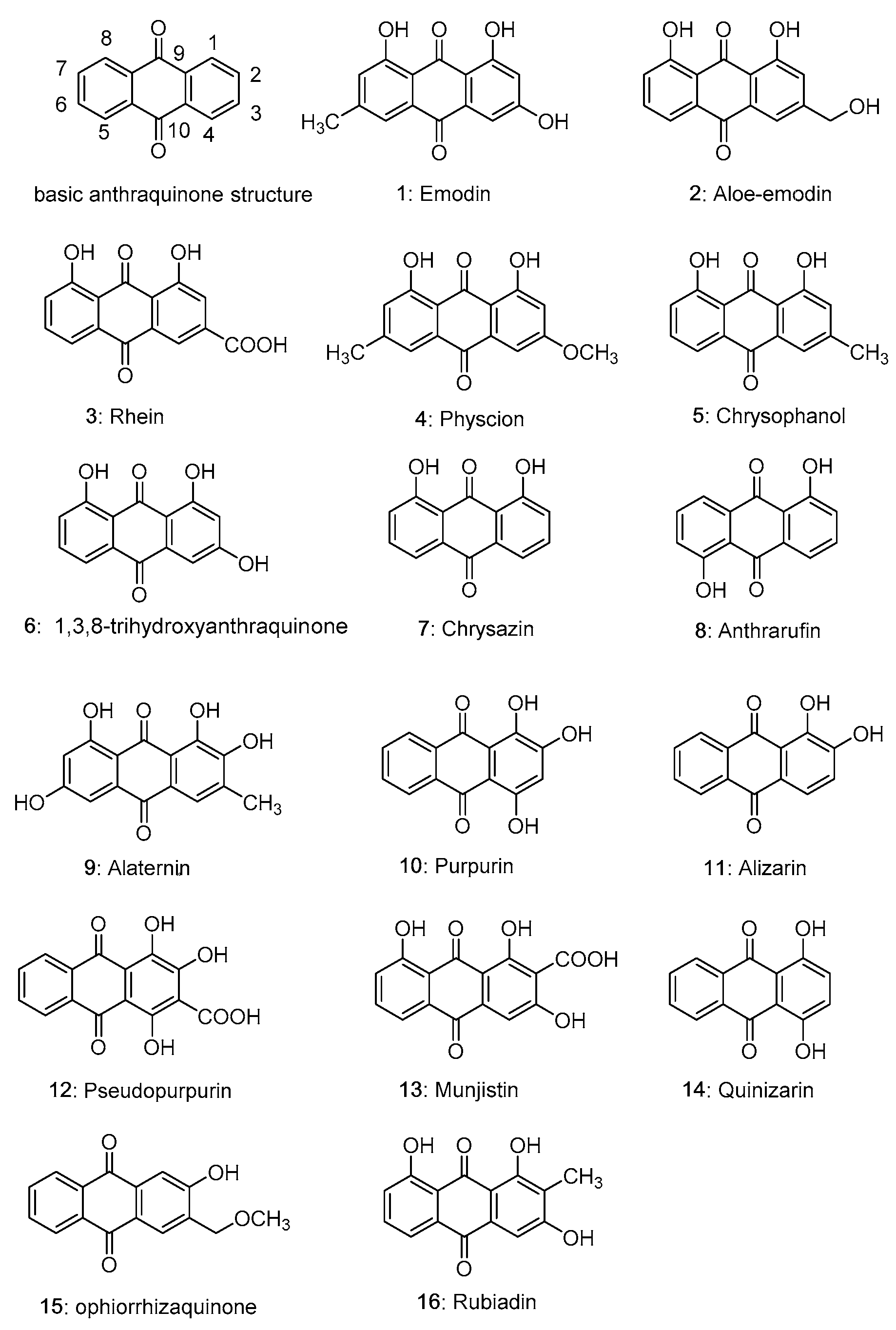
Figure 1. Structures of common natural anthraquinone compounds.
Molecules 28 08139 g001
Figure 2. The main line of this work.
Molecules 28 08139 g002
Figure 3. The structure of aloesone, aloe-emodin and isoeleutheol.
Molecules 28 08139 g003
Figure 4. The chelation of anthraquinone and metals.
Molecules 28 08139 g004
Figure 5. Summary of the effects of 1,5-bisacyloxy-anthraquinones with O–linked substituents (17, 17a11e) and 1,5-bisacyloxy-anthraquinones with S–linked substituents (18) on superoxide production in human neutrophils. Data are expressed as inhibition (%) at 100 μM. Values represent the means ± s.e.m. of 3–6 experiments performed on different days using cells from different donors.
Molecules 28 08139 g005
Table 1. Antioxidant activities of anthraquinone compounds with cellular experiments.
Comp.ModelMajor Discoveries and Proposed Mechanisms aDosage of AdministrationRefs.
Emodin (1)human embryonic kidney cells (HEK 293) ↑ SOD, CAT, GPX, GR, GST0.5 μM[68]
Aloe-emodin (2)H2O2-induced PC12 cells↓ AChE
↓ Oxidative stress
-[78]
Rhein (3)H2O2-induced human umbilical vein endothelial cells↑ NO, NOS, SOD, GSH-PX
↓ Caspase-3, -8, -9 mRNA, MDA, LDH
2, 4, 8, 16 μM[79]
Chrysophanol (5)BV-2 mucin microglia↓ Drp1 (S637)
↓ MAPK, NF-κB, ROS
10 μM[80]
Purpurin (10)primary hepatocytes and WRL-68 cells↑ Nrf2, GST, GPX, GR, CAT, SOD30, 100 μM[81]
a “↑” upregulating or activation. “↓” downregulating or inhibition.
Table 3. Metabolic pathways and metabolites of anthraquinones.
Comp.ModelsDosage of AdministrationMetabolic PathwayMetabolitesRefs.
Emodin (1)Sprague-Dawley rats10 mL/kg rhubarb decoctionglucuronidation
oxidation
hydroxylation
hydrogenation
sulfation
emodin-O-diglucuronides, emodin-O-glucoside-O-glucuronide,
1,8-dihydroxy-3-carboxy-6-methylanthraquinone-1 or 8-O-glucoside,
emodin-1 or 8-O-glucuronide-3-O-sulfate or emodin-1 or 8-O-sulfate-3-O-glucuronide,
1,3,8-trihydroxy-6-methyl-10-oxanthranol glucuronide, emodin-O-diglucuronides, 1,3,8-trihydroxy-6-(glucuronidyl)methylanthrquinone, emodin acid-O-glucuronide,
emodin-2-C-glucuronide, emodin-3-O-glucuronide
[132]
Sprague-Dawley rats0.0156 mg/mLtranshydroxylation
hydroxylation
reduction
oxidation
dihydroxylation
hydroxy-emodin, dihydroxy-emodin,
hydroxy-aloe-emodin, hydroxy-rhein,
aloe-emodin isomer, aloe-emodin, emodin,
1,3,8-trihydroxy-6-methyl-9-oxanthranol/1,3,8-trihydroxy-6-methyl-10-oxanthranol
[133]
Aloe-emodin (2)Sprague-Dawley rats10 mg/kg rhubarb decoctionglucuronidation
oxidation
hydrogenation
hydroxylation
aloe-emodin-8-O-glucoside-1-O-glucuronide or aloe-emodin-1-O-glucoside-8-O-glucuronide,
2-hydroxyaloe-emodin-ω-O-glucuronide
[132]
Sprague-Dawley rats0.035 mg/mLhydroxylation
reduction
oxidation
dihydroxy-aloe-emodin, hydroxy-aloe-emodin, hydroxy-rhein,
hydroxyl-1,8-dihydroxy-3-hydroxymethyl-9-oxanthranol/hydroxyl-1,
8-dihydroxy-3-hydroxymethyl-10-oxanthranol, aloe-emodin, rhein isomer
[133]
Rhein (3)Sprague-Dawley rats0.195 mg/mLhydroxylation
reduction
rhein, rhein isomer,
dihydroxyl-1,8-dihydroxy-3-carboxyl-9-oxanthranol/dihydroxyl-1,8-dihydroxy-3-carboxyl-10-oxanthranol
[133]
Physcion (4)Sprague-Dawley rats0.16 mg/mLdemethylation
hydroxylation
reduction
emodin isomer, hydroxy-emodin, emodin,
dihydroxy-1,8-dihydroxy-3-methoxy-6-methyl-9-oxanthranol/1,8-dihydroxy-3-methoxy-6-methyl-10-oxanthranol
[133]
Sprague-Dawley rats10 mg/kg rhubarb decoctionglucuronidation
sulfation
physcion-1-O-glucoside-8-O-glucuronide or physcion-8-O-glucoside-1-O-glucuronide,
physcion-1,8-O-diglucuronides
[132]
Chrysophanol (5)Sprague-Dawley rats10 mg/kg rhubarb decoctionglucuronidation
sulfation
chrysophanol-1-O-glucoside-8-O-glucuronide,
chrysophanol-8-O-glucoside-1-O-glucuronide,
chrysophanol-1,8-biglucuronides,
chrysophanol-1-O-glucuronide,
chrysophanol-8-O-glucuroniede
[132]
Sprague-Dawley rats0.0755 mg/mLhydroxylation
acetylation
demethylation,
reduction,
oxidation
dihydroxy-chrysophanol,
chrysophanol,
dihydroxyl-1,8-dihydroxy-3-methyl-9-oxanthranol/dihydroxyl-1, 8-dihydroxy-3-methyl-10-oxanthranol,
hydroxy-chrysophanol,
rhein
[133]
Table 4. Anthraquinone levels in botanical and botanical usage.
Comp.Source BotanticalBotanical Daily Intake a (g/day)Neutraceutical
Properties
Refs.
1
Emodin
Rhubarb (Da Huang, Rheum officinale,
Rheum palmatum, Rheum tanguticum), root or rhizome
3–30
[141]
Tumor,
Inflammation,
Gastrointestinal disease,
Hepatoprotective activity,
Diabetic nephropathy,
Atherosclerosis
[142,143,144]
Fo-Ti (Polygonum multiflorum), root3–12
[145]
Alzheimer’s disease,
Parkinson’s disease,
Hyperlipidaemia,
Inflammation,
Cancer
[146]
2
Aloe-emodin
Rhubarb (Da Huang, Rheum officinale,
Rheum palmatum, Rheum tanguticum), root or rhizome
3–30
[141]
Tumor,
Inflammation,
Gastrointestinal disease,
Hepatoprotective activity,
Diabetic nephropathy,
Atherosclerosis
[142,143,144]
Fo-Ti (Polygonum multiflorum), root3–12
[145]
Alzheimer’s disease,
Parkinson’s disease,
Hyperlipidaemia,
Inflammation,
Cancer
[146]
Aloe (Aloe vera, Aloe barbadensis),
leaf
0.005–174
National Institutes of Health, (2019) [147]
Inflammation,
Cancer
[148]
3
Rhein
Rhubarb (Da Huang, Rheum officinale,
Rheum palmatum, Rheum tanguticum), root or rhizome
3–30
[141]
Tumor,
Inflammation,
Gastrointestinal disease,
Hepatoprotective activity,
Diabetic nephropathy,
Atherosclerosis
[142,143,144]
Fo-Ti (Polygonum multiflorum), root3–12
[145]
Alzheimer’s disease,
Parkinson’s disease,
Hyperlipidaemia,
Inflammation,
Cancer
[146]
a Values used to calculate to estimate the daily intake of compounds 1, 2 and 3.
Disclaimer/Publisher’s Note: The statements, opinions and data contained in all publications are solely those of the individual author(s) and contributor(s) and not of MDPI and/or the editor(s). MDPI and/or the editor(s) disclaim responsibility for any injury to people or property resulting from any ideas, methods, instructions or products referred to in the content.