


Determination of Chemical Composition and Investigation of Biological Activities of Ocimum basilicum L.

 , ,
, ,  ,
,  , , , , ,
  ,
,  , and
, and 
Abstract
1. Introduction
2. Material and Methods
2.1. Plant Collection and Extraction
2.2. GC-MS Analysis of Essential Oils
2.3. Evaluation of Antimicrobial Activity
2.3.1. Microorganisms Tested
2.3.2. Inoculum Preparation
2.3.3. Disc Diffusion Assay
2.3.4. Determination of Minimum Inhibitory Concentration
2.3.5. Determination of Minimum Bactericidal Concentration
2.4. Antioxidant Activity
2.5. In Vitro Anti-Diabetic Assay
2.6. 5-Lipoxygenase (5-LOX) Inhibition Assay
2.7. Dermatoprotective Activity
2.8. Toxicological Study
2.8.1. Experimental Animals
2.8.2. Acute Oral Toxicity Study
2.8.3. Chronic Toxicity Study
2.9. Molecular Docking of EO Compounds against E. coli
2.9.1. Molecular Structure Preparation
2.9.2. Pharmacokinetic, Toxicology, and Lipinski Test
2.9.3. Docking Analysis
2.10. Statistical Analysis
3. Results
3.1. Chemical Composition of EOs
3.2. Antibacterial Activity
3.3. Antioxidant Activity
3.4. Antidiabetic Activity
3.5. Anti-Inflammatory and Dermatoprotective Activities
3.6. Toxicological Investigation
3.7. Molecular Docking
3.7.1. Lipinski Test
3.7.2. Pharmacokinetic and Toxicological Properties (ADME/Tox)
3.7.3. Molecular Docking
4. Discussion
5. Conclusions and Perspectives
Author Contributions
Funding
Institutional Review Board Statement
Informed Consent Statement
Data Availability Statement
Acknowledgments
Conflicts of Interest
Sample Availability
Abbreviations
| AA: | Amino Acids |
| ABTS: | 2,2-Azinobis(3-ethyl-Benzothiazoline-6-Sulfonic acid |
| ATCC: | American Type Culture Collection |
| BBB: | Blood-Brain Barrier |
| CCl4: | Carbon Tetrachloride |
| DMSO: | Dimethyl Sulfoxide |
| DNA: | Deoxyribonucleic Acid |
| DPPH: | 2,2- Diphenyl-1-Picrylhydrazyl |
| EM: | Energy Minimization |
| EO: | Essential Oil |
| FRAP: | Ferric Reducing Antioxidant Power |
| GC/MS: | Gas Chromatography/Mass Spectrometry |
| GI: | Gastrointestinal |
| GLUT4: | Glucose Transporter Type 4 |
| hERG: | Human Ether-À-Go-Go Related Gene |
| HPLC: | High-Performance Liquid Chromatography |
| IC50: | Inhibitory Concentration 50% |
| IL-1: | Interleukin-1 |
| ImKCs: | Immortalized mouse Kupffer Cells |
| iNOS: | inducible Nitric Oxide Synthase |
| LPS: | Lipopolysaccharide |
| MBC: | Minimum Bactericidal Concentration |
| MCD: | Methionine-Choline-Deficient |
| NCIM: | National Collection of Industrial Microorganisms |
| MDR: | Multidrug-Resistant |
| MHA: | Mueller-Hinton Agar |
| MHB: | Mueller-Hinton Broth |
| MIC: | Minimum Inhibitory Concentration |
| mRNA: | messenger Ribonucleic Acid |
| MS: | Mass Spectrum |
| NA: | Nicotinamide |
| NASH: | Non-Alcoholic Steatohepatitis |
| NF-κB: | Nuclear Factor-κB |
| NT: | Not Tested |
| OBEO: | Ocimum Basilicum Essential Oil |
| OD: | Optical Density |
| OECD: | Organization of Economic Co-operation and Development |
| OGTT: | Oral Glucose Tolerance Test |
| NO: | Nitric Oxide |
| PDB: | Protein Data Bank |
| P-gp: | P-glycoprotein |
| RO5: | Rule Of Five |
| RT: | Retention Time |
| SA: | Sabouraud’s Agar |
| SD: | Standard Deviation |
| SDF: | Structure-Data File |
| SIRT1: | Sirtuin 1 |
| TNF-α: | Tumor Necrosis Factor-α |
| TSA: | Tryptone Soy Agar |
| WHO: | World Health Organization |
| 5-LOX: | 5-Lipoxygenase |
References
- Mrabti, H.N.; Bouyahya, A.; Ed-Dra, A.; Kachmar, M.R.; Mrabti, N.N.; Benali, T.; Shariati, M.A.; Ouahbi, A.; Doudach, L.; Faouzi, M.E.A. Polyphenolic Profile and Biological Properties of Arbutus unedo Root Extracts. Eur. J. Integr. Med. 2021, 42, 101266. [Google Scholar] [CrossRef]
- WHO. WHO Guidelines on Good Herbal Processing Practices for Herbal Medicines. In WHO Technical Report Series; World Health Organization: Geneva, Switzerland, 2018; pp. 81–152. [Google Scholar]
- Yepes-Perez, A.F.; Herrera-Calderón, O.; Oliveros, C.A.; Flórez-Álvarez, L.; Zapata-Cardona, M.I.; Yepes, L.; Aguilar-Jimenez, W.; Rugeles, M.T.; Zapata, W. The Hydroalcoholic Extract of Uncaria Tomentosa (Cat’s Claw) Inhibits the Infection of Severe Acute Respiratory Syndrome Coronavirus 2 (SARS-CoV-2) In Vitro. Evid. -Based Complementary Altern. Med. 2021, 2021, 6679761. [Google Scholar] [CrossRef] [PubMed]
- Wu, X.M.; Tan, R.X. Interaction between Gut Microbiota and Ethnomedicine Constituents. Nat. Prod. Rep. 2019, 36, 788–809. [Google Scholar] [CrossRef]
- Sharifi-Rad, J.; Dey, A.; Koirala, N.; Shaheen, S.; El Omari, N.; Salehi, B.; Goloshvili, T.; Cirone Silva, N.C.; Bouyahya, A.; Vitalini, S. Cinnamomum Species: Bridging Phytochemistry Knowledge, Pharmacological Properties and Toxicological Safety for Health Benefits. Front. Pharmacol. 2021, 12, 600139. [Google Scholar] [CrossRef] [PubMed]
- Bouyahya, A.; Abrini, J.; Bakri, Y.; Dakka, N. Phytochemical Screening and Evaluation of Antioxidant and Antibacterial Activities of Origanum compactum Extracts. Phytothérapie 2017, 15, 379–383. [Google Scholar] [CrossRef]
- Bouyahya, A.; Chamkhi, I.; Benali, T.; Guaouguaou, F.-E.; Balahbib, A.; El Omari, N.; Taha, D.; Belmehdi, O.; Ghokhan, Z.; El Menyiy, N. Traditional Use, Phytochemistry, Toxicology, and Pharmacology of Origanum majorana L. J. Ethnopharmacol. 2021, 265, 113318. [Google Scholar] [CrossRef]
- Khouchlaa, A.; Talbaoui, A.; El Idrissi, A.E.Y.; Bouyahya, A.; Ait Lahsen, S.; Kahouadji, A.; Tijane, M. Determination of Phenol Content and Evaluation of in Vitro Litholytic Effects on Urolithiasis of Moroccan Zizyphus lotus L. Extract. Phytothérapie 2017, 16, 14–19. [Google Scholar] [CrossRef]
- Bouyahya, A.; Belmehdi, O.; Abrini, J.; Dakka, N.; Bakri, Y. Chemical Composition of Mentha suaveolens and Pinus halepensis Essential Oils and Their Antibacterial and Antioxidant Activities. Asian Pac. J. Trop. Med. 2019, 12, 117. [Google Scholar] [CrossRef]
- Benali, T.; Bouyahya, A.; Habbadi, K.; Zengin, G.; Khabbach, A.; Hammani, K. Chemical Composition and Antibacterial Activity of the Essential Oil and Extracts of Cistus ladaniferus Subsp. Ladanifer and Mentha suaveolens against Phytopathogenic Bacteria and Their Ecofriendly Management of Phytopathogenic Bacteria. Biocatal. Agric. Biotechnol. 2020, 28, 101696. [Google Scholar] [CrossRef]
- Benali, T.; Khabbach, A.; Ennabili, A.; Hammani, K. Ethnopharmacological Prospecting of Medicinal Plants from the Province of Guercif (NE of Morocco). Moroc. J. Biol. 2017, 14, 1–14. [Google Scholar]
- Bouyahya, A.; Abrini, J.; Et-Touys, A.; Bakri, Y.; Dakka, N. Indigenous Knowledge of the Use of Medicinal Plants in the North-West of Morocco and Their Biological Activities. Eur. J. Integr. Med. 2017, 13, 9–25. [Google Scholar] [CrossRef]
- Khabbach, A.; Libiad, M.; Ennabili, A.; Bousta, D. Medicinal and Cosmetic Use of Plants from the Province of Taza, Northern Morocco. Bol. Latinoam. Caribe Plantas Med. Aromat. 2012, 11, 46–60. [Google Scholar]
- Simon, J.E.; Morales, M.R.; Phippen, W.B.; Vieira, R.F.; Hao, Z. Basil: A Source of Aroma Compounds and a Popular Culinary and Ornamental Herb. Perspect. New Crops New Uses 1999, 16, 499–505. [Google Scholar]
- Siddiqui, B.S.; Bhatti, H.A.; Begum, S.; Perwaiz, S. Evaluation of the Antimycobacterium Activity of the Constituents from Ocimum basilicum against Mycobacterium tuberculosis. J. Ethnopharmacol. 2012, 144, 220–222. [Google Scholar] [CrossRef] [PubMed]
- Bantis, F.; Ouzounis, T.; Radoglou, K. Artificial LED Lighting Enhances Growth Characteristics and Total Phenolic Content of Ocimum basilicum, but Variably Affects Transplant Success. Sci. Hortic. 2016, 198, 277–283. [Google Scholar] [CrossRef]
- Gülçin, I.; Elmastaş, M.; Aboul-Enein, H.Y. Determination of Antioxidant and Radical Scavenging Activity of Basil (Ocimum basilicum L. Family Lamiaceae) Assayed by Different Methodologies. Phytother. Res. Int. J. Devoted Pharmacol. Toxicol. Eval. Nat. Prod. Deriv. 2007, 21, 354–361. [Google Scholar] [CrossRef]
- Makinen, S.M.; Paakkonen, K.K. Processing and Use of Basil in Foodstuffs, Beverages and in Food Preparation. In Basil: The Genus Ocimum; Harwood Academic Publishers: Amsterdam, The Netherlands, 1999; pp. 137–152. [Google Scholar]
- Suppakul, P.; Miltz, J.; Sonneveld, K.; Bigger, S.W. Antimicrobial Properties of Basil and Its Possible Application in Food Packaging. J. Agric. Food Chem. 2003, 51, 3197–3207. [Google Scholar] [CrossRef]
- Nguyen, P.M.; Niemeyer, E.D. Effects of Nitrogen Fertilization on the Phenolic Composition and Antioxidant Properties of Basil (Ocimum basilicum L.). J. Agric. Food Chem. 2008, 56, 8685–8691. [Google Scholar] [CrossRef]
- Ekren, S.; Sönmez, Ç.; Özçakal, E.; Kurttaş, Y.S.K.; Bayram, E.; Gürgülü, H. The Effect of Different Irrigation Water Levels on Yield and Quality Characteristics of Purple Basil (Ocimum basilicum L.). Agric. Water Manag. 2012, 109, 155–161. [Google Scholar] [CrossRef]
- Asadollahi, A.; Mirza, M.; Abbaszadeh, B.; Azizpour, S.; Keshavarzi, A. Comparison of Essential Oil from Leaves and Inflorescence of Three Basil (Ocimum basilicum L.) Populations under Drought Stress. Int. J. Agron. Plant Prod. 2013, 4, 2764–2767. [Google Scholar]
- Joshi, R.K. Chemical Composition and Antimicrobial Activity of the Essential Oil of Ocimum basilicum L.(Sweet Basil) from Western Ghats of North West Karnataka, India. Anc. Sci. Life 2014, 33, 151. [Google Scholar] [CrossRef] [PubMed]
- Sethuraman, J.; Nehru, H.; Shanmugam, K.; Balakrishnan, P. Evaluation of potent phytochemicals and antidiabetic activity of Ficus racemosa Linn. World J. Pharm. Res. 2017, 6, 909–920. [Google Scholar]
- Srinivasan Ramalingam, P.; Raj, M.A.S.; Ravichandran, P.; Shanmugam, K.; Nagarasan, S.; Balakrishnan, P. Lipid Peroxidation and Anti-Obesity Activity of Nigella sativa Seeds. World J. Pharm. Res. 2017, 6, 882–892. [Google Scholar]
- Shahrajabian, M.H.; Sun, W.; Cheng, Q. Chemical Components and Pharmacological Benefits of Basil (Ocimum basilicum): A Review. Int. J. Food Prop. 2020, 23, 1961–1970. [Google Scholar] [CrossRef]
- Mahmoud, E.; Starowicz, M.; Ciska, E.; Topolska, J.; Farouk, A. Determination of Volatiles, Antioxidant Activity, and Polyphenol Content in the Postharvest Waste of Ocimum basilicum L. Food Chem. 2022, 375, 131692. [Google Scholar] [CrossRef]
- Nguyen, V.T.; Nguyen, N.Q.; Thi, N.Q.N.; Thi, C.Q.N.; Truc, T.T.; Nghi, P.T.B. Studies on Chemical, Polyphenol Content, Flavonoid Content, and Antioxidant Activity of Sweet Basil Leaves (Ocimum basilicum L.). In IOP Conference Series: Materials Science and Engineering; IOP Publishing: Bristol, UK, 2021; Volume 1092, p. 012083. [Google Scholar]
- Teofilović, B.; Tomas, A.; Martić, N.; Stilinović, N.; Popović, M.; Čapo, I.; Grujić, N.; Ilinčić, B.; Rašković, A. Antioxidant and Hepatoprotective Potential of Sweet Basil (Ocimum basilicum L.) Extract in Acetaminophen-Induced Hepatotoxicity in Rats. J. Funct. Foods 2021, 87, 104783. [Google Scholar] [CrossRef]
- Anwar, S.L.; Lehmann, U. MicroRNAs: Emerging Novel Clinical Biomarkers for Hepatocellular Carcinomas. J. Clin. Med. 2015, 4, 1631–1650. [Google Scholar] [CrossRef]
- Gürgan, M.; Adiloğlu, S. Increasing Concentrations of Iron Fertilizer Affect Antibacterial Activity of Basil (Ocimum basilicum L.). Ind. Crops Prod. 2021, 170, 113768. [Google Scholar] [CrossRef]
- Ikram, A.; Saleem, S.; Imran, M.; Ghazal, A. Antimicrobial Activity by Solvents Extracted from Ocimum basilicum Herb against Multidrug Resistant Gram Negative Rods. J. Islamabad Med. Dent. Coll. 2021, 10, 200–206. [Google Scholar] [CrossRef]
- Rubab, S.; Bahadur, S.; Hanif, U.; Durrani, A.I.; Sadiqa, A.; Shafique, S.; Zafar, U.; Shuaib, M.; Urooj, Z.; Nizamani, M.M. Phytochemical and Antimicrobial Investigation of Methanolic Extract/Fraction of Ocimum basilicum L. Biocatal. Agric. Biotechnol. 2021, 31, 101894. [Google Scholar] [CrossRef]
- Sharaf, M.H.; Abdelaziz, A.M.; Kalaba, M.H.; Radwan, A.A.; Hashem, A.H. Antimicrobial, Antioxidant, Cytotoxic Activities and Phytochemical Analysis of Fungal Endophytes Isolated from Ocimum basilicum. Appl. Biochem. Biotechnol. 2021, 194, 1271–1289. [Google Scholar] [CrossRef]
- Dolghi, A.; Buzatu, R.; Dobrescu, A.; Olaru, F.; Popescu, G.A.; Marcovici, I.; Pinzaru, I.; Navolan, D.; Cretu, O.M.; Popescu, I. Phytochemical Analysis and In Vitro Cytotoxic Activity against Colorectal Adenocarcinoma Cells of Hippophae rhamnodies L., Cymbopogon citratus (DC) Stapf, and Ocimum basilicum L. Essential Oils. Plants 2021, 10, 2752. [Google Scholar] [CrossRef]
- Hanachi, P.; Rezaei Fakhrnezhad, F.; Zarringhalami, R.; Erdogan Orhan, I. Cytotoxicity of Ocimum basilicum and Impatiens Walleriana Extracts on AGS and SKOV-3 Cancer Cell Lines by Flow Cytometry Analysis. Int. J. Cancer Manag. 2021, 14, 102610. [Google Scholar] [CrossRef]
- Okazaki, K.; Nakayama, S.; Kawazoe, K.; Takaishi, Y. Antiaggregant Effects on Human Platelets of Culinary Herbs. Phytother. Res. Int. J. Devoted Pharmacol. Toxicol. Eval. Nat. Prod. Deriv. 1998, 12, 603–605. [Google Scholar] [CrossRef]
- Freire, C.M.M.; Marques, M.O.M.; Costa, M. Effects of Seasonal Variation on the Central Nervous System Activity of Ocimum gratissimum L. Essential Oil. J. Ethnopharmacol. 2006, 105, 161–166. [Google Scholar] [CrossRef]
- Tohti, I.; Tursun, M.; Umar, A.; Turdi, S.; Imin, H.; Moore, N. Aqueous Extracts of Ocimum basilicum L.(Sweet Basil) Decrease Platelet Aggregation Induced by ADP and Thrombin in Vitro and Rats Arterio–Venous Shunt Thrombosis In Vivo. Thromb. Res. 2006, 118, 733–739. [Google Scholar] [CrossRef]
- Amrani, S.; Harnafi, H.; Gadi, D.; Mekhfi, H.; Legssyer, A.; Aziz, M.; Martin-Nizard, F.; Bosca, L. Vasorelaxant and Anti-Platelet Aggregation Effects of Aqueous Ocimum basilicum Extract. J. Ethnopharmacol. 2009, 125, 157–162. [Google Scholar] [CrossRef]
- Raina, P.; Deepak, M.; Chandrasekaran, C.V.; Agarwal, A.; Wagh, N.; Kaul-Ghanekar, R. Comparative Analysis of Anti-Inflammatory Activity of Aqueous and Methanolic Extracts of Ocimum basilicum (Basil) in RAW264. 7, SW1353 and Human Primary Chondrocytes in Respect of the Management of Osteoarthritis. J. Herb. Med. 2016, 6, 28–36. [Google Scholar] [CrossRef]
- Nguyen, P.M.; Kwee, E.M.; Niemeyer, E.D. Potassium Rate Alters the Antioxidant Capacity and Phenolic Concentration of Basil (Ocimum basilicum L.) Leaves. Food Chem. 2010, 123, 1235–1241. [Google Scholar] [CrossRef]
- Bouyahya, A.; El Omari, N.; Elmenyiy, N.; Guaouguaou, F.-E.; Balahbib, A.; Belmehdi, O.; Salhi, N.; Imtara, H.; Mrabti, H.N.; El-Shazly, M. Moroccan Antidiabetic Medicinal Plants: Ethnobotanical Studies, Phytochemical Bioactive Compounds, Preclinical Investigations, Toxicological Validations and Clinical Evidences; Challenges, Guidance and Perspectives for Future Management of Diabetes Worldwide. Trends Food Sci. Technol. 2021, 115, 147–254. [Google Scholar]
- Bouyahya, A.; Guaouguaou, F.-E.; El Omari, N.; El Menyiy, N.; Balahbib, A.; El-Shazly, M.; Bakri, Y. Anti-Inflammatory and Analgesic Properties of Moroccan Medicinal Plants: Phytochemistry, in Vitro and in Vivo Investigations, Mechanism Insights, Clinical Evidences and Perspectives. J. Pharm. Anal. 2021, 12, 35–57. [Google Scholar] [CrossRef] [PubMed]
- Assaggaf, H.M.; Naceiri Mrabti, H.; Rajab, B.S.; Attar, A.A.; Alyamani, R.A.; Hamed, M.; El Omari, N.; El Menyiy, N.; Hazzoumi, Z.; Benali, T.; et al. Chemical Analysis and Investigation of Biological Effects of Salvia officinalis Essential Oils at Three Phenological Stages. Molecules 2022, 27, 5157. [Google Scholar] [CrossRef] [PubMed]
- Al-Mijalli, S.H.; Mrabti, H.N.; Assaggaf, H.; Attar, A.A.; Hamed, M.; Baaboua, A.E.; Omari, N.E.; Menyiy, N.E.; Hazzoumi, Z.; Sheikh, R.A. Chemical Profiling and Biological Activities of Pelargonium graveolens Essential Oils at Three Different Phenological Stages. Plants 2022, 11, 2226. [Google Scholar] [CrossRef]
- Dhama, K.; Sharun, K.; Gugjoo, M.B.; Tiwari, R.; Alagawany, M.; Iqbal Yatoo, M.; Thakur, P.; Iqbal, H.M.; Chaicumpa, W.; Michalak, I. A Comprehensive Review on Chemical Profile and Pharmacological Activities of Ocimum basilicum. Food Rev. Int. 2021, 2021, 1900230. [Google Scholar] [CrossRef]
- Filip, S. Basil (Ocimum basilicum L.) a Source of Valuable Phytonutrients. Int. J. Clin. Nutr. Diet 2017, 3, 118. [Google Scholar] [CrossRef]
- Mekkaoui, M.; Assaggaf, H.; Qasem, A.; El-Shemi, A.; Abdallah, E.M.; Bouidida, E.H.; Naceiri Mrabti, H.; Cherrah, Y.; Alaoui, K. Ethnopharmacological Survey and Comparative Study of the Healing Activity of Moroccan Thyme Honey and Its Mixture with Selected Essential Oils on Two Types of Wounds on Albino Rabbits. Foods 2022, 11, 28. [Google Scholar] [CrossRef]
- Ed-Dra, A.; Filali, F.R.; Lo Presti, V.; Zekkori, B.; Nalbone, L.; Bouymajane, A.; Trabelsi, N.; Lamberta, F.; Bentayeb, A.; Giuffrida, A.; et al. Chemical Composition, Antioxidant Capacity and Antibacterial Action of Five Moroccan Essential Oils against Listeria monocytogenes and Different Serotypes of Salmonella enterica. Microb. Pathog. 2020, 149, 104510. [Google Scholar] [CrossRef]
- Hu, F.; Tu, X.F.; Thakur, K.; Hu, F.; Li, X.L.; Zhang, Y.S.; Zhang, J.G.; Wei, Z.J. Comparison of Antifungal Activity of Essential Oils from Different Plants against Three Fungi. Food Chem. Toxicol. 2019, 134, 110821. [Google Scholar] [CrossRef]
- Ed-Dra, A.; Nalbone, L.; Filali, F.R.; Trabelsi, N.; El Majdoub, Y.O.; Bouchrif, B.; Giarratana, F.; Giuffrida, A. Comprehensive Evaluation on the Use of Thymus vulgaris Essential Oil as Natural Additive against Different Serotypes of Salmonella enterica. Sustainability 2021, 13, 4594. [Google Scholar] [CrossRef]
- Bouyahya, A.; Et-Touys, A.; Bakri, Y.; Talbaui, A.; Fellah, H.; Abrini, J.; Dakka, N. Chemical Composition of Mentha Pulegium and Rosmarinus Officinalis Essential Oils and Their Antileishmanial, Antibacterial and Antioxidant Activities. Microb. Pathog. 2017, 111, 41–49. [Google Scholar] [CrossRef]
- Bouyahya, A.; Bakri, Y.; Et-Touys, A.; Assemian, I.C.C.; Abrini, J.; Dakka, N. In Vitro Antiproliferative Activity of Selected Medicinal Plants from the North-West of Morocco on Several Cancer Cell Lines. Eur. J. Integr. Med. 2018, 18, 23–29. [Google Scholar] [CrossRef]
- Naceiri Mrabti, H.; Doudach, L.; Kachmar, M.R.; Ed-Dra, A.; Khalil, Z.; Naceiri Mrabti, N.; Benrahou, K.; Harraqui, K.; Zengin, G.; Bouyahya, A.; et al. Phenolic Content, Antibacterial, Antioxidant, and Toxicological Investigations of Erodium guttatum (Geraniaceae) Collected from the Northeast of Morocco. Turk. J. Bot. 2021, 45, 739–749. [Google Scholar] [CrossRef]
- Mrabti, H.N.; Sayah, K.; Jaradat, N.; Kichou, F.; Ed-Dra, A.; Belarj, B.; Cherrah, Y.; Faouzi, M.E.A. Antidiabetic and Protective Effects of the Aqueous Extract of Arbutus unedo L. in Streptozotocin-Nicotinamide-Induced Diabetic Mice. J. Complementary Integr. Med. 2018, 15, 2017-0165. [Google Scholar] [CrossRef]
- Hu, B.; Cui, F.; Yin, F.; Zeng, X.; Sun, Y.; Li, Y. Caffeoylquinic Acids Competitively Inhibit Pancreatic Lipase through Binding to the Catalytic Triad. Int. J. Biol. Macromol. 2015, 80, 529–535. [Google Scholar] [CrossRef]
- Andrade, C.; Ferreres, F.; Gomes, N.G.; Duangsrisai, S.; Srisombat, N.; Vajrodaya, S.; Pereira, D.M.; Gil-Izquierdo, A.; Andrade, P.B.; Valentão, P. Phenolic Profiling and Biological Potential of Ficus curtipes Corner Leaves and Stem Bark: 5-Lipoxygenase Inhibition and Interference with NO Levels in LPS-Stimulated RAW 264.7 Macrophages. Biomolecules 2019, 9, 400. [Google Scholar] [CrossRef]
- Bouyahya, A.; Belmehdi, O.; El Jemli, M.; Marmouzi, I.; Bourais, I.; Abrini, J.; Faouzi, M.E.A.; Dakka, N.; Bakri, Y. Chemical Variability of Centaurium erythraea Essential Oils at Three Developmental Stages and Investigation of Their In Vitro Antioxidant, Antidiabetic, Dermatoprotective and Antibacterial Activities. Ind. Crops Prod. 2019, 132, 111–117. [Google Scholar] [CrossRef]
- OECD. OECD Acute Oral Toxicity—Fixed Dose Procedure (Chptr). In Guidelines for the Testing of Chemicals; OECD: Paris, France, 2001; pp. 1–14. [Google Scholar]
- El Kabbaoui, M.; Chda, A.; El-Akhal, J.; Azdad, O.; Mejrhit, N.; Aarab, L.; Bencheikh, R.; Tazi, A. Acute and Sub-Chronic Toxicity Studies of the Aqueous Extract from Leaves of Cistus ladaniferus L. in Mice and Rats. J. Ethnopharmacol. 2017, 209, 147–156. [Google Scholar] [CrossRef]
- Hearnshaw, S.J.; Chung, T.T.-H.; Stevenson, C.E.M.; Maxwell, A.; Lawson, D.M. The Role of Monovalent Cations in the ATPase Reaction of DNA Gyrase. Acta Cryst D 2015, 71, 996–1005. [Google Scholar] [CrossRef]
- Lipinski, C.A.; Lombardo, F.; Dominy, B.W.; Feeney, P.J. Experimental and Computational Approaches to Estimate Solubility and Permeability in Drug Discovery and Development Settings. Adv. Drug Deliv. Rev. 1997, 23, 3–25. [Google Scholar] [CrossRef]
- Brvar, M.; Perdih, A.; Renko, M.; Anderluh, G.; Turk, D.; Solmajer, T. Structure-Based Discovery of Substituted 4, 5′-Bithiazoles as Novel DNA Gyrase Inhibitors. J. Med. Chem. 2012, 55, 6413–6426. [Google Scholar] [CrossRef]
- Ahmed, A.F.; Attia, F.A.; Liu, Z.; Li, C.; Wei, J.; Kang, W. Antioxidant Activity and Total Phenolic Content of Essential Oils and Extracts of Sweet Basil (Ocimum basilicum L.) Plants. Food Sci. Hum. Wellness 2019, 8, 299–305. [Google Scholar] [CrossRef]
- Ghannam, I.A.; Abd El-Meguid, E.A.; Ali, I.H.; Sheir, D.H.; El Kerdawy, A.M. Novel 2-Arylbenzothiazole DNA Gyrase Inhibitors: Synthesis, Antimicrobial Evaluation, QSAR and Molecular Docking Studies. Bioorganic Chem. 2019, 93, 103373. [Google Scholar] [CrossRef] [PubMed]
- Al-Maskri, A.Y.; Hanif, M.A.; Al-Maskari, M.Y.; Abraham, A.S.; Al-sabahi, J.N.; Al-Mantheri, O. Essential Oil from Ocimum basilicum (Omani Basil): A Desert Crop. Nat. Prod. Commun. 2011, 6, 1487–1490. [Google Scholar] [CrossRef] [PubMed]
- Rezzoug, M.; Bakchiche, B.; Gherib, A.; Roberta, A.; Kilinçarslan, Ö.; Mammadov, R.; Bardaweel, S.K. Chemical Composition and Bioactivity of Essential Oils and Ethanolic Extracts of Ocimum basilicum L. and Thymus algeriensis Boiss. & Reut. from the Algerian Saharan Atlas. BMC Complementary Altern. Med. 2019, 19, 146. [Google Scholar] [CrossRef]
- Tamfu, A.N.; Kucukaydin, S.; Ceylan, O.; Sarac, N.; Duru, M.E. Phenolic Composition, Enzyme Inhibitory and Anti-Quorum Sensing Activities of Cinnamon (Cinnamomum zeylanicum Blume) and Basil (Ocimum basilicum Linn). Chem. Afr. 2021, 4, 759–767. [Google Scholar] [CrossRef]
- Faiza Fedoul, F.; Meddah, B.; Larouci, M.; Tir Touil, A.; Merazi, Y.; Bekhti, N.; Piras, A.; Falconieri, D.; Cakmak, Y.S. Medicinal Applications, Chemical Compositions, and Biological Effects of Algerian Ocimum basilicum L. var Genovese with the Conversion of Experimental Doses to Humans. J. Appl. Biotechnol. Rep. 2022, 9, 671–683. [Google Scholar]
- Shoker, R.M.; Raheema, R.H.; Shamkhi, I.J. Antimicrobial Activity, HPLC Analysis of Phenolic Extract of Ocimum basilicum and Ocimum sanctum. Biochem. Cell. Arch. 2021, 21, 1–8. [Google Scholar]
- Hussain, A.I.; Anwar, F.; Hussain Sherazi, S.T.; Przybylski, R. Chemical Composition, Antioxidant and Antimicrobial Activities of Basil (Ocimum basilicum) Essential Oils Depends on Seasonal Variations. Food Chem. 2008, 108, 986–995. [Google Scholar] [CrossRef]
- Shirazi, M.T.; Gholami, H.; Kavoosi, G.; Rowshan, V.; Tafsiry, A. Chemical Composition, Antioxidant, Antimicrobial and Cytotoxic Activities of Tagetes minuta and Ocimum basilicum Essential Oils. Food Sci. Nutr. 2014, 2, 146–155. [Google Scholar] [CrossRef]
- Hossain, M.A.; Kabir, M.J.; Salehuddin, S.M.; Rahman, S.M.M.; Das, A.K.; Singha, S.K.; Alam, M.K.; Rahman, A. Antibacterial Properties of Essential Oils and Methanol Extracts of Sweet Basil Ocimum basilicum Occurring in Bangladesh. Pharm. Biol. 2010, 48, 504–511. [Google Scholar] [CrossRef]
- Jaber, H.; Oubihi, A.; Ouryemchi, I.; Boulamtat, R.; Oubayoucef, A.; Bourkhiss, B.; Ouhssine, M. Chemical Composition and Antibacterial Activities of Eight Plant Essential Oils from Morocco against Escherichia coli Strains Isolated from Different Turkey Organs. Biochem. Res. Int. 2021, 2021, e6685800. [Google Scholar] [CrossRef]
- Malapermal, V.; Botha, I.; Krishna, S.B.N.; Mbatha, J.N. Enhancing Antidiabetic and Antimicrobial Performance of Ocimum basilicum, and Ocimum sanctum (L.) Using Silver Nanoparticles. Saudi J. Biol. Sci. 2017, 24, 1294–1305. [Google Scholar] [CrossRef]
- Ahmad, K.; Khalil, A.T.; Somayya, R. Antifungal, Phytotoxic and Hemagglutination Activity of Methanolic Extracts of Ocimum basilicum. J. Tradit. Chin. Med. 2016, 36, 794–798. [Google Scholar] [CrossRef]
- de Lira Mota, K.S.; de Oliveira Pereira, F.; de Oliveira, W.A.; Lima, I.O.; de Oliveira Lima, E. Antifungal Activity of Thymus Vulgaris L. Essential Oil and Its Constituent Phytochemicals against Rhizopus Oryzae: Interaction with Ergosterol. Molecules 2012, 17, 14418–14433. [Google Scholar] [CrossRef]
- Ibrahim, S.Y.; Abd El-Salam, M.M. Anti-Dermatophyte Efficacy and Environmental Safety of Some Essential Oils Commercial and in Vitro Extracted Pure and Combined against Four Keratinophilic Pathogenic Fungi. Environ. Health Prev. Med. 2015, 20, 279–286. [Google Scholar] [CrossRef]
- Al Abbasy, D.W.; Pathare, N.; Al-Sabahi, J.N.; Khan, S.A. Chemical Composition and Antibacterial Activity of Essential Oil Isolated from Omani Basil (Ocimum basilicum Linn.). Asian Pac. J. Trop. Dis. 2015, 5, 645–649. [Google Scholar] [CrossRef]
- Rodríguez-González, Á.; Álvarez-García, S.; González-López, Ó.; Da Silva, F.; Casquero, P.A. Insecticidal Properties of Ocimum basilicum and Cymbopogon Winterianus against Acanthoscelides obtectus, Insect Pest of the Common Bean (Phaseolus vulgaris, L.). Insects 2019, 10, 151. [Google Scholar] [CrossRef]
- Miao, Q.; Zhao, L.; Wang, Y.; Hao, F.; Sun, P.; He, P.; Liu, Y.; Huang, J.; Liu, X.; Liu, X.; et al. Microbial Metabolomics and Network Analysis Reveal Fungistatic Effect of Basil (Ocimum basilicum) Oil on Candida Albicans. J. Ethnopharmacol. 2020, 260, 113002. [Google Scholar] [CrossRef]
- Ademiluyi, A.O.; Oyeleye, S.I.; Oboh, G. Biological Activities, Antioxidant Properties and Phytoconstituents of Essential Oil from Sweet Basil (Ocimum basilicum L.) Leaves. Comp. Clin. Pathol. 2016, 25, 169–176. [Google Scholar] [CrossRef]
- El-Beshbishy, H.A.; Bahashwan, S.A. Hypoglycemic Effect of Basil (Ocimum basilicum) Aqueous Extract Is Mediated through Inhibition of α-Glucosidase and α-Amylase Activities: An in Vitro Study. Toxicol. Ind. Health 2012, 28, 42–50. [Google Scholar] [CrossRef]
- Mohammed, A.B.; Yagi, S.; Tzanova, T.; Schohn, H.; Abdelgadir, H.; Stefanucci, A.; Mollica, A.; Mahomoodally, M.F.; Adlan, T.A.; Zengin, G. Chemical Profile, Antiproliferative, Antioxidant and Enzyme Inhibition Activities of Ocimum basilicum L. and Pulicaria undulata (L.) CA Mey. Grown in Sudan. S. Afr. J. Bot. 2020, 132, 403–409. [Google Scholar] [CrossRef]
- Shanak, S.; Bassalat, N.; Albzoor, R.; Kadan, S.; Zaid, H. In Vitro and In Silico Evaluation for the Inhibitory Action of O. basilicum Methanol Extract on α-Glucosidase and α-Amylase. Evid. -Based Complementary Altern. Med. 2021, 2021, 5515775. [Google Scholar] [CrossRef] [PubMed]
- Ezeani, C.; Ezenyi, I.; Okoye, T.; Okoli, C. Ocimum basilicum Extract Exhibits Antidiabetic Effects via Inhibition of Hepatic Glucose Mobilization and Carbohydrate Metabolizing Enzymes. J. Intercult. Ethnopharmacol. 2017, 6, 22. [Google Scholar] [CrossRef] [PubMed]
- de Souza, E.M.; de Souza, R.C.; Melo, J.F.; da Costa, M.M.; de Souza, A.M.; Copatti, C.E. Evaluation of the Effects of Ocimum basilicum Essential Oil in Nile Tilapia Diet: Growth, Biochemical, Intestinal Enzymes, Haematology, Lysozyme and Antimicrobial Challenges. Aquaculture 2019, 504, 7–12. [Google Scholar] [CrossRef]
- Soliman, M.E.; Ali, A.F.; Khair, N.; Mahmoud, R.A. The Protective Effect of Ocimum basilicum on the Induced Diabetic Colon of Adult Male Albino Rats: A Histological, Histochemical and Immunohistochemical Study. Egypt. J. Histol. 2019, 42, 608–623. [Google Scholar] [CrossRef]
- Widjaja, S.S.; Rusdiana, M.S. Glucose Lowering Effect of Basil Leaves in Diabetic Rats. Open Access Maced. J. Med. Sci. 2019, 7, 1415. [Google Scholar] [CrossRef]
- Mousavi, L.; Salleh, R.M.; Murugaiyah, V. Phytochemical and Bioactive Compounds Identification of Ocimum tenuiflorum Leaves of Methanol Extract and Its Fraction with an Anti-Diabetic Potential. Int. J. Food Prop. 2018, 21, 2390–2399. [Google Scholar] [CrossRef]
- Ibrahim, G.M.; Ahmed, O.M.; Abbas, N.H.; El Fateh, M.M. Evaluation of the Anti-Diabetic Effects of Epicatechin and/or Gallic Acid in STZ/NA-Induced Diabetic Wister Rats. Res. J. Appl. Biotechnol. 2018, 4, 87–104. [Google Scholar] [CrossRef]
- Takahashi, M.; Ozaki, M.; Tsubosaka, M.; Kim, H.-K.; Sasaki, H.; Matsui, Y.; Hibi, M.; Osaki, N.; Miyashita, M.; Shibata, S. Effects of Timing of Acute and Consecutive Catechin Ingestion on Postprandial Glucose Metabolism in Mice and Humans. Nutrients 2020, 12, 565. [Google Scholar] [CrossRef]
- Mechchate, H.; Es-Safi, I.; Haddad, H.; Bekkari, H.; Grafov, A.; Bousta, D. Combination of Catechin, Epicatechin, and Rutin: Optimization of a Novel Complete Antidiabetic Formulation Using a Mixture Design Approach. J. Nutr. Biochem. 2021, 88, 108520. [Google Scholar] [CrossRef]
- Singh, B.; Kumar, A.; Singh, H.; Kaur, S.; Arora, S.; Singh, B. Protective Effect of Vanillic Acid against Diabetes and Diabetic Nephropathy by Attenuating Oxidative Stress and Upregulation of NF-ΚB, TNF-α and COX-2 Proteins in Rats. Phytother. Res. 2022, 36, 1338–1352. [Google Scholar] [CrossRef]
- Debnath, P.; Singh, K.S.; Devi, T.S.; Singh, S.S.; Butcher, R.J.; Sieroń, L.; Maniukiewicz, W. Synthesis, Characterization, Crystal Structures and Anti-Diabetic Activity of Organotin (IV) Complexes with 2-(4-Hydroxynaphthylazo)-Benzoic Acid. Inorg. Chim. Acta 2020, 510, 118736. [Google Scholar] [CrossRef]
- Topal, M.; Gulcin, İ. Evaluation of the In Vitro Antioxidant, Antidiabetic and Anticholinergic Properties of Rosmarinic Acid from Rosemary (Rosmarinus officinalis L.). Biocatal. Agric. Biotechnol. 2022, 43, 102417. [Google Scholar] [CrossRef]
- Benedec, D.; Pârvu, A.E.; Oniga, I.; Toiu, A.; Tiperciuc, B. Effects of Ocimum basilicum L. Extract on Experimental Acute Inflammation. Rev. Med. Chir. Soc. Med. Nat. Iasi. 2007, 111, 1065–1069. [Google Scholar]
- Bayala, B.; Bassole, I.H.N.; Gnoula, C.; Nebie, R.; Yonli, A.; Morel, L.; Figueredo, G.; Nikiema, J.-B.; Lobaccaro, J.-M.A.; Simpore, J. Chemical Composition, Antioxidant, Anti-Inflammatory and Anti-Proliferative Activities of Essential Oils of Plants from Burkina Faso. PLoS ONE 2014, 9, e92122. [Google Scholar] [CrossRef]
- Li, H.; Ge, Y.; Luo, Z.; Zhou, Y.; Zhang, X.; Zhang, J.; Fu, Q. Evaluation of the Chemical Composition, Antioxidant and Anti-Inflammatory Activities of Distillate and Residue Fractions of Sweet Basil Essential Oil. J. Food Sci. Technol. 2017, 54, 1882–1890. [Google Scholar] [CrossRef]
- Aye, A.; Jeon, Y.-D.; Lee, J.-H.; Bang, K.-S.; Jin, J.-S. Anti-Inflammatory Activity of Ethanol Extract of Leaf and Leaf Callus of Basil (Ocimum basilicum L.) on RAW 264.7 Macrophage Cells. Orient. Pharm. Exp. Med. 2019, 19, 217–226. [Google Scholar] [CrossRef]
- Selvakkumar, C.; Gayathri, B.; Vinaykumar, K.S.; Lakshmi, B.S.; Balakrishnan, A. Potential Anti-Inflammatory Properties of Crude Alcoholic Extract of Ocimum basilicum L. in Human Peripheral Blood Mononuclear Cells. J. Health Sci. 2007, 53, 500–505. [Google Scholar] [CrossRef]
- Rodrigues, L.B.; Martins, A.O.B.P.B.; Cesário, F.R.A.S.; e Castro, F.F.; de Albuquerque, T.R.; Fernandes, M.N.M.; da Silva, B.A.F.; Júnior, L.J.Q.; da Costa, J.G.M.; Coutinho, H.D.M. Anti-Inflammatory and Antiedematogenic Activity of the Ocimum basilicum Essential Oil and Its Main Compound Estragole: In Vivo Mouse Models. Chem. -Biol. Interact. 2016, 257, 14–25. [Google Scholar] [CrossRef]
- Tjahjono, Y.; Karnati, S.; Foe, K.; Anggara, E.; Gunawan, Y.N.; Wijaya, H.; Suyono, H.; Esar, S.Y.; Hadinugroho, W.; Wihadmadyatami, H. Anti-Inflammatory Activity of 2-((3-(Chloromethyl) Benzoyl) Oxy) Benzoic Acid in LPS-Induced Rat Model. Prostaglandins Other Lipid Mediat. 2021, 154, 106549. [Google Scholar] [CrossRef]
- Arowoogun, J.; Akanni, O.O.; Adefisan, A.O.; Owumi, S.E.; Tijani, A.S.; Adaramoye, O.A. Rutin Ameliorates Copper Sulfate-Induced Brain Damage via Antioxidative and Anti-Inflammatory Activities in Rats. J. Biochem. Mol. Toxicol. 2021, 35, e22623. [Google Scholar] [CrossRef] [PubMed]
- Kim, E.J.; Seo, J.B.; Yu, J.S.; Lee, S.; Lim, J.S.; Choi, J.U.; Lee, C.-M.; Rashan, L.; Kim, K.H.; Cho, Y.-C. Anti-Inflammatory Effects of a Polyphenol, Catechin-7, 4′-O-Digallate, from Woodfordia Uniflora by Regulating NF-ΚB Signaling Pathway in Mouse Macrophages. Pharmaceutics 2021, 13, 408. [Google Scholar] [CrossRef] [PubMed]
- Mani, V.M.; Soundari, A.J.P.G.; Balasubramanian, B.; Park, S.; Issara, U.; Preethi, K.; Liu, W.-C. Evaluation of Dimer of Epicatechin from an Endophytic Fungus Curvularia australiensis FC2AP on Acute Toxicity Levels, Anti-Inflammatory and Anti-Cervical Cancer Activity in Animal Models. Molecules 2021, 26, 654. [Google Scholar] [CrossRef] [PubMed]
- Komeili-Movahhed, T.; Bassirian, M.; Changizi, Z.; Moslehi, A. SIRT1/NFκB Pathway Mediates Anti-Inflammatory and Anti-Apoptotic Effects of Rosmarinic Acid on in a Mouse Model of Nonalcoholic Steatohepatitis (NASH). J. Recept. Signal Transduct. 2022, 42, 241–250. [Google Scholar] [CrossRef] [PubMed]
- Ibrahim, S.S.; Abd-Allah, H. Spanlastic Nanovesicles for Enhanced Ocular Delivery of Vanillic Acid: Design, In Vitro Characterization, and In Vivo Anti-Inflammatory Evaluation. Int. J. Pharm. 2022, 625, 122068. [Google Scholar] [CrossRef]
- Parvez, S.; Kang, M.; Chung, H.-S.; Bae, H. Naturally Occurring Tyrosinase Inhibitors: Mechanism and Applications in Skin Health, Cosmetics and Agriculture Industries. Phytother. Res. Int. J. Devoted Pharmacol. Toxicol. Eval. Nat. Prod. Deriv. 2007, 21, 805–816. [Google Scholar] [CrossRef]
- Avetisyan, A.; Markosian, A.; Petrosyan, M.; Sahakyan, N.; Babayan, A.; Aloyan, S.; Trchounian, A. Chemical Composition and Some Biological Activities of the Essential Oils from Basil Ocimum Different Cultivars. BMC Complementary Altern. Med. 2017, 17, 60. [Google Scholar] [CrossRef]
- Coelho, J.; Veiga, J.; Karmali, A.; Nicolai, M.; Pinto Reis, C.; Nobre, B.; Palavra, A. Supercritical CO2 Extracts and Volatile Oil of Basil (Ocimum basilicum L.) Comparison with Conventional Methods. Separations 2018, 5, 21. [Google Scholar] [CrossRef]
- Touiss, I.; Ouahhoud, S.; Harnafi, M.; Khatib, S.; Bekkouch, O.; Amrani, S.; Harnafi, H. Toxicological Evaluation and Hepatoprotective Efficacy of Rosmarinic Acid-Rich Extract from Ocimum basilicum L. Evid. -Based Complementary Altern. Med. 2021, 2021, 6676998. [Google Scholar] [CrossRef]
- El Omari, N.; El Blidi, O.; Bouyahya, A.; Sayah, K.; Bakrim, S.; Fettach, S.; Tahri, R.; Taghzouti, K.; Chokairi, O.; Barkiyou, M. Toxicological Investigations of Aristolochia Longa Root Extracts. J. Toxicol. 2020, 2020, 7643573. [Google Scholar] [CrossRef]


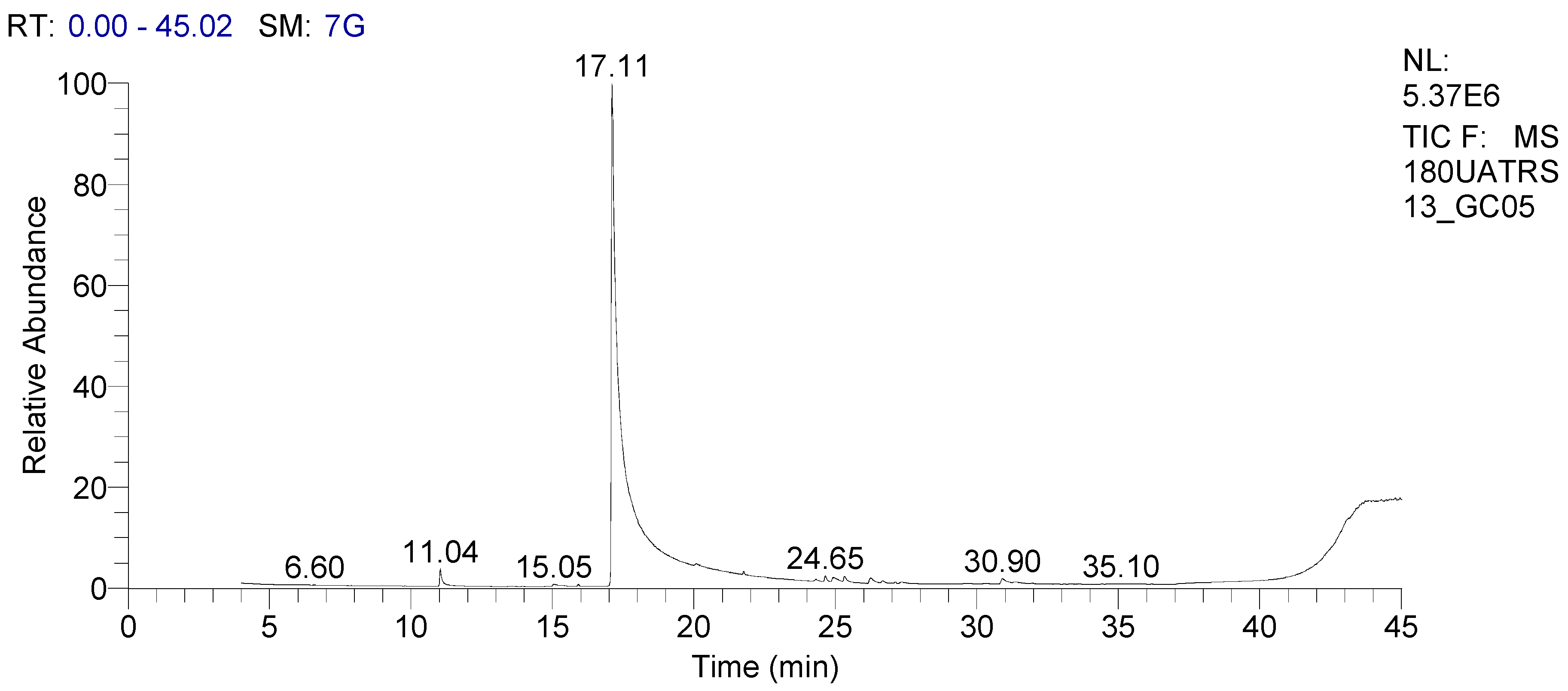
| Compounds | RT (min) | % |
|---|---|---|
| Eucalyptol | 11.05 | 0.74 |
| α-campholene aldehyde | 15.06 | 0.23 |
| Cyclopentasiloxane, decamethyl- | 15.92 | 0.05 |
| Methyl chavicol | 17.12 | 86.40 |
| Trans-anethol | 20.07 | 8.31 |
| Cis-anethol | 21.76 | 1.64 |
| Calarene | 24.65 | 0.18 |
| α-Longipinene | 24.93 | 0.35 |
| Azulene | 25.33 | 0.31 |
| Aristolene | 26.25 | 0.33 |
| Cedrene | 26.67 | 0.09 |
| Aristolene | 27.35 | 0.11 |
| Azulene | 30.91 | 0.48 |
| Phytochemical Compound | Reference |
|---|---|
| Rosmarinic acid, Chicoric acid, Caftaric acid, Caffeic acid | [65] |
| Caftaric acid, gentisic acic, caffeic acid, chlorogenic acid, p-coumaric acid, ferulic acid, isoquercitrin, rosmarinic acid, rutin, quercitrin, quercetin, luteolin | [66] |
| Caffeic acid, caftaric acid, chicoric acid, gentisic acid, rosmarinic acid | [67] |
| Caftaric acid, caffeic acid, ferulic acid, chicoric acid, rosmarinic acid | [68] |
| Chlorogenic acid, vanillic acid, epicatechin, rutin, cinnamic acid, 2,5 dihydroxybenzoic acid, 4-hydroxy benzoic acid, p-coumaric acid, caffeic acid, chlorogenic acid, 3,4-dihydroxy benzoic acid, gallic acid | [69] |
| Caffeic, caftaric, chicoric, gentisic, p-coumaric, and rosmarinic acids | [70] |
| Microorganisms | Ocimum basilicum | Controls | ||
|---|---|---|---|---|
| OBEO | Aqueous Extract | Chloramphenicol | Nystatin | |
| Escherichia coli ATCC 25922 | 16.5 ± 0.20 | 12.6 ± 0.20 | 21.9 ± 0.20 | NT |
| Proteus mirabilis ATCC 25933 | 15.4 ± 0.25 | 11.6 ± 0.15 | 22.2 ± 0.15 | NT |
| Salmonella enterica Typhimurium ATCC700408 | 12.1 ± 0.05 | 8.0 ± 0.50 | 13.5 ± 0.15 | NT |
| Bacillus subtilis ATCC 6633 | 20.4 ± 0.30 | 14.1 ± 0.05 | 15.5 ± 0.15 | NT |
| Staphylococcus aureus ATCC 29213 | 17.3 ± 0.17 | 13.3 ± 0.15 | 25.0 ± 0.25 | NT |
| Listeria monocytogenes ATCC 13932 | 19.0 ± 0.25 | 13.1 ± 0.10 | 27.9 ± 0.15 | NT |
| Candida albicans | 17.3 ± 0.15 | 12.3 ± 0.20 | NT | 29.7 ± 0.20 |
| Trichophyton rubrum | 15.2 ± 0.36 | 10.4 ± 0.20 | NT | 24.9 ± 0.26 |
| Aspergillus niger | 17.1 ± 0.26 | 11.0 ± 0.20 | NT | 26.4 ± 0.10 |
| Microorganisms | O. basilicum in % (v/v) | Controls (µg/mL) | ||||
|---|---|---|---|---|---|---|
| Essential Oil | Aqueous Extract | Chloramphenicol | Nystatin | |||
| MIC | MBC | MIC | MBC | |||
| Escherichia coli ATCC 25922 | 0.5 | 1 | 1 | 2 | 4 | NT |
| Proteus mirabilis ATCC 25933 | 0.5 | 1 | 1 | 2 | 4 | NT |
| Salmonella enterica Typhimurium ATCC 700408 | 1 | 2 | 4 | >4 | 64 | NT |
| Bacillus subtilis ATCC 6633 | 0.25 | 0.25 | 1 | 1 | 32 | NT |
| Staphylococcus aureus ATCC 29213 | 0.5 | 0.5 | 1 | 1 | 4 | NT |
| Listeria monocytogenes ATCC 13932 | 0.25 | 0.25 | 1 | 1 | 2 | NT |
| Candida albicans | 1 | >4 | 2 | NT | NT | 4 |
| Trichophyton rubrum | 2 | >4 | 4 | NT | NT | 16 |
| Aspergillus niger | 2 | >4 | 4 | NT | NT | 16 |
| Ocimum basilicum | DPPH | ABTS | FRAP |
|---|---|---|---|
| Essential oils | 6.41 ± 0.02 a | 32.58 ± 0.53 a | 74.66 ± 0.65 a |
| Aqueous extract | 4.12 ± 0.08 b | 21.00 ± 0.06 b | 33.13 ± 0.17 b |
| Trolox | 2.23 ± 0.02 c | 9.33 ± 0.07 c | 8.52 ± 0.17 c |
| IC50 (μg/mL) | α-Amylase | α-Glucosidase | Lipase |
|---|---|---|---|
| Essential oil | 50.51 ± 0.32 a | 39.84 ± 1.2 a | 43.24 ± 0.01 a |
| Aqueous extract | 82 ± 0.26 b | 56 ± 4.21 b | 74.28 ± 0.02 b |
| Acarbose | 28.24 ± 0.06 c | 19 ± 1.12 c | |
| Orlistat | 18.27 ± 0.03 c |
| Assays | Ocimum basilicum IC50 (μg/mL) | Control | |
|---|---|---|---|
| Essential Oil | Aqueous Extract | Quercetin | |
| 5-Lipoxygenase | 18.28 ± 0.03 a | 24.8 ± 0.01 b | 4.19 ± 0.0 2 c |
| Tyrosinase | 68.58 ± 0.03 a | 118.37 ± 0.05 b | 25.08 ± 0.12 c |
| Days | Control | 0.25 g/kg | 0.5 g/kg |
|---|---|---|---|
| D0 | 217.97 ± 0.03 a | 209.46 ± 0.02 b | 209.26 ± 09.04 b |
| D30 | 233.93 ± 0.01 a | 215.37 ± 0.01 b | 225.31 ± 02.14 c |
| D60 | 244.62 ± 0.02 a | 226.17 ± 0.02 b | 254.93 ± 18.03 c |
| D90 | 262.21 ± 0.04 a | 237.58 ± 0.01 b | 275.42 ± 39.02 c |
| Control | 0.25 g/kg | 0.5 g/kg | |
|---|---|---|---|
| Red blood cells (106 μL−1) | 8.2 ± 0.1 a | 8.4 ± 0.2 b | 8.5 ± 0.1 b |
| White blood cells (103 μL−1) | 12.4 ± 0.2 a | 12.8 ± 0.1 b | 12.9 ± 0.3 b |
| Hemoglobin (g/dL) | 11.8 ± 0.2 a | 12.2 ± 0.4 b | 12.3 ± 0.5 b |
| Hematocrit (vol %) | 42.2 ± 0.8 a | 45.5 ± 1.4 b | 47.8 ± 1.3 c |
| Platelets (×104 L−1) | 82.8 ± 0.2 a | 84.6 ± 0.3 b | 85.9 ± 0.1 c |
| Lymphocytes (%) | 78.31 ± 0.3 a | 79.45 ± 0.5 b | 79.68 ± 0.3 b |
| Neutrophils (%) | 15.5 ± 0.3 a | 15.6 ± 0.8 a | 15.9 ± 0.5 a |
| Ligand Name | Molecular Weight (g/mol) | XLogP3 Count | Hydrogen Bond Donor | Hydrogen Bond Acceptor Count | Agreement with RO5 |
|---|---|---|---|---|---|
| Methyl chavicol | 148. 2 | 3.4 | 0 | 1 | Yes |
| Trans-anethole | 148.2 | 3.3 | 0 | 1 | Yes |
| Ligand | GI Abs | Blood-Brain Barrier Permeant | P-Glycoprote in Substrate | CYP1A2 Inhibitor | CYP2C19 Inhibitor | CYP2C9 Inhibitor | CYP2D6 Inhibitor | CYP3A4 Inhibitor | Log Kp | Carcino_Mous | Carcino_ Rat | hERC Inhibition |
|---|---|---|---|---|---|---|---|---|---|---|---|---|
| Methyl chavicol | High | Yes | No | Yes | No | No | No | No | −4.73 | Positive | Positive | Medium risk |
| Trans-anethole | High | Yes | No | Yes | No | No | No | No | −4.87 | Positive | Positive | Medium risk |
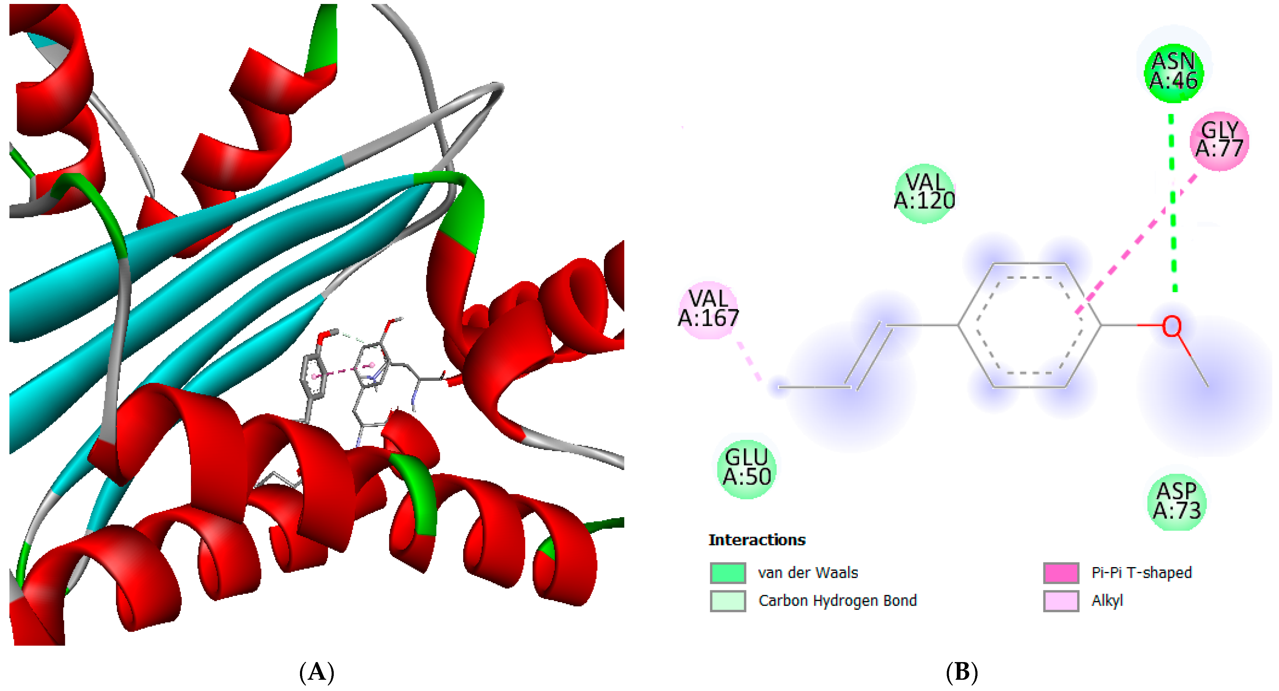
| Ligand ID | Ligand Name | Final Intermolecular Energy (kcal.mol) | Final Total Internal Energy (kcal.mol) | Torsional FreeEnergy (kcal.mol) | Unbound System Energy (kcal.mol) | Estimated Free Energy of Binding (kcal/mol) |
|---|---|---|---|---|---|---|
| 8815 | Methyl chavicol | −5.28 | −0.19 | 0.86 | −0.16 | −5.21 |
| 637563 | Trans-anethole | −6.08 | −0.18 | 0.89 | −0.15 | −5.22 |
Disclaimer/Publisher’s Note: The statements, opinions and data contained in all publications are solely those of the individual author(s) and contributor(s) and not of MDPI and/or the editor(s). MDPI and/or the editor(s) disclaim responsibility for any injury to people or property resulting from any ideas, methods, instructions or products referred to in the content. |
© 2023 by the authors. Licensee MDPI, Basel, Switzerland. This article is an open access article distributed under the terms and conditions of the Creative Commons Attribution (CC BY) license (https://creativecommons.org/licenses/by/4.0/).
Share and Cite
Qasem, A.; Assaggaf, H.; Mrabti, H.N.; Minshawi, F.; Rajab, B.S.; Attar, A.A.; Alyamani, R.A.; Hamed, M.; Mrabti, N.N.; Baaboua, A.E.; et al. Determination of Chemical Composition and Investigation of Biological Activities of Ocimum basilicum L. Molecules 2023, 28, 614. https://doi.org/10.3390/molecules28020614
Qasem A, Assaggaf H, Mrabti HN, Minshawi F, Rajab BS, Attar AA, Alyamani RA, Hamed M, Mrabti NN, Baaboua AE, et al. Determination of Chemical Composition and Investigation of Biological Activities of Ocimum basilicum L. Molecules. 2023; 28(2):614. https://doi.org/10.3390/molecules28020614
Chicago/Turabian StyleQasem, Ahmed, Hamza Assaggaf, Hanae Naceiri Mrabti, Faisal Minshawi, Bodour S. Rajab, Ammar A. Attar, Reema A. Alyamani, Munerah Hamed, Nidal Naceiri Mrabti, Aicha El Baaboua, and et al. 2023. "Determination of Chemical Composition and Investigation of Biological Activities of Ocimum basilicum L." Molecules 28, no. 2: 614. https://doi.org/10.3390/molecules28020614
APA StyleQasem, A., Assaggaf, H., Mrabti, H. N., Minshawi, F., Rajab, B. S., Attar, A. A., Alyamani, R. A., Hamed, M., Mrabti, N. N., Baaboua, A. E., Omari, N. E., Alshahrani, M. M., Awadh, A. A. A., Sheikh, R. A., Ming, L. C., Goh, K. W., & Bouyahya, A. (2023). Determination of Chemical Composition and Investigation of Biological Activities of Ocimum basilicum L. Molecules, 28(2), 614. https://doi.org/10.3390/molecules28020614