Article

Design, Synthesis, Docking Study, and Antiproliferative Evaluation of Novel Schiff Base–Benzimidazole Hybrids with VEGFR-2 Inhibitory Activity

by
Hany M. Abd El-Lateef
1,2,*,
Mohammed A. I. Elbastawesy
3,*,
Tamer Mohamed Abdelghani Ibrahim
4,5,
Mai M. Khalaf
1,2,
Mohamed Gouda
1,
Mariam G. F. Wahba
6,
Islam Zaki
7 and
Martha M. Morcoss
8
1
Department of Chemistry, College of Science, King Faisal University, Al-Ahsa 31982, Saudi Arabia
2
Department of Chemistry, Faculty of Science, Sohag University, Sohag 82524, Egypt
3
Department of Pharmaceutical Organic Chemistry, Faculty of Pharmacy, Al-Azhar University, Assiut 71524, Egypt
4
Community Engagement Development Administration, Vice-Presidency for Studies, Development and Community Service, King Faisal University, Al-Ahsa 31982, Saudi Arabia
5
Faculty of Social Work, Helwan University, Helwan 11795, Egypt
6
Department of Pharmacology and Toxicology, Faculty of pharmacy, Nahda University, Beni-Suef 62513, Egypt
7
Pharmaceutical Organic Chemistry Department, Faculty of Pharmacy, Port Said University, Port Said 42526, Egypt
8
Department of Pharmaceutical Chemistry, Faculty of Pharmacy, Nahda University, Beni-Suef 62513, Egypt
*
Authors to whom correspondence should be addressed.
Molecules 2023, 28(2), 481; https://doi.org/10.3390/molecules28020481
Submission received: 1 December 2022 / Revised: 18 December 2022 / Accepted: 29 December 2022 / Published: 4 January 2023
(This article belongs to the Special Issue Synthesis, Characterization and Application of Surfactants II)

Abstract

:
A new series of Schiff–benzimidazole hybrids 3ao has been designed and synthesized. The structure of the target compounds was proved by different spectroscopic and elemental analysis tools. The target compounds were evaluated for their in vitro cytotoxic activity against 60 cancer cell lines according to NCI single- and five-dose protocols. Consequently, four compounds were further examined against the most sensitive lung cancer A549 and NCI-H460 cell lines. Compounds 3e and 3g were the most active, achieving 3.58 ± 0.53, 1.71 ± 0.17 and 1.88 ± 0.35, 0.85 ± 0.24 against A549 and NCI-H460 cell lines, respectively. Moreover, they showed remarkable inhibitory activity on the VEGFR-2 TK with 86.23 and 89.89%, respectively, as compared with Sorafenib (88.17%). Moreover, cell cycle analysis of NCI-H460 cells treated with 3e and 3g showed cellular cycle arrest at both G1 and S phases (supported by caspases-9 study) with significant pro-apoptotic activity, as indicated by annexin V-FITC staining. The binding interactions of these compounds were confirmed through molecular docking studies; the most active compounds displayed complete overlay with, and a similar binding mode and pose to, Sorafenib, a reference VEGFR-2 inhibitor.

1. Introduction

Heterocycles, especially heterocycles containing nitrogen, are considered the most fundamental structural units in medicinal chemistry research and development [1,2]. Among several heterocyclic structures, syntheses and biological applications of benzimidazole have been subjected to various studies from many research groups [3,4,5,6].
On another chemical front, the Schiff hydrazone (C=N-) framework is an exceptionally adaptable drug-like moiety which has recently been utilized in the development of cancer treatments or cellular apoptosis [7,8]. The structures containing this bioactive moiety showed remarkable anticancer activities via the restraint of numerous kinds of enzymes, proteins, and/or receptors that plays essential roles in cell growth and survival [9]. Consequently, Schiff-fused chemical compounds, because of their wide scope of biological activities and synthetic applications, have been developed as a target heterocyclic framework in the research and development of medicinal chemistry. (Compound 1, 3, Figure 1) [10,11]
Controlling the angiogenesis process is the basic factor of the growth or inhibition of the majority of cancer cell types [12,13]. Angiogenesis is simply defined as the process by which new blood capillaries have arisen from the existing vasculature, and this process can be stimulated through the activation of various chemical signals [14]. Tyrosine kinases (TKs) are one of the main regulators of tumor angiogenesis [15,16]. It has been reported that many TKs receptors, especially VEGFR-2, are over-expressed in many cancer cells. In response to its stimulation, VEGFR-2 can promote a consecutive series of successive signals that control cell growth, survival, and proliferation [17]. The over-stimulation of VEGFR-2 was reported in plenty of cancer cell types when compared to normal cells [18]. Consequently, and depending on the previous facts, many medicinal chemistry researchers target the inhibition of such an enzyme in order to obtain more safe or more selective candidates that treat or operate on cancer angiogenesis with no effect on normal cells [19]. The process of hindering the VEGF pathway can be carried out by blocking the VEGFR-2 receptors activation using novel VEGFR-2 inhibitors [20].
Apoptosis simply means the regulation of programming cell death; it plays a fundamental role in normal cell development or tissue homeostasis [21]. Many researchers showed that the apoptotic process is closely related to the survival of tumors. Moreover, caspases are broadly known of their role in the apoptotic process [22]. Dysregulation of apoptotic caspases leads directly to the inactivation or over-activation of bioactive substrates, the generation of a cascade of signaling events, authorized the controlled demolition of cellular components, and proliferation; therefore, targeting the cellular apoptotic cascades of cancer cells has gained great attention due to its clinically beneficial effects in cancer therapy [23].
Recently, Meguid et al. reported the design and synthesis of a new series of benzimidazole derivatives with promising dual inhibition action on EGFR and VEGFR-2 kinases [24]. Among their tested compounds, compound 2 (see Figure 1) displayed remarkable inhibition activity against VEGFR-2 with IC50 69.62 μM. Furthermore, a notable cytotoxicity activity was found against the HeLa cancer cell line with IC50 of 1.44 μM compared to the standard [24].
Finally, and in continuation of our previous research and efforts to synthesize novel heterocyclic compounds bearing various bioactive scaffolds of efficient anticancer activity [25,26], the present work depends on the hybridization of the well-known biologically active entities Schiff base and benzimidazole scaffolds. Our synthesized compounds were tested as anti-cancer candidates targeting VEGFR-2 inhibitors with the analysis of their apoptotic anti-proliferative activities, in the hope that this synergistic hybridization may lead to a more selective, safe, and efficient drug.

2. Results

2.1. Chemistry

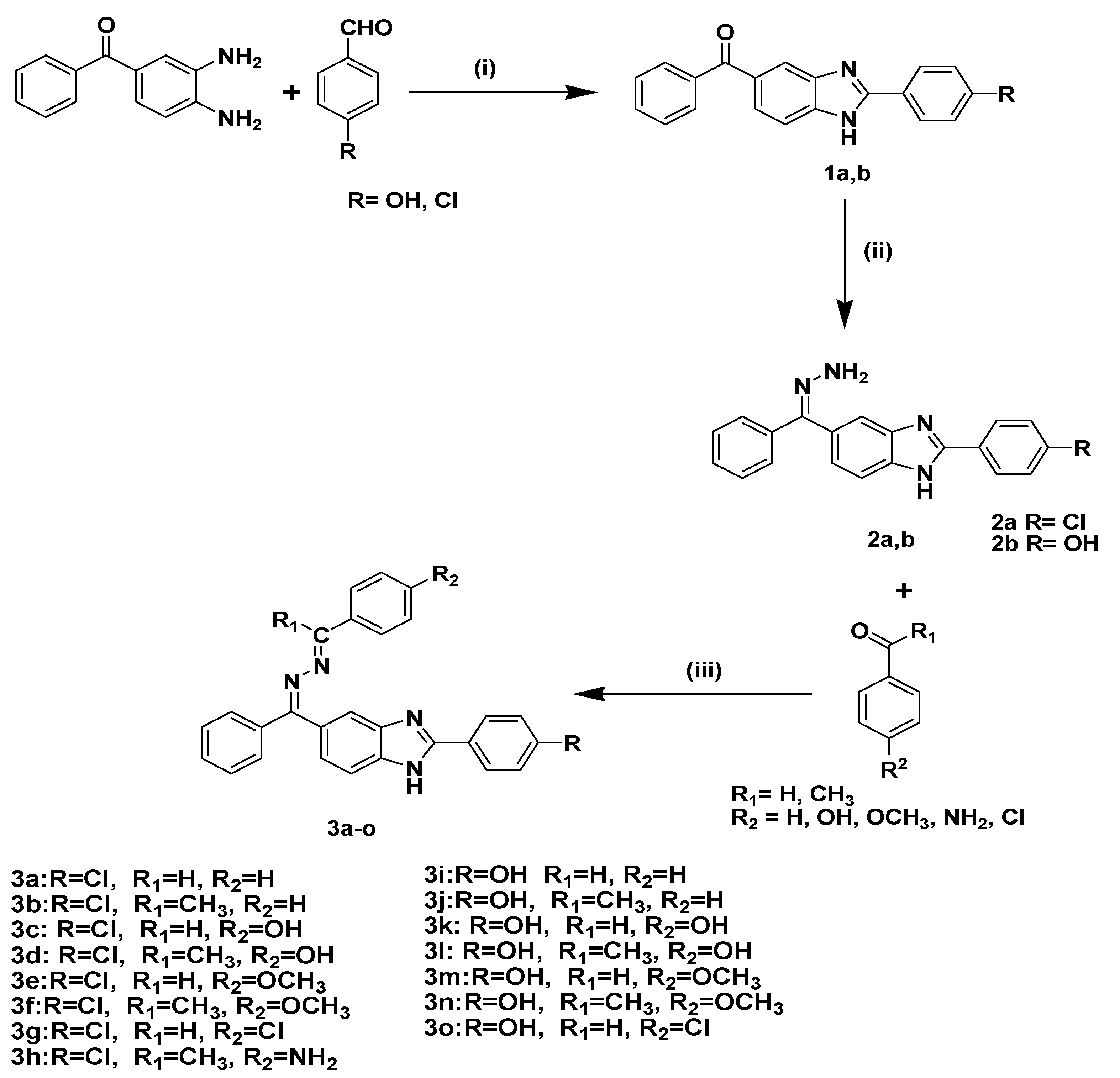
Target compounds 3ao were chemically synthesized as shown in Scheme 1. Compounds 1a,b were prepared according to the reported methods [27]. The chemical structures of such compounds were proven by matching their spectral data with the reported data [27]. The key intermediates compounds, benzimidazole-hydrazone 2a,b, were prepared by heating compounds 1a,b with hydrazine hydrate at reflux temperature for 12 h. The final target benzimidazole–Shiff hybrids 3ao were prepared by reaction of the benzimidazole hydrazone derivatives 2a,b with the appropriate acetophenone or aldehyde derivatives in ethanol in the presence of few drops of acetic acid as catalyst under reflux conditions. The spectral data, in addition to elemental analyses, showed that all derivatives of 3ao underwent the reaction smoothly to give the predicted Schiff base hybrid structure in good yields. The 1H NMR spectra experienced the disappearance of NH2 signal, which indicates the condensation process. On other hand, the appearance of doublet signals with their characteristic pattern in the aromatic δ6.70–8.00 ppm region augments the formation of new hybrids.
As a representative example, compound 3m NMR spectra showed a broad singlet signal at δH 10.39 ppm for phenolic OH proton. Moreover, as mentioned, a characteristic pattern for the additional four phenyl protons at δH 8.04 and 7.06 ppm represented di para-substituted phenyl. Moreover, 1H singlet signal at δ 4.20 is distinctive as an NH proton and could be exchanged using D2O, in addition to the singlet 3 proton of the methoxy group at δ 3.83. The distinguished hydrazone proton appeared at δ 8.64 ppm. Moreover, the 13C spectrum showed a characteristic peak appearing at δC 148.09, representing the (C=N) imine carbon. Moreover, the obtained elemental results of compound 3m align with the calculated data for such a compound.

2.2. Biology

2.2.1. Primary In Vitro One-Dose Anticancer Assay

Benzimidazole compounds 3ao were selected by the National Cancer Institute (NCI), Bethesda, USA, according to the protocols of drug evaluation for in vitro anticancer screening [16]. Primary in vitro one-dose screening assay was performed in full panels of 60 human tumor cell lines derived from nine tumor cell lines, including leukemia, melanoma, lung, colon, CNS, ovarian, renal, prostate, and breast cancer cell lines. The selected benzimidazole hybrids were added at a single concentration (10−5 M) and the culture was incubated for 48 h. Analysis of the single dose 60 cell panel assay results showed that benzimidazole molecules 3a, 3b, 3d, 3f, 3l and 3n were found to be inactive while other benzimidazole molecules were active against some tumor cell lines. Benzimidazole molecules 3hk and 3m displayed moderate cell growth inhibition activity only against leukemia CCRF-CEM, HL-60(TB) and RPMI-8226 cell lines with percentage growth between 21.39–39.31%. Compound 3c achieved high cell growth inhibitory activity against leukemia CCRF-CCEM, HL-60(TB), K-562, MOLT-4 and RPMI-8226, non-small cell lung cancer NCI-H23, NCI-H522, colon cancer HCC-2998, KM-12, SW-620, CNS cancer U-251, melanoma MALME-3M, M14, SK-MEL-5, ovarian cancer OVCAR-8, renal cancer RXF 393, SN 12C and breast cancer BT-549 with percentage growth values between 1.86–11.73%. A complete cell death was recorded for the leukemia SR, non-small cell lung cancer NCI-H460, colon cancer COLO 205, HCT-116, HT-29, melanoma LOX IMVI, and breast cancer MCF-7 and MDA-MB-468 cells where the growth percent was −3.17, −43.84, −50.64, −37.23, −0.83, −52.93, −4.44 and −32.95%, respectively. The obtained data revealed an obvious activity profile for the compound 3e toward leukemia K-562, SR, non-small cell lung cancer HOP-62, colon cancer HCT-116, CNS cancer SF-268, SF-295, melanoma SK-MEL-28, ovarian cancer OVCAR-4, OVCAR-8, renal cancer 786-0, and breast cancer HS-578T cancer cell lines with growth percentage 4.08–14.80%. Compound 3e revealed complete cell death against non-small lung cancer HOP-92, NCI-H460, CNS cancer U-251, melanoma LOX IMVI, MALME-3M, and renal cancer RXF 393 cells with growth percentage −2.97, −47.73, −51.51, −26.04, −9.02, −17.67%, respectively. In addition, compound 3g displayed marvelous anticancer activity against non-small cell lung cancer NCI-H23, CNS cancer SF-539, melanoma SK-MEL-28, ovarian cancer IGROV 1, OVCAR-4, renal cancer ACHN, and breast cancer MCF-7 cancer cells where the growth percentage values were in the range of 0.75–12.48%. Additionally, compound 3g showed complete cell death toward non-small cell lung cancer A549/ATCC, NCI-H460, colon cancer HCT-116, HT-29, CNS cancer U251, melanoma LOX IMVI, MALME-3M, renal cancer 786-0, RXF 393, and prostate cancer DU-145 cells with growth percentage values of −3.93, −64.39, −21.05, −58.89, −62.08, −12.27, −49.93, −32.04, −12.30 and −22.13%, respectively Furthermore, compound 3o showed remarkable anticancer activity against leukemia K-562, MOLT-4, non-small cell lung cancer EKVX, NCI-H226, CNS cancer SNB-19, melanoma MDA-MB-435, UACC-257, UACC-62, and breast cancer with growth percentage values between 0.08–11.93%. Furthermore, compound 3o displayed complete cell death toward most of the remaining cell lines with growth percentage values between −5.56 to −86.78%. The obtained results indicate that compounds 3c, 3e, 3g, and 3o exhibited the highest ability to inhibit the growth of different cancer cell lines compared to other benzimidazole compounds. It could be concluded that Schiff bases attached to the benzimidazole ring might contribute to the activity of the prepared compounds. The presence of an electron donating group (OH or OCH3) or an electron withdrawing group (Cl) has better anticancer activity against cancer cell lines over the unsubstituted phenyl derivatives. In addition, replacement of arylidenehydrazono substituent with the arylethylidene group resulted in sharp decrease in growth percentage inhibition. Furthermore, regarding groups present at C-2 of benzimidazole ring, 4-chlorophenyl exhibited the higher activity among the tested groups, indicating that the common C-2-(4-chlorophenyl) has a better contribution in growth percentage inhibitory activity than the 4-hydroxyphenyl group (see the supplementary materials).

2.2.2. Full In Vitro Five-Dose Anticancer Assay

Benzimidazole–Schiff hybrids 3c, 3e, 3g, and 3o, which demonstrated noticeable activities against most tested cell lines, were additionally selected for the five doses testing against the full panel of 60 human tumor cell lines by NCI. In the context, compound 3c showed high activities against most of the tested cell lines with GI50 values ranging from 1.46–7.97 µM. The compound exhibited GI50 values in the range of 1–3 µM in 53 tested subpanels. The highest growth inhibition activity was observed against leukemia HL-60(TB) with a GI50 value of 1.46 µM. On the other hand, compound 3e showed obvious sensitivity toward leukemia cell lines, except for the leukemia SR cell line (GI50 value 8.94 µM) with GI50 values ranging from 1.05–2.04 µM. Concerning non-small cell lung cancer, the compound showed high activity against A549, HOP-62, NCI-H23, and NCI-H522, with GI50 values less than 2 µM. All the remaining subpanels showed marvelous sensitivity profiles with GI50 not more than 2.56 µM. In addition, compound 3g displayed GI50 values ranging from 1.62–4.30 µM against all the tested subpanels except for the leukemia cancer CCRF-CEM cell line where it showed a GI50 value of 6.91 µM. The highest growth inhibitory activity was observed against the renal cancer RXF-393 cell line with a GI50 value of 1.62 µM. The obtained data revealed a good sensitivity profile for benzimidazole molecule 3g toward colon cancer HT-29 and renal cancer UO-31 subpanels with a GI50 value of 1.70 µM. Furthermore, compound 3o showed high anticancer activity against most of the tested cell lines with GI50 values between 1.02–9.95 µM. With regard to sensitivity against some individual subpanels, compound 3o revealed noticeable activity against NCI-H226, SF-539, SNB-19, and SK-MEL-28, with GI50 values 1.23, 1.02, 1.03, and 1.09 µM, respectively. Moreover, the obtained data showed high activity against ovarian cancer OVCAR-5 and breast cancer MDA-MB-231 subpanels with GI50 values of 1.40 and 1.93 µM, respectively (see the supplementary materials).

2.2.3. MTT Assay against Lung Cancer Cell Lines

To determine IC50, benzimidazole hybrids 3c, 3e, 3g, and 3o were further analyzed using the standard MTT colorimetric assay against lung cancer A549 and NCI-H460 cell lines [28], Appendix A. Sorafenib was chosen as a reference control in the present study. The choice of lung cancer cell lines was based on their sensitivity to tested compounds in the NCI-60 cell line assay. The in vitro results showed that the test benzimidazole molecules showed significant anticancer activity against test lung cancer cell lines (Table 1). Benzimidazole molecules 3e and 3g showed 1.3–2.1-fold more potent cytotoxic activity than Sorafenib. The cytotoxic activity correlation of the test benzimidazole molecules showed that compounds 3e and 3g showed more potent cytotoxic effects as concluded from their IC50 values against test lung cancer cell lines when compared to compounds 3c or 3o. It could be concluded that, regarding aryl groups present at C-2 of benzimidazole ring, 4-chlorophenyl moiety was favorable and exhibited higher cytotoxic activity than the 4-hydroxyphenyl group.

2.2.4. In Vitro VEGFR-2 Inhibition Assay

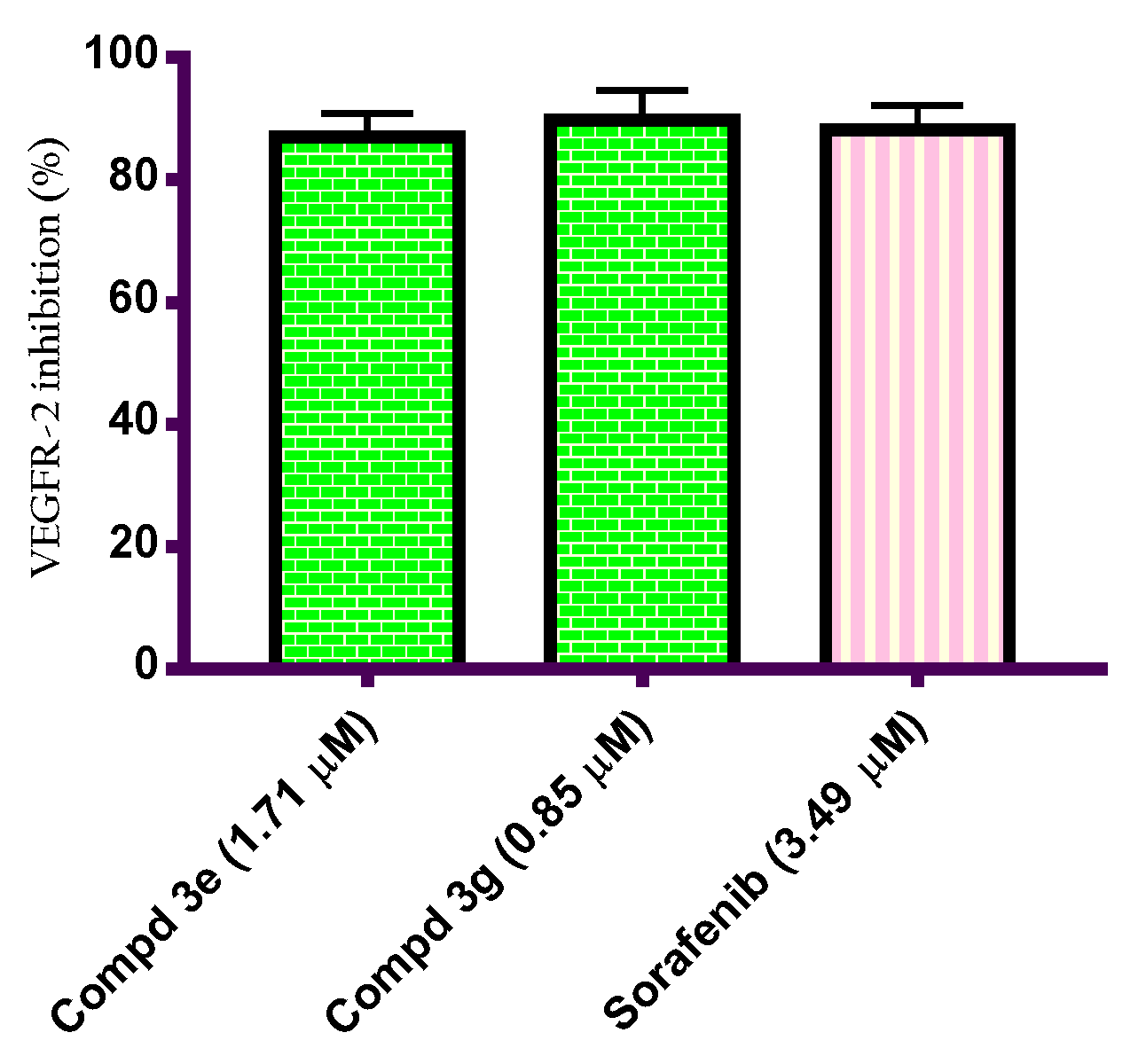
VEGFR-2, a key endothelial RTK, functions as a major positive signal transducer for both physiological and pathological angiogenesis. Regulation of VEGFR-2 activation is one of the major important mechanisms that are essential for proteostasis in endothelial cells under pathological conditions [29]. In order to prove the mechanism of the antiproliferative activity of the prepared benzimidazole molecules, compounds 3e and 3g were subjected to in vitro VEGFR-2 inhibition activity using ELISA analysis [30]. Sorafenib is a reference VEGFR-2 inhibitor and was included as positive control in this study. The results showed that compounds 3e and 3g displayed significantly decreased VEGFR-2 activity compared with untreated NCI-H460 cells. Benzimidazole hybrids 3e and 3g subsequently showed 86.23 and 89.89% VEGFR-2 inhibition activity, which were nearly equipotent to or more potent than reference Sorafenib (88.17% inhibition activity). In conclusion, data indicate that benzimidazoles 3e and 3g were potent inhibitors of VEGFR-2 in NCI-H460 cells and 4-chlorobenzylidene diazinyl benzimidazole 3g exhibited higher VEGFR-2 inhibitory activity than 4-methoxybenzylidene diazinyl benzimidazole 3e, (Figure 2).

2.2.5. Cell Cycle Analysis of Benzimidazole Hybrids 3e and 3g

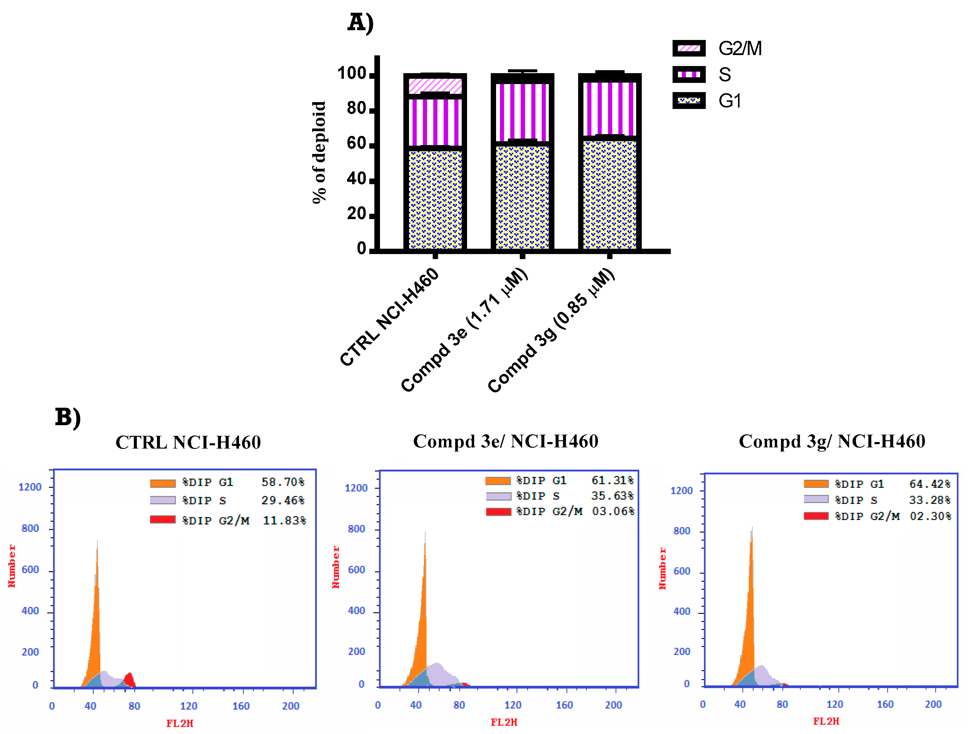
The effect of the most potent compounds 3e and 3g on NCI-H460 lung cell line was studied. Treatment of NCI-H460 cells with benzimidazole molecules 3e and 3g at a concentration equal to their IC50 concentration dose values (IC50 = 1.71 and 0.85, subsequently), resulted in significant alteration in cellular cycle phases [10] (Figure 3). A significant increase in the percentage of cells at G1 phase (61.31 and 64.42, subsequently) compared with untreated control (58.70%) was observed. Furthermore, an increase in the S phase percent (3e: 35.63%; 3g: 33.28%) compared with untreated NCI-H460 cells (29.46%) confirmed that both benzimidazole molecules induce cell growth arrest at both G1 and S phases. Additionally, a concurrent reduction in the percentage of cells at G2/M phase (3e: 3.06%; 3g: 2.30%) compared with untreated lung cells (11.83%) was observed. From the obtained results, it could be concluded that the tested benzimidazole hybrids inhibit the cell proliferation through cellular cycle arrest at both G1 and S phases.

2.2.6. Annexin V/ FITC Apoptosis Staining Assay

Induction of apoptotic cascade in cancerous cells is a crucial determinant in the outcome of therapy. The mode of cellular death induced by compounds 3e and 3g was further studied to declare whether death is due to apoptosis or necrosis. Accordingly, compounds 3e and 3g were selected to be further investigated for their impact on induction of apoptosis in the NCI-H460 lung cancer cell line. The NCI-H460 cells were treated with compounds 3e and 3g for 24 h at a concentration equal to their IC50 value and then analyzed for the apoptosis percentage via FACS detection using Annexin V-FITC and PI dual staining [25]. As illustrated in Figure 4, the tested cancer cells displayed an increase in the percentage of apoptotic cells following exposure treatment with compounds 3e and 3g as observed with untreated control cells. NCI-H460 cells treated with compounds 3e and 3g showed 11.17% and 4.36% of cells in the early apoptotic cells, respectively. Additionally, the percentage of late apoptotic cells in NCI-H460 cells after treatment with compounds 3e and 3g was 16.54% and 20.74%, respectively. Furthermore, the percentages of early and late apoptotic cells in the untreated NCI-H460 cells were 0.46% and 0.28%, respectively. All the obtained results indicate that benzimidazole hybrids 3e and 3g could induce a marked apoptosis in NCI-H460 lung cancer cells.

2.2.7. Caspase Assay

Apoptosis is a programmed cellular death and is a crucial regulator of physiological growth. The activation of apoptosis signal transduction pathways in cancerous cells is the main mechanism of action of the current available chemotherapy or immunotherapy [31]. To investigate whether the cytotoxic activity of benzimidazole [32,33] compounds 3e and 3g against NCI-H460 lung cancer cells is secondary to its ability to activate apoptotic cascade, H-460 cells were treated with compounds 3e and 3g at a concentration equal to their IC50 concentration for 48 h and then subjected to ELISA analysis as shown in Figure 5. The results showed that compounds 3e and 3g caused a significant increase in the level of active caspase-9 compared to untreated H-460 cells. It is worth mentioning that benzimidazole molecules 3e and 3g were 13.31 and 11.73-fold more than untreated NCI-H460 cells. The results suggested that the sample induced apoptosis in NCI-H460 cells via activation of caspase-9.

3. Docking Study

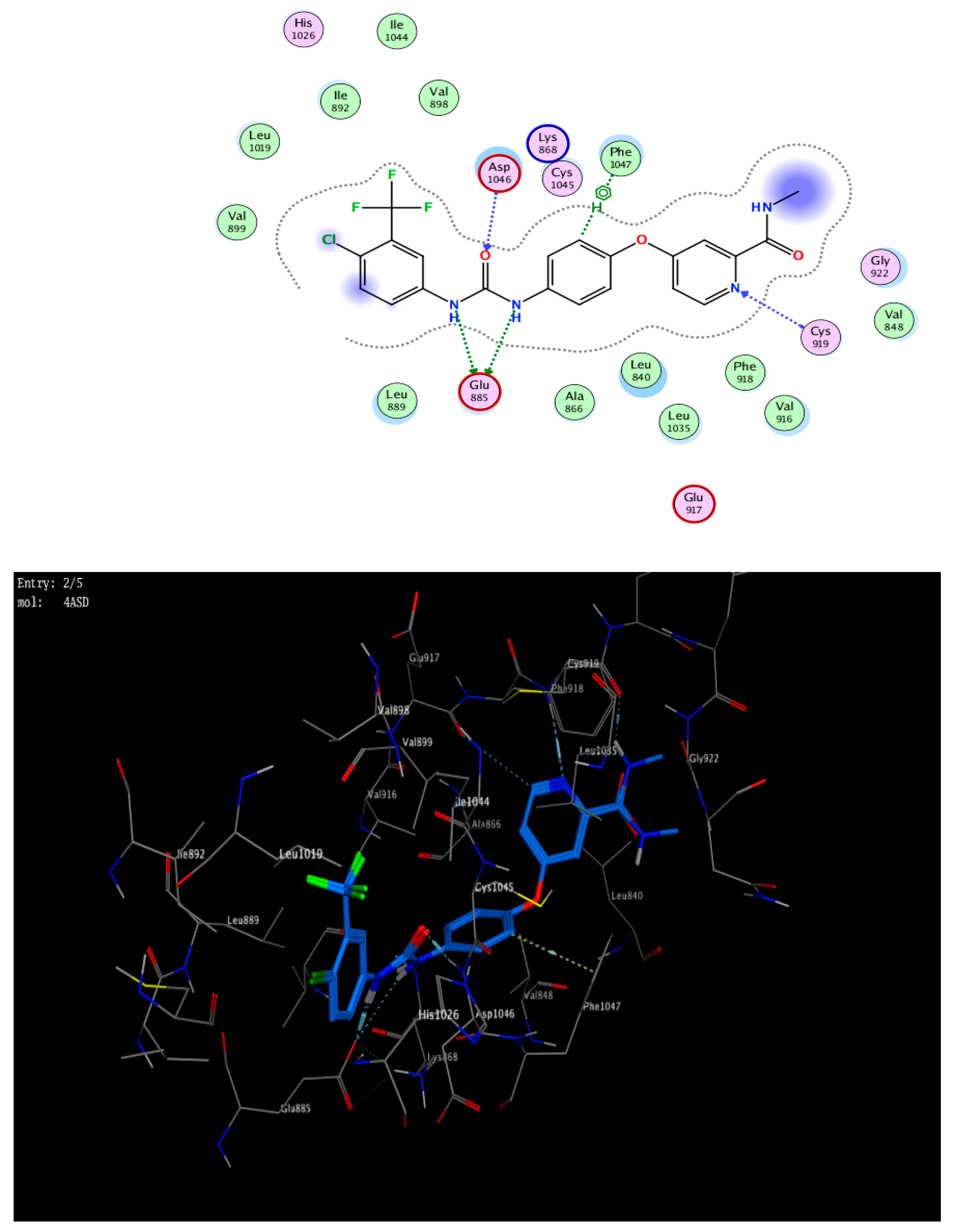
Based on promising antiproliferative activity shown by test compound 3g against cancer lines and inhibitory activity exerted on the VEGFR-2 enzyme, we ran molecular docking simulations to explore the possible mode of inhibition of such a class of compounds. Docking simulations within the active site of VEGFR-2 (PDB ID: 4ASD) revealed an interesting binding profile that could be used as an explanation for their inhibitory activity. Table 2 shows that the docking scores of 3g with the best antiproliferative activity were better than the co-crystallized ligand. Compound 3g was fitted within the VEGFR-2 active site with high affinity (−8.59 Kcal/mol) in comparison with Sorafenib (−8.74 kcal/mol) (Table 2).
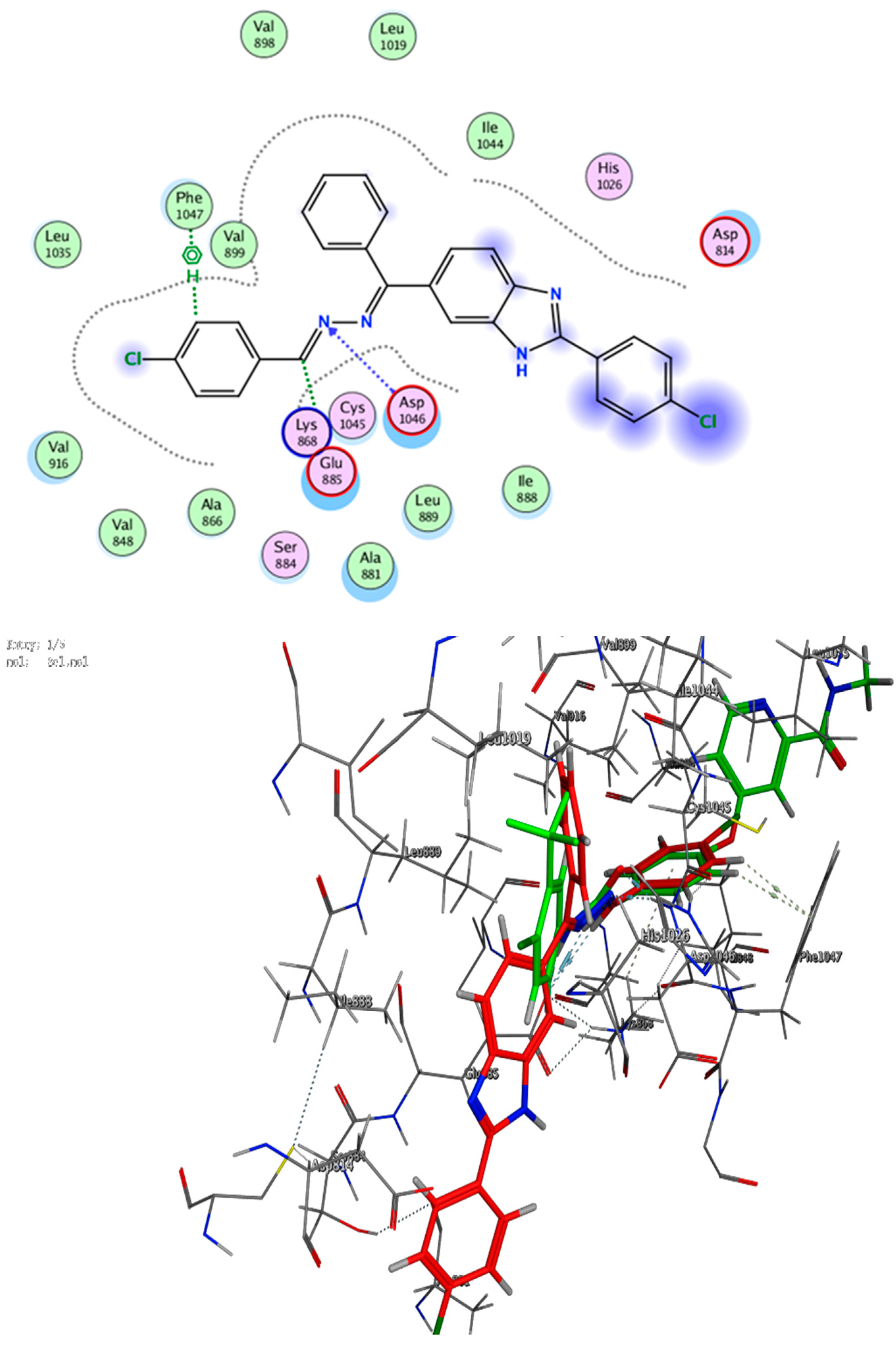
Moreover, Visual inspection of the best docking poses of the test compound revealed a number of binding interactions with key amino acid residues which could help predict its mode of inhibition. Compound 3g showed good binding through an aryl ring attached to the hydrazone moiety via hydrophobic interaction with Phe 1047 amino acid. In addition to another notable connection via H-donor and H-acceptor, the bioactive hydrazone moiety of 3g was also bounded to Glu 885 and Asp 1046 through C24 and N23, respectively (Figure 6). Thus, the molecular docking results suggest that compounds 3g may be bonded to the VEGFR-2 active site with the same manner as reference Sorafenib (Figure 6 and Figure 7).

4. Conclusions

In conclusion, a new series of Schiff base–benzimidazole molecules was designed so as to obtain potential anticancer hybrids. The designed compounds were constructed and structurally confirmed on the basis of 1H-NMR and 13C-NMR spectroscopy as well as elemental microanalysis. All the constructed benzimidazole hybrids were selected for their in vitro anticancer activity according to the NCI-60 single-dose analysis. Compounds 3c, 3e, 3g, and 3o were further selected for NCI five-dose anticancer analysis. Results indicated that benzimidazole molecules 3c, 3e, 3g, and 3o were potent anticancer agents showing broad spectrum anticancer activity against the tested human cancer subpanels. The anticancer activity of tested benzimidazole hybrids is correlated to VEGFR-2 enzyme inhibition where benzimidazoles 3e and 3g were potent VEGFR-2 enzyme inhibitors (86.23 and 89.89% inhibition activity, respectively) as compared with Sorafenib (88.17% inhibition activity). Moreover, the cellular cycle flow cytometry analysis demonstrated that benzimidazole molecules 3e and 3g induce cellular cycle arrest at both G1 and S phases of NCI-H460 cells. In addition, benzimidazole molecule is a strong inducer of apoptosis as elicited by the results of the Annexin staining analysis. Furthermore, ELISA measurements for caspase-9 showed that benzimidazole hybrids 3e and 3g boosted the level of active caspase-9 by 13.31 and 11.73-fold, respectively, compared with untreated NCI-H460 cells. In addition, the binding interaction of 3g was also confirmed through molecular docking studies; the compound displayed complete overlay with, and a similar binding mode and pose to, Sorafenib, a reference VEGFR-2 inhibitor.

5. Experimental Section

5.1. Chemistry

General details: refer to (Supplementary File). Synthesis and analytical data of compounds 1 and 2 were as reported [27].

5.1.1. General Synthesis of Compounds 3ao

To a suspension of benzimidazole hydrazone 2 (1.0 mmol) in ethanol (20 mL), an appropriate aryl ketone or aryl aldehyde derivative (1.0 mmol) with 2 drops of glacial acetic acid was added. The reaction mixture was heated under reflux for 8–10 h. After cooling, the formed precipitate was filtered, washed with diethyl ether, and crystallized from ethanol to give pure compound 3ao (see supplementary material Figures S1–S49).

6-((Z)-((E)-Benzylidenehydrazono)(phenyl)methyl)-2-(4-chlorophenyl)-1H-benzo[d]imidazole (3a)

Yield: 0.34 g (79%); mp: 210–211 °C,1H NMR (400 MHz, DMSO-d6) δ ppm 8.72 (s, 1H, HC=N), 8.23 (d, J = 8.1 Hz, 2H, Ar-H), 8.10 (s, 1H, Ar-H), 7.88 (d, J = 7.4 Hz, 1H, Ar-H), 7.84–7.69 (m, 9H, Ar-H), 7.60 (t, J = 7.6 Hz, 2H, Ar-H), 7.53 (d, J = 7.8 Hz, 2H, Ar-H), 4.92 (s, 1H, NH). 13C NMR (101 MHz, DMSO-d6) δ ppm 163.18, 148.67, 142.48, 141.84, 139.94, 135.60, 134.65, 134.41, 134.34, 133.53, 130.74, 129.37, 128.80, 127.88, 127.67, 12.698, 126.72, 123.83, 119.16, 111.84. Anal. Calcd for C27H19ClN4 (434.92): C, 74.56; H, 4.40; N, 12.88. Found: C, 74.66; H, 4.49; N, 12.75.

2-(4-Chlorophenyl)-5-((Z)-phenyl((E)-(1-phenylethylidene)hydrazono)methyl)-1H-benzo[d]imidazole (3b)

Yield: 0.30 g (68%); mp: 200–202 °C,1H NMR (400 MHz, DMSO-d6) δ ppm 8.23 (t, J = 7.6 Hz, 3H, Ar-H), 8.01 (s, 1H, Ar-H), 7.84–7.72 (m, 10H, Ar-H), 7.61 (d, J = 7.4 Hz, 2H, Ar-H), 7.53 (s, 1H, Ar-H), 5.21 (s, 1H, NH), 1.11 (s, 3H, CH3). 13C NMR (101 MHz, DMSO-d6) δ ppm 162.67, 159.30, 152.07, 149.48, 142.05, 135.55, 134.69, 134.22, 133.24, 129.07, 128.65, 127.82, 126.65, 124.76, 124.12, 121.85, 118.66, 113.13, 112.26, 112.26, 111.13, 21.77. Anal. Calcd for C28H21ClN4 (448.95): C, 74.91; H, 4.71; N, 12.48. Found: C, 75.01; H, 4.50; N, 12.65.

4-((E)-((Z)-((2-(4-Chlorophenyl)-1H-benzo[d]imidazol-6-yl)(phenyl) methylene)hydrazono)methyl)phenol (3c)

Yield: 0.37 g (84%); mp: 178–180 °C, 1H NMR (400 MHz, DMSO-d6) δ ppm 8.56 (s, 1H, HC=N), 8.24 (d, J = 7.8 Hz, 2H, Ar-H), 8.01 (s, 1H, Ar-H), 7.88 (d, J = 7.4 Hz, 1H, Ar-H), 7.84–7.69 (m, 10H, Ar-H), 7.60 (t, J = 6.8 Hz, 2H, Ar-H), 4.41 (s, 1H, NH). 13C NMR (101 MHz, DMSO-d6) δ ppm 164.40, 160.45, 148.24, 148.02, 145.36, 141.42, 135.63, 133.42, 129.70, 129.45, 128.92, 128.44, 128.26, 127.90, 127.67, 124.82, 124.56, 124.29, 124.18, 123.89. Anal. Calcd for C27H19ClN4O (450.92): C, 71.92; H, 4.25; N, 12.43. Found: C, 71.71; H, 4.33; N, 12.45.

4-((E)-1-((Z)-((2-(4-Chlorophenyl)-1H-benzo[d]imidazol-5-yl)(phenyl)methylene)hydrazono)ethyl)phenol (3d)

Yield: 0.38 g (81%); mp: 181–183 °C,1H NMR (400 MHz, DMSO-d6) δ ppm 8.90 (s, 1H, OH), 7.55 (d, J = 8.3 Hz, 2H, Ar-H), 7.47 (d, J = 6.9 Hz, 1H, Ar-H), 7.07–7.00 (m, 7H, Ar-H), 6.82 (s, 2H, Ar-H), 6.77–6.70 (m, 3H, Ar-H), 6.57 (d, J = 8.6 Hz, 1H, Ar-H), 4.82 (s, 1H, NH), 1.80 (s, 3H, CH3). 13C NMR (101 MHz, DMSO-d6) δ ppm 162.26, 155.06, 151.72, 151.01, 146.45, 138.58, 131.61, 129.65, 129.56, 128.95, 128.67, 128.27, 127.02, 126.61, 122.06, 116.61, 116.48, 116.28, 114.86, 114.03, 113.49, 21.52. Anal. Calcd for C28H21ClN4O(464.95): C, 72.33; H, 4.55; N, 12.05. Found: C, 72.11; H, 4.37; N, 11.95.

2-(4-Chlorophenyl)-6-((Z)-((E)-(4-methoxybenzylidene)hydrazono)(phenyl)methyl)-1H-benzo[d]imidazole (3e)

Yield: 0.36 g (82%); mp: 182–184 °C, 1H NMR (400 MHz, DMSO-d6) δ ppm 8.63 (s, 1H, HC=N), 8.23 (d, J = 8.4 Hz, 2H, Ar-H), 8.04 (s, 1H, Ar-H), 7.88 (d, J = 8.5 Hz, 1H, Ar-H), 7.82–7.78 (m, 6H, Ar-H), 7.71 (t, J = 7.4 Hz, 1H, Ar-H), 7.60 (t, J = 7.6 Hz, 3H, Ar-H), 7.05 (d, J = 8.7 Hz, 2H, Ar-H), 4.79 (s, 1H, NH), 3.83 (s, 3H, OCH3). 13C NMR (101 MHz, DMSO-d6) δ ppm 162.16 160.94, 152.52, 152.42, 138.21, 138.06, 137.03, 136.80, 132.93, 131.28, 130.47, 130.03, 130.00, 129.52, 129.01, 126.99, 126.42, 125.98, 117.86, 115.20, 114.87, 55.86. Anal. Calcd for C28H21ClN4O (464.95): C, 72.33; H, 4.55; N, 12.05. Found: C, 72.21; H, 4.48; N, 12.14.

2-(4-Chlorophenyl)-5-((Z)-((E)-(1-(4-methoxyphenyl)ethylidene)hydrazono) (phenyl)methyl)-1H-benzo[d]imidazole (3f)

Yield: 0.36 g (76%); mp: 198–200 °C,1H NMR (400 MHz, DMSO-d6) δ ppm 8.22 (d, J = 7.0 Hz, 3H, HC=N and Ar-H), 7.99 (s, 1H, Ar-H), 7.87–7.64 (m, 11H, Ar-H), 7.60 (t, J = 7.6 Hz, 3H, Ar-H), 3.68 (br s, 1H, NH), 3.49 (s, 3H, OCH3), 2.51 (s, 3H, CH3). 13C NMR (101 MHz, DMSO-d6) δ ppm 163.14, 161.14, 150.77, 147.22, 146.17, 146.17, 136.53, 135.10, 134.24, 133.64, 133.01, 130.90, 130.51, 129.57, 129.46, 129.28, 128.96, 128.81, 128.58, 126.70, 123.32, 56.52, 21.81. Anal. Calcd for C29H23ClN4O (478.97): C, 72.72; H, 4.84; N, 11.70. Found: C, 72.51; H, 4.58; N, 11.45.

6-((Z)-((E)-(4-Chlorobenzylidene)hydrazono)(phenyl)methyl)-2-(4-chlorophenyl)-1H-benzo[d]imidazole (3g)

Yield: 0.38 g (82%); mp: 201–203 °C, 1H NMR (400 MHz, DMSO-d6) δ ppm 8.25 (d, J = 7.6 Hz, 3H, HC=N and Ar-H), 8.01 (s, 1H, Ar-H), 7.96–7.79 (m, 9H, Ar-H), 7.63 (t, J = 7.00 Hz, 1H, Ar-H), 7.61–7.58 (m, 3H, Ar-H), 4.49 (s, 1H, NH). 13C NMR (101 MHz, DMSO-d6) δ ppm 164.40, 148.62, 148.24, 148.02, 145.36, 141.21, 136.10, 135.63, 129.70, 129.45, 129.06, 128.92, 128.85, 128.44, 128.26, 129.90, 124.82, 124.56, 124.18, 124.04, 123.89. Anal. Calcd for C27H18Cl2N4 (469.36): C, 69.09; H, 3.87; N, 11.94. Found: C, 69.11; H, 3.53; N, 11.75.

4-((E)-1-((Z)-((2-(4-Chlorophenyl)-1H-benzo[d]imidazol-5-yl)(phenyl)methylene)hydrazono)ethyl)aniline (3h)

Yield: 0.38 g (81%); mp: 181–183 °C, 1H NMR (400 MHz, DMSO-d6) δ ppm 8.21 (d, J = 7.4 Hz, 2H, Ar-H), 8.01 (s, 1H, Ar-H), 7.84–7.69 (m, 10H, Ar-H), 7.48–7.46 (m, 3H, Ar-H), 7.41 (s, 2H, NH2), 4.43 (s, 1H, NH), 2.52 (s, 3H, CH3). 13C NMR (101 MHz, DMSO-d6) δ ppm 161.09, 151.88, 146.14, 136.00, 134.08, 133.14, 130.64, 129.64, 128.98, 128.71, 128.53, 128.07, 126.76, 126.12, 121.42, 117.87, 117.25, 115.08, 114.13, 113.77, 111.26, 21.59. Anal. Calcd for C28H22ClN5 (463.96): C, 72.48; H, 4.78; N, 15.09. Found: C, 72.19; H, 4.67; N, 15.25.

4-(6-((Z)-((E)-benzylidenehydrazono)(phenyl)methyl)-1H-benzo[d]imidazol-2-yl)phenol (3i)

Yield: 0.34 g (83%); mp: 217–219 °C,1H NMR (400 MHz, DMSO-d6) δ ppm 10.56 (s, 1H, OH), 8.09 (s, 1H, HC=N), 8.06 (d, J = 7.0 Hz, 2H, Ar-H), 8.00 (s, 1H, Ar-H), 7.83–7.78 (m, 6H, Ar-H), 7.74 (t, J = 7.4 Hz, 2H, Ar-H), 7.62–7.59 (m, 3H, Ar-H), 7.07–7.04 (m, 3H, Ar-H), 3.92 (s, 1H, NH). Anal. Calcd for C27H20N4O (416.47): C, 77.87; H, 4.48; N, 13.45. Found: C, 77.66; H, 4.39; N, 13.70.

4-(5-((Z)-Phenyl((E)-(1-phenylethylidene)hydrazono)methyl)-1H-benzo[d]imidazol-2-yl)phenol (3j)

Yield: 0.33 g (78%); mp: 201–203 °C,1H NMR (400 MHz, DMSO-d6) δ ppm 10.55 (s, 1H, OH), 8.09 (d, J = 7.4 Hz, 2H, Ar-H), 8.00 (s, 1H, Ar-H), 7.84–7.77 (m, 6H, Ar-H), 7.70 (t, J = 7.2 Hz, 2H, Ar-H), 7.60–7.58 (m, 3H, Ar-H), 7.03 (d, 3H, Ar-H), 4.21 (s, 1H, NH), 2.54 (s, 3H, CH3). 13C NMR (101 MHz, DMSO-d6) δ ppm 161.86, 153.46, 138.22, 138.02, 137.57, 136.92, 136.62, 132.97, 130.27, 130.02, 129.34, 129.02, 128.70, 128.41, 126.13, 121.05, 117.90, 116.80, 116.60, 115.04, 115.04, 114.60, 21.45. Anal. Calcd for C28H22N4O (430.50): C, 78.12; H, 5.15; N, 13.01. Found: C, 78.01; H, 5.30; N, 12.95.

4-(6-((Z)-((E)-(4-hydroxybenzylidene)hydrazono)(phenyl)methyl)-1H-benzo[d]imidazol-2-yl)phenol (3k)

Yield: 0.35 g (82%); mp: 188–190 °C, 1H NMR (400 MHz, DMSO-d6) δ ppm 1H NMR (400 MHz, DMSO-d6) δ 10.52 (s, 1H, OH), 8.08 (d, J = 8.5 Hz, 3H, HC=N and Ar-H), 7.99 (s, 1H, Ar-H), 7.79 (s, 5H, Ar-H), 7.70 (t, J = 7.2 Hz, 2H, Ar-H), 7.60 (d, J = 7.5 Hz, 3H, Ar-H), 7.04 (d, J = 8.5 Hz, 3H, Ar-H), 4.53 (s, 1H, NH). 13C NMR (101 MHz, DMSO-d6) δ ppm 163.42, 157.10, 156.49, 152.92, 15195, 151.38, 148.83, 135.54, 129.68, 129.02, 128.94, 128.87, 122.08, 112.26, 111.54, 111.50, 110.94. Anal. Calcd for C27H20N4O2 (432.47): C, 74.98; H, 4.66; N, 12.95. Found: C, 74.81; H, 4.63; N, 12.85.

4-((E)-1-((Z)-((2-(4-Hydroxyphenyl)-1H-benzo[d]imidazol-5-yl)(phenyl)methylene)hydrazono)ethyl)phenol (3l)

Yield: 0.32 g (72%); mp: 187–189 °C, 1H NMR (400 MHz, DMSO-d6) δ ppm 8.24 (d, J = 7.4 Hz, 2H, Ar-H), 8.01 (s, 1H, Ar-H), 7.82–7.70 (m, 10H, Ar-H), 7.62–7.60 (m, 3H, Ar-H), 7.43 (s, 1H, OH), 3.79 (s, 1H, NH), 3.19 (s, 3H, CH3). 13C NMR (101 MHz, DMSO-d6) δ ppm 163.18, 158.68, 150.89, 148.67, 144.03, 142.48, 135.60, 134.65, 134.41, 134.34, 133.53, 130.74, 129.79, 129.37, 128.80, 127.88, 127.67, 126.98, 126.91, 126.72, 119.16, 21.80. Anal. Calcd for C28H22N4O2 (446.50): C, 75.32; H, 4.97; N, 12.55. Found: C, 75.21; H, 5.08; N, 12.75.

4-(6-((Z)-((E)-(4-Methoxybenzylidene)hydrazono)(phenyl)methyl)-1H-benzo[d]imidazol-2-yl)phenol (3m)

Yield: 0.36 g (82%); mp: 191–193 °C, 1H NMR (400 MHz, DMSO-d6) δ ppm 10.39 (s, 1H, OH), 8.64 (s, 1H, HC=N), 8.08 (d, J = 7.8 Hz, 2H, Ar-H), 7.96 (s, 1H, Ar-H), 7.83–7.58 (m, 7H, Ar-H), 7.06 (d, J = 7.8 Hz, 2H, Ar-H), 7.00 (d, 4H, Ar-H), 4.20 (s, 1H, NH), 3.83 (s, 3H, OCH3). 13C NMR (101 MHz, DMSO-d6) δ ppm 163.93, 161.43, 152.57, 148.09, 147.61, 136.24, 135.42, 133.17, 132.33, 131.90, 130.72, 129.58, 128.79, 128.21, 127.17, 126.83, 123.25, 115.69, 114.44, 114.15, 55.91. Anal. Calcd for C28H22N4O2 (446.50): C, 75.32; H, 4.97; N, 12.55. Found: C, 75.19; H, 4.73; N, 12.80.

4-(5-((Z)-((E)-(1-(4-Methoxyphenyl)ethylidene)hydrazono)(phenyl)methyl)-1H-benzo[d]imidazol-2-yl)phenol (3n)

Yield: 0.36 g (79%); mp: 167–169 °C,1H NMR (400 MHz, DMSO-d6) δ ppm 10.60 (s, 1H, OH), 8.08 (d, J = 7.0 Hz, 3H, Ar-H), 8.00 (s, 1H, Ar-H), 7.85–7.73 (m, 7H, Ar-H), 7.60 (t, 3H, Ar-H), 7.05 (d, 3H, Ar-H), 4.33 (s, 1H, NH), 3.96 (s, 3H, OCH3), 2.51 (s, 3H, CH3).13C NMR (101 MHz, DMSO-d6) δ ppm 162.17, 160.94, 152.42, 153.40, 138.06, 137.40, 137.03, 132.93, 130.47, 130.03, 130.00, 129.52, 129.01, 126.99, 125.98, 118.00, 117.80, 115.20, 114.87, 55.86, 56.49, 21.59. Anal. Calcd for C29H24N4O2 (460.53): C, 75.63; H, 5.25; N, 12.17. Found: C, 75.61; H, 5.15; N, 12.25.

4-(6-((Z)-((E)-(4-chlorobenzylidene)hydrazono)(phenyl)methyl)-1H-benzo[d]imidazol-2-yl)phenol (3o)

Yield: 0.35 g (78%); mp: 159–161 °C, 1H NMR (400 MHz, DMSO-d6) δ ppm 10.33 (s, 1H, OH), 8.07 (d, J = 7.8 Hz, 3H, HC=N and Ar-H), 7.95 (s, 1H, Ar-H), 7.90 (d, J = 7.0 Hz, 1H, Ar-H), 7.79–7.57 (m, 10H, Ar-H), 7.00 (d, J = 7.8 Hz, 3H, Ar-H), 4.20 (s, 1H, NH). 13C NMR (101 MHz, DMSO-d6) δ ppm 164.21, 156.38, 152.78, 148.09, 147.17, 146.59, 135.95, 135.60, 134.93, 133.80, 132.80, 129.99, 129.69, 129.42, 129.19, 129.09, 128.90, 128.81, 122.95, 117.89, 113.00. Anal. Calcd for C27H19 ClN4O (450.92): C, 71.92; H, 4.25; N, 12.43. Found: C, 71.99; H, 4.53; N, 12.49.

5.2. Biological Evaluation

5.2.1. NCI Screening Assay

As mentioned, the methodology of the NCI procedure for the primary anticancer assay was detailed on their site (http://www.dtp.nci.nih.gov, accessed on 1 January 2020). However, briefly, the protocol was performed in full panels of 60 human tumor cell lines derived from 9 different neoplastic diseases. NCI-60 testing is performed in two parts: first, a single concentration is tested in all 60 cell lines at a single dose of 10−5 molar or 15 µg/mL in accordance with the protocol of the Drug Evaluation Branch, National Cancer Institute, 37 Convent Dr, Bethesda, MD 20814, USA. If the results obtained meet the selection criteria, then, second, the compound is tested again in all 60 cell lines in 5 × 10-fold dilution with the top dose being 10−4 molar or 150 µg/mL.

5.2.2. MTT Assay for Cell Viability

To investigate the effect of the newly synthesized compounds on lung cancer cells, an MTT assay was performed against A549 and NCI-H460 cell lines (see Appendix A).

5.2.3. VEGFR Inhibitory Assay

A VEGFR-2 assay was performed by the established reported method using a VEGFR-2 (KDR) Kinase Assay Kit (Catalog # 40325, BPS Bioscience, Biotechnology company, San Diego, CA, USA) for selected synthetic compounds 3e and 3g. Details are summarized in Appendix A.

5.2.4. Cell Cycle Analysis and Apoptotic Assay

Cell Apoptosis and Apoptotic Detection

Studies on the effect of compound 3e and 3g on cell cycle development and induction of apoptosis in the NCI-H460 lung cell wetr done using the fluorescent Annexin V-FITC/ PI detection kit (BioVision EZCellTM Cell Cycle Analysis Kit Catalog #K920, Milpitas Blvd., Milpitas, CA 95035 USA) by flow cytometry assay. For more details, see Appendix A.

5.2.5. Activation of Caspases

For more a deeper and more systematic investigation on cell apoptosis, the effect of compounds 3e and 3g on caspase-9 was evaluated compared to control. Details are summarized in Appendix A.

5.2.6. Docking Study

Molecular Operating Environment (MOE) version 2021 is used to perform the molecular modeling study. The structure of 3g was built in the MOE database (see Appendix A).

Supplementary Materials

The following supporting information can be downloaded at: https://www.mdpi.com/article/10.3390/molecules28020481/s1, Figure S1: 1H-NMR spectrum of compound 3a; Figure S2: 13C-NMR spectrum of compound 3a; Figure S3: 1H-NMR spectrum of compound 3b; Figure S4: 13C-NMR spectrum of compound 3b; Figure S5: 1H-NMR spectrum of compound 3c; Figure S6: 13C-NMR spectrum of compound 3c; Figure S7: 1H-NMR spectrum of compound 3d; Figure S8: 13C-NMR spectrum of compound 3d; Figure S9: 1H-NMR spectrum of compound 3e; Figure S10: 13C-NMR spectrum of compound 3e; Figure S11: 1H-NMR spectrum of compound 3f; Figure S12: 13C-NMR spectrum of compound 3f; Figure S13: 1H-NMR spectrum of compound 3g; Figure S14: 13C-NMR spectrum of compound 3g; Figure S15: 1H-NMR spectrum of compound 3h; Figure S16: 13C-NMR spectrum of compound 3h; Figure S17: 1H-NMR spectrum of compound 3i; Figure S18: 13C-NMR spectrum of compound 3i; Figure S19: 1H-NMR spectrum of compound 3j; Figure S20: 13C-NMR spectrum of compound 3j; Figure S21: 1H-NMR spectrum of compound 3k; Figure S22: 13C-NMR spectrum of compound 3k; Figure S23: 1H-NMR spectrum of compound 3l; Figure S24: 13C-NMR spectrum of compound 3l; Figure S25: 1H-NMR spectrum of compound 3m; Figure S26: 13C-NMR spectrum of compound 3m; Figure S27: 1H-NMR spectrum of compound 3n; Figure S28: 13C-NMR spectrum of compound 3n; Figure S29: 1H-NMR spectrum of compound 3o; Figure S30: 13C-NMR spectrum of compound 3o; Figure S31: Results of primary in vitro one-dose anticancer assay of compound 3a; Figure S32: Results of primary in vitro one-dose anticancer assay of compound 3b; Figure S33: Results of primary in vitro one-dose anticancer assay of compound 3c; Figure S34: Results of primary in vitro one-dose anticancer assay of compound 3d; Figure S35: Results of primary in vitro one-dose anticancer assay of compound 3e; Figure S36: Results of primary in vitro one-dose anticancer assay of compound 3f; Figure S37: Results of primary in vitro one-dose anticancer assay of compound 3g; Figure S38: Results of primary in vitro one-dose anticancer assay of compound 3h; Figure S39: Results of primary in vitro one-dose anticancer assay of compound 3i; Figure S40: Results of primary in vitro one-dose anticancer assay of compound 3j; Figure S41: Results of primary in vitro one-dose anticancer assay of compound 3k; Figure S42: Results of primary in vitro one-dose anticancer assay of compound 3l; Figure S43: Results of primary in vitro one-dose anticancer assay of compound 3m; Figure S44: Results of primary in vitro one-dose anticancer assay of compound 3n; Figure S45: Results of primary in vitro one-dose anticancer assay of compound 3o; Figure S46: Dose response curve of the in vitro NCI five dose anticancer assay of compound 3c; Figure S47: Dose response curve of the in vitro NCI five dose anticancer assay of compound 3e; Figure S48: Dose response curve of the in vitro NCI five dose anticancer assay of compound 3g; Figure S49: Dose response curve of the in vitro NCI five dose anticancer assay of compound 3o.

Author Contributions

Data curation, M.A.I.E., M.G.F.W., I.Z. and M.M.M.; Formal analysis, M.A.I.E., M.G.F.W., I.Z. and M.M.M.; Funding acquisition, H.M.A.E.-L., T.M.A.I., M.M.K. and M.G.; Investigation, M.A.I.E., M.G.F.W., I.Z. and M.M.M.; Methodology, M.A.I.E., M.G.F.W., I.Z. and M.M.M.; Project administration, M.A.I.E., M.G. and M.G.F.W.; Resources, M.A.I.E. and M.M.M.; Software, M.M.M.; Validation, M.A.I.E., M.G.F.W. and M.M.M.; Visualization, M.A.I.E., I.Z. and M.M.M.; Writing—original draft, H.M.A.E.-L., M.A.I.E., T.M.A.I., M.M.K., M.G., M.G.F.W., I.Z. and M.M.M.; Writing—review & editing, H.M.A.E.-L., M.A.I.E., M.M.K., M.G.F.W., I.Z. and M.M.M. All authors have read and agreed to the published version of the manuscript.

Funding

This research was funded by Deanship of Scientific Research, King Faisal University, Saudi Arabia, through GRANT No. 2084.

Institutional Review Board Statement

Not applicable.

Informed Consent Statement

Not applicable.

Data Availability Statement

The raw/processed data generated in this work are available upon request from the corresponding author.

Acknowledgments

This work was supported by the Deanship of Scientific Research, Vice Presidency for Graduate Studies and Scientific Research, King Faisal University, Saudi Arabia [GRANT2084], through its KFU Research Summer initiative. The authors extend their appreciation to the faculty of science for funding this work through project No. FC-2201201.

Conflicts of Interest

The authors declare no conflict of interest. The authors declare that they have no known competing interest.

Sample Availability

Samples of the compounds are available from the authors.

Appendix A

Appendix A.1. Biological Studies

Appendix A.1.1. Cytotoxic Activity Evaluation against Lung Cancer Cell Lines

To determine IC50, benzimidazole molecules 3c, 3e, 3g and 3o were further analyzed using the standard MTT colorimetric assay against lung cancer A549 and NCI-H460 cell lines. Cells at density of 1 × 10⁴ were seeded in a 96-well plate at 37 °C for 24 h under 5% CO2. After incubation, the cells were treated with different concentrations of the test compounds 3c, 3e, 3g and 3o and incubated for 24 h, then 20 μL of MTT solution at 5 mg/mL was applied and incubated for 4 h at 37 °C. Dimethyl sulphoxide (DMSO) in volume of 100 μL was added to each well to dissolve the purple formazan that had formed. The color intensity of the formazan product, which represents the growth condition of the cells, is quantified by using an ELISA plate reader (EXL 800, USA) at 570 nm absorbance. The experimental conditions were carried out with at least three replicates, and the experiments were repeated at least three times.

Appendix A.1.2. VEGFR-2 Inhibition Assay

Benzimidazole compounds 3e and 3g and Sorafenib were evaluated for their VEGFR-2 inhibitory activity according to manufacturer’s instructions using # VEGFR-2 (KDR) Kinase Assay Kit Catalog # 40325 (BPS Bioscience).

Appendix A.1.3. Cell Cycle Analysis of Compounds 3e and 3g

Cell cycle analysis in NCI-H460 cells was investigated using fluorescent Annexin V-FITC/PI detection kit (BioVision EZCellTM Cell Cycle Analysis Kit Catalog #K920) by flow cytometry assay. NCI-H460 cells at a density of 2 × 10⁵ per well were harvested and washed twice in PBS. After that, the cells were incubated at 37 °C and 5% CO2. The medium was incubated with the tested compounds 3e and 3g at their IC50 (μM) for 24 h, washed twice in PBS, fixed with 70% ethanol, rinsed again with PBS. Afterward, medium was stained with DNA fluorochrome PI for 15 min at 37 °C. The samples were immediately analyzed using Facs Calibur flow cytometer (Becton and Dickinson, Heidelberg, Germany).

Appendix A.1.4. Apoptosis Assay for Compounds 3e and 3g

Apoptosis in NCI-H460 cells was investigated using fluorescent Annexin V-FITC/ PI detection kit (BioVision Annexin V-FITC Apoptosis Detection Kit, Catalog #: K101) by flow cytometry assay. NCI-H460 cells at a density of 2 × 10⁵ per well were treated with compounds 3e and 3g at their IC50 (μM) for 24 h, then the cells were harvested and stained with Annexin V-FITC/PI dye for 15 min in the dark at 37 °C. The samples were immediately analyzed using FACS Calibur flow cytometer (Becton and Dickinson, Heidelberg, Germany).

Appendix A.1.5. Caspase 9 Assay for Compounds 3e and 3g

To determine the effect of the synthesized benzimidazole hybrids 3e and 3g on apoptosis, the active caspase 9 level was measured using ELISA analysis according to manufacturer’s instructions. Briefly, NCI-H460 cells at a density of 2 × 10⁴ per well were treated with compounds 3e and 3g at their IC50 (μM) for 24 h, then the cells were lysed with cell extraction buffer. This lysate was diluted in standard diluent buffer over the range of the assay. The optical density of each well was determined within 30 min using a microplate plate reader set at 450 nm to determine the human active caspase 9 level.

Appendix A.1.6. Molecular Modeling Study

Docking studies, calculations, and investigations was conducting using Molecular Operating Environment MOE version MOE (1 January 2021 Chemical Computing Group, Canada Software) to perform molecular modeling study. Structure of 3g was built in database of MOE. The X-ray crystal structure of Sorafenib bound to the VEGFR-2 enzyme binding site (PDB ID: 4ASD) active site was acquired from protein data bank at research collaboration for Structural Bioinformatics (RSCB) protein database [PDB].

References

  1. Aly, A.A.; Ramadan, M.; Abuo-Rahma, G.E.-D.A.; Elshaier, Y.A.; Elbastawesy, M.A.; Brown, A.B.; Bräse, S. Quinolones as prospective drugs: Their syntheses and biological applications. In Advances in Heterocyclic Chemistry; Academic Press: Cambridge, MA, USA, 2020; pp. 147–196. [Google Scholar]
  2. Tiglani, D.; Salahuddin; Mazumder, A.; Yar, M.S.; Kumar, R.; Ahsan, M.J. Benzimidazole-quinoline hybrid scaffold as promising pharmacological Agents: A review. Polycycl. Aromat. Compd. 2022, 42, 5044–5066. [Google Scholar] [CrossRef]
  3. Brishty, S.R.; Hossain, M.; Khandaker, M.U.; Faruque, M.R.I.; Osman, H.; Rahman, S. A comprehensive account on recent progress in pharmacological activities of benzimidazole derivatives. Front. Pharmacol. 2021, 1, 2863. [Google Scholar] [CrossRef] [PubMed]
  4. Faheem, M.; Rathaur, A.; Pandey, A.; Kumar Singh, V.; Tiwari, A.K. A review on the modern synthetic approach of benzimidazole candidate. ChemistrySelect 2020, 5, 3981–3994. [Google Scholar] [CrossRef]
  5. Choudhary, S.; Arora, M.; Verma, H.; Kumar, M.; Silakari, O. Benzimidazole based hybrids against complex diseases: A catalogue of the SAR profile. Eur. J. Pharmacol. 2021, 899, 174027. [Google Scholar] [CrossRef]
  6. Satija, G.; Sharma, B.; Madan, A.; Iqubal, A.; Shaquiquzzaman, M.; Akhter, M.; Parvez, S.; Khan, M.A.; Alam, M.M. Benzimidazole based derivatives as anticancer agents: Structure activity relationship analysis for various targets. J. Heterocycl. Chem. 2022, 59, 22–66. [Google Scholar] [CrossRef]
  7. Khan, S.; Alhumaydhi, F.A.; Ibrahim, M.M.; Alqahtani, A.; Alshamrani, M.; Alruwaili, A.S.; Hassanian, A.A.; Khan, S. Recent advances and therapeutic journey of Schiff base complexes with selected metals (Pt, Pd, Ag, Au) as potent anticancer agents: A review. Anti-Cancer Agents Med. Chem. 2022, 22, 3086–3096. [Google Scholar] [CrossRef]
  8. Korkmaz, A.; Bursal, E. Benzothiazole sulfonate derivatives bearing azomethine: Synthesis, characterization, enzyme inhibition, and molecular docking study. J. Mol. Struct. 2022, 1257, 132641. [Google Scholar] [CrossRef]
  9. Matela, G. Schiff bases and complexes: A review on anti-cancer activity. Anti-Cancer Agents Med. Chem. 2020, 20, 1908–1917. [Google Scholar] [CrossRef]
  10. Elbastawesy, M.A.; Ramadan, M.; El-Shaier, Y.A.; Aly, A.A.; Abuo-Rahma, G.E.-D.A. Arylidenes of Quinolin-2-one scaffold as Erlotinib analogues with activities against leukemia through inhibition of EGFR TK/STAT-3 pathways. Bioorg. Chem. 2020, 96, 103628. [Google Scholar] [CrossRef]
  11. El-Khouly, O.A.; Henen, M.A.; El-Sayed, M.A.-A.; El-Messery, S.M. Design, synthesis and computational study of new benzofuran hybrids as dual PI3K/VEGFR2 inhibitors targeting cancer. Sci. Rep. 2022, 12, 17104. [Google Scholar] [CrossRef]
  12. Lugano, R.; Ramachandran, M.; Dimberg, A. Tumor angiogenesis: Causes, consequences, challenges and opportunities. Cell. Mol. Life Sci. 2020, 77, 1745–1770. [Google Scholar] [CrossRef] [PubMed] [Green Version]
  13. Chu, H.; Wang, Y. Therapeutic angiogenesis: Controlled delivery of angiogenic factors. Ther. Deliv. 2012, 3, 693–714. [Google Scholar] [CrossRef] [PubMed] [Green Version]
  14. Teleanu, R.I.; Chircov, C.; Grumezescu, A.M.; Teleanu, D.M. Tumor angiogenesis and anti-angiogenic strategies for cancer treatment. J. Clin. Med. 2019, 9, 84. [Google Scholar] [CrossRef] [PubMed] [Green Version]
  15. Shibuya, M. Tyrosine kinase receptor Flt/VEGFR family: Its characterization related to angiogenesis and cancer. Genes Cancer 2010, 1, 1119–1123. [Google Scholar] [CrossRef] [Green Version]
  16. Elbastawesy, M.A.; Aly, A.A.; Ramadan, M.; Elshaier, Y.A.; Youssif, B.G.; Brown, A.B.; Abuo-Rahma, G.E.-D.A. Novel pyrazoloquinolin-2-ones: Design, synthesis, docking studies, and biological evaluation as antiproliferative EGFR-TK inhibitors. Bioorg. Chem. 2019, 90, 103045. [Google Scholar] [CrossRef]
  17. S Sharma, P.; Sharma, R.; Tyagi, T. VEGF/VEGFR pathway inhibitors as anti-angiogenic agents: Present and future. Curr. Cancer Drug Targets 2011, 11, 624–653. [Google Scholar] [CrossRef]
  18. Poliaková, M.; Aebersold, D.M.; Zimmer, Y.; Medová, M. The relevance of tyrosine kinase inhibitors for global metabolic pathways in cancer. Mol. Cancer 2018, 17, 27. [Google Scholar] [CrossRef] [Green Version]
  19. Liu, Y.; Li, Y.; Wang, Y.; Lin, C.; Zhang, D.; Chen, J.; Ouyang, L.; Wu, F.; Zhang, J.; Chen, L. Recent progress on vascular endothelial growth factor receptor inhibitors with dual targeting capabilities for tumor therapy. J. Hematol. Oncol. 2022, 15, 89. [Google Scholar] [CrossRef]
  20. Soltan, O.M.; Shoman, M.E.; Abdel-Aziz, S.A.; Narumi, A.; Konno, H.; Abdel-Aziz, M. Molecular hybrids: A five-year survey on structures of multiple targeted hybrids of protein kinase inhibitors for cancer therapy. Eur. J. Med. Chem. 2021, 225, 113768. [Google Scholar] [CrossRef]
  21. Obeng, E. Apoptosis (programmed cell death) and its signals—A review. Braz. J. Biol. 2020, 81, 1133–1143. [Google Scholar] [CrossRef]
  22. Jan, R. Understanding apoptosis and apoptotic pathways targeted cancer therapeutics. Adv. Pharm. Bull. 2019, 9, 205. [Google Scholar] [CrossRef] [PubMed] [Green Version]
  23. Carneiro, B.A.; El-Deiry, W.S. Targeting apoptosis in cancer therapy. Nat. Rev. Clin. Oncol. 2020, 17, 395–417. [Google Scholar] [CrossRef] [PubMed]
  24. Abd El-Meguid, E.A.; El-Deen, E.M.M.; Nael, M.A.; Anwar, M.M. Novel benzimidazole derivatives as anti-cervical cancer agents of potential multi-targeting kinase inhibitory activity. Arab. J. Chem. 2020, 13, 9179–9195. [Google Scholar] [CrossRef]
  25. Zaki, I.; Moustafa, A.M.; Beshay, B.Y.; Masoud, R.E.; Elbastawesy, M.A.; Abourehab, M.A.; Zakaria, M.Y. Design and synthesis of new trimethoxylphenyl-linked combretastatin analogues loaded on diamond nanoparticles as a panel for ameliorated solubility and antiproliferative activity. J. Enzym. Inhib. Med. Chem. 2022, 37, 2679–2701. [Google Scholar] [CrossRef]
  26. El-Sheref, E.M.; Elbastawesy, M.A.; Brown, A.B.; Shawky, A.M.; Gomaa, H.A.; Bräse, S.; Youssif, B.G. Design and Synthesis of (2-oxo-1,2-Dihydroquinolin-4-yl)-1,2,3-triazole Derivatives via Click Reaction: Potential Apoptotic Antiproliferative Agents. Molecules 2021, 26, 6798. [Google Scholar] [CrossRef]
  27. Morcoss, M.M.; Abdelhafez, E.S.; Abdel-Rahman, H.M.; Abdel-Aziz, M.; El-Ella, A.; Dalal, A. Novel Benzimidazole/Hydrazone Derivatives as Promising Anticancer Lead Compounds: Design, Synthesis, and Molecular Docking Study. J. Adv. Biomed. Pharm. Sci. 2020, 3, 45–52. [Google Scholar] [CrossRef]
  28. Zha, G.-F.; Qin, H.-L.; Youssif, B.G.; Amjad, M.W.; Raja, M.A.G.; Abdelazeem, A.H.; Bukhari, S.N.A. Discovery of potential anticancer multi-targeted ligustrazine based cyclohexanone and oxime analogs overcoming the cancer multidrug resistance. Eur. J. Med. Chem. 2017, 135, 34–48. [Google Scholar] [CrossRef] [PubMed]
  29. Ceci, C.; Atzori, M.G.; Lacal, P.M.; Graziani, G. Role of VEGFs/VEGFR-1 signaling and its inhibition in modulating tumor invasion: Experimental evidence in different metastatic cancer models. Int. J. Mol. Sci. 2020, 21, 1388. [Google Scholar] [CrossRef] [Green Version]
  30. Ahmed, M.F.; Santali, E.Y.; El-Haggar, R. Novel piperazine–chalcone hybrids and related pyrazoline analogues targeting VEGFR-2 kinase; design, synthesis, molecular docking studies, and anticancer evaluation. J. Enzym. Inhib. Med. Chem. 2021, 36, 308–319. [Google Scholar] [CrossRef]
  31. Avrutsky, M.I.; Troy, C.M. Caspase-9: A multimodal therapeutic target with diverse cellular expression in human disease. Front. Pharmacol. 2021, 12, 1728. [Google Scholar] [CrossRef]
  32. Morcoss, M.M.; El Shimaa, M.; Ibrahem, R.A.; Abdel-Rahman, H.M.; Abdel-Aziz, M.; Abou El-Ella, D.A. Design, synthesis, mechanistic studies and in silico ADME predictions of benzimidazole derivatives as novel antifungal agents. Bioorg. Chem. 2020, 101, 103956. [Google Scholar] [CrossRef] [PubMed]
  33. Mostafa, A.S.; Gomaa, R.M.; Elmorsy, M.A. Design and synthesis of 2-phenyl benzimidazole derivatives as VEGFR-2 inhibitors with anti-breast cancer activity. Chem. Biol. Drug Des. 2019, 93, 454–463. [Google Scholar] [CrossRef] [PubMed]
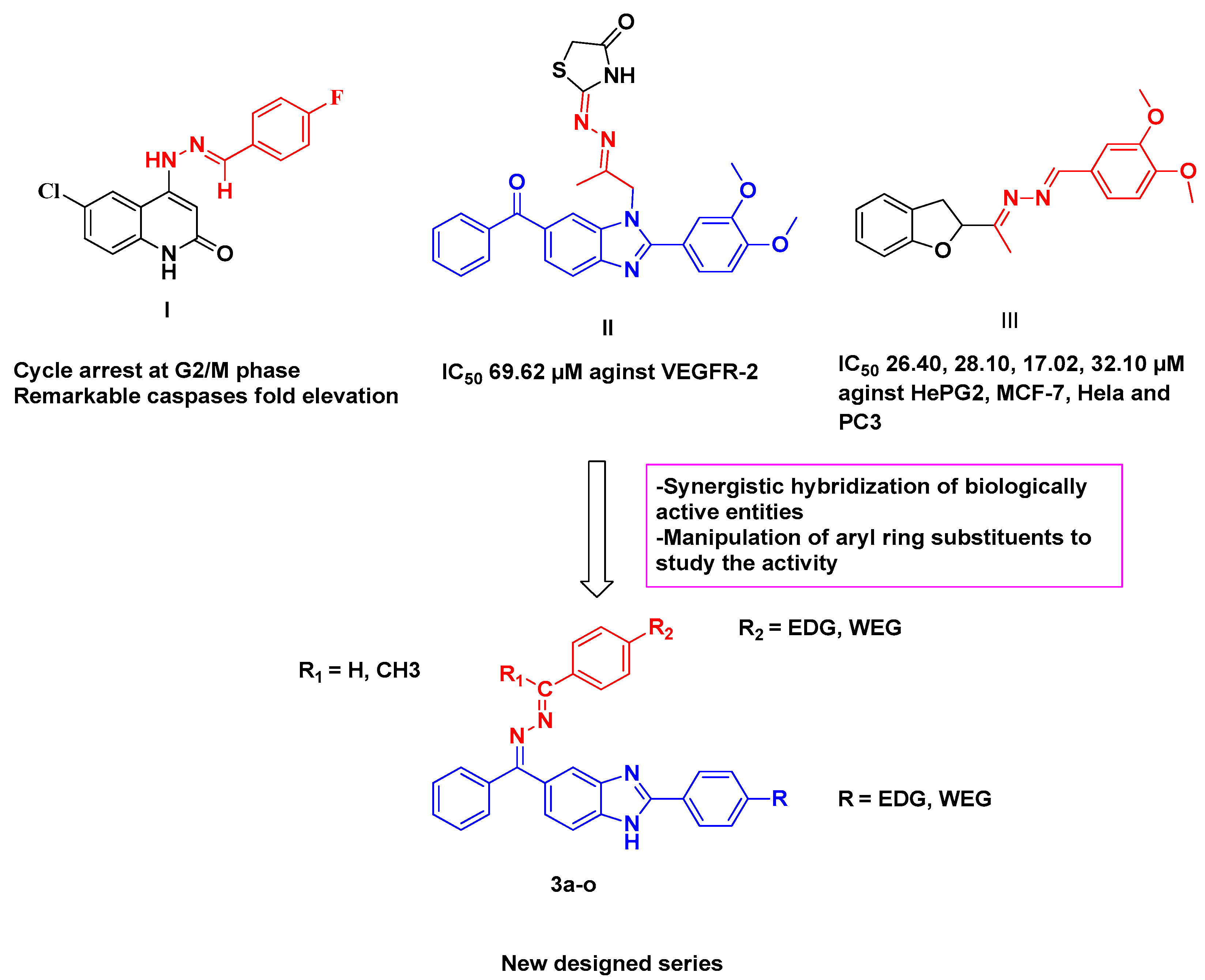
Figure 1. Rational design of the new hybrid target compounds.
Molecules 28 00481 g001
Scheme 1. Reagent and condition: (i) DMF, Na2S2O5, reflux 24 h; (ii) NH2.NH2.H2O, reflux 12 h; (iii) AcOH, ethanol, reflux 8–10 h.
Molecules 28 00481 sch001
Figure 2. Graphical representation of the in vitro VEGFR-2 kinase inhibition activity (%) in NCI-H460 cells treated with benzimidazole molecules 3e and 3g and Sorafenib at their IC50 concentration (µM).
Molecules 28 00481 g002
Figure 3. (A) FACS analysis of cell cycle distribution percentage of NCI-H460 cells after treatment with compounds 3e and 3g at their IC50 (μM) for 24 h relative to untreated control. (B) Graphical representation of FACS analysis of cell cycle distribution percentage of NCI-H460 cells after treatment with compounds 3e and 3g at their IC50 (μM) for 24 h relative to untreated control.
Molecules 28 00481 g003
Figure 4. (A) Graphical representation of effect of compounds 3e and 3g on induction of apoptosis in NCI-H460 cells after 24 h. (B) Effect of compounds 3e and 3g on induction of apoptosis in NCI-H460 cells after 24 h.
Molecules 28 00481 g004
Figure 5. ELISA results of active caspase-9 in NCI-H460 cells treated with benzimidazole molecules 3e and 3g at their IC50 concentration (μM) after 24 h.
Molecules 28 00481 g005
Figure 6. Two-dimensional and three-dimensional models of the binding of 3g with the active site of VEGFR-2.
Molecules 28 00481 g006
Figure 7. Two-dimensional and three-dimensional models of the binding of Sorafenib with the active site of VEGFR-2.
Molecules 28 00481 g007
Table 1. Cytotoxicity screening of the tested benzimidazole molecules 3c, 3e, 3g, and 3o against A549 and NCI-H460 lung cancer cell lines. Data expressed as the mean ± SD.
Comp NoIC50 Value (μM)
A549NCI-H460
3c4.95 ± 0.613.07 ± 0.42
3e3.58 ± 0.531.71 ± 0.17
3g1.88 ± 0.350.85 ± 0.24
3o5.98 ± 0.555.22 ± 0.42
Sorafenib4.84 ± 0.233.49 ± 0.18
Table 2. Molecular docking data for compound 3g and Sorafenib in VEGFR-2 active site (PDB ID: 4ASD).
Comp. No.VEGFR-2
Affinity
kcal/mol
Distance (in Ao) from
Main Residue
Amino Acids ResidueFunctional
Group
Type of Interactions
3g−8.593.38Glu 885C24H-donor
3.09ASP 1046N23H-acceptor
3.72PHE 10476-ringpi-H
Sorafenib−8.742.91Glu 885N12H-donor
3.45Glu 885N14H-donor
2.90Asp 1046O15H-acceptor
3.25Cys 919N26H-acceptor
3.67PHE 10475-ringpi-H
Disclaimer/Publisher’s Note: The statements, opinions and data contained in all publications are solely those of the individual author(s) and contributor(s) and not of MDPI and/or the editor(s). MDPI and/or the editor(s) disclaim responsibility for any injury to people or property resulting from any ideas, methods, instructions or products referred to in the content.