Abstract
Hazardous dyes in industrial wastewater are an internationally recognized issue for community health. Nanoparticles synthesized through green protocols are a fascinating research field with numerous applications. The current study mainly aimed to investigate the degradation of Congo red (CR) dye under UV light in the presence of H2O2 and the photocatalytic activity of copper oxide nanoparticles (CuONPs). For CuONP formation, Citrus maxima extract contains a high number of phytochemical constituents. The size of CuONPs ranges between 25 and 90 nm. The photocatalytic activity of CuONPs with the addition of H2O2 was observed and analyzed under UV light to eliminate CR dye. The UV light caused the decomposition of H2O2, which produced ·OH radicals. The results revealed a significant increment in dye degradation during the presence of H2O2. The effect of concentration on the degradation of the CR dye was also studied. The degradation pathway of organic pollutants was reputable from the hydroxy radical medicated degradation of CR. Advanced Oxidation Treatment depends on the in situ production of reactive ·OH species and is presented as the most effective procedure for decontamination. The biological activity of CuONPs was evaluated against Escherichia coli Bacillus subtillis, Staphylococcus aureus, Shigella flexenari, Acinetobacter Klebsiella pneumonia, Salmonella typhi and Micrococcus luteus. The newly synthesised nanomaterials showed strong inhibition activity against Escherichia coli (45%), Bacillus subtilis (42%) and Acinetobacter species (25%). The activity of CuONPs was also investigated against different fungus species such as: Aspergillus flavus, A. niger, Candida glabrata, T. longifusus, M. Canis, C. glabrata and showed a good inhibition zone against Candida glabrata 75%, Aspergillus flavus 68%, T. longifusus 60%. The materials showed good activity against C. glaberata, A. flavus and T. longifusus. Furthermore, CuONPs were tested for antioxidant properties using 2, 2 diphenyl-1-picrylhydrazyl) (DPPH).
1. Introduction
Industrial effluents from factories comprise significant amounts of pigments and other organic pollutants that are highly stable; however, they can cause severe health issues (kidney disorders, cancer, etc.) and are challenging to remediate [1,2,3,4,5,6,7]. Nanoparticles (NPs) have gained significant attention over time due to their unique properties and high surface-to-volume ratios [1,8,9]. Nanotechnology is an ever-growing discipline dedicated to synthesizing and promoting the use of NPs [10,11,12,13,14]. The antimicrobial properties of NPs such as chitosan, AgNPs, photocatalytic TiO2 and other carbon nanotubes (CNT) have already been reported [15,16,17]. Nanotechnology is widely used for developing effective catalysts with high biochemical activity to control microbial infections and diseases [18,19,20,21]. Nanotechnology is a multidisciplinary field of engineering [22,23,24] medicine with a wide range of applications for molecular imaging, diagnosis and targeted therapy [25]. This new field is making a substantial contribution to medical science as an innovative and useful tool [24,25,26,27]. The use of nanomaterials for diagnostic and screening purposes [27,28,29,30,31], the development of artificial proteins, receptors, DNA and protein sequencing with nano-pores and nano-sprays [32,33,34], novel drug delivery systems (DDS), gene therapy and tissue-engineering applications [13] are all examples of nanomedicine. Water pollution by hazardous chemicals is threatening human life. Many pigments (dyes) released from industries are not degradable [35,36,37]. Many sectors use dyes as a primary chemical, such as the textile, food and cosmetics industries. Their by-products are directly disposed of in water bodies [3,36].
To disinfect pollutants, reverse osmosis and attachment to activated carbons are the most frequently used processes [29]. There are several methods to treat wastewater, but they are not cost-efficient and are less reliable [13]. Green synthesis is a good process for metal nanoparticle synthesis that plays a significant role in wastewater treatment. Nanoparticles are vital in the medical field; dyes and inorganic and organic contaminants are eliminated compared to their bulk structure [13]. Because of their superior applications and multi-purpose properties such as sensors [1], memory devices [38], photo catalysis [39], drug delivery [24], catalysis [23], magnetic resonance imaging [40,41] in the treatment of cancer cells [42,43] and CoAl2O4 nanocrystals [44,45], there has been a growing interest in synthesizing nanoparticles of Fe, Co and Ni in recent years. Nickel nanoparticles play important electrocatalytic, cytotoxic, antimicrobial [46], sensor, photo- and magmatic device roles [13]. Different methods can be used to synthesize CuONPs, as they are extremely hard materials that lack the ductility and malleability of bulk copper. Plant-mediated NP synthesis plays an important role in the reduction of metal ion. Plant extracts are used as a powerful redox agent [47,48,49,50,51]. The present research is focused on investigating the photocatalytic, CuONPs/H2O2 effect, advanced oxidation process and toxicity of water-soluble dye and its daughter products (DPs). Green synthesized CuONPs were used with H2O2 to enhance catalytic activity. Further, the antibacterial and anti-fungal activities were studied against different bacteria and fungi [52,53,54].
2. Results and Discussion
2.1. Ultraviolet-Visible Spectroscopic Analysis
The wavelength for the maximum CuONP formation SPR was determined through a UV/vis system as shown in Figure 1 [5,24]. The single plasmonic band shows the spherical shape of the CuONPs [5,25]. The valence electron excites from HOMO to LUMO through UV/Vis spectroscopy. The electrons move from a lower energy level to a higher energy level (n-σ*, σ-σ*, n-π* and π-π*) when measured under ultraviolet spectroscopy and gaps of energy formed among HOMO and LUMO and transition states are determined by absorption. For the validation of the CuONP synthesis, UV/visible spectra are conducted. The biosynthesis of the CuONPs is confirmed by the absorption spectrum (Figure 1). A UV/Vis spectrometer is used to confirm the synthesis of CuONPs by measuring the range of 470–475 nm. The presence of CuONPs in the reaction mixture clearly illustrates the SPR peak at 389 nm [16,38]. The low-intensity absorption of the plant extract indicates the formation of a minor quantity of CuONPs. The broadness and the concentration of the absorption band depend on time. The Plasmon peak demonstrates high intensity using 20 mL of the extract. However, the particle size also increases with the increment in plant extracts [13]. The % of hydroxyl and bioactive compounds affect the plant materials in particle size and shape for NP synthesis and Cu reduction. These -OH and bioactive compounds cause the reduction of metallic ions [14].
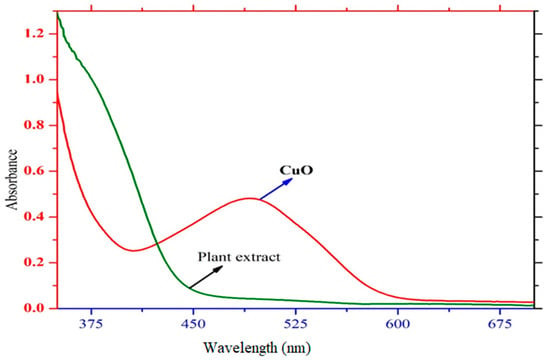
Figure 1.
UV/Vis spectrum of CuONPs.
2.2. Fourier Transform Infrared Spectroscopy Analysis
To assist in CuONP stabilization and capping, the FT-IR spectrum expresses the possible organic complex adherent [13,25,38]. This method mainly depends on the absorption of small molecules. It also measures the functional groups and evaluates the atom’s vibrations and the compound’s structure in a molecule [46,55]. A 4000–400 cm−1 wavenumber region of the IR absorption bands was attained in this process [56]. The absorption peaks’ ranges at 3234, 2060, 1707, 1075 and 800 cm−1 were demonstrated by FTIR spectroscopy, Figure 2. The absorption bands at 2066 and 1607 cm−1 appear because of the aldehyde carbon-hydrogen stretching and the aromatic carbon and carbon vibration, respectively [57]. Due to the ethylene group, the stretching vibrations responsible for the 1075 cm−1 band and the absorption band at 800 cm−1 are ascribed to the C-O carboxylic acid and –C-O-C- ether stretching vibrations. The absorption band at 1040 cm−1 is mainly due to the stretching vibration of the C-H bond [32].
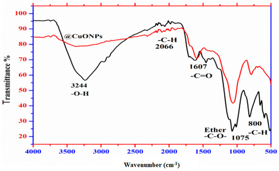
Figure 2.
The FTIR spectrum of CuONPs.
2.3. X-ray Diffraction
The crystalline structure of the CuONPs was examined by advanced physical technique XRD, as shown in Figure 3 [27]. An analytical and nondestructive X-ray diffraction method was used to characterize various crystalline matter structures and compositions. The diffraction of the X-rays occurs in the lattice planes at a typical angle in a sample. Atomic distribution measures the peak intensities within a lattice [58,59]. X-ray diffraction analysis at 10°–70° values confirmed that the CuONPs were crystalline [59]. The CuONPs were identified through the presence of a peak at 2θ = 36.21 [14,57]. At lattice planes of (110), (111), (002), (112), (202), (202), (020), (202), (113), (311) and (022) (JCPDS 48-1548) [59] the XRD pattern determined the highest purity of the CuONPs. In cases where other peaks were not assigned and there was capping of the biogenic substances at the CuONP surface using the Scherer formula, the shapes and sizes of particles were calculated according to Equation (1) [38,59].
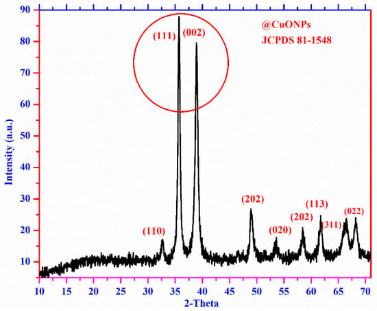
Figure 3.
The XRD spectrum of CuONPs.
During analysis, D denotes crystalline; whereas X-ray represents λ, the full width at half maximum is expressed as ß, and the diffraction angle is expressed as θ.
2.4. Scanning Electron Microscopy (SEM)
The Scanning Electron Microscopy (SEM) technique is mainly used to measure the surface activity of particles. The SEM of the green synthesised CuONPs of different scales is shown in Figure 4A,B [27], which provides detailed information on the sample surface by tracing the specimen in a raster with an en stream of light. The activity increases as the size remains small and the surface area is larger. Hence, due to the existence of colorants in the contaminated water, the particles showed good adsorption properties [5,13].
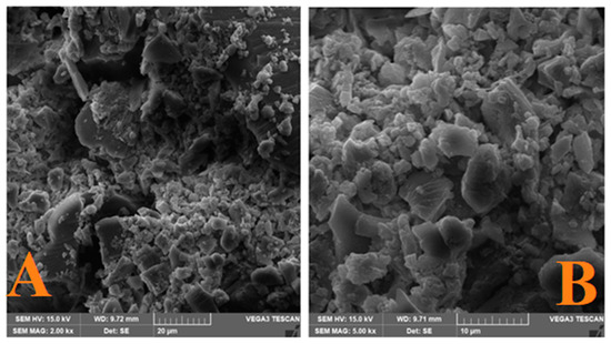
Figure 4.
The SEM Images of CuONPs at (A) 20 µm and (B) 10 µm.
2.5. High-Resolution Transmission Electron Microscopy (HRTEM)
The results showed that CuONPs are spherical and exhibit a small size and are not aggregated but well dispersed, which is closely associated with SPR absorption in UV spectra, as shown in Figure 5. Substantial activity in various fields was shown by nanoparticles having a larger surface area. In the chemo, electrochemistry and medical fields, well-dispersed and small-size NPs show significant activity [5,60,61].
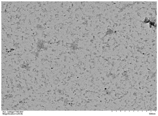
Figure 5.
The HRTEM Images of CuONPs.
2.6. Energy-Dispersive X-ray Spectroscopy (EDX)
The elemental conformation and analysis of CuONPs were confirmed by EDX, as shown in Figure 6. The arrangement of the EDX demonstrated that the Cu solution reduction with Citrus maxima extracts yielded CuONPs in crystalline form.
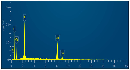
Figure 6.
The EDX spectrum of CuONPs.
To determine the particle size of the @CuONPs, a histogram was used. For the determination of particle size, ImageJ software was used [14], as shown in Figure 7. The histogram graph gives the information that the @CuONPs were of a small size and well dispersed. The particle size of the @CuONPs is calculated in the range of 0.6–85 nm with an average size of up to 35 nm.
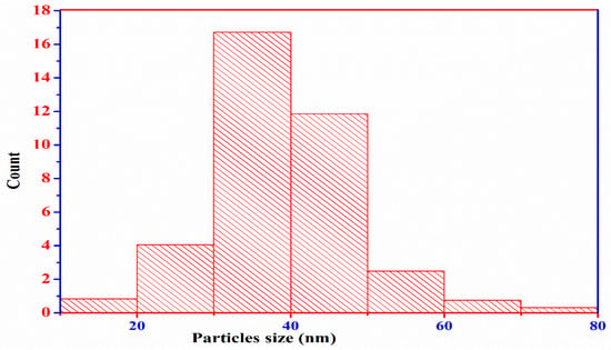
Figure 7.
Histogram spectrum of CuONPs.
2.7. Antibacterial and Antifungal Activities of CuONPs
The synthesised CuONPs have significant antimicrobial properties against Escherichia coli Bacillus subtillis, Staphylococcus aureus, Shigella flexenari, Acinetobacter Klebsiella pneumonia, Salmonella typhi and Micrococcus luteus [62]. The respective inhibition zones are as 45, 42, 25, 34, 35 and 11 mm compared to the standard 45 mm. The biogenic CuONPs have significant antibacterial activity because of their smaller spherical size, with high dispersion without aggregation. As a consequence of the decomposition of the CuONPs, Cu2+ ion concentration is increased [63]. The antibacterial activity of the CuONPs is increased due to the deposition of radicals at the surface of the CuONPs that cause damage to the membrane. Moreover, CuONPs entering and depositing in the bacterial cytoplasm cause cell death [64]. The Fenton reagent-generated •OH radical increases the antimicrobial activities of the CuONPs and causes damage to the DNA [64]. Superoxide reduces Cu2+ and releases from ferritin protein according to Equation (2). Table 1 shows the zone of inhibition against different bacteria.

Table 1.
Antibacterial activity of CuONPs.
Reactive Cu2+ creates reactive [O] species, as shown in Equation (3).
Antifungal activities were studied against Aspergillus flavus, Aspergillus fumigatus, Asper gillusniger and the reference drug Terbinafine [65]. To investigate the linear growth of the fungus strain, useable tubes were incubated at room temperature for one week. Mycelia growth inhibition was measured with the fungal growth area and was shown in percent inhibition [64,65,66,67]. The CuONPs showed significant properties against these fungi. Table 2 shows the results of the anti-fungal activities.

Table 2.
Anti-fungal activity of CuO NPs.
2.8. Antioxidant Activity
The CuONPs showed significant scavenging properties against DPPH as shown in Table 3. The antioxidant activity mainly relies on the reducing influence of DPPH [68]. The CuONPs showed effective free radical inhibition at 25, 50, 75, 100 and IC50 μg/mL was measured with IC50 values of 55.02 ± 0.05, 65.7 ± 0.15 and 69.01 ± 0.21 (uM/mL) [69]. The antioxidant activities increase with the increase in concentration of CuONPs. The smaller particle size and large surface area have significant antioxidant activity. The antiradical activity mainly relies on the DPPH-reducing power [69]. Many antioxidants have overcome the action of free radicals [70] to improve the body’s immune system [70,71].

Table 3.
Antioxidant activity against DPPH.
2.9. Dilapidation of CR in the Presence of H2O2
The photodegradation of the CR dye was studied under UV light with CuONPs and UV light with H2O2 to investigate the effect of H2O2 in the removal of the CR dye, as shown in Figure 8A,B. During the reaction of UV/H2O2, hydroxy free radicals are generated, which play important role in the elimination of the CR dye [72,73,74]. The UV/H2O2 enhanced the photodegradation of the CR in comparison with UV radioactivity. With the variation in H2O2 directly related to the degradation rate of dyes during the process, maximum degradation was achieved [73]. During the Fenton-type reaction, ●OH is generated and acts as a strong oxidizing agent as an electron scavenger [73,74,75].
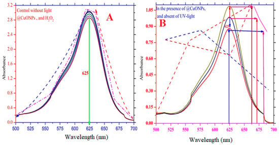
Figure 8.
Photo-degradation of Congo red dye: (A) Effect of light control; (B) Effect of catalyst.
2.10. Effect of H2O2 Concentration in the Degradation of CR Dyes
The concentration effect of UV/H2O2 was investigated in the degradation of the CR dye. The reports showed that UV/H2O2 plays a vital role in CR dye removal. The effect of the concertation of the H2O2 in the removal of CR dyes is shown in Figure 9A,B. The production of the ●OH radical under UV light stimulates the rate of reaction. The effect of the ●OH radical in the removal of CR dyes was studied using different concentrations of H2O2 (i.e., 150 µM to 250 µM), while the CR concentration was kept constant. The results showed that the increase in H2O2 dosage under UV light caused a significant removal of CR dye. It is reported [74] that, with the increase in oxidant concentration, the rate of ·OH radical production increases, which might help remove organic pollutants.
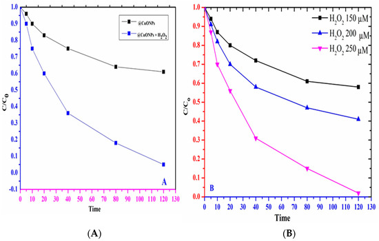
Figure 9.
(A) Effect and removal of CR dye by UV/ H2O2; (B) Effect of H2O2 concentration in the degradation of CR dyes.
2.11. Photocatalytic Activity of CuONPs
The as-synthesized CuONPs showed significant photocatalytic activity [75,76]. Different types of organic pollutants and toxic materials are present in industrial effluents, which on release to water bodies cause many adverse environmental impacts [77]. Congo red dye shows resistance to heat and light for their removal, because of having a bulky group structure [77]. The LC analysis of Aqueous Congo red dye solution treated with UV/visible light proposed the formation of 8 degradation products (DPs). The photocatalytic activity using UV light increases with the increase in catalyst concertation and H2O2 dosage up to some limits [78,79]. The UV light shows excellent results in the degradation of CR [80,81,82]. It is suggested that, at the surface of CuONPs, CR adsorbs UV light and reacts with •OH free radical, causing the degradation of CR dyes as shown in Figure 10. The mechanism of the degradation of CR by CuONPs under UV light follows: (i) CR absorbed physically at the CuONPs; (ii) the •OH radical generated during the Fenton-type reaction initiating the degradation of CR under radiation. The proposed reductive and oxidative mechanistic degradation pathway of the CR dyes is represented in Scheme 1. During the heterogeneous photocatalytic degradation, the formation of a positive hole (h+vb) takes place. The catalytic reaction under UV light is shown in reaction 7. During this process, the valence electron jumps to an excited and higher energy level from the valance band [80]. In the degradation process, h+vb react with hydroxy, hydrogen and water. Due to the high reactivity of the ●OH toward CR dyes, degradation occur as shown in Equations (4)–(11) [81].
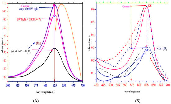
Figure 10.
Photo dilapidation of CR dye: (A) Effect of light control, light effect, light and @CuONPs and @CuONPs and H2O2 effect; (B) Effect and comparison of H2O2 on the degradation of Congo red dye.
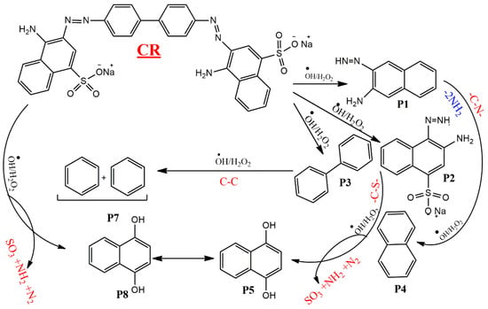
Scheme 1.
Proposed mechanism of degradation of Congo red dye.
The degradation of the dye was 25% through CuONPs in the absence of UV as compared to the presence of UV light (91%), as shown in Figure 10A,B. The catalyst CuONP concentration ranged from 0.5 to 1.5 g/L. However, at a catalyst concentration (1.0 g/L) of about (91%), the removal of the CR dye occurs. The degradation of the CR dye was studied previously [72]; here, the effect of H2O2 along with catalyst CuONPs in the removal of CR was studied. The result shows that with the addition of H2O2, with CuONPs the catalytic efficiency increases much more than without H2O2. Due to the creation of the ●OH radical, the degradation of the dye increases because the hydroxy radical oxidative reagent causes degradation. The higher dye degradation with CuONPs is because of the ●OH radical generated during UV light. The ●OH radicals have a high redox potential (2.8 V), are extremely reactive oxidants and react rapidly at high rates with the target contaminants [55,72]. Tert-butanol was used as a scavenger for % ●OH and the production of % ●OH. The removal effectiveness inhibition of the dye in the alcohol’s presence recommends the creation of % ●OH from CuONPs; photo light has a high contribution in the dye degradation. The production of various degradation products (P) results from the degradation of the dye (P1) started by % ●OH [14,25]. Because of the significant acidic behavior of the H at P1, C-35 illustrates great reactivity toward the hydroxyl radical and is therefore abstracted easily. As illustrated in Scheme 1, P2 upon functional group interconversion is transformed into P3. Moreover, the % ●OH reaction with P3 results in the transformation of P3 into P4 through the removal of -SO3H from C-54 [72]. A further oxidation process occurs at the C=C group change into P5. P5 is changed to high reactive species P6 when the hydroxyl radical abstracts -H from C-50. By eliminating many functional groups, P6 is converted to P7 by participating in the % ●OH group [55,72]. Furthermore, the reaction paths in the solution and gas phases were evaluated on the reaction time scale [55]. By the adduct of –O-O-, the generalization of -H from the –N-N- bond from hydrazones [72] and based on (FGI) P7 is converted to P8. The CR dyes are degraded into smaller molecules as the reaction continues and does not stop. Scheme 1 represents the complete process of the degradation of CR. It shows that the morphology of the CuONPs is significant in the photocatalytic activity. Without CuONPs, there was no progress in the reaction [72].
3. Materials and Methods
3.1. Preparation of Plant Extract from Citrus maxima
The Citrus maxima plant was collected from the field and was washed several times with clean water to remove the dust material. The plant extract contains phenolic constituents and other bioactive organic compounds. The plant biomass was kept under shade to dry at room temperature and ground into a fine powder [25]. To obtain the plant extract, 20 g of plant powder was soaked in 150 mL DW and heated at 50 °C for 12 h with constant stirring. After filtration, the plant extract was used for the synthesis of CuONPs [5].
3.2. Synthesis of CuONPs
Citrus maxima plant extract (15 mL) was mixed with 50 mL of 6 × 10⁻³ solution of CuSO4 to synthesize CuONPs [1,13,24,25] at 35–40 °C with continuous stirring. During the stirring process, the dark color of the solution changed to a greenish color. UV/Visible spectroscopy was used to investigate the plasmonic peak and synthesis process of CuONPs [14]. After the UV confirmation, the suspension was centrifuged for 15 min at 5000 rpm. The suspension was dried under a vacuum and CuONPs were isolated [38]. The reduction of to was caused by the phytochemicals of C. maxima. The phenolic constituents in plant extract, especially alcoholic compounds, caused the reduction and stabilization of metallic ions. The phenolic and alcoholic constituents of the medicinal plant oxidized rapidly due to through autoxidation. The autoxidation of to Cu0 through phenolic constituents is represented in Scheme 2.
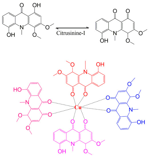
Scheme 2.
Schematic and proposed mechanistic reduction of Cu2+ into zero valent ion.
3.3. Antibacterial Response of CuONPs
The antibacterial activity of CuONPs was screened through AWD (Agar Well Diffusion) method. All the bacterial strains were grown using the nutritive broth (Oxide) at 37 °C in the incubator for 24 h until the level of turbidity touched 0.5 (using the McFarland standard) [5]. For streaking, inocula of Escherichia coli, Bacillus subtillis, Staphylococcus aureus, Shigella flexenari, Acinetobacter Klebsiella pneumonia, Salmonella typhi and Micrococcus luteus using Muller Hinton Agar were spread in PD (Petri dishes) to confirm the strained growth. Using cork, 8 mm wells were bored in the Petri plates [24]. Biogenic CuONPs were placed in plates and wells at 25 °C. After 24 h growth at 37 °C, the subsequent region’s diameter of inhibition was determined. The biosynthesized CuONPs exhibited substantial activity, which was confirmed by studies from the literature [1,5,13,24,38]. The experiment was performed in triplicate and the results obtained were expressed as means ± SD.
3.4. ROS Generation by CuONPs
To identify reactive oxygen class (ROS) (•OH) within the cells, 2, 7-dichlorodihydrofluorescein diacetate (DCFH-DA) was used, which is a moderately useful indicator. The biological activity of CuONPs was investigated using several bacterial strains, Escherichia coli, Bacillus subtillis, Staphylococcus aureus, Shigella flexenari, Acinetobacter Klebsiella pneumonia and Salmonella typhi species. The strains were incubated at 300 rpm for 180 min and 10,000 for 5 min. The suspension of pellets in 1 mL of buffer solution was preserved with one mM (DCFH-DA) reagent for about 40 min. The phosphate buffer solution was used in the cells several times to diminish the excess of dyes from the surface of the cells [14,25,38].
3.5. Antifungal Activity of CuONPs
Sabouraud dextrose agar was used to calculate the antifungal properties of biosynthesized CuONPs against various species of fungi, i.e., Aspergillus flavus, A. niger, Candida glaberata, T. longifusus, M. Canis, C. glaberata at 25 ± 1 °C incubation temperature for 7 days [14]. The dispersion of nanoparticles at 100 μg/mL concentration in medium was confirmed by ultrasonic treatment, as they were included in pure dimethyl sulfoxide (DMSO). To ensure sterile conditions, sabouraud dextrose agar was used to pack the test tubes and then exposed to an autoclave. After autoclaving, 100 μL of nanoparticle suspension was added to test tubes and retained in an inclined position. To detect the linear growth of fungus strains, 5 mm of the fungal colony was retained at the slant end, and all test tubes were incubated at 25 ± 1 °C for 7 days [24]. Using Terbinafineas, a fungicide reference, Equation (12) was applied to estimate mycelial growth (%) from the fungal growth area (cm2) [15].
In Equation (1), the growth of mycelia (control) is represented by Gc and the mycelia growth with the treatment of CuONPs is represented by Gt.
3.6. Free Radical Scavenging Assay (DPPH)
The antioxidant activity of the synthesized CuONPs was determined using DPPH [5,13,14,24,27,38]. Optima Sp-300 (Optima, Tokyo, Japan) displayed the maximum absorption at 517 nm for Free DPPH nm and decreased dramatically with antioxidants. The DPPH solution of various concentrations (25–500) was made with methanol (CH3OH) with constant stirring and ascorbic acid was used as standard. The control was DPPH methanol without a sample. Antioxidant activity was calculated using Equation (13).
3.7. Photocatalytic Activity
The photocatalytic behavior of CuONPs under UV light was examined for CR dye removal [14]. The CR dye solution (15 mL) with 10 mg of CuONPs was mixed to prepare the reaction mixture. The solution was magnetically stirred under UV light for some time before being centrifuged to eliminate the catalyst [29]. The Hg lamp of lower pressure with monochromatic emanation at 245 nm was used as a light source. To find out the photocatalytic degradation behavior of CuONPs, CR dye solution sample was taken after five min to study the absorbance through UV light. For the establishment of equilibrium of CuONPs in the mixture, the suspending solution was gently stirred for thirty minutes in dark media instead of being exposed to light. The photodegradation of CR was studied at different concentrations of CuONPs over time (5–25 min). The CR degradation (%) was calculated with Equation (14).
where Ac denotes the absorbance of CR (without CuONPs) and At denotes the absorbance of the test solution (CR and CuONPs). A blank solution was run in parallel to analyze the degradation of CR under UV light.
4. Conclusions
In the present research, CuONPs were synthesised through a greener method and tested against Congo red dye and biological activities. The capping agent on the surface and the CuONP characteristic peaks were confirmed by FTIR analysis. X-ray diffraction showed that the CuONPs were crystalline. The SEM and HRTEM gave information about the structure, shape and size of the CuONPs. The HRTEM images show that CuONPs are small with a large surface area and have a particle size between 25–90 nm. The synthesized particles proved very effective for dye adsorption from polluted water. Moreover, the newly synthesised material showed excellent antibacterial, anti-fungal and antioxidant activities due to its small size, spherical shape and well-dispersed nature. The CuONPs had the highest degradation ability for CR under UV light (catalyst). The H2O2 further improved the photocatalytic behavior of the CuONPs due to the generation of highly reactive •OH species. The removal of the organic pollutant depended upon the concentration of the oxidants and was stimulated with a higher amount of H2O2 that initiated the rate of reaction due to a greater inhibition of ecb -hvb+ recombination and a higher production of the hydroxyl radical. The proposed degradation mechanism for CR chain reactions yielded hydroxyl radical formation and dye degradation resulting in smaller fragments.
Author Contributions
Conceptualization, S.L.; methodology, F.A.; A.M.A.; software, A.H.A.; formal analysis, M.M.A., investigation, and resources, A.A.A. All authors have read and agreed to the published version of the manuscript.
Funding
This research received no external funding.
Institutional Review Board Statement
Not applicable.
Informed Consent Statement
Not applicable.
Data Availability Statement
All the data, including characterizations required for this research are presented in this manuscript.
Acknowledgments
The authors acknowledge and thank the University of Ha’il, Saudi Arabia.
Conflicts of Interest
The authors declare no conflict of interest.
Sample Availability
Samples of the compounds are available from the authors.
References
- Faisal, S.; Jan, H.; Shah, S.A.; Shah, S.; Khan, A.; Akbar, M.T.; Rizwan, M.; Jan, F.; Wajidullah Akhtar, N.; Khattak, A. Green synthesis of zinc oxide (ZnO) nanoparticles using aqueous fruit extracts of Myristica fragrans: Their characterisations and biological and environmental applications. ACS Omega 2021, 6, 9709–9722. [Google Scholar] [CrossRef] [PubMed]
- Park, H.-S.; Koduru, J.; Choo, K.-H.; Lee, B. Activated carbons impregnated with iron oxide nanoparticles for enhanced removal of bisphenol A and natural organic matter. J. Hazard. Mater. 2015, 286, 315–324. [Google Scholar] [CrossRef] [PubMed]
- Ezeigwe, E.R.; Tan, M.T.; Khiew, P.S.; Siong, C.W. One-step green synthesis of graphene/ZnO nanocomposites for electrochemical capacitors. Ceram. Int. 2015, 41, 715–724. [Google Scholar] [CrossRef]
- Bozetine, H.; Wang, Q.; Barras, A.; Li, M.; Hadjersi, T.; Szunerits, S.; Boukherroub, R. Green chemistry approach for the synthesis of ZnO–carbon dots nanocomposites with good photocatalytic properties under visible light. J. Colloid Interface Sci. 2016, 465, 286–294. [Google Scholar] [CrossRef] [PubMed]
- Herlekar, M.; Barve, S.; Kumar, R. Plant-mediated green synthesis of iron nanoparticles. J. Nanopart. 2014, 2014, 140614. [Google Scholar] [CrossRef]
- Salem, W.; Leitner, D.R.; Zingl, F.G.; Schratter, G.; Prassl, R.; Goessler, W.; Reidl, J.; Schild, S. Antibacterial activity of silver and zinc nanoparticles against Vibrio cholerae and enterotoxic Escherichia coli. Int. J. Med. Microbiol. 2015, 305, 85–95. [Google Scholar] [CrossRef]
- Pelgrift, R.Y.; Friedman, A.J. Nanotechnology as a therapeutic tool to combat microbial resistance. Adv. Drug Deliv. Rev. 2013, 65, 1803–1815. [Google Scholar] [CrossRef]
- Kalaiselvi, A.; Roopan, S.M.; Madhumitha, G.; Ramalingam, C.; Elango, G. Synthesis and characterisation of palladium nanoparticles using Catharanthus roseus leaf extract and its application in the photocatalytic degradation. Spectrochim. Acta Part A Mol. Biomol. Spectrosc. 2015, 135, 116–119. [Google Scholar] [CrossRef]
- Elango, G.; Roopan, S.M.; Dhamodaran, K.I.; Elumalai, K.; Al-Dhabi, N.A.; Arasu, M.V. Spectroscopic investigation of biosynthesised nickel nanoparticles and its larvicidal, pesticidal activities. J. Photochem. Photobiol. B Biol. 2016, 162, 162–167. [Google Scholar] [CrossRef]
- Wicki, A.; Witzigmann, D.; Balasubramanian, V.; Huwyler, J. Nanomedicine in cancer therapy: Challenges, opportunities, and clinical applications. J. Control. Release 2015, 200, 138–157. [Google Scholar] [CrossRef]
- Kumar, D.A.; Palanichamy, V.; Roopan, S.M. One step production of AgCl nanoparticles and its antioxidant and photo catalytic activity. Mater. Lett. 2015, 144, 62–64. [Google Scholar] [CrossRef]
- Kumar, S.; Meena, V.K.; Hazari, P.P.; Sharma, R.K. PEG coated and doxorubicin loaded multimodal Gadolinium oxide nanoparticles for simultaneous drug delivery and imaging applications. Int. J. Pharm. 2017, 527, 142–150. [Google Scholar] [CrossRef]
- Sukumar, S.; Rudrasenan, A.; Padmanabhan Nambiar, D. Green-synthesized rice-shaped copper oxide nanoparticles using Caesalpinia bonducella seed extract and their applications. ACS Omega 2020, 5, 1040–1051. [Google Scholar] [CrossRef]
- Peternela, J.; Silva, M.F.; Vieira, M.F.; Bergamasco, R.; Vieira, A.M. Synthesis and impregnation of copper oxide nanoparticles on activated carbon through green synthesis for water pollutant removal. Mater. Res. 2017, 21. [Google Scholar] [CrossRef]
- Mortazavi-Derazkola, S.; Salavati-Niasari, M.; Khojasteh, H.; Amiri, O.; Ghoreishi, S.M. Green synthesis of magnetic Fe3O4/SiO2/HAp nanocomposite for atenolol delivery and in vivo toxicity study. J. Clean. Prod. 2017, 168, 39–50. [Google Scholar] [CrossRef]
- Sajadi, S.M.; Kolo, K.; Hamad, S.M.; Mahmud, S.A.; Barzinjy, A.A.; Hussein, S.M. Green synthesis of the Ag/Bentonite nanocomposite UsingEuphorbia larica extract: A reusable catalyst for efficient reduction of nitro compounds and organic dyes. ChemistrySelect 2018, 3, 12274–12280. [Google Scholar] [CrossRef]
- Safardoust-Hojaghan, H.; Salavati-Niasari, M. Degradation of methylene blue as a pollutant with N-doped graphene quantum dot/titanium dioxide nanocomposite. J. Clean. Prod. 2017, 148, 31–36. [Google Scholar] [CrossRef]
- Huh, A.J.; Kwon, Y.J. “Nanoantibiotics”: A new paradigm for treating infectious diseases using nanomaterials in the antibiotics resistant era. J. Control. Release 2011, 156, 128–145. [Google Scholar] [CrossRef]
- Salavati-Niasari, M.; Davar, F.; Loghman-Estarki, M.R. Controllable synthesis of thioglycolic acid capped ZnS(Pn)0.5 nanotubes via simple aqueous solution route at low temperatures and conversion to wurtzite ZnS nanorods via thermal decompose of precursor. J. Alloys Compd. 2010, 494, 199–204. [Google Scholar] [CrossRef]
- Haritha, E.; Roopan, S.M.; Madhavi, G.; Elango, G.; Al-Dhabi, N.A.; Arasu, M.V. Green chemical approach towards the synthesis of SnO2 NPs in argument with photocatalytic degradation of diazo dye and its kinetic studies. J. Photochem. Photobiol. B Biol. 2016, 162, 441–447. [Google Scholar] [CrossRef]
- Kar, S.; Chaudhuri, S. Controlled synthesis and photoluminescence properties of ZnS nanowires and nanoribbons. J. Phys. Chem. B 2005, 109, 3298–3302. [Google Scholar] [CrossRef] [PubMed]
- Cho, M.; Chung, H.; Choi, W.; Yoon, J. Different inactivation behaviors of MS-2 phage and Escherichia coli in TiO2 photocatalytic disinfection. Appl. Environ. Microbiol. 2005, 71, 270–275. [Google Scholar] [CrossRef] [PubMed]
- Roopan, S.M.; Khan, F.R.N. SnO2 nanoparticles mediated nontraditional synthesis of biologically active 9-chloro-6, 13-dihydro-7-phenyl-5H-indolo [3, 2-c]-acridine derivatives. Med. Chem. Res. 2011, 20, 732–737. [Google Scholar] [CrossRef]
- Yousaf, M.; Ahmad, M.; Zhao, Z.P. Rapid and highly selective conversion of CO2 to methanol by heterometallic porous ZIF-8. J. CO2 Util. 2022, 64, 102172. [Google Scholar] [CrossRef]
- Alhalili, Z. Green synthesis of copper oxide nanoparticles CuO NPs from Eucalyptus Globoulus leaf extract: Adsorption and design of experiments. Arab. J. Chem. 2022, 15, 103739. [Google Scholar] [CrossRef]
- Wang, J.Z.; Zhong, C.; Wexler, D.; Idris, N.H.; Wang, Z.X.; Chen, L.Q.; Liu, H.K. Graphene-encapsulated Fe3O4 nanoparticles with 3D laminated structure as superior anode in lithium ion batteries. Chem.–A Eur. J. 2011, 17, 661–667. [Google Scholar] [CrossRef]
- Strong, F.M.; Carpenter, L.E. Preparation of samples for microbiological determination of riboflavin. Ind. Eng. Chem. Anal. Ed. 1942, 14, 909–913. [Google Scholar] [CrossRef]
- Zaman, M.I.; Mustafa, S.; Khan, S.; Xing, B. Effect of phosphate complexation on Cd2+ sorption by manganese dioxide (β-MnO2). J. Colloid Interface Sci. 2009, 330, 9–19. [Google Scholar] [CrossRef]
- Dhaka, S.; Kumar, R.; Lee, S.H.; Kurade, M.B.; Jeon, B.H. Degradation of ethyl paraben in aqueous medium using advanced oxidation processes: Efficiency evaluation of UV-C supported oxidants. J. Clean. Prod. 2018, 180, 505–513. [Google Scholar] [CrossRef]
- Rahim, M.; Iram, S.; Khan, M.S.; Khan, M.S.; Shukla, A.R.; Srivastava, A.; Ahmad, S.J.C.; Biointerfaces, S.B. Glycation-assisted synthesised gold nanoparticles inhibit growth of bone cancer cells. Colloids Surf. B Biointerfaces 2014, 117, 473–479. [Google Scholar] [CrossRef]
- Bhushan, B.; Khanadeev, V.; Khlebtsov, B.; Khlebtsov, N.; Gopinath, P. Impact of albumin based approaches in nanomedicine: Imaging, targeting and drug delivery. Adv. Colloid Interface Sci. 2017, 246, 13–39. [Google Scholar] [CrossRef] [PubMed]
- Amini, S.M. Preparation of antimicrobial metallic nanoparticles with bioactive compounds. Mater. Sci. Eng. C 2019, 103, 109809. [Google Scholar] [CrossRef] [PubMed]
- Pendashteh, A.; Mousavi, M.F.; Rahmanifar, M.S. Fabrication of anchored copper oxide nanoparticles on graphene oxide nanosheets via an electrostatic coprecipitation and its application as supercapacitor. Electrochim. Acta 2013, 88, 347–357. [Google Scholar] [CrossRef]
- Sharma, J.K.; Akhtar, M.S.; Ameen, S.; Srivastava, P.; Singh, G. Green synthesis of CuO nanoparticles with leaf extract of Calotropis gigantea and its dye-sensitised solar cells applications. J. Alloys Compd. 2015, 632, 321–325. [Google Scholar] [CrossRef]
- Prasad, A.R.; Ammal, P.R.; Joseph, A. Effective photocatalytic removal of different dye stuffs using green synthesised zinc oxide nanogranules. Mater. Res. Bull. 2018, 102, 116–121. [Google Scholar] [CrossRef]
- Volkert, O.; Schulte-Frohlinde, D. Mechanism of homolytic aromatic hydroxylation III. Tetrahedron Lett. 1968, 9, 2151–2154. [Google Scholar] [CrossRef]
- Lu, W.; Singh, A.K.; Khan, S.A.; Senapati, D.; Yu, H.; Ray, P.C. Gold nano-popcorn-based targeted diagnosis, nanotherapy treatment, and in situ monitoring of photothermal therapy response of prostate cancer cells using surface-enhanced Raman spectroscopy. J. Am. Chem. Soc. 2010, 132, 18103–18114. [Google Scholar] [CrossRef]
- Kim, T.W.; Yang, Y.; Li, F.; Kwan, W.L. Electrical memory devices based on inorganic/organic nanocomposites. NPG Asia Mater. 2012, 4, e18. [Google Scholar] [CrossRef]
- Gao, Y.; An, T.; Ji, Y.; Li, G.; Zhao, C. Eco-toxicity and human estrogenic exposure risks from OH-initiated photochemical transformation of four phthalates in water: A computational study. Environ. Pollut. 2015, 206, 510–517. [Google Scholar] [CrossRef]
- Malaikozhundan, B.; Vaseeharan, B.; Vijayakumar, S.; Pandiselvi, K.; Kalanjiam, M.A.R.; Murugan, K.; Benelli, G. Biological therapeutics of Pongamia pinnata coated zinc oxide nanoparticles against clinically important pathogenic bacteria, fungi and MCF-7 breast cancer cells. Microb. Pathog. 2017, 104, 268–277. [Google Scholar] [CrossRef]
- Banumathi, B.; Malaikozhundan, B.; Vaseeharan, B. In vitro acaricidal activity of ethnoveterinary plants and green synthesis of zinc oxide nanoparticles against Rhipicephalus (Boophilus) microplus. Vet. Parasitol. 2016, 216, 93–100. [Google Scholar] [CrossRef]
- Choi, C.W.; Kim, S.C.; Hwang, S.S.; Choi, B.K.; Ahn, H.J.; Lee, M.Y.; Park, S.H.; Kim, S.K. Antioxidant activity and free radical scavenging capacity between Korean medicinal plants and flavonoids by assay-guided comparison. Plant Sci. 2002, 163, 1161–1168. [Google Scholar] [CrossRef]
- Cho, M.; Cho, W.-S.; Choi, M.; Kim, S.J.; Han, B.S.; Kim, S.H.; Kim, H.O.; Sheen, Y.Y.; Jeong, J. The impact of size on tissue distribution and elimination by single intravenous injection of silica nanoparticles. Toxicol. Lett. 2009, 189, 177–183. [Google Scholar] [CrossRef]
- Owens, E.A.; Henary, M.; El Fakhri, G.; Choi, H.S. Tissue-specific near-infrared fluorescence imaging. Acc. Chem. Res. 2016, 49, 1731–1740. [Google Scholar] [CrossRef]
- Choi, J.Y. Treatment of bone metastasis with bone-targeting radiopharmaceuticals. Nucl. Med. Mol. Imaging 2018, 52, 200–207. [Google Scholar] [CrossRef]
- Tozer, D.J. Relationship between long-range charge-transfer excitation energy error and integer discontinuity in Kohn–Sham theory. J. Chem. Phys. 2003, 119, 12697–12699. [Google Scholar] [CrossRef]
- Yousaf, M.; Ahmad, M.; Batool, A.; Zhao, Z.P. Highly-stable, bifunctional, binder-free & stand-alone photoelectrode (FexNi1−xO@ a-CC) for natural waters splitting into hydrogen. Int. J. Hydrogen Energy 2022, 47, 36032–36045. [Google Scholar]
- Wang, S. Surface Characterisation of Chemically Modified Fiber, Wood and Paper. 2014. Available online: https://www.doria.fi/handle/10024/96382 (accessed on 1 November 2022).
- Chen, S.; Liu, J.; Zeng, H. Structure and antibacterial activity of silver-supporting activated carbon fibers. J. Mater. Sci. 2005, 40, 6223–6231. [Google Scholar] [CrossRef]
- TKulisic; Radonic, A.; Katalinic, V.; Milos, M. Use of different methods for testing antioxidative activity of oregano essential oil. Food Chem. 2004, 85, 633–640. [Google Scholar] [CrossRef]
- Obied, H.K.; Allen, M.S.; Bedgood, D.R.; Prenzler, P.D.; Robards, K.; Stockmann, R. Bioactivity and analysis of biophenols recovered from olive mill waste. J. Agric. Food Chem. 2005, 53, 823–837. [Google Scholar] [CrossRef]
- Huang, B.; Ban, X.; He, J.; Tong, J.; Tian, J.; Wang, Y. Hepatoprotective and antioxidant activity of ethanolic extracts of edible lotus (Nelumbo nucifera Gaertn.) leaves. Food Chem. 2010, 120, 873–878. [Google Scholar] [CrossRef]
- Klibanov, A.L.; Maruyama, K.; Torchilin, V.P.; Huang, L. Amphipathic polyethyleneglycols effectively prolong the circulation time of liposomes. FEBS Lett. 1990, 268, 235–237. [Google Scholar] [CrossRef] [PubMed]
- Sabu, M.; Kuttan, R. Anti-diabetic activity of medicinal plants and its relationship with their antioxidant property. J. Ethnopharmacol. 2002, 81, 155–160. [Google Scholar] [CrossRef] [PubMed]
- Hammouda, S.B.; Zhao, F.; Safaei, Z.; Srivastava, V.; Ramasamy, D.L.; Iftekhar, S.; Sillanpää, M. Degradation and mineralisation of phenol in aqueous medium by heterogeneous monopersulfate activation on nanostructured cobalt based-perovskite catalysts ACoO3 (A = La, Ba, Sr and Ce): Characterisation, kinetics and mechanism study. Appl. Catal. B Environ. 2017, 215, 60–73. [Google Scholar] [CrossRef]
- Li, L.; Gao, H.; Yi, Z.; Wang, S.; Wu, X.; Li, R.; Yang, H. Comparative investigation on synthesis, morphological tailoring and photocatalytic activities of Bi2O2CO3 nanostructures. Colloids Surf. A Physicochem. Eng. Asp. 2022, 644, 128758. [Google Scholar] [CrossRef]
- Shi, L.B.; Tang, P.F.; Zhang, W.; Zhao, Y.P.; Zhang, L.C.; Zhang, H. Green synthesis of CuO nanoparticles using Cassia auriculata leaf extract and in vitro evaluation of their biocompatibility with rheumatoid arthritis macrophages (RAW 264.7). Trop. J. Pharm. Res. 2017, 16, 185–192. [Google Scholar] [CrossRef]
- Riehemann, K.; Schneider, S.W.; Luger, T.A.; Godin, B.; Ferrari, M.; Fuchs, H. Nanomedicine—Challenge and perspectives. Angew. Chem. Int. Ed. 2009, 48, 872–897. [Google Scholar] [CrossRef]
- Tamaekong, N.; Liewhiran, C.; Phanichphant, S. Synthesis of thermally spherical CuO nanoparticles. J. Nanomater. 2014, 2014, 507978. [Google Scholar] [CrossRef]
- Das, S.; Maiti, S.; Saha, S.; Das, N.S.; Chattopadhyay, K.K. Template free synthesis of mesoporous CuO nano architects for field emission applications. J. Nanosci. Nanotechnol. 2013, 13, 2722–2728. [Google Scholar] [CrossRef]
- Nasrollahzadeh, M.; Sajadi, S.M.; Rostami-Vartooni, A.; Hussin, S.M. Green synthesis of CuO nanoparticles using aqueous extract of Thymus vulgaris L. leaves and their catalytic performance for N-arylation of indoles and amines. J. Colloid Interface Sci. 2016, 466, 113–119. [Google Scholar] [CrossRef]
- Espitia, P.J.P.; de Fátima Ferreira Soares, N.; dos Reis Coimbra, J.S.; de Andrade, N.J.; Cruz, R.S.; Medeiros, E.A.A. Zinc oxide nanoparticles: Synthesis, antimicrobial activity and food packaging applications. Food Bioprocess Technol. 2012, 25, 1447–1464. [Google Scholar] [CrossRef]
- Shalini, D.; Senthilkumar, S.; Rajaguru, P. Effect of size and shape on toxicity of zinc oxide (ZnO) nanomaterials in human peripheral blood lymphocytes. Toxicol. Mech. Methods 2018, 28, 87–94. [Google Scholar] [CrossRef]
- Abu-Kharma, M.; Awwad, A. Green synthesis of copper oxide nanoparticles using Bougainvillea leaves aqueous extract and antibacterial activity evaluation. Int. Sci. Organ. 2021, 7, 155–162. [Google Scholar]
- Das, S.; Mitra, S.; Khurana, S.P.; Debnath, D. Nanomaterials for biomedical applications. Biol. Mater. Sci. 2013, 7, 90–98. [Google Scholar] [CrossRef]
- Nandi, S.K.; Bandyopadhyay, S.; Das, P.; Samanta, I.; Mukherjee, P.; Roy, S.; Kundu, B. Understanding osteomyelitis and its treatment through local drug delivery system. Biotechnol. Adv. 2016, 34, 1305–1317. [Google Scholar] [CrossRef]
- Bagamboula, C.; Uyttendaele, M.; Debevere, J. Antimicrobial effect of spices and herbs on Shigella sonnei and Shigella flexneri. J. Food Prot. 2003, 66, 668–673. [Google Scholar] [CrossRef]
- Vimala, K.; Sundarraj, S.; Paulpandi, M.; Vengatesan, S.; Kannan, S. Green synthesised doxorubicin loaded zinc oxide nanoparticles regulates the Bax and Bcl-2 expression in breast and colon carcinoma. Process Biochem. 2014, 49, 160–172. [Google Scholar] [CrossRef]
- Tepe, B.; Daferera, D.; Sokmen, A.; Sokmen, M.; Polissiou, M. Antimicrobial and antioxidant activities of the essential oil and various extracts of Salvia tomentosa Miller (Lamiaceae). Food Chem. 2005, 90, 333–340. [Google Scholar] [CrossRef]
- Aruoma, O.I. Methodological considerations for characterising potential antioxidant actions of bioactive components in plant foods. Mutat. Res./Fundam. Mol. Mech. Mutagen. 2003, 523, 9–20. [Google Scholar] [CrossRef]
- Tajik, S.; Beitollahi, H.; Nejad, F.G.; Safaei, M.; Zhang, K.; Van Le, Q.; Varma, R.S.; Jang, H.W.; Shokouhimehr, M. Developments and applications of nanomaterial-based carbon paste electrodes. RSC Adv. 2020, 10, 21561–21581. [Google Scholar] [CrossRef]
- Anipsitakis, G.P.; Dionysiou, D.D. Radical generation by the interaction of transition metals with common oxidants. Environ. Sci. Technol. 2004, 38, 3705–3712. [Google Scholar] [CrossRef]
- Arshad, R.; Bokhari, T.; Javed, T.; Bhatti, I.; Rasheed, S.; Iqbal, M.; Nazir, A.; Naz, S.; Khan, M.; Khosa, M. Degradation product distribution of Reactive Red-147 dye treated by UV/H2O2/TiO2 advanced oxidation process. J. Mater. Res. Technol. 2020, 9, 3168–3178. [Google Scholar] [CrossRef]
- Manikandan, A.; Durka, M.; Antony, S.A. Magnetically recyclable spinel MnxZn1–x Fe2O4 (0.0 ≤ x ≤ 0.5) nano-photocatalysts. Adv. Sci. Eng. Med. 2015, 7, 33–46. [Google Scholar] [CrossRef]
- Pourgholi, M.; Jahandizi, R.M.; Miranzadeh, M.; Beigi, O.H.; Dehghan, S. Removal of Dye and COD from textile wastewater using AOP (UV/O3, UV/H2O2, O3/H2O2 and UV/H2O2/O3). J. Environ. Health Sustain. Dev. 2018, 3, 621–629. [Google Scholar] [CrossRef]
- Chang, F.; Yang, C.; Wang, X.; Zhao, S.; Wang, J.; Yang, W.; Dong, F.; Zhu, G.; Kong, Y. Mechanical ball-milling preparation and superior photocatalytic NO elimination of Z-scheme Bi12SiO20-based heterojunctions with surface oxygen vacancies. J. Clean. Prod. 2022, 380, 135167. [Google Scholar] [CrossRef]
- Chang, F.; Yan, W.; Wang, X.; Peng, S.; Li, S.; Hu, X. Strengthened photocatalytic removal of bisphenol a by robust 3D hierarchical np heterojunctions Bi4O5Br2-MnO2 via boosting oxidative radicals generation. Chem. Eng. J. 2022, 428, 131223. [Google Scholar] [CrossRef]
- Buxton, G.V.; Greenstock, C.L.; Helman, W.P.; Ross, A.B. Critical Review of rate constants for reactions of hydrated electrons, hydrogen atoms and hydroxyl radicals ( OH/ O− in Aqueous Solution. J. Phys. Chem. Ref. Data 1988, 17, 513–886. [Google Scholar] [CrossRef]
- Arunadevi, R.; Kavitha, B.; Rajarajan, M.; Suganthi, A.; Jeyamurugan, A. Investigation of the drastic improvement of photocatalytic degradation of Congo red by monoclinic Cd, Ba-CuO nanoparticles and its antimicrobial activities. Surf. Interfaces 2018, 10, 32–44. [Google Scholar] [CrossRef]
- Güy, N.; Özacar, M. The influence of noble metals on photocatalytic activity of ZnO for Congo red degradation. Int. J. Hydrogen Energy 2016, 41, 20100–20112. [Google Scholar] [CrossRef]
- Li, L.; Sun, X.; Xian, T.; Gao, H.; Wang, S.; Yi, Z.; Wu, X.; Yang, H. Template-free synthesis of Bi2O2CO3 hierarchical nanotubes self-assembled from ordered nanoplates for promising photocatalytic applications. Phys. Chem. Chem. Phys. 2022, 24, 8279–8295. [Google Scholar] [CrossRef]
- Gu, Y.; Guo, B.; Yi, Z.; Wu, X.; Zhang, J.; Yang, H. Morphology modulation of hollow-shell ZnSn(OH)6 for enhanced photodegradation of methylene blue. Colloids Surf. A Physicochem. Eng. Asp. 2022, 653, 129908. [Google Scholar] [CrossRef]
Disclaimer/Publisher’s Note: The statements, opinions and data contained in all publications are solely those of the individual author(s) and contributor(s) and not of MDPI and/or the editor(s). MDPI and/or the editor(s) disclaim responsibility for any injury to people or property resulting from any ideas, methods, instructions or products referred to in the content. |
© 2023 by the authors. Licensee MDPI, Basel, Switzerland. This article is an open access article distributed under the terms and conditions of the Creative Commons Attribution (CC BY) license (https://creativecommons.org/licenses/by/4.0/).