
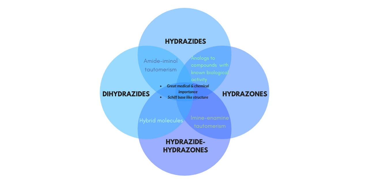
Different Schiff Bases—Structure, Importance and Classification


Abstract

1. Introduction
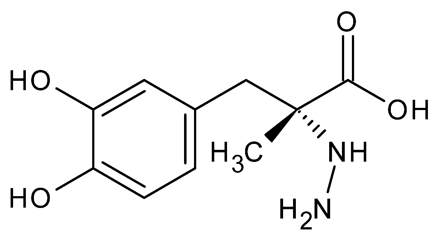
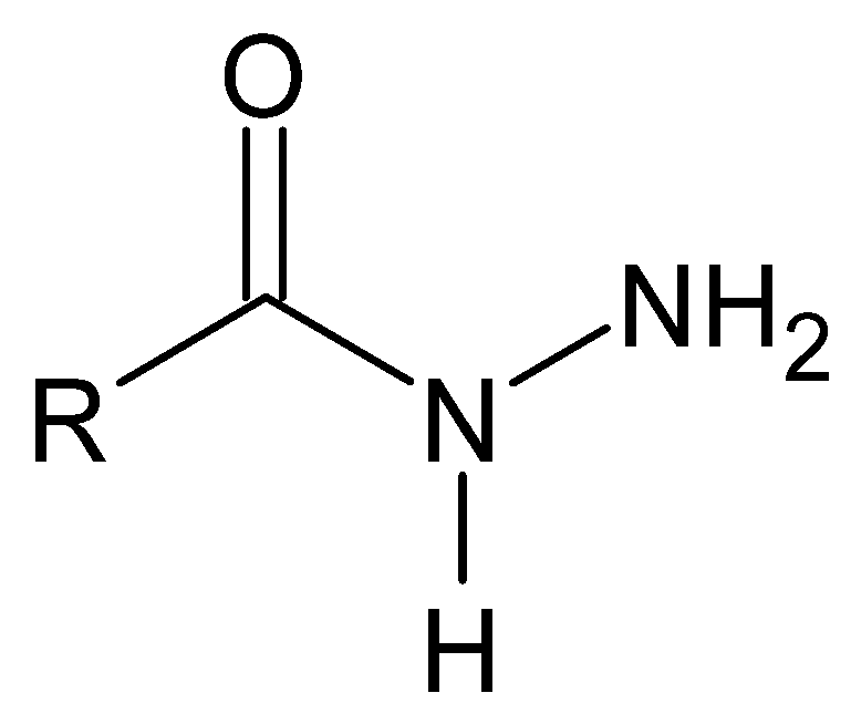
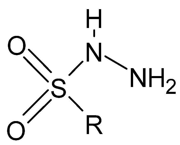
2. Hydrazides
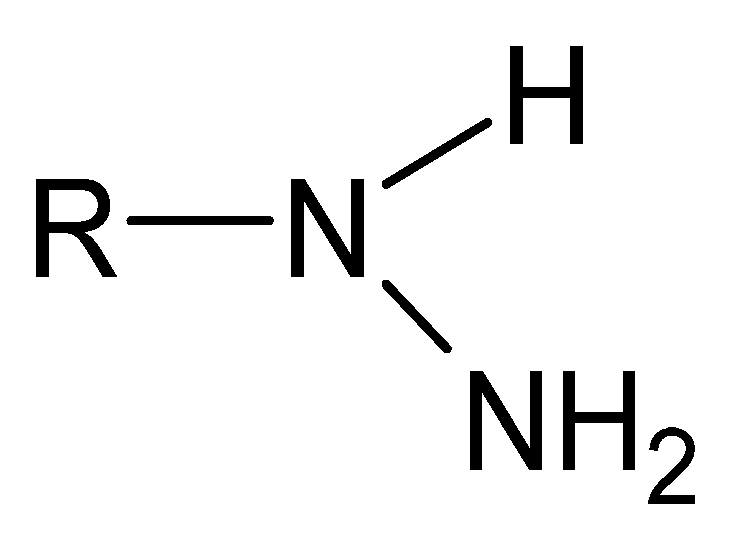
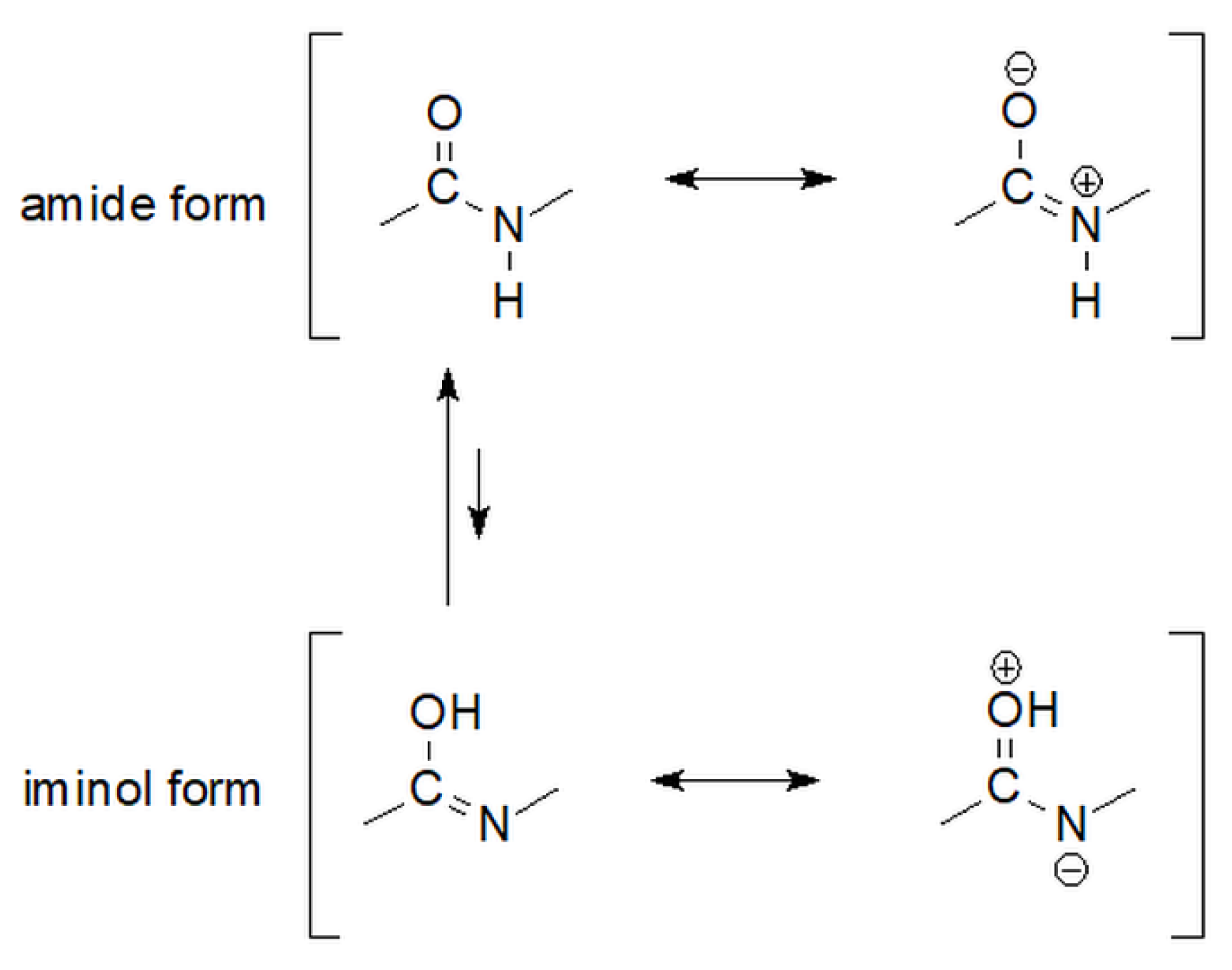
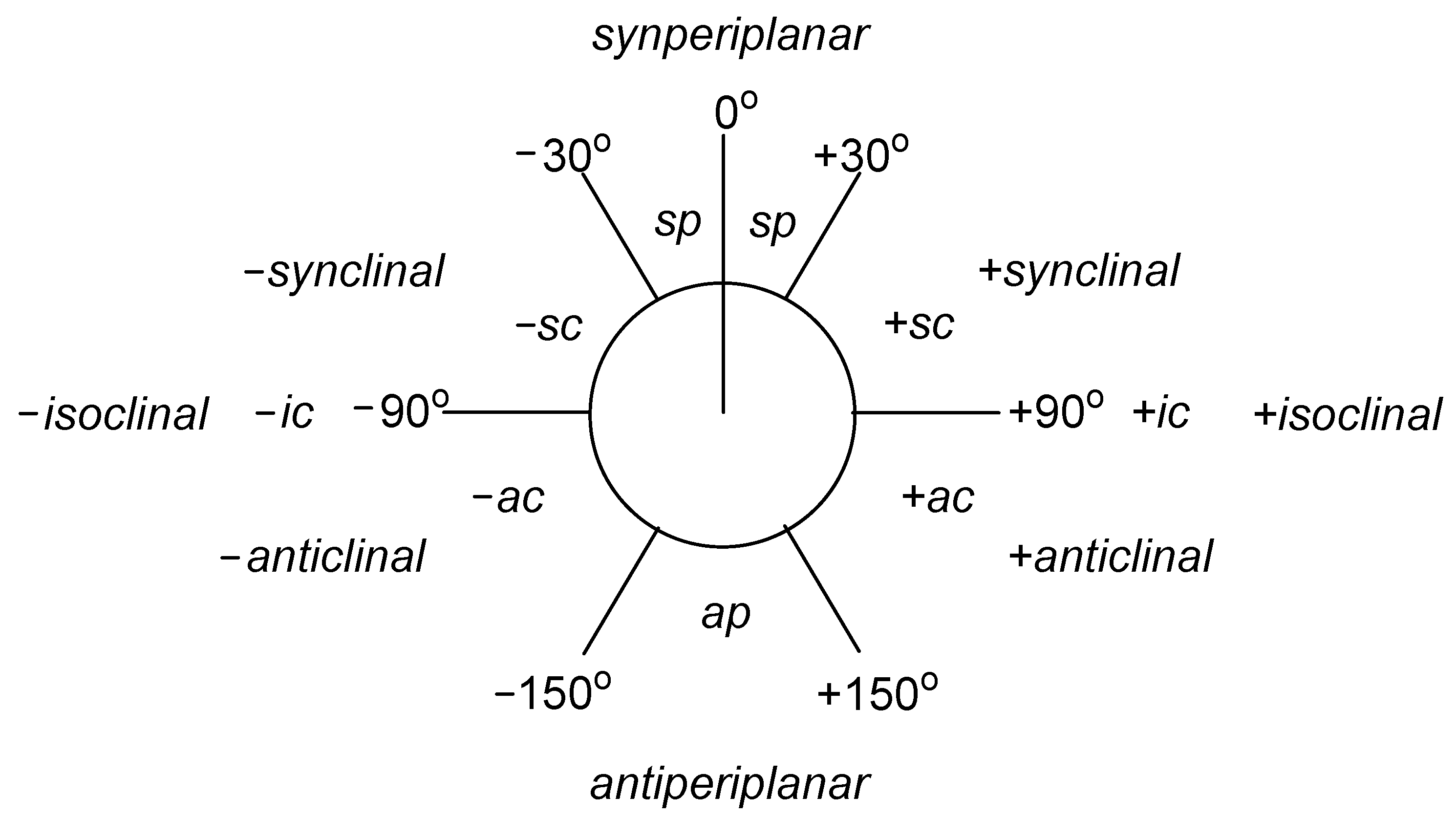
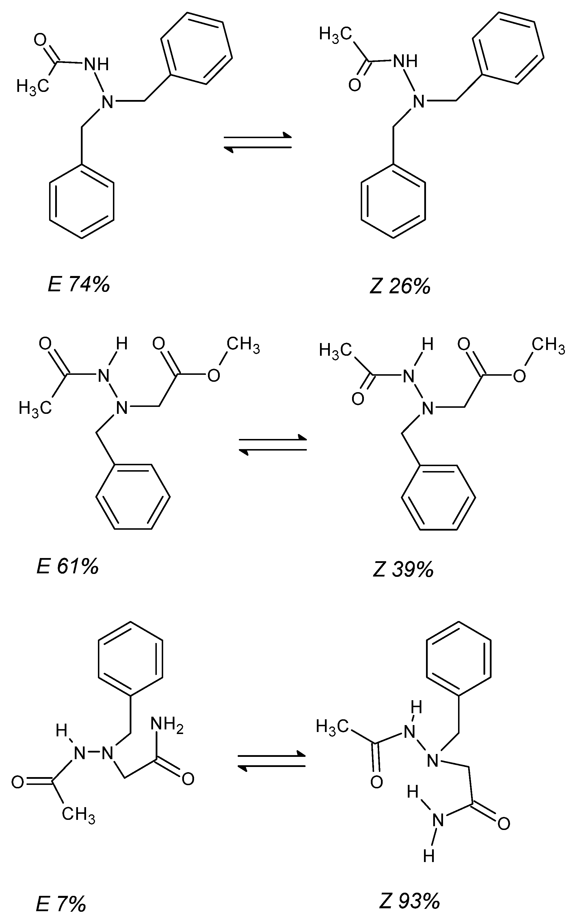
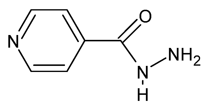
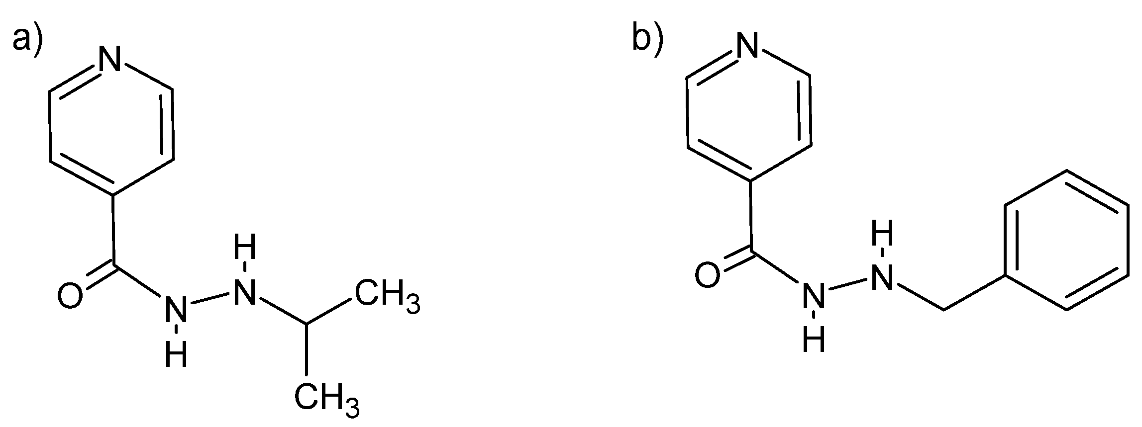
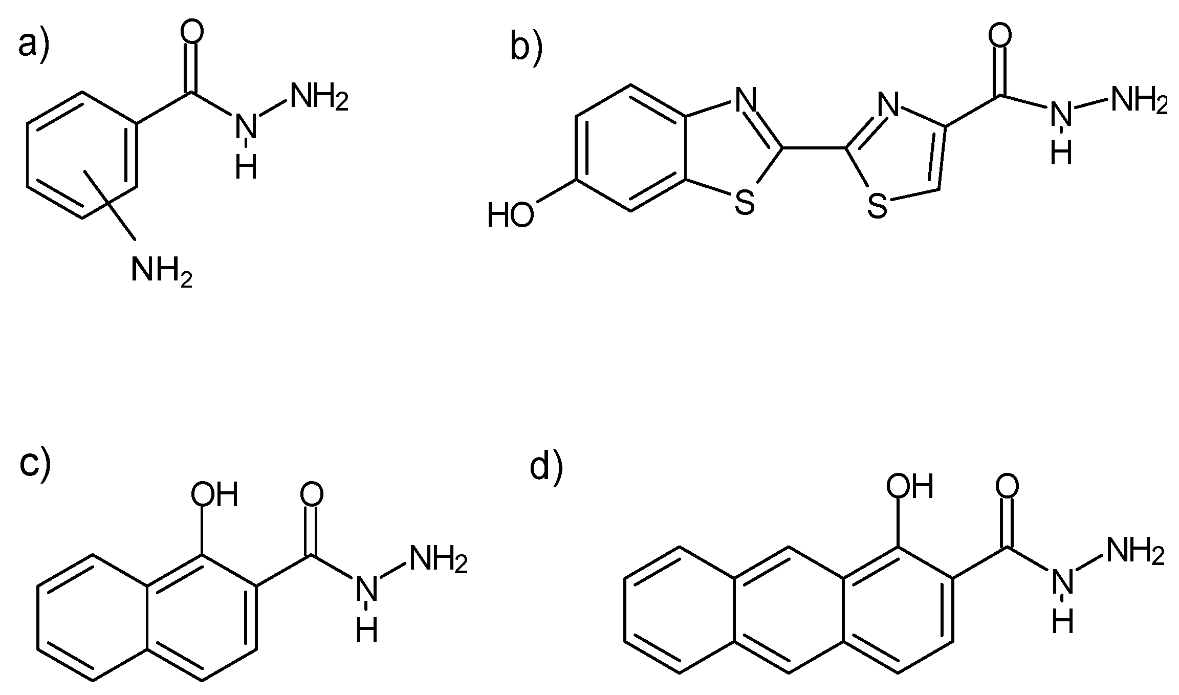
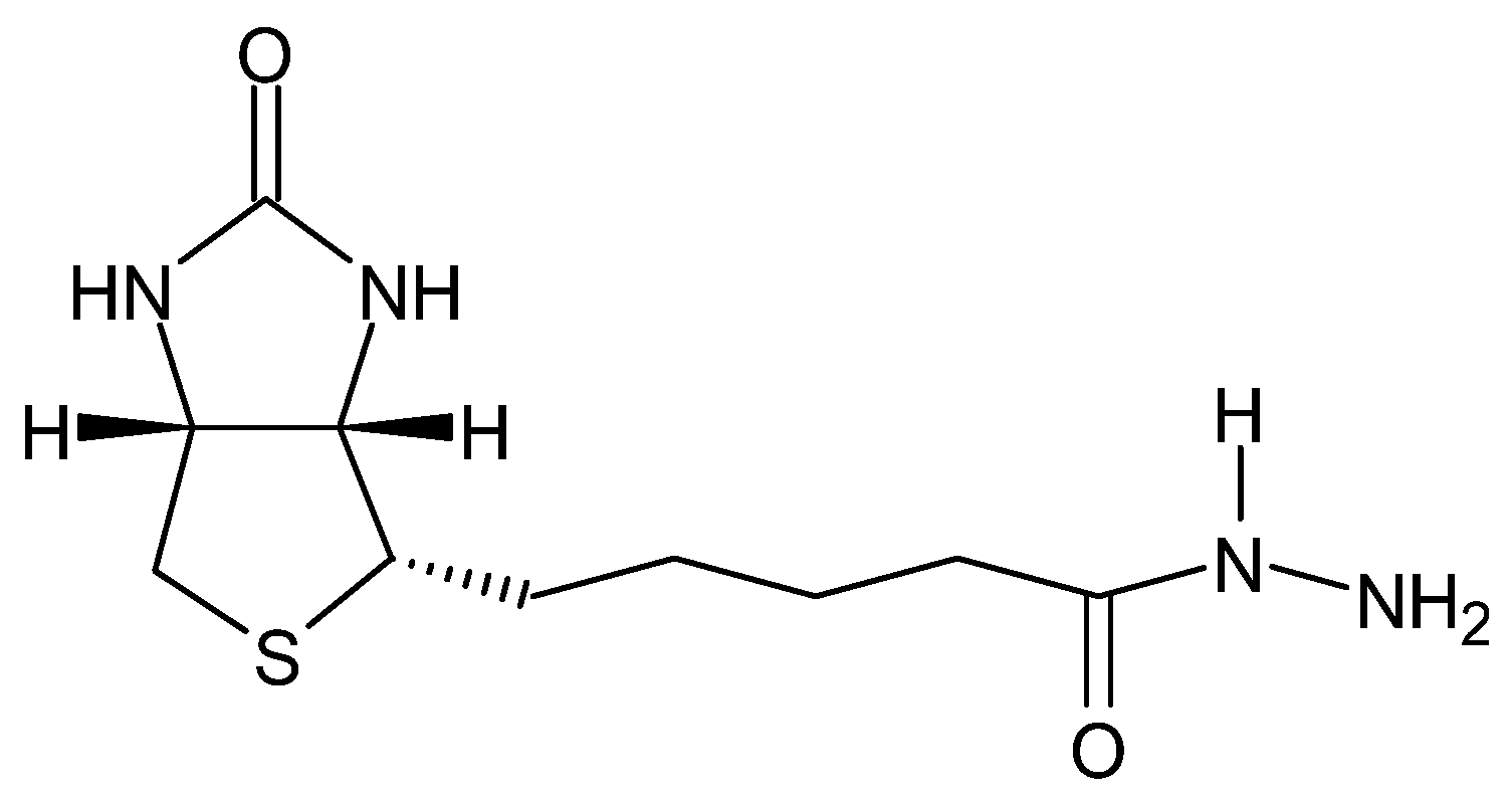
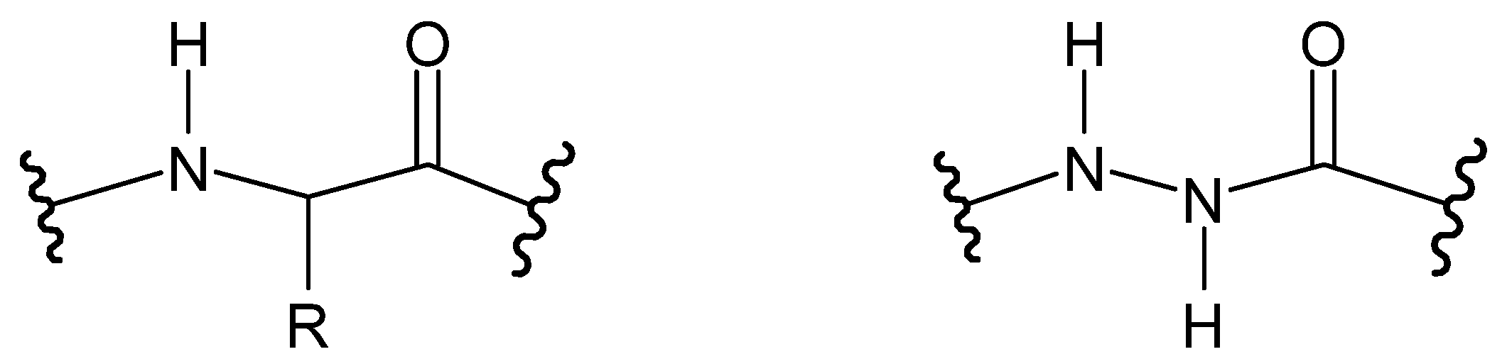
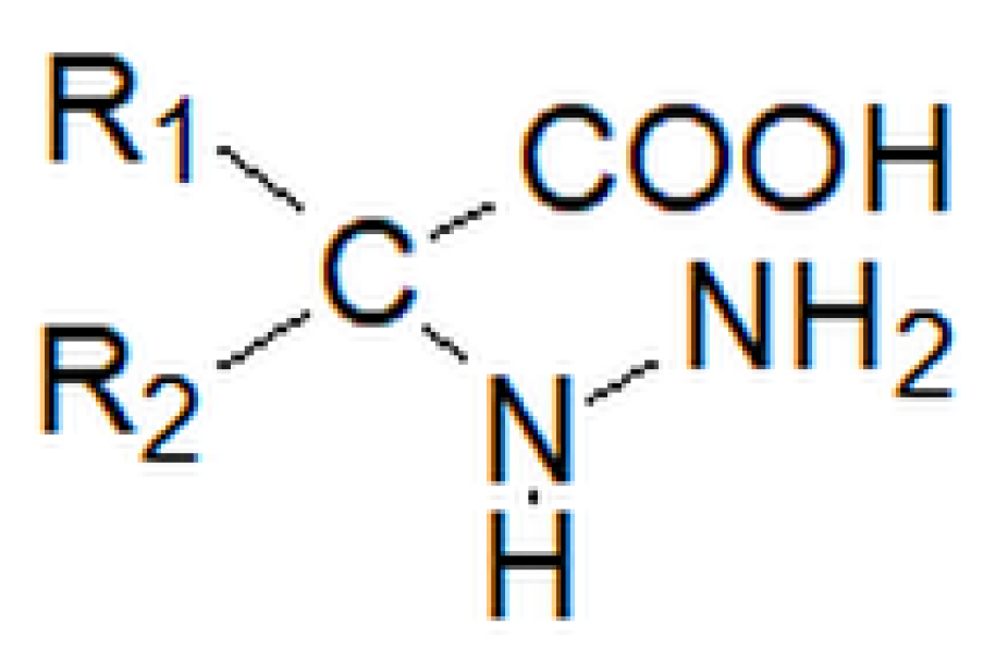
2.1. Structure
2.2. Importance
2.3. Classification
3. Dihydrazides
3.1. Structure
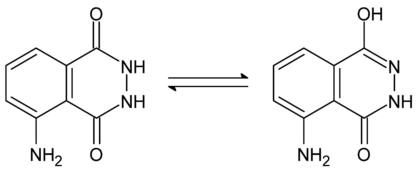
3.2. Importance
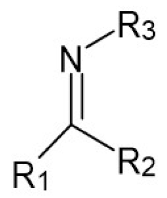
4. Hydrazones
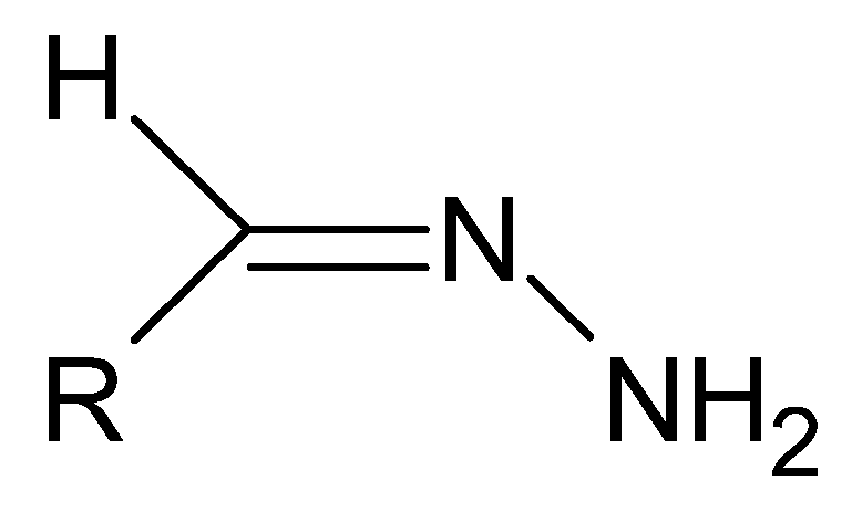
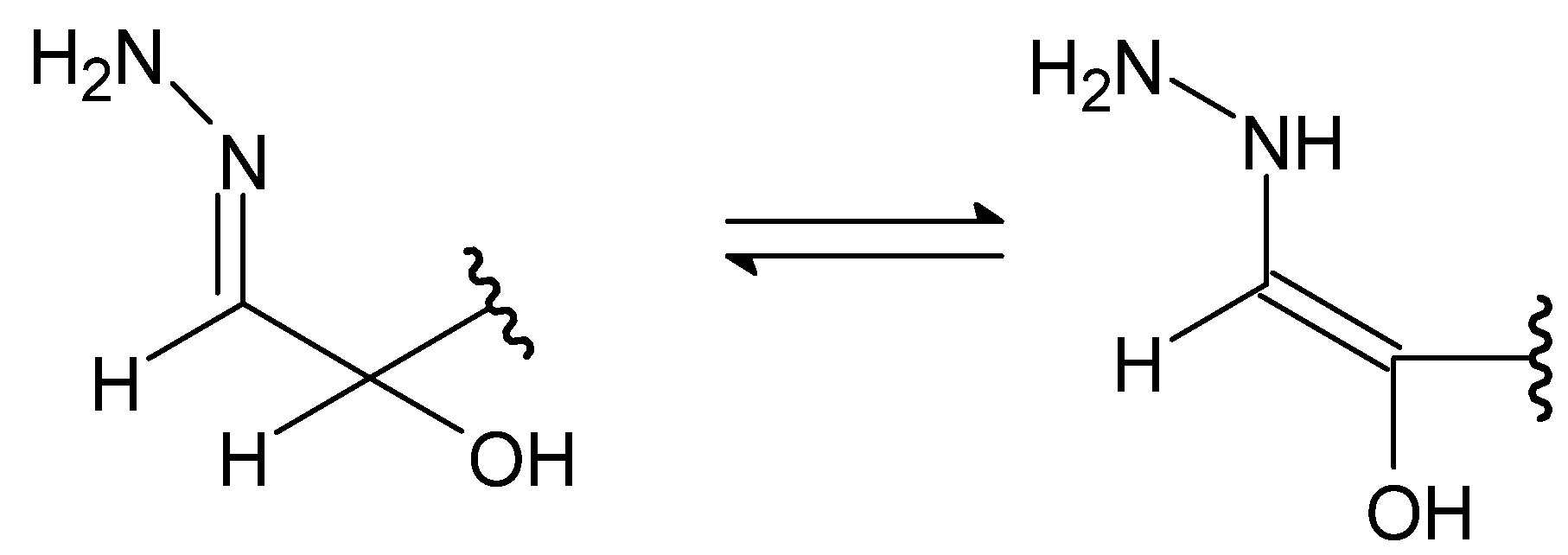
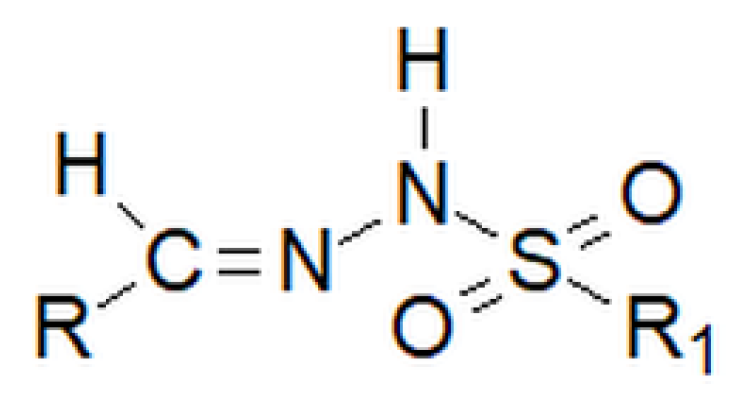
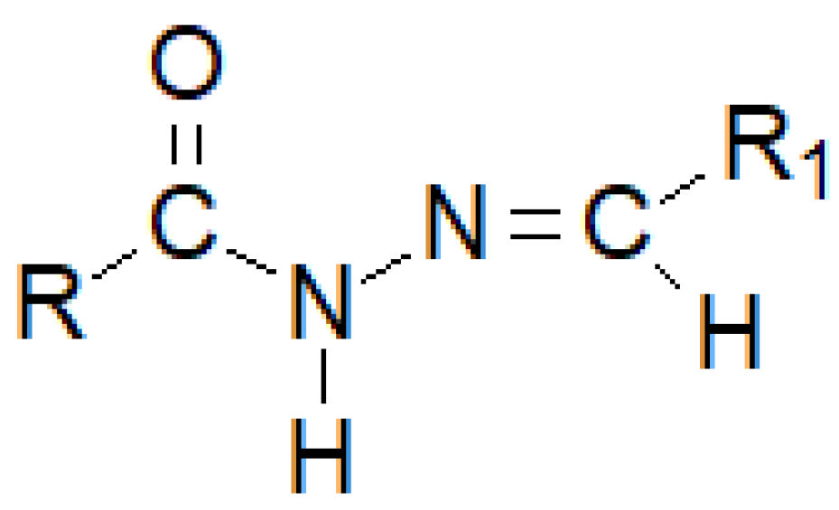
4.1. Structure
4.2. Importance
4.3. Classification
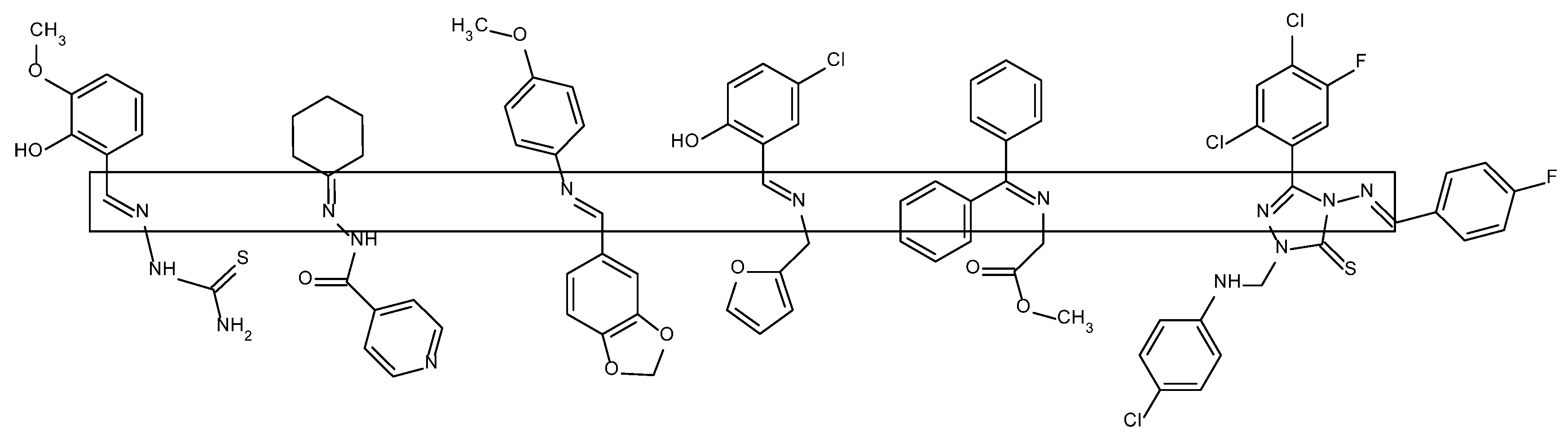
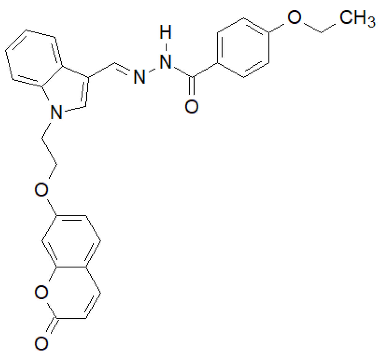
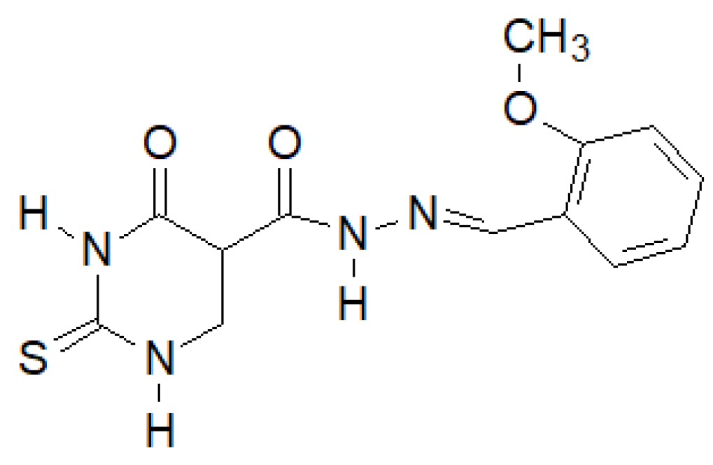
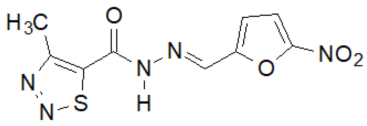
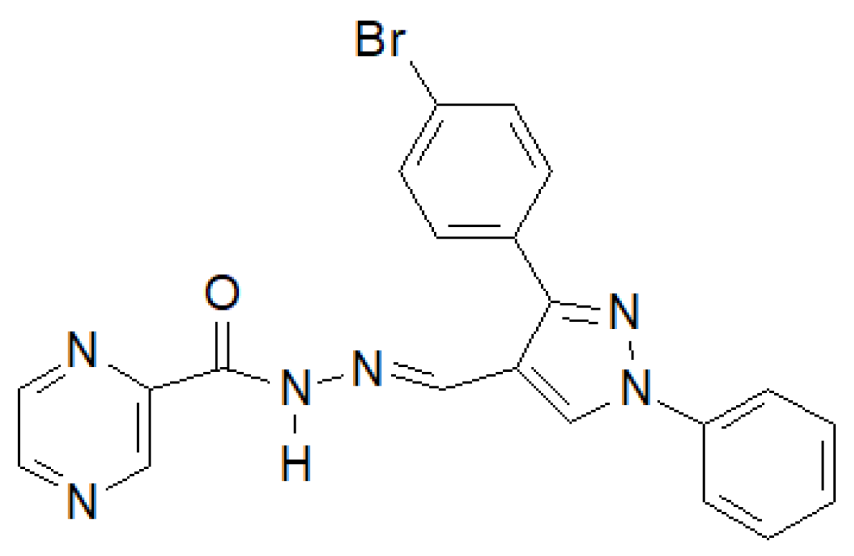
5. Hydrazide–Hydrazones
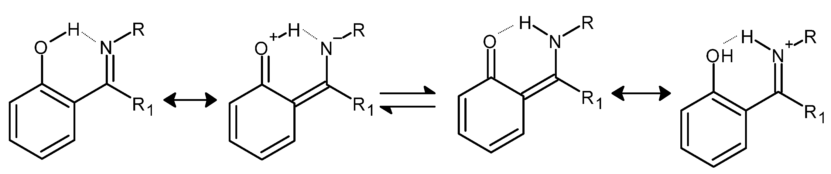
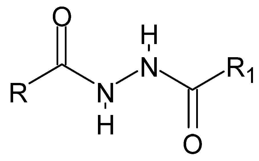
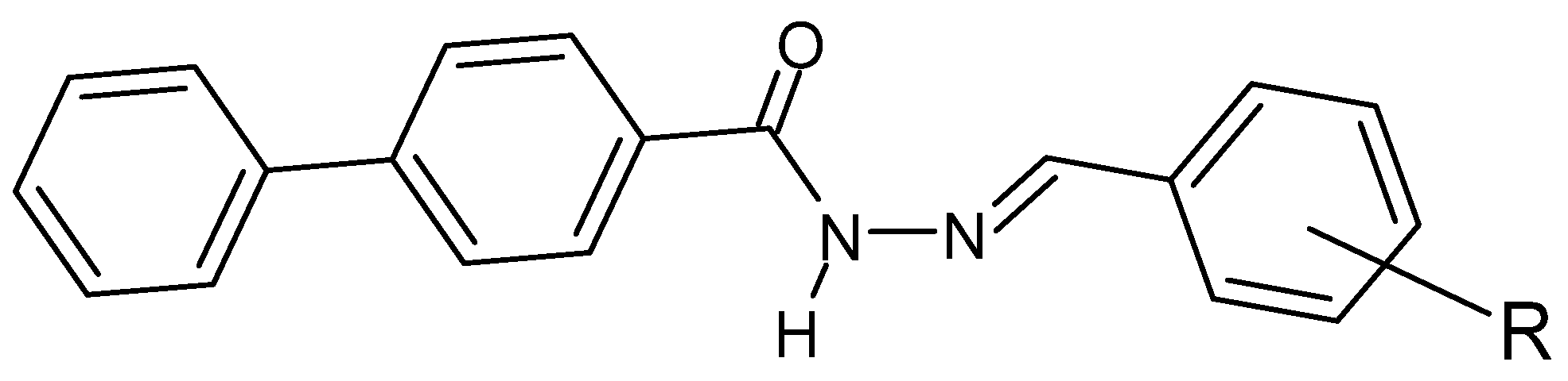
5.1. Structure
5.2. Importance
6. Conclusions
Author Contributions
Funding
Institutional Review Board Statement
Informed Consent Statement
Conflicts of Interest
References
- Schiff, H. Mittheilungen aus dem Universitäts-laboratorium in Pisa: 2. Eine neue Reihe organischer Basen [Communications from the university laboratory in Pisa: 2. A new series of organic bases]. Ann. Der Chem. Und Pharm. 1864, 131, 118–119. (In German) [Google Scholar] [CrossRef]
- Moss, G.P.; Smith, P.A.S.; Tavernier, D. Glossary of class names of organic compounds and reactivity intermediates based on structure (IUPAC Recommendations 1995). Pure Appl. Chem. 1995, 67, 1307–1375. [Google Scholar] [CrossRef]
- Pfeiffer, P.; Breith, E.; Llibbe, E.; Tsumaki, T. Tricyclische orthokondensierte Nebenvalenzringe. Justus Liebigs Ann. Chem. 1933, 503, 84–130. [Google Scholar] [CrossRef]
- Hunter, L.; Marriott, J.A. Co-ordinated copper and nickel compounds of salicylidene derivatives. J. Chem. Soc. 1937, 422, 2000–2003. [Google Scholar] [CrossRef]
- Sacconi, L.; Ciampolini, M.; Maggio, F.; Cavasini, F.P. Studies in Coordination Chemistry. IX.1Investigation of the Stereochemistry of Some Complex Compounds of Cobalt(II) with N-Substituted Salicylaldimines. J. Am. Chem. Soc. 1962, 84, 3246–3248. [Google Scholar] [CrossRef]
- Holm, R.H.; Swaminathan, K. Studies on Nickel(II) Complexes. III. Bis-(N-arylsalicylaldimine) Complexes. Inorg. Chem. 1962, 1, 599–607. [Google Scholar] [CrossRef]
- Percy, G.C.; Thornton, D.A. N-aryl salicylaldimine complexes: Infrared and PMR spectra of the ligands and vibrational frequencies of their metal (II) chelates. J. Inorg. Nucl. Chem. 1972, 34, 3357–3367. [Google Scholar] [CrossRef]
- Lundgren, R.L.; Stradiotto, M. Ligand Design in Metal Chemistry: Reactivity and Catalysis; Key Concepts in Ligand Design: An Introduction; John Wiley & Sons, Ltd.: Chichester, UK; Hoboken, NJ, USA, 2016; pp. 1–13. [Google Scholar]
- Fryzuk, M.D.; Haddad, T.S.; Berg, D.J.; Rettig, S.J. Phosphine complexes of the early metals and the lanthanoids. Pure Appl. Chem. 1991, 63, 845–850. [Google Scholar] [CrossRef]
- Dilli, S.; Maitra, A.M.; Patsalides, E. Oxidative transformations in nickel(II) chelates of tetradentate Schiff bases. Inorg. Chem. 1982, 21, 2832–2838. [Google Scholar] [CrossRef]
- Camp, C.; Chatelain, L.; Mougel, V.; Pecaut, J.; Mazzanti, M. Ferrocene-Based Tetradentate Schiff Bases as Supporting Ligands in Uranium Chemistry. Inorg. Chem. 2015, 54, 5774–5783. [Google Scholar] [CrossRef]
- Zoubi, W.A. Biological Activities of Schiff Bases and Their Complexes: A Review of Recent Works. Int. J. Org. Chem. 2013, 3, 73–95. [Google Scholar] [CrossRef]
- Donzelli, A.; Metushi, I.; Potvin, P.G. Titanium(IV) Complexes of Disulfide-Linked Schiff Bases. Inorg. Chem. 2012, 51, 5138–5145. [Google Scholar] [CrossRef]
- Chaudhary, N.K.; Mishra, P. Metal complexes of a novel Schiff base based on penicillin: Characterization, molecular modeling, and antibacterial activity study. Bioinorg. Chem. Appl. 2017, 2017, 6927675. [Google Scholar] [CrossRef]
- Chaudhary, N.K.; Mishra, P. Bioactivity of some divalent M(II) complexes of penicillin based Schiff base ligand: Synthesis, spectroscopic characterization, and thermal study. J. Saudi Chem. Soc. 2018, 22, 601–613. [Google Scholar] [CrossRef]
- Md Yusof, E.N.; Ravoof, T.B.S.A.; Tiekink, E.R.T.; Veerakumarasivam, A.; Crouse, K.A.; Tahir, M.I.M.; Ahmad, H. Synthesis, characterization and biological evaluation of transition metal complexes derived from N, S bidentate ligands. Int. J. Mol. Sci. 2015, 16, 11034–11054. [Google Scholar] [CrossRef]
- Sridhar, G.; Bilal, M.; Easwaramoorthy, D.; Rani, K.; Kumar, S.; Manohar, C.S. Synthesis, Characterization and Antimicrobial Activities of Copper, Nickel, Cobalt, Chromium Complexes Derived from (Z)-4-Fluoro-N-(2,7-dimethylhept-6-enylidene) benzenamine. J. Braz. Chem. Soc. 2017, 28, 756–767. [Google Scholar] [CrossRef]
- Moustafa, S.A.; Ali, M.M.; El-rashedy, A.A. Synthesis, anticancer activity and molecular docking study of Schiff base complexes containing thiazole moiety. J. Basic Appl. Sci. 2016, 5, 85–96. [Google Scholar] [CrossRef]
- El-Boraey, H.A.; EL-Gammal, O.A. Novel (N4) Macrocyclic Metal Complexes: Synthesis, Characterization, Spectral Studies and Anticancer Activity. Open Chem. J. 2018, 5, 51–63. [Google Scholar] [CrossRef]
- Hu, K.; Liu, C.; Li, J.; Liang, F. Copper(II) complexes based on quinoline-derived Schiff-base ligands: Synthesis, characterization, HSA/DNA binding ability, and anticancer activity. MedChemComm 2018, 9, 1663–1672. [Google Scholar] [CrossRef]
- Chioma, F.; Ekennia, A.C.; Osowole, A.A.; Okafor, S.N.; Ibeji, C.U.; Onwudiwe, D.C.; Ujam, O.T. Synthesis, characterization, in-vitro antimicrobial properties, molecular docking and DFT studies of 3-{(E)-[(4,6-dimethylpyrimidin-2-yl)imino]methyl} naphthalen-2-ol and Heteroleptic Mn(II), Co(II), Ni(II) and Zn(II) complexes. Open Chem. 2018, 16, 184–200. [Google Scholar] [CrossRef]
- Kuate, M.; Conde, M.A.; Nchimi, K.N.; Paboudam, A.G.; Ntum, S.-J.E.; Ndifon, P.T. Synthesis, characterization and antimicrobial studies of Co(II), Ni(II), Cu(II) and Zn(II) complexes of (E)-2-(4-dimethylbenzydimino)-Glycylglycine, (Glygly-DAB) a Schiff Base Derived from 4-Dimethylaminobenzaldehyde and glycylglycine. Int. J. Org. Chem. 2018, 8, 298–308. [Google Scholar] [CrossRef]
- Abu-khadra, A.S.; Afify, A.S.; Mohamed, A.; Farag, R.S.; Hassan, Y. Preparation, characterization and antimicrobial activity of Schiff base of (E)-N-(4-(Thiophen-2ylmethyleneamino) Phenylsulfonyl) Acetamide metal complexes. Open Bioact. Compd. J. 2018, 6, 1–10. [Google Scholar] [CrossRef]
- Festus, C.; Okafor, S.N.; Ekennia, A.C. Heteroleptic metal complexes of a Pyrimidinyl based Schiff base ligand incorporating 2,2′-Bipyridine moiety: Synthesis, characterization, and biological studies. Front. Chem. 2019, 7, 1–12. [Google Scholar] [CrossRef]
- Al-zaidi, B.H.; Hasson, M.M.; Ismail, A.H. New complexes of chelating Schiff base: Synthesis, spectral investigation, antimicrobial, and thermal behavior studies. J. Appl. Pharm. Sci. 2019, 9, 45–57. [Google Scholar] [CrossRef][Green Version]
- Kumar, S.; Hansda, A.; Chandra, A.; Kumar, A.; Kumar, M.; Sithambaresan, M.; Faizi, M.S.H.; Kumar, V. Co(II), Ni(II), Cu(II) and Zn(II) complexes of acenaphthoquinone 3-(4-benzylpiperidyl) thiosemicarbazone: Synthesis, structural, electrochemical and antibacterial studies. Polyhedron 2017, 134, 11–21. [Google Scholar] [CrossRef]
- Ott, I. On the medicinal chemistry of gold complexes as anticancer drugs. Coord. Chem. Rev. 2009, 253, 1670–1681. [Google Scholar] [CrossRef]
- Mihorianu, M.; Franz, M.H.; Jones, P.G.; Freytag, M.; Kelter, G.; Fiebig, H.-H.; Tamm, M.; Neda, I. N-Heterocyclic carbenes derived from imidazo-[1,5-a]pyridines related to natural products: Synthesis, structure and potential biological activity of some corresponding gold(I) and silver(I) complexes. Appl. Organometal. Chem. 2016, 30, 581–589. [Google Scholar] [CrossRef]
- Al-aghbari, S.A.; Al-shuja, O.M.; Al-badani, R.; Japir, A.A.M. Synthesis, characterization and anticancer activity studies of new Schiff base Pt (II) complex. J. Mater. Sci. Chem. Eng. 2019, 7, 94137. [Google Scholar] [CrossRef]
- Deng, J.; Yu, P.; Zhang, Z.; Zhang, J.; Sun, Z.; Cai, M.; Yuan, H.; Liang, H.; Yang, F. Novel Pt(II) complexes with modified aroyl-hydrazone Schiff- base ligands: Synthesis, cytotoxicity and action mechanism. Metallomics 2019, 11, 1847–1863. [Google Scholar] [CrossRef]
- Adeleke, A.A.; Zamisa, S.J.; Islam, M.S.; Olofinsan, K.; Salau, V.F.; Mocktar, C.; Omondi, B. Quinoline Functionalized Schiff Base Silver (I) Complexes: Interactions with Biomolecules and In Vitro Cytotoxicity, Antioxidant and Antimicrobial Activities. Molecules 2021, 26, 1205. [Google Scholar] [CrossRef]
- Al-Masoudi, N.A.; Aziz, N.; Mohammed, A. Synthesis and In vitro anti-HIV activity of some new Schiff base ligands derived from 5-Amino-4-phenyl-4H-1,2,4-triazole-3- thiol and their metal complexes. Phosphorus Sulfur Silicon Relat. Elem. 2009, 184, 2891–2901. [Google Scholar] [CrossRef]
- Westheimer, F.H.; Taguchi, K. Catalysis by molecular sieves in the preparation of ketimines and enamines. J. Org. Chem. 1971, 36, 1570–1572. [Google Scholar] [CrossRef]
- Król-Starzomska, I.; Filarowski, A.; Rospenk, M.; Koll, A.; Melikova, S. Proton Transfer Equilibria in Schiff Bases with Steric Repulsion. J. Phys. Chem. A 2004, 108, 2131–2138. [Google Scholar] [CrossRef]
- Chakraborti, A.K.; Bhagat, S.; Rudrawar, S. Magnesium perchlorate as an efficient catalyst for the synthesis of imines and phenylhydrazones. Tetrahdron Lett. 2004, 45, 7641–7644. [Google Scholar] [CrossRef]
- Dalpozzo, R.; de Nino, A.; Nardi, M.; Russo, B.; Procopio, A. Erbium(III) triflate: A valuable catalyst for the synthesis of aldimines, ketimines and enaminones. Synthesis 2006, 7, 1127–1132. [Google Scholar] [CrossRef]
- Naeimi, H.; Salimi, F.; Rabiei, K. Mild and convenient one pot synthesis of Schiff bases in the presence of P2O5/Al2O3 as new catalyst under solvent-free conditions. J. Mol. Catal. A Chem. 2006, 260, 100–104. [Google Scholar] [CrossRef]
- Barluenga, J.; Aznar, F.; Valdes, C. N-trialkylsilylimines as coupling partners for Pd-catalyzed C-N bond-forming reactions: One-step synthesis of imines and azadienes from aryl and alkenyl bromides. Angew. Chem. Int. Ed. 2004, 116, 347–349. [Google Scholar] [CrossRef]
- Arluenga, J.B.; Jimenez-Aquino, A.; Fernandez, M.A.; Aznar, F.; Valdes, C. Multicomponent and one-pot synthesis of trisubstituted pyridines through a Pd catalyzed cross-coupling/cross-coupling/cycloaddition sequence. Tetrahedron Lett. 2007, 64, 778–786. [Google Scholar] [CrossRef]
- Jiang, L.; Jin, L.; Tian, H.; Yuan, X.; Yu, X.; Xu, Q. Direct and mild palladium-catalyzed aerobic oxidative synthesis of imines from alcohols and amines under ambient conditions. Chem. Commun. 2011, 47, 10833–10835. [Google Scholar] [CrossRef]
- Huang, B.; Tian, H.; Lin, S.; Xie, M.; Yu, X.; Xu, Q. Cu(I)/TEMPO-catalyzed aerobic oxidative synthesis of imines directly from primary and secondary amines under ambient and neat conditions. Tetrahedron Lett. 2013, 54, 2861–2864. [Google Scholar] [CrossRef]
- Shiraishi, Y.; Ikeda, M.; Tsukamoto, D.; Tanaka, S.; Hirai, T. One-pot synthesis of imines from alcohols and amines with TiO2 loading Pt nanoparticles under UV irradiation. Chem. Commun. 2011, 47, 4811–4813. [Google Scholar] [CrossRef] [PubMed]
- Largeron, M.; Fleury, M.B. Bioinspired oxidation catalysts. Science 2013, 339, 43–44. [Google Scholar] [CrossRef] [PubMed]
- Lan, Y.S.; Liao, B.S.; Liu, Y.H.; Peng, S.M.; Liu, S.T. Preparation of imines by oxidative coupling of benzyl alcohols with amines catalysed by dicopper complexes. Eur. J. Org. Chem. 2013, 2013, 5160–5164. [Google Scholar] [CrossRef]
- Largeron, M. Protocols for the catalytic oxidation of primary amines to imines. Eur. J. Org. Chem. 2013, 2013, 5225–5235. [Google Scholar] [CrossRef]
- Joshi, H.H.; Kamounah, F.S.; Gooijer, C.; Zwan, G.; Antonov, L. Excited state intramolecular proton transfer in some tautomeric azo dyesand schiff bases containing an intramolecular hydrogen bond. J. Photochem. Photobiol. B 2002, 152, 183–191. [Google Scholar] [CrossRef]
- Abdel Aziz, A.A.; Salem, A.N.M.; Sayed, M.A.; Aboaly, M.M. Synthesis, structural characterization, thermal studies, catalytic efficiency and antimicrobial activity of some M(II) complexes with ONO tridentate Schiff base N-salicylidene Oaminophenol (saphH2). J. Mol. Struct. 2012, 1010, 130–138. [Google Scholar] [CrossRef]
- Saravanan, G.; Pannerselvam, P.; Prakash, C.R. Synthesis and anti-microbial screening of novel Schiff bases of 3-amino-2-methyl quinazolin 4-(3H)-one. J. Adv. Pharm. Technol. Res. 2010, 1, 320–325. [Google Scholar] [CrossRef]
- Gungor, O.; Gurkan, P. Synthesis and characterization of higher amino acid Schiff bases, as monosodium salts and neutral forms. Investigation of the intramolecular hydrogen bonding in all Schiff bases, antibacterial and antifungal activities of neutral forms. J. Mol. Struct. 2014, 1074, 62–70. [Google Scholar] [CrossRef]
- Kumar, K.S.; Ganguly, S.; Veerasamy, R.; De Clercq, E. Synthesis, antiviral activity and cytotoxicity evaluation of Schiff bases of some 2-phenyl quinazoline-4 (3) H-ones. Eur. J. Med. Chem. 2010, 45, 5474–5479. [Google Scholar] [CrossRef]
- Sriram, D.; Yogeswari, P.; Myneedu, N.S.; Saraswat, V. Abacavir prodrugs: Microwave-assisted synthesis and their evaluation of anti-HIV activities. Bioorg. Med. Chem. Lett. 2006, 16, 2127–2129. [Google Scholar] [CrossRef]
- Hu, G.; Wang, G.; Duan, N.; Wen, X.; Cao, T.; Xie, S.; Huang, W. Design, synthesis and antitumor activities of fluoroquinolone C-3 heterocycles (IV): S-triazole Schiff–Mannich bases derived from ofloxacin. Acta Pharm. Sin. B 2012, 2, 312–317. [Google Scholar] [CrossRef]
- El-wakiel, N.; El-keiy, M.; Gaber, M. Synthesis, spectral, antitumor, antioxidant and antimicrobial studies on Cu (II), Ni (II) and Co (II) complexes of 4-[(1HBenzoimidazol-2-ylimino)-methyl]-benzene-1, 3-diol. Spectrochim. Acta A Mol. Biomol. Spectrosc. 2015, 147, 117–123. [Google Scholar] [CrossRef] [PubMed]
- Pontiki, E.; Hadjipavlou-Litina, D.; Chaviara, A. Evaluation of anti-inflammatory and antioxidant activities of copper (II) Schiff mono-base and copper (II) Schiff base coordination compounds of dien with heterocyclic aldehydes and 2-amino-5-methylthiazole. J. Enzym. Inhib. Med. Chem. 2008, 23, 1011–1017. [Google Scholar] [CrossRef] [PubMed]
- Rathelot, P.; Vanelle, P.; Gasquet, M.; Delmas, F.; Crozet, M.P.; Timon-David, P.J. Maldonado, Synthesis of novel functionalized 5-nitroisoquinolines and evaluation of in vitro Antimalarial activity. Eur. J. Med. Chem. 1995, 30, 503–508. [Google Scholar] [CrossRef]
- Bensaber, S.M.; Allafe, H.; Ermeli, N.B.; Mohamed, S.B.; Zetrini, A.A.; Alsabri, S.G.; Erhuma, M.; Hermann, A.; Jaeda, M.I.; Gbaj, A.M. Chemical synthesis, molecular modelling, and evaluation of anticancer activity of some pyrazol-3-one Schiff base derivatives. Med. Chem. Res. 2014, 23, 5120–5134. [Google Scholar] [CrossRef]
- Desai, S.B.; Desai, P.B.; Desai, K.R. Synthesis of some Schiff bases, thiazolidinones and azetidinones derived from 2,6-diaminobenzo1,2-d: 4,5-d’ bisthiazole and their anticancer activities. Heterocycl. Commun. 2001, 7, 83–90. [Google Scholar] [CrossRef]
- Przybylski, P.; Huczynski, A.; Pyta, K.; Brzezinski, B.; Bartl, F. Biological properties of Schiff bases and azo derivatives of phenols. Curr. Org. Chem. 2009, 13, 124–148. [Google Scholar] [CrossRef]
- Bluhm, M.E.; Ciesielski, M.; Gorls, H.; Walter, O.; Doring, M. Complexes of Schiff Bases and Intermediates in the Copper-Catalyzed Oxidative Heterocyclization by Atmospheric Oxygen. Inorg. Chem. 2003, 42, 8878–8885. [Google Scholar] [CrossRef]
- Wang, L.; Qin, W.; Tang, X.; Dou, W.; Liu, W.; Teng, Q.; Yao, X. A selective, cell-permeable fluorescent probe for Al3+ in living cells. Org. Biomol. Chem. 2010, 8, 3751–3757. [Google Scholar] [CrossRef]
- Zoubi, W.A.; Kandii, F.; Chebani, K. Active transport of metal ions by using Schiff bases. Phys. Sci. Res. Int. 2014, 2, 12–23. [Google Scholar]
- Velezheva, V.; Brennan, P.; Ivanov, P.; Kornienko, A.; Lyubimov, S.; Kazarian, K.; Nikonenko, B.; Majorov, K.; Apt, A. Synthesis, Spectroscopic, Molecular Modeling and Anti-Fungal Studies of Some Divalent Metal Complexes of 4-Hydroxyacetophenone Isonicotinoyl Hydrazone. Bioorg. Med. Chemi. Lett. 2016, 26, 978–985. [Google Scholar] [CrossRef] [PubMed]
- Gutowsky, H.S.; Holm, C.H. Rate Processes and Nuclear Magnetic Resonance Spectra II. Hindered Internal Rotation of Amides. J. Chem. Phys. 1956, 25, 1228–1234. [Google Scholar] [CrossRef]
- Grel, P.L.; Salaün, A.; Mocquet, C.; Grel, B.L.; Roisnel, T.; Potel, M. Postsynthetic Modification of C3-Symmetric Aza-β3-Cyclohexapeptides. J. Org. Chem. 2011, 76, 8756–8767. [Google Scholar] [CrossRef]
- Gloaguen, E.; Brenner, V.; Alauddin, M.; Tardivel, B.; Mons, M.; Zehnacker-Rentien, A.; Aitken, D.J. Direct Spectroscopic Evidence of Hyperconjugation Unveils the Conformational Landscape of Hydrazides. Angew. Chem. Int. Ed. 2014, 53, 13756–13759. [Google Scholar] [CrossRef]
- Takahashi, O.; Kirikoshi, R. Intramolecular cyclization of aspartic acid residues assisted by three water molecules: A density functional theory study. Comput. Sci. Discov. 2014, 7, 015005. [Google Scholar] [CrossRef]
- Knapp, S.; Toby, B.H.; Sebastian, M.; Krogh-Jespersen, K.; Potenza, J.A. Relative reactivity and structures of benzoyltrimethylhydrazine and 1-benzoyl-2-methylpyrazolidine. J. Org. Chem. 1981, 46, 2490–2497. [Google Scholar] [CrossRef]
- Stackhouse, J.; Baechler, R.D.; Mislow, K. Pyramidal inversion barriers: The significance of ground state geometry. Tetrahedron Lett. 1971, 12, 3437–3440. [Google Scholar] [CrossRef]
- Andrade, L.A.F.; Silla, J.M.; Cormanich, R.A.; Freitas, M.P. Infrared Fingerprints of nN → σ*NH Hyperconjugation in Hydrazides. J. Org. Chem. 2017, 82, 12181–12187. [Google Scholar] [CrossRef]
- Lii, J.-H.; Chen, K.-H.; Allinger, N.L. Alcohols, Ethers, Carbohydrates, and Related Compounds Part V.2 The Bohlmann Torsional Effect. J. Phys. Chem. A 2004, 108, 3006–3015. [Google Scholar] [CrossRef]
- Bohlmann, F. Zur Konfigurationsbestimmung von Chinolizin-Derivaten. Agnew. Chem. 1957, 69, 641–642. [Google Scholar] [CrossRef]
- Gribble, G.W.; Nelson, R.B. Conformational Requirements for the Existence of Bohlmann Bands in the Infrared Spectra of Indolo[2,3-a]quinolizidines. I. cis- and trans-2-tert-Butyl Derivatives. J. Org. Chem. 1973, 38, 2831–2834. [Google Scholar] [CrossRef]
- Skolik, J.; Krueger, P.J.; Wiewiorowski, M. Correlation between the stereochemistry of quinolizidine alkaloids and their infrared spectra from 2840-2600 CM−1. Tetrahedron 1968, 24, 5439–5456. [Google Scholar] [CrossRef]
- Wolfe, S.; Schlegel, H.B.; Whangbo, M.-H.; Bernardi, F. On the origin of the Bohlmann bands. Can. J. Chem. 1974, 52, 3787–3792. [Google Scholar] [CrossRef]
- Ernstbrunner, E.E.; Hudec, J. Bohlmann bands—A reassessment. J. Mol. Struct. 1973, 17, 249–256. [Google Scholar] [CrossRef]
- Moss, G.P. Basic terminology of stereochemistry (IUPAC Recommendations 1996). Pure Appl. Chem. 1996, 68, 2193–2222. [Google Scholar] [CrossRef]
- Patil, S.; Kuman, M.M.; Palvai, S.; Sengupta, P.; Basu, S. Impairing Powerhouse in Colon Cancer Cells by Hydrazide−Hydrazone-Based Small Molecule. ACS Omega 2018, 3, 1470–1481. [Google Scholar] [CrossRef]
- Hastings, J.; Owen, G.; Dekker, A.; Ennis, M.; Kale, N.; Muthukrishnan, V.; Turner, S.; Swainston, N.; Mendes, P.; Steinbeck, C. Improved services and an expanding collection of metabolites. Nucleic Acids Res. 2016, 44, D1214–D1219. [Google Scholar] [CrossRef]
- Tiec, C.L.; Barrail, A.; Goujard, C.; Taburet, A.-M. Clinical pharmacokinetics and summary of efficacy and tolerability of atazanavir. Clin. Pharmacokinet. 2005, 44, 1035–1050. [Google Scholar] [CrossRef]
- Ventura, C.; Martins, F. Application of Quantitative Structure−Activity Relationships to the Modeling of Antitubercular Compounds. 1. The Hydrazide Family. J. Med. Chem. 2008, 51, 612–624. [Google Scholar] [CrossRef]
- Nagy, J.M.; Cass, A.E.G.; Brown, K.A. Purification and Characterization of Recombinant Catalase-Peroxidase, Which Confers Isoniazid Sensitivity in Mycobacterium tuberculosis. J. Biol. Chem. 1997, 272, 31265–31271. [Google Scholar] [CrossRef]
- Ragnarsson, U. Synthetic methodology for alkyl substituted hydrazines. Chem. Soc. Rev. 2001, 30, 205–213. [Google Scholar] [CrossRef]
- Cheng, C.; Sun, J.; Wang, C.; Zhang, Y.; Wei, S.; Jiang, F.; Wu, Y. Protonated N′-benzyl-N′-prolyl proline hydrazide as highly enantioselective catalyst for direct asymmetric aldol reaction. Chem. Commun. 2006, 37, 215–217. [Google Scholar] [CrossRef] [PubMed]
- Xiong, X.; Jiang, Y.; Ma, D. Assembly of N,N-Disubstituted Hydrazines and 1-Aryl-1H-indazoles via Copper-Catalyzed Coupling Reactions. Org. Lett. 2012, 14, 2552–2555. [Google Scholar] [CrossRef] [PubMed]
- Zhang, Y.-G.; Liu, X.-L.; He, Z.-Y.; Li, X.-M.; Kang, H.-J.; Tian, S.-K. Palladium/Copper-Catalyzed Oxidative Arylation of Terminal Alkenes with Aroyl Hydrazides. Chem. Eur. J. 2014, 20, 2765–2769. [Google Scholar] [CrossRef] [PubMed]
- Yang, F.-L.; Ma, X.-T.; Tian, S.-K. Oxidative Mizoroki-Heck-Type Reaction of Arylsulfonyl Hydrazides for a Highly Regio- and Stereoselective Synthesis of Polysubstituted Alkenes. Chem. Eur. J. 2012, 18, 1582–1585. [Google Scholar] [CrossRef] [PubMed]
- Wang, T.-T.; Yang, F.-L.; Tian, S.-K. Copper-Catalyzed Sulfenylationof Boronic Acids with Sulfonyl Hydrazides. Adv. Synth. Catal. 2015, 357, 928–932. [Google Scholar] [CrossRef]
- Eldridge, G.M.; Weiss, G.A. Hydrazide Reactive Peptide Tags for Site-Specific Protein Labeling. Bioconjugate Chem. 2011, 22, 2143–2153. [Google Scholar] [CrossRef]
- Huang, J.-P.; Bian, X.-Z.; Chang, K.; Hou, L.-J.; Feng, H.-T. Capture and Analysis of Cell Surface N-Glycans by Hydrazide-Modified Magnetic Beads and CE-LIF. Chromatographia 2019, 82, 1079–1088. [Google Scholar] [CrossRef]
- Wang, L.; Aryal, U.K.; Dai, Z.; Mason, A.C.; Monroe, M.E.; Tian, Z.-X.; Zhou, J.-Y.; Su, D.; Weitz, K.K.; Liu, T.; et al. Mapping N-Linked Glycosylation Sites in the Secretome and Whole Cells of Aspergillus niger Using Hydrazide Chemistry and Mass Spectrometry. J. Proteome Res. 2012, 11, 143–156. [Google Scholar] [CrossRef]
- Roveda, J.-G.; Clavette, C.; Hunt, A.D.; Gorelsky, S.I.; Whipp, C.J.; Beauchemin, A.M. Hydrazides as Tunable Reagents for Alkene Hydroamination and Aminocarbonylation. J. Am. Chem. Soc. 2009, 131, 8740–8741. [Google Scholar] [CrossRef]
- White, E.H.; Roswell, D.F. Chemiluminescence of organic hydrazides. Acc. Chem. Res. 1970, 3, 54–62. [Google Scholar] [CrossRef]
- Jin, Y.; Sun, Y.; Li, C.; Yang, C. A highly selective chemiluminescent probe for the detection of chromium(VI). Spectrochim. Acta A Mol. Biomol. Spectrosc. 2018, 192, 82–87. [Google Scholar] [CrossRef] [PubMed]
- Arakawa, H.; Maeda, M.; Tsuji, A.; Takahashi, T. Highly Sensitive Biotin-Labelled Hybridization Probe. Chem. Pharm. Bull 1989, 37, 1831–1833. [Google Scholar] [CrossRef] [PubMed]
- Begum, A.; Sujatha, D.; Prasad, K.V.S.R.G.; Bharathi, K. A Review on Azapeptides: The Promising Peptidomimetics. Asian J. Chem. 2017, 29, 1879–1887. [Google Scholar] [CrossRef]
- Verhelst, S.H.L.; Witte, M.D.; Arastu-Kapur, S.; Fonovic, M.; Bogyo, M. Novel Aza Peptide Inhibitors and Active-Site Probes of Papain-Family Cysteine Proteases. ChemBioChem 2006, 7, 943–950. [Google Scholar] [CrossRef]
- Lee, H.-J.; Ahn, I.-A.; Ro, S.; Choi, K.-H.; Choi, Y.-S.; Lee, K.-B. Role of azaamino acid residue in β-turn formation stability in designed peptide. J. Peptide Res. 2000, 56, 35–46. [Google Scholar] [CrossRef]
- Słomiak, K.; Łazarenkow, A.; Checinska, L.; Kusz, J.; Ochocki, J.; Nawrot-Modranka, J. Synthesis, Spectroscopic Analysis and Assessment of the Biological Activity of New Hydrazine and Hydrazide Derivatives of 3-Formylchromone. Molecules 2018, 23, 2067. [Google Scholar] [CrossRef]
- Zhang, X.; Breslav, M.; Grimm, J.; Guan, K.; Huang, A.; Liu, F.; Maryanoff, C.A.; Palmer, D.; Patel, M.; Qian, Y.; et al. A new procedure for preparation of carboxylic acid hydrazides. J. Org. Chem. 2002, 67, 9471–9474. [Google Scholar] [CrossRef]
- Curtius, T.; Schöfer, G.; Schwan, N. Hydrazide und Azide organischer Säuren. IV. Abhandlung. 26. Ueber einige Hydrazide einbasischer und zweibasischer Säuren der Fettreihe. J. Prakt. Chem. 1895, 51, 180–196. [Google Scholar] [CrossRef]
- Slagel, R.C. Aminimides VI. Synthesis of aminimides from carboxylic acid esters, unsymmetrically disubstituted hydrazines, and epoxides. J. Org. Chem. 1968, 33, 1374–1378. [Google Scholar] [CrossRef]
- Han, H.; Janda, K.D. Azatides: Solution and Liquid Phase Syntheses of a New Peptidomimetic. J. Am. Chem. Soc. 1996, 118, 2539–2544. [Google Scholar] [CrossRef]
- Grel, P.L.; Salaün, A.; Potel, M.; Grel, B.L.; Lassagne, F. Aza-β3-Cyclohexapeptides: Pseudopeptidic Macrocycles with Interesting Conformational and Configurational Properties Slow Pyramidal Nitrogen Inversion in 24-Membered Rings! J. Org. Chem. 2006, 71, 5638–5645. [Google Scholar] [CrossRef] [PubMed]
- Samdal, S.; Møllendal, H. The Structural and Conformational Properties of Formic Hydrazide (Formylhydrazine) Studied by Microwave Spectroscopy and Quantum Chemical Calculations. J. Phys. Chem. A 2003, 107, 8845–8850. [Google Scholar] [CrossRef]
- Elian, M.; Hoffmann, R. Bonding Capabilities of Transition Metal Carbonyl Fragment. Inorg. Chem. 1975, 14, 1058–1076. [Google Scholar] [CrossRef]
- Hoffmann, R. Building Bridges Between Inorganic and Organic Chemistry (Nobel Lecture). Angew. Chem. Int. Ed. Engl. 1982, 21, 711–724. [Google Scholar] [CrossRef]
- Licandro, E.; Perdicchia, D. N-Acylhydrazines: Future Perspectives Offered by New Syntheses and Chemistry. Eur. J. Org. Chem. 2004, 4, 665–675. [Google Scholar] [CrossRef]
- Pradier, C.M.; Salmain, M.; Boujday, S. Biointerface Characterization by Advanced IR Spectroscopy; Elsevier: Amsterdam, The Netherlands, 2011; Chapter 7; pp. 167–216. [Google Scholar]
- Bhatt, V. Essentials of Coordination Chemistry; Academic Press: Cambridge, MA, USA, 2016; Chapter 8; pp. 191–236. [Google Scholar]
- Shriver, D.F. Encyclopædia Britannica. 2018. Available online: https://www.britannica.com/science/organometallic-compound (accessed on 5 December 2020).
- Licandro, E.; Maiorana, S.; Vandoni, B.; Perdicchia, D.; Paravidino, P.; Baldoli, C. Synthesis of Medium-Sized N-Heterocycles through RCM of Fisher-type Hydrazino Carbene Complexes. Synlett 2001, 6, 757–760. [Google Scholar] [CrossRef]
- Nielsen, P.E.; Egholm, M.; Buchardt, O. Peptide nucleic acid (PNA). A DNA mimic with a peptide backbone. Bioconjugate Chem. 1994, 5, 3–7. [Google Scholar] [CrossRef]
- Cauteruccio, S.; Licandro, E.; Panigati, M.; D’Alfonso, G.; Maiorana, S. Modifying the properties of organic molecules by conjugation with metal complexes: The case of peptide nucleic acids and of the intrinsically chiral thiahelicenes. Coord. Chem. Rev. 2019, 386, 119–137. [Google Scholar] [CrossRef]
- Cavier, R.; Rips, R. Dihydrazides, a New Class of Anthelmintics. J. Med. Chem. 1965, 8, 706–708. [Google Scholar] [CrossRef]
- Zadykowicz, B.; Romanowska, A.; Pieńkos, M. Photophyysical basis of chemiluminescent labeling—A modern medical diagnostic tool. Wiadomości Chem. 2018, 72, 887–906. [Google Scholar]
- Albrecht, H.O. Uber die Chemiluminescenz der Aminophthalhydrazid. Z. Für Phys. Chem. 1928, 136, 321–330. [Google Scholar] [CrossRef]
- Wang, J.; Yang, Q.; Song, H.; Zhang, W. A fluorescent probe of N′-formyl-rhodamine B hydrazide: Structure and spectral properties of protonation behaviour. Org. Biomol. Chem. 2012, 10, 7677–7680. [Google Scholar] [CrossRef] [PubMed]
- Kricka, L.J. Chemiluminescent and bioluminescent techniques. Clin. Chem. 1991, 37, 1472–1481. [Google Scholar] [CrossRef] [PubMed]
- Bedia, K.-K.; Elçin, O.; Seda, U.; Fatma, K.; Nathaly, S.; Sevim, R.; Dimoglo, A. Synthesis and characterization of novel hydrazide-hydrazones and the study of their structure-antituberculosis activity. Eur. J. Med. Chem. 2006, 41, 1253–1261. [Google Scholar] [CrossRef]
- Enders, D.; Schubert, H.; Nübling, C. Enantioselective Synthesis of α-Substituted Primary Amines by Nucleophilic Addition to Aldehyde-SAMP Hydrazones. Angew. Chem. Int. Ed. Engl. 1986, 25, 1109–1110. [Google Scholar] [CrossRef]
- Perdicchia, D.; Licandro, E.; Maiorana, S.; Baldoli, C.; Giannini, C. A new ‘one-pot’ synthesis of hydrazidesby reduction of hydrazones. Tetrahedron 2003, 59, 7733–7742. [Google Scholar] [CrossRef]
- Lazny, R.; Nodzewska, A. N,N-dialkylhydrazones in organic synthesis. From simple N,N-dimethylhydrazones to supported chiral auxiliaries. Chem. Rev. 2010, 110, 1386–1434. [Google Scholar] [CrossRef]
- Curti, C.; Battistini, L.; Sartori, A.; Zanardi, F. New Developments of the Principle of Vinylogy as Applied to π-Extended Enolate-Type Donor Systems. Chem. Rev. 2020, 120, 2448–2612. [Google Scholar] [CrossRef]
- Lei, Y.; Li, T.-Z.; Fu, C.; Guan, X.-L.; Tan, Y. Synthesis, crystal structures, and antibacterial activity of a series of hydrazone compounds derived from 4-methylbenzohydrazide. J. Chil. Chem. Soc. 2015, 60, 2961–2965. [Google Scholar] [CrossRef]
- Navakoski de Oliveira, K.; Chiaradia, L.D.; Alves Martins, P.G.; Mascarello, A.; Sechini Cordeiro, M.N.; Carvalho Guido, R.V.; Andricopulo, A.D.; Yunes, R.A.; Nunes, R.J.; Vernalb, J.; et al. Sulfonyl-hydrazones of cyclic imides derivatives as potent inhibitors of the Mycobacterium tuberculosisprotein tyrosine phosphatase B (PtpB). Med. Chem. Commun. 2011, 2, 500–504. [Google Scholar] [CrossRef]
- Cunha, M.R.; Tavares, M.T.; Carvalho, C.F.; Silva, N.A.T.; Souza, A.D.F.; Pereira, G.J.V.; Ferreira, F.F.; Parise-Filho, R. Environmentally Safe Condition for the Synthesis of Aryl and Alkyl Sulfonyl Hydrazones via One-Pot Reaction. Sustain. Chem. Eng. 2016, 4, 1899–1905. [Google Scholar] [CrossRef]
- La Regina, G.; Gatti, V.; Piscitelli, F.; Silvestri, R. Open Vessel and Cooling while Heating Microwave-Assisted Synthesis of Pyridinyl N-Aryl Hydrazones. ACS Comb. Sci. 2011, 13, 2–6. [Google Scholar] [CrossRef] [PubMed]
- Cao, W.; Liu, Y.; Zhang, T.; Jia, J. Synthesis, characterization, theoretical and antimicrobial studies of tridentate hydrazone metal complexes of Zn(II), Cd(II), Cu(II) and Co(III). Polyhedron 2018, 147, 62–68. [Google Scholar] [CrossRef]
- Anacona, J.R.; Rincones, M. Tridentate hydrazone metal complexes derived from cephalexin and 2-hydrazinopyridine: Synthesis, characterization and antibacterial activity. Spectrochim. Acta A Mol. Biomol. Spectrosc. 2015, 141, 169–175. [Google Scholar] [CrossRef]
- Aly, S.A.; Fathalla, S.K. Preparation, characterization of some transition metal complexes of hydrazone derivatives and their antibacterial and antioxidant activities. Arab. J. Chem. 2020, 13, 3735–3750. [Google Scholar] [CrossRef]
- Özmen, Ü.Ö.; Olgun, G. Synthesis, characterization and antibacterial activity of new sulfonyl hydrazone derivatives and their nickel(II) complexes. Spectrochim. Acta A 2008, 70, 641–645. [Google Scholar] [CrossRef]
- Friedman, L.; Litle, R.L.; Reichle, W.R. p-Toluenesulfonylhydrazide. Org. Synth. 1960, 40, 93. [Google Scholar] [CrossRef]
- Popiołek, Ł. Hydrazide–hydrazones as potential antimicrobial agents: Overview of the literature since 2010. Med. Chem. Res. 2017, 26, 287–301. [Google Scholar] [CrossRef]
- Küçükgüzel, S.G.; Mazi, A.; Sahin, F.; Öztürk, S.; Stables, J. Synthesis and Biological Activities of Diflunisal Hydrazide-Hydrazones. Eur. J. Med. Chem. 2003, 38, 1005–1013. [Google Scholar] [CrossRef]
- Deep, A.; Jain, S.; Sharma, P.C.; Verma, P.; Kumar, M.; Dora, C.P. Design and biological evaluation of biphenyl-4-carboxylic acid hydrazide-hydrazone for antimicrobial activity. Acta Pol. Pharm. 2010, 67, 255–259. [Google Scholar] [PubMed]
- Kodisundaram, P.; Amirthaganesan, S.; Balasankar, T. Antimicrobial evaluation of a set of heterobicyclic methylthiadiazole hydrazones: Synthesis, characterization, and SAR studies. J. Agric. Food Chem. 2013, 61, 11952–11956. [Google Scholar] [CrossRef] [PubMed]
- Brentnall, M.; Rodriguez-Menocal, L.; De Guevara, R.L.; Cepero, E.; Boise, L.H. Caspase-9, caspase-3 and caspase-7 have distinct roles during intrinsic apoptosis. BMC Cell Biology 2013, 14, 32. [Google Scholar] [CrossRef]
- Ling, A.; Plewe, M.; Gonzalez, J.; Madsen, P.; Sams, C.K.; Lau, J.; Gregor, V.; Murphy, D.; Teston, K.; Kuki, A.; et al. Human Glucagon Receptor Antagonists Based on Alkylidene Hydrazides. Bioorg. Med. Chem. Lett. 2002, 12, 663–666. [Google Scholar] [CrossRef]
- Madsen, P.; Ling, A.; Plewe, M.; Sams, C.K.; Knudsen, L.B.; Sidelman, U.G.; Ynddal, L.; Brand, C.; Andersen, B.; Murphy, D.; et al. Optimization of alkylidene hydrazide based human glucagon receptor antagonists. Discovery of the highly potent and orally available 3-cyano-4-hydroxybenzoic acid [1-(2,3,5,6-tetramethylbenzyl)-1H-indol-4-ylmethylene]hydrazide. J. Med. Chem. 2002, 45, 5755–5775. [Google Scholar] [CrossRef]
- Noshiranzadeh, N.; Heidari, A.; Haghi, F.; Bikas, R.; Lis, T. Chiral lactic hydrazone derivatives as potential bioactive antibacterial agents: Synthesis, spectroscopic, structural and molecular docking studies. J. Mol. Struct. 2017, 1128, 391–399. [Google Scholar] [CrossRef]
- Ajani, O.O.; Iyaye, K.T.; Aderohunmu, D.V.; Olanrewaju, I.O.; Germann, M.W.; Olorunshola, S.J.; Bello, B.L. Microwaveassisted synthesis and antibacterial propensity of N0-s-benzylidene-2-propylquinoline-4-carbohydrazide and N0-((s-1H-pyrrol-2-yl)methylene)-2-propylquinoline-4-carbohydrazide motifs. Arab. J. Chem. 2020, 13, 1809–1820. [Google Scholar] [CrossRef]
- Salem, M.A.; Ragab, A.; El-Khalafawy, A.; Makhlouf, A.H.; Askar, A.A.; Ammar, Y.A. Design, synthesis, in vitro antimicrobial evaluation and molecular docking studies of indol-2-one tagged with morpholinosulfonyl moiety as DNA gyrase inhibitors. Bioorg. Chem. 2020, 96, 103619. [Google Scholar] [CrossRef]
- Tiwari, S.; Kirar, S.; Banerjee, U.C.; Neerupudi, K.B.; Singh, S.; Wani, A.A.; Bharatam, P.V.; Singh, I.P. Synthesis of N-substituted indole derivatives as potential antimicrobial and antileishmanial agents. Bioorg. Chem. 2020, 99, 103787. [Google Scholar] [CrossRef]
- El-Etrawy, A.-A.; Sherbiny, F.F. Design, synthesis, biological evaluation and molecular modeling investigation of new N0-(2-Thiouracil-5-oyl) hydrazone derivatives as potential anti-breast cancer and anti-bacterial agents. J. Mol. Struct. 2021, 1232, 129993. [Google Scholar] [CrossRef]
- Paruch, K.; Popiołek, Ł.; Biernasiuk, A.; Berecka-Rycerz, A.; Malm, A.; Gumieniczek, A.; Wujec, M. Novel Derivatives of 4-Methyl-1,2,3-Thiadiazole-5-Carboxylic Acid Hydrazide: Synthesis, Lipophilicity, and In Vitro Antimicrobial Activity Screening. Appl. Sci. 2021, 11, 1180. [Google Scholar] [CrossRef]
- Rohane, S.H.; Chauhan, A.J.; Fuloria, N.K.; Fuloria, S. Synthesis and in vitro antimycobacterial potential of novel hydrazones of eugenol. Arab. J. Chem. 2020, 13, 4495–4504. [Google Scholar] [CrossRef]
- Reis, R.C.N.; Oda, S.C.; De Almeida, M.V.; Lourenco, M.C.S.; Vicente, F.R.C.; Barbosa, N.R.; Trevizani, R.; Santos, P.L.C.; Le Hyaric, M. Synthesis and Antimicrobial Activity of Amphiphilic Carbohydrate Derivatives. J. Braz. Chem. Soc. 2008, 19, 1065–1072. [Google Scholar] [CrossRef]


| No. | Type of Substitution; Ligand/Complex | MIC Values (μg/mL) | |||
|---|---|---|---|---|---|
| B. subtilis | B. magaterium | S. aureus | S. enteritidis | ||
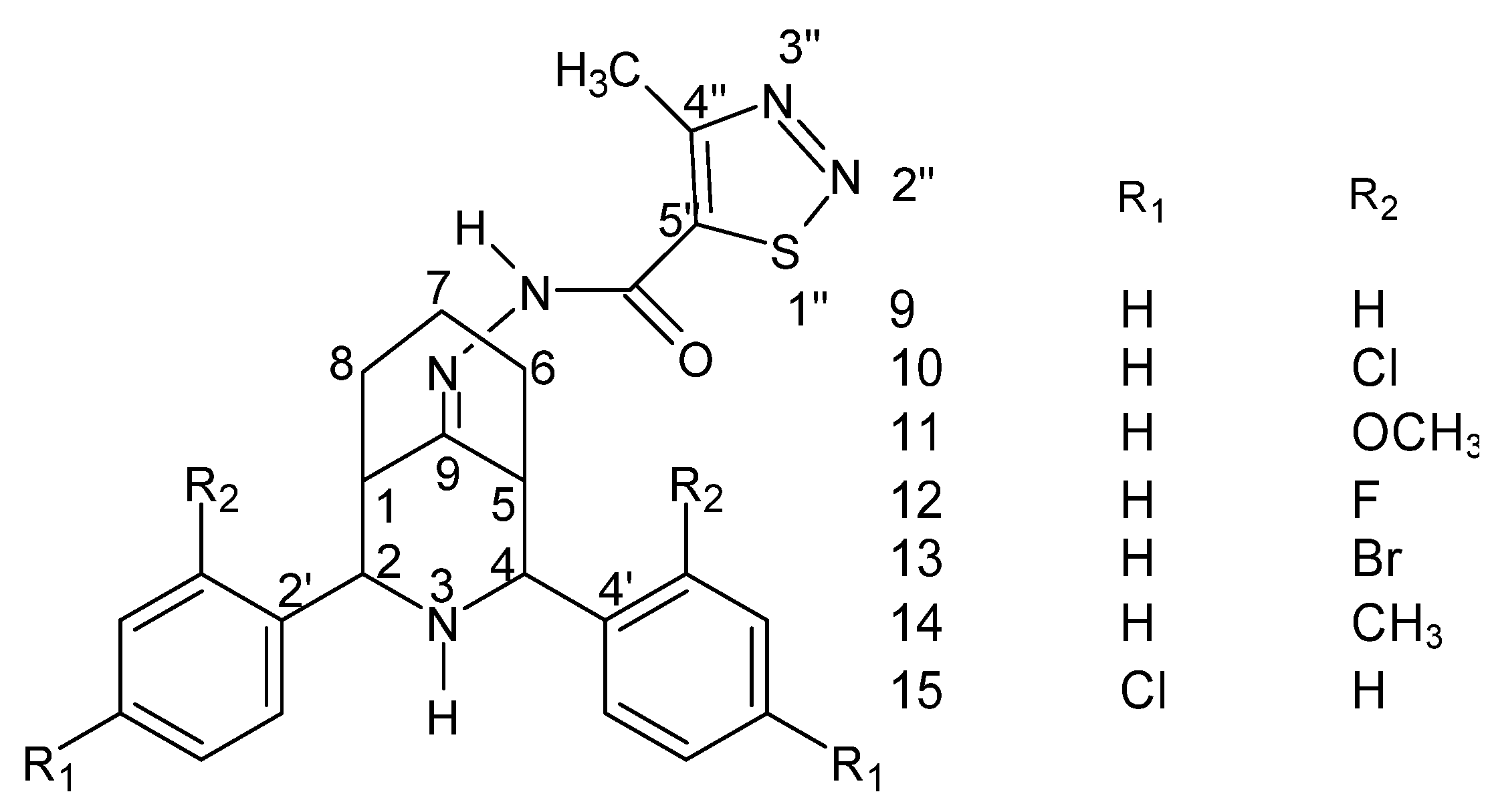
| 1 | R1=R2=H; ligand | 266 | 242 | 145 | 242 |
| R1=R2=H; complex | 595 | 541 | 541 | 595 | |
| 2 | R1=H, R2=CH3; ligand | 282 | 256 | 154 | 256 |
| R1=H, R2=CH3; complex | 569 | 683 | 626 | 626 | |
| 3 | R1=CH3, R2=H; ligand | 256 | 307 | 256 | 282 |
| R1=CH3, R2=H; complex | 569 | 626 | 569 | 626 | |
| 4 | R1=R2=CH3; ligand | 324 | 297 | 270 | 297 |
| R1=R2=CH3; complex | 717 | 657 | 657 | 597 | |
| No. | Type of Substitution/Reference Compound | MIC Value (μg/mL) | ||
|---|---|---|---|---|
| B. subtilis | K. pneumoniae | E. coli | ||
| 1 | para-Cl | 6.25 | 6.25 | 12.5 |
| 2 | para-F | 6.25 | 6.25 | 12.5 |
| 3 | para-Br | 6.25 | 6.25 | 12.5 |
| 4 | streptomycin | 12.5 | 12.5 | 25.0 |
| No. | Type of Substitution/Reference Compound | MIC Value (μg/mL) | |||
|---|---|---|---|---|---|
| A. flavus | A. niger | C. albicans | Candida6 | ||
| 1 | para-Cl | 12.5 | 12.5 | 6.25 | 6.25 |
| 2 | orto-Cl | 12.5 | 12.5 | 6.25 | 6.25 |
| 3 | para-F | 12.5 | 12.5 | 6.25 | 6.25 |
| 4 | para-Br | 12.5 | 12.5 | 6.25 | 25.0 |
| 5 | fluconazole | 25.0 | 25.0 | 12.5 | 25.0 |
Publisher’s Note: MDPI stays neutral with regard to jurisdictional claims in published maps and institutional affiliations. |
© 2022 by the authors. Licensee MDPI, Basel, Switzerland. This article is an open access article distributed under the terms and conditions of the Creative Commons Attribution (CC BY) license (https://creativecommons.org/licenses/by/4.0/).
Share and Cite
Raczuk, E.; Dmochowska, B.; Samaszko-Fiertek, J.; Madaj, J. Different Schiff Bases—Structure, Importance and Classification. Molecules 2022, 27, 787. https://doi.org/10.3390/molecules27030787
Raczuk E, Dmochowska B, Samaszko-Fiertek J, Madaj J. Different Schiff Bases—Structure, Importance and Classification. Molecules. 2022; 27(3):787. https://doi.org/10.3390/molecules27030787
Chicago/Turabian StyleRaczuk, Edyta, Barbara Dmochowska, Justyna Samaszko-Fiertek, and Janusz Madaj. 2022. "Different Schiff Bases—Structure, Importance and Classification" Molecules 27, no. 3: 787. https://doi.org/10.3390/molecules27030787
APA StyleRaczuk, E., Dmochowska, B., Samaszko-Fiertek, J., & Madaj, J. (2022). Different Schiff Bases—Structure, Importance and Classification. Molecules, 27(3), 787. https://doi.org/10.3390/molecules27030787