Article

Anisotropic Thermal Expansion and Electronic Structure of LiInSe2

by
Victor V. Atuchin
1,2,3,4,5,*,
Ludmila I. Isaenko
6,7,
Sergei I. Lobanov
6,7,
Alina A. Goloshumova
6,7,
Maxim S. Molokeev
8,9,10,
Zhaoming Zhang
11,
Xingyu Zhang
12,13,
Xingxing Jiang
12 and
Zheshuai Lin
12,13
1
Laboratory of Optical Materials and Structures, Institute of Semiconductor Physics, SB RAS, Novosibirsk 630090, Russia
2
Department of Applied Physics, Novosibirsk State University, Novosibirsk 630090, Russia
3
Research and Development Department, Kemerovo State University, Kemerovo 650000, Russia
4
Department of Industrial Machinery Design, Novosibirsk State Technical University, Novosibirsk 630073, Russia
5
R&D Center “Advanced Electronic Technologies”, Tomsk State University, Tomsk 634034, Russia
6
Laboratory of Crystal Growth, Sobolev Institute of Geology and Mineralogy, SB RAS, Novosibirsk 630090, Russia
7
Laboratory of Functional Materials, Novosibirsk State University, Novosibirsk 630090, Russia
8
Laboratory of Crystal Physics, Kirensky Institute of Physics, Federal Research Center KSC SB RAS, Krasnoyarsk 660036, Russia
9
Institute of Engineering Physics and Radioelectronic, Siberian Federal University, Krasnoyarsk 660041, Russia
10
Department of Physics, Far Eastern State Transport University, Khabarovsk 680021, Russia
11
Australian Science and Technology Organisation (ANSTO), Lucas Heights, NSW 2234, Australia
12
Functional Crystals Laboratory, Technical Institute of Physics and Chemistry, Chinese Academy of Sciences, Beijing 100190, China
13
University of the Chinese Academy of Sciences, Beijing 100049, China
*
Author to whom correspondence should be addressed.
Molecules 2022, 27(16), 5078; https://doi.org/10.3390/molecules27165078
Submission received: 29 June 2022 / Revised: 26 July 2022 / Accepted: 4 August 2022 / Published: 10 August 2022
(This article belongs to the Section Computational and Theoretical Chemistry)

Abstract

:
Optical quality cm-sized LiInSe2 crystals were grown using the Bridgman–Stockbarger method, starting from pure element reagents, under the conditions of a low temperature gradient of 5–6 degrees/cm and a slight melt overheating. The phase purity of the grown crystal was verified by the powder XRD analysis. The thermophysical characteristics of LiInSe2 were determined by the XRD measurements in the temperature range of 303–703 K and strong anisotropy of the thermal expansion coefficients was established. The following values of thermal expansion coefficients were determined in LiInSe2: αa = 8.1 (1), αb = 16.1 (2) and αc = 5.64 (6) MK−1. The electronic structure of LiInSe2 was measured by X-ray photoelectron spectroscopy. The band structure of LiInSe2 was calculated by ab initio methods.

1. Introduction

Complex chalcogenide compounds have interesting structural, chemical, and physical properties, and the materials are widely applied in modern optical and electronic technologies [1,2,3,4,5,6,7,8,9,10]. Among such compounds, the crystals with general composition LiMX2 (M = Al, In, Ga; X = S, Se, Te) have attracted particular research interests because of their valuable combination of optical and electronic characteristics, including thermal and chemical stabilities, wide transparency range, appropriate nonlinear optical coefficients, birefringence values and optical damage thresholds [2,11,12,13,14,15,16,17,18]. From the structural point of view, two main structure types are known for compounds LiMX2, as listed in Table 1 [19,20,21,22,23,24,25]: type I, space group Pna21, is observed in sulfides and selenides and type II, space group I-42d, is common in tellurides. Moreover, the formation of trigonal high-temperature modification, space group P3m1, was detected for LiAlTe2 [25]. All these structures are without inversion center, and accordingly, the materials are characterized by valuable combinations of attractive physical properties [26]. As of now, the LiMX2 crystals are widely applied for the optical frequency conversion in visible-IR spectral ranges [2,15,27,28,29,30,31,32,33,34] and as a working medium in neutron detectors [35,36,37,38,39]. Accordingly, high-quality single crystals are urgently needed and the growth technology should be further developed to provide large-sized low-defect LiMX2 materials.
The thermal and thermophysical characteristics of LiMX2 materials are of great importance in the single crystal growth to avoid crystal cracking and defect generation [12,17,18,40,41,42]. However, such information is scarce in the literature and, in some cases, is doubtful. The thermophysical parameters earlier reported for LiMX2 crystals are summarized in Table 2 [43,44,45,46]. As it is seen, the behavior of type I and type II crystals is principally different, and in tellurides, the thermal expansion along crystallographic direction c is negative on heating. Comparatively, in sulfides and selenides, thermal expansion is positive in all crystallographic directions with increasing temperature. However, a detailed analysis of the thermophysical parameters is complicated by the following obstacles. As for the practically valuable LiInSe2, the thermal expansion coefficients were measured only in one study and a contradiction is found in the reported values [43]. The possible error ranges given in Figure 5 of [43] are above the integrated effect over the considered temperature range. At the same time, the possible error ranges reported for the numerical values of thermal expansion coefficients seem to be reasonable (Table 2). Moreover, the atomic mechanism of the thermal expansion in type I LiMX2 crystals has not been considered up to now. To avoid the uncertainty, the present work is aimed at the precise determination of the structural parameters of LiInSe2 on temperature using both experimental and theoretical methods, including a detailed analysis of thermal expansion mechanism. To attain this, a high-quality single crystal of LiInSe2 was grown by the developed technology and its structural parameters and electronic structure were determined. The electronic structure was observed in parallel by X-ray photoelectron spectroscopy (XPS) and theoretical methods, where XPS is extremely sensitive to the chemical state of the crystal surface and the theoretical model is a powerful tool to consider the relations between the crystal structure and physical properties.

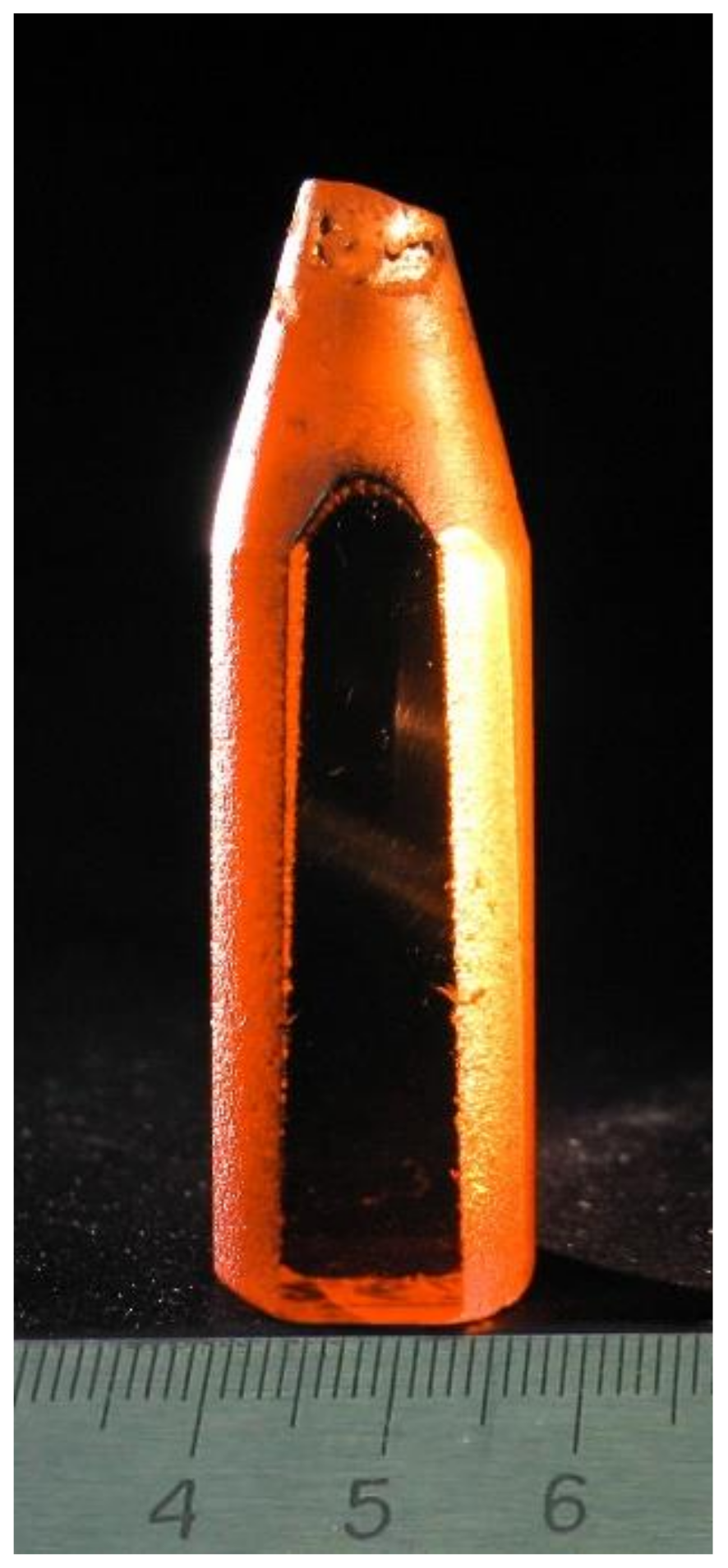
2. Experimental Methods

LiInSe2 crystals were grown using the Bridgman–Stockbarger method, as described in [12]. Li (3N, Novosibirsk Chemical Concentrates Plant, Novosibirsk, Russia), In (5N, Alfa Aesar GmbH, Haverill, MA, USA), and Se (5N, Reachem, Moscow, Russia) were used as starting materials. The melting point of the batch was about 915 °C. Since chalcogenide compounds contain volatile components and chemically aggressive lithium, the synthesis and growth were carried out in glass-graphite containers. In the process of chalcogenide compound synthesis and crystal growth, incongruent evaporation takes place at temperatures above the melting point [47,48,49,50]. As a result, there is a deviation of the composition from the stoichiometric one, but it can be reduced under the conditions of a low temperature gradient at the crystallization front and a small excess pressure in the ampoule. Deviation from stoichiometry is the cause of spot defects and changes in the LiInSe2 sample color from greenish and yellow to red with the variation of cell parameters [51]. In the first group, the crystal composition is close to stoichiometric composition with lithium and selenium vacancies (VLi, VSe). The presence of InLi + 2VLi interatomic substitution with a charge compensator and interstitial Sei atoms leads to an increase in red color intensity [52]. In this work, in contrast to the methodology described in [12], the crystals were grown under the conditions of a low gradient of 5–6 degrees/cm and a slight melt overheating (no more than 50 degrees above the melting point). The grown boules were annealed at 800 °C for 2 h. As a result, yellowish single crystals up to 17 mm in diameter 40 mm long of high optical quality were obtained (Figure 1). The applied method of crystal growth under the low thermal gradient conditions makes it possible to improve the crystal homogeneity along the ingot length and reduce the number of inclusions in the crystal volume. At the 5 cm−1 absorption level, the transparency range for the crystal is 0.47–13 μm.
The powder diffraction data of LiInSe2 were collected at room temperature with a Bruker D8 ADVANCE powder diffractometer (Cu-Kα radiation) and a linear VANTEC detector. The step size of 2θ was 0.016°, and the counting time was 1.5 s per step. The 2θ range of 10–70° was measured with a 0.6 mm divergence slit, while the 2θ range of 70–140° was measured with a 2 mm divergence slit. Larger slits allow a noticeably increased intensity of high-angle peaks without the loss of resolution because the high-angle peaks are broad enough not to be affected by a more diverged beam. The esd’s σ (Ii) of all points on patterns were calculated using intensities Ii: σ (Ii) = Ii1/2. The intensities and obtained esd’s were further normalized: Iinorm = Ii × 0.6/(slit width), σnorm (Ii) = σ (Ii) × 0.6/(slit width), taking into account the actual divergence slit width value, which was used to measure each particular intensity Ii, and saved in xye-type file. Such transformed powder XRD patterns can be viewed in the whole 2θ range of 10–140°, but all high-angle points have small esd’s. To prepare the powder sample for the XRD measurements, a piece of LiInSe2 crystal was ground with the help of mortar and pestle.
The XPS analysis was performed in ultra-high vacuum with a VGESCALAB 220i-XL system employing a monochromatic Al Kα (1486.6 eV) X-ray source. The X-ray gun was operated at 120 W, and the spectrometer pass energy was set at 20 and 100 eV for regional and survey scans, respectively. The diameter of the area under analysis was approximately 500 μm, and the probed surface layer thickness was ~5 nm. A low-energy electron flood gun was used to neutralize the surface charge buildup. The binding energies (BEs) were calibrated by fixing the saturated hydrocarbon component of the C 1s peak at 285.0 eV. The peak fitting of the overlapping Li 1s and Se 3d region was performed using the CasaXPS software package [53]. The XPS measurements were carried out for a single crystal sample.

3. Computation Methods

First-principles band structure and lattice dynamics properties were calculated to analyze the thermal behavior of LiInSe2. The calculation was carried out by CASTEP [54], a package based on the plane-wave pseudopotential density functional theory [55]. The functions developed by the Perdew, Burke, Emzerhof (PBE) [56] in the form of generalized gradient approximation (GGA) [57] were chosen to describe the exchange-correlation interaction, and optimized norm-conserving pseudopotentials [58] were adopted to model the effective interaction between the atomic cores and the valence electrons. To guarantee the precise calculation, the cutoff energy was set as 800 eV and the energy convergence tolerance for a self-consistent field calculation was set as 1 × 10⁻⁸ eV/atom. The intensive Monkhorst−Pack k-point meshes [59] spanning less than 0.07Å−3 were chosen. The phonon characters were calculated by the linear response mechanism [60]. To get the phonon modes contribution to the thermal expansion of the respective axis, the phonon frequency was first calculated on the optimized structure with the cell parameter fixed on the experimental values at 303 K. Then, the respective axis was stretched to the experimental values at 703 K, and the phonon frequency was calculated. Finally, the Gruneisen parameter (g) was calculated by the formula g = (V/ΔV) × (Δω/ω).

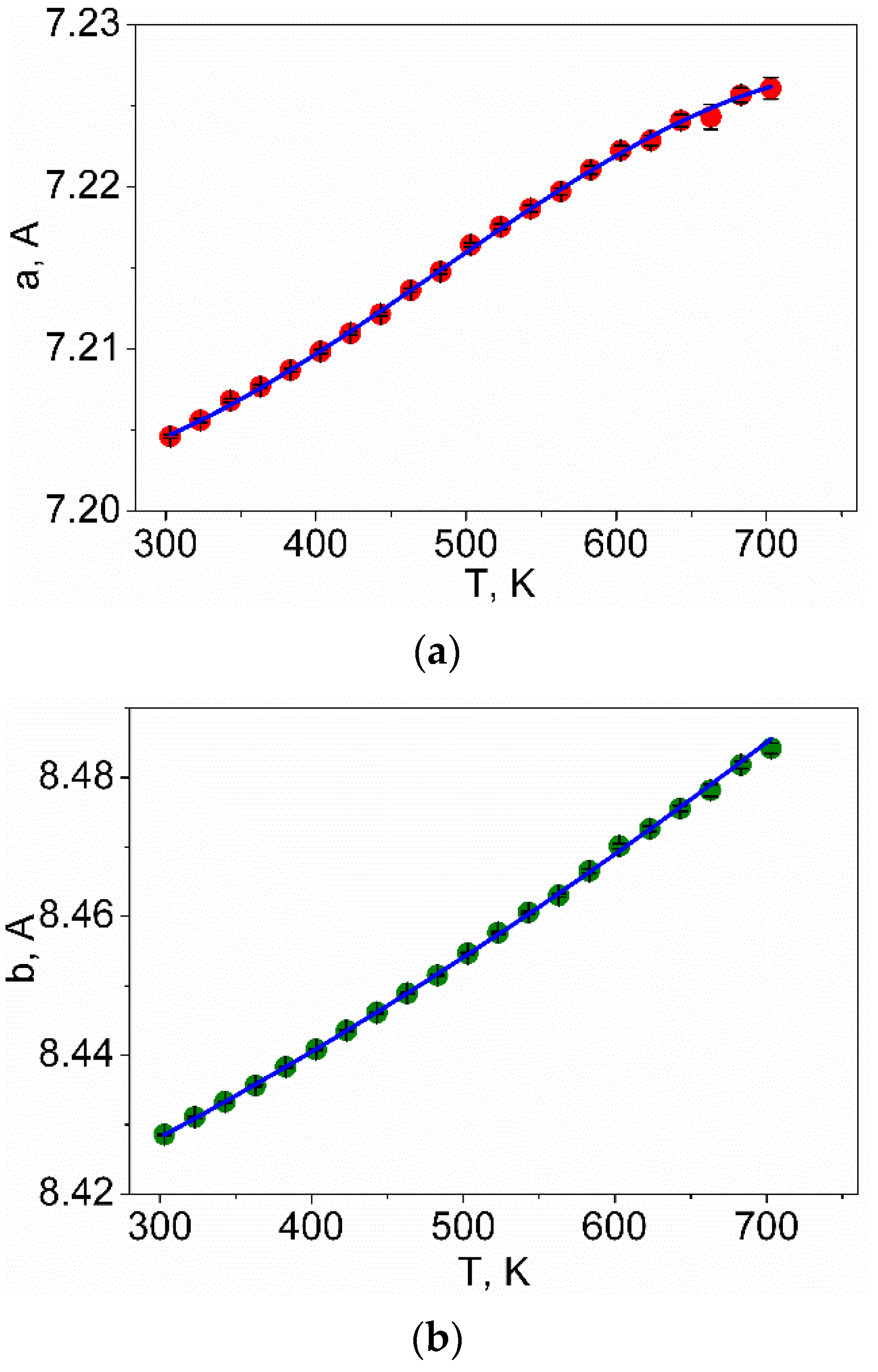
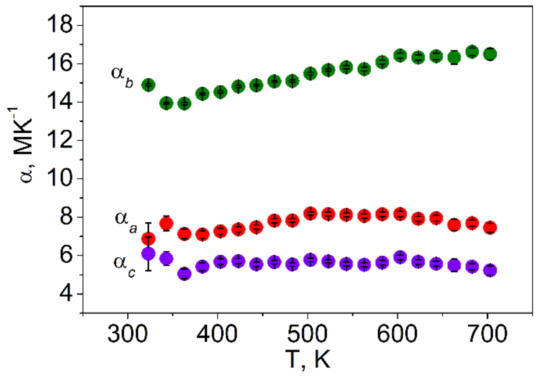
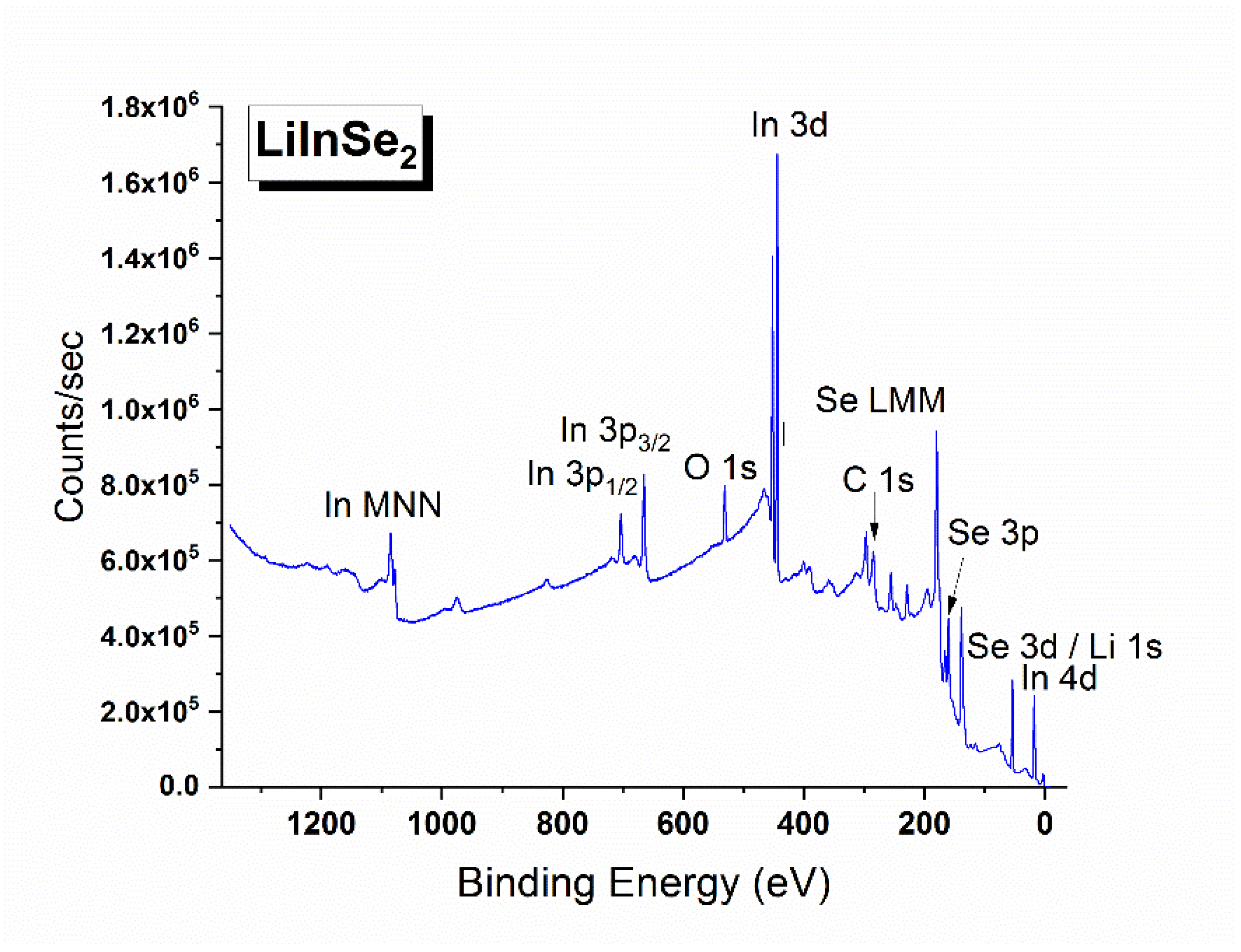
4. Results and Discussion

Rietveld refinement was performed by using TOPAS 4.2 [61] which accounts esd’s of each point by a special weight scheme. All peaks were indexed by an orthorhombic cell (Pna21) with parameters close to those previously reported for LiInSe2 [23], and the refinement was stable resulting in low R-factors (Table 3, Figure 2). The obtained coordinates of atoms and main bond lengths are listed in Supplementary Materials, Tables S1 and S2, respectively, and the crystal structure is presented in Figure 3. To get the information on the temperature dependence of the unit cell parameters (Figure 4), 22 X-ray patterns in the 2θ range of 5–120° were collected from 303 to 723 K with 20 K step: (303, 323, 343, 363, 383, 403, 423, 443, 463, 483, 503, 523, 543, 563, 583, 603, 623, 643, 663, 683, 703, and 723 K) spending 35 min for each pattern (Figure S1). At each selected point, the temperature was fixed and controlled with precision of ±0.2 °C during the pattern measurement. The powder pattern recorded at 723 K showed a large amount of impurity and pattern intensities were not stable during the experiment. So, we stopped at this temperature because the sample decomposition was observed. Almost all peaks of all patterns were indexed by LiInSe2, besides small amount of impurity peaks (Figure S1) marked by asterisk (appeared at 623 K and increased their intensities under further heating) and arrow (appeared at 503 K and disappeared at 663 K). These impurity phases were not identified, but their appearance did not influence the main phase cell parameter refinement. One can see that all a, b, and c cell parameters and the cell volume of the main phase LiInSe2 increase upon heating. Therefore, LiInSe2 has thermal expansion behavior along all crystallographic directions. The related thermal expansion coefficients of LiInSe2 are reported in Table 2. As it is seen in Figure 5, the thermal expansion coefficients of LiInSe2 remain nearly constant with increased temperature. It should be pointed that the thermal expansion of LiInSe2 is strongly anisotropic, and the ratio αbc is as high as ~2.9.
The X-ray photoelectron survey spectrum recorded from LiInSe2 is shown in Figure 6. Besides the photoemission and Auger lines of the constituent elements, two low intensity C1s and O1s peaks were also detected. These “foreign” species are attributed to the presence of adventitious hydrocarbons and hydroxyl groups adsorbed from the ambient environment and the results of surface chemical reaction with oxygen-bearing components of the air. The detailed regional spectra were measured for the valence band and all elemental core levels detected in the survey spectrum. The overlapping spectral region of the Li 1s core level and the Se 3d doublet is shown in Figure 7. Comparing our results with those published for LiGaSe2 [62] and LiGa0.5In0.5Se2 [63], it is obvious that the spectral resolution in the current study is much better as the overlapping peaks are partially resolved in our study in contrast to the single symmetric peak observed in the previous studies. Owing to the superior resolution, it is possible for us to determine the BE values of the Li 1s, Se 3d5/2, and 3d3/2 components by employing the curve fitting procedure in CasaXPS [53]. After background subtraction, all three components were fitted with a mixed 50% Gaussian—50% Lorentzian peak. To improve the reliability of curve-fitting, the intensity ratio of the two Se spin-orbit splitting peaks, I (3d5/2)/I (3d3/2), was fixed to the theoretical value of 1.5 (i.e., ratio of the respective degeneracies 2j + 1), and the amount of the spin-orbit splitting was fixed to be 0.85 eV (based on the value reported in a high resolution XPS study of a single crystal CuInSe2 [64]), but no other constraints were imposed. The In 3d doublet region is presented in Figure 8. The energy positions of the constituent element core levels and Auger lines observed for LiInSe2 are presented in Table 4 (based on the C 1s level being fixed at 285.0 eV). In Table 5, our XPS results of LiInSe2 are compared to those of the representative LiMX2 crystals, with their electronic structures reported in the literature [45,62,63,65]. As mentioned above, the overlapping peaks of Li 1s and Se 3d were not resolved for LiGaSe2 [62] or LiGa0.5In0.5Se2 [63]. Therefore, the reported BE for the combined Li 1s/Se 3d peak is an overestimation for the actual value of Se 3d and underestimation of the actual value of Li 1s in [62,63]. Nevertheless, a reasonable agreement is evident, especially given the possibility of differential charging effects.
The experimental XPS spectrum obtained for the valence band of LiInSe2 is displayed in Figure 9. For comparison, the first-principles electronic density of state (DOS) and partial density of state (PDOS) projected in the electronic orbitals of the constituent atoms are also shown. Because the inner-shell electrons were excluded in the electronic structure calculations, the XPS spectrum and DOS/PDOS are exhibited only for the valence electrons. A good agreement is reached between the experimental and calculated spectra, especially for the energy position of the respective electronic orbitals, indicating the validity of the plane-wave pseudopotential method for the studied crystal. The measured spectrum is slightly broadened compared to the calculated results, which might be attributed to the thermal effect, as well as instrumental broadening associated with the experiment. From the calculated PDOS, the feature of anionic crystal for LiInSe2 can be clearly deduced: almost all the electronic orbitals are strongly localized and the hybridization with the others is very small.
The anharmonicity of lattice vibration phonon is the principal source of thermal expansion of solids. To shed light on the mechanism of the thermal expansion anisotropy in LiInSe2, Grüneisen constant, the parameter that characterizes the phonon anharmonicity was calculated. Accordingly, no imaginary frequency is observed in the phonon spectrum, which demonstrates the dynamical stability of LiInSe2. According to the Grüneisen–constant-colored phonon dispersion, one can see that for all the three axes, the phonon modes in the range of 250~300 cm−1 have the maximum Grüneisen constants (Figure 10a–c), i.e., maximum phonon anharmonicity. This suggests that these phonon modes make the major contribution to the thermal expansion along all the three modes. By subtracting the Grüneisen constants along the maximum (c-axis) and minimum (a-axis) thermal expansion coefficient, the phonon modes accounting for the thermal expansion anisotropy can be obtained. Accordingly, as shown in Figure 10d, the modes with the largest Grüneisen constant difference are mainly located around 260 cm−1. Atomic vibrational assignment reveals that these modes mainly arise from the vibration of lithium atoms, and this demonstrates that lithium atoms play a key role in determining the thermal expansion anisotropy in LiInSe2.

5. Conclusions

In this study, a big-sized high-quality LiInSe2 crystal was grown by the developed Bridgman–Stockbarger method. The anisotropic thermal expansion behavior of the LiInSe2 crystal was measured for the first time. Combining the experimental characterization and theoretical calculations, the huge thermal expansion anisotropy was attributed to the vibration of lithium atoms. In addition, the electronic structure of LiInSe2 crystal was measured by XPS and the recorded valence band is in a good agreement with the theoretical electronic density of states. Moreover, the Grüneisen parameters were also calculated in the theory to reveal the dominant source of the thermal expansion anisotropy. These results indicate that LiInSe2, besides its well-known pronounced linear and nonlinear optical properties in the IR spectral range, possesses specific structural effects.

Supplementary Materials

The following supporting information can be downloaded at: https://www.mdpi.com/article/10.3390/molecules27165078/s1, Figure S1: X-ray patterns collected from 303 to 703 K. The major impurity peaks appeared under heating are marked by asterisk and arrow; Table S1: Fractional atomic coordinates and isotropic displacement parameters (Å2) of LiInSe2; Table S2: Main bond lengths (Å) of LiInSe2.

Author Contributions

Conceptualization, V.V.A. and X.J.; methodology, L.I.I., S.I.L. and X.Z.; software, M.S.M., X.Z. and X.J.; formal analysis, M.S.M., Z.Z., X.Z. and X.J.; investigation, S.I.L. and A.A.G.; data curation, M.S.M. and Z.Z.; writing—original draft preparation, V.V.A., L.I.I., M.S.M., Z.Z. and X.Z.; writing—review and editing, V.V.A., L.I.I. and X.J.; supervision, Z.L. All authors have read and agreed to the published version of the manuscript.

Funding

This work was partly supported by the Ministry of Education and Science of the Russian Federation (grant FSUS-2020-0036), state assignment of IGM SB RAS (preliminary crystal charge composition analysis), Russian Science Foundation (grants #19-12-00085-P, crystal growth, and 21-19-00046, conceptualization), National Scientific Foundations of China (Grants 51702330, 11974360 and 51872297), the Young Elite Scientist Sponsorship Program by CAST (YESS), and the CAS Project for Young Scientists in Basic Research (Grants YSBR-024) and the Government of the Russian Federation (075-15-2022-1132). The XPS measurements were carried out at the Surface Analysis Laboratory of the University of New South Wales, Sydney, Australia.

Institutional Review Board Statement

Not applicable.

Informed Consent Statement

Not applicable.

Data Availability Statement

Data are available on request to authors.

Conflicts of Interest

The authors declare no conflict of interest.

Sample Availability

Samples of the compounds are not available from the authors.

References

  1. Ohmer, M.C.; Goldstein, J.T.; Zelmon, D.E.; Saxler, A.W.; Hegde, S.M.; Wolf, J.D.; Schunemann, P.G.; Pollak, T.M. Infrared properties of AgGaTe2, a nonlinear optical chalcopyrite semiconductor. J. Appl. Phys. 1999, 86, 94–99. [Google Scholar] [CrossRef]
  2. Fossier, S.; Salaün, S.; Mangin, J.; Bidault, O.; Thénot, I.; Zondy, J.-J.; Chen, W.; Rotermund, F.; Petrov, V.; Petrov, P.; et al. Optical, vibrational, thermal, electrical, damage, and phase-matching properties of lithium thioindate. J. Opt. Soc. Am. B 2004, 21, 1981–2007. [Google Scholar] [CrossRef] [Green Version]
  3. Yin, W.; Feng, K.; He, R.; Mei, D.; Lin, Z.; Yao, J.; Wu, Y. BaGa2MQ6 (M = Si, Ge; Q = S, Se): A new series of promising IR nonlinear optical materials. Dalton Trans. 2012, 41, 5653–5661. [Google Scholar] [CrossRef] [PubMed]
  4. Reshak, A.H.; Kityk, I.V.; Parasyuk, O.V.; Kamarudin, H.; Auluck, S. Influence of replacing Si by Ge in the chalcogenide quaternary sulfides Ag2In2Si(Ge)S6 on the chemical bonding, linear and nonlinear optical susceptibilities, and hyperpolarizability. J. Phys. Chem. B 2013, 117, 2545–2553. [Google Scholar] [CrossRef] [PubMed]
  5. Coughlan, C.; Ibáñez, M.; Dobrozhan, O.; Singh, A.; Cabot, A.; Ryan, K.M. Compound Copper Chalcogenide Nanocrystals. Chem. Rev. 2017, 117, 5865–6109. [Google Scholar] [CrossRef] [PubMed]
  6. Sengar, B.S.; Garg, V.; Kumar, A.; Awasthi, V.; Kumar, S.; Atuchin, V.V.; Mukherjee, S. Band alignment of Cd-free (Zn, Mg)O layer with Cu2ZnSn(S,Se)4 and its effect on the photovoltaic properties. Opt. Mater. 2018, 84, 748–756. [Google Scholar] [CrossRef]
  7. Yelisseyev, A.P.; Molokeev, M.S.; Jiang, X.; Krinitsin, P.G.; Isaenko, L.I.; Lin, Z. Structure and Optical Properties of the Li2In2GeSe6 Crystal. J. Phys. Chem. C 2018, 122, 17413–17422. [Google Scholar] [CrossRef]
  8. Yelisseyev, A.; Lobanov, S.; Molokeev, M.; Zhang, S.; Pugachev, A.; Lin, Z.; Vedenyapin, V.; Kurus, A.; Khamoyam, A.; Isaenko, L. A new nonlinear optical selenide crystal AgLiGa2Se4 with good comprehensive performance in mid-infrared region. Adv. Opt. Mater. 2020, 9, 2001856. [Google Scholar] [CrossRef]
  9. Azarapin, N.O.; Aleksandrovsky, A.S.; Atuchin, V.V.; Gavrilova, T.A.; Krylov, A.S.; Molokeev, M.S.; Mukherjee, S.; Oreshonkov, A.S.; Andreev, O.V. Synthesis, structural and spectroscopic properties of orthorhombic compounds BaLnCuS3 (Ln = Pr, Sm). J. Alloy. Compd. 2020, 832, 153134. [Google Scholar] [CrossRef]
  10. Garg, V.; Sengar, B.S.; Siddharth, G.; Kumar, S.; Atuchin, V.V.; Mukherjee, S. Insights into the sputter-instigated valence plasmon oscillations in CIGSe thin films. Surf. Interfaces 2021, 25, 101146. [Google Scholar] [CrossRef]
  11. Petrov, V.; Yelisseyev, A.; Isaenko, L.; Lobanov, S.; Titov, A.; Zondy, J.-J. Second harmonic generation and optical parametric amplification in the mid-IR with orthorhombic biaxial crystals LiGaS2 and LiGaSe2. Appl. Phys. B 2004, 78, 543–546. [Google Scholar] [CrossRef]
  12. Isaenko, L.; Vasilyeva, I.; Merkulov, A.; Yelisseyev, A.; Lobanov, S. Growth of new nonlinear crystals LiMX2 (M = Al, In, Ga; X = S, Se, Te) for the mid-IR optics. J. Cryst. Growth 2005, 275, 217–223. [Google Scholar] [CrossRef]
  13. Andreev, Y.; Atuchin, V.; Lanskii, G.; Pervukhina, N.; Popov, V.; Trocenco, N. Linear optical properties of LiIn(S1–xSex)2 crystals and tuning of phase matching conditions. Solid State Sci. 2005, 7, 1188–1193. [Google Scholar] [CrossRef]
  14. Isaenko, L.; Krinitsin, P.; Vedenyapin, V.; Yelisseyev, A.; Merkulov, A.; Zondy, A.J.-J.; Petrov, V. LiGaTe2: A New Highly Nonlinear Chalcopyrite Optical Crystal for the Mid-IR. Cryst. Growth Des. 2005, 5, 1325–1329. [Google Scholar] [CrossRef]
  15. Petrov, V.; Zondy, J.; Bidault, O.; Isaenko, L.; Vedenyapin, V.; Yelisseyev, A.; Chen, W.; Tyazhev, A.; Lobanov, S.; Marchev, G.; et al. Optical, thermal, electrical, damage, andphase-matching properties of lithium selenoindate. J. Opt. Soc. Am. B 2010, 27, 1902–1927. [Google Scholar] [CrossRef]
  16. Kato, K.; Petrov, V.; Umemura, N. Sellmeier and thermo-optic dispersion formulas for LiInSe2. Appl. Opt. 2014, 53, 1063. [Google Scholar] [CrossRef]
  17. Drebushchak, V.A.; Isaenko, L.I.; Lobanov, S.I.; Krinitsin, P.G.; Grazhdannikov, S.A. Experimental heat capacity of LiInS2, LiInSe2, LiGaS2, LiGaSe2, and LiGaTe2 from 180 to 460 K. J. Therm. Anal. 2017, 129, 103–108. [Google Scholar] [CrossRef]
  18. Kurus, A.; Yelisseyev, A.; Lobanov, S.; Plyusnin, P.; Molokeev, M.; Solovyev, L.; Samoshkin, D.; Stankus, S.; Melnikova, S.; Isaenko, L. Thermophysical properties of lithium thiogallate that are important for optical applications. RSC Adv. 2021, 11, 39177–39187. [Google Scholar] [CrossRef]
  19. Kish, Z.Z.; Kanishcheva, A.S.; Mikhailov, Y.N.; Lazarev, V.B.; Semrad, E.E.; Peresh, E.Y. Synthesis and crystal structure of lithium thioindate. Dokl. Akad. Nauk. SSSR 1985, 280, 398–401. [Google Scholar]
  20. Leal-Gonzalez, J.; Melibary, S.; Smith, A. Structure of lithium gallium sulfide, LiGaS2. Acta Cryst. 1990, 46, 2017–2019. [Google Scholar] [CrossRef]
  21. Isaenko, L.; Yelisseyev, A.; Lobanov, S.; Titov, A.; Petrov, V.; Zondy, J.-J.; Krinitsin, P.; Merkulov, A.; Vedenyapin, V.; Smirnova, J. Growth and properties of LiGaX2 (X = S, Se, Te) single crystals for nonlinear optical applications in the mid-IR. Cryst. Res. Technol. 2003, 38, 379–387. [Google Scholar] [CrossRef]
  22. Kuriyama, K.; Nozaki, T. Single-crystal growth and characterization of LiGaSe2. J. Appl. Phys. 1981, 52, 6441–6443. [Google Scholar] [CrossRef]
  23. Neumann, H.; Hönle, W.; Kuhn, G. Die Kristallstruktur von LiInSe2. Z. Anorg. Allg. Chem. 1986, 543, 161–168. [Google Scholar]
  24. Kühn, G.; Schumann, B.; Oppermann, D.; Neumann, H.; Sobotta, H. Preparation, structure, and infrared lattice vibrations of LilnTe2. Z. Anorg. Allg. Chem. 1985, 531, 61–66. [Google Scholar] [CrossRef]
  25. Kim, J.; Hughbanks, T. ChemInform Abstract: Synthesis and Structures of New Ternary Aluminum Chalcogenides: LiAlSe2, α-LiAlTe2, and β-LiAlTe2. ChemInform 2000, 39, 3092–3097. [Google Scholar] [CrossRef]
  26. Kidyarov, B.I.; Atuchin, V.V. Universal crystal classification system “Point symmetry—Physical property”. Ferroelectrics 2007, 360, 96–99. [Google Scholar] [CrossRef]
  27. Huang, J.-J.; Atuchin, V.; Andreev, Y.M.; Lanskii, G.; Pervukhina, N. Potentials of LiGa(S1−xSex)2 mixed crystals for optical frequency conversion. J. Cryst. Growth 2006, 292, 500–504. [Google Scholar] [CrossRef]
  28. Tyazhev, A.; Marchev, G.; Vedenyapin, V.; Kolker, D.; Yelisseyev, A.; Lobanov, S.; Isaenko, L.; Zondy, J.; Petrov, V. LiInSe2 nanosecond optical parametric oscillator tunable from 4.7 to 8.7 μm. Proc. SPIE 2010, 7582, 75820E. [Google Scholar]
  29. Takeya, K.; Takemoto, Y.; Kawayama, I.; Murakami, H.; Matsukawa, T.; Yoshimura, M.; Mori, Y.; Tonouchi, M. Terahertz emission from coherent phonons in lithium ternary chalcopyrite crystals illuminated by 1560 nm femtosecond laser pulses, EPL 91. Eur. Lett. 2010, 91, 20004. [Google Scholar] [CrossRef]
  30. Beutler, M.; Rimke, I.; Büttner, E.; Petrov, V.; Isaenko, L. Femtosecond mid-IR difference-frequency generation in LiInSe2. Opt. Mater. Express 2013, 3, 1834–1838. [Google Scholar] [CrossRef]
  31. Isaenko, L.; Yelisseyev, A.; Lobanov, S.; Vedenyapin, V.; Krinitsyn, P.; Petrov, V. Properties of LiGa0.5In0.5Se2: A Quaternary Chalcogenide Crystal for Nonlinear Optical Applications in the Mid-IR. Crystals 2016, 6, 85. [Google Scholar] [CrossRef] [Green Version]
  32. Wang, S.; Dai, S.; Jia, N.; Zong, N.; Li, C.; Shen, Y.; Yu, T.; Qiao, J.; Gao, Z.; Peng, Q.; et al. Tunable 7–12 μm picosecond optical parametric amplifier based on a LiInSe2 mid-infrared crystal. Opt. Lett. 2017, 42, 2098–2101. [Google Scholar] [CrossRef] [PubMed]
  33. Dai, S.-B.; Jia, N.; Chen, J.-K.; Shen, Y.; Yang, S.; Li, Y.-J.; Liu, Q.; Yang, F.; Zong, N.; Wang, Z.-M.; et al. Picosecond mid-infrared optical parametric amplifier based on LiInSe2 with tenability extending from 3.6 to 4.8 μm. Opt. Express 2017, 25, 12860–12866. [Google Scholar] [CrossRef]
  34. Kato, K.; Petrov, V.; Miyata, K. Phase-matching properties of LiIn(SxSe1-x)2. Proc. SPIE 2020, 11264, 112641W. [Google Scholar]
  35. Tupitsyn, E.; Bhattacharya, P.; Rowe, E.; Matei, L.; Groza, M.; Wiggins, B.; Burger, A.; Stowe, A. Single crystal of LiInSe2 semiconductor for neutron detector. Appl. Phys. Lett. 2012, 101, 202101. [Google Scholar] [CrossRef]
  36. Egner, J.C.; Groza, M.; Burger, A.; Stassun, K.G.; Buliga, V.; Matei, L.; Bodnarik, J.G.; Stowe, A.C.; Prettymane, T.H. Integration of a 6LilnSe2 thermal neutron detector into a CubeSat instrument. J. Astronom. Telescopes Instrum. Syst. 2016, 2, 046001. [Google Scholar] [CrossRef] [Green Version]
  37. Belushkin, A.V.; Bogdzel, A.A.; Goloshumova, A.A.; Isaenko, L.I.; Lobanov, S.I.; Milkov, V.M.; Tarasova, A.Y.; Yelisseyev, A.P. Study of LiInSe2 Single Crystals for the Thermal Neutron Detection. Synchrotron Neutron Tech. 2020, 14 (Suppl. S1), S15–S18. [Google Scholar] [CrossRef]
  38. Lukosi, E.; Hamm, D.; Preston, J.; Hausladen, P.; Brune, C.; Massey, T.; Jacobs, D.; Burger, A.; Stowe, A. First evaluation of fast neutron imaging with LiInSe2 semiconductors. Nucl. Instrum. Methods Phys. Res. Sect. A 2020, 976, 164254. [Google Scholar] [CrossRef]
  39. Matei, L.; Hawrami, R.; Buliga, V.; Babalola, S.; Duff, M.C.; Inabinet, L.; Baldwin, T.; Jandeska, A.; Burger, A. Lithium indium diselenide—An advanced material for neutron detection. Nucl. Instrum. Methods Phys. Res. Sect. A 2021, 1020, 165898. [Google Scholar] [CrossRef]
  40. Jia, N.; Wang, S.; Gao, Z.; Wu, Q.; Li, C.; Zhang, X.; Yu, T.; Lu, Q.; Tao, X. Optimized Growth of Large-Sized LiInSe2 Crystals and the Electric–Elastic Properties. Cryst. Growth Des. 2017, 17, 5875–5880. [Google Scholar] [CrossRef]
  41. Grazhdannikov, S.A.; Krinitsyn, P.G.; Kurus’, A.F.; Isaenko, L.I.; Yelisseyev, A.P.; Molokeev, M.S. LiGaTe2 (LGT) nonlinear crystal: Synthesis and crystal growth processes exploration. Mater. Sci. Semicond. Process. 2017, 72, 52–59. [Google Scholar] [CrossRef]
  42. Jia, N.; Xiong, X.; Wang, S.; Yu, T.; Han, B.; Qiao, J.; Li, C.; Tao, X. Optimized oriented seed growth and optical properties of high-quality LiInSe2 crystals. CrystEngComm 2018, 48, 7802–7808. [Google Scholar] [CrossRef]
  43. Yelisseyev, A.P.; Drebushchak, V.; Titov, A.S.; Isaenko, L.I.; Lobanov, S.I.; Lyapunov, K.M.; Gruzdev, V.A.; Komarov, S.G.; Petrov, V.; Zondy, J.-J. Thermal properties of the midinfrared nonlinear crystal LiInSe2. J. Appl. Phys. 2004, 96, 3659–3665. [Google Scholar] [CrossRef]
  44. Yang, L.-Q.; Jiang, X.-M.; Guo, G.-C. LiGa0.54In0.46S2: A new infrared nonlinear optical material with large laser damage threshold designed by gallium substitution in LiInS2. Inorg. Chem. Commun. 2020, 115, 107852. [Google Scholar] [CrossRef]
  45. Atuchin, V.V.; Liang, F.; Grazhdannikov, S.; Isaenko, L.I.; Krinitsin, P.G.; Molokeev, M.S.; Prosvirin, I.P.; Jiang, X.; Lin, Z. Negative thermal expansion and electronic structure variation of chalcopyrite type LiGaTe2. RSC Adv. 2018, 8, 9946–9955. [Google Scholar] [CrossRef] [PubMed] [Green Version]
  46. Krinitsin, P.; Yelisseyev, A.; Jiang, X.; Isaenko, L.; Molokeev, M.; Lin, Z.; Pugachev, A. Growth, structure, and optical properties of nonlinear LiGa0.55In0.45Te2 single crystals. Cryst. Growth Des. 2019, 19, 1805–1814. [Google Scholar] [CrossRef]
  47. Andreev, Y.M.; Atuchin, V.V.; Lanskii, G.V.; Morozov, A.N.; Pokrovsky, L.D.; Sarkisov, S.Y.; Voevodina, O.V. Growth, real structure and applications of GaSe1−xSx crystals. Mater. Sci. Eng. B 2006, 128, 205–210. [Google Scholar] [CrossRef]
  48. Atuchin, V.V.; Golyashov, V.A.; Kokh, K.A.; Korolkov, I.V.; Kozhukhov, A.S.; Kruchinin, V.N.; Makarenko, S.V.; Pokrovsky, L.D.; Prosvirin, I.P.; Romanyuk, K.N.; et al. Formation of inert Bi2Se3(0001) cleaved surface. Cryst. Growth Des. 2011, 11, 5507–5514. [Google Scholar] [CrossRef]
  49. Kokh, K.A.; Atuchin, V.V.; Adichtchev, S.V.; Gavrilova, T.A.; Bakhadur, A.M.; Klimov, A.O.; Korolkov, I.V.; Kuratieva, N.V.; Mukherjee, S.; Pervukhina, N.V.; et al. Cu2ZnSnS4 crystal growth using an SnCl2 based flux. CrystEngComm 2021, 22, 1025–1032. [Google Scholar] [CrossRef]
  50. Vasilyeva, I.G.; Nikolaev, R.E. Non-stoichiometry and point native defects in non-oxide non-linear optical large single crystals: Advantages and problems. CrystEngComm 2022, 24, 1495–1506. [Google Scholar] [CrossRef]
  51. Siemek, K.; Yelisseyev, A.; Horodek, P.; Lobanov, S.; Goloshumova, A.; Belushkin, A.; Isaenko, L. Optical and positron annihilation studies of structural defects in LiInSe2 single crystals. Opt. Mater. 2020, 109, 110262. [Google Scholar] [CrossRef]
  52. Cui, Y.; Bhattacharya, P.; Buliga, V.; Tupitsyn, E.; Rowe, E.; Wiggins, B.; Johnstone, D.; Stowe, A.; Burger, A. Defects in 6LiInSe2 neutron detector investigated by photo-induced current transient spectroscopy and photoluminescence. Appl. Phys. Lett. 2013, 103, 092104. [Google Scholar] [CrossRef]
  53. Fairley, N.; Fernandez, V.; Richard-Plouet, M.; Guillot-Deudon, C.; Walton, J.; Smith, E.; Flahaut, D.; Greiner, M.; Biesinger, M.; Tougaard, S.; et al. Systematic and collaborative approach to problem solving using X-ray photoelectron spectroscopy. Appl. Surf. Sci. Adv. 2021, 5, 100112. [Google Scholar] [CrossRef]
  54. Clark, S.J.; Segall, M.D.; Pickard, C.J.; Hasnip, P.J.; Probert, M.I.J.; Refson, K.; Payne, M.C. First Principles Methods Using CASTEP. Z. Krist.-Cryst. Mater. 2005, 220, 567–570. [Google Scholar] [CrossRef] [Green Version]
  55. Kohn, W.; Sham, L.J. Self-consistent equations including exchange and correlation effects. Phys. Rev. 1965, 140, A1133–A1138. [Google Scholar] [CrossRef] [Green Version]
  56. Perdew, J.P.; Burke, K.; Ernzerhof, M. Generalized gradient approximation made simple. Phys. Rev. Lett. 1996, 77, 3865. [Google Scholar] [CrossRef] [Green Version]
  57. Perdew, J.P.; Chevary, J.A.; Vosko, S.H.; Jackson, K.A.; Pederson, M.R.; Singh, D.J.; Fiolhais, C. Atoms, molecules, solids, and surfaces—Applications of the generalized gradient approximation for exchange and correlation. Phys. Rev. B 1992, 46, 6671–6687. [Google Scholar] [CrossRef]
  58. Hamann, D.R.; Schlüter, M.; Chiang, C. Norm-conserving pseudopotentials. Phys. Rev. Lett. 1979, 43, 1494–1497. [Google Scholar] [CrossRef]
  59. Monkhorst, H.J.; Pack, J.D. Special points for Brillouin-zone integrations. Phys. Rev. B 1976, 13, 5188. [Google Scholar] [CrossRef]
  60. Baroni, S.; de Gironcoli, S.; Corso, A.D.; Giannozzi, P. Phonons and related crystal properties from density-functional perturbation theory. Rev. Mod. Phys. 2001, 73, 515–562. [Google Scholar] [CrossRef] [Green Version]
  61. Bruker AXS. TOPAS V4: General Profile and Structure Analysis Software for Powder Diffraction Data—User’s Manual; Bruker AXS: Karlsruhe, Germany, 2008. [Google Scholar]
  62. Lavrentyev, A.; Gabrelian, B.; Vu, V.; Ananchenko, L.; Isaenko, L.; Yelisseyev, A.; Khyzhun, O. Electronic structure and optical properties of noncentrosymmetric LiGaSe2: Experimental measurements and DFT band structure calculations. Opt. Mater. 2017, 66, 149–159. [Google Scholar] [CrossRef]
  63. Lavrentyev, A.; Gabrelian, B.; Vu, T.V.; Isaenko, L.; Yelisseyev, A.; Khyzhun, O. Electronic structure and optical properties of LiGa0.5In0.5Se2 single crystal, a nonlinear optical mid-IR material. Opt. Mater. 2018, 80, 12–21. [Google Scholar] [CrossRef]
  64. Sobol, P.E.; Nelson, A.J.; Schwerdtfeger, C.R.; Stickle, W.F.; Moulder, J.F. Single crystal CulnSe2 analysis by high resolution XPS. Surf. Sci. Spectra 1992, 1, 393. [Google Scholar] [CrossRef]
  65. Atuchin, V.; Isaenko, L.; Kesler, V.; Lobanov, S. Core level photoelectron spectroscopy of LiGaS2 and Ga–S bonding in complex sulfides. J. Alloy. Compd. 2010, 497, 244–248. [Google Scholar] [CrossRef]
Figure 1. Digital image of the LiInSe2 crystal.
Molecules 27 05078 g001
Figure 2. Observed, calculated (using the Rietveld method) and difference XRD patterns obtained for the LiInSe2 sample.
Molecules 27 05078 g002
Figure 3. Crystal structure of LiInSe2. Unit cell is outlined. Lone atoms are omitted for clarity.
Molecules 27 05078 g003
Figure 4. Cell parameter dependence on temperature: (a) a(T), (b) b(T), (c) c(T), and (d) V(T).
Molecules 27 05078 g004aMolecules 27 05078 g004b
Figure 5. Thermal expansion coefficient dependence on temperature.
Molecules 27 05078 g005
Figure 6. Survey X-ray photoelectron spectrum of LiInSe2.
Molecules 27 05078 g006
Figure 7. Detailed XPS spectrum of the Se 3d and Li 1s region in LiInSe2.
Molecules 27 05078 g007
Figure 8. Detailed XPS spectrum of the In 3d doublet in LiInSe2.
Molecules 27 05078 g008
Figure 9. Comparison of the experimental XPS spectrum and ab initio calculated distributions of electronic states.
Molecules 27 05078 g009
Figure 10. (ac) The phonon dispersion of the structure with the perturbation of axes a, b, and c, respectively, in which the Grüneisen values of different vibration mode were presented by the color from blue to red. (d) The phonon dispersion of the structure at 303 K, in which the subtracted Grüneisen values of a-axis and c-axis were presented by the color from blue to red. The inset in (d) is the graphic vibration mode of around 260 cm−1, and the vibration vectors are shown as blue arrows.
Molecules 27 05078 g010
Table 1. Structural types known in compounds LiMX2 (M = Al, In, Ga; X = S, Se, Te).
MSSeTe
InP21nb (Pna21) [19]Pna21 [23]I-42d [24]
GaPna21 [20,21]Pna21 [21,22]I-42d [21]
Al Pna21 [25]I-42d and P3m1 [25]
Table 2. Thermal expansion coefficients of LiMT2 (M = Ga, In; T = Se, Te) crystals.
CompoundTypeαa MK−1αb MK−1αc MK−1αV MK−1Reference
LiGaTe2II19.119.1−8.629.4[45]
LiGa0.55In0.45Te2II18.918.9−5.732.3[46]
LiGa0.54In0.46S2I11.715.812.7 [44]
LiInS2I8.916.16.6 [2]
LiInSe2I11.5 ± 1.720.4 ± 2.48.9 ± 2.4 [43]
LiInSe2I8.1 (1)16.1 (2)5.64 (6)29.9 (3)This work
Table 3. Main parameters of processing and refinement of the LiInSe2 sample.
CompoundLiInSe2
Sp. Gr.Pna21
a, Å7.20442 (7)
b, Å8.42826 (8)
c, Å6.80491 (6)
V, Å3413.199 (7)
Z4
-range, °10–140
Rwp, %5.63
Rp, %4.71
Rexp, %2.67
χ22.11
RB, %2.16
Table 4. Core level binding energies and Auger lines in LiInSe2.
LineBinding Energy, eV
In 4d5/218.06
In 4d3/218.93
Se 3d5/254.04
Se 3d3/254.89
Li 1s54.97
Se L3M45M45179.95
C 1s285.0 (fixed)
O 1s531.46
In 3d5/2444.60
In 3d3/2452.15
In M4N45N451078.70
Table 5. BE values of representative core levels measured in LiMX2 materials.
CrystalGa 3dLi 1sIn 3d5/2S 2pSe 3d5/2Te 3d5/2Ref.
LiGaS220.155.3-162.0--[65]
LiGaSe219.7254.23 *--54.23 *-[62]
LiGaTe219.355.0---572.3[45]
LiGa0.5In0.5Se218.26 **54.23 *444.82-54.23 *-[63]
LiInSe2-54.97444.60-54.04-This study
* superposition (Li 1s + Se 3d). ** superposition (Ga 3d + In 4d). Note that values reported in [45] were shifted by +0.2 eV due to their C 1s level being fixed at 284.8 eV, and values in [62,63,65] were shifted by +0.4 eV because their C 1s level was fixed at 284.6 eV.
Publisher’s Note: MDPI stays neutral with regard to jurisdictional claims in published maps and institutional affiliations.

