Article

64CuCl2 PET Imaging of 4T1-Related Allograft of Triple-Negative Breast Cancer in Mice

1
Nuclear Medicine and Molecular Imaging Department, Institut de Cancérologie de Strasbourg Europe (ICANS), 17 Rue Albert Calmette, 67200 Strasbourg, France
2
Institut Pluridisciplinaire Hubert Curien, Université de Strasbourg, 23 Rue du Loess, 67037 Strasbourg, France
3
CNRS, UMR7178, 23 Rue du Loess, 67037 Strasbourg, France
4
Department of Pathology, Hôpitaux Universitaires de Strasbourg, 1 Avenue Molière, 67200 Strasbourg, France
*
Author to whom correspondence should be addressed.
Molecules 2022, 27(15), 4869; https://doi.org/10.3390/molecules27154869
Submission received: 19 June 2022 / Revised: 27 July 2022 / Accepted: 28 July 2022 / Published: 29 July 2022
(This article belongs to the Special Issue Radiopharmaceuticals for PET Imaging - Issue B)

Abstract

:
64CuCl2 is an economic radiotracer for oncologic PET investigations. In the present study, we characterized the uptake of 64CuCl2 in vivo by µPET/CT in an allograft 4T1-related mouse model (BALB/c) of advanced breast cancer. 18F-FDG was used as a comparator. Twenty-two animals were imaged 7–9 days following 4T1-cell implantation inside mammary glands. Dynamic 64CuCl2 µPET/CT acquisition or iterative static images up to 8 h p.i. were performed. Animal biodistribution and tumor uptake were first evaluated in vivo by µPET analysis and then assessed on tissue specimens. Concerning 18F-FDG µPET, a static acquisition was performed at 15 min and 60 min p.i. Tumor 64CuCl2 accumulation increased from 5 min to 4 h p.i., reaching a maximum value of 5.0 ± 0.20 %ID/g. Liver, brain, and muscle 64CuCl2 accumulation was stable over time. The tumor-to-muscle ratio remained stable from 1 to 8 h p.i., ranging from 3.0 to 3.7. Ex vivo data were consistent with in vivo estimations. The 18F-FDG tumor accumulation was 8.82 ± 1.03 %ID/g, and the tumor-to-muscle ratio was 4.54 ± 1.11. 64CuCl2 PET/CT provides good characterization of the 4T1-related breast cancer model and allows for exploration of non-glycolytic cellular pathways potentially of interest for theragnostic strategies.

1. Introduction

Copper is a metallic element involved in the regulation of multiple physiological processes in humans, such as cell growth and neurotransmitter synthesis. After internalization by the high-affinity transmembrane protein hCTR-1, copper accumulates in intracellular organelles via various chaperone proteins, such as CCS, ATOX1, Cox17, SCO1, and SCO2 [1,2,3]. Copper seems to play a key role in tumor growth, neoangiogenesis, and metastatic spread [4,5,6]; overexpression of hCTR-1 in various cancers (prostate, lung, melanoma, glioblastoma, etc.) has been revealed by several studies [3].
Copper-64 (64Cu) is a cyclotron-produced radionuclide that is ideally suited for positron emission computed tomography (PET) investigation, enabling image acquisition over 48 h [7]. Copper radioisotopes such as 64Cu and 67Cu offer an interesting opportunity for diagnosis and related radiometabolic treatment. The use of 64Cu permits prospective dosimetry or dose personalization of 67Cu, opening new possibilities in the theragnostic field. 64Cu and 67Cu have optimal half-lives (12.7 h and 2.5 d, respectively) and energies appropriate for use with biologic drugs. Moreover, the energy of 67Cu is equivalent to that of 177Lu, which is routinely used for peptide receptor radionuclide therapy (PRRT) in patients with metastatic neuroendocrine tumors [8].
The ionic form of copper chloride (64CuCl2) offers economic and logistic advantages, with no need for complex chemistry to be produced, and can be directly administered in vivo in an aqueous solution. The results from clinical studies underline the feasibility of 64CuCl2 use as a metabolic radiotracer for PET/CT investigations in patients with prostate cancer relapse [9]. Several preclinical analyses have also been performed with promising results using cellular lines and animal models of prostate, colon, ovarian, lung, brain, and head-and-neck tumors. However, clinical and preclinical data concerning 64CuCl2 PET investigations in breast cancer are currently scarce. 4T1 is an interesting cell line derived from murine breast carcinoma [10] and can grow in culture or after allografting in immunocompetent mice. As a consequence, it is widely used as a surrogate for late-stage triple-negative breast cancer (TNBC) in humans due to its highly invasive potential and substantial molecular similarity (lack of Esr1, Erbb2, and Pgr expression) [11]. As a human TNBC model, 4T1-related tumors in mice have shown intense 18F-fluorodeoxyglucose (18F-FDG) uptake; thus, 18F-FDG PET is often used as a comparator in preclinical imaging studies [12]. Due to the recent development of therapeutics, several molecular alternatives investigating nonglycolytic pathways have been researched for breast cancer imaging [13,14]. To our knowledge, in no case has 64Cu been used as a labeling isotope or 64CuCl2 been used as a diagnostic agent for TNBC.
Therefore, in the present study, we have characterized the uptake of 64CuCl2 in an allograft 4T1-related mouse model of advanced breast cancer by in vivo PET. 18F-FDG PET was used as an image comparator.

2. Results

Twenty-nine female BALB/c mice were used for the study: 10 for 64CuCl2 PET investigations, 13 for 64CuCl2 ex vivo biodistribution assessment, and 6 for 18F-FDG PET images and ex vivo studies. Among the 22 mice imaged by µPET/CT, 6/22 underwent 64CuCl2 dynamic studies, 10/22 underwent 64CuCl2 static acquisitions, and 6/22 underwent 18F-FDG static imaging.

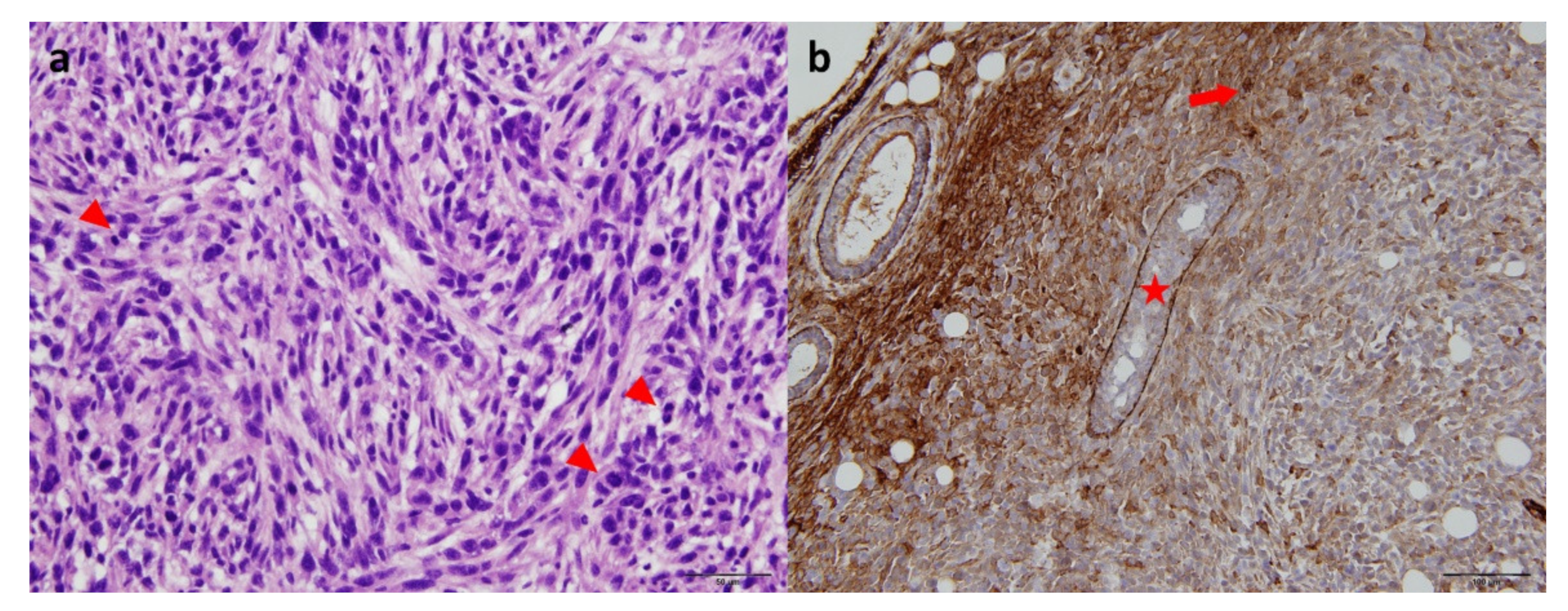
2.1. Tumor Size and Histology

Seven to nine days after 4T1 cells were injected into the mammary gland, tumors reached an average volume of 315 ± 175 mm3. Tumors developed locally without detection of extramammary metastases upon visual investigation. Pathological reports revealed hyperchromatic cells with polymorphic nuclei, restricted cytoplasms, and numerous mitoses. Tumors were positive for cytokeratin 5/6 (Figure 1).

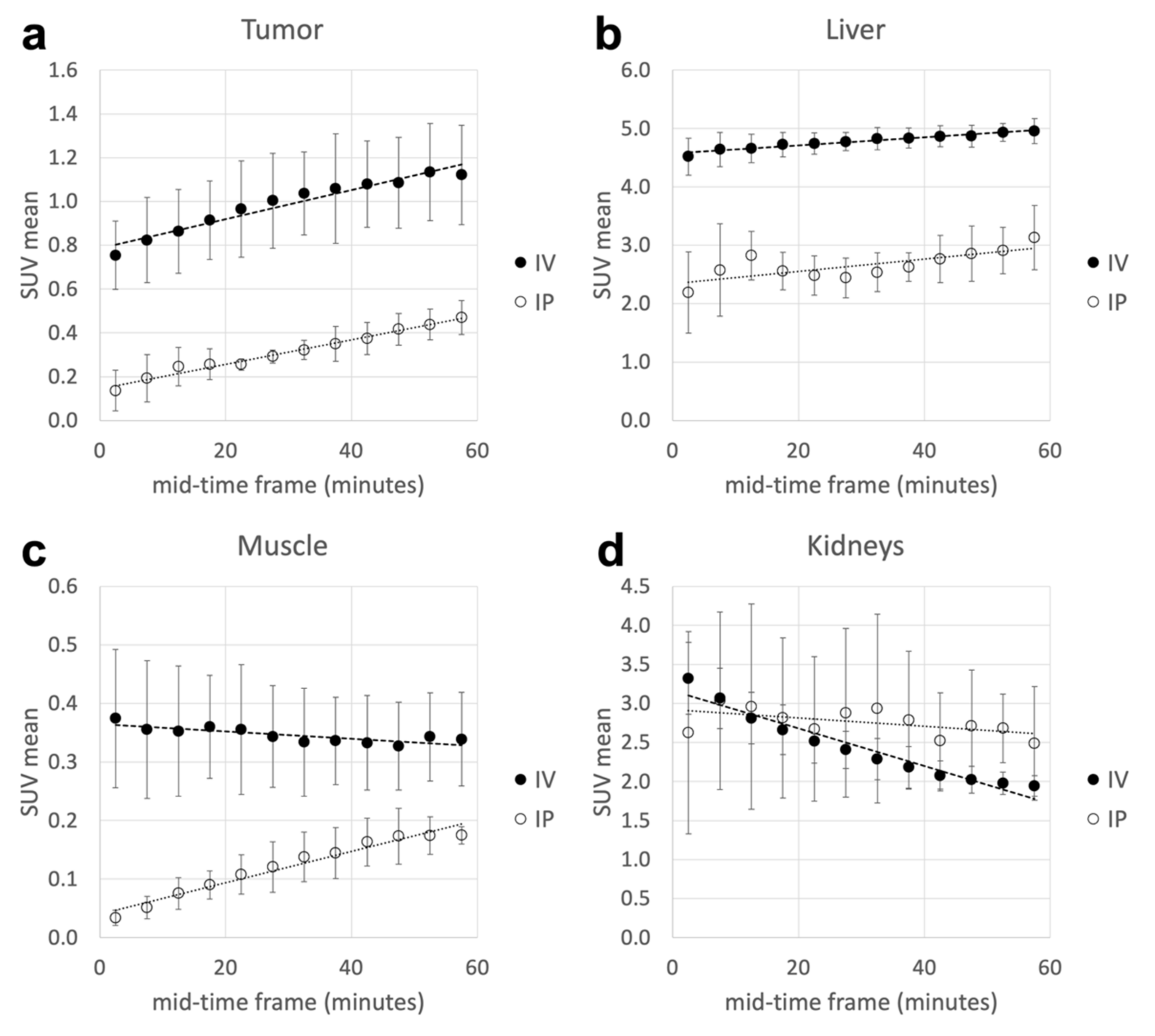
2.2. 64CuCl2 Dynamic µPET/CT Acquisitions

Six mice were imaged from 5 to 60 min after intravenous (i.v., n = 3) or intraperitoneal (i.p., n = 3) 64CuCl2 administration (Figure 2). Activity inside tumors and organs was higher for intravenous injection. The tumor SUVmean increased linearly from 0.75 ± 0.16 at 5 min p.i. to 1.12 ± 0.23 at 60 min p.i. for i.v. injected mice; the corresponding percentages of injected doses per gram were 3.50 ± 0.37 and 5.20 ± 0.31, respectively. The same trend was observed in mice that underwent i.p. injections (0.79 ± 0.57 to 2.68 ± 0.63 %ID/g at 5 and 60 min p.i., respectively). The tumor mean SUV was higher in animals that underwent intravenous 64CuCl2 injection (5 min p.i, p < 0.01). The liver and brain displayed a slight activity increase within the first hour after injection: SUVmean varied from 4.52 ± 0.32 (liver) and 0.47 ± 0.01 (brain) at 5 min p.i. to 4.95 ± 0.22 (liver) and 0.77 ± 0.34 (brain) at 60 min p.i. for i.v. injected mice. In contrast, muscle showed a globally stable SUVmean over time (from 0.37 ± 0.12 at 5 min p.i. to 0.34 ± 0.08 at 60 min p.i.) for i.v. injected mice, while there was increasing activity over time for i.p. injected mice (0.03 ± 0.01 at 5 min p.i. to 0.18 ± 0.02 at 60 min p.i.). Even in these cases, SUV values were higher in animals that underwent i.v. injection (5 min p.i, p < 0.04). Kidney biodistribution was somewhat different: there was a significant decrease in the mean SUV from 5 to 60 min p.i. (p = 0.03) in mice with i.v. injection (corresponding to 15.6 ± 2.1 to 9.1 ± 0.6 %ID/g at 5 min and 60 min p.i., respectively), while activity remained stable in mice with i.p. injection.

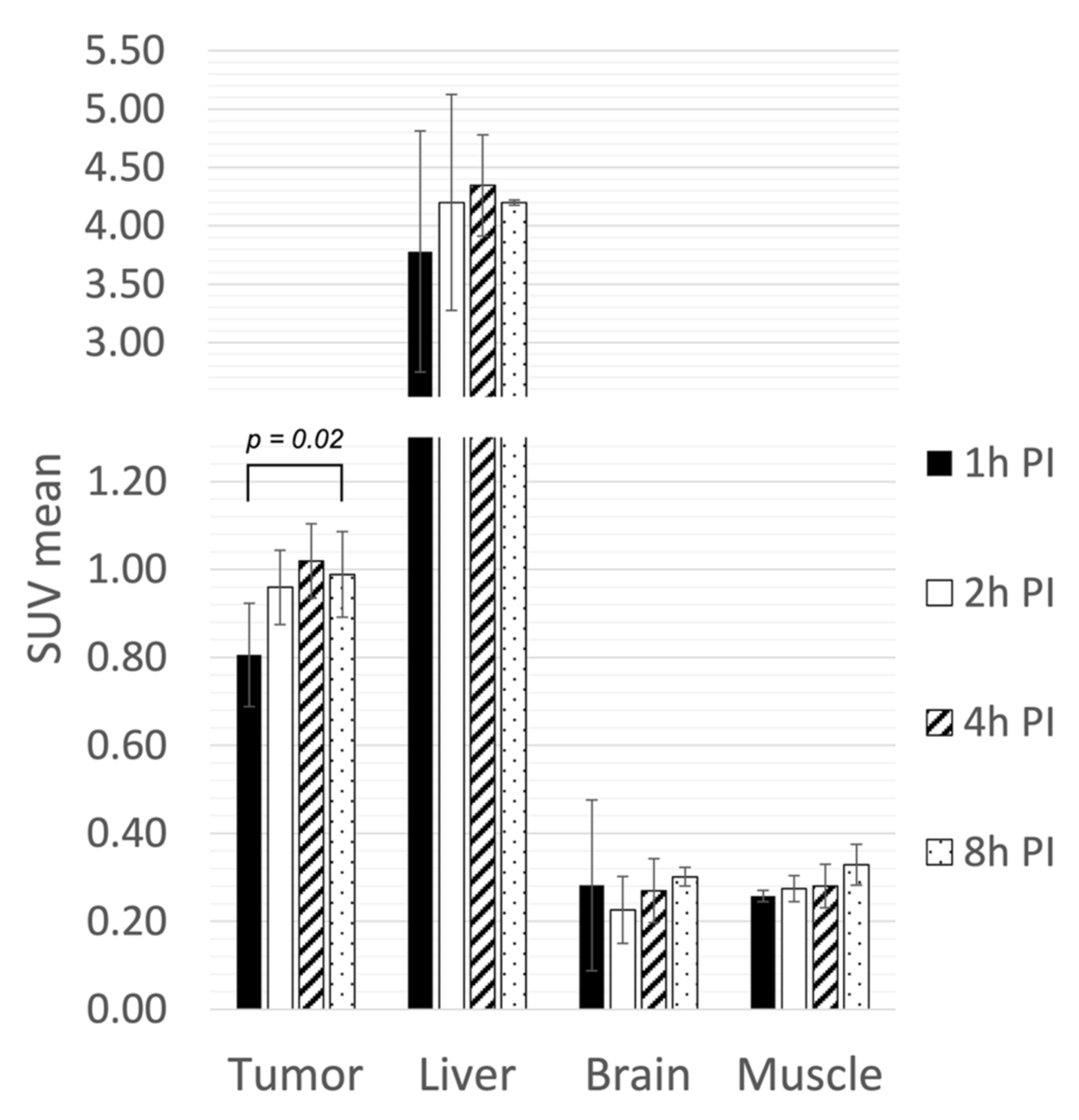
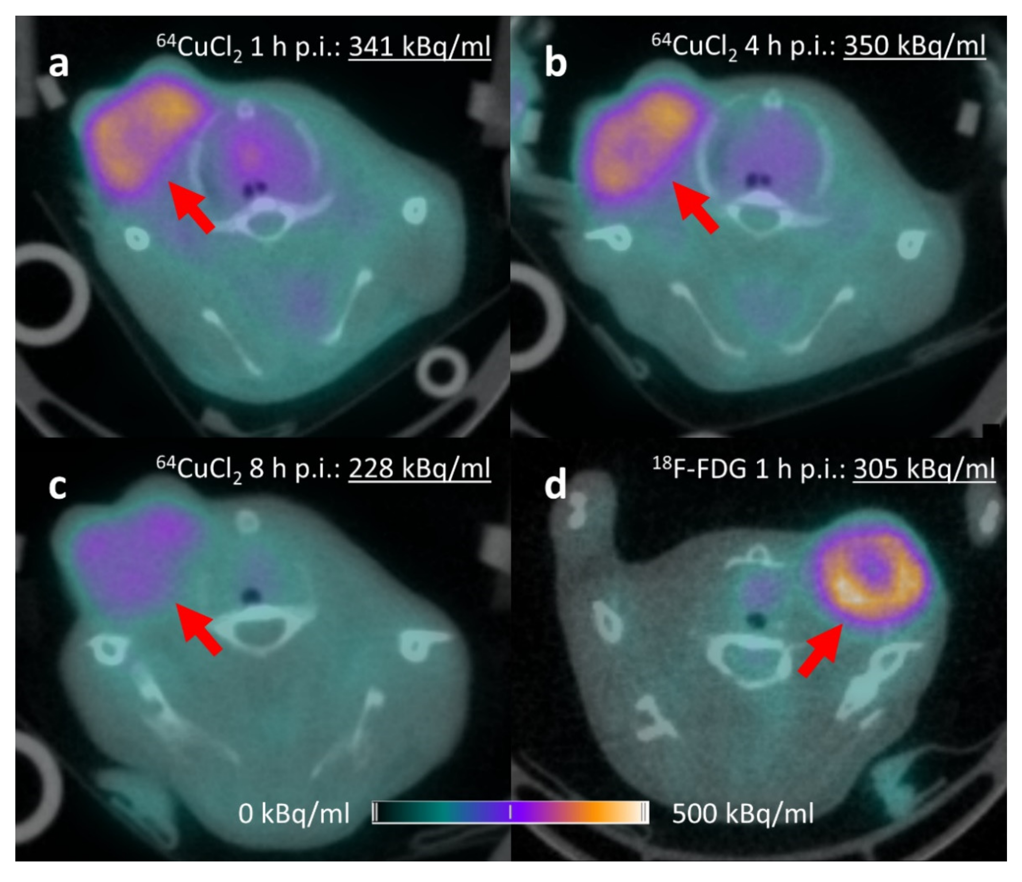
2.3. 64CuCl2 µPET/CT Static Acquisitions

Four i.v. injected mice were imaged from 1 h to 8 h p.i. (Figure 3). A mild but significant increase in activity was observed within tumors. SUVmean ranged from 0.81 ± 0.12 at 1 h p.i. to a maximum of 1.02 ± 0.08 at 4 h p.i. (p = 0.02), corresponding to 5.0 ± 0.20 %ID/g. The liver had the highest 64CuCl2 accumulation, showing an SUVmean from 3.78 ± 1.03 at 1 h p.i. to 4.35 ± 0.43 at 4 h p.i. (18.5 ± 5.6 to 21.3 ± 2.8 %ID/g). Brain and muscle activity variation was minimal from 1 h to 8 h p.i., showing the highest activity at 8 h p.i. of 1.5 ± 0.1 %ID/g for brain and 1.6 ± 0.26 %ID/g for muscle. The tumor-to-muscle ratio exhibited a nonsignificant increase from 1 h (3.13 ± 0.32) to 4 h p.i. (3.71 ± 0.64) (p = 0.23).

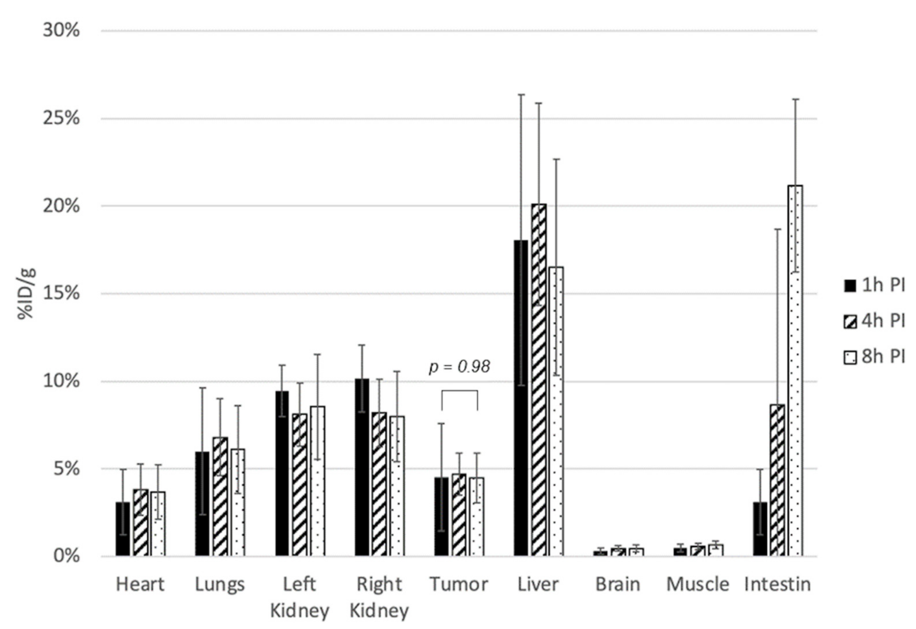
2.4. 64CuCl2 Ex Vivo Biodistribution Study

Ex vivo biodistribution studies were conducted in 13 mice with tumor grafts and sacrificed at 1 h (n = 5), 4 h (n = 4), and 8 h (n = 4) after 64CuCl2 i.v. administration (Figure 4). The mean weight of the examined tissue specimens was 0.36 ± 0.03 g for the brain, 0.11 ± 0.02 g for the heart, 0.19 ± 0.05 g for the lungs, 0.24 ± 0.07 g for the liver, 1.95 ± 0.55 g for the intestine, 0.16 ± 0.02 g for the kidney, and 0.13 ± 0.05 g for the muscle. The mean excised tumor weight was 0.19 ± 0.06 g.
No significant variation in tumor activity was observed from 1 h p.i. (4.52 ± 3.07 %ID/g) to 8 h p.i. (4.47 ± 1.42 %ID/g) (p = 0.98). The liver had the highest 64CuCl2 accumulation (20.10 ± 5.77 %ID/g at 4 h p.i.). The intestine was the only organ displaying an important temporal variation in 64CuCl2 uptake mainly due to intestinal excretion (3.10 ± 1.88 %ID/g at 1 h, 21.16 ± 4.92 %ID/g at 8 h p.i.). Kidney cortical activity averaged 10.15 ± 1.91 %ID/g at 1 h p.i (right kidney measurement), with a slight decrease over time without reaching significance (p = 0.22). The brain and muscle were characterized by the lowest tracer accumulation: 0.31 ± 0.18 %ID/g and 0.48 ± 0.21 %ID/g (1 h p.i.), respectively.

2.5. 18F-FDG µPET/CT and Ex Vivo Biodistribution Study

Six mice were scanned at 15 and 60 min after 18F-FDG i.v. administration and then sacrificed and dissected for the biodistribution study. The tumor-to-muscle 18F-FDG uptake ratios were 2.11 ± 0.31 at 20 min and 4.54 ± 1.11 at 65 min p.i. Tumor SUVmean values were 0.69 ± 0.07 at 20 min p.i. and 1.94 ± 0.25 at 65 min p.i. SUVmean at 65 min p.i. of brain and muscle were 1.11 ± 0.11 and 0.44 ± 0.08, respectively. From ex vivo measurements, the mean tumor 18F-FDG accumulation at 70 min p.i. was 8.82 ± 1.03 %ID/g.

3. Discussion

Our study first assessed the uptake profile of 64CuCl2 in a 4T1 allograft mouse model of TNBC considering 18F-FDG as a comparator. 64CuCl2 accumulation was more than 3-fold higher than that in the muscle background, allowing clear tumor detection and characterization in all examined animals. 64CuCl2 tumor accumulation was maximal approximately 4 h after 64CuCl2 administration, and tumors were detectable from 5 min to 8 h p.i. The tumor accumulation of 64CuCl2 remains stable and close to 5 %ID/g at 1 h, 4 h, and 8 h p.i. These results are encouraging and support the feasibility of 64CuCl2 as a PET radiotracer for 4T1-related TNBC imaging.
Copper-64 is most often used to label biologically active molecules such as proteins, antibodies, and nanoparticles through bifunctional chelators (DOTA, TETA). Nonetheless, this approach is complex, requires highly specific activities, and has numerous technical drawbacks, such as poor in vivo stability and chemical reactions with serum proteins. Conversely, copper-64—in its ionic form—achieves high in vivo stability, does not require complex radiochemistry, is not involved in degradation issues, and can be easily available through cyclotron-based production [15].
The 4T1 tumor cell line was previously screened as a potential candidate among 12 different tumor lines in a study designed to evaluate the diagnostic value of 64CuCl2 [16]. Assuming that 64CuCl2 uptake would be correlated with the quantity of the main copper transporter, each of these cell lines was tested in vitro for CTR-1 expression. The CTR-1/actin ratio ranged from approximately 0.1 (PC-3) to >1.0 (MDA-MB-435). The expression ratio of the 4T1 cell line was approximately 0.5, suggesting moderate overexpression of CTR-1 that, according to our observations, seems to be sufficient for tumor visualization by 64CuCl2 PET studies.
Jørgensen et al. evaluated 64CuCl2 binding among 5 human tumor lines (A2780, FaDu, H727, HT-29, U87MG) xenografted into NMRI mice. In vivo imaging was performed at 1 h and 22 h p.i., and the results were correlated with CTR-1 expression [17]. At 1 h p.i., 64CuCl2 activity ranged from 1.53 ± 0.07 %ID/g (A2780) to 3.48 ± 0.35 %ID/g (U87MG). FaDu, H727, and U87MG cell lines showed a significant 64CuCl2 uptake increase over time (up to 4.92 ± 0.17 for H727), but no correlation was found between 64CuCl2, tumor size, and CTR-1 expression. In our study, 64CuCl2 biodistribution remained stable between 1 h and 8 h p.i., allowing PET imaging to be performed during a large timeframe. Moreover, the important 64CuCl2 accumulation in 4T1 allografts (4.52 ± 3.07 %ID/g as early as 1 h p.i) suggests that TNBC is among the tumors with the highest 64CuCl2 avidity.
As 18F-FDG is the preferred radiotracer for TNBC [18], it was important to assess to what extent 64CuCl2 could provide satisfying PET images compared to 18F-FDG in 4T1-bearing mice. All tumors were clearly visualized by 64CuCl2 and 18F-FDG PET (Figure 5). In both cases, a photopenic area was present at the center of the largest tumors. This pattern is well known in PET clinical practice and is usually attributed to altered vascularization and tissue necrosis inside voluminous tumoral lesions.
Despite good tumor visualization, 18F-FDG accumulation was almost 2-fold higher than 64CuCl2 uptake (8.82 %ID/g at 65 min p.i. vs. 4.70 %ID/g at 4 h p.i.). However, the interest of 64CuCl2 could potentially reside in its ability to act as a therapeutic agent due to its β and Auger electron emission (in addition to diagnostic β+ emission). This hypothesis is based on data suggesting that nuclear Cu+ internalization is mediated by ATOX1 only in p53-positive cell lines [19]. This behavior allows for the potential therapeutic application of radioactive copper [20,21]. Ferrari et al. investigated a glioblastoma–astrocytoma animal model in athymic nude male BALB/c mice by 64CuCl2 PET [20]. Adequate tumor PET visualization was achieved after the injection of 12 MBq of 64CuCl2, reaching a tumor-to-healthy tissue ratio of 2.88, similar to what we have observed with 4T1-related tumors. After therapeutic injection of 64CuCl2 (single 333 MBq injection or 6 daily injections of 55.5 MBq), the tumor volume showed a reduction of 64–94% between the 1st and 20th weeks after injection. The survival rate at the 20th week drastically improved, from 0% in untreated mice to 73% in mice treated with multiple injections of 64CuCl2. Hematological toxicity (40% reduction in leukocytes) was moderate and reversible 5 weeks after 64CuCl2 administration.
The biodistribution of copper in humans has been widely studied. Ingested copper is absorbed by the proximal intestine, travels through the portal vein and undergoes a first hepatic passage. Once inside the blood, the copper circulates in the plasma compartment and is mostly bound to ceruloplasmin (60–95%) and albumin (10%). Its elimination is mainly hepato-biliary, explaining the diffuse hyperfixation of the liver and intestines [22]. As previously reported, kidneys displayed an important accumulation of 64CuCl2. This fact could be linked to the presence of superoxide dismutase, a ubiquitous enzyme present in the cytosol of eukaryotic cells and abundant in the liver and kidney, for which copper acts as a cofactor [23]. Renal uptake was predominant in the cortex, without significant bladder filling over time, rejecting the hypothesis of 64CuCl2 urinary elimination [24]. Relative 64CuCl2 kidney uptake was even higher from 30 min to 3 h p.i. in the study from Manrique-Arias et al. in Wistar rats [25]. These data contrast with our observations, as kidneys were only the third most accumulating organ after the liver and intestines. Using 64CuCl2 for therapeutic purposes would lead to higher injected doses, and identifying dose-limiting organs would be of paramount importance, as radionuclide therapy may lead to a wide spectrum of radiation-induced side effects [26].
Radiomic analysis on PET tumoral data could improve biological characterization using artificial intelligence frameworks. With the help of machine-learning, non-FDG PET explorations may lead to better understanding of tumoral features and related prognosis factors [27]. In vivo theranostic strategies could also benefit from nanomedicines, able to carry both diagnostic (e.g., 64Cu for PET, Gd(III) for MRI) and therapeutic compounds [28].
The main limitation of our work is the lack of complementary immunohistochemical characterization of 4T1 tumors. Determining the expression level of several proteins involved in copper uptake, transport, and efflux (particularly CTR-1, ATP7A/B, ATOX1) remains necessary to define the mechanism of 64CuCl2 uptake. A better understanding of the underlying molecular mechanisms should help to assess which pathways are involved in 64Cu homeostasis. To evaluate the potential role of 64CuCl2 for therapeutic purposes, ATOX-mediated Cu+ internalization within the nucleus should be further investigated.

4. Materials and Methods

4.1. Radiotracer Production

For 64CuCl2 production, the target material isotopically enriched 64Ni was purchased from ChemGas (Boulogne, France). Ni plating was performed based on the method reported by Wesley and Carey [29]. Target irradiation was performed using a TR24 cyclotron from ACSI, which delivered an extracted energy proton beam between 16 MeV and 25 MeV. A dedicated degrader was developed to obtain proton energy in the range of 11 MeV on target (optimal production yield of 64Cu for thin target). A few hours after bombardment, the plated Ni layer was dissolved in a concentrated 9 M HCl solution. The solution was transferred onto a 1.5 × 15 cm AG1-X8 anion exchange column. The column was washed with 6 M HCl solution to collect 64Ni. Then, 0.1 M HCl was added to elute 64Cu in 64CuCl2 form. Using a heating block, the eluted 64CuCl2 was evaporated to dryness and then diluted in saline solution (pH > 5.5). The good-manufacturing-practices-compliant 18F-FDG was provided by Curium (Dijon, France).

4.2. 4T1 Cell Culture and Animal Model

4T1 cells were obtained from Weizmann Institute of Science (Rehovot, Israel). Culture was performed under the following conditions: constant temperature (37 °C), 5% CO2, DMEM with 4.5 g of glucose supplemented with 10% fetal bovine serum and 1% antibiotics (penicillin/streptomycin). A suspension of 106 4T1 cells in 100 µL of PBS was injected into the mammary gland of 29 female BALB/c mice after anesthesia inhalation (2% isoflurane). Tumor width and length were measured with a caliper to calculate the tumor volume. Animals were maintained at constant temperature (22 °C) and humidity (40%) with 12 h light/dark cycles and unlimited access to water and food. Individual cages were ventilated with HEPA filters in the dedicated IPHC facility in Strasbourg.

4.3. PET/CT Imaging Acquisition and Analysis

PET imaging was performed 7 to 9 days after 4T1 cell allografting. All PET/CT acquisitions were performed on an IRIS PET/CT device (Inviscan, Strasbourg, France) equipped with 2 octagonal rings of 8 block detectors each, a matrix of 702 LYSO:Ce crystals of 1.6 × 1.6 × 12 mm each per module, and 64 Hamamatsu H8500 photomultiplier anodes. The geometrical configuration provides an axial coverage of 95 mm and a transverse field of view of 80 mm. The spatial resolution of the system is below 1.5 mm, with an absolute sensitivity close to 8% (NEMA standard) [30].
The 64CuCl2 PET acquisition protocol included a dynamic phase of 11 frames of 5 min each starting 5 min after the injection of 351 ± 56 kBq/g (18F equivalent) of 64CuCl2 and a 10-min static image acquired at 1 h, 2 h, 4 h, and 8 h p.i. 64CuCl2 was injected into the mouse tail vein (intravenous) or inside the peritoneum to compare both injection modalities. Concerning 18F-FDG PET, a 10-min static acquisition was performed at 15 min and 60 min after the injection of 278 ± 45 kBq/g radiotracer in the tail vein. All 64CuCl2 and 18F-FDG PET data were reconstructed using the OSEM algorithm (8 iterations, 8 subsets) in a 3D volume of 201 × 201 × 120. CT acquisition (80 kV, 0.9 mA) was performed for anatomical correlation. PET data were corrected for random coincidences. No attenuation or scatter corrections were applied. The results of PET images were qualitatively interpreted as positive or negative on a dedicated workstation (Syngo.via VB30; Siemens, Erlangen, Germany). A focal nonphysiologic increase in radiotracer uptake in the area of tumor development was considered to be a positive PET result. For a semiquantitative assessment of tumoral uptake, an elliptic volume of interest (VOI) was drawn to assess the mean activity of each organ and tumor. Muscle activity was determined from the left posterior triceps. Finally, time–activity curves were extracted from VOIs for the entire dynamic PET acquisition. SUVs were calculated from the mean voxel value within the VOIs. SUVs were recorded and used to generate time–activity curves.
To obtain 64CuCl2 biodistribution over time, 13 4T1-bearing BALB/c mice were sacrificed and dissected at 1 h (n = 4), 4 h (n = 5), or 8 h (n = 4) postinjection (p.i.). After excision of the tumor and organs of interest (liver, brain, intestines, kidneys, lungs, heart, and muscle), each histological specimen was weighed and placed in an automatic gamma counter (Hidex, Turku, Finland) for 1 min. The count obtained was converted to absolute activity using a calibration curve and then corrected for radioactive decay to an activity value arbitrarily taken at the time of animal sacrifice. The results are expressed as a percentage of the injected dose per gram of tissue (%ID/g).

4.4. Data Presentation

The quantitative data are reported as the mean values and standard deviations. Box plots were used for graphical representation of the results. Means were compared using Student’s t-test (for paired and independent values). p < 0.05 was considered significant. Statistical analysis was performed using the online BiostaTGV (https://biostatgv.sentiweb.fr, accessed on 10 March 2022).

5. Conclusions

64CuCl2 is easily available through cyclotron-based production and can be efficiently used as a PET radiotracer in 4T1-related TNBC. Although 64CuCl2 uptake is lower than 18F-FDG uptake, the method allows for nonglycolytic molecular characterization of TNBC in an animal model. Complementary studies are needed to correlate 64CuCl2 uptake to cell expression of proteins involved in copper transportation, particularly to enable theragnostic strategies.

Author Contributions

A.L., F.B., A.O., L.T., A.I. and D.B. designed the study, collected and analyzed the data, and were involved in original draft preparation. G.A. provided histology analyses. A.L., F.B., A.O., G.A., L.T., A.I. and D.B. reviewed the final draft. F.B., A.I. and D.B. carried out overall supervision. All authors have read and agreed to the published version of the manuscript.

Funding

The work has been supported in part by a grant from the French National Agency for research called “Investissements d’Avenir” Labex IRON n ANR-11-LABX-0018-01.

Institutional Review Board Statement

All animal experiments were performed in accordance with the European Institutes of Health Guidelines regarding the care and use of animals for experimental procedures. The study protocol was approved by the Alsace Regional Ethics Committee for Animal Experimentation (Approval ID: APAFIS#15255).

Informed Consent Statement

Not applicable.

Data Availability Statement

Not applicable.

Acknowledgments

The technical assistance of CYRCé platform members (IPHC, Strasbourg, France) is gratefully acknowledged.

Conflicts of Interest

The authors declare no conflict of interest.

References

  1. Inesi, G. Molecular features of copper binding proteins involved in copper homeostasis: Copper Binding Proteins. IUBMB Life 2017, 69, 211–217. [Google Scholar] [CrossRef] [PubMed]
  2. Devi, S.R.B.; Dhivya, M.A.; Sulochana, K.N. Copper transporters and chaperones: Their function on angiogenesis and cellular signalling. J. Biosci. 2016, 41, 487–496. [Google Scholar] [CrossRef] [PubMed]
  3. Boschi, A.; Martini, P.; Janevik-Ivanovska, E.; Duatti, A. The emerging role of copper-64 radiopharmaceuticals as cancer theranostics. Drug Discov. Today 2018, 23, 1489–1501. [Google Scholar] [CrossRef] [PubMed]
  4. Fuchs, A.G.; de Lustig, E.S. Localization of Tissue Copper in Mouse Mammary Tumors. Oncology 1989, 46, 183–187. [Google Scholar] [CrossRef]
  5. Ishida, S.; Andreux, P.; Poitry-Yamate, C.; Auwerx, J.; Hanahan, D. Bioavailable copper modulates oxidative phosphorylation and growth of tumors. Proc. Natl. Acad. Sci. USA 2013, 110, 19507–19512. [Google Scholar] [CrossRef] [Green Version]
  6. Denoyer, D.; Masaldan, S.; La Fontaine, S.; Cater, M.A. Targeting copper in cancer therapy: ‘Copper That Cancer’. Metallomics 2015, 7, 1459–1476. [Google Scholar] [CrossRef]
  7. Brasse, D.; Nonat, A. Radiometals: Towards a new success story in nuclear imaging? Dalton Trans. 2015, 44, 4845–4858. [Google Scholar] [CrossRef]
  8. Gutfilen, B.; Souza, S.; Valentini, G. Copper-64: A real theranostic agent. Drug Des. Dev. Ther. 2018, 12, 3235–3245. [Google Scholar] [CrossRef] [Green Version]
  9. Piccardo, A.; Paparo, F.; Puntoni, M.; Righi, S.; Bottoni, G.; Bacigalupo, L.; Zanardi, S.; DeCensi, A.; Ferrarazzo, G.; Gambaro, M.; et al. 64CuCl2 PET/CT in Prostate Cancer Relapse. J. Nucl. Med. 2018, 59, 444–451. [Google Scholar] [CrossRef] [Green Version]
  10. Pulaski, B.A.; Ostrand-Rosenberg, S. Mouse 4T1 Breast Tumor Model. Curr. Protoc. Immunol. 2000, 39, 20–22. [Google Scholar] [CrossRef]
  11. Schrörs, B.; Boegel, S.; Albrecht, C.; Bukur, T.; Bukur, V.; Holtsträter, C.; Ritzel, C.; Manninen, K.; Tadmor, A.; Vormehr, M.; et al. Multi-Omics Characterization of the 4T1 Murine Mammary Gland Tumor Model. Front. Oncol. 2020, 10, 1195. [Google Scholar] [CrossRef]
  12. Capozza, M.; Anemone, A.; Dhakan, C.; Della Peruta, M.; Bracesco, M.; Zullino, S.; Villano, D.; Terreno, E.; Longo, D.R.; Aime, S. GlucoCEST MRI for the Evaluation Response to Chemotherapeutic and Metabolic Treatments in a Murine Triple-Negative Breast Cancer: A Comparison with [18F]F-FDG-PET. Mol. Imaging Biol. 2022, 24, 126–134. [Google Scholar] [CrossRef]
  13. Kaloudi, A.; Lymperis, E.; Giarika, A.; Dalm, S.; Orlandi, F.; Barbato, D.; Tedesco, M.; Maina, T.; de Jong, M.; Nock, B.A. NeoBOMB1, a GRPR-Antagonist for Breast Cancer Theragnostics: First Results of a Preclinical Study with [67Ga]NeoBOMB1 in T-47D Cells and Tumor-Bearing Mice. Molecules 2017, 22, 1950. [Google Scholar] [CrossRef] [Green Version]
  14. Ding, F.; Huang, C.; Liang, C.; Wang, C.; Liu, J.; Tang, D. 68Ga-FAPI-04 vs. 18F-FDG in a longitudinal preclinical PET imaging of metastatic breast cancer. Eur. J. Nucl. Med. Mol. Imaging 2021, 49, 290–300. [Google Scholar] [CrossRef]
  15. Chakravarty, R.; Chakraborty, S.; Dash, A. 64Cu2+ Ions as PET Probe: An Emerging Paradigm in Molecular Imaging of Cancer. Mol. Pharm. 2016, 13, 3601–3612. [Google Scholar] [CrossRef]
  16. Qin, C.; Liu, H.; Chen, K.; Hu, X.; Ma, X.; Lan, X.; Zhang, Y.; Cheng, Z. Theranostics of Malignant Melanoma with 64CuCl2. J. Nucl. Med. 2014, 55, 812–817. [Google Scholar] [CrossRef] [Green Version]
  17. Jørgensen, J.T.; Persson, M.; Madsen, J.; Kjær, A. High tumor uptake of 64Cu: Implications for molecular imaging of tumor characteristics with copper-based PET tracers. Nucl. Med. Biol. 2013, 40, 345–350. [Google Scholar] [CrossRef]
  18. Tchou, J.; Sonnad, S.S.; Bergey, M.R.; Basu, S.; Tomaszewski, J.; Alavi, A.; Schnall, M. Degree of Tumor FDG Uptake Correlates with Proliferation Index in Triple Negative Breast Cancer. Mol. Imaging Biol. 2010, 12, 657–662. [Google Scholar] [CrossRef]
  19. Eiblmaier, M.; Meyer, L.A.; Anderson, C.J. The role of p53 in the trafficking of copper-64 to tumor cell nuclei. Cancer Biol. Ther. 2008, 7, 63–69. [Google Scholar] [CrossRef] [Green Version]
  20. Ferrari, C.; Niccoli Asabella, A.; Villano, C.; Giacobbi, B.; Coccetti, D.; Panichelli, P.; Rubini, G. Copper-64 Dichloride as Theranostic Agent for Glioblastoma Multiforme: A Preclinical Study. Biomed Res. Int. 2015, 2015, 129764. [Google Scholar] [CrossRef] [Green Version]
  21. Pinto, C.I.G.; Bucar, S.; Alves, V.; Fonseca, A.; Abrunhosa, A.J.; da Silva, C.L.; Guerreiro, J.F.; Mendes, F. Copper-64 Chloride Exhibits Therapeutic Potential in Three-Dimensional Cellular Models of Prostate Cancer. Front. Mol. Biosci. 2020, 7, 609172. [Google Scholar] [CrossRef]
  22. Kjærgaard, K.; Sandahl, T.D.; Frisch, K.; Vase, K.H.; Keiding, S.; Vilstrup, H.; Ott, P.; Gormser, L.C.; Munk, O.L. Intravenous and oral copper kinetics, biodistribution and dosimetry in healthy humans studied by [64Cu]copper PET/CT. EJNMMI Radiopharm. Chem. 2020, 5, 15. [Google Scholar] [CrossRef]
  23. Bass, L.A.; Wang, M.; Welch, M.J.; Anderson, C.J. In Vivo Transchelation of Copper-64 from TETA-Octreotide to Superoxide Dismutase in Rat Liver. Bioconjug. Chem. 2000, 11, 527–532. [Google Scholar] [CrossRef]
  24. Avila-Rodriguez, M.A.; Rios, C.; Carrasco-Hernandez, J.; Manrique-Arias, J.C.; Martinez-Hernandez, R.; García-Pérez, F.O.; Jalilian, A.R.; Martinez-Rodriguez, E.; Romero-Piña, M.E.; Diaz-Ruiz, A. Biodistribution and radiation dosimetry of [64Cu] copper dichloride: First-in-human study in healthy volunteers. EJNMMI Res. 2017, 7, 98. [Google Scholar] [CrossRef]
  25. Manrique-Arias, J.C.; Carrasco-Hernández, J.; Reyes, P.G.; Ávila-Rodríguez, M.A. Biodistribution in rats and estimates of doses to humans from 64CuCl2, a potential theranostic tracer. Appl. Radiat. Isot. 2016, 115, 18–22. [Google Scholar] [CrossRef]
  26. Iyer, R. Radiation injury: Imaging findings in the chest, abdomen and pelvis after therapeutic radiation. Cancer Imaging 2006, 6, S131–S139. [Google Scholar] [CrossRef] [Green Version]
  27. Thompson, R.F.; Valdes, G.; Fuller, C.D.; Carpenter, C.M.; Morin, O.; Aneja, S.; Lindsay, W.D.; Aerts, H.J.W.L.; Agrimson, B.; Deville, C.; et al. Artificial Intelligence in Radiation Oncology Imaging. Int. J. Radiat. Oncol. Biol. Phys. 2018, 102, 1159–1161. [Google Scholar] [CrossRef] [PubMed]
  28. Xue, X.; Qu, H.; Li, Y. Stimuli-Responsive Crosslinked Nanomedicine for Cancer Treatment. In Exploration; John Wiley & Sons, Inc.: Hoboken, NJ, USA, 2022; p. 20210134. [Google Scholar]
  29. Wesley, W.A.; Carey, J.W. The Electrodeposition of Nickel from Nickel Chloride Solutions. Trans. Electrochem. Soc. 1939, 75, 209. [Google Scholar] [CrossRef]
  30. Belcari, N.; Camarlinghi, N.; Ferretti, S.; Iozzo, P.; Panetta, D.; Salvadori, P.A.; Sportelli, G.; Del Guerra, A. NEMA NU-4 Performance Evaluation of the IRIS PET/CT Preclinical Scanner. IEEE Trans. Radiat. Plasma Med. Sci. 2017, 1, 301–309. [Google Scholar] [CrossRef]
Figure 1. Histological sections of 4T1-related allografts. (a) Hematoxylin-eosin staining (×400) showing fusocellular patterns with numerous mitoses (arrowheads). (b) Cytokeratin 5/6 staining (×200) revealing tumoral cells (arrow) and basal cells (star).
Molecules 27 04869 g001
Figure 2. In vivo dynamic PET assessment (from 5 to 60 min p.i.) of 64CuCl2 activity expressed as SUVmean inside mammary tumor and organs of interest. (a) Tumor. (b) Liver. (c) Muscle. (d) Kidneys. IV: intravenous; IP: intraperitoneal.
Molecules 27 04869 g002
Figure 3. In vivo static PET assessment (from 1 to 8 h p.i.) of 64CuCl2 activity expressed as SUVmean inside mammary tumor and organs of interest. IV: intravenous; IP: intraperitoneal.
Molecules 27 04869 g003
Figure 4. Ex vivo assessment of 64CuCl2 activity in tumors and organs of interest at 1 h, 4 h, and 8 h p.i.
Molecules 27 04869 g004
Figure 5. Mammary tumors assessed with 64CuCl2 PET/CT over time, with 18F-FDG PET/CT as a reference. 64CuCl2 tumor uptake expressed as mean kBq/mL inside VOI was higher at 4 h p.i. and slightly reduced over time. (a) Axial fused 64CuCl2 PET/CT, 1 h p.i. (b) Axial fused 64CuCl2 PET/CT, 4 h p.i. (c) Axial fused 64CuCl2 PET/CT, 8 h p.i. (d) Axial fused 18F-FDG PET/CT, 1 h p.i.
Molecules 27 04869 g005
Publisher’s Note: MDPI stays neutral with regard to jurisdictional claims in published maps and institutional affiliations.

