Review

Timeline of the Development of Skin-Lightening Active Ingredients in Japan

School of Bioscience and Biotechnology, Tokyo University of Technology, 1404-1 Katakura, Hachioji, Tokyo 192-0982, Japan
Molecules 2022, 27(15), 4774; https://doi.org/10.3390/molecules27154774
Submission received: 29 June 2022 / Revised: 20 July 2022 / Accepted: 22 July 2022 / Published: 26 July 2022

Abstract

:
Japanese pharmaceutical cosmetics, often referred to as quasi-drugs, contain skin-lightening active ingredients formulated to prevent sun-induced pigment spots and freckles. Their mechanisms of action include suppressing melanin production in melanocytes and promoting epidermal growth to eliminate melanin more rapidly. For example, arbutin and rucinol are representative skin-lightening active ingredients that inhibit melanin production, and disodium adenosine monophosphate and dexpanthenol are skin-lightening active ingredients that inhibit melanin accumulation in the epidermis. In contrast, oral administration of vitamin C and tranexamic acid in pharmaceutical products can lighten freckles and melasma, and these products are more effective than quasi-drugs. On the basis of their clinical effectiveness, skin-lightening active ingredients can be divided into four categories according to their effectiveness and adverse effects. This review discusses academic research and development regarding skin-lightening ingredients in Japan.

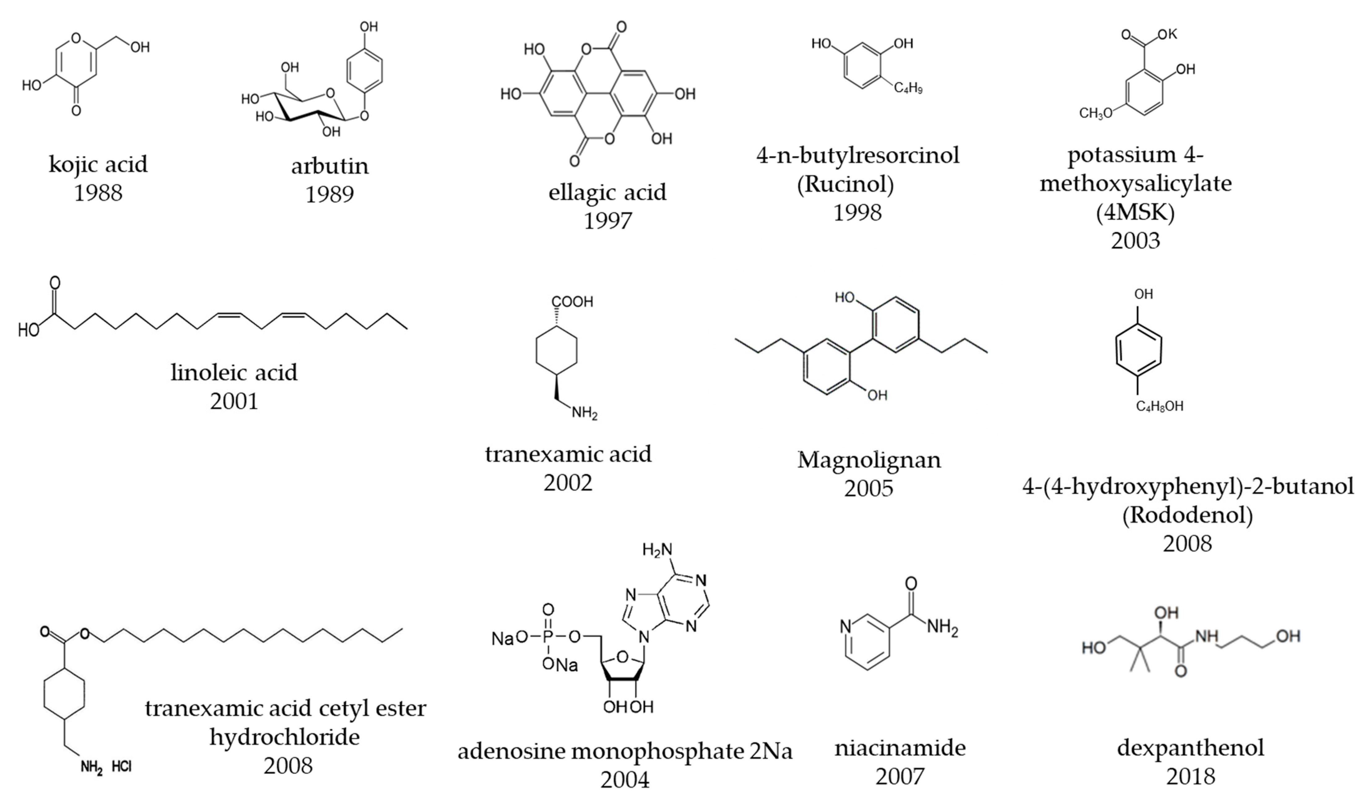
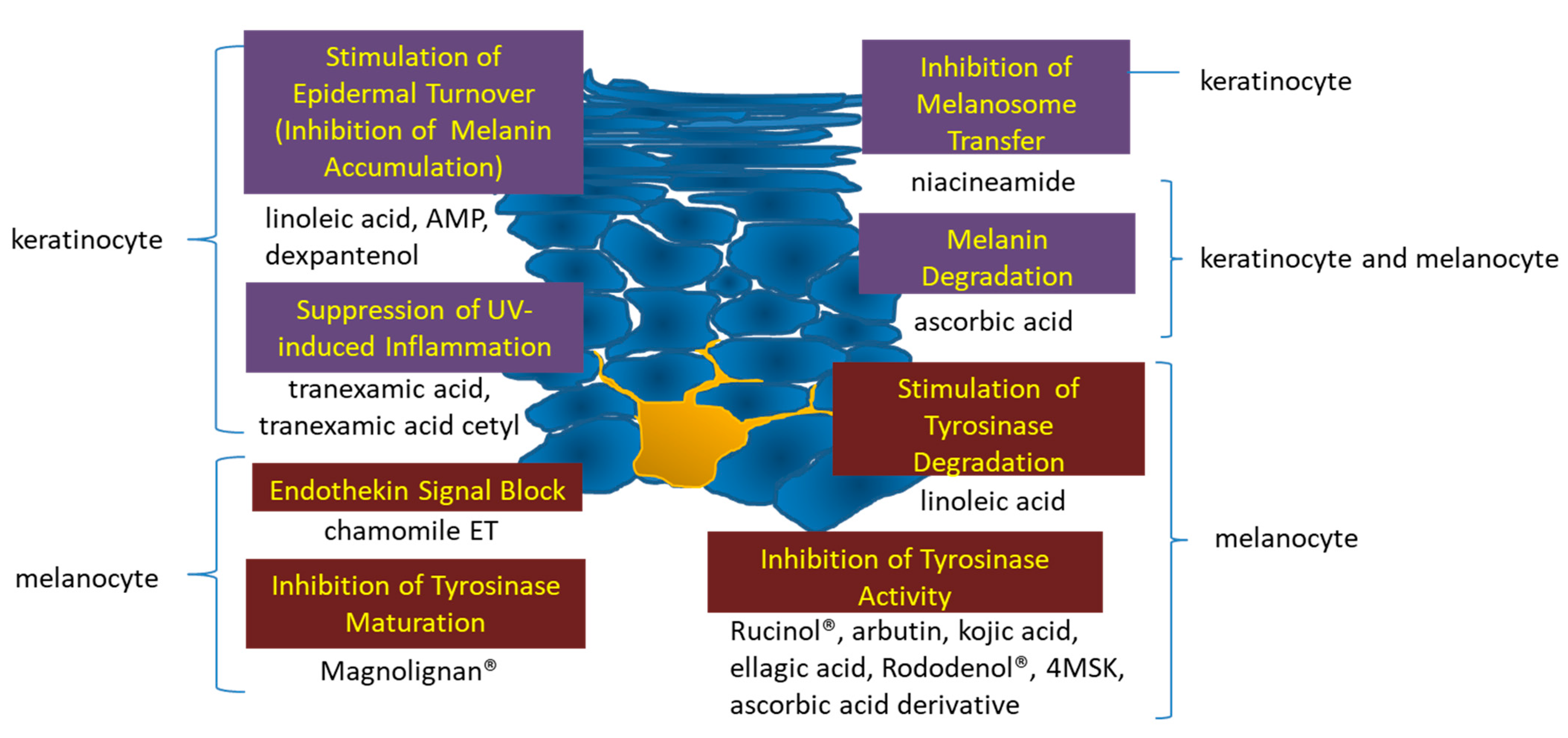
1. Development of Skin-Lightening Active Ingredients

Japanese pharmaceutical cosmetics are required to have one of the following purposes of use: (1) cleansing, (2) beautifying, (3) increasing attractiveness, (4) changing appearance, and (5) maintaining healthy skin or hair. Whether or not the product has the above purpose of use should be clarified by the efficacy or effectiveness, usage, and dosage of the product. If a product is determined not to meet the specified purpose of use on these bases of its efficacy or effectiveness, usage, and dosage, it is considered a “drug.” Examples of indications include “spots, freckles, pigmentation due to sunburn, etc.” (internal use), “spots, freckles, pigmentation due to sunburn/rash” (internal use), “skin pigmentation, senile pigmentation” (external use), “spots” (internal use), “Riehl melanosis, post-inflammatory hyperpigmentation” (for injection), and “melasma, freckles, post-inflammatory hyperpigmentation” (for internal use and injection). If the product has only the above efficacy or effectiveness, usage, and dosage, it cannot be considered a pharmaceutical cosmetic (quasi-drug).
In Japan, the efficacy for pharmaceutical cosmetics was changed in 2019 from “prevents sun spots and freckles” to “prevents sun spots and freckles by suppressing melanin production.” In 2004, the action of the ingredients in the formulation was changed to “prevents spots and freckles,” and, on the basis of the mechanism of action of the ingredients in the formula, the indication of “suppressing the accumulation of melanin and preventing spots and freckles” was approved. Thus, it is possible to apply for approval of pharmaceutical cosmetics for new efficacy within the scope of quasi-drug efficacy, on the basis of a clear scientific rationale.
The Japanese skincare market can be divided into the following functional categories: moisturizing, skin-lightening, anti-aging, sensitive skin, and pore and acne care. Lightening of the skin accounts for approximately 30% of the market and is the category with the most rapid growth over the past 35 years, particularly in terms of research and development [1]. Approximately twenty active ingredients have been developed for lightening-related quasi-drugs, including chemical substances and plant extracts with excellent inhibitory effects on tyrosinase activity and melanin production. A chronology of the development of skin-lightening active ingredients is presented in Table 1. The chemical structures of skin-lightening active ingredients in Japan are presented in Figure 1. Recent research on anti-aging skin-lightening has revealed new mechanisms and methods for the treatment of skin lightening. This section introduces the history of the skin-lightening ingredients developed in Japan.

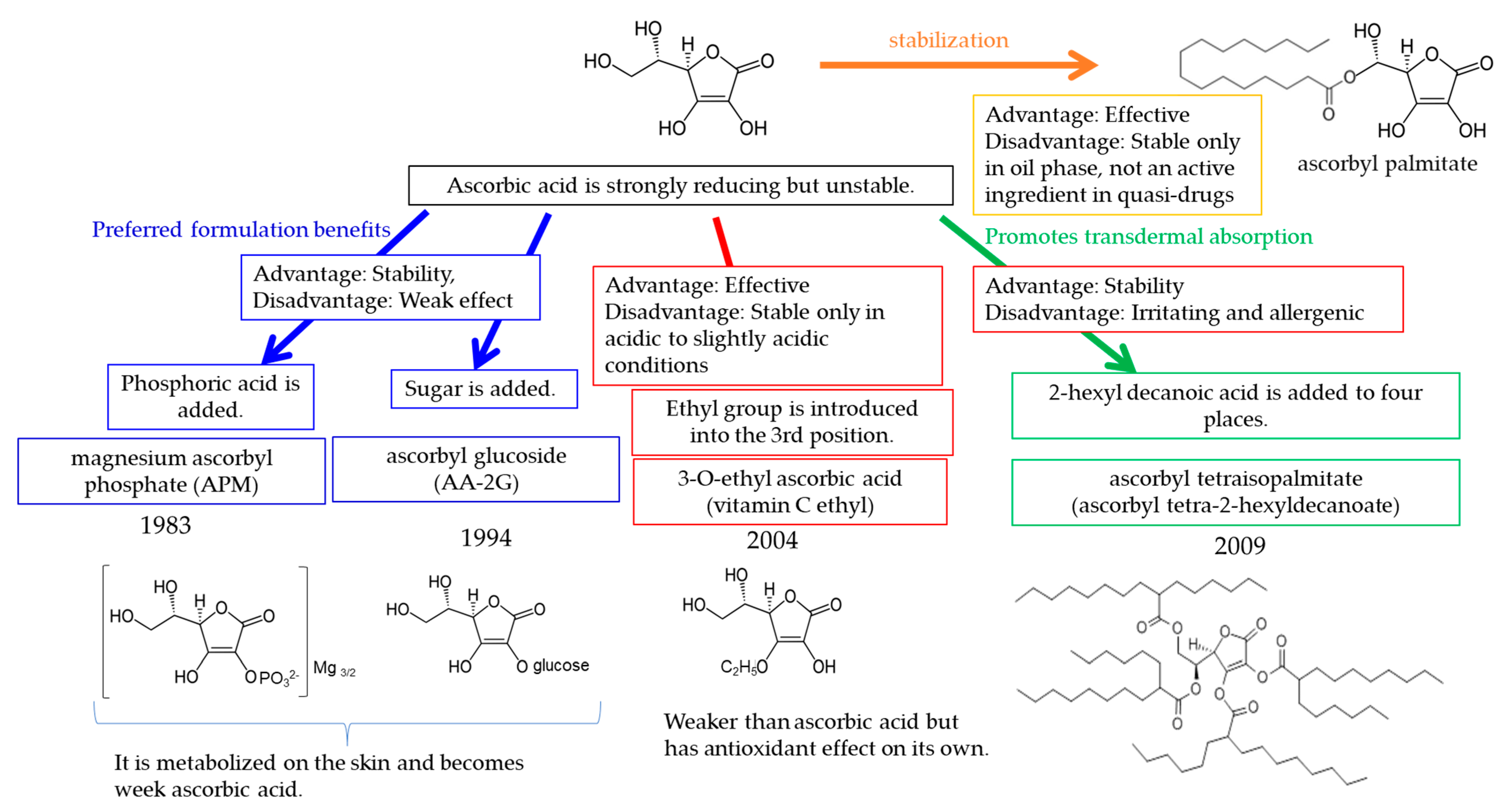
1.1. Ascorbic Acid Derivatives

Figure 2 shows the developmental chronology of ascorbic acid derivatives used in Japanese skin-lightening pharmaceutical cosmetics. The ascorbic acid derivative magnesium L-ascorbyl-2-phosphate (magnesium ascorbyl phosphate; APM) was developed as the first skin-lightening ingredient more than 40 years ago. Because ascorbic acid easily discolors over time when used in formulations, thus decreasing its content, more stable ascorbic acid fatty acid esters need to be developed, such as such as ascorbyl stearate or ascorbyl palmitate. However, even these fatty acid esters are not sufficiently stable in formulations containing water, such as emulsions; thus, browning occurs. A more stable APM was developed by phosphorylation of the hydroxyl group at position 2 of ascorbic acid to form magnesium salt; this compound was approved for use in Japanese quasi-drugs in the 1980s. APM has been used for the treatment of hyperpigmentation (melasma and Riehl melanosis) since 1969 [2], and its effectiveness in the treatment of sunburn-induced hyperpigmentation has been demonstrated in well-controlled studies since 1983 [3,4]. To make APM more stable under weakly acidic conditions, glucose has been attached to the hydroxyl group at position 2 to form 2-O-α-D-glucopyranosyl-L-ascorbic acid (ascorbyl glucoside; AA-2G), a compound that is stable around pH 6 that was approved as an active ingredient in quasi-drugs in the 1990s [5,6]. Unlike AA-2G, 3-O-ethyl-L-ascorbic acid (3-O-ethyl ascorbic acid; vitamin C ethyl), a vitamin C derivative with antioxidant and skin-lightening effects, was developed and approved as an active ingredient in quasi-drugs in 2004 [7,8]. Although ultraviolet A radiation contributes to persistent pigmentation [9], and DHICA polymerization is involved in the mechanism [10], vitamin C ethyl inhibits both polymerization of DHICA and persistent pigmentation formation [7]. 3-O-ethyl ascorbic acid manufactured by Nippon Hypox Laboratories Inc. is not allergenic, and no evidence of induced allergic contact dermatitis was recorded during a study by the National Industrial Chemicals Notification and Assessment Scheme (NICNAS) [11]. However, allergic contact dermatitis has been reported for other manufactured ingredients [12]. This may be due to impurities in other manufacturing materials. AA-2G is stabilized by modification of the hydroxyl group at position 2, whereas 3-O-ethyl ascorbic acid differs in that the ethyl group is ether-linked to the hydroxyl group at position 3. The fat-soluble vitamin C derivative ascorbyl tetra-2-hexyldecanoate (ascorbyl tetraisopalmitate) has also been developed [13], but it has been associated with allergic contact dermatitis [14,15,16].

1.2. Placenta Extract

Placenta extract, which dates back to ancient Egypt, is used mainly in cosmetic medicine for injections and internal administration. Placenta has been used for more than 60 years since its approval as a pharmaceutical injection in 1956 in Japan. It has been used as an OTC drug for frostbite and chapped skin and, for the purpose of enhancing cell division and promoting metabolism. The use of 3% human placenta extract preparation for treating facial hyperpigmentation in women was reported in 1982 in 47 cases of melasma, and two cases of freckles [17,18,19].
Placental extract used in pharmaceutical cosmetics is obtained by aseptic extraction of water from the placenta of pigs [Sus scrofa domesticus Erxleben (Suidae)] by freezing, thawing, or other methods, and removal of the high molecular weight fraction containing 0.01–0.40% nitrogen [placenta extract (1)] or 0.02–0.15% of nitrogen, as well as the macromolecular fraction containing 100 KAU or more of alkaline phosphatase [placenta extract (2)]. In addition, an extract is obtained by aseptic extraction of water from the placenta of pigs by enzymolysis or other methods, thus yielding a fraction containing 0.15–0.48% nitrogen [placenta extract (3)]. Common constituents include amino acids; vitamins such as ascorbic acid, thiamine, pyridoxine, and niacin; nucleic acid components such as uracil, adenine, and guanine; carbohydrates; lipids; and minerals.
Placenta extract promotes tissue metabolism and inhibits tyrosinase activity [20]. Moreover, the protein/peptide and lipid fractions of human placenta have melanogenesis-promoting effects [21,22]. Inhibitory or promoting effects on melanogenesis have been observed, depending on the extracted biomolecules, and the mechanisms of action are still largely unknown.

1.3. Kojic Acid

Kojic acid (5-hydroxy-2-hydroxymethyl-4-pyrone) is a skin-lightening ingredient obtained by cultivating yeast used in the production of soy sauce and miso. Topical application of a 1% or 2.5% kojic acid formulation has been shown to be effective in treating senile pigmentation and melasma [23,24,25,26], and kojic acid was approved as an active ingredient in quasi-drugs in 1988. In 2003, however, oral administration of kojic acid to p53-deficient mice was reported to induce liver cancer [27]. The Ministry of Health, Labour and Welfare of Japan suspended its use from March 2003, pending further research to clarify whether kojic acid is carcinogenic or genotoxic. A cosmetics manufacturer has conducted additional tests on the cosmetic properties of kojic acid and found it to be safe [28]. Accordingly, the Ministry of Health, Labour and Welfare issued an opinion at the Subcommittee on Safety Measures for Medicinal Products of the Pharmaceutical Affairs and Food Sanction Council on 2 November 2005, stating: “When used appropriately in quasi-drugs, there should be no particular concern about safety”. Subsequently, the aforementioned notice of discontinuance of use was withdrawn, and the resumption of production and marketing of quasi-drugs containing kojic acid was approved. However, simultaneous administration of ascorbic acid has been reported to enhance the liver tumor-promoting effect of kojic acid in rats induced with diethyl nitrosamine [29].
In a human transdermal absorption study in which 500 mg of cream containing 1% kojic acid was applied daily to the faces of six healthy women, the plasma concentration (limit of quantification 1 ng/mL) was highest 3–6 h after application, and the mean maximum plasma concentration in the six participants was 1.54 ng/mL. The highest plasma concentration averaged 1.54 ng/mL in the six participants. After more than 10 years of experience in using kojic acid in humans, no adverse health effects have been reported [30] Thus, the risk of carcinogenicity is extremely low for concentrations of kojic acid below 3%, and kojic acid is unlikely to exhibit genotoxicity that would harm living organisms. Furthermore, because kojic acid at a 1% concentration is unlikely to be absorbed into the body through the skin, and no adverse health effects have been reported, the safety of kojic acid is not of concern in cosmetics and pharmaceutical cosmetics under normal conditions of use. Products containing kojic acid are being sold as before.

1.4. Hydroquinone Derivatives and Phenolic Compounds

Focusing on the hydroquinone and the phenolic hydroxyl group of kojic acid, a hydroquinone derivative and a lightening agent with a phenolic hydroxyl group have been developed that have a stronger inhibitory effect on melanogenesis than kojic acid. Arbutin (hydroquinone β-D-glucopyranoside) was developed and approved as an active ingredient in quasi-drugs in 1989, on the basis of clinical results of its effectiveness against melasma [31]. Arbutin, a hydroquinone glycoside, is found in the leaves of Arctostaphylos uva ursi, with a content of 5–7.5%, as listed in the Japanese Pharmacopoeia. Arbutin inhibits melanogenesis at concentrations that do not cause cytotoxicity to pigment cells through antagonistic inhibition of tyrosinase activity [32,33]. Arbutin inhibits tyrosinase activity in a reversible manner and therefore is safer than the nonspecific or irreversible inhibition mediated by other tyrosinase inhibitors [32,33]. A variety of active ingredients have been developed to control tyrosinase activity in cosmetics since then, including ellagic acid [34,35,36], 4-n-butylresorcinol (Rucinol) [37,38,39,40], 5,5′-dipropyl-biphenyl-2,2′-diol (Magnolignan) [41,42,43], 4-(4-hydroxyphenyl)-2-butanol (Rhododenol) [44], and potassium 4-methoxysalicylate. Ellagic acid is a natural phenolic antioxidant, whereas the other five components are chemically synthesized. Magnolignan inhibits tyrosinase maturation, and cases of allergic contact dermatitis have been reported [45]. Rhododenol interferes with several steps of melanin production, including inhibiting tyrosinase activity, accelerating tyrosinase degradation, and targeting tyrosinase-related proteins, thus decreasing eumelanin levels. Rhododenol and Magnolignan both release hydroxyl radicals in the presence of tyrosinase in melanosomes and therefore are cytotoxic to pigmented cells [46,47]. Rhododenol is no longer used in pharmaceutical cosmetics, because of its leukoderma-inducing properties. Magnolignan is also no longer included in pharmaceutical cosmetics. Consequently, a 1-year safety review of all new quasi-drugs used in skin-lightening products is required before their approval.

1.5. Natural Compounds Contained in Plants

Research and development regarding natural compounds contained in plants and cosmetic raw materials to inhibit melanin production has been actively pursued, and many plant extracts have been incorporated into pharmaceutical skin-lightening cosmetics as well as quasi-drugs. For example, chamomile (Matricaria chamomilla L.) extract is an active ingredient in quasi-drugs that inhibits the action of endothelin [48,49,50,51], and safflower oil contains a high content of linoleic acid [52,53,54,55], which has been reported to promote both epidermal turnover and tyrosinase degradation [53,55]. The extract of Arnica montana, which is used as a cosmetic ingredient in Japan, has been found to inhibit melanogenesis; the active ingredient has been identified as a traxastane-type triterpene [56]. In pigmented cells, suppression of the expression of tyrosinase, tyrosinase-related protein-1 (Tyrp1), tyrosinase-related protein-2 (Tyrp2), and Pmel17 has been proposed to be the mechanism underlying melanogenesis inhibition [56]. Scutellarein, a flavonoid found in the medicinal plant Scutellaria baicalensis Georgi, inhibits melanin production by suppressing tyrosinase activity and microphthalmia-associated transcription factor, a protein required for the development of melanocytes [57].

1.6. Tanexamic Acid, Adenosine Monophosphate and Dexpantenol

Skin-lightening ingredients that act on keratinocytes without acting on melanocytes include tranexamic acid, adenosine monophosphate, and dexpanthenol.
In the 2000s, comprehensive skin-lightening research efforts focused on the various processes involved in melanogenesis, as opposed to only direct inhibition of melanogenesis. Phospholipase A2, arachidonic acid metabolites, and histamine were found to promote melanin synthesis [58,59,60], and tranexamic acid was found to be effective against ultraviolet B (UVB)-induced skin redness and pigmentation in guinea pigs [61]. Oral tranexamic acid was found to be effective for melasma [62] and was marketed as an over-the-counter option for treating melasma in Japan in 2007. Several other studies have demonstrated the effectiveness of tranexamic acid in oral administration [63,64,65] as well as its utility as a topical agent [66,67,68,69]. The skin-lightening effects of tranexamic acid are believed to result from both prostaglandin (PG) production mediated by plasmin [70,71] and cytokine production [72]. As of 2008, tranexamic acid cetyl ester hydrochloride (TXC), which is tranexamic acid esterified with cetyl alcohol, was also approved as an active ingredient [73,74]. TXC is hydrolyzed by an esterase and is likely to be converted to tranexamic acid in the subepidermal layer. Because both TXC and tranexamic acid suppress PGE2 production, TXC has been suggested to inhibit melanogenesis by preventing the production of melanocyte activating factors, particularly PGE2 production. Disodium adenosine phosphate was also developed as an active ingredient to suppress melanin accumulation by promoting epidermal turnover [75]. Furthermore, vitamin B3, niacinamide, has been reported to block the transfer of melanosomes at the surfaces of epidermal cells [76,77,78,79]. Some of these skin-lightening ingredients are also used in Korea and China. Another provitamin B5 called dexpanthenol was also approved in 2018 for use as a quasi-drug to decrease melanin accumulation and prevent spots and freckles, because of its ability to convert metabolites into energy and promote the turnover of epidermal cells [80,81].
Figure 3 illustrates their mechanisms of action. Since approximately 40 years ago, technological development has progressed from active ingredients that act on tyrosinase to those that act on tyrosinase genes, melanocytes, and the epidermis, thus leading to a wide variety of active ingredients and mechanisms of action in current formulations. Research on the delivery of active ingredients in skin-lightening products is also progressing. In contrast to sunburn pigment, which fades spontaneously as melanin is expelled during epidermal cell turnover, senile pigmentation does not fade spontaneously but instead worsens and progresses gradually, thereby suggesting a different mechanism from that in sunburn. Technological advances will continue to produce new active ingredients with high effectiveness in skin-lightening products in the future.

2. Report on the Effectiveness of a Formulation Containing a Lightening Agent and a Spot Remedy for Senile Pigmented Lesions

Clinical studies on senile pigmentation have investigated a formulation containing magnesium ascorbate phosphate salt, arbutin, and ellagic acid, which are active ingredients in quasi-drugs that prevent melanin production and freckles; a formulation containing kojic acid, which is no longer used in quasi-drugs because of concerns over its carcinogenic properties; a formulation containing vitamin A acid [82,83,84], which is involved in regulating epidermal turnover and is used in the United States to treat diseases such as acne vulgaris and psoriasis, and is effective in treating wrinkles and pigmentation caused by sun damage; and a formulation containing 2% 4-hydroxyanisole and 0.01% vitamin A acid, which is marketed in the United States as a treatment for senile pigmentation [85,86]. The effectiveness of these compounds is summarized in Table 2.
In an open study of 34 patients, including 17 with senile pigmentation, patients were given a formulation containing 10% magnesium L-ascorbyl-2-phosphate—a content exceeding the 3% in most quasi-drugs. A total of 88.3% of patients with senile pigmentation showed slight or even higher effectiveness, as assessed by skin color measurement [4]. A 7% arbutin formulation demonstrated an effectiveness rate of 81.3% on senile pigmentation after 3 months in an open test, and effectiveness was observed for all patients when the treatment period was extended to 6 months and 1 year [87]. An open study in 13 women treated with a 0.5% formulation of ellagic acid for 1–3 months found improved effectiveness on senile pigmentation in 69.2% of participants [36]. In a study in 18 patients (5 men and 13 women) with senile pigmentation who received topical application of 1% kojic acid and 0.1% oil-soluble licorice extract over a 16-week period, 77.8% of participants showed an improved effectiveness rate [26]. Additionally, 2% 4-hydroxyanisole and 0.01% vitamin A acid have been found to be more effective than 3% hydroquinone in treating senile pigmentation [85]. In a double-blind study investigating the topical effectiveness of 2% 4-hydroxyanisole for 6 months in 421 patients, the improved effectiveness was 84.1%. Complete disappearance of pigmented lesions was observed in some cases, thus indicating high effectiveness, but some adverse effects including erythema and irritation were also observed [86].
Reported effectiveness rates differ depending on whether the effectiveness criteria were “effective”, “somewhat effective”, higher. In each study, different criteria are used to determine the level of improvement as “effective” or “somewhat effective”. In some studies, the criteria for determining effectiveness are unclear, and placebos may or may not be used to control effectiveness. Therefore, caution should be exercised in comparing effectiveness rates across studies in which the criteria for determining effectiveness are unclear or in open studies lacking placebo controls.
Table 2. Comparison of clinical trials of skin-lightening agents and drugs for the treatment of age spots.
10% Magnesium Ascorbyl Phosphate Formulation7% Arbutin Formulation0.5% Ellagic Acid Formulation1% Kojic Acid and 0.1% Oil-Soluble Licorice Extract Formulation2% 4-Hydroxyanisole and 0.01% Vitamin A Acid Formulation
Test designOpen studyOpen studyOpen studyOpen studyDouble-blind controlled study
Number of cases17161318420421
SexWomen5 men, 13 womenMen and women
AgeUnknown28–50 years old (average 39 years old)34–85 years old (average 62.6 years old)
LocationUnknownFaceForearmFace
Period3 months to 1 year1 to 3 months8 and 16 weeks24 weeksObservation up to 48 weeks after 24 weeks of application
Effectiveness judgmentSkin color value (color difference meter)Visual observationVisual observationClose-up photograph determination and skin color value (image analysis)Visual observationVisual observation
Effectiveness ratioEffective (improved or much improved) or higher58.80%3 months 0%,
6 months 15.4%,
1 year 66.7%
30.80%8 weeks 5.6%,
16 weeks 22.2%
52.60%56.30%
Slightly effective (slightly improved) or more88.20%3 months 81.2%,
6 months 100%,
1 year 100%
69.20%8 weeks 66.7%,
16 weeks 77.8%
79.30%84.10%
Adverse effectsNoneNoneIrritation in a few cases, but no serious adverse effectsRedness 56%, burning 34%, desquamation 24%,
itching 16%, irritation 7%, decoloration 9%.
References[4][87][36][26][86][86]

3. Effectiveness Indices of Lightening Ingredients Developed in Japan

Most Japanese tests of the effectiveness of pharmaceutical skin-lightening cosmetics have assessed the degree of prevention of skin darkening caused by artificial UVB irradiation. Skin-lightening effects are exclusively evaluated on skin that darkens when exposed to ultraviolet light in humans. There are two types of evaluation methods: the anti-pigmentation test and the accelerated pigmentation disappearance test. The former is a double-blind controlled study in which a test sample is applied before, during, or after the UV irradiation period and the degree of pigmentation formation is compared with a placebo. The latter is a double-blind controlled trial in which the test sample is applied to the pigmented area and the degree of fading of the pigmentation is compared with a placebo. Both are assessed by visual evaluation and instrumental measurement based on superiority or inferiority comparisons or score judgments.
Consumers expect that skin lightening products will “lighten spots and freckles”. Moreover, many consumers expect these products to “eliminate spots and freckles” [88] and want skin-lightening cosmetics to have an effect on spots that have already formed, rather than preventing formation, or promoting the fading, of pigmentation caused by ultraviolet rays. However, problems exist regarding the lightening effects of such cosmetics when actually used on spots and freckles: test results published in scientific journals are lacking, and a possibility exists that these cosmetics might cause skin problems.
Skin-lightening active ingredients can be divided into four categories according to their clinical effectiveness and adverse effects. This review discusses academic research and development regarding skin-lightening ingredients in Japan. We have compiled a table of effectiveness indices for Japanese skin-lightening ingredients, on the basis of the published scientific literature (Table 3).
Category A: Effectiveness of the same concentration of cosmetic formulations on human pigment spots has been described in scientific journals and is highly recommended. Examples include tranexamic acid, arbutin, 3-O-ethyl ascorbic acid, magnesium ascorbyl phosphate (APM), ellagic acid, kojic acid, linoleic acid, 4-n-butylresorcinol, chamomile extract, and adenosine monophosphate.
Category B: Effectiveness of higher concentrations than those used in cosmetics on human pigment spots has been described in scientific journals and is recommended. Examples include oil-soluble licorice extract containing 50% glabridin, niacinamide, placenta extract, retinol, ascorbyl glucoside (AA-2G), and azelaic acid.
Category C: No effectiveness for human pigment spots has been described in scientific journals but may be considered; however, evidence is insufficient. Examples include potassium 4-methoxysalicylic acid and dexpanthenol.
Category D: Not recommended, because of toxicity data described in scientific journals. Examples include Rhododenol, Magnolignan, and ascorbyl tetra-2-hexyldecanoate.
Table 3. Effectiveness indices of lightening ingredients developed in Japan.
Effectiveness IndicesSkin-Lightening IngredientsTest Concentration (%)General Purpose or Japanese Cosmetics CompanyScientific Articles Providing Evidence
AEffectiveness of same concentration of cosmetic formulations on human pigment spots has been published in scientific journals and is highly recommended.tranexamic acid2General purpose[66]
arbutin3General purpose[31]
3-O-ethyl ascorbic acid (vitamin C ethyl)1General purpose[8]
magnesium L-ascorbyl-2-phosphate (APM)3General purpose[2]
ellagic acid 0.5General purpose[36]
kojic acid2.5, 0.5General purpose[23,24]
linoleic acid0.1General purpose[52]
4-n-butyl resorcinol, 0.3General purpose[37]
chamomile extract0.5Kao Corporation[51]
adenosine monophosphate3Otsuka Pharmaceutical Co., Ltd.[75]
BEffectiveness of higher concentrations than those used in cosmetics on human pigment spots has been published in scientific journals and is recommended.oil-soluble licorice extract containing 50% glabridin0.2General purpose[89]
niacinamide5, 4General purpose[77,78]
placenta extract3General purpose[17]
retinol0.15General purpose[90]
ascorbic acid 2-O-α-glucoside (AA-2G)20 (iontophoresis)General purpose[91]
azelaic acid.20General purpose[92]
CNo effectiveness for human pigment spots has been published in scientific journals and may be considered, but evidence is insufficientpotassium 4-methoxysalicylate 1, 3Shiseido Co. Ltd.
dexpanthenol POLA ORBIS HOLDINGS INC.
DNot recommended, because of toxicity data published in scientific journalsRhododenol2Kanebo Cosmetics Inc.[46]
Magnolignan0.5Kanebo Cosmetics Inc.[46]
ascorbyl tetra-2-hexyldecanoate Nikko Chemicals Co. Ltd.[14,15,16]
The use of a hydrophilic ointment containing 3% APM for hyperpigmentation (melasma and Riehl melanosis) was reported in 1969 [2]. It was reported in 1996 that APM 10% cream was found to be significantly beneficial in 19 of 34 patients with melasma and senile freckles and in 3 of 25 patients with normal skin [4]. The use of 3% human placenta extract for treating facial hyperpigmentation in women (47 cases of melasma and 2 cases of freckles) was reported in 1982 [17]. Other topically applied products with good results for melasma include 1% or 2.5% kojic acid [23], 3% arbutin [31], 1% tranexamic acid [66], 0.1% linoleic acid S [52], 0.3% Rucinol [37], and 0.5% ellagic acid [36]. Examples of topically applied products with good results for senile pigmentation include 3% APM [2], 10% APM [3,4], 1% or 2.5% kojic acid [23,24,25], 0.5% ellagic acid [36], 7% arbutin [87], and 0.5% chamomile extract [51]. Topical application of 1% or 2.5% kojic acid [23], 0.5% ellagic acid [36], 1% tranexamic acid [66], and 7% arbutin [87] has been used with good results in the treatment of freckles. In some cases, 1% vitamin C ethyl has shown effectiveness in pigmentation after natural light exposure and burns [8]. The effectiveness of these pharmaceutical skin-lightening cosmetics was not evaluated in a comparative study with a placebo, but instead was assessed in a study that examined the effectiveness of the formulations before and after continuous use, without double-blinding. In examining effectiveness, attention must be paid to changes in skin tone with seasonal variations and the effects of the base agent.
Examples of topical effectiveness in double-blind comparative studies include 0.1% retinoic acid (Tretinoin) [83], 0.2% oil-soluble licorice extract [89], and 20% azelaic acid [92] for melasma. Topical 0.1% retinol has been reported to be effective in photodamaged skin in a double-blind, controlled study [90]. Iontophoresis of 20% AA-2G has been reported to be effective in melasma and postinflammatory hyperpigmentation [91]. For senile pigmentation, both 0.1% retinoic acid (tretinoin) [84] and 2% 4-methoxyphenol/0.01% retinoic acid [85,86] have shown topical effectiveness in double-blind comparative studies, but these are not active ingredients in pharmaceutical cosmetics. Thus, a pharmaceutical skin-lightening cosmetic product is defined as a pharmaceutical product if its effectiveness against melasma and senile pigmentation is statistically demonstrated in actual use in a double-blind comparison test.

4. Issues in Methods for Evaluation of Skin-Lightening Effects

In Japan, the effectiveness of pharmaceutical skin-lightening quasi-drugs must be statistically demonstrated to prevent or promote the fading of UVB-induced pigmentation, or promote the fading of pigmentation in a UVB-induced pigmentation prevention test or a UVB-induced pigmentation improvement test through double-blind comparison. However, currently different active ingredients are evaluated in various ways to determine their skin-lightening effectiveness. For a quasi-drug to be approved for efficacy, its effectiveness must have been evaluated with an appropriate evaluation method. Moreover, the effectiveness must be properly evaluated with respect to what is used to transmit the efficacy to the consumer.
Visual evaluation using a skin color chart for more objective and quantitative evaluation is a valuable assessment method [93,94]. For instrumental measurement, the following methods are used: (1) use of a reflectance spectrophotometer (the reflected light is finely spectrophotographed, and the reflectance is measured every 5–10 nm to analyze the color in detail); (2) use of a color chromameter (which measures colors with three sensors—red, blue, and green—close to the sensitivity of the human eye and obtains tristimulus values in the color space); (3) use of a three-wavelength melanin index meter; and (4) analysis of digital images. However, evaluation and analysis methods are advancing rapidly [95,96,97,98]. Importantly, with these methods, data at each time point during the test must be collected under the same conditions for both participants and measurements. Skin color measurements are affected by factors including measurement time, posture, temperature, illumination, and probe pressing force. Therefore, for accurate measurements, nearby normal skin should be used as a control, and the probe pressing force should be kept constant. For images, an illuminance meter or a cast match should be used to confirm that the images are taken at a constant brightness and color.
The most commonly used instrumental measurement is the L*a*b* color system (L*: lightness; a*: chromaticity of red-green coordinates; b*: chromaticity of yellow-blue coordinates) standardized by the Commission Internationale de l’Eclairage (CIE). L* values are often used for skin-lightening evaluation but have the disadvantage of being affected by skin redness [98]. Notably, because the measured value is an average within the range of the measurement area, it cannot be accurately measured if the target area is small. The Mexameter MX18 (Courage + Khazaka, Germany), which measure the melanin index and erythema index by irradiating three wavelengths of light from a probe and measuring the light reflected from the skin, has slightly different hemoglobin indexes. It can assess pigmentation more accurately, although it is slightly affected by hemoglobin. Another method involves taking images of the entire face or part of the face with a CCD camera, calculating the tristimulus XYZ values from the values of the three primary colors of light (red, green, blue) at each pixel, and estimating the amount of melanin from the formula relating these values to the amount of melanin and hemoglobin [98]. The imaging method has the advantage of displaying the distribution of melanin over a wide area of the face. In addition, an ultraviolet camera can capture images of “hidden” pigmentation spots that cannot be recognized by the naked eye [97]. If quantitative measurement and analysis methods are used, this technology may be applied to skin-lightening evaluation. In vivo confocal microscopy can be used to obtain images of melanin granules by using the reflection of light of a certain wavelength, and the location of melanin granules can be determined on the basis of the brightness of the images [99]. In the future, we expect that analysis of skin-lightening effects will advance beyond indirect measurements of light reflected from the skin to noninvasive quantitative measurements of melanin in the skin in vivo.
Because instrumental measurement technology for skin surface conditions and color is continually advancing, and guidelines have been published for evaluating methods of skin-lightening and pigment spot improvement in humans [100], objective evaluation is required not only for pharmaceutical applications but also for those used to communicate efficacy to consumers. In addition, guidelines should be established for in vitro evaluation methods to ensure the appropriateness of methods for measuring inhibition of tyrosinase activity, melanin production and cytotoxicity in cultured cells, because the results differ depending on the evaluation method [41,44,46,101].

5. Future Development of Skin-Lightening Ingredients

Because the sale of cosmetic ingredients and cosmetics that have been tested on animals is prohibited, the skin-lightening effect of existing cosmetic ingredients must be demonstrated in clinical trials. Furthermore, skin-lightening ingredient that have high efficacy but cannot be monopolized to increase corporate profits are simply not used as active ingredients in quasi-drugs, because investments are not made in their development. Exfoliating ingredients and parabens currently used in cosmetics are candidates. Exfoliating ingredients, such as glycolic acid and lactic acid, that are used in cosmetics have been demonstrated to lighten skin by removing melanin-deposited stratum corneum [102,103], thus demonstrating that cosmetic ingredients can also lighten skin sufficiently. Parabens, which are commonly used as preservatives in cosmetics and foods, inhibit melanin production more effectively than the active ingredients used in quasi-drugs to lighten skin [104]. Several common cosmetic ingredients can also lighten skin; therefore, the effect of the active ingredients in quasi-drugs must be greater than the skin-lightening effect of cosmetic ingredients, excluding those in makeup cosmetics. Japanese pharmaceutical skin-lightening cosmetics must have “a mitigating effect on freckles and spots,” within the scope of efficacy of quasi-drugs. Current quasi-drugs suppress melanin production and prevent freckles and dark spots. Large melanosome complexes are increased in keratinocytes of solar lentigo [105]. Epidermal turnover time is involved in melanin disappearance [106]. Adenosine monophosphate and dexpanthenol, owing to their mechanisms of action, effectively promotes melanin excretion from the epidermis by promoting epidermal turnover. In line with the actual use scenarios of skin-lightening cosmetics by consumers, who expect their spots and freckles to fade and disappear [88], a specialist committee for evaluation of skin-lightening effectiveness of the Japanese Cosmetic Science Society has proposed “gentle improvement of spot and freckle pigmentation” as a new efficacy standard [100]. The proposal was made at a meeting of the cosmetic functional evaluation methodology review committee of the Japan Cosmetic Science Society. Moreover, to avoid problems with leukoderma, in Japan, as in other countries, third-party test results, rather than the cosmetics company’s own test results, should be evaluated for approval. The definition of skin-lightening, efficacy, and evaluation methods will be better suited to consumer needs through academic collaboration among universities, dermatologists, and companies in the future.

6. Conclusions

In Japan, approximately 20 active ingredients for skin brightening have been developed and approved, and sales of pharmaceutical skin lightening cosmetics containing these ingredients have also been approved. Some treatments have no apparent effect on spots and freckles, cause leukoderma, or cause allergic reactions. One of the reasons for these problems may be that the results of in-house effectiveness and safety tests have not been publicly reviewed. Objective evaluation by a third-party organization is needed to solve these problems. Because the sale of cosmetic ingredients and cosmetics that have been tested on animals is prohibited, pharmaceutical skin-lightening cosmetics containing active ingredients that have been demonstrated to be effective in clinical trials on existing cosmetic ingredients are expected to be marketed in the future.

Funding

This review received no external funding.

Institutional Review Board Statement

Not applicable.

Informed Consent Statement

Not applicable.

Data Availability Statement

Not applicable.

Conflicts of Interest

The authors declare no conflict of interest.

References

  1. General chapter. Functional Cosmetics Marketing Handbook 2019–2020; Fuji Keizai Group Co., Ltd.: Tokyo, Japan, 2019; pp. 7–32. (In Japanese) [Google Scholar]
  2. Ichikawa, H.; Kawase, H.; Aso, K.; Takeuchi, K. Topical treatment f pigmented dermatoses by modified ascorbic acid. Jpn. J. Clin. Dermatol. 1969, 23, 327–331. (In Japanese) [Google Scholar]
  3. Tagawa, M.; Murata, T.; Onuma, T.; Kameyama, K.; Sakai, C.; Kondo, S.; Yonemoto, K.; Quigley, J.; Dorsky, A.; Bucks, D.; et al. Inhibitory effects of magnesium ascorbyl phosphate on melanogenesis. SCCJ J. 1993, 27, 409–414. (In Japanese) [Google Scholar]
  4. Kameyama, K.; Sakai, C.; Kondoh, S.; Yonemoto, K.; Nishiyama, S.; Tagawa, M.; Murata, T.; Ohnuma, T.; Quigley, J.; Dorsky, A.; et al. Inhibitory effect of magnesium L-ascorbyl-2-phosphate (VC-PMG) on melanogenesis in vitro and in vivo. J. Am. Acad. Dermatol. 1996, 34, 29–33. [Google Scholar] [CrossRef]
  5. Miyai, E.; Yamamoto, I.; Akiyama, J.; Yanagida, M. Inhibitory effect of ascorbic acid 2-O-α-glucoside on the pigmentation of skin by exposure to ultraviolet light. Nishinihon J. Dermatol. 1996, 58, 439–443. (In Japanese) [Google Scholar] [CrossRef]
  6. Kumano, Y.; Sakamoto, T.; Egawa, M.; Iwai, I.; Tanaka, M.; Yamamoto, I. In vitro and in vivo prolonged biological activities of novel vitamin C derivative, 2-O-α-D-glucopyranosyl-L-ascorbic acid (AA-2G), in cosmetic fields. J. Nutr. Sci. Vitaminol. 1998, 44, 345–359. [Google Scholar] [CrossRef] [Green Version]
  7. Maeda, K.; Inoue, Y.; Nishikawa, H.; Miki, S.; Urushibata, O.; Miki, T.; Hatao, M. Involvement of melanin monomers in the skin persistent UVA-pigmentation and effectiveness of vitamin C ethyl on UVA-pigmentation. J. Jpn. Cosmet. Sci. Soc. 2003, 27, 257–268. (In Japanese) [Google Scholar]
  8. Miki, S.; Nishikawa, H. Effectiveness of vitamin C ethyl, Anti-Aging Series 2; N.T.S.: Tokyo, Japan, 2006; pp. 265–278. (In Japanese) [Google Scholar]
  9. Maeda, K. Action spectrum on UVA irradiation for formation of persistent pigmentation in normal Japanese individuals. Cosmetics 2017, 4, 55. [Google Scholar] [CrossRef] [Green Version]
  10. Maeda, K.; Hatao, M. Involvement of photo-oxidation of melanogenic precursors in prolonged pigmentation induced by UVA. J. Investig. Dermatol. 2004, 122, 503–509. [Google Scholar] [CrossRef] [PubMed] [Green Version]
  11. NICNAS. L-Ascorbic acid 3-O-ethyl-(INCI Name: 3-O-ethyl ascorbic acid), Public Report LTD/1879; National Industrial Chemicals Notification and Assessment Scheme (NICAS): Sydney, Australia, 2016; pp. 1–25. [Google Scholar]
  12. Romita, P.; Foti, C.; Barlusconi, C.; Mercurio, S.; Hansel, K.; Stingeni, L. Allergic contact dermatitis to 3-O-ethyl-L-ascorbic acid: An underrated allergen in cosmetics? Contact Dermat. 2020, 83, 63–64. [Google Scholar] [CrossRef]
  13. Obayashi, K.; Ochiai, Y.; Masaki, H.; Kurata, Y. Lipid-solble VC derivative ascorbic acid tetra-2-hexyldecanoate (VC-IP) as an anti-aging agent. In Proceedings of the SCSK Conference, Seoul, Korea, 22–24 September 2003; pp. 313–319. [Google Scholar]
  14. Swinnen, I.; Goossens, A. Allergic contact dermatitis caused by ascorbyl tetraisopalmitate. Contact Dermat. 2011, 64, 241–242. [Google Scholar] [CrossRef] [PubMed]
  15. Assier, H.; Wolkenstein, P.; Grille, C.; Chosidow, O. Contact dermatitis caused by ascorbyl tetraisopalmitate in a cream used for the management of atopic dermatitis. Contact Dermat. 2014, 71, 60–61. [Google Scholar] [CrossRef]
  16. Scheman, A.; Fournier, E.; Kerchinsky, L. Allergic contact dermatitis to two eye creams containing tetrahexyldecyl ascorbate. Contact Dermat. 2022, 86, 556–557. [Google Scholar] [CrossRef]
  17. Shimokawa, Y.; Kamisasanuki, S.; Tashiro, M. Treatment of facial dysmelanosis with cosmetics containing Placen A. Nishinihon J. Dermatol. 1982, 44, 1027–1029. (In Japanese) [Google Scholar]
  18. Imahara, K.; Ito, Y. Influence of placenta extract on melanin formation. SCCJ J. 1982, 16, 10–14. (In Japanese) [Google Scholar]
  19. Mallick, S.; Singh, S.K.; Sarkar, C.; Saha, B.; Bahdra, R. Human placental lipid induces melanogenesis by increasing the expression of tyrosinase and its related proteins in vitro. Pigment Cell Res. 2005, 18, 25–33. [Google Scholar] [CrossRef] [PubMed]
  20. Yamasaki, M.; Hasegawa, S.; Takahashi, H.; Kobayashi, Y.; Sakai, C.; Ashizawa, Y.; Asai, Y.; Kanzaki, M.; Fukui, T. Placental extracts induce the expression of antioxidant enzyme genes and suppress melanogenesis in B16 melanoma cells. Nat. Prod. Res. 2015, 29, 2103–2106. [Google Scholar] [CrossRef] [PubMed]
  21. Sarkar, C.; Singh, S.K.; Mandal, S.K.; Saha, B.; Bera, R.; Ratha, J.; Datta, P.K.; Bhadra, R. Human placental protein/peptides stimulate melanin synthesis by enhancing tyrosinase gene expression. Mol. Cell Biochem. 2006, 285, 133–142. [Google Scholar] [CrossRef]
  22. Saha, B.; Singh, S.K.; Sarkar, C.; Mallick, S.; Bera, R.; Bhadra, R. Transcriptional activation of tyrosinase gene by human placental sphingolipid. Glycoconj. J. 2006, 23, 259–268. [Google Scholar] [CrossRef]
  23. Mishima, Y.; Ohyama, Y.; Shibata, T.; Seto, H.; Hatae, S. Inhibitory action of kojic acid on melanogenesis and its therapeutic effect for various human hyper-pigmentation disorders. Ski. Res. 1994, 36, 134–150. (In Japanese) [Google Scholar]
  24. Nakayama, H.; Sakurai, M.; Kume, A.; Hanada, S.; Iwanaga, A. The effect of kojic acid application on various facial pigmentary disorders. Nishinihon J. Dermatol. 1994, 56, 1172–1181. (In Japanese) [Google Scholar] [CrossRef]
  25. Yamamoto, K.; Ebihara, T.; Nakayama, H.; Okubo, A.; Higa, Y. The result of long term use test of kojic acid mixture pharmaceutical. Nishinihon J. Dermatol. 1998, 60, 849–852. (In Japanese) [Google Scholar]
  26. Harada, T. Clinical evaluation of whitening cream containing kojic acid and oil-soluble licorice extract for senile pigment fleckle on the face. Ski. Res. 2000, 42, 270–275. (In Japanese) [Google Scholar]
  27. Takizawa, T.; Mitsumori, K.; Tamura, T.; Nasu, M.; Ueda, M.; Imai, T.; Hirose, M. Hepatocellular tumor induction in heterozygous p53-deficient CBA mice by a 26-week dietary administration of kojic acid. Toxicol. Sci. 2003, 73, 287–293. [Google Scholar] [CrossRef] [Green Version]
  28. Higa, Y.; Kawabe, M.; Nabae, K.; Toda, Y.; Kitamoto, S.; Hara, T.; Tanaka, N.; Kariya, K.; Takahashi, M. Kojic acid-Absence of tumor-initiating activity in rat liver, and of carcinogenic and photo-genotoxic potential in mouse skin. J. Toxicol. Sci. 2007, 32, 143–159. [Google Scholar] [CrossRef] [Green Version]
  29. Takabatake, M.; Shibutani, M.; Dewa, Y.; Nishimura, J.; Yasuno, H.; Jin, M.; Muguruma, M.; Kono, T.; Mitsumori, K. Concurrent administration of ascorbic acid enhances liver tumor-promoting activity of kojic acid in rats. J. Toxicol. Sci. 2008, 33, 127–140. [Google Scholar] [CrossRef] [Green Version]
  30. Quasi-Drugs Containing Kojic Acid, Second Meeting of the Pharmaceutical Affairs and Food Sanitation Council in 2005. Ministry of Health, Labour and Welfare, 2 November 2005. Available online: http://www.mhlw.go.jp/shingi/2005/11/dl/s1102-8c.pdf (accessed on 16 July 2022). (In Japanese)
  31. Sugai, T. Clinical effects of arbutin in patients with chloasma. Ski. Res. 1992, 34, 522–529. (In Japanese) [Google Scholar]
  32. Akiu, S.; Suzuki, Y.; Asahara, T.; Fujinuma, Y.; Fukuda, M. Inhibitory effect of arbutin on melanogenesis--biochemical study using cultured B16 melanoma cells. Nihon Hifuka Gakkai Zasshi 1991, 101, 609–613. (In Japanese) [Google Scholar]
  33. Maeda, K.; Fukuda, M. arbutin: Mechanism of its depigmenting action in human melanocyte culture. J. Pharmacol. Exp. Ther. 1996, 276, 765–769. [Google Scholar]
  34. Kamide, R.; Arase, S.; Takiwaki, H.; Watanabe, S.; Watanabe, Y.; Kageyama, S. Clinical effects of XSC-29 formulation on UV-induced pigmentation. Nishinihon J. Dermatol. 1995, 57, 136–142. (In Japanese) [Google Scholar] [CrossRef]
  35. Shimogaki, H.; Tanaka, Y.; Tamai, H.; Masuda, M. In vitro and in vivo evaluation of ellagic acid on melanogenesis inhibition. Int. J. Cosmet. Sci. 2000, 22, 291–303. [Google Scholar] [CrossRef]
  36. Yokoyama, M. Clinical evaluation of the use of whitening cream containing ellagic acid for the treatment of skin pigmentation conditions. Ski. Res. 2001, 43, 286–291. (In Japanese) [Google Scholar]
  37. Harada, S.; Matsushima, T.; Toda, K.; Takemura, T.; Kawashima, M.; Sugawara, M.; Mizuno, J.; Iijima, M.; Miyakawa, S. Efficacy of rusinol (4-n-butylresorcinol) on chloasma. Nishinihon J. Dermatol. 1999, 61, 813–819. (In Japanese) [Google Scholar] [CrossRef]
  38. Katagiri, T.; Okubo, T.; Oyobikawa, M.; Futaki, K.; Shaku, M.; Kawai, M.; Takenouchi, M. Inhibitory action of 4-n-butylresorcinol (Rucinol®) on melanogenesis and its skin whitening effects. J. Cosmet. Chem. Jpn. 2001, 35, 42–49. (In Japanese) [Google Scholar] [CrossRef]
  39. Kim, D.S.; Kim, S.Y.; Park, S.H.; Choi, Y.G.; Kwon, S.B.; Kim, M.K.; Na, J.I.; Youn, S.W. Inhibitory effects of 4-n-butylresorcinol on tyrosinase activity and melanin synthesis. Biol. Pharm. Bull. 2005, 28, 2216–2219. [Google Scholar] [CrossRef] [Green Version]
  40. Huh, S.Y.; Shin, J.W.; Na, J.I.; Huh, C.H.; Youn, S.W.; Park, K.C. The Efficacy and Safety of 4-n-butylresorcinol 0.1% Cream for the Treatment of Melasma: A Randomized Controlled Split-face Trial. Ann Dermatol. 2010, 22, 21–25. [Google Scholar] [CrossRef] [Green Version]
  41. Nakamura, K.; Yoshida, M.; Uchiwa, H.; Kawa, Y.; Mizoguchi, M. Down-regulation of melanin synthesis by a biphenyl derivative and its mechanism. Pigment Cell Res. 2003, 16, 494–500. [Google Scholar] [CrossRef]
  42. Takeda, K.; Yokota, T.; Ikemoto, T.; Kakishima, H.; Matsuo, T. Inhibitory effect of a formuloation containing 0.5% Magnolignan® (5, 5′-dipropyl-biphenyl-2, 2′-diol) on UV-induced skin pigmentation. Nishinihon J. Dermatol. 2006, 68, 288–292. (In Japanese) [Google Scholar] [CrossRef]
  43. Takeda, K.; Arase, S.; Sagawa, Y.; Shikata, Y.; Okada, H.; Watanabe, S.; Yokota, T.; Ikemoto, T.; Kakishima, H.; Matsuo, T. Clinical evaluation of the topical application of Magnolignan® (5, 5′-dipropyl-biphenyl-2, 2′-diol) for hyperpigmentation on the face. Nishinihon J. Dermatol. 2006, 68, 293–298. (In Japanese) [Google Scholar] [CrossRef]
  44. Sasaki, M.; Kondo, M.; Sato, K.; Umeda, M.; Kawabata, K.; Takahashi, Y.; Suzuki, T.; Matsunaga, K.; Inoue, S. Rhododendrol, a depigmentation-inducing phenolic compound, exerts melanocyte cytotoxicity via a tyrosinase-dependent mechanism. Pigment. Cell Mmelanoma Res. 2014, 27, 754–763. [Google Scholar] [CrossRef]
  45. Suzuki, K.; Yagami, A.; Matsunaga, K. Allergic contact dermatitis caused by a skin-lightening agent, 5,5’-dipropylbiphenyl-2,2’-diol. Contact Dermat. 2012, 66, 51–52. [Google Scholar] [CrossRef]
  46. Gu, L.; Zeng, H.; Takahashi, T.; Maeda, K. In vitro methods for predicting chemical leukoderma caused by quasi-drug cosmetics. Cosmetics 2017, 4, 31. [Google Scholar] [CrossRef] [Green Version]
  47. Gu, L.; Maeda, K. Metabolism of enantiomers of rhododendrol in human skin homogenate. Metabolites 2022, 12, 412. [Google Scholar] [CrossRef]
  48. Imokawa, G.; Kobayashi, T.; Miyagishi, M.; Higashi, K.; Yada, Y. The role of endotheln-1 in epidermal hyperpigmentation and signaling mechanisms of mitogenesis and melanogenesis. Pigment Cell Res. 1997, 10, 218–228. [Google Scholar] [CrossRef]
  49. Ichihashi, M.; Kobayashi, A.; Okuda, M.; Imokawa, G. Effect of chamomilla extracts application on UV-induced pigmentation. Ski. Res. 1999, 41, 475–480. (In Japanese) [Google Scholar]
  50. Kawashima, M.; Okuda, M.; Kobayashi, A.; Imokawa, G. Inhibitory effect of chamomilla extracts on UV-induced pigmentation. Nishinihon J. Dermatol. 1999, 61, 682–685. (In Japanese) [Google Scholar] [CrossRef]
  51. Kawashima, M.; Imokawa, G. Mechanism of pigment enhancement in UVB-induced melanosis and lentigo senilis, and anti-spot effect of Chamomile ET. Mon. Book Derma 2005, 98, 43–61. (In Japanese) [Google Scholar]
  52. Clinical trial group for linoleic acid-containing gel. Clinical trial for liver spots using a linoleic acid-containing gel. Nishinihon J. Dermatol. 1998, 60, 537–542. (In Japanese) [Google Scholar]
  53. Ando, H.; Ryu, A.; Hashimoto, A.; Oka, M.; Ichihashi, M. Linoleic acid and α-linolenic acid lightens ultraviolet-induced hyperpigmentation of the skin. Arch. Dermatol. Res. 1998, 290, 375–381. [Google Scholar] [CrossRef] [PubMed]
  54. Shigeta, Y.; Imanaka, H.; Ando, H.; Ryu, A.; Oku, N.; Baba, N.; Makino, T. Skin whitening effect of linoleic acid is enhanced by liposomal formulations. Biol. Pharm. Bull. 2004, 27, 591–594. [Google Scholar] [CrossRef] [Green Version]
  55. Ando, H.; Watabe, H.; Valencia, J.C.; Yasumoto, K.; Furumura, M.; Funasaka, Y.; Oka, M.; Ichihashi, M.; Hearing, V.J. Fatty acids regulate pigmentation via proteasomal degradation of tyrosinase: A new aspect of ubiquitin-proteasome function. J. Biol. Chem. 2004, 279, 15427–15433. [Google Scholar] [CrossRef] [Green Version]
  56. Maeda, K.; Naitou, T.; Umishio, K.; Fukuhara, T.; Motoyama, A. A novel melanin inhibitor: Hydroperoxy traxastane-type triterpene from flowers of Arnica montana. Biol. Pharm. Bull. 2007, 30, 873–879. [Google Scholar] [CrossRef] [Green Version]
  57. Dai, L.; Gu, L.; Maeda, K. Inhibitory effect and mechanism of scutellarein on melanogenesis. Cosmetics 2021, 8, 15. [Google Scholar] [CrossRef]
  58. Maeda, K.; Tomita, Y.; Naganuma, M.; Tagami, H. Phospholipases induce melanogenesis in organ-cultured skin. Photochem. Photobiol. 1996, 64, 220–223. [Google Scholar] [CrossRef] [PubMed]
  59. Tomita, Y.; Maeda, K.; Tagami, H. Melanocyte-stimulating properties of arachidonic acid metabolites: Possible role in postinflammatory pigmentation. Pigment Cell Res. 1992, 5, 357–361. [Google Scholar] [CrossRef]
  60. Tomita, Y.; Maeda, K.; Tagami, H. Histamine stimulates normal human melanocytes in vitro: One of the possible inducers of hyperpigmentation in urticaria pigmentosa. J. Dermatol. Sci. 1993, 6, 146–154. [Google Scholar] [CrossRef]
  61. Maeda, K.; Naganuma, M. Topical trans-4-aminomethylcyclohexanecarboxylic acid prevents ultraviolet radiation-induced pigmentation. J. Photochem. Photobiol. B 1998, 47, 136–141. [Google Scholar] [CrossRef]
  62. Kawashima, M.; Kawada, A.; Takiwaki, H.; Mizuno, A.; Torii, H.; Hayashi, N.; Nogita, T.; Akiyoshi, E.; Yoshikawa, N.; Watanabe, C.; et al. Clinical efficacy of DH-4243 for Chloasma: A multi-center randomized controlled trial. Rinsho Hifuka 2007, 61, 735–743. (In Japanese) [Google Scholar]
  63. Na, J.I.; Choi, S.Y.; Yang, S.H.; Choi, H.R.; Kang, H.Y.; Park, K.C. Effect of tranexamic acid on melasma: A clinical trial with histological evaluation. J. Eur. Acad. Dermatol. Venereol. 2013, 27, 1035–1039. [Google Scholar] [CrossRef]
  64. Lee, H.C.; Thing, T.G.; Goh, C.L. Oral tranexamic acid (TA) in the treatment of melasma: A retrospective analysis. J. Am. Acad. Dermatol. 2016, 75, 385–392. [Google Scholar] [CrossRef]
  65. Del Rosario, E.; Florez-Pollack, S.; Zapata, L., Jr.; Hernandez, K.; Tovar-Garza, A.; Rodrigues, M. Randomized, placebo-controlled, double-blind study of oral tranexamic acid in the treatment of moderate to severe melasma. J. Am. Acad. Dermatol. 2018, 78, 63–369. [Google Scholar] [CrossRef]
  66. Kondo, S.; Okada, Y.; Tomita, Y. Clinical study of effect of tranexamic acid emulsion on melasma and freckles. Ski. Res. 2007, 6, 309–315. (In Japanese) [Google Scholar]
  67. Ebrahimi, B.; Naeini, F.F. Topical tranexamic acid as a promising treatment for melasma. J. Res. Med. Sci. 2014, 19, 753–757. [Google Scholar]
  68. Banihashemi, M.; Zabolinejad, N.; Jaafari, M.R.; Salehi, M.; Jabari, A. Comparison of therapeutic effects of liposomal tranexamic acid and conventional hydroquinone on melasma. J. Cosmet. Dermatol. 2015, 14, 174–177. [Google Scholar] [CrossRef]
  69. Na Ayuthaya, P.K.; Niumphradit, N.; Manosroi, A.; Nakakes, A. Topical 5% tranexamic acid for the treatment of melasma in Asians: A double-blind randomized controlled clinical trial. J. Cosmet. Laser Ther. 2012, 14, 150–154. [Google Scholar] [CrossRef]
  70. Chung, W.C.; Shi, G.Y.; Chow, Y.H.; Chang, L.C.; Hau, J.S.; Lin, M.T.; Jen, C.J.; Wing, L.Y.; Wu, H.L. Human plasmin induces a receptor-mediated arachidonate release coupled with G proteins in endothelial cells. Am. J. Physiol. 1993, 264, C271–C281. [Google Scholar] [CrossRef]
  71. Kamio, N.; Hashizume, H.; Nakao, S.; Matsushima, K.; Sugiya, H. Plasmin is involved in inflammation via protease-activated receptor-1 activation in human dental pulp. Biochem. Pharmacol. 2008, 75, 1974–1980. [Google Scholar] [CrossRef]
  72. Wang, N.; Zhang, L.; Miles, L.; Hoover-Plow, J. Plasminogen regulates pro-opiomelanocortin processing. J. Thromb. Haemost. 2004, 2, 785–796. [Google Scholar] [CrossRef] [Green Version]
  73. Examination Report (1), Cream TX, Examination Report and Summary of Application Materials. Pharmaceuticals and Medical Devices Agency, 16 October 2008. Available online: https://www.pmda.go.jp/quasi_drugs/2009/Q200900001/340109000_22100DZX00896000_Q100_1.pdf (accessed on 28 June 2022). (In Japanese)
  74. Examination Report (2), Cream TX, Examination Report and Summary of Application Materials. Pharmaceuticals and Medical Devices Agency, 16 October 2008. Available online: https://www.pmda.go.jp/quasi_drugs/2009/Q200900001/340109000_22100DZX00896000_Q101_1.pdf (accessed on 28 June 2022). (In Japanese)
  75. Kawashima, M.; Mizuno, A.; Murata, Y. Improvement of hyperpigmentation based on accelerated epidermal turnover: Clinical effects of disodium adenosine monophosphate in patients with melasma. Jpn. J. Clin. Dermatol. 2008, 62, 250–257. (In Japanese) [Google Scholar]
  76. Bissett, D. Topical niacinamide and barrier enhancement. Cutis 2002, 70, 8–12. [Google Scholar]
  77. Hakozaki, T.; Minwalla, L.; Zhuang, J.; Chhoa, M.; Matsubara, A.; Miyamoto, K.; Greatens, A.; Hillebrand, G.G.; Bissett, D.L.; Boissy, R.E. The effect of niacinamide on reducing cutaneous pigmentation and suppression of melanosome transfer. Br. J. Dermatol. 2002, 147, 20–31. [Google Scholar] [CrossRef]
  78. Navarrete-Solís, J.; Castanedo-Cázares, J.P.; Torres-Álvarez, B.; Oros-Ovalle, C.; Fuentes-Ahumada, C.; González, F.J.; Martínez-Ramírez, J.D.; Moncada, B. A double-blind, randomized clinical trial of niacinamide 4% versus hydroquinone 4% in the treatment of melasma. Derm. Res. Pract. 2011, 2011, 379173. [Google Scholar] [CrossRef]
  79. Wohlrab, J.; Kreft, D. Niacinamide-Mechanisms of action and its topical use in dermatology. Ski. Pharmacol. Physiol. 2014, 27, 311–315. [Google Scholar] [CrossRef]
  80. Proksch, E.; De Bony, R.; Trapp, S.; Boudon, S. Topical use of dexpanthenol: A 70th anniversary article. J. Dermatolog. Treat. 2017, 28, 766–773. [Google Scholar] [CrossRef]
  81. News release Pola Orbis Holding. 22 April 2019. Available online: http://www.pola-rm.co.jp/pdf/release_20190422.pdf (accessed on 28 June 2022). (In Japanese).
  82. Olsen, E.A.; Katz, H.I.; Levine, N.; Shupack, J.; Billys, M.M.; Prawer, S.; Gold, J.; Stiller, M.; Lufrano, L.; Thorne, E.G. Tretinoin emollient cream: A new therapy for photodamaged skin. J. Am. Acad. Dermatol. 1992, 26, 215–224. [Google Scholar] [CrossRef]
  83. Griffiths, C.E.; Finkel, L.J.; Ditre, C.M.; Hamilton, T.A.; Ellis, C.N.; Voorhees, J.J. Topical tretinoin (retinoic acid) improves melasma. A vehicle-controlled, clinical trial. Br. J. Dermatol. 1993, 129, 415–421. [Google Scholar] [CrossRef]
  84. Griffiths, C.E.; Goldfarb, M.T.; Finkel, L.J.; Roulia, V.; Bonawitz, M.; Hamilton, T.A.; Ellis, C.N.; Voorhees, J.J. Topical tretinoin (retinoic acid) treatment of hyperpigmented lesions associated with photoaging in Chinese and Japanese patients: A vehicle-controlled trial. J. Am. Acad. Dermatol. 1994, 30, 76–84. [Google Scholar] [CrossRef]
  85. Jarratt, M. Mequinol 2%/tretinoin 0.01% solution: An effective and safe alternative to hydroquinone 3% in the treatment of solar lentigines. Cutis 2004, 74, 319–322. [Google Scholar]
  86. Fleischer, A.B., Jr.; Schwartzel, E.H.; Colby, S.I.; Altman, D.J. The combination of 2% 4-hydroxyanisole (Mequinol) and 0.01% tretinoin is effective in improving the appearance of solar lentigines and related hyperpigmented lesions in two double-blind multicenter clinical studies. J. Am. Acad. Dermatol. 2000, 42, 459–467. [Google Scholar] [CrossRef]
  87. Naganuma, M. Whitening cosmetics and its effectiveness in Japan. Ski. Surg. 1999, 8, 2–7. (In Japanese) [Google Scholar]
  88. Koide, C.; Suzuki, T.; Mizutani, Y.; Hori, K.; Nakajima, A.; Uchiwa, H.; Sasaki, M.; Ifuku, O.; Maeda, K.; Iwabuchi, H.; et al. A questionnaire on pigmented disorders and use of whitening cosmetics in Japanese women. J. Jpn. Cosmet. Sci. Soc. 2006, 30, 306–310. (In Japanese) [Google Scholar]
  89. Haramoto, I.; Mizoguchi, M. Clinical evaluation of oil-soluble licorice extracts cream on chloasma. Nishinihon J. Dermatol. 1995, 57, 601–608. (In Japanese) [Google Scholar] [CrossRef] [Green Version]
  90. Tucker-Samaras, S.; Zedayko, T.; Cole, C.; Miller, D.; Wallo, W.; Leyden, J.J. A stabilized 0.1% retinol facial moisturizer improves the appearance of photodamaged skin in an eight-week, double-blind, vehicle-controlled study. J. Drugs Dermatol. 2009, 8, 932–936. [Google Scholar] [PubMed]
  91. Taylor, M.B.; Yanaki, J.S.; Draper, D.O.; Shurtz, J.C.; Coglianese, M. Successful short-term and long-term treatment of melasma and postinflammatory hyperpigmentation using vitamin C with a full-face iontophoresis mask and a mandelic/malic acid skin care regimen. J. Drugs Dermatol. 2013, 12, 45–50. [Google Scholar]
  92. Verallo-Rowell, V.M.; Verallo, V.; Graupe, K.; Lopez-Villafuerte, L.; Garcia-Lopez, M. Double-blind comparison of azelaic acid and hydroquinone in the treatment of melasma. Acta. Derm. Venereol. Suppl. 1989, 143, 58–61. [Google Scholar]
  93. Konishi, N.; Kawada, A.; Morimoto, Y.; Watake, A.; Matsuda, H.; Oiso, N.; Kawara, S. New approach to the evaluation of skin color of pigmentary lesions using Skin Tone Color Scale. J. Dermatol. 2007, 34, 441–446. [Google Scholar] [CrossRef]
  94. De Rigal, J.; Abella, M.L.; Giron, F.; Caisey, L.; Lefebvre, M.A. Development and validation of a new Skin Color Chart. Ski. Res. Technol. 2007, 13, 101–109. [Google Scholar] [CrossRef]
  95. Takiwaki, H.; Shirai, S.; Kanno, Y.; Watanabe, Y.; Arase, S. Quantification of erythema and pigmentation using a videomicroscope and a computer. Br. J. Dermatol. 1994, 131, 85–92. [Google Scholar] [CrossRef]
  96. Akimoto, M.; Koshiishi, Y.; Ikeda, H.; Maeda, K.; Hata, M. Skin color measurements: Usefulness of the metric hue angle of uniform color spaces for dermatological treatment. In Proceedings of the Progress in Electromagnetics Research Symposium, Guangzhou, China, 25–28 August 2014; pp. 187–191. [Google Scholar]
  97. Maeda, K. Analysis of pigmentation and microcirculation of skin by spectral polarization imaging technology. Med. Imaging Technol. 2011, 30, 3–10. (In Japanese) [Google Scholar]
  98. Ly, B.C.K.; Dyer, E.B.; Feig, J.L.; Chien, A.L.; Del Bino, S. Research Techniques Made Simple: Cutaneous Colorimetry: A Reliable Technique for Objective Skin Color Measurement. J. Investig. Dermatol. 2020, 140, 3–12.e1. [Google Scholar] [CrossRef] [Green Version]
  99. Ulrich, M.; Lange-Asschenfeldt, S. In vivo confocal microscopy in dermatology: From research to clinical application. J. Biomed. Opt. 2013, 18, 061212. [Google Scholar] [CrossRef]
  100. Task Force Committee for Evaluation of Whitening Function. Guidelines for evaluation of quasi-drug whitening products for new efficacy claims. J. Jpn. Cosmet. Sci. Soc. 2007, 31 (Suppl. S4), 432–438. (In Japanese) [Google Scholar]
  101. Maeda, K.; Tomita, Y. In vitro effectiveness of several whitening cosmetic components in human melanocytes. J. Soc. Cosmet. Chem. 1991, 42, 361–368. [Google Scholar]
  102. Javaheri, S.M.; Handa, S.; Kaur, I.; Kumar, B. Safety and efficacy of glycolic acid facial peel in Indian women with melisma. Int. J. Dermatol. 2001, 40, 354–357. [Google Scholar] [CrossRef]
  103. Sharquie, K.E.; Al-Tikreety, M.M.; Al-Mashhadani, S.A. Lactic acid as a new therapeutic peeling agent in melisma. Dermatol. Surg. 2005, 31, 149–154. [Google Scholar] [CrossRef]
  104. Zeng, H.; Harashima, A.; Kato, K.; Gu, L.; Motomura, Y.; Otsuka, R.; Maeda, K. Degradation of tyrosinase by melanosomal pH change and a new mechanism of whitening with propylparaben. Cosmetics 2017, 4, 43. [Google Scholar] [CrossRef]
  105. Maeda, K. Large melanosome complex is increased in keratinocytes of solar lentigo. Cosmetics 2017, 4, 49. [Google Scholar] [CrossRef] [Green Version]
  106. Maeda, K. New method of measurement of epidermal turnover in humans. Cosmetics 2017, 4, 47. [Google Scholar] [CrossRef] [Green Version]
Figure 1. Chemical structure of skin-lightening active ingredients in Japan.
Molecules 27 04774 g001
Figure 2. Vitamin C derivatives in skin-lightening ingredients in Japan.
Molecules 27 04774 g002
Figure 3. Point of action of skin-lightening active ingredients on the epidermis.
Molecules 27 04774 g003
Table 1. List of skin-lightening active ingredients approved in Japan.
Approved YearGeneric NameDevelopment CompanyChemical Name/Substance NameMain Mechanism of Action
placenta extract
1983magnesium ascorbyl phosphate (APM)Takeda Pharmaceutical Co., Ltd.magnesium L-ascorbyl-2-phosphate tyrosinase inhibition
1988kojic acidSansho Seiyaku Co., Ltd.kojic acidtyrosinase inhibition
1989arbutinShiseido Co., Ltd.hydroquinone-β-D-glucopyranosidetyrosinase inhibition
1994ascorbyl glucoside (AA-2G)Hayashibara Co., Ltd., Kaminomoto Co., Ltd., Shiseido Co., Ltd.L- ascorbic acid 2-O-α-glucosidetyrosinase inhibition
1997ellagic acidLion Corporationellagic acidtyrosinase inhibition
1998Rucinol®Kurarey Co., Ltd.
POLA Chemical industries, Inc.
4-n-butylresorcinoltyrosinase inhibition
1999Chamomile ETKao CorporationMatricaria chamomilla L Extractendothelin blocker
2001linoleic acid SSunstar Inc.linoleic acidtyrosinase degradation, stimulation of epidermal turn over
2002tranexamic acid
(t-AMCHA)
Shiseido Co., Ltd.trans-4-aminocyclohexane carboxylic acidinhibition of prostaglandin E2 production by anti-plasmin
20034MSKShiseido Co., Ltd.potassium 4-methoxysalicylatetyrosinase inhibition
2004Vitamin C ethylNippon Hypox Laboratories, Inc.3-O-ethyl ascorbic acid tyrosinase inhibition
2004Energy signal AMP®Otsuka Pharmaceutical Co., Ltd.adenosine mono phosphatestimulation of epidermal turnover
2005Magnolignan®Kanebo Cosmetics Inc.5,5-dipropyl-biphenyl-2,2-diolinhibition of tyrosinase maturation, cytotoxicity to melanocytes
2007D-Melano (niacinamide W)P&G Maxfactorniacinamidesuppression of melanosome transfer
2008Rhododenol®Kanebo Cosmetics Inc.4-(4-hydroxyphenyl)-2-butanol, Rhododendroltyrosinase inhibition, cytotoxicity of melanocytes
2008TXCCHANELtranexamic acid cetyl ester hydrochlorideinhibition of prostaglandin E2 production
2009ascorbyl tetraisopalmitateNikko Chemicals Co., Ltd.ascorbyl tetra-2-hexyldecanoatetyrosinase inhibition
2018dexpanthenol W (PCE–DP)POLA ORBIS Holdings Inc.dexpanthenolenhance energy production of epidermal cells
Publisher’s Note: MDPI stays neutral with regard to jurisdictional claims in published maps and institutional affiliations.

