
Rational Design of Peptides Derived from Odorant-Binding Proteins for SARS-CoV-2-Related Volatile Organic Compounds Recognition


Abstract
:1. Introduction
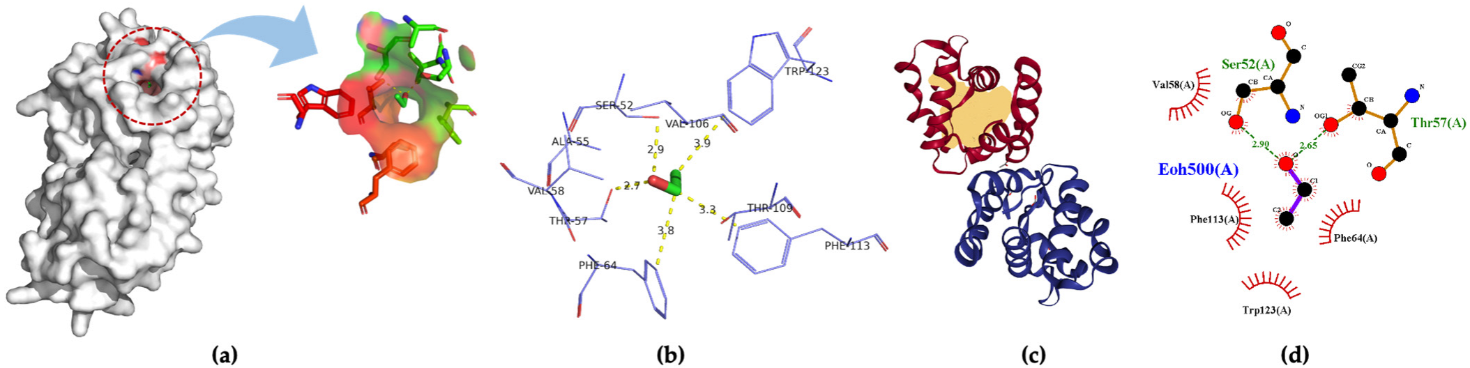
2. Results
3. Discussion
4. Materials and Methods
5. Conclusions
Supplementary Materials
Author Contributions
Funding
Institutional Review Board Statement
Informed Consent Statement
Data Availability Statement
Conflicts of Interest
Sample Availability
References
- Xun, G.; Lane, S.T.; Petrov, V.A.; Pepa, B.E.; Zhao, H. A rapid, accurate, scalable, and portable testing system for COVID-19 diagnosis. Nat. Commun. 2021, 12, 1–9. [Google Scholar] [CrossRef]
- Inagaki, H.; Saito, A.; Sugiyama, H.; Okabayashi, T.; Fujimoto, S. Rapid inactivation of SARS-CoV-2 with Deep-UV LED irradiation. Emerg. Microbes Infect. 2020, 9, 1744–1747. [Google Scholar] [CrossRef]
- Baden, L.R.; El Sahly, H.M.; Essink, B.; Kotloff, K.; Frey, S.; Novak, R.; Diemert, D.; Spector, S.A.; Rouphael, N.; Creech, C.B.; et al. Efficacy and Safety of the mRNA-1273 SARS-CoV-2 Vaccine. N. Engl. J. Med. 2021, 384, 403–416. [Google Scholar] [CrossRef] [PubMed]
- Torrente-Rodríguez, R.M.; Lukas, H.; Tu, J.; Min, J.; Yang, Y.; Xu, C.; Rossiter, H.B.; Gao, W. SARS-CoV-2 RapidPlex: A Graphene-Based Multiplexed Telemedicine Platform for Rapid and Low-Cost COVID-19 Diagnosis and Monitoring. Matter 2020, 3, 1981–1998. [Google Scholar] [CrossRef] [PubMed]
- Premkumar, L.; Segovia-Chumbez, B.; Jadi, R.; Martinez, D.R.; Raut, R.; Markmann, A.J.; Cornaby, C.; Bartelt, L.; Weiss, S.; Park, Y.; et al. The receptor-binding domain of the viral spike protein is an immunodominant and highly specific target of antibodies in SARS-CoV-2 patients. Sci. Immunol. 2020, 5, 1–10. [Google Scholar] [CrossRef] [PubMed]
- Cho, S.Y.; Jin, X.; Gong, X.; Yang, S.; Cui, J.; Strano, M.S. Antibody-Free Rapid Detection of SARS-CoV-2 Proteins Using Corona Phase Molecular Recognition to Accelerate Development Time. Anal. Chem. 2021, 93, 14685–14693. [Google Scholar] [CrossRef]
- Wolfe, M.; Webb, S.; Chushak, Y.; Krabacher, R.; Liu, Y.; Swami, N.; Harbaugh, S.; Chávez, J. A high-throughput pipeline for design and selection of peptides targeting the SARS-CoV-2 Spike protein. Sci. Rep. 2021, 11, 1–10. [Google Scholar] [CrossRef]
- Kesarwani, V.; Gupta, R.; Vetukuri, R.R.; Kushwaha, S.K.; Gandhi, S. Identification of Unique Peptides for SARS-CoV-2 Diagnostics and Vaccine Development by an In Silico Proteomics Approach. Front. Immunol. 2021, 12, 1–14. [Google Scholar] [CrossRef]
- Svobodova, M.; Skouridou, V.; Jauset-Rubio, M.; Viéitez, I.; Fernández-Villar, A.; Cabrera Alvargonzalez, J.J.; Poveda, E.; Bofill, C.B.; Sans, T.; Bashammakh, A.; et al. Aptamer Sandwich Assay for the Detection of SARS-CoV-2 Spike Protein Antigen. ACS Omega 2021, 6, 35657–35666. [Google Scholar] [CrossRef]
- Coronavirus (COVID-19) Update: FDA Authorizes First COVID-19 Diagnostic Test Using Breath Samples. RELEASE, FDA NEWS. Available online: https://www.fda.gov/news-events/press-announcements/coronavirus-covid-19-update-fda-authorizes-first-covid-19-diagnostic-test-using-breath-samples (accessed on 14 April 2022).
- Qu, C.; Yang, Z.; Wang, S.; Zhao, H.; Li, F.; Yang, X.; Luo, C. Binding Affinity Characterization of Four Antennae-Enriched Odorant-Binding Proteins From Harmonia axyridis (Coleoptera: Coccinellidae). Front. Physiol. 2022, 13, 1–14. [Google Scholar] [CrossRef]
- Cali, K.; Persaud, K.C. Modification of an Anopheles gambiae odorant binding protein to create an array of chemical sensors for detection of drugs. Sci. Rep. 2020, 10, 1–13. [Google Scholar] [CrossRef] [PubMed] [Green Version]
- Pelosi, P.; Zhu, J.; Knoll, W. Odorant-binding proteins as sensing elements for odour monitoring. Sensors 2018, 18, 3248. [Google Scholar] [CrossRef] [PubMed] [Green Version]
- Barbosa, A.J.M.; Oliveira, A.R.; Roque, A.C.A. Protein- and Peptide-Based Biosensors in Artificial Olfaction. Trends Biotechnol. 2018, 36, 1244–1258. [Google Scholar] [CrossRef] [PubMed] [Green Version]
- Rihani, K.; Ferveur, J.F.; Briand, L. The 40-year mystery of insect odorant-binding proteins. Biomolecules 2021, 11, 509. [Google Scholar] [CrossRef]
- Arima, A.; Harlisa, I.H.; Yoshida, T.; Tsutsui, M.; Tanaka, M.; Yokota, K.; Tonomura, W.; Yasuda, J.; Taniguchi, M.; Washio, T.; et al. Identifying Single Viruses Using Biorecognition Solid-State Nanopores. J. Am. Chem. Soc. 2018, 140, 16834–16841. [Google Scholar] [CrossRef]
- Hober, A.; Tran-Minh, K.H.; Foley, D.; McDonald, T.; Vissers, J.P.C.; Pattison, R.; Ferries, S.; Hermansson, S.; Betner, I.; Uhlén, M.; et al. Rapid and sensitive detection of SARS-CoV-2 infection using quantitative peptide enrichment lc-ms analysis. Elife 2021, 10, 1–13. [Google Scholar] [CrossRef]
- Oh, J.-W.; Chung, W.-J.; Heo, K.; Jin, H.-E.; Lee, B.Y.; Wang, E.; Zueger, C.; Wong, W.; Meyer, J.; Kim, C.; et al. Biomimetic virus-based colourimetric sensors. Nat. Commun. 2014, 5, 3043. [Google Scholar] [CrossRef] [Green Version]
- Okochi, M.; Muto, M.; Yanai, K.; Tanaka, M.; Onodera, T.; Wang, J.; Ueda, H.; Toko, K. Array-Based Rational Design of Short Peptide Probe-Derived from an Anti-TNT Monoclonal Antibody. ACS Comb. Sci. 2017, 19, 625–632. [Google Scholar] [CrossRef]
- Kuang, Z.; Kim, S.N.; Crookes-goodson, W.J.; Farmer, B.L.; Naik, R.R. Biomimetic Chemosensor: Designing Peptide Recognition Elements for Surface Functionalization of Carbon Nanotube Field Effect Transistors. ACS Nano 2010, 4, 452–458. [Google Scholar] [CrossRef]
- Park, J.; Lee, J.M.; Chun, H.; Lee, Y.; Hong, S.J.; Jung, H.; Kim, Y.J.; Kim, W.G.; Devaraj, V.; Choi, E.J.; et al. Optical bioelectronic nose of outstanding sensitivity and selectivity toward volatile organic compounds implemented with genetically engineered bacteriophage: Integrated study of multi-scale computational prediction and experimental validation. Biosens. Bioelectron. 2021, 177, 112979. [Google Scholar] [CrossRef]
- Harini, K.; Sowdhamini, R. Computational approaches for decoding select odorant-olfactory receptor interactions using mini-virtual screening. PLoS ONE 2015, 10, 1–30. [Google Scholar] [CrossRef] [PubMed]
- Beaufays, J.; Lins, L.; Thomas, A.; Brasseur, R. In silico predictions of 3D structures of linear and cyclic peptides with natural and non-proteinogenic residues. J. Pept. Sci. 2012, 18, 17–24. [Google Scholar] [CrossRef] [PubMed]
- Elekofehinti, O.O.; Aladenika, Y.V.; Alli-Smith, Y.R.; Ejelonu, O.C.; Lawal, A.O. Molecular modeling, dynamics simulation and characterization of human inositol hexakisphosphate kinase 1 (IP6K1) related to diabetes. J. Appl. Sci. Environ. Manag. 2019, 23, 461. [Google Scholar] [CrossRef]
- Pizzoni, D.; Mascini, M.; Lanzone, V.; Del Carlo, M.; Di Natale, C.; Compagnone, D. Selection of peptide ligands for piezoelectric peptide based gas sensors arrays using a virtual screening approach. Biosens. Bioelectron. 2014, 52, 247–254. [Google Scholar] [CrossRef]
- Hwang, K.S.; Lee, M.H.; Lee, J.; Yeo, W.S.; Lee, J.H.; Kim, K.M.; Kang, J.Y.; Kim, T.S. Peptide receptor-based selective dinitrotoluene detection using a microcantilever sensor. Biosens. Bioelectron. 2011, 30, 249–254. [Google Scholar] [CrossRef]
- Wang, J.; Muto, M.; Yatabe, R.; Tahara, Y.; Onodera, T.; Tanaka, M.; Okochi, M.; Toko, K. Highly Selective Rational Design of Peptide-Based Surface Plasmon Resonance Sensor for Direct Determination of 2,4,6-trinitrotoluene (TNT) Explosive. Sens. Actuators B Chem. 2018, 264, 279–284. [Google Scholar] [CrossRef]
- Wang, J. Near infrared optical biosensor based on peptide functionalized single-walled carbon nanotubes hybrids for 2,4,6-trinitrotoluene (TNT) explosive detection. Anal. Biochem. 2018, 550, 49–53. [Google Scholar] [CrossRef]
- Snitz, K.; Andelman-Gur, M.; Pinchover, L.; Weissgross, R.; Weissbrod, A.; Mishor, E.; Zoller, R.; Linetsky, V.; Medhanie, A.; Shushan, S.; et al. Proof of concept for real-time detection of SARS-CoV-2 infection with an electronic nose. PLoS ONE 2021, 16, 1–12. [Google Scholar] [CrossRef]
- Leong, S.X.; Leong, Y.X.; Tan, E.X.; Sim, H.Y.F.; Koh, C.S.L.; Lee, Y.H.; Chong, C.; Ng, L.S.; Chen, J.R.T.; Pang, D.W.C.; et al. Noninvasive and Point-of-Care Surface-Enhanced Raman Scattering (SERS)-Based Breathalyzer for Mass Screening of Coronavirus Disease 2019 (COVID-19) under 5 min. ACS Nano 2022, 16, 2629–2639. [Google Scholar] [CrossRef]
- Chen, H.; Qi, X.; Zhang, L.; Li, X.; Ma, J.; Zhang, C.; Feng, H.; Yao, M. COVID-19 screening using breath-borne volatile organic compounds. J. Breath Res. 2021, 15, 047104. [Google Scholar] [CrossRef]
- Ruszkiewicz, D.M.; Sanders, D.; O’Brien, R.; Hempel, F.; Reed, M.J.; Riepe, A.C.; Bailie, K.; Brodrick, E.; Darnley, K.; Ellerkmann, R.; et al. Diagnosis of COVID-19 by analysis of breath with gas chromatography-ion mobility spectrometry—A feasibility study. EClinicalMedicine 2020, 29–30, 100609. [Google Scholar] [CrossRef] [PubMed]
- Allouche, A. Software News and Updates Gabedit—A Graphical User Interface for Computational Chemistry Softwares. J. Comput. Chem. 2012, 32, 174–182. [Google Scholar] [CrossRef] [PubMed]
- Tunyasuvunakool, K.; Adler, J.; Wu, Z.; Green, T.; Zielinski, M.; Žídek, A.; Bridgland, A.; Cowie, A.; Meyer, C.; Laydon, A.; et al. Highly accurate protein structure prediction for the human proteome. Nature 2021, 596, 590–596. [Google Scholar] [CrossRef] [PubMed]
- Varadi, M.; Anyango, S.; Deshpande, M.; Nair, S.; Natassia, C.; Yordanova, G.; Yuan, D.; Stroe, O.; Wood, G.; Laydon, A.; et al. AlphaFold Protein Structure Database: Massively expanding the structural coverage of protein-sequence space with high-accuracy models. Nucleic Acids Res. 2022, 50, D439–D444. [Google Scholar] [CrossRef]
- Lamiable, A.; Thévenet, P.; Rey, J.; Vavrusa, M.; Derreumaux, P.; Tufféry, P. PEP-FOLD3: Faster de novo structure prediction for linear peptides in solution and in complex. Nucleic Acids Res. 2016, 44, W449–W454. [Google Scholar] [CrossRef] [Green Version]
- Thévenet, P.; Shen, Y.; Maupetit, J.; Guyon, F.; Derreumaux, P.; Tufféry, P. PEP-FOLD: An updated de novo structure prediction server for both linear and disulfide bonded cyclic peptides. Nucleic Acids Res. 2012, 40, W288–W293. [Google Scholar] [CrossRef] [Green Version]
- Shen, Y.; Maupetit, J.; Derreumaux, P.; Tufféry, P. Improved PEP-FOLD Approach for Peptide and Miniprotein Structure Prediction. J. Chem. Theory Comput. 2014, 10, 4745–4758. [Google Scholar] [CrossRef]
- Wu, T.Z.; Lo, Y.R.; Chan, E.C. Exploring the recognized bio-mimicry materials for gas sensing. Biosens. Bioelectron. 2001, 16, 945–953. [Google Scholar] [CrossRef]
- Wasilewski, T.; Szulczyński, B.; Wojciechowski, M.; Kamysz, W.; Gębicki, J. Determination of long-chain aldehydes using a novel quartz crystal microbalance sensor based on a biomimetic peptide. Microchem. J. 2020, 154, 104509. [Google Scholar] [CrossRef]
- Manoharan, M.; Chong, M.N.F.; Vaïtinadapoulé, A.; Frumence, E.; Sowdhamini, R.; Offmann, B. Comparative genomics of odorant binding proteins in Anopheles gambiae, Aedes aegypti, and Culex quinquefasciatus. Genome Biol. Evol. 2013, 5, 163–180. [Google Scholar] [CrossRef] [Green Version]


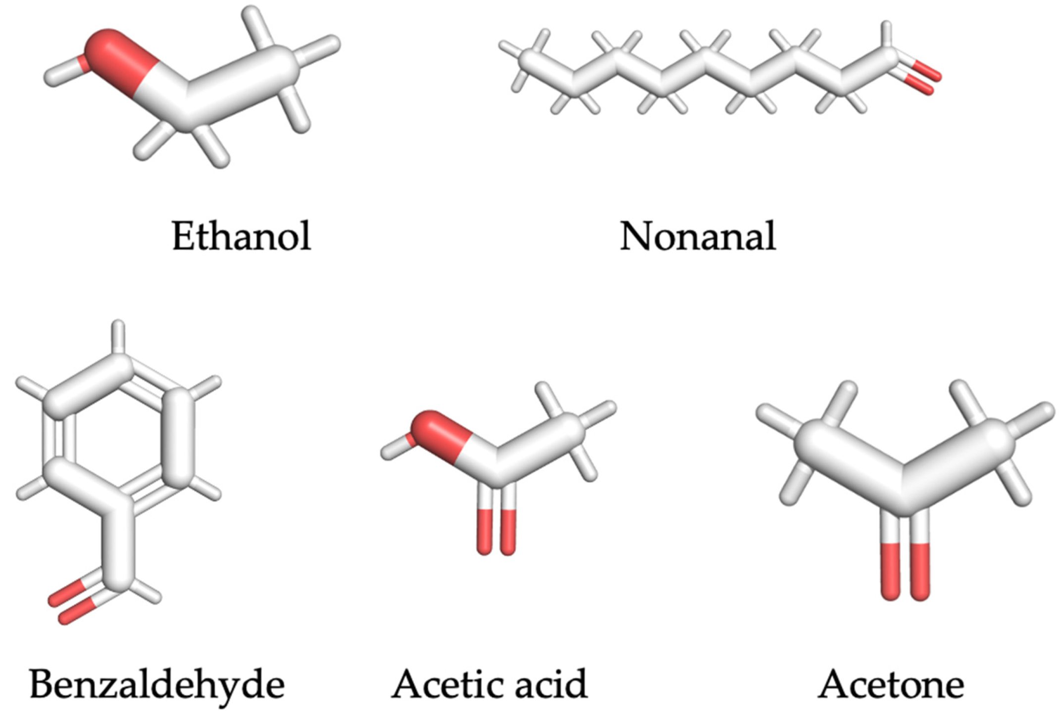
| VOC Ligand | Molecular Formula | Molecular Weight (g/mol) | Odor Description | Vapor Pressure (mmHg) | Flash Point (°F) |
|---|---|---|---|---|---|
| ethanol | C2H6O | 46.07 | Weak, ethereal, vinous odor | 59.27 | 57.2 |
| nonanal | C9H18O | 142.24 | Orange-rose odor | 0.37 | 147 |
| benzaldehyde | C7H6O | 106.12 | Odor resembling oil of bitter almond | 1.27 | 145 |
| acetic acid | C2H4O2 | 60.05 | Sour, vinegar-like odor | 15.73 | 103 |
| acetone | C3H6O | 58.08 | Fruity odor | 231.53 | 1.42 |
| VOC Ligand | Peptide Sequence | Molecular Weight (g/mol) | Theoretical pI | GRAVY | Instability Index |
|---|---|---|---|---|---|
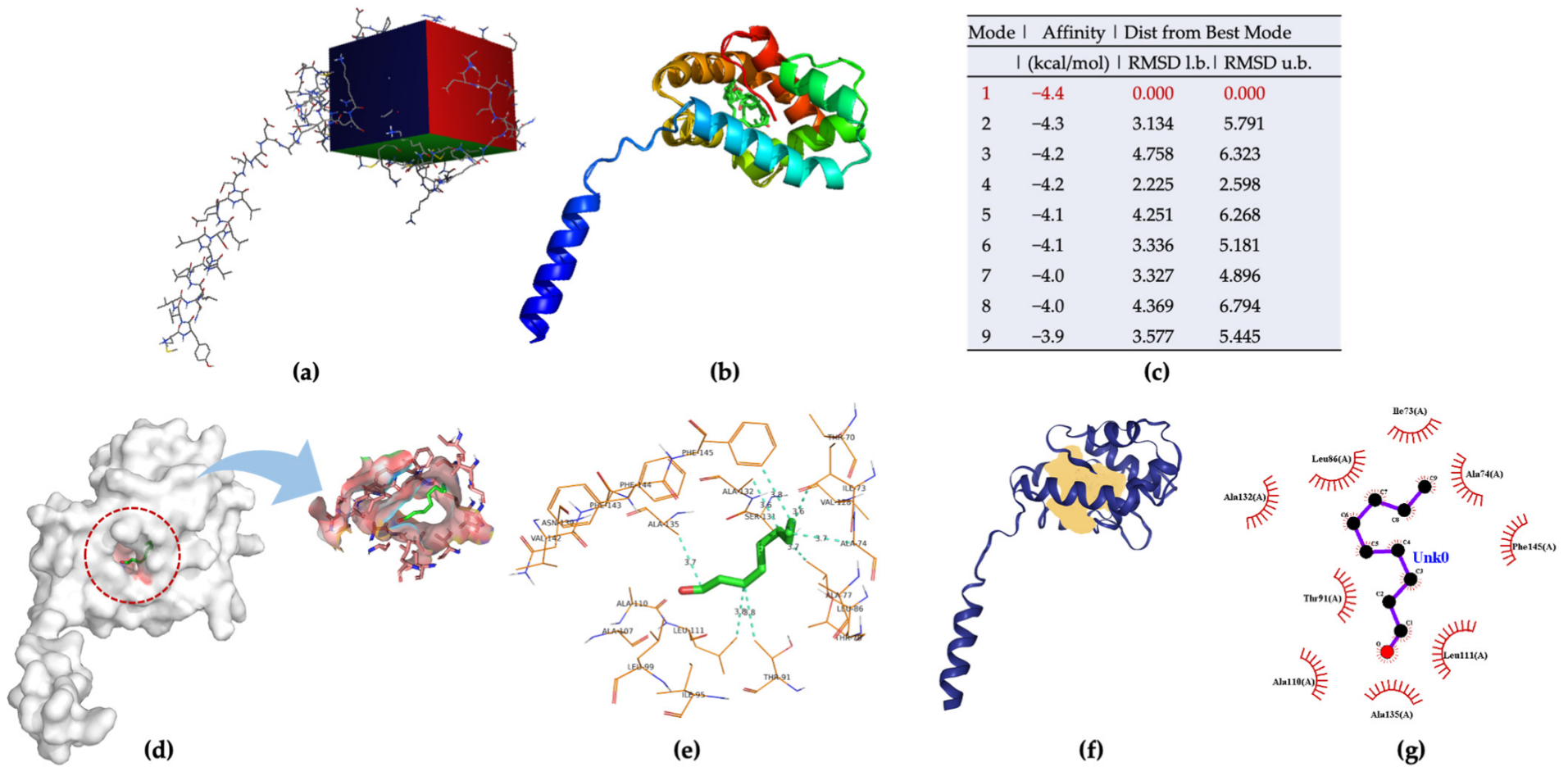
| nonanal | TIAATLTILAALVSAANVFFF | 2154.58 | 5.19 | 1.981 | 18.70 |
| benzaldehyde | VRGLLYVNAYAFLY | 1661.96 | 8.47 | 0.993 | 20.14 |
| acetone | TPFLVFVVYLFL | 1457.82 | 5.18 | 2.400 | 18.93 |
| ethanol | SATVFVTFW | 1057.21 | 5.24 | 1.411 | 13.17 |
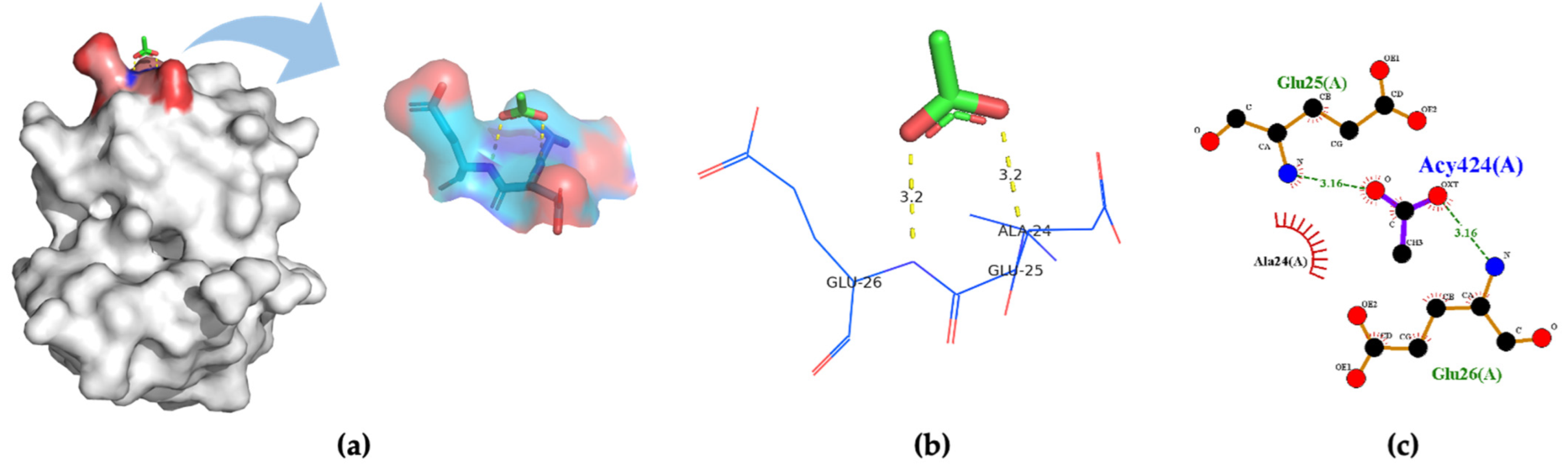
| acetic acid | AEE | 347.32 | - | - | - |
Publisher’s Note: MDPI stays neutral with regard to jurisdictional claims in published maps and institutional affiliations. |
© 2022 by the authors. Licensee MDPI, Basel, Switzerland. This article is an open access article distributed under the terms and conditions of the Creative Commons Attribution (CC BY) license (https://creativecommons.org/licenses/by/4.0/).
Share and Cite
Wang, J.; Sakai, K.; Kiwa, T. Rational Design of Peptides Derived from Odorant-Binding Proteins for SARS-CoV-2-Related Volatile Organic Compounds Recognition. Molecules 2022, 27, 3917. https://doi.org/10.3390/molecules27123917
Wang J, Sakai K, Kiwa T. Rational Design of Peptides Derived from Odorant-Binding Proteins for SARS-CoV-2-Related Volatile Organic Compounds Recognition. Molecules. 2022; 27(12):3917. https://doi.org/10.3390/molecules27123917
Chicago/Turabian StyleWang, Jin, Kenji Sakai, and Toshihiko Kiwa. 2022. "Rational Design of Peptides Derived from Odorant-Binding Proteins for SARS-CoV-2-Related Volatile Organic Compounds Recognition" Molecules 27, no. 12: 3917. https://doi.org/10.3390/molecules27123917
APA StyleWang, J., Sakai, K., & Kiwa, T. (2022). Rational Design of Peptides Derived from Odorant-Binding Proteins for SARS-CoV-2-Related Volatile Organic Compounds Recognition. Molecules, 27(12), 3917. https://doi.org/10.3390/molecules27123917