Article

Structural Characterization, Antimicrobial Activity and BSA/DNA Binding Affinity of New Silver(I) Complexes with Thianthrene and 1,8-Naphthyridine

by
Darko P. Ašanin
1,
Sanja Skaro Bogojevic
2,
Franc Perdih
3,
Tina P. Andrejević
4,
Dusan Milivojevic
2,
Ivana Aleksic
2,
Jasmina Nikodinovic-Runic
2,*,
Biljana Đ. Glišić
4,*,
Iztok Turel
3,* and
Miloš I. Djuran
5,*
1
Department of Science, Institute for Information Technologies Kragujevac, University of Kragujevac, Jovana Cvijića bb, 34000 Kragujevac, Serbia
2
Institute of Molecular Genetics and Genetic Engineering, University of Belgrade, Vojvode Stepe 444a, 11042 Belgrade, Serbia
3
Department of Chemistry and Biochemistry, Faculty of Chemistry and Chemical Technology, University of Ljubljana, Večna pot 113, SI-1000 Ljubljana, Slovenia
4
Department of Chemistry, Faculty of Science, University of Kragujevac, R. Domanovića 12, 34000 Kragujevac, Serbia
5
Serbian Academy of Sciences and Arts, Knez Mihailova 35, 11000 Belgrade, Serbia
*
Authors to whom correspondence should be addressed.
Molecules 2021, 26(7), 1871; https://doi.org/10.3390/molecules26071871
Submission received: 9 March 2021 / Revised: 23 March 2021 / Accepted: 23 March 2021 / Published: 26 March 2021
(This article belongs to the Special Issue New Transition Metal Complexes as Chemotherapeutic Agents)

Abstract

:
Three new silver(I) complexes [Ag(NO3)(tia)(H2O)]n (Ag1), [Ag(CF3SO3)(1,8-naph)]n (Ag2) and [Ag2(1,8-naph)2(H2O)1.2](PF6)2 (Ag3), where tia is thianthrene and 1,8-naph is 1,8-naphthyridine, were synthesized and structurally characterized by different spectroscopic and electrochemical methods and their crystal structures were determined by single-crystal X-ray diffraction analysis. Their antimicrobial potential was evaluated against four bacterial and three Candida species, and the obtained results revealed that these complexes showed significant activity toward the Gram-positive Staphylococcus aureus, Gram-negative Pseudomonas aeruginosa and the investigated Candida species with minimal inhibitory concentration (MIC) values in the range 1.56–7.81 μg/mL. On the other hand, tia and 1,8-naph ligands were not active against the investigated strains, suggesting that their complexation with Ag(I) ion results in the formation of antimicrobial compounds. Moreover, low toxicity of the complexes was detected by in vivo model Caenorhabditis elegans. The interaction of the complexes with calf thymus DNA (ct-DNA) and bovine serum albumin (BSA) was studied to evaluate their binding affinity towards these biomolecules for possible insights into the mode of antimicrobial activity. The binding affinity of Ag13 to BSA was higher than that for DNA, indicating that proteins could be more favorable binding sites for these complexes in comparison to the nucleic acids.

Graphical Abstract

1. Introduction

Infections caused by bacteria and fungi threaten human health on a daily basis because many microbes have developed resistance to the commercial antimicrobial agents over time [1]. It is estimated that antimicrobial resistance causes at least 50 thousand deaths each year in Europe and the United States and that it will cause 10 million deaths worldwide per year by 2050 [2]. Considering this, the problems related to the microbial infections and resistance occurrence demand urgent and effective solution, which is based on the development of novel antimicrobial agents. It is well known that silver(I) compounds have shown significant antimicrobial activity, such as silver(I) nitrate which was used for the prevention of eye infections in newborns [3], and silver(I) sulfadiazine (AgSD) applied for the treatment of bacterial infections in burns [4]. Silver(I) complexes have also shown antiproliferative [5] and antiviral [6,7] activities, exhibiting acceptable toxicity towards human cells [8,9]. The mechanism of antimicrobial activity of the silver(I) complexes is explained by their slow release of Ag(I) ions [10,11]. These ions can interact with the bacterial cell surfaces, enabling their penetration into the cell. Once inside the cell, Ag(I) ions could interact with the biomolecules in its interiors, such as DNA and proteins. They can also cause the production of reactive oxygen species (ROS), leading to bacterial cell death [10,11]. Due to this multi-targeting mechanism, the silver(I) complexes act as antimicrobial agents, which might overcome the problem of bacterial resistance [12].
Depending on the type of heteroatom present in heterocyclic compounds, they may show a wide spectrum of pharmacological activities [13,14,15,16,17]. For instance, the United States Food and Drug Administration (FDA) approved several heterocycles containing sulfur or/and nitrogen donor atoms, such as raloxifene as anticancer [18], rosiglitazone as anti-diabetes [19], thiabendazole and clotrimazole as antifungal [20,21], and chloroquine as antimalarial agent [22]. Considering this, these compounds represent the important class of ligands for the synthesis of biologically active silver(I) complexes. For example, the silver(I) complexes with aromatic nitrogen-containing heterocycles (N-heterocycles), such as pyridazine, pyrimidine, pyrazine, phthalazine, quinazoline, quinoxaline, and phenazine, have shown significant activity against the Gram-negative bacterium Pseudomonas aeruginosa [23,24,25]. On the other hand, an excellent ability to inhibit the growth of Candida strains has been observed for the silver(I) complexes with phenanthrolines [26,27,28,29,30] and 1,5-naphtyridine [31]. The significant antimicrobial activity has also been manifested by silver(I) complexes with N-methylbenzothiazole-2-thione (mbtt), [(mbtt)2Ag(μ-mbtt)2Ag(mbtt)2](NO3)2 and [Ag(mbtt)3]CF3SO3 against certain Gram-negative (Xanthomonas campestris, Escherichia coli) and Gram-positive (Bacillus subtilis, Bacillus cereus, Staphylococcus aureus) bacteria [32]. Similarly, silver(I) complexes with N-substituted imidazolidine-2-thiones, purine-6-thione, 2-thiouracil, pyrimidine-2-thione, and pyridine-2-thione have shown moderate to good antimicrobial activity against Gram-positive (methicillin-resistant Staphylococcus aureus MRSA and Staphylococcus aureus), and Gram-negative (Staphylococcus epidermidis, Enterococcus faecalis, Shigella flexneri) bacteria and a yeast Candida albicans [33].
Considering all these facts, in the present study, we used two heterocycles, thianthrene (tia; S-heterocycle) and 1,8-naphthyridine (1,8-naph; N-heterocycle) for the synthesis of three new silver(I) complexes [Ag(NO3)(tia)(H2O)]n (Ag1), [Ag(CF3SO3)(1,8-naph)]n (Ag2) and [Ag2(1,8-naph)2(H2O)1.2](PF6)2 (Ag3). The synthesized complexes were characterized by spectroscopy (NMR, IR, and UV-Vis), mass spectrometry, cyclic voltammetry, and single-crystal X-ray diffraction analysis. These complexes were further evaluated for their in vitro antimicrobial activity and in vivo toxicity in Caenorhabditis elegans, an important animal model for rapid toxicity assessment of the novel compounds [34]. The interactions of the complexes Ag13 with calf thymus DNA (ct-DNA) and bovine serum albumin (BSA) were studied with the aim to check their binding affinity towards these potential biological targets.

2. Results

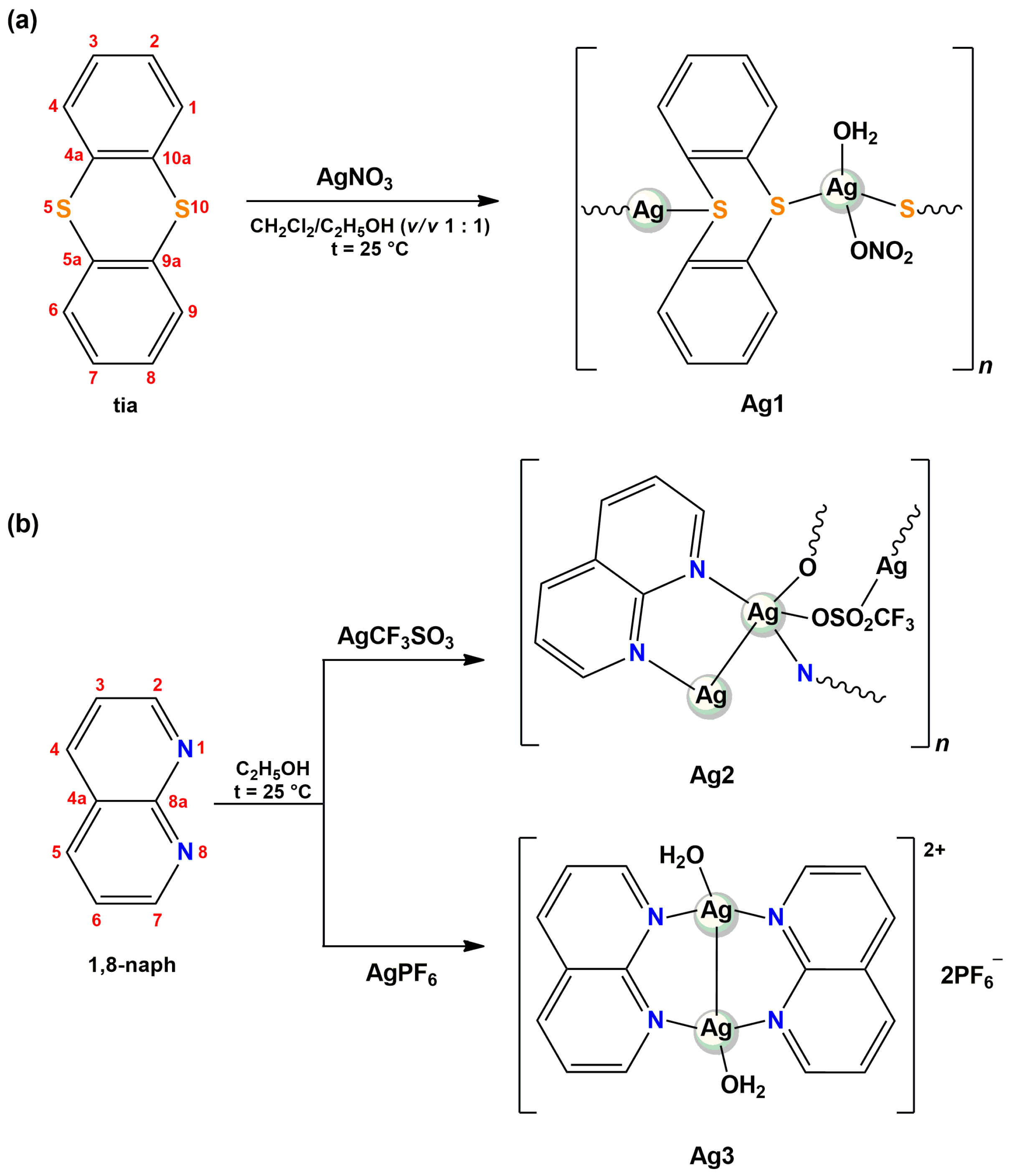
2.1. Synthesis and Characterization of Silver(I) Complexes Ag13

Silver(I) complexes Ag13 were synthesized according to the route presented in Scheme 1. The reaction of AgNO3 with an equimolar amount of thianthrene (tia) in ethanol/dichloromethane (v/v 1:1) at ambient temperature yielded polynuclear [Ag(NO3)(tia)(H2O)]n complex (Ag1), while by reacting 1,8-naphthyridine (1,8-naph) with AgCF3SO3 and AgPF6 in ethanol, polynuclear [Ag(CF3SO3)(1,8-naph)]n (Ag2) and dinuclear [Ag2(1,8-naph)2(H2O)1.2](PF6)2 (Ag3) complexes, respectively, were formed. NMR (1H and 13C), ultraviolet–visible (UV-Vis) and IR spectroscopy, mass spectrometry, and cyclic voltammetry were applied for characterization of the obtained complexes, while a single-crystal X-ray diffraction analysis was used for the determination of their structures.

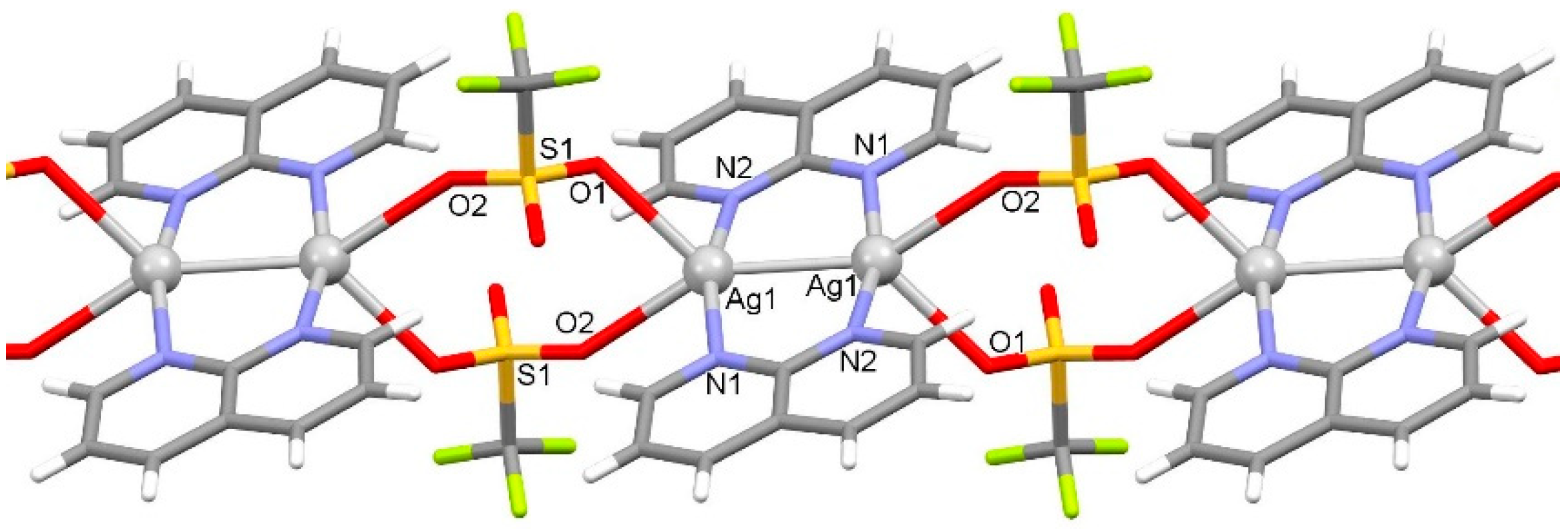
2.1.1. Solid-State Characterization

Silver(I) complexes Ag1 and Ag2 are polynuclear species with infinite chains and complex Ag3 is dinuclear species (Figure 1). Complex Ag1 has a polynuclear structure with tetrahedrally coordinated Ag(I) ion via two thianthrene sulfur atoms (Ag–S distances 2.6198(7) and 2.6747(8) Å), one nitrate oxygen atom (Ag–O distance 2.328(2) Å) and one water oxygen atom (Ag–O distance 2.360(2) Å) (Figure 2, Table 1). Thianthrene acts as a bridging ligand. Hydrogen bonding between coordinated water molecules of one polynuclear chain with the nitrato ligand of the adjacent polynuclear chain enables the formation of an infinite belt. The polynuclear belt is supported also by C3–H3···O1 interactions (Table 2). Belts are connected into a supramolecular structure through C5–H5···O2/O4 interactions.
Complex Ag2 has a polynuclear structure with Ag2(1,8-naph)2 units connected into a chain via bridging triflato ligands (Figure 3). Within the Ag2(1,8-naph)2 unit, the 1,8-naph is a bridging ligand with Ag–N distances of 2.200(2) and 2.218(2) Å, and this unit is supported also by argentophilic interaction with Ag1···Ag1i distance of 2.7871(5) Å representing the 81% of the sum of van der Waals radii (Table 1). Two triflato ligands connect two adjacent Ag2(1,8-naph)2 units into a chain through an Ag1–O1 bond distance of 2.555(2) Å and through a somewhat longer Ag1–O2 contact of 2.645(2) Å. The polynuclear chain is supported also by C1–H1···O2 and C7–H7···F2 interactions (Table 2). The crystal structure is stabilized by π···π interactions between 1,8-naph rings of adjacent chains with a centroid-to-centroid distance of 3.6330(17) Å and ring slippage of 1.061 Å as well as by C8–H8···O3 interactions.
Complex Ag3 has a dinuclear structure with [Ag2(1,8-naph)2(H2O)1.2]2+ cation and two PF6 counter-anions. In [Ag2(1,8-naph)2(H2O)1.2]2+ cation, the 1,8-naph acts as a bridging ligand with almost identical Ag–N distances (2.186(3) and 2.184(3) Å) and, also, this unit is supported by argentophilic interaction with Ag1···Ag1i distance of 2.7235(6) Å representing the 79% of the sum of van der Waals radii (Table 1). The Ag(I) metal center is coordinated also by a water molecule with Ag–O distance of 2.449(4) Å. However, the water molecule was found to possess a 0.60 occupancy ratio. The crystal structure is stabilized by π···π interactions between 1,8-naph rings of adjacent cations with a centroid-to-centroid distance of 3.602(3) Å and ring slippage of 1.035 Å as well as by various O–H···F and C–H···F interactions (Table 2).
The IR spectroscopic data of the complexes Ag13 are following their structures determined by a single-crystal X-ray diffraction analysis. The IR spectra of complexes Ag1 and Ag3 exhibited a broad band at ~ 3440 cm−1 due to the stretching vibration of the –OH group, confirming the presence of coordinated water to Ag(I) ion in these complexes. The nitrate coordination in the Ag1 complex is confirmed by a very strong band at 1384 cm−1 with a tendency to split into two bands [35], while in the case of Ag2, strong bands in the 1300–1000 cm−1 region originate from a coordinated triflate in this complex [36,37]. More precisely, the bands at 1163 and 1134 cm−1 can be attributed to the symmetric and asymmetric stretching vibrations of the –CF3 group, while the bands at 1254, and 1044 and 1032 cm−1 are due to the asymmetric and symmetric stretching of the –SO3 group in triflate, respectively [36]. Regarding the Ag3 complex, the presence of a strong band at 831 cm−1 assigned to the stretching vibrations of the PF6 is a consequence of its existence as a counter-anion in the crystal lattice of this complex [38,39].

2.1.2. Solution Behavior

The NMR spectra of the synthesized silver(I) complexes and corresponding ligands (Supplementary Materials) were assigned based on the previously reported NMR spectroscopic data for tia [40] and 1,8-naph [41]. 1H- and 13C-NMR spectra of the presently investigated silver(I) complexes contain the same number of signals as those for the uncoordinated tia (Ag1) and 1,8-naph (Ag2 and Ag3) ligands, being in accordance with the bidentate bridging coordination mode of these ligands to Ag(I) ion. While in the case of Ag1 complex, the resonances in both 1H- and 13C-NMR spectra are almost unshifted in respect to those for the uncoordinated tia ligand (vide infra in Materials and Methods section), the chemical shifts of 1H- and 13C-NMR resonances for Ag2 and Ag3 differ significantly from those of 1,8-naph. All 1H resonances for coordinated 1,8-naph in Ag2 and Ag3 are downfield shifted with respect to those for the free ligand. The chemical shifts of 1H-NMR resonances in these complexes are strongly dependent on the proton position with respect to the nitrogen donor atoms. As can be expected, the largest coordination shift (Δδ = δcomplexδ1,8-naph) of + 0.32 (for both complexes) were observed for H2/H7 protons, which are adjacent to the nitrogen atoms. On the other hand, there is no strict rule in the shifting of 13C resonances of the silver(I) complexes Ag2 and Ag3 with respect to the carbon position relative to the nitrogen atoms.
In the mass spectra of the investigated silver(I) complexes, the major doublet peaks are those centered at m/z = 540.9176 (Ag1) and 367.0107 (Ag2 and Ag3), which are consistent with the presence of mononuclear [Ag(tia)2]+ and [Ag(1,8-naph)2]+ cations, respectively. As it was found previously, the mass spectra of silver(I) complexes show doublet peaks with almost equal intensity for a mononuclear species or triplet peaks with approximately 1:2:1 intensity ratios for dinuclear silver(I) species, as a consequence of the nearly equal abundance of two silver isotopes, 107Ag (51.84%) and 109Ag (48.16%) [29].
The UV-Vis spectra of the complexes Ag13, recorded in dimethyl sulfoxide (DMSO), are similar to those of the uncoordinated tia and 1,8-naph ligands, suggesting that the absorption maxima (λmax) at 259 nm for Ag1 and 307 nm for Ag2 and Ag3 are due to the characteristic π→π* transitions in the corresponding heterocyclic ligand [42,43]. In order to check the stability of complexes Ag13, their UV-Vis spectra were also recorded 24 h and 48 h after dissolution. As an illustration, time-dependent UV-Vis spectra of the Ag3 complex were shown in Figure S1. For Ag2 and Ag3, the observed transitions remained unmodified over 48 h at room temperature, implying their stability in the solution during this time. Nevertheless, a slight decrease in the intensity of the absorption maximum of 12% was noticed for Ag1, although without significant modifications of the spectrum shape and position of the absorption maxima, indicating that this complex is the least stable amongst the synthesized silver(I) complexes.
With the aim to investigate the air/light stability of complexes Ag13, which is important for their possible external application in the form of ointments, gels, and coating materials of dressings [44], sterile cellulose discs impregnated with their DMSO solutions were exposed to air and light for 48 h. The obtained results were compared with those for silver(I) salts used for the synthesis of the complexes (AgNO3, AgCF3SO3, and AgPF6) (Figure S2). As can be seen, all investigated silver(I) complexes were less darkened than the salts during 48 h, indicating the slightly higher air/light stability of the complexes Ag1–3. Among the complexes, Ag1 turned out to be the least stable under the investigated experimental conditions.
The electrochemical behavior of the metal complexes is highly relevant for a better understanding of their stability and biological activity, as well as the way of interaction with biomolecules [45,46]. In accordance with this, the cyclic voltammograms (CV) of complexes Ag13 were recorded at the glassy carbon (GC) electrode in DMSO and 0.1 M tetrabutylammonium hexafluorophosphate (TBAHP) as a supporting electrolyte under the following conditions, Ebegin = −2.0 V and Eend = 2.0 V (Figure 4). As can be seen from this figure, a broad anodic peak, which can be attributed to Ag(I)→Ag(II) oxidation process, was observed [47,48]. When the cyclic voltammogram was recorded in the cathodic direction, two distinctive reduction peaks were detected for all complexes (Table 3), which were assigned to Ag(II)→Ag(I) and Ag(I)→Ag(0) reduction processes, in accordance with the other silver(I) complexes where electrochemical behavior was previously investigated [47,49].

2.2. Biological Evaluation of the Silver(I) Complexes Ag13

2.2.1. Antimicrobial and Antiproliferative Effect of Silver(I) Complexes Ag13

Given the traditional antimicrobial properties shown by silver(I) complexes [23,24,25,26,27,28,29,30,31,32,33], the activity of silver(I) complexes Ag13 and the ligands, tia and 1,8-naph used for their synthesis, was determined against two Gram-positive (S. aureus and L. monocytogenes), two Gram-negative (E. coli and P. aeruginosa ATCC 10332 and BK25H [50]) bacteria, and three Candida species (C. albicans, C. krusei, and C. parapsilosis; Table 4). The antimicrobial activity of different AgX salts against these bacterial and fungal strains was previously evaluated [25]. Although these salts have shown significant antimicrobial activity, their use has been limited due to their precipitation in the form of AgCl under physiological conditions. As a consequence of this process, Ag(I) ions could not reach the infected sites and manifest antimicrobial activity [10].
Overall, presently investigated silver(I) complexes Ag13 showed significant antimicrobial activity toward the Gram-positive S. aureus, two Gram-negative P. aeruginosa species, and tested Candida species, while the lower sensitivity was detected in the case of L. monocytogenes and E. coli (Table 4). The most sensitive species was C. krusei, with MIC values being 1.56 µg/mL for all tested silver(I) complexes (3.6, 4.0, and 1.0 µM for Ag13, respectively). On the other hand, both tia and 1,8-naph ligands did not affect the microbial growth at 200 µg/mL (925 and 1537 µM, respectively), suggesting that the observed activity of the complexes is due to the presence of Ag(I) ions. As suggested previously, the antimicrobial activity of silver(I) complexes are connected to the complex ability to release free Ag(I) ions that may exert their antimicrobial action through different pathways [11]. On the other hand, the antiproliferative effect of these complexes was considerably higher but comparable to the observed MIC values (Table 4). On some level, this may limit the antimicrobial application of these complexes, but it is worth mentioning that silver(I) complexes with different classes of ligands have been studied for their antitumor potential and some of them showed antiproliferative activities higher than cisplatin [51].
These results are in line with our previous work in which we showed that the silver(I) complexes with a 1,5-naphthyridine ligand, which is a structural isomer of 1,8-naph, have remarkable activity against a range of Candida species, while for the tested bacterial species, MIC values obtained in this study are 2–3 times lower than in our previously published work (Table 4) [31]. Considerable anti-Candida activity has been also reported for the silver(I) complexes with 1,7- and 4,7-phenanthroline [29,30], while, on the other hand, silver(I) complexes with metronidazole (mtz), [Ag(mtz)2(NO3)] and [Ag2(mtz)4](BF4)2, showed better activity against the tested bacterial species and very low activity against C. albicans [44].
The different antibacterial behavior of silver(I) complexes obtained in this study might be related to their mode of action [52]. Gram-positive and Gram-negative bacteria have a different peptidoglycan cell wall and therefore, it can be assumed that the different bactericidal behavior of the silver(I) complexes can be explained by their ability to interact with the cell wall and pass through the membrane causing bacterial growth inhibition [52,53,54].

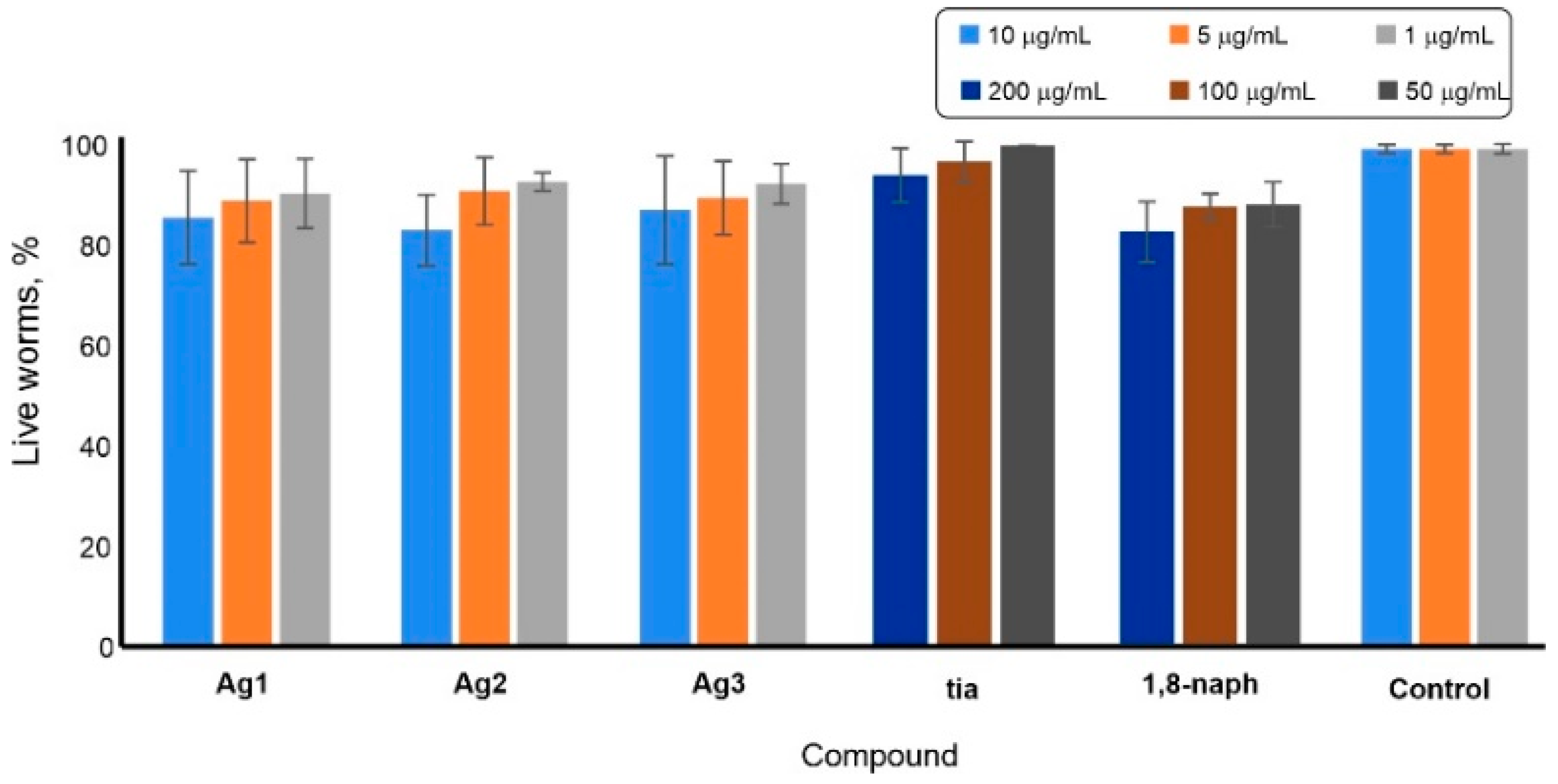
2.2.2. C. elegans Toxicity

A genetically tractable, multicellular nematode C. elegans, is a widely used model organism as a high throughput platform for toxicity assessment and more recently for the discovery of new antimicrobial compounds [55,56]. The tested complexes Ag13 and the corresponding ligands showed low toxicity against C. elegans in concentrations relevant to the observed MICs and cytotoxicity on MRC-5 cells (Figure 5 and Table 4). The highest percentage of dead worms was observed upon treatment with the highest concentrations of Ag2 complex and 1,8-naph, being 17 and 18%, respectively (Figure 5). The moderate toxicity against C. elegans was also previously observed for silver(I) complexes with different pyridine-4,5-dicarboxylate esters as ligands, which showed significant activity on the cow mastitis-associated pathogens [49]. The differences in the two toxicity models are possibly due to the more complex uptake system present in nematode in comparison to the direct application to cells in in vitro model.

2.3. BSA Binding Study

Serum albumin (SA) is one of the most abundant proteins present in the blood plasma and plays a crucial function in the transport of various endo- and exogenous compounds, including pharmacologically used agents [57]. Therefore, the distribution, free concentration, and metabolism of these agents can be changed as a result of their binding to serum albumin. Considering this, the study of the binding interaction of this protein with potential therapeutic agents could contribute to an understanding of the mechanism of their transport and distribution in the human body and identification of the dynamics of their action [57]. Bovine serum albumin (BSA) has been widely used as a model protein for this purpose due to its structural similarity with human serum albumin (HSA) of approximately 76%, low cost, and some unusual binding properties [57,58]. Fluorescence spectroscopy is one of the most convenient methods for the investigation of BSA binding interactions [59]. The intrinsic fluorescence of this protein is mainly due to tryptophan (Trp), tyrosine (Tyr), and phenylalanine (Phe) residues, whereas the binding interaction between the investigated compound and BSA will cause fluorescence quenching [59].
The emission spectra of BSA were recorded in the absence and presence of an increasing amount of the investigated silver(I) complexes Ag13. In all cases, a remarkable quenching of a fluorophore was observed with the gradual addition of the complex, indicating that its interaction with BSA occurred [57,58,59,60,61]. As can be seen from Figure 6, upon addition of an increasing concentration of Ag1 complex to BSA solution of a constant concentration, a significant decrease of fluorescence intensity at 367 nm was observed, leading to the formation of a BSA-complex system.
In order to study the quenching mechanism, Stern-Volmer and Scatchard equations were applied for the analysis of the fluorescence quenching data and the obtained data (Stern-Volmer constants (Ksv), quenching rate constants (Kq), binding constants (KA), and the number of binding sites per BSA (n)) are presented in Table 5. The values of Ksv constants indicate that the Ag1 complex containing tia ligand has a higher affinity for BSA compared to the remaining two complexes Ag2 and Ag3 with 1,8-naph ligand. The Ksv values for Ag2 and Ag3 are similar to those calculated for silver(I) complex with 1,2-bis(4-pyridyl)ethene [62], as well as for those with N-methyl-1,3,5-triaza-7-phosphaadamantane and tris(pyrazol-1-yl)methanesulfonate [63]. The Kq constant depends on the probability of a collision between fluorophore and quencher and is a measure of the tryptophan residues exposure to the investigated compound [64]. As can be seen from Table 5, the Kq values for the complexes Ag13 are in accordance with their good quenching ability of the BSA fluorescence. The Kq values for all investigated complexes are significantly higher than 2 × 10¹⁰ M−1·s−1 (the value of maximum diffusion collision quenching rate constant), indicating the static quenching mechanism [63,65]. The KA values for Ag13 are in an optimal range, i.e., these constants are high enough to suggest that the complexes can be efficiently carried in the blood (which is an important property of a drug [57]), but not so high to prevent their release from the BSA upon arrival to the target cell [66]. Finally, the calculated n values for complexes suggest their binding to only one binding site per protein molecule.

2.4. Lipophilicity Assay

The lipophilicity of a compound is in accordance with its cellular uptake efficiency and is of great importance for the new drug candidate design [67,68]. The octanol-water partition coefficient (logP) is a measure of lipophilicity and can be determined by the flask-shaking method [69]. Thus, the logP values for complexes Ag13 are 0.72, −0.14, and 0.37, respectively, being in accordance with the logP values range of −0.4 to 5.6 for new pharmacophores in the comprehensive medicinal chemistry [70]. The greater logP values of complexes Ag1 and Ag3 in respect of Ag2 indicate that these two complexes are mainly distributed in the octanol phase, further implying their higher cellular uptake efficiency.

2.5. DNA Interaction

Interactions of metal ions and their complexes with DNA are of great importance for the design of novel metal-based therapeutic agents [71]. Moreover, it is known that the mechanism of antimicrobial activity of silver(I) complexes can be related to their interaction with different cellular biomolecules, including DNA [10]. Firstly, the binding affinity of Ag13 complexes toward DNA was investigated by fluorescence spectroscopy by performing competitive binding experiments based on the displacement of ethidium bromide (EthBr) from DNA. It is well known that EthBr intercalates between adjacent base pairs in the DNA double helix resulting in the enhancement of its fluorescence [63]. After the addition of the investigated complex, a decrease in the fluorescence intensity of the EthBr-DNA system will occur if this complex intercalates into DNA or after its binding to EthBr-DNA, which leads to the formation of a new nonfluorescent EthBr-DNA-complex system [63,65]. In the case of complexes Ag13, their addition to the EthBr-DNA solution causes the reduction in its emission intensity, indicating the interaction of complexes with DNA (as an illustration, the fluorescence emission spectra of EthBr–DNA system in the presence of an increasing concentration of Ag2 complex were shown in Figure 7a). However, the calculated binding constants for all complexes (KA, Table 6) are significantly lower than that for EthBr (KA = 2 × 10⁶ M−1) [65], implying that the reason for the reduction in the emission intensity of the EthBr-DNA system in the presence of silver(I) complexes Ag13 could be their binding to the EthBr-DNA and the formation of a new nonfluorescent EthBr-DNA-complex system.
The Ksv values for the silver(I) complexes Ag13 are low and suggest that they bind to ct-DNA through the non-intercalative (electrostatic) mode, which can be also concluded from the percentage of hypochromism up to 15%. For comparison, the percentage of hypochromism of 50% was previously obtained for lucigenin, which is proven as a DNA intercalator [72]. The values of Ksv constants for the presently investigated complexes are slightly lower than those obtained for silver(I) complexes {[Ag(bpa)]NO3}n and {[Ag(bpa)2]CF3SO3·H2O}n, where bpa stands for 1,2-bis(4-pyridyl)ethane [62]. On the other hand, silver(I) complex [Ag(daf)(1,10-phen)]NO3 (daf = 4,5-diazafluoren-9-one and 1,10-phen = 1,10-phenanthroline) had high affinity for the intercalation between DNA base pairs, what can be concluded from the value of its Ksv constant of 0.97 × 10⁵ M−1 at 37 °C [73]. From the values of Kq constants higher than 2 × 10¹⁰ M−1·s−1, it can be seen that the mechanism of interaction between silver(I) complexes Ag13 and DNA is a static quenching [65].
To further corroborate the possibility of complexes Ag13 toward DNA interaction, gel electrophoresis methodology was also applied (Figure 7b). The slight reduction of the emission of the EthBr-DNA system was observable upon incubation with the tested complexes (Figure 7b), suggesting their weak interaction. The significant effect was only noticed for the Ag3 complex at concentrations of 400 and 200 µM, as determined by Image J analysis (Figure S3).

3. Materials and Methods

3.1. Materials

All chemicals were of reagent-grade quality or higher and used without further purification. Solvents were used as received. The silver(I) salts (AgNO3, AgCF3SO3, and AgPF6), thianthrene (tia), 1,8-naphthyridine (1,8-naph), ethanol, dichloromethane, acetonitrile, dimethyl sulfoxide (DMSO), deuterated dimethyl sulfoxide (DMSO-d6), acetonitrile (CD3CN), and chloroform (CDCl3), deuterium oxide (D2O), calf thymus DNA (ct-DNA), phosphate buffer saline (PBS), ethidium bromide (EthBr) and bovine serum albumin (BSA) were purchased from the Sigma-Aldrich (Munich, Germany).

3.2. Measurements

Elemental microanalyses of complexes Ag13 for carbon, hydrogen, and nitrogen were done using a Perkin-Elmer 2400 Series II instrument (CHN) (Waltham, MA, USA). The ESI-HRMS spectra in the positive mode were recorded after dissolving 0.1 mg of silver(I) complexes Ag13 in 1.0 mL acetonitrile with an Agilent 62224 accurate mass spectrometer (Santa Clara, CA, USA). The NMR (1H and 13C) spectra were recorded at room temperature on a Varian Gemini 2000 spectrometer (1H at 200 MHz, 13C at 50 MHz). Five milligrams of a compound were dissolved in 0.6 mL of CDCl3 (Ag1) and CD3CN/D2O (v/v 1:9) (Ag2 and Ag3) and transferred into a 5 mm NMR tube. Chemical shifts were calibrated relative to those of the solvent. The abbreviations for the peak multiplicities are the follows dd (doublet of doublets) and m (multiplet). In order to investigate the stability of silver(I) complexes in solution, the 1H-NMR spectra were recorded immediately after their dissolution in DMSO-d6/D2O (v/v 1:9), as well as after 24 h and 48 h standing in the dark at room temperature. The IR spectra were recorded as KBr pellets on a Perkin-Elmer Spectrum 100 spectrometer (Shelton, CT, USA)) over the wavenumber range of 4000–450 cm−1. The UV-Vis spectra were recorded over the wavelength range of 900–200 nm on a Shimadzu UV-1800 spectrophotometer (Duisburg, Germany) after dissolving the silver(I) complexes in DMSO at room temperature, immediately and 24 h and 48 h after dissolution. The cyclic voltammetry (CV) measurements were performed using a potentiostat/galvanostat AutoLab PGSTAT204 (Utrecht, Netherlands). The cell (5.0 mL) consisted of a three-electrode system, a glassy carbon (GC) electrode as a working electrode, Ag/AgCl (saturated KCl) as a reference electrode, and a platinum wire as a counter electrode. All reported potentials are referred versus the Ag/AgCl (saturated KCl) reference electrode. The electrode surface was renewed before every measurement by polishing with Al2O3 micro-powder and with a piece of cotton due to the strong adsorption of the complexes. The concentration of the solutions of complexes Ag13 in DMSO used for these measurements was 1 × 10⁻³ M. The emission spectra for DNA and BSA interactions of the complexes were recorded using Jasco FP-6600 spectrophotometer (Pfungstadt, Germany).

3.3. Synthesis of Silver(I) Complexes Ag13

Silver(I) complexes, [Ag(NO3)(tia)(H2O)]n (Ag1), [Ag(CF3SO3)(1,8-naph)]n (Ag2) and [Ag2(1,8-naph)2(H2O)1.2](PF6)2 (Ag3), were synthesized according to the modified procedure for the synthesis of silver(I) complexes with diazanaphthalenes [25] and their preliminary data have been presented [74,75]. The solution of 1.0 mmol of the corresponding silver(I) salt (169.9 mg of AgNO3 for Ag1, 256.9 mg of AgCF3SO3 for Ag2, and 252.8 mg of AgPF6 for Ag3) in 5.0 mL of ethanol was added slowly under stirring to the solution containing an equimolar amount of tia (216.3 mg) in 5.0 mL of dichloromethane for Ag1 and 1,8-naph (130.2 mg) in 5.0 mL of ethanol for Ag2 and Ag3. The reaction mixture was stirred for 3 h in the dark at room temperature, and then, the precipitate was filtered off and recrystallized in acetonitrile in the case of Ag2 and Ag3. The obtained solutions were left at room temperature and after several days, colorless crystals of complexes Ag13 were formed (Ag1 complex crystallized from the mother solution). These crystals were filtered off and dried in the dark at ambient temperature. Yield (calculated on basis of the corresponding heterocyclic ligand): 315.3 mg (78%) for Ag1, 251.6 mg (65%) for Ag2 and 267.8 mg (68%) for Ag3.
Anal. calcd for Ag1 = C12H10AgNO4S2 (MW = 404.21): C, 35.66; H, 2.49; N, 3.47. Found: C, 35.39; H, 2.35; N, 3.29%. ESI-HRMS (CH3CN): m/z calcd for [Ag(tia)2]+: 540.5120; found: 540.9176; m/z calcd for [Ag(tia)(CH3CN)]+: 365.2420; found: 365.9370. 1H-NMR (200 MHz, CDCl3): δ = 7.23 (m, H2, H3, H7 and H8), 7.49 (m, H1, H4, H6 and H9) ppm. 13C-NMR (50 MHz, CDCl3): δ = 127.6 (C2, C3, C7 and C8), 128.6 (C1, C4, C6 and C9), 135.6 (C4a, C5a, C9a and C10a) ppm. IR (KBr, ν, cm−1): 3455 br (ν(O–H)), 3053 w (ν(Car–H)), 1630 w, 1617 w, 1553 w, 1490 w, 1439 m, 1432 m (ν(Car=Car)), 1384 vs (νas(NO3)), 761 m (γ(Car–H)), 751 m (γ(C–S)). UV-Vis (DMSO, λmax, nm): 259 (ε = 6.7 × 10⁴ M−1·cm−1).
Anal. calcd for Ag2 = C9H6AgF3N2O3S (MW = 387.08): C, 27.93; H, 1.56; N, 7.24. Found: C, 28.00; H, 1.41; N, 7.23%. ESI-HRMS (CH3CN): m/z calcd for [Ag(1,8-naph)2]+: 367.0113; found: 367.0107; m/z calcd for [Ag(1,8-naph)(CH3CN)]+: 277.9847; found: 277.9843. 1H-NMR (200 MHz, D2O/CD3CN, v/v 1:9): δ 7.91 (dd, J = 8.2, 4.7 Hz, H4 and H5), 8.73 (dd, J = 8.3, 1.8 Hz, H3 and H6), 9.34 (dd, J = 4.7, 1.6 Hz, H2 and H7) ppm. 13C-NMR (50 MHz, D2O/CD3CN, v/v 1:9): δ = 124.3 (C4 and C5), 124.7 (C4a), 140.6 (C3 and C6), 155.9 (C2 and C7), 156.4 (C8a) ppm. IR (KBr, ν, cm−1): 3075 w (ν(Car–H)), 1603 m, 1575 w, 1498 m, 146 w, 1457 w, 1441 w, 1411 w (ν(Car=Car) and ν(Car=N)), 1254 vs (νas(SO3)), 1163 s (νs(CF3)), 1134 m (νas(CF3)), 1044 m, 1032 s (νs(SO3)), 833 m, 799 m, 636 m (γ(Car–H)). UV-Vis (DMSO, λmax, nm): 307 (ε = 5.0 × 10³ M−1·cm−1).
Anal. calcd for Ag3 = C16H14.4Ag2F12N4O1.2P2 (MW = 787.58): C, 24.40; H, 1.84; N, 7.11. Found: C, 24.66; H, 1.69; N, 7.26%. ESI-HRMS (CH3CN): m/z calcd for [Ag(1,8-naph)2]+: 367.0113; found: 367.0107. 1H-NMR (200 MHz, D2O/CD3CN, v/v 1:9): δ 7.89 (dd, J = 8.2, 4.7 Hz, H4 and H5), 8.72 (dd, J = 8.2, 1.8 Hz, H3 and H6), 9.34 (dd, J = 4.7, 1.8 Hz, H2 and H7) ppm. 13C-NMR (50 MHz, D2O/CD3CN, v/v 1:9): δ = 124.6 (C4 and C5), 125.3 (C4a), 141.3 (C3 and C6), 152.8 (C2 and C7), 156.8 (C8a) ppm. IR (KBr, ν, cm−1): 3436 br (ν(O–H)), 3125 w, 3089 w (ν(Car–H)), 1605 m, 1596 w, 1508 m, 1499 w, 1468 w, 1407 w (ν(Car=Car) and ν(Car=N)), 831 vs (ν(PF6)), 847 vs, 799 s (γ(Car–H)). UV-Vis (DMSO, λmax, nm): 307 (ε = 1.1 × 10⁴ M−1·cm−1).

3.4. Air/Light Stability

The air/light stability of complexes Ag13 and the silver(I) salts used for their synthesis (AgNO3, AgCF3SO3, and AgPF6) were studied in direct light in an air atmosphere at room temperature [44]. For this purpose, sterile cellulose discs were impregnated with the corresponding silver(I) complex (5.0 μL of 50 mg/mL DMSO stock solution) and exposed to air and light. The stability was monitored visually over 48 h.

3.5. Crystallographic Data Collection and Refinement of the Structures

Single-crystal X-ray diffraction data were collected on an Agilent Technologies SuperNova Dual diffractometer (Yarnton, Oxfordshire, UK) with an Atlas detector using monochromated Mo-Kα radiation (λ = 0.71073 Å) at 150 K. The data were processed using CrysAlis Pro [76]. The structure was solved by the SHELXT program [77] and refined by a full-matrix least-squares procedure based on F2 with SHELXL [78] using the Olex2 program suite [79]. All non-hydrogen atoms were refined anisotropically. All the hydrogen atoms were readily located in difference Fourier maps and were subsequently treated as riding atoms in geometrically idealized positions with Uiso(H) = kUeq(C,O), where k = 1.5 for OH groups and 1.2 for all other H atoms unless otherwise noted. In Ag1, the hydrogen atom H1A bonded to water molecule O1 was refined restraining the O–H bond length, while the hydrogen atom H1B was refined freely. In Ag3, the water molecule O1 was refined with a fixed 0.60 occupation factor. The crystallographic data are listed in Table S1.

3.6. Antimicrobial Studies

The minimum inhibitory concentration (MIC) values of silver(I) complexes Ag13 alongside corresponding ligands, tia, and 1,8-naph, were determined according to the standard broth microdilution assays, recommended by the Standards of European Committee on Antimicrobial Susceptibility Testing (v 7.3.1: the method for the determination of broth dilution minimum inhibitory concentrations of antifungal agents for yeasts [80]) for Candida spp. (C. albicans ATCC 10,231, C. parapsilosis ATCC 22,019 and C. krusei ATCC 6258), and the National Committee for Clinical Laboratory Standards (M07-A8) for bacteria (Pseudomonas aeruginosa ATCC 10,332, P. aeruginosa BK25H [50], Staphylococcus aureus ATCC 25,923, Listeria monocytogenes NCTC 11,994 and Escherichia coli NCTC 9001). Stock solutions of silver(I) complexes were prepared in DMSO at a final concentration of 50 µg/mL and the highest tested concentration was 250 µg/mL (making sure that the final concentration of the DMSO in the assay was lower than 0.05%, v/v). The inoculums were 1 × 10⁵ colony forming units (cfu)/mL for Candida species, and 5 × 10⁵ cfu/mL for bacteria. The MIC value was recorded as the lowest concentration that inhibited the growth after 24 h at 37 °C, using the Tecan Infinite 200 Pro multiplate reader (Tecan Group Ltd., Männedorf, Switzerland).

3.7. Toxicity Assessment

3.7.1. MTT Assay

Antiproliferative activity of silver(I) complexes Ag13 was determined by 3-(4,5-dimethylthiazol-2-yl)-2,5-diphenyltetrazolium bromide (MTT) assay [81] on human lung fibroblasts cells (MRC-5, obtained from American Type Culture Collection (ATCC)). 1 × 10⁴ cells per well were cultured in the complete RPMI 1640 medium (Sigma-Aldrich, Munich, Germany) as a monolayer, and incubated with the silver(I) complexes, at a concentration from a maximum of 100 µg/mL, in a humidified atmosphere of 95% air and 5% CO2 at 37 °C for 48 h. The extent of MTT reduction was measured spectrophotometrically at 540 nm using Tecan Infinite 200 Pro multiplate reader (Tecan Group Ltd., Männedorf, Switzerland). Cytotoxicity was expressed as the concentration of the compound inhibiting cell growth by 50% (IC50) in comparison with the control (DMSO-treated cells).

3.7.2. C. elegans Survival Assay

Caenorhabditis elegans N2 (glp-4; sek-1) was propagated under standard conditions, synchronized by hypochlorite bleaching, and cultured on nematode growth medium using E. coli OP50 as a food source, as described previously [82]. The C. elegans survival assay was carried out as described previously with some modifications [83,84]. In brief, synchronized worms (L4 stage) were suspended in a medium containing 95% M9 buffer (3.0 g of KH2PO4, 6.0 g of Na2HPO4, 5.0 g of NaCl, and 1 mL of 1 M MgSO4 × 7H2O in 1 L of water), 5% LB (Luria-Bertani) broth (Oxoid, Basingstoke, UK), and 10 μg of cholesterol (Sigma-Aldrich, Munich, Germany) per mL. The experiment was carried out in 96-well flat bottomed microtiter plates (Sarstedt, Nümbrecht, Germany) in the final volume of 100 μL per well. 25 μL of this suspension of nematodes (25–35 nematodes) were transferred to the wells of a 96-well microtiter plate, where 50 μL of the medium was previously added. Next, 25 μL of a solvent control (DMSO) or 25 μL of a concentrated solution was added to the test wells. Final concentrations of the complexes were 10, 5, and 1 µg/mL and 200, 100, and 50 µg/mL for ligands, made out from the stock solutions (50 mg/mL in DMSO) of each compound. Subsequently, the plates were incubated at 25 °C for 2 days. The fraction of dead worms was determined after 48 h by counting the number of dead worms and the total number of worms in each well, using a stereomicroscope (SMZ143-N2GG, Motic, Germany). The compounds were tested at least three times in each assay and each assay was repeated at least two times (n ≥ 6). As a negative control experiment, nematodes were exposed to the medium containing 1% DMSO.

3.8. BSA Binding Study

The BSA (16 μM) binding study was performed by carrying out tryptophan fluorescence quenching experiments in PBS solution (pH = 7.4). The quenching of the emission intensity of BSA at 366 nm was monitored using the increasing concentration of the complexes Ag13 (0–310 μM). Fluorescence spectra were recorded in the range 280–500 nm with an excitation wavelength of 275 nm. The binding constants of the complexes (KA) and binding sites (n) were calculated using the following equation [63,85]:
log(F0 − F)/F = logKA + nlog[complex]
where KA is the binding constant of the silver(I) complex with BSA, while n represents the number of binding sites of the complex per BSA molecule.

3.9. Lipophilicity Assay

The lipophilicity of the silver(I) complexes Ag13 was determined by the flask-shaking method [69]. The complexes were dissolved in DMSO, added to the water/n-octanol system, and vortexed for 1 h at room temperature. Thereafter, the solutions were allowed to stand for 24 h until the separation of the two phases was achieved. The concentration of complexes in both phases was determined by measuring absorbance values using previously determined calibration curves. logP values were calculated according to the equation:
logP = log(c0/cw)
where c0 and cw are the concentrations of the complex in the n-octanol and water phase, respectively.

3.10. DNA Interaction

3.10.1. Fluorescence Emission Spectroscopy

The silver(I) complexes Ag13 were dissolved in DMSO (10 mM). A stock solution of ct-DNA was prepared in PBS, and the concentration of this solution was determined from UV absorbance at 260 nm using the molar extinction coefficient ε = 6.6 × 10³ M−1·cm−1 [86]. A stock solution of ethidium bromide (EthBr) was prepared in DMSO (1.01 × 10⁻² M) and kept at 4 °C prior to use. The competitive binding studies were carried out in PBS (pH = 7.4) by keeping [DNA]/[EthBr] = 10, while increasing the concentration of the complexes. Measurements were performed in the wavelength range of 525–800 nm with an excitation wavelength of 520 nm. The Stern–Volmer constants (Ksv) were calculated using the following equation [63]:
F0/F = 1 + Kqτ0[complex] = 1 + Ksv[complex]
where F0 and F stand for the fluorescence intensity in the absence and presence of the silver(I) complex, respectively, Kq represents the bimolecular quenching constant, and τ0 (10−8 s) is the average fluorescence lifetime of the fluorophore in the absence of the quencher. The binding constants (KA) and apparent binding sites (n) were calculated as described above for BSA binding study [63,85].

3.10.2. Gel Electrophoresis Assay

DNA interaction assay using gel electrophoresis was performed according to the previously published procedure [29] with commercial lambda bacteriophage DNA (300 ng, Thermo Scientific™). DNA solution of the final concentration 20 ng/μL was incubated with 400, 200 and 100 µM of silver(I) complexes Ag13 and corresponding ligands, tia, and 1,8-naph, in 15 μL of reaction volume, for 1 h at 37 °C. 300 ng per lane of samples were run on 0.8% agarose gel with EthBr against a HyperLadder™ 1 kb DNA Ladder plus (FastGene) at 60 V for 1 h. Gels were visualized and analyzed using the Gel Doc EZ system (Bio-Rad, Life Sciences, Hercules, CA, USA), equipped with the Image Lab™ Software.

4. Conclusions

In this study, three new silver(I) complexes [Ag(NO3)(tia)(H2O)]n (Ag1), [Ag(CF3SO3)(1,8-naph)]n (Ag2) and [Ag2(1,8-naph)2(H2O)1.2](PF6)2 (Ag3), were synthesized, structurally characterized and biologically evaluated as potential antimicrobial agents. The present study confirms that the sulfur- and nitrogen-containing heterocycles, thianthrene and 1,8-naphthyridine, respectively, act as effective bridging ligands between two Ag(I) ions forming poly- or dinuclear complexes. The investigated complexes showed considerable activity against the Gram-positive S. aureus, two Gram-negative P. aeruginosa species and the tested Candida species, with low toxicity in vivo on the C. elegans nematode model. The type of bridging ligand in the investigated complexes plays an important role in determining their affinity toward DNA and BSA biomolecules. Thus, the Ag1 complex with sulfur-containing heterocycle as bridging ligand has a higher binding affinity toward BSA in respect to Ag2 and Ag3 complexes having nitrogen-containing bridging ligand. On the other hand, the DNA interaction of the latter two complexes is more significant than with the Ag1 complex. Nevertheless, it can be concluded that proteins could be more favorable binding sites for all three complexes in comparison to the nucleic acids. The obtained results from this study also suggest that the synthesized silver(I) complexes with thianthrene and 1,8-naphthyridine as ligands could be further evaluated as agents for the treatment of the mixed Candida-Pseudomonas aeruginosa and Candida-Staphylococcus aureus infections.

Supplementary Materials

The following are available online. CCDC 2065519–2065521 contains the supplementary crystallographic data for this paper. These data can be obtained free of charge via www.ccdc.cam.ac.uk/data_request/cif or by emailing [email protected] or by contacting The Cambridge Crystallography Data Centre, 12 Union Road, Cambridge CB2 1EZ, UK; fax: +44-1223-336033. 1H and 13C-NMR spectra for Ag13, thianthrene (tia) and 1,8-naphthyridine (1,8-naph). Experimental data for thianthrene (tia) and 1,8-naphthyridine (1,8-naph). Figure S1: Time stability of Ag3 complex followed by UV-Vis spectrophotometry at room temperature in DMSO, Figure S2: Air/light stability of silver(I) complexes Ag13 and corresponding silver(I) salts used for their synthesis, Figure S3: Quantification of the interaction of silver(I) complexes Ag13 with commercial lambda bacteriophage DNA by gel electrophoresis done in the Excel program. ImageJ program was used for figure analysis, Table S1: Details of the crystal structure determination for complexes Ag13.

Author Contributions

Conceptualization, D.P.A., J.N.-R., B.Đ.G., I.T., and M.I.D.; methodology, D.P.A., S.S.B., F.P., T.P.A., D.M., and I.A.; software, F.P., T.P.A. and D.M.; validation, J.N.-R., B.Đ.G., I.T., and M.I.D.; investigation, D.P.A., S.S.B., F.P., T.P.A., D.M., and I.A.; resources, J.N.-R., I.T., and M.I.D.; writing—original draft preparation, D.P.A., S.S.B., F.P., T.P.A., D.M., and I.A.; writing—review and editing, J.N.-R., B.Đ.G., I.T., and M.I.D.; visualization, D.P.A., F.P., T.P.A., D.M. and I.A.; supervision, D.P.A., J.N.-R., B.Đ.G., I.T., and M.I.D. All authors have read and agreed to the published version of the manuscript.

Funding

This research has been financially supported by the Ministry of Education, Science and Technological Development of the Republic of Serbia (Agreement No. 451-03-68/2021-14/200042, 451-03-68/2021-14/200122, and 451-03-68/2021-14/200378) and by the Slovenian Research Agency (grant P1-0175; funding in 2019-20). The EN→FIST Centre of Excellence, Trg OF 13, SI-1000 Ljubljana, Slovenia, is acknowledged for the use of the SuperNova diffractometer. This research has also received funding from the Serbian Academy of Sciences and Arts under a strategic projects program grant (Agreement No. 01-2019-F65) and a project of this institution No. F128.

Institutional Review Board Statement

Not applicable.

Informed Consent Statement

Not applicable.

Data Availability Statement

The experimental data used to support the findings of this study are available on request from the corresponding author.

Conflicts of Interest

The authors declare no conflict of interest. The funders had no role in the design of the study; in the collection, analyses, or interpretation of data; in the writing of the manuscript, or in the decision to publish the results.

Sample Availability

Samples of the compounds Ag13 are available from the authors.

References

  1. Tacconelli, E.; Pezzani, M.D. Public health burden of antimicrobial resistance in Europe. Lancet Infect. Dis. 2019, 19, 4–6. [Google Scholar] [CrossRef] [Green Version]
  2. O’Neill, J. Review on Antimicrobial Resistance Antimicrobial Resistance: Tackling a Crisis for the Health and Wealth of Nations; Wellcome Trust: London, UK, 2014. [Google Scholar]
  3. Barillo, D.J.; Marx, D.E. Silver in medicine: A brief history BC 335 to present. Burns 2014, 40, S3–S8. [Google Scholar] [CrossRef] [PubMed]
  4. Dai, T.; Huang, Y.-Y.; Sharma, S.K.; Hashmi, J.T.; Kurup, D.B.; Hamblin, M.R. Topical antimicrobials for burn wound infections. Recent Pat. Antiinfect. Drug Discov. 2010, 5, 124–151. [Google Scholar] [CrossRef] [PubMed] [Green Version]
  5. Nunes, J.H.B.; Bergamini, F.R.G.; Lustri, W.R.; de Paiva, P.P.; Ruiz, A.L.T.G.; de Carvalho, J.E.; Corbi, P.P. Synthesis, characterization and in vitro biological assays of a silver(I) complex with 5-fluorouracil: A strategy to overcome multidrug resistant tumor cells. J. Fluor. Chem. 2017, 195, 93–101. [Google Scholar] [CrossRef]
  6. Cavicchioli, M.; Massabni, A.C.; Heinrich, T.A.; Costa-Neto, C.M.; Abrão, E.P.; Fonseca, B.A.L.; Castellano, E.E.; Corbi, P.P.; Lustri, W.R.; Leite, C.Q.F. Pt(II) and Ag(I) complexes with acesulfame: Crystal structure and a study of their antitumoral, antimicrobial and antiviral activities. J. Inorg. Biochem. 2010, 104, 533–540. [Google Scholar] [CrossRef] [PubMed]
  7. Zachariadis, P.C.; Hadjikakou, S.K.; Hadjiliadis, N.; Skoulika, S.; Michaelides, A.; Balzarini, J.; De Clercq, E. Synthesis, characterization and in vitro study of the cytostatic and antiviral activity of new polymeric silver(I) complexes with ribbon structures derived from the conjugated heterocyclic thioamide 2-mercapto-3,4,5,6-tetra- hydropyrimidine. Eur. J. Inorg. Chem. 2004, 2004, 1420–1426. [Google Scholar] [CrossRef]
  8. Lansdown, A.B.G. Silver in health care: Antimicrobial effects and safety in use. Curr. Probl. Dermatol. 2006, 33, 17–34. [Google Scholar] [PubMed] [Green Version]
  9. Rizzello, L.; Pompa, P.P. Nanosilver-based antibacterial drugs and devices: Mechanisms, methodological drawbacks, and guidelines. Chem. Soc. Rev. 2014, 43, 1501–1518. [Google Scholar] [CrossRef]
  10. Medici, S.; Peana, M.; Nurchi, V.M.; Zoroddu, M.A. Medical uses of silver: History, myths, and scientific evidence. J. Med. Chem. 2019, 62, 5923–5943. [Google Scholar] [CrossRef]
  11. Eckhardt, S.; Brunetto, P.S.; Gagnon, J.; Priebe, M.; Giese, B.; Fromm, K.M. Nanobio silver: Its interactions with peptides and bacteria, and its uses in medicine. Chem. Rev. 2013, 113, 4708–4754. [Google Scholar] [CrossRef] [PubMed] [Green Version]
  12. Nunes, J.H.B.; de Paiva, R.E.F.; Cuin, A.; Lustri, W.R.; Corbi, P.P. Silver complexes with sulfathiazole and sulfamethoxazole: Synthesis, spectroscopic characterization, crystal structure and antibacterial assays. Polyhedron 2015, 85, 437–444. [Google Scholar] [CrossRef]
  13. Tomašić, T.; Mašič, L.P. Rhodanine as a privileged scaffold in drug discovery. Curr. Med. Chem. 2009, 16, 1596–1629. [Google Scholar] [CrossRef] [PubMed]
  14. Matysiak, J. Biological and pharmacological activities of 1,3,4-thiadiazole based compounds. Mini Rev. Med. Chem. 2015, 15, 762–775. [Google Scholar] [CrossRef] [PubMed]
  15. Yadav, G.; Ganguly, S. Structure activity relationship (SAR) study of benzimidazole scaffold for different biological activities: A mini-review. Eur. J. Med. Chem. 2015, 97, 419–443. [Google Scholar] [CrossRef]
  16. Pathania, S.; Narang, R.K.; Rawal, R.K. Role of sulphur-heterocycles in medicinal chemistry: An update. Eur. J. Med. Chem. 2019, 180, 486–508. [Google Scholar] [CrossRef]
  17. Kerru, N.; Gummidi, L.; Maddila, S.; Gangu, K.K.; Jonnalagadda, S.B. A review on recent advances in nitrogen-containing molecules and their biological applications. Molecules 2020, 25, 1909. [Google Scholar] [CrossRef]
  18. Kim, D.E.; Kim, Y.; Cho, D.-H.; Jeong, S.-Y.; Kim, S.-B.; Suh, N.; Lee, J.S.; Choi, E.K.; Koh, J.-Y.; Hwang, J.J. Raloxifene induces autophagy-dependent cell death in breast cancer cells via the activation of AMP-activated protein kinase. Mol. Cells 2015, 38, 138–144. [Google Scholar] [CrossRef] [Green Version]
  19. Herdeiro, M.T.; Soares, S.; Silva, T.; Roque, F.; Figueiras, A. Impact of rosiglitazone safety alerts on oral antidiabetic sales trends: A countrywide study in Portugal. Fundam. Clin. Pharmacol. 2016, 30, 440–449. [Google Scholar] [CrossRef]
  20. Séïde, M.; Marion, M.; Mateescu, M.A.; Averill-Bates, D.A. The fungicide thiabendazole causes apoptosis in rat hepatocytes. Toxicol. In Vitro 2016, 32, 232–239. [Google Scholar] [CrossRef]
  21. Zhang, L.; Peng, X.-M.; Damu, G.L.V.; Geng, R.-X.; Zhou, C.-H. Comprehensive review in current developments of imidazole-based medicinal chemistry. Med. Res. Rev. 2013, 34, 340–437. [Google Scholar] [CrossRef]
  22. Jain, S.; Chandra, V.; Jain, P.K.; Pathak, K.; Pathak, D.; Vaidya, A. Comprehensive review on current developments of quinoline-based anticancer agents. Arabian J. Chem. 2019, 12, 4920–4946. [Google Scholar] [CrossRef] [Green Version]
  23. Savić, N.D.; Glišić, B.Đ.; Wadepohl, H.; Pavic, A.; Senerovic, L.; Nikodinovic-Runic, J.; Djuran, M.I. Silver(I) complexes with quinazoline and phthalazine: Synthesis, structural characterization and evaluation of biological activities. MedChemComm 2016, 7, 282–291. [Google Scholar] [CrossRef]
  24. Savić, N.D.; Milivojevic, D.R.; Glišić, B.Đ.; Ilic-Tomic, T.; Veselinovic, J.; Pavic, A.; Vasiljevic, B.; Nikodinovic-Runic, J.; Djuran, M.I. A comparative antimicrobial and toxicological study of gold(III) and silver(I) complexes with aromatic nitrogen-containing heterocycles: Synergistic activity and improved selectivity index of Au(III)/Ag(I) complexes mixture. RSC Adv. 2016, 6, 13193–13206. [Google Scholar] [CrossRef] [Green Version]
  25. Glišić, B.Đ.; Senerovic, L.; Comba, P.; Wadepohl, H.; Veselinovic, A.; Milivojevic, D.R.; Djuran, M.I.; Nikodinovic-Runic, J. Silver(I) complexes with phthalazine and quinazoline as effective agents against pathogenic Pseudomonas aeruginosa strains. J. Inorg. Biochem. 2016, 155, 115–128. [Google Scholar] [CrossRef] [PubMed]
  26. Coyle, B.; Kavanagh, K.; McCann, M.; Devereux, M.; Geraghty, M. Mode of anti-fungal activity of 1,10-phenanthroline and its Cu(II), Mn(II) and Ag(I) complexes. Biometals 2003, 16, 321–329. [Google Scholar] [CrossRef] [PubMed]
  27. McCann, M.; Coyle, B.; McKay, S.; McCormack, P.; Kavanagh, K.; Devereux, M.; McKee, V.; Kinsella, P.; O’Connor, R.; Clynes, M. Synthesis and X-ray crystal structure of [Ag(phendio)2]ClO4 (phendio = 1,10-phenanthroline-5,6-dione) and its effects on fungal and mammalian cells. Biometals 2004, 17, 635–645. [Google Scholar] [CrossRef] [PubMed] [Green Version]
  28. Rowan, R.; Moran, C.; McCann, M.; Kavanagh, K. Use of Galleria mellonella larvae to evaluate the in vivo anti-fungal activity of [Ag2(mal)(phen)3]. Biometals 2009, 22, 461–467. [Google Scholar] [CrossRef] [PubMed] [Green Version]
  29. Savić, N.D.; Vojnovic, S.; Glišić, B.Đ.; Crochet, A.; Pavic, A.; Janjić, G.V.; Pekmezović, M.; Opsenica, I.M.; Fromm, K.M.; Nikodinovic-Runic, J.; et al. Mononuclear silver(I) complexes with 1,7-phenanthroline as potent inhibitors of Candida growth. Eur. J. Med. Chem. 2018, 156, 760–773. [Google Scholar] [CrossRef] [PubMed] [Green Version]
  30. Pavic, A.; Savić, N.D.; Glišić, B.Đ.; Crochet, A.; Vojnovic, S.; Kurutos, A.; Stanković, D.M.; Fromm, K.M.; Nikodinovic-Runic, J.; Djuran, M.I. Silver(I) complexes with 4,7-phenanthroline efficient in rescuing the zebrafish embryos of lethal Candida albicans infection. J. Inorg. Biochem. 2019, 195, 149–163. [Google Scholar] [CrossRef] [Green Version]
  31. Đurić, S.; Vojnovic, S.; Pavic, A.; Mojicevic, M.; Wadepohl, H.; Savić, N.D.; Popsavin, M.; Nikodinovic-Runic, J.; Djuran, M.I.; Glišić, B.Đ. New polynuclear 1,5-naphthyridine-silver(I) complexes as potential antimicrobial agents: The key role of the nature of donor coordinated to the metal center. J. Inorg. Biochem. 2020, 203, 110872. [Google Scholar] [CrossRef]
  32. Aslanidis, P.; Hatzidimitriou, A.G.; Andreadou, E.G.; Pantazaki, A.A.; Voulgarakis, N. Silver(I) complexes of N-methylbenzothiazole-2-thione: Synthesis, structures and antibacterial activity. Mat. Sci. Eng. C 2015, 50, 187–193. [Google Scholar] [CrossRef] [PubMed]
  33. Aulakh, J.K.; Lobana, T.S.; Sood, H.; Arora, D.S.; Kaur, R.; Singh, J.; Garcia-Santos, I.; Kaure, M.; Jasinski, J.P. Silver derivatives of multi-donor heterocyclic thioamides as antimicrobial/anticancer agents: Unusual bio-activity against methicillin resistant S. aureus, S. epidermidis, and E. faecalis and human bone cancer MG63 cell line. RSC Adv. 2019, 9, 15470–15487. [Google Scholar] [CrossRef] [Green Version]
  34. Leung, M.C.K.; Williams, P.L.; Benedetto, A.; Au, C.; Helmcke, K.J.; Aschner, M.; Meyer, J.N. Caenorhabditis elegans: An emerging model in biomedical and environmental toxicology. Toxicol. Sci. 2008, 106, 5–28. [Google Scholar] [CrossRef] [PubMed]
  35. Potapov, A.S.; Nudnova, E.A.; Khlebnikov, A.I.; Ogorodnikov, V.D.; Petrenko, T.V. Synthesis, crystal structure and electrocatalytic activity of discrete and polymeric copper(II) complexes with bitopic bis(pyrazol-1-yl)methane ligands. Inorg. Chem. Commun. 2015, 53, 72–75. [Google Scholar] [CrossRef]
  36. Johnston, D.H.; Shriver, D.F. Vibrational study of the trifluoromethanesulfonate anion: Unambiguous assignment of the asymmetric stretching modes. Inorg. Chem. 1993, 32, 1045–1047. [Google Scholar] [CrossRef]
  37. Van Albada, G.A.; Smeets, W.J.J.; Spek, A.L.; Reedijk, J. Synthesis, spectroscopic properties and X-ray crystal structures of two dinuclear alkoxo-bridged copper(II) compounds with the ligand bis(1-methyl-2-benzimidazolyl) propane. A unique alkoxo-bridged Cu(II) dinuclear compound with an additional bidentate bridging triflate anion. Inorg. Chim. Acta 1997, 260, 151–161. [Google Scholar]
  38. El Hamdani, H.; El Amane, M.; Duhayon, C. Studies on the syntheses, structural characterization, antimicrobial of the co-crystal 1,10-phenanthrolin-1-ium(1,10-phenH+)-caffeine(caf)-hexafluorophosphate. J. Mol. Struct. 2018, 1155, 789–796. [Google Scholar] [CrossRef]
  39. Nakajima, Y.; Shiraishi, Y.; Tsuchimoto, T.; Ozawa, F. Synthesis and coordination behavior of CuI bis(phosphaethenyl)pyridine complexes. Chem. Commun. 2011, 47, 6332–6334. [Google Scholar] [CrossRef] [Green Version]
  40. Munakata, M.; Yan, S.G.; Ino, I.; Kuroda-Sowa, T.; Maekawa, M.; Suenaga, Y. Synthesis and structure of a novel bis(μ-η2-thianthrene )disilver(I) bis(perchlorate). Inorg. Chim. Acta 1998, 271, 145–150. [Google Scholar] [CrossRef]
  41. Glišić, B.Đ.; Warżajtis, B.; Hoffmann, M.; Rychlewska, U.; Djuran, M.I. Mononuclear gold(III) complexes with diazanaphthalenes: The influence of the position of nitrogen atoms in the aromatic rings on the complex crystalline properties. RSC Adv. 2020, 10, 44481–44493. [Google Scholar] [CrossRef]
  42. Zhang, J.-A.; Pan, M.; Zhang, J.-Y.; Zhang, H.-K.; Fan, Z.-J.; Kang, B.-S.; Su, C.-Y. Syntheses, structures and bioactivities of silver(I) complexes with a tridentate heterocyclic N- and S-ligand. Polyhedron 2009, 28, 145–149. [Google Scholar] [CrossRef]
  43. Jiang, Y.; Zhu, C.-F.; Zheng, Z.; He, J.-B.; Wang, Y. Synthesis, characterization and antibacterial activity of a biocompatible silver complex based on 2,2′-bipyridine and 5-sulfoisophthalate. Inorg. Chim. Acta 2016, 451, 143–147. [Google Scholar] [CrossRef]
  44. Kalinowska-Lis, U.; Felczak, A.; Chęcińska, L.; Zawadzka, K.; Patyna, E.; Lisowska, K.; Ochocki, J. Synthesis, characterization and antimicrobial activity of water-soluble silver(I) complexes of metronidazole drug and selected counter-ions. Dalton Trans. 2015, 44, 8178–8189. [Google Scholar] [CrossRef] [PubMed]
  45. Cardoso, J.M.S.; Correia, I.; Galvão, A.M.; Marques, F.; Carvalho, M.F.N.N. Synthesis of Ag(I) camphor sulphonylimine complexes and assessment of their cytotoxic properties against cisplatin-resistant A2780cisR and A2780 cell lines. J. Inorg. Biochem. 2017, 166, 55–63. [Google Scholar] [CrossRef]
  46. Sirajuddin, M.; Ali, S.; Badshah, A. Drug–DNA interactions and their study by UV–Visible, fluorescence spectroscopies and cyclic voltametry. J. Photochem. Photobiol. B Biol. 2013, 124, 1–19. [Google Scholar] [CrossRef]
  47. Wu, J.-Y.; Pan, Y.-L.; Zhang, X.-J.; Sun, T.; Tian, Y.-P.; Yang, J.-X.; Chen, Z.-N. Synthesis, photoluminescence and electrochemical properties of a series of carbazole-functionalized ligands and their silver(I) complexes. Inorg. Chim. Acta 2007, 360, 2083–2091. [Google Scholar] [CrossRef]
  48. Połczyński, P.; Jurczakowski, R.; Grochala, W. Strong and long-lived free-radical oxidizer based on silver(II). Mechanism of Ag(I) electrooxidation in concentrated H2SO4. J. Phys. Chem. C 2013, 117, 20689–20696. [Google Scholar] [CrossRef]
  49. Andrejević, T.P.; Milivojevic, D.; Glišić, B.Đ.; Kljun, J.; Stevanović, N.L.; Vojnovic, S.; Medic, S.; Nikodinovic-Runic, J.; Turel, I.; Djuran, M.I. Silver(I) complexes with different pyridine-4,5-dicarboxylate ligands as efficient agents for the control of cow mastitis associated pathogens. Dalton Trans. 2020, 49, 6084–6096. [Google Scholar] [CrossRef] [PubMed]
  50. Milivojevic, D.; Šumonja, N.; Medić, S.; Pavic, A.; Moric, I.; Vasiljevic, B.; Senerovic, L.; Nikodinovic-Runic, J. Biofilm-forming ability and infection potential of Pseudomonas aeruginosa strains isolated from animals and humans. Pathog. Dis. 2018, 76, fty041. [Google Scholar] [CrossRef]
  51. Banti, C.N.; Hadjikakou, S.K. Anti-proliferative and anti-tumor activity of silver(I) compounds. Metallomics 2013, 5, 569–596. [Google Scholar] [CrossRef]
  52. Dakal, T.C.; Kumar, A.; Majumdar, R.S.; Yadav, V. Mechanistic basis of antimicrobial actions of silver nanoparticles. Front. Microbiol. 2016, 7, 1831. [Google Scholar] [CrossRef] [PubMed] [Green Version]
  53. Ortego, L.; Gonzalo-Asensio, J.; Laguna, A.; Villacampa, M.D.; Gimeno, M.C. (Aminophosphane)gold(I) and silver(I) complexes as antibacterial agents. J. Inorg. Biochem. 2015, 146, 19–27. [Google Scholar] [CrossRef] [PubMed] [Green Version]
  54. Francius, G.; Domenech, O.; Mingeot-Leclercq, M.P.; Dufrêne, Y.F. Direct observation of Staphylococcus aureus cell wall digestion by lysostaphin. J. Bacteriol. 2008, 190, 7904–7909. [Google Scholar] [CrossRef] [Green Version]
  55. Xiong, H.; Pears, C.; Woollard, A. An enhanced C. elegans based platform for toxicity assessment. Sci. Rep. 2017, 7, 9839. [Google Scholar] [CrossRef] [Green Version]
  56. Hunt, P.R. The C. elegans model in toxicity testing. J. Appl. Toxicol. 2017, 37, 50–59. [Google Scholar] [CrossRef]
  57. Shi, J.-H.; Pan, D.-Q.; Jiang, M.; Liu, T.-T.; Wang, Q. In vitro study on binding interaction of quinapril with bovine serum albumin (BSA) using multi-spectroscopic and molecular docking methods. J. Biomol. Struct. Dyn. 2017, 35, 2211–2223. [Google Scholar] [CrossRef]
  58. Shi, J.-H.; Zhu, Y.-Y.; Wang, J.; Chen, J.; Shen, Y.-J. Intermolecular interaction of prednisolone with bovine serum albumin: Spectroscopic and molecular docking methods. Spectrochim. Acta A Mol. Biomol. Spectrosc. 2013, 103, 287–294. [Google Scholar] [CrossRef]
  59. Anbazhagan, V.; Renganathan, R. Study on the binding of 2,3-diazabicyclo [2.2.2]oct-2-ene with bovine serum albumin by fluorescence spectroscopy. J. Lumin. 2008, 128, 1454–1458. [Google Scholar] [CrossRef]
  60. Li, Z.; Niu, M.; Chang, G.; Zhao, G.C. Chiral manganese(IV) complexes derived from Schiff base ligands: Synthesis, characterization, in vitro cytotoxicity and DNA/BSA interaction. J. Photochem. Photobiol. B Biol. 2015, 153, 473–482. [Google Scholar] [CrossRef] [PubMed]
  61. Bhat, S.S.; Kumbhar, A.A.; Heptullah, H.; Khan, A.A.; Gobre, V.V.; Gejji, S.P.; Puranik, V.G. Synthesis, electronic structure, DNA and protein binding, DNA cleavage, and anticancer activity of fluorophore-labeled copper(II) complexes. Inorg. Chem. 2011, 50, 545–558. [Google Scholar] [CrossRef]
  62. Đurić, S.Ž.; Vojnovic, S.; Andrejević, T.P.; Stevanović, N.L.; Savić, N.D.; Nikodinovic-Runic, J.; Glišić, B.Đ.; Djuran, M.I. Antimicrobial activity and DNA/BSA binding affinity of polynuclear silver(I) complexes with 1,2-bis(4-pyridyl)ethane/ethene as bridging ligands. Bioinorg. Chem. Appl. 2020, 2020, 3812050. [Google Scholar] [CrossRef] [Green Version]
  63. Smolénski, P.; Pettinari, C.; Marchetti, F.; Guedes da Silva, M.F.C.; Lupidi, G.; Patzmay, G.V.B.; Petrelli, D.; Vitali, L.A.; Pomberio, A.J.L. Syntheses, structures, and antimicrobial activity of new remarkably light-stable and water-soluble tris(pyrazolyl)methanesulfonate silver(I) derivatives of N-methyl-1,3,5-triaza-7-phosphaadamantane salt—[mPTA]BF4. Inorg. Chem. 2015, 54, 434–440. [Google Scholar] [CrossRef] [PubMed]
  64. Timerbaev, A.R.; Hartinger, C.G.; Aleksenko, S.S.; Keppler, B.K. Interactions of antitumor metallodrugs with serum proteins:  advances in characterization using modern analytical methodology. Chem. Rev. 2006, 106, 2224–2248. [Google Scholar] [CrossRef]
  65. Shi, Y.; Guo, C.; Sun, Y.; Liu, Z.; Xu, F.; Zhang, Y.; Wen, Z.; Li, Z. Interaction between DNA and microcystin-LR studied by spectra analysis and atomic force microscopy. Biomacromolecules 2011, 12, 797–803. [Google Scholar] [CrossRef]
  66. Rajendiran, V.; Karthik, R.; Palaniandavar, M.; Stoeckli-Evans, H.; Periasamy, V.S.; Akbarsha, M.A.; Srinag, B.S.; Krishnamurthy, H. Mixed-ligand copper(II)-phenolate complexes:  effect of coligand on enhanced DNA and protein binding, DNA cleavage, and anticancer activity. Inorg. Chem. 2007, 46, 8208–8221. [Google Scholar] [CrossRef] [PubMed]
  67. Fetzer, L.; Boff, B.; Ali, M.; Xiangjun, M.; Collin, J.-P.; Sirlin, C.; Gaiddon, C.; Pfeffer, M. Library of second-generation cycloruthenated compounds and evaluation of their biological properties as potential anticancer drugs: Passing the nanomolar barrier. Dalton Trans. 2011, 40, 8869–8878. [Google Scholar] [CrossRef]
  68. Li, J.J.; Tian, M.; Tian, Z.; Zhang, S.; Yan, C.; Shao, C.; Liu, Z. Half-sandwich iridium(III) and ruthenium(II) complexes containing P^P-chelating ligands: A new class of potent anticancer agents with unusual redox features. Inorg. Chem. 2018, 57, 1705–1716. [Google Scholar] [CrossRef]
  69. Puckett, C.A.; Barton, J.K. Methods to explore cellular uptake of ruthenium complexes. J. Am. Chem. Soc. 2007, 129, 46–47. [Google Scholar] [CrossRef] [Green Version]
  70. Ghose, A.K.; Viswanadhan, V.N.; Wendoloski, J.J. A knowledge-based approach in designing combinatorial or medicinal chemistry libraries for drug discovery. 1. A qualitative and quantitative characterization of known drug databases. J. Comb. Chem. 1999, 1, 55–68. [Google Scholar] [CrossRef]
  71. Turel, I.; Kljun, J. Interactions of metal ions with DNA, its constituents and derivatives, which may be relevant for anticancer research. Curr. Top. Med. Chem. 2011, 11, 2661–2687. [Google Scholar] [CrossRef] [PubMed]
  72. Wu, H.-L.; Li, W.-Y.; He, X.-W.; Miao, K.; Liang, H. Spectral studies of the binding of lucigenin, a bisacridinium derivative, with double-helix DNA. Anal. Bioanal. Chem. 2002, 373, 163–168. [Google Scholar] [CrossRef] [PubMed]
  73. Movahedi, E.; Rezvani, A.R.; Razmazma, H. Binding interaction of a heteroleptic silver(I) complex with DNA: A joint experimental and computational study. Int. J. Biol. Macromol. 2019, 126, 1244–1254. [Google Scholar] [CrossRef]
  74. Ašanin, D.P.; Andrejević, T.P.; Skaro-Bogojevic, S.; Stevanović, N.L.; Aleksic, I.; Milivojevic, D.; Perdih, F.; Turel, I.; Djuran, M.I.; Glišić, B.Đ. Polynuclear silver(I) complex with thianthrene: Structural characterization, antimicrobial activity and interaction with biomolecules. Proceedings 2020, 67, 4. [Google Scholar] [CrossRef]
  75. Ašanin, D.P.; Andrejević, T.P.; Skaro-Bogojevic, S.; Perdih, F.; Turel, I.; Nikodinovic-Runic, J.; Djuran, M.I.; Glišić, B.Đ. Antimicrobial activity and DNA/BSA binding study of new silver(I) complexes with 1,8-naphthyridine. In Proceedings of the 6th International Electronic Conference on Medicinal Chemistry, Session General: Presentations, 1–30 November 2020. [Google Scholar] [CrossRef]
  76. Agilent Technologies Ltd. CrysAlisPro, Version 1.171.39.46; Agilent Technologies: Yarnton, UK, 2013. [Google Scholar]
  77. Sheldrick, G.M. SHELXT—Integrated space-group and crystal-structure determination. Acta Cryst. 2015, A71, 3–8. [Google Scholar] [CrossRef] [PubMed] [Green Version]
  78. Sheldrick, G.M. Crystal structure refinement with SHELXL. Acta Cryst. 2015, C71, 3–8. [Google Scholar]
  79. Dolomanov, O.V.; Bourhis, L.J.; Gildea, R.J.; Howard, J.A.K.; Puschmann, H. OLEX2: A complete structure solution, refinement and analysis program. J. Appl. Cryst. 2009, 42, 339–341. [Google Scholar] [CrossRef]
  80. Arendrup, M.C.; Cuenca-Estrella, M.; Lass-Flörl, C.; Hope, W. EUCAST-AFST. EUCAST technical note on the EUCAST definitive document EDef 7.2: Method for the determination of broth dilution minimum inhibitory concentrations of antifungal agents for yeasts EDef 7.2 (EUCAST-AFST). Clin. Microbiol. Infect. 2012, 18, E246–E247. [Google Scholar] [CrossRef] [Green Version]
  81. Hansen, M.B.; Nielsen, S.E.; Berg, K. Re-examination and further development of a precise and rapid dye method for measuring cell growth/cell kill. J. Immunol. Methods 1989, 119, 203–210. [Google Scholar] [CrossRef]
  82. Stiernagle, T. Maintenance of C. elegans. In WormBook; Oxford University Press: Oxford, UK, 2006; pp. 1–11. [Google Scholar]
  83. Scoffone, V.C.; Chiarelli, L.R.; Makarov, V.; Brackman, G.; Israyilova, A.; Azzalin, A.; Forneris, F.; Riabova, O.; Savina, S.; Coenye, T.; et al. Discovery of new diketopiperazines inhibiting Burkholderia cenocepacia quorum sensing in vitro and in vivo. Sci. Rep. 2016, 6, 32487. [Google Scholar] [CrossRef] [Green Version]
  84. Brackman, G.; Cos, P.; Maes, L.; Nelis, H.J.; Coenye, T. Quorum sensing inhibitors increase the susceptibility of bacterial biofilms to antibiotics in vitro and in vivo. Antimicrob. Agents Chemother. 2011, 55, 2655–2661. [Google Scholar] [CrossRef] [Green Version]
  85. Wolfe, A.; Shimer, G.H., Jr.; Meehan, T. Polycyclic aromatic hydrocarbons physically intercalate into duplex regions of denatured DNA. Biochemistry 1987, 26, 6392–6396. [Google Scholar] [CrossRef]
  86. Bera, R.; Sahoo, B.K.; Ghosh, K.S.; Dasgupta, S. Studies on the interaction of isoxazolcurcumin with calf thymus DNA. Int. J. Biol. Macromol. 2008, 42, 14–21. [Google Scholar] [CrossRef] [PubMed]
Scheme 1. Schematic presentation of the reaction route for the synthesis of silver(I) complexes with tia (thianthrene) (a) and 1,8-naph (1,8-naphthyridine) (b). Numeration of atoms in the ligands is in accordance with IUPAC recommendations for fused ring systems and used for NMR characterization of complexes Ag13.
Molecules 26 01871 sch001
Figure 1. Crystal structures of complexes Ag13. Thermal ellipsoids are drawn at the 50% probability level. For Ag3, only one anion is presented for clarity.
Molecules 26 01871 g001
Figure 2. Hydrogen bonded belt in Ag1. Hydrogen bonds are drawn by dashed blue lines.
Molecules 26 01871 g002
Figure 3. Polynuclear chain in Ag2.
Molecules 26 01871 g003
Figure 4. Cyclic voltammograms of the silver(I) complexes Ag13 at GC electrode in DMSO and 0.1 M TBAHP as a supporting electrolyte with a scan rate of 50 mV/s. The conditions are given as follows: Ebegin = −2.0 V, Eend = 2.0 V and Estep = 0.002 V.
Molecules 26 01871 g004
Figure 5. Survival of C. elegans in the presence of silver(I) complexes Ag13 alongside tia and 1,8-naph ligands (%, average ± the SD) after 48 h of treatment.
Molecules 26 01871 g005
Figure 6. Fluorescence emission spectra of BSA in the presence of an increasing amount of Ag1 complex. The arrow shows the intensity changes upon the gradual addition of the complex. Inserted graph: Stern-Volmer plots of F0/F vs. complex.
Molecules 26 01871 g006
Figure 7. (a) Fluorescence emission spectra of EthBr–DNA system in the presence of an increasing amount of Ag2 complex. The arrow shows the intensity changes upon the gradual addition of the complex. Inserted graph: Stern-Volmer plots of F0/F vs. complex. (b) Interaction of silver(I) complexes Ag13 with commercial lambda bacteriophage DNA assessed by gel electrophoresis. 0.8% agarose gel with EthBr against a HyperLadder™ 1 kb DNA Ladder plus (FastGene) (Mw), DMSO is a control sample (C). Gel visualization by the Gel Doc EZ system (Bio-Rad, Life Sciences, Hercules, USA), equipped with the Image Lab™ Software.
Molecules 26 01871 g007
Table 1. Selected bond distances (Å) and bond angles (°) for complexes Ag13.
Distance (Å)Ag1 Distance (Å)Ag2Ag3
Ag1–O12.360(2) Ag1–O12.555(2)2.449(4)
Ag1–O22.328(2) Ag1···O2 ii2.645(2)
Ag1–S12.6198(7) Ag1–N12.200(2)2.186(3)
Ag1–S2 i2.6747(8) Ag1–N2 i2.218(2)2.184(3)
Ag1···Ag1 i2.7871(5)2.7235(6)
Angle (°) Angle (°)
O1–Ag1–O2108.07(7) O1–Ag1–N1112.75(8)91.94(14)
O1–Ag1–S1105.18(5) O1–Ag1–N2 i82.66(8)96.78(14)
O2–Ag1–S1124.24(5) N1–Ag1–N2 i164.29(10)169.76(12)
O1–Ag1–S2 i103.56(5)
O2–Ag1–S2 i121.54(5)
S1–Ag1–S2 i91.44(2)
Symmetry codes for Ag1: (i) 1 + x, y, z; for Ag2: (i) 1 − x, 1 − y, 1 – z; (ii) 1 − x, 2 − y, 1 – z; for Ag3: (i) 1 − x, 1 − y, 2 − z.
Table 2. Hydrogen-bonding interactions in complexes Ag13.
D–H···AD–H (Å)H···A (Å)D···A (Å)D–H···A (o)
Ag1
O1–H1A···O3 i0.820(18)2.007(18)2.825(3)175(3)
O1–H1B···O4 ii0.82(3)2.06(3)2.849(3)160(3)
C3–H3···O1 iii0.952.503.428(3)166.6
C5–H5···O2 iv0.952.553.202(3)125.6
C5–H5···O4 v0.952.513.279(4)137.8
Ag2
C1–H1···O2 ii0.952.433.169(4)134
C7–H7···F2 iii0.952.463.301(4)148
C8–H8···O3 iv0.952.403.299(4)157
Ag3
O1–H1A···F10.912.122.787(7)129.0
O1–H1A···F1 ii0.912.112.810(7)133.0
O1–H1B···F50.922.493.121(6)126.6
O1–H1B···F5 iii0.922.513.084(6)121.2
C1–H1···O10.952.453.152(6)130.3
C8–H8···F5 iv0.952.523.141(5)123.0
Symmetry codes for Ag1: (i) 1 − x, 1 − y, 1 − z; (ii) 2 − x, 1 − y, 1 − z; (iii) −1 + x, y, z; (iv) 1 − x, ½ + y, 3/2 − z; (v) 2 − x, ½ + y, 3/2 – z; for Ag2: (ii) 1 − x, 2 − y, 1 − z; (iii) −1 + x, −1 + y, z; (iv) x, −1 + y, z; for Ag3: (ii) 1 − x, 1 − y, 1 − z; (iii) 2 − x, 1 − y, 1 − z; (iv) −1 + x, y, 1 + z.
Table 3. Positions and redox reactions associated with observed cathodic and anodic peaks in cyclic voltammograms of complexes Ag13 recorded at GC electrode in DMSO and 0.1 M TBAHP as a supporting electrolyte with a scan rate of 50 mV/s.
Silver(I) ComplexOxidation Process (E, V)Reduction Processes (E, V)
Ag(I)→Ag(II)Ag(II)→Ag(I)Ag(I)→Ag(0)
Ag1+1.25−0.43−1.26
Ag2+1.38−0.27−0.92
Ag3+1.12−0.19-0.82
Table 4. Minimum inhibitory concentrations (MIC, µg/mL and µM) and cytotoxicity against healthy human fibroblasts MRC-5 (IC50, µg/mL and µM) of Ag13 complexes and ligands against fungal and bacterial strains. Silver(I) complexes with 1,5-naphthyridine (1,5-naph) and silver(I) sulfadiazine (AgSD) were given for comparative purposes [30,31]. NT = Non tested. Reproduced with permissions from refs. [30,31]. Copyright (2019) Elsevier.
Compound
Test
tiaAg11,8-naphAg2Ag31,5-naph [Ag(NO3)
(1,5-naph)]n
[Ag(CF3COO)
(1,5-naph)]n
[Ag(CF3SO3)
(1,5-naph)]n
AgSD
C. albicansµg/mL>2007.81>2003.913.91>2503.13.11.253.6
µM>92519.3>153710.15.0>192110.38.83.210
C. kruseiµg/mL>2001.56>2001.561.56>2500.781.561.250.89
µM>9253.6>15374.01.0>19212.64.43.22.5
C. parapsilosisµg/mL>2003.91>2003.917.81>2506.253.12.50.89
µM>9259.7>153710.19.9>192120.88.86.52.5
S. aureusµg/mL>2503.91>2507.817.81>25050502527
µM>11579.7>192120.29.9>19211671426575
L. monocytogenesµg/mL>25015.62>25015.62125>250NTNTNTNT
µM>115738.6>192140.4160>1921NTNTNTNT
E. coliµg/mL>25015.62>25031.2515.62>2502512.512.57.14
µM>115738.6>192180.719.8>192183363220
P. aeruginosa
ATCC 10332
µg/mL>2006.25>2003.133.13>2502525258.93
µM>92515.5>15378.14.0>192183716525
P. aeruginosa BK25Hµg/mL>2503.91>2503.913.91>250NTNTNTNT
µM>11579.7>192110.15.0>1921NTNTNTNT
MRC-5 cellsµg/mL>1004.25>1003.753.65>250NTNTNT3.6
µM>46210.5>7689.74.6>1921NTNTNT10
Table 5. Values of the binding constants of complexes Ag13 with BSA.
ComplexKsv (M−1)Hypochromism (%)Kq (M−1·s−1)KA (M−1)n
Ag1(1.56 ± 0.01) × 10578.01.56 × 10133.05 × 1061.41
Ag2(1.34 ± 0.01) × 10470.21.34 × 10122.88 × 1041.12
Ag3(2.70 ± 0.05) × 10468.72.70 × 10126.08 × 1051.37
Table 6. Values of the binding constants of complexes Ag1Ag3 with ct-DNA.
ComplexKsv (M−1)Hypochromism (%)Kq (M−1·s−1)KA (M−1)n
Ag1(7.33 ± 0.33) × 10212.77.33 × 10101.05 × 1020.76
Ag2(1.29 ± 0.03) × 10315.21.29 × 10114.83 × 1020.90
Ag3(1.15 ± 0.07) × 10315.11.15 × 10114.33 × 1020.88
Publisher’s Note: MDPI stays neutral with regard to jurisdictional claims in published maps and institutional affiliations.

