Review

Molecular Modeling of Histamine Receptors—Recent Advances in Drug Discovery

Biological and Chemical Research Centre, Faculty of Chemistry, University of Warsaw, 02-093 Warsaw, Poland
*
Author to whom correspondence should be addressed.
Molecules 2021, 26(6), 1778; https://doi.org/10.3390/molecules26061778
Submission received: 22 January 2021 / Revised: 17 March 2021 / Accepted: 19 March 2021 / Published: 22 March 2021
(This article belongs to the Special Issue G Protein-Coupled Receptors)

Abstract

:
The recent developments of fast reliable docking, virtual screening and other algorithms gave rise to discovery of many novel ligands of histamine receptors that could be used for treatment of allergic inflammatory disorders, central nervous system pathologies, pain, cancer and obesity. Furthermore, the pharmacological profiles of ligands clearly indicate that these receptors may be considered as targets not only for selective but also for multi-target drugs that could be used for treatment of complex disorders such as Alzheimer’s disease. Therefore, analysis of protein-ligand recognition in the binding site of histamine receptors and also other molecular targets has become a valuable tool in drug design toolkit. This review covers the period 2014–2020 in the field of theoretical investigations of histamine receptors mostly based on molecular modeling as well as the experimental characterization of novel ligands of these receptors.

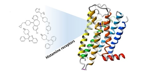
Graphical Abstract

1. Introduction

The most recent scientific technologies have crucial applications for drug discovery and design [1,2,3,4]. In silico approaches such as virtual screening and molecular docking have been widely applied to diverse proteins including G protein-coupled receptors (GPCRs) which constitute the largest family of cell surface receptors in the human body comprising, among others, histamine, dopamine, adenosine or adrenergic receptors that play a key role in cellular signaling [5,6,7,8]. Developments in GPCR structural biology provide insights into GPCR-ligand binding. In the recent years, a tremendous progress has been made in the crystallization of GPCRs representing different GPCR families and subfamilies, including aminergic GPCRs such as the histamine H1 receptor (H1R). Crystallographic information concerning GPCRs is essential for understanding the possible ligand-protein interactions and diverse conformational changes associated with multiple downstream signaling paths. Obtaining crystallographic data for GPCRs has been a very difficult task due to their conformational flexibility and heterogeneity of this superfamily. The crystal structures of GPCRs proved to be very useful for structure-based ligand design methods, screening of compound libraries and building homology models of not yet crystallized GPCRs [5,6,9,10]. With the crystal structure of H1R, a new opportunity has emerged to prepare the homology models of other histamine receptor subtypes (H2R, H3R and H4R) together with a combination of ligand-based and structure-based drug design [11,12]. Structural information provided by H1R as well as adrenergic β2-AR and other templates was used to resolve the structures of HRs in the inactive and active states in order to understand GPCR functionality and generate more effective drug discovery strategies. Recent investigations on the role of HRs in (patho)physiology and the use of receptor antagonists in in vivo disease models reveal a vast potential of histamine receptors in the treatment of e.g., allergic inflammation, neuropathic pain, and cancer [13,14,15,16,17,18,19,20,21]. The search for new and potent HR antagonists contributes to a steadily increasing number of potent and structurally diverse compounds [6,22,23,24,25,26,27,28]. Furthermore, development of ligands that are able to bind to two or more HR subtypes offers another opportunity to achieve a synergistic clinical effect mostly for allergic inflammations and neuropsychiatric disorders [29,30,31,32,33]. Taken together, the HRs field is showing a lot of potential to deliver another generation of potent drugs for clinical studies. In this review, we focus on the applicability of molecular modeling including drug discovery and design procedures such as molecular docking and virtual screening performed on HRs. We also review relevant clinical candidates and their therapeutic potential for the treatment of various inflammatory and allergic diseases.

1.1. Overall Structure of the Histamine Receptors

Histamine receptors, which belong to class A of the GPCR superfamily contain a bundle of seven antiparallel transmembrane helices, TM1 to TM7, connected by three extracellular loops, ECL1 to ECL3, and three intracellular loops, ICL1 to ICL3. Some helices contain highly conserved sequence motifs which are necessary to conduct activation steps. Furthermore, in all helices there are evolutionarily conserved residues required to preserve proper structure and function of GPCRs. Those residues are denoted by the number “50” in each helix in the Ballesteros-Weinstein numbering scheme: N1.50, D2.50, R3.50, W4.50, P5.50, P6.50 and P7.50 [34,35,36]. In this numbering scheme each residue of GPCR is recognized by two numbers separated by a dot; the first number indicates the transmembrane helix while the second number the position of the residue relative to the most conserved residue (assigned the number 50) on the same helix in a sequential order [34]. The most recognized sequence motifs in class A GPCRs are associated with molecular switches existing also in HRs; they are: the ionic lock associated with the DR3.50(Y/F) motif in TM3, the transmission switch in TM6 associated with the CWxP6.50 motif, and the tyrosine toggle switch in TM7 (NP7.50xxY motif) [37,38]. In the H1R receptor crystal structure the classical ionic lock is absent and R3.50, instead forming a salt bridge with a negatively charged residue in TM6, forms a hydrogen bond with Q6.36. The hydrogen bond can also link helices TM3 and TM6 but lack of the ionic interaction is intriguing and can modulate the activation of HRs. Since the activated structure of histamine receptors has not been determined the details of the activation steps remain unknown. Nonetheless, this structure must be similar to that of other amine-activated GPCRs, since some residues typical for this group, such as D3.32 and W7.40, are also present in all HRs [39]. D3.32 is directly bound to the ligand, while W7.40 is not in contact with the ligand but is located just behind the 3–7 lock [37,38] indicating importance of this molecular switch for activation of amine-activated receptors. Apart from differences in the ligand binding sites there are also other differences in a sequence and the average sequence identity between HR subtypes is only about 20–30% [11]. HRs are divided into four subtypes, H1R-H4R, that present also some differences related to tissue expression, ligand specificity and the final cellular effects.
Since the publication of the first crystal structure of GPCR describing the 3D structure of rhodopsin in 2000, multiple GPCRs have been crystallized including one histamine receptor [40]. The structure-based drug design using HR homology modeling has only been made possible after determination of the 3D-structure of bovine rhodopsin. The crystal structure of the histamine H1 receptor was obtained in 2011 and is still the only structure of histaminergic receptor deposited in the Protein Data Bank [41]. The structure of H1R provided important insights into the ligand binding mode in HRs and was successfully applied for drug discovery and design purposes [42]. The structures of GPCRs greatly contributed to and continue to provide great opportunities for the discovery and design of novel ligands using the structure-based approaches [43,44,45].

1.2. Recent Publications on Computational Studies Targeting HRs

The research papers describing in-silico approaches applied for the discovery of novel ligand chemotypes targeting HRs published in the recent years (2014–2020) have been compiled in Figure 1. These research papers were located in PubMed, Google Scholar and SciFinder using the expressions “histamine receptor docking” and “histamine receptor virtual screening”, and then filtering out those papers which did not directly deal with HRs. This search yielded over 80 novel research papers. Figure 1a depicts the number of research papers published yearly on each receptor as well as the number of publications covering all HRs. It can be observed that the number of research publications in the histamine H3R field shows an increasing trend after 2015 while the year 2019 saw a boom in the number of drug discovery projects in HR field with the exception of H2R and H4R. In 2020 the coronavirus pandemic was probably a direct reason for a much smaller number of papers. Figure 1b depicts the total number of papers published on each HR in the 2014–2020 time period. In the following sections, we review the most recent research publications describing computational approaches applied for the discovery of novel HR ligands and residues responsible for ligand binding and receptor activity.
Recently, Zobayer and Hossain studied the physicochemical and structural properties of HRs. The 3D models of HRs were developed through the homology modeling methodology using the I-TASSER webserver and the best model for each receptor was selected by applying various structure-validation tools [46]. The homology models of all GPCRs were created and made available in the GPCRdb webserver which is the information hub for GPCRs and their complexes [47]. The availability of the crystal structure of H1R, along with the homology models of other HRs, have resulted in the development of HRs-targeted ligands exemplified in Table 1. The crystal structure of H1R provides the highest hit rates while the hit rates for homology models vary to some extent.

2. HR-Targeted Ligands and Receptor Binding Site of Inactive/Active States of HRs

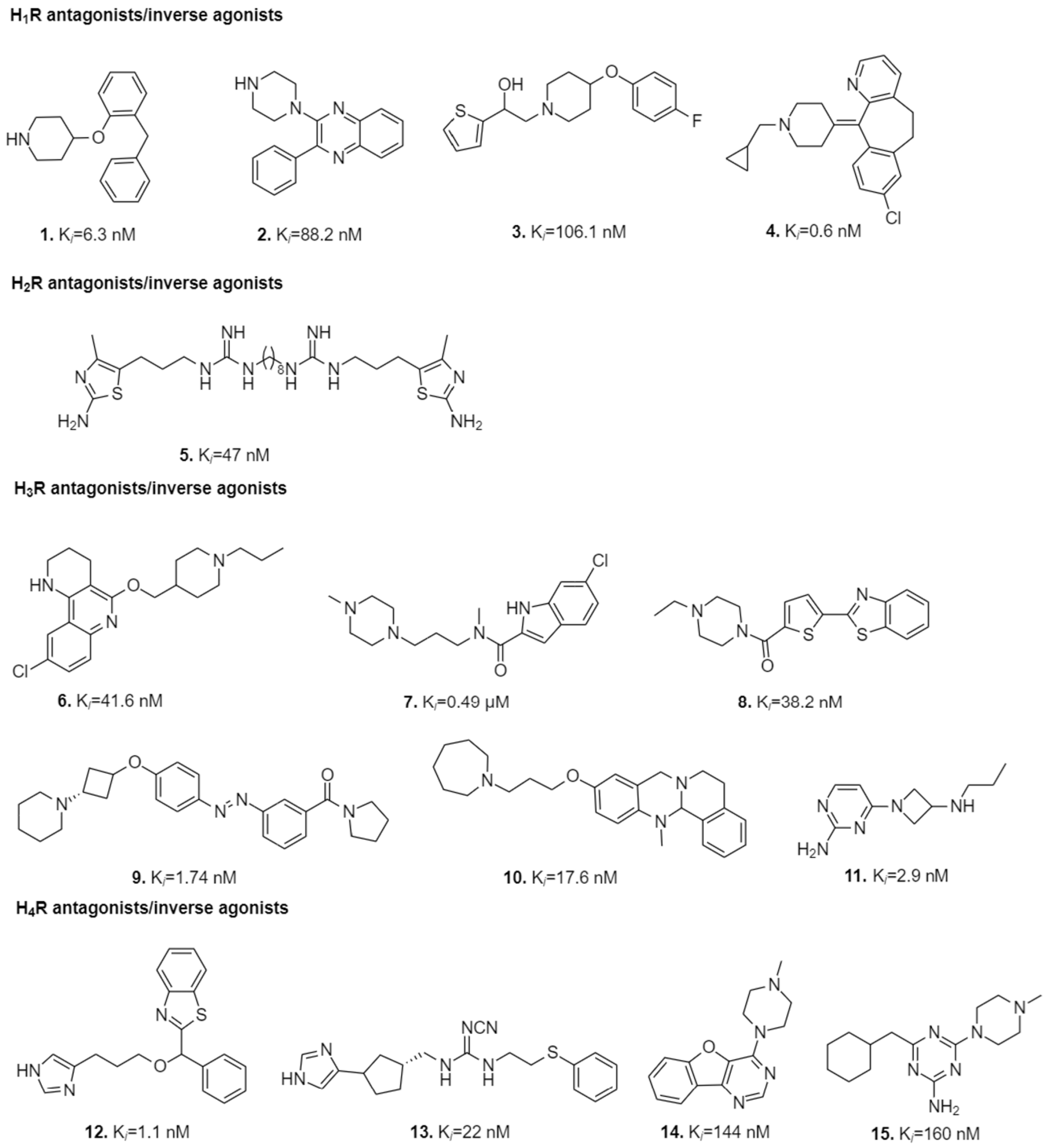
Virtual screenings and molecular docking have led the path towards development of novel HR-targeted ligands with high affinities for particular subtypes of HRs (Figure 2). The novel ligands display predicted interactions with crucial residues in the binding sites of HRs. In Table 2 these residues are presented with their original numbers from particular receptors and also in B-W numbering scheme.

2.1. Computational Studies on Histamine H1 Receptor and Its ligands

2.1.1. Structural Aspects of Histamine H1 Receptor

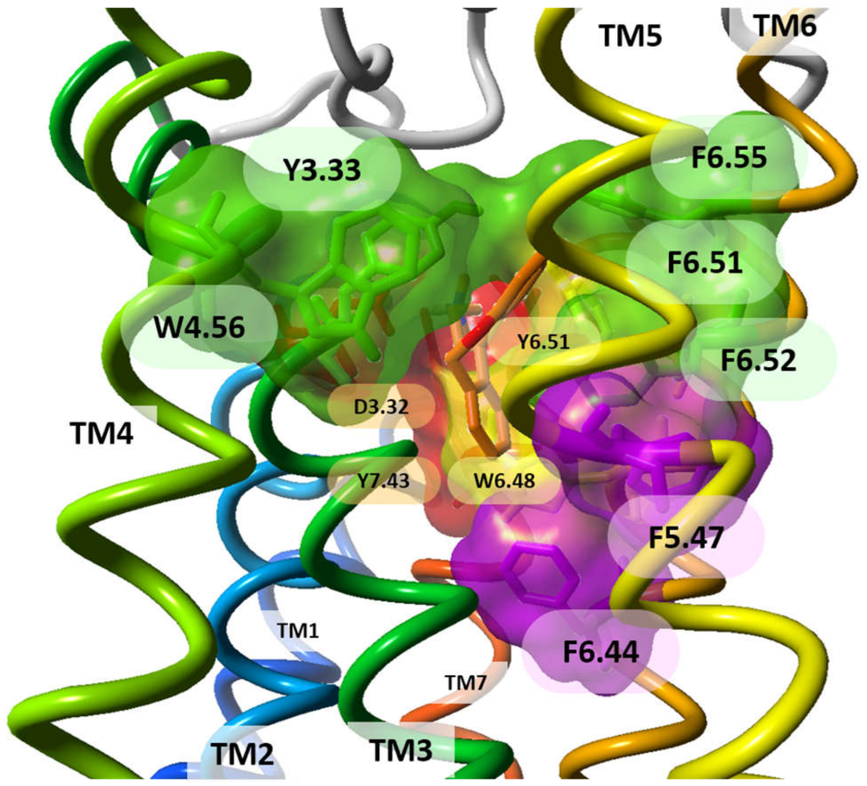
The first HR deposited in Protein Data Bank in 2011 was H1R in an inactive conformational state (PDB id:3RZE) [41]. The orthosteric binding site of this receptor is confined between the upper regions of the transmembrane helices (TMHs) and the extracellular loops (ECLs). H1R (UniProt id:P35367, 487 amino acids) crystal structure has a disulphide bond linking C180 (ECL2) with the extracellular end of TM3 (C3.25) but it is lacking the palmitoylation site at the end of helix H8 which is present in many other GPCRs. The antagonist orthosteric binding site in the H1R structure is divided into three regions lined by crucial residues as suggested by several site-directed mutagenesis studies: (i) the amine-binding region (D3.32, W6.48, Y6.51, I7.39, and Y7.43); (ii) the upper aromatic region (Y3.33, W4.56, Y6.51, F6.52, and F6.55), and (iii) the lower aromatic region (F5.47, F6.44, and W6.48), positioned deep in the TMHs [72] (Figure 3). The first-generation H1R antagonist doxepin sits deep in the ligand-binding pocket and directly interacts with W6.48, a highly conserved key residue required for GPCR activation, which is stabilized upon antagonist binding. The amine moiety of doxepin interacts with residue D3.32 present in all aminergic GPCRs. Both the upper and lower aromatic regions accommodate the butterfly-shaped hydrophobic aromatic moieties of doxepin. All these three regions were investigated to unravel H1R molecular determinants and visualize the binding hotspots in order to determine high affinity H1R ligands. The combined WaterFLAP calculations (approach to predict the binding site waters to guide ligand docking) and site-directed mutagenesis studies emphasized the crucial role of residue 7.39 (Table 2), a highly variable residue in aminergic GPCRs, as a determinant of specific N-methyl effects in amine ligand binding and responsible for stereo- and subtype-selectivity [72]. The second generation H1R antagonists containing unique carboxyl groups interacted with K5.39 and/or K179ECL2 in the anion-binding region of H1R leading to improved receptor pharmacology. This region is not conserved in other aminergic receptors illuminating the molecular basis of H1R antagonistic specificity and selectivity. The TM4 of the aminergic histamine H1R is also constricted, directing W4.56 (an important residue for H1R-ligand binding, based on mutation studies) towards the aromatic ligand binding pocket.

2.1.2. H1R Targeted Ligands and Their Interactions in the Ligand-Binding Pocket

Receptor-Based in Silico Approaches Targeting H1R

Several researchers have pioneered in the field of discovery of HR antagonists, especially those of H1R, as its crystal structure has been available since 2011. Both mutagenesis and computational studies indicated the importance of interactions with D3.32, Y3.33, T5.42, N5.46, W6.48, Y6.51, F6.52, and F6.55 in the orthosteric pocket for histamine and doxepin binding, and underlined the crucial electrostatic interaction with the side chain of D3.32 [75]. The other residues present in H1R and bound to the ligand were found to be K179ECL2, K5.39, H7.35 and Y7.43 [56]. Multiple walker metadynamics-simulation protocol was used for the identification of the preferential binding mode of the physiological ligand histamine obtained from 92% of conformational ensembles [99]. Pose re-scoring of doxepin at H1R followed by multiple linear regression using Prime software of Schrodinger and MD simulations proved to be essential for predictive modeling of receptor-ligand interactions [84]. Enrichments in virtual screenings have been improved using Interaction Fingerprints (IFPs) such as the SYBYL software capturing atom–atom interactions and SPLIF (Structural Protein-Ligand Interaction Fingerprints) capturing fragment–fragment co-occurrences [5]. Also, the virtual screenings of fragment-like compounds, using consensus energy-based docking scoring approach including both IFP (≥0.75) and PLANTS (≤−90), and the ionic interaction with residue D3.32 used as a filter proved to be better than individual scoring algorithms with increased hit rate of 73%. These attempts led to the efficient identification of chemically novel H1R antagonists (ECFP-4 similarity cut-off of 0.4) with nanomolar affinities and potencies such as Compounds 13 (Figure 2) [9,72].
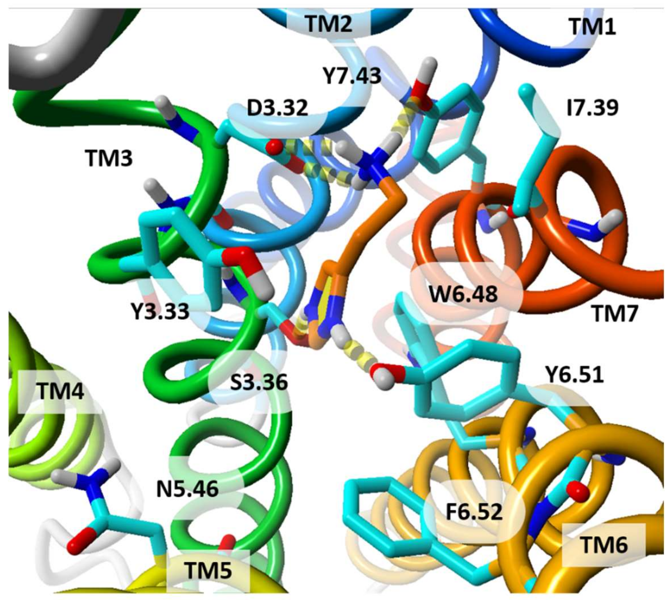
To demonstrate differences in the orthosteric binding sites of histamine receptors we have docked doubly protonated histamine to explore strong binding modes. The protonation state and the chosen pose of histamine are used for illustrative purposes only. The ligand histamine has been selected since the histamine molecule is small, so it does not change the binding site much, and is able to bind to all histamine receptors. The most likely binding mode of doubly protonated histamine in hH1R is shown in Figure 4.
Diverse multi-target compounds, including indolecarboxamides and alkyl/aryl piperidyl indoles, were reported as H1R, serotonin-5HT and CCR2 antagonists [65,69] while phenothiazines (promethazine, chlorpromazine, 2-chlorophenothiazine, thioridazine, trifluoperazine) were reported to be MRGPRX2 activators and H1R antagonists [100]. Chlorpromazine has high affinity to other GPCRs including dopamine, norepinephrine and muscarinic receptors [101,102]. The polypharmacology effect [103,104] of such ligands highlights the importance of other GPCRs, not only H1R, as potential templates for homology modeling of HRs. Indolecarboxamides were reported to form hydrogen bonds with N443ECL3, R176ECL2, and I6.58 [65] while alkyl/aryl piperidyl indole formed electrostatic interactions with conserved Y6.51 residue [69]. Several other compounds have been docked to the H1R structure such as aminomethylenepyrimidine-2,4,6-triones, N1-alkyltheobromine, N-methylanthranilates, azabicyclic isoxazoline acylhydrazones, substituted tetrazole-incorporated quinoline derivative, synthesized rupatadine and desloratadine analogues [70,73,76,105]. Elbayaa [70] designed a series of substituted aminomethylenepyrimidine-2,4,6-trione derivatives generated from a four-featured pharmacophore model with an aromatic or π-ring system, hydrophobic group, a H-bond donor and a H-bond acceptor group derived from five H1R antagonists and validated this model using six other H1R antagonists. These antagonists were mapped on the pharmacophore model with a good fitting score and low RMSD and then were docked using Molegro Virtual Docker on the H1R model obtained from SWISS-MODEL. They were reported to be a promising template for designing novel non-sedating H1R antihistaminic agents [70]. Docking studies, using AutoDock, showed that derivatives of N1-alkyltheobromine, which exhibited H1R antihistaminic activity comparable to doxepin and less CNS depressant side effects than olapatadine, interacted electrostatically and formed hydrogen bonds with residues D3.32, Y3.33, S3.36, T3.37, K5.39 and Y6.51 [71]. In one study, quinoline derivative (QS-15), astemizole and diclofenac sodium have been shown to interact with residues D3.32, W6.48 and F6.52 [73]. Additionally, although the fragment VUF13816 is structurally different from the reported H1R ligands obtained from virtual screening, it was observed to have similar contacts to the receptor as doxepin (residues H7.35 and D3.32). A series of three fluorescent ligands was designed based on this small fragment that retained similar affinity towards H1R as the parent compound [74].
In another study [55], fexofenadine, a potent non-sedative third-generation hH1R antagonist was proved to be beneficial in treating H1R related allergic conditions of dogs and cats. In this study, the homology models of dog and cat H1R isoforms were built and fexofenadine was subsequently docked to human, dog and cat H1R. In total, it interacted with 23 residues in all the three receptors, and the most crucial molecular determinants in hH1R were W6.48, F6.52, Y3.33, N5.46 and T5.42 [55]. The potential phytochemical inhibitors targeting both H1R and cytosolic phospholipase A2, with good pharmaceutical drug-like properties, such as 3′,4′,7-trihydroxyflavone, calycosin, geraldone, licoflavanone and epidistenin were reported to interact with D3.32, S3.36, Y6.51, H7.35 as well as T182ECL2 and D183ECL2 [48]. Also, the anti-allergic and anti-inflammatory potential of phytocomponents was analyzed by computational docking analysis on H1R [87,97,106]. The examined compounds included: β-pinene, thymol and carvacrol, present in the Siddha formulation Oma Legium, the standard cetrizine and bioactive phytotherapeutics such as ascorbic acid, β-sitosterol, sesquiterpene, and tocopherolpresent in the medicinal herb Corallocarpus epigaeus, as well as flavones like kaempferol and kaempferol-3-glucuronide found in Centratherum punctatum along with desloratadine as a reference standard. The flavones and desloratadine showed similar ligand-protein interactions with residues Y6.51 and F6.52 [87]. Curcumin has also been shown to possess H1R antagonistic activity [56]. About 22 synthesized rupatadine and desloratadine analogues, including Compound 4 (Figure 2), were docked to H1R and, guided by docking studies, the steric constraints within the binding pocket were found to explain the observed differences in affinity of ligands. The limiting residues were I7.39 and Y7.43 located next to the amine-binding region [98]. Methyl (MMA), propyl (PMA) and isopropyl (IMA) N-methylanthranilate, originally found in the leaf essential oil of Choisya ternata, were reported to establish interactions with D3.32, Y3.33, S3.36, I3.40, W4.56, N5.46, F5.47, F6.44, W6.48, Y6.51, F6.52, F6.55 and I7.39 [76]. In 2020, a set of 35 antihistamines was designed using cloperastine as the core molecule in docking and molecular dynamics studies, however, no experimental binding studies were performed [107]. Another study involving in silico design, synthesis, ADME profiling and evaluation of antagonistic effects of 1,8-naphthyridine-3-carboxylic acid analogues was carried out using chlorpheniramine as the standard drug. Ligand dockings using Auto Dock Vina elucidated the crucial interactions in the binding pocket of H1R, involving residues D3.32, Y3.33, S3.36 and Y6.51, for the ligands possessing satisfactory ADME profiles [108].

Ligand-Based Computational Approaches in Search for Potential H1R Ligands

Fragment-based drug discovery proved to be a propitious approach for the development of novel chemically and therapeutically active leads. The identification of small fragment-like biologically active dual H1R/H4R antagonists has been made possible through prospective ligand-based virtual screening (LBVS) using 14 different chemical similarity descriptors and consensus scoring approaches [27]. It was evident through this study that the performance of the similarity descriptors decreases with decreasing self-similarity of the actives. Also, the Molecular ACCess System (MACCS) turned out to be one of the three best performing similarity descriptors for H1R and H4R, while piDAPH3 and piDAPH4 being the worst. Certain consensus scoring methods achieve better enrichments, with best results in both the max-value or ranked-by-vote consensus methods as became evident in study of H1R and H4R virtual screening enrichments [27]. Even the ligand-based comparative molecular similarity indices analysis (CoMSIA) model (Q2 = 0.525, R2ncv = 0.891, R2pred = 0.807), using 129 reported H1R antagonists, had good predictive quality for predicting the bioactivities of new chemicals. Subsequent molecular docking and simulation of these reported antagonists unraveled their binding modes in the active site of H1R [64].

2.2. Computational Studies of H2R and Its Ligands

Although H2R is a promising drug target, the computational modeling of H2R (UniProt id:P25021, 359 amino acids) has not been studied extensively due to the lack of the crystal structure which hampered the drug discovery efforts. This shortcoming makes development of protein models for structure-based approach a necessity [109]. The quality of the generated homology models largely depends on the selection of template(s) and the sequence alignment while robustness is judged on the basis of their ability to differentiate between known actives and decoys [110,111].

2.2.1. Homology Modeling of H2R

Homology modeling that combines multiple templates usually yields better receptor structures for drug discovery processes. Since hH1R and hH2R have low sequence similarity and identity, the β1AR (PDB id:4BVN), hβ2AR (PDB id:2RH1), hD3R (PDB id:3PBL) and hH1R (PDB id:3RZE) were chosen as templates, depending upon TM similarities and identities for H2R homology modeling, as for example, in the study of Saxena et al. [66]. Krzan et al. [85] generated a H2R homology model employing multiple webservers and programs: I-TASSER [112], MODELLER [113], SWISS MODEL [114] and Pyre2 [115] using the following templates: hH1R (PDB id:3RZE), the neurokinin 1-receptor (PDB id:2KS9), human β2-adrenergic receptor (PDB id:2RH1), human β1-adrenoceptor (PDB id:4BVN), and M3 muscarinic acetylcholine receptor (PDB id:4DAJ). The best model was selected using the statistics for non-bonded interactions generated by the ERRAT tool [116] and stereochemical properties obtained from PROCHECK [117]. Chaudhary et al. [118] screened a range of phytochemicals present in Ficus religiosa for binding to hH2R. For this study a homology model of hH2R was generated on the basis of similarity search using four structures of β1-adrenergic receptor (PDB ids:2VT4, 2Y00, 4BVN and 5A8E) as templates. In another study, a template of β1-AR (PDB id:2VT4) was used for homology modeling and a model quality was assessed by Ramachandran plot while AutoDock was used for docking of compounds [119]. Recently, Boddupally et al. [120] also generated a hH2R model based on a β1-AR template (PDB id:6H7J) using MODELLER and evaluated it by PROCHECK and Ramachandran plot. The modeling was followed by docking of twenty natural flavonoid compounds to the receptor in AutoDock. In the above three studies only β1-AR was used as a template so the modeled structure of H2R showed some limitations in model quality.

2.2.2. H2R-Targeted Ligands and Their Interactions at H2R Active Site

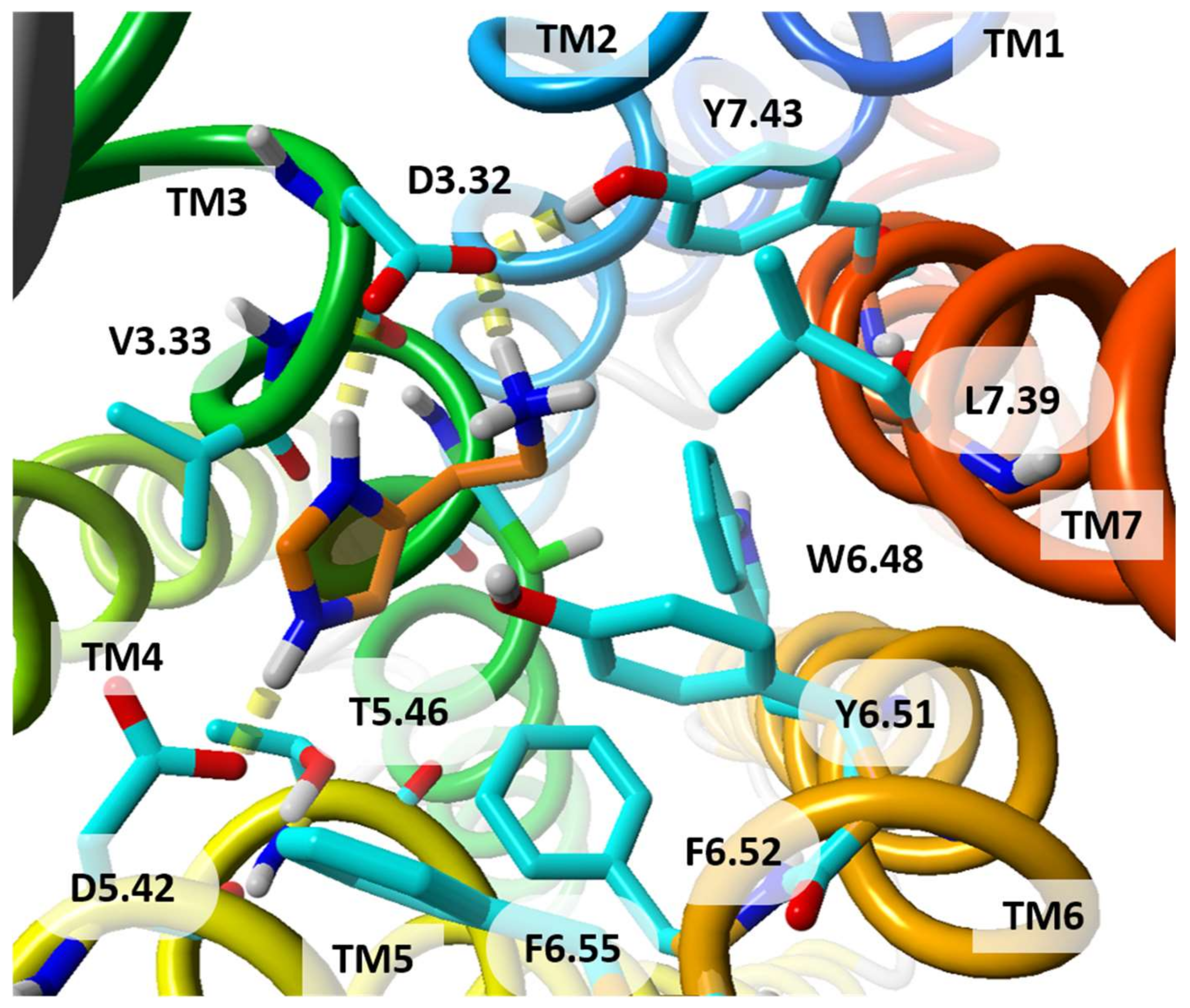
In an effort to design, develop and optimize selective H2R as well as dual H1R and H2R ligands, molecular docking using the Schrodinger package provided insights on how to rationalize the binding of octahydropyrazinopyridoindole class of compounds [66]. Additionally, docking the standard H2R antagonists such as metiamide, cimetidine, ranitidine and famotidine to the homology modeled hH2R enabled comparison of properties of selective ligands of H1 and H2 receptors. It has been found that hydrophobic regions are important for selective hH1R antagonists whereas polar features for selectivity of hH2R antagonists. However, the hydrophobic interaction in vicinity of polar region was also discriminating for hH2R ligands due to the presence of V3.33 residue in H2R instead of Y3.33 in H1R and other histamine receptors. Details of the orthosteric binding site of homology model of hH2R are shown in Figure 5. Analysis of the residue properties further confirmed TM5 to be the most dissimilar region between hH1R and hH2R, followed by TM6 and TM3. A mutagenesis study revealed that the residue D3.32 is vital for histamine and antagonist binding, while D5.42 and T5.46 are important for the selectivity and kinetics of histamine binding. The presence of V3.33 and D5.42 in hH2R introduces a bulkier space and negatively charged environment at this position as compared to Y3.33 and T5.42 in hH1R, respectively. Other important hydrophobic residues, W6.48, Y6.51, F6.52 and F6.55, are the same in both H1R and H2R. The representative class of hH2R antagonists was found to form hydrogen bonds with D3.32, D5.42, Y6.51, and N159ECL2 and hydrophobic contacts with the aromatic triad in H2R (W6.48, Y6.51, and F6.52). Analysis of the structure-based pharmacophore model for H1R and H2R indicates that hydrophobic features are important for selective H1R antagonism while polar groups are preferable for selective H2R antagonism.
Histamine docked to the optimized model of H2R asserted the importance of three crucial residues D3.32, D5.42 and Y6.51 for histamine binding. A proton transfer from the charged ethylamino group of histamine to D3.32 allowed K175ECL2 to undergo large conformational change and approach the D3.32 residue. Additionally, from performed quantum-chemical calculations, it was evident that deuteration increased affinity of histamine towards H2R binding [85]. Pockes et al. [77] performed studies on dimeric hetarylpropylguanidine-type derivatives which were docked to H2R while the monovalent ligands were docked to hH3R and hH4R. Docking studies were followed by 30 ns MD simulations and the lowest free-energy conformations of compounds formed the strongest H-bond contacts with the residues of the orthosteric binding site: D3.32 and D5.42 in hH2R; D3.32 in hH3R; and D3.32, E5.46, E163ECL2 and T5.42 in hH4R. The residue D5.42 accounts for hH2R selectivity since it is not present in other histamine subtypes. Different steric effects of residues enclosing the orthosteric binding pocket may be at play since residues V3.33, V176ECL2-Q177ECL2 of hH2R are less voluminous when compared to Y3.33, F184ECL2-Y185ECL2 of hH1R; Y3.33, F192ECL2-F193ECL2 of hH3R; and Y3.33, F168ECL2-F169ECL2 of hH4R. During the study, it was observed that dimeric compounds showed better affinity towards hH2R while monomeric ligands showed better affinity towards hH3R and hH4R [77]. A representative of the former ligands is Compound 5 shown in Figure 2.

2.3. Computational Studies on H3R and Its Ligands

2.3.1. Homology Modeling and Structural Aspects of H3R

The homology models of H3R (UniProt id:Q9Y5N1, 445 amino acids) were generated using a variety of templates starting from bovine rhodopsin (PDB id:1U19) [59]. In many studies the crystal structure of inactive hH1R (PDB id:3RZE) was used as a template for construction of the homology model of inactive hH3R since their sequences possess 31.4% identical residues [57,58,79,81]. The homology model of H3R was also built using M3 muscarinic acetylcholine receptor (PDB id: 4DAJ) [51,62,94,95]. Recently, researchers have described a H3R homology model taking the crystal structure of M2 muscarinic acetylcholine receptor as a template (PDB id: 3UON) [67,121,122].
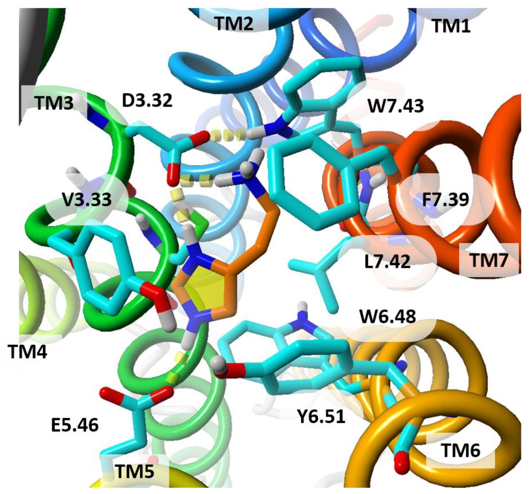
Jonczyk et al. [80] generated H3R homology models and used a hybrid assessment of these models based on knowledge-based scoring algorithm and two-step docking protocols including GOLD and Glide. The models also passed the quality analysis performed using BCL::Score, QMEAN and PSVS methods. A model built on the M3R template was preferred as compared to H1R or models built on mixed template alignments—they were characterized by significant differences in the most conformationally diversified ECL2 loop. Most models generated by MODELLER proved to be much better that those of SwissModel, I-TASSER and Jackal. In the best H3R model, 3-7 lock switch was considered as the interaction between the side chains D3.32 and W7.43, whose breakup can promote receptor activation. Additionally, the perpendicular position of residue W6.48, with respect to the helix axis forced by L7.42, favored recognition of H3R-specific ligands and interaction with E5.46. Inactive conformation of H3R is maintained by residues L2.43, L2.46, I3.43, I3.46 and I6.40 in the center of the receptor creating a hydrophobic barrier inside the receptor as in other GPCRs. Breakdown of this barrier is an important step in receptor activation and opens a gate for a continuous intrinsic water pathway [123,124].
The multiple template approach has also been applied for H3R homology modeling by using H1R (PDB id:3RZE), M2R (PDB id:3UON) and M3R (PDB id:4U15) as templates while ECL2 was built on M2R and M3R. The models were ranked for possessing a crucial ionic protein-ligand interaction with residue D3.32 considered essential for ligand binding. It was also found that a conformation of residue E5.46 was more advantageous for ligand binding when it was pointing toward the binding pocket [52]. Another multiple template approach was employed by Hauwert et al. [60] for modeling H3R using H1R (PDB id:3RZE), M3R (PDB id:4U15), dopamine D3R (PDB id:3PBL), serotonin 5-HT1BR (PDB id:4IAR) and serotonin 5-HT2BR (PDB id:4IB4) receptors as templates and the MODELLER program.

2.3.2. Ligands Targeting H3R and Their Interactions

Like other GPCRs H3R is sodium sensitive, as it was made evident by using a mathematical model and MD simulations. It was also calculated that an H3R inverse agonist thioperamide binds to the orthosteric binding site of hH3R preferentially in a presence of Na+. In the presence of Na+, the positively charged imidazole moiety of thioperamide is located “above” the highly conserved residue D3.32 while in its absence the same moiety is located “below” D3.32 and directed toward the highly conserved D2.50, which forms the allosteric site for binding a sodium ion [58]. Thioperamide spans horizontally between two negatively charged residues D3.32 and E5.46 and the ligand is embedded in a pocket between helices TM3, TM5 and TM6 [58,59]. However, in a study [80] based on another template, M3 muscarinic receptor, thioperamide was docked vertically and interacted only with D3.32. There are two negatively charged residues, D3.32 and E5.46, in the orthosteric ligand binding pocket of hH3R (similarly to hH4R). The former residue can bind to the protonated amine and is essential for interactions with agonists, including histamine. The second acidic binding point is created by E5.46 and adjacent tyrosine Y6.51. In a study by Jonczyk et al. [80], a series of amine antagonists including JNJ5207852 was docked to hH3R in vertical poses, and the protonated amines of these ligands were bound to E5.46, Y3.33 and Y6.51. It was also found that antagonist clobenpropit with protonated imidazole ring and isothiourea fragments, used both acidic points, D3.32 and E5.46, in the ligand binding space: imidazole interacted with D3.32 while isothiourea group, as a second protonated system, created a salt bridge with E5.46. It is in contrast to results obtained by Kim et al. [125] which employed a model of hH3R based on the human β2-adrenergic receptor. Doubly charged clobenpropit was bound to D3.32 by its isothiourea group while the imidazole ring was bound to E5.46 and adjacent residues. Although it cannot be excluded that both poses are allowed, the future experimentally determined structures of histamine receptors with ligands will help docking studies enormously. Details of the orthosteric binding site of homology model of hH3R are shown in Figure 6.
Binding of more complex antagonists of H3R was possible by extending the ligands toward the external allosteric site residues, R6.58, Y7.35 and residues of the ECL2 loop (Y189, A190, F193 and Y194) as documented by Kumar et al. [62] for novel 4-aryl-6-methyl-5,6,7,8-tetrahydroquinazolinamines as anti-obesity candidate drugs. In the study performed by the Gloriam group [50] the pharmacophores were constructed based on the residue-ligand fragments from the GPCR crystal structures. Thanks to the chemogenomic techniques that detect local similarities within the transmembrane binding pocket the whole scaffolds or ligands can be exchanged between targets. Performing such binding pocket comparisons is reasonable since many high-quality GPCR crystal structures with ligands are currently known. Using the above methodology for the fragment-based prospective virtual screening employing pharmacophores and molecular docking methodology they identified five novel neutral antagonists and one inverse agonist which were later confirmed in Gq-coupled pharmacological assay. They also found that residue E5.46 contributes to selectivity in H3R and H4R as compared to H1Rs/H2Rs while residues at positions 2.64, 2.65, 5.42, 6.52, 6.55, and 7.36 confer H3R/H4R selectivity [50].
Diverse screening protocols were applied for discovery of H3R targeted ligands including ligand-based approaches exploiting the similarities with respect to known actives as well as structure-based approaches exploiting the 3D-chemical interactions between ligands and the target structure [126]. Ghamari et al. [127] employed individual and combined hybrid structural similarity (FP2 fingerprint in 2D search, and also Electroshape and Spectrophores methods in 3D search) and pharmacophore-based approaches using the ZINCPharmer [128] and ZINC15 database followed by lead optimization, and identified potential drug-like anti-H3R ligands in micromolar and submicromolar activity range (including Compound 7 in Figure 2). The geometric center of residues D3.32, T3.37, Y189ECL2, F5.38, E5.46, W6.48, and Y6.51 was set as the center of the binding site. Docking of pitolisant and other ligands was performed with two distance constraints with E5.46 and Y189ECL2 in GOLD. The phenyl ring of pitolisant was found sandwiched between Y189ECL2 and F7.39 while piperidine nitrogen formed an ionic bond with E5.46 and a H-bond with Y3.33. Four-featured 3D-pharmacophore model comprising two hydrophobic regions, a positively ionizable moiety, and a hydrogen bond acceptor was generated from a docked pitolisant-H3R complex using LigandScout [129]. The obtained hits possessed a common scaffold containing a basic moiety and an aromatic/hydrophobic moiety joined by a linker. The basic moiety interacted with E5.46, the linker interacted with Y3.33 and Y6.51, while the hydrophobic/aromatic moiety was observed to interact with Y189ECL2 and F7.39 [51,127]. In another study a quantitative structure-activity relationship (QSAR) model, employing a genetic algorithm coupled with partial least square and stepwise multiple linear regression methods, was used with two major descriptors: connectivity information and mean absolute charge in predicting H3R antagonistic activity. The additional molecular docking revealed the crucial role of residues Y3.33, Y189ECL2, F193ECL2, L5.39, E5.46, W6.48, Y6.51, M6.55, Y7.35 and F7.39 in the interaction with H3R targeted ligands [91]. Schaller et al. [52] used a ligand-guided homology modeling strategy that resulted in identification of two H3R ligands with nanomolar affinity, including Compound 8 (Z27743747) in Figure 2. Pharmacophore models were generated by taking nine reported H3R antagonists and the virtual screening was performed using the Enamine library. The high-quality model of H3R was used for docking while keeping constraints focused on interactions with D3.32 and E5.46. The obtained hits interacted with D3.32 and Y3.33 as well as with several other hydrophobic residues. The additional π-cation interaction with F7.39 was predicted to contribute to the superior H3R affinity of ligands [52].
The H3R antagonists have been classified as imidazole- and non-imidazole-based compounds. Preliminary studies have focused on the development of the imidazole type compounds which are structurally similar to histamine but, due to some undesirable features, they failed to enter the pharmaceutical market [130]. Hence, researchers turned their attention to imidazole ring bioisosteres in an attempt to develop non-imidazole type compounds such as piperidine, piperazine and pyrimidine. This approach resulted in e.g., pitolisant. The ligands with diverse scaffolds have been docked to H3R models, including amino acetylenic benzophenone derivatives [59], flavones [79], isoflavones [90], tert-amylphenoxyalkyl (homo)piperidine derivatives [88], piperazine derivatives including tert-butyl/tert-pentylphenoxyalkyl, acetyl/propionyl phenoxyalkyl, 4-pyridyl, and (dihydro)benzofuranyl [29,67,86,89], tetrahydroquinazolinamines [62] or pramepexole carbamodithiolate metal complexes [131]. A series of flavone derivatives was designed and docked to H3R model. The flavones were observed to form a salt bridge with E5.46, π-anion bond with D3.32, H-bond with Y6.51, and the hydrophobic interactions with Y3.33, C3.36, A5.42, W6.48, Y6.51, and L7.42 [79] while isoflavones formed a salt bridge with E5.42 and π–π interactions with Y3.33 and F5.38 [90]. In another study, the tert-amylphenoxyalkyl (homo)piperidine derivatives were docked to H3R model built on M3 muscarinic acetylcholine receptor in GPCRM structure modeling server [132]. It was observed while docking these derivatives that a salt bridge was formed with residue E5.46, and π–π stacking with Y3.33, F5.39 and/or Y189ECL2 [88]. In the case of a novel series of piperazine derivatives (tert-butyl/tert-pentylphenoxyalkyl, acetyl/propionyl phenoxyalkyl and 4-pyridyl), the ligands were observed to form H-bond/salt bridge with E5.46, and H-bond with Y6.51 while forming π–π interactions with W6.48 and F5.47. Additionally, the ligands were found to be stabilized through π–π stacking with at least one of the following residues: Y3.33, Y7.35 and F193ECL2 [67]. An additional hydrogen bond with residue R6.58 could have a positive influence on H3R affinity [89]. Ligands with 4-pyridyl piperazine scaffold were found to be H3R selective antagonists as compared to H3R, D2R, M1R and α1-adrenergic receptors [61]. Dihydrobenzofuranyl-piperazines analogs with 5-phenyl and allylpiperazine modifications were recently found to be the potent hH3R antagonists with nanomolar affinity [86].
Wagner et al. [81] have evaluated a series of 4-(3-aminoazetidin-1-yl)pyrimidin-2-amines as high affinity H3R partial agonists obtained from an in-house screening campaign (Compound 11). All these ligands along with histamine, when docked using PLANTS and after MD simulations as H3R complex, have maintained stable crucial interactions with D3.32, E5.46 and Y6.51 [81]. Two bidirectional GPCR photoswitchable antagonists with substituted azobenzene scaffold (VUF14738 and VUF14862 (Compound 9 in Figure 2) were developed by Hauwert et al. They possessed 10–100 fold higher H3R selectivity over H1R and no measurable H4R affinity [60]. Recently, Z3303614736 have been found to be new strong inverse agonist of H3R that inactivates the receptor in a nanomolar concentration range. It was identified by using the intramolecular H3R biosensor based on bioluminescence resonance energy transfer (BRET) that is sensitive to the conformational dynamics of H3R and can be used to screen for the receptor agonists and inverse agonists in a live cell [133]. Nonimidazole antagonists/inverse agonists of H3R with nanomolar activity as anticonvulsant drugs were obtained by Song et al. [134] by linking the H3R pharmacophore (aliphatic amine with propyloxy chain) with the 1,2,4-triazole moiety. Docking studies indicated that these ligands were bound to Y3.33 and E5.46, and additionally to R6.58, F193ECL2 and M6.55 [134].

2.3.3. Multi-Target H3R Ligands

Recently, the multi-target-directed ligands of histamine receptors gained a lot of attention [45,57,68,78,94,95] that led to development of multi-targeted ligands of H3R, serotonin receptors, as well as acetylcholinesterases (AChE and BuChE), for potential treatment of neurodegenerative diseases such as Alzheimer’s disease. Jonczyk et al. [68] identified multi-targeted piperazine derivatives to be blockers of AChE and BuChE as well as of hH3R. The ligands were bound to E5.46, as this was suggested to be a crucial interaction for H3R antagonistic activity, while the other interactions were with residues F163ECL2, Y3.33 and Y6.51 [68]. Lepailleur et al. [49] performed ligand-based pharmacophore-guided virtual screening of CERMN chemical compound library (17,194 compounds) using six active H3R ligands with the purpose of designing dual targeting H3R/5-HT4R ligands. Ligand docking was performed using the Schrodinger Glide Induced Fit procedure. The best ligands appeared to be benzo[h] [1,6] naphthyridine derivatives. The pharmacophoric features correlated well with the interacting residues: protonated amine (basic center) formed a salt bridge with D3.32, the aromatic system formed a π−π interaction with indole ring of W6.48, the hydrophobic part was surrounded by residues L3.29, W3.28 and F7.39, and the NH of the tricyclic system served as a H-bond donor bound to the side chain of E5.46. The ligands including the most potent Compound 6 in Figure 2 possessed two positively ionizable groups (imidazole, alkylamine) that were sometimes supposed to interact with residues D2.50 or with E175ECL2 and E191ECL2 [49]. Darras et al. [57] performed computational studies followed by synthesis and biological evaluation for synergistic effects of tri- and tetracyclic nitrogen bridgehead compounds reported as dual acting hH3R antagonists and AChE inhibitors. The tricyclic partial structure of the most potent and selective H3R compound in this study remained stably bound in a pocket formed by residues L3.29, Y3.33, C3.36, W6.48, Y6.51, M6.55, and F193ECL2. Two stable ionic interactions between D3.32 and a positively charged piperidine moiety of compounds, as well as between E5.46 and a positively charged amidine moiety were observed during entire MD simulation. Also, an aromatic interaction was observed between an aromatic moiety of a docked Compound 10 with Y3.33 and F193ECL2 [57].
A series of twenty new chlorophenoxyalkylamine derivatives was reported as dual acting hH3R antagonists and AChE/BuChE inhibitors [94,95]. The hH3R model was developed using GPCRM webserver and validated using pitolisant as a reference H3R ligand. All these compounds were docked to target proteins using Schrodinger Glide software. It was observed that irrespective of alkyl chain length, chloro substituted phenyl rings were in close contact with ECL2 residues (W174, L177, A190, E191, Y194 and W196) [94,95]. The multitarget-directed ligands with H3R antagonistic activity coupled with the ability to inhibit acetyl/butyrylcholinesterases and monoamine oxidases A/B, potentially suitable for the treatment of Alzheimer’s or Parkinson’s disease, were studied by Bautista-Aguilera et al. [78]. All the studied compounds revealed an interesting neuroprotection profile against oligomycin A, okadaic acid (as a model of the hyperphosphorylation of tau), and β-amyloid peptide Aβ25–35. Of all ligands the non-imidazole ligand, contilisant, had the best properties. Recently, 26 non-imidazole histamine H3R ligands and 23 xanthone derivatives, rationally designed using a pharmacophore model for H3R antagonists/inverse agonists, have been confirmed as potential anti-Alzheimer agents by docking to H3R and acetyl- and butyrylcholinesterases. The most promising derivatives combined the flavone moiety via a six carbon atom linker with a heterocyclic moiety, such as azepane, piperidine or 3-methylpiperidine [121,122].

2.4. Computational Studies on H4R and Its Ligands

2.4.1. Homology Modeling and Structural Aspects of H4R

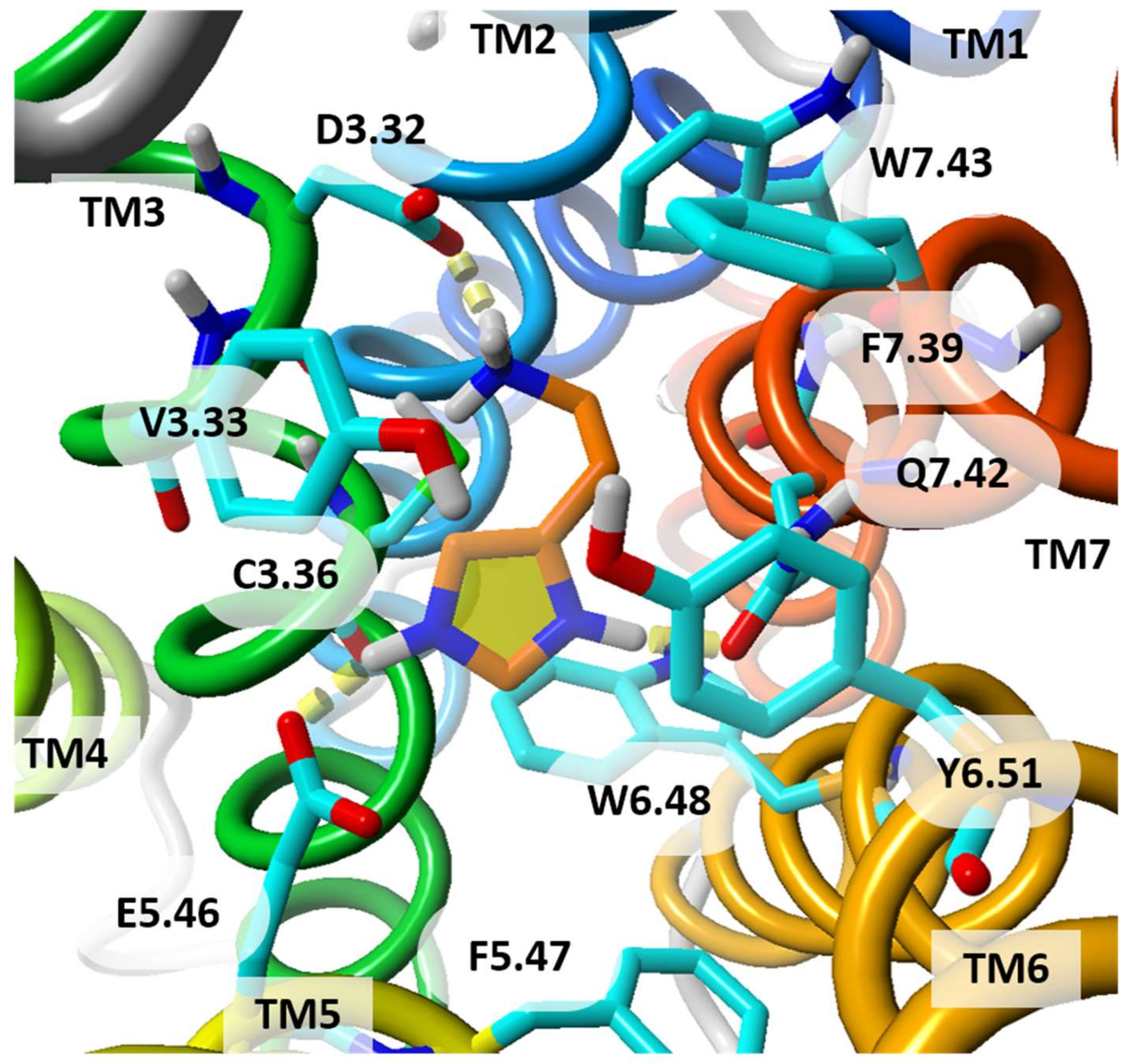
H4R (UniProt id:Q9H3N8, 390 amino acids) possess 40% identity in TM region to H1R which led many researchers to generate H4R homology models using H1R crystal structure as a template. However, there are substantial differences in their binding sites as the residues K5.39, N5.46 and G7.42 in H1R are equivalent to L5.39, E5.46 and Q7.42 in H4R. The large sequence similarity (37%) and structural similarity (58%) in the binding site between H3R and H4R, confirmed by mutagenesis studies, emphasized a crucial role of identical residues: D3.32, E5.46, Y3.33, W6.48 and Y6.51 in ligand binding that results in similar ligand poses in the binding site and can explain the mechanism of dual H3R/H4R antagonism [36,57]. Importance of specific residues for ligand binding was confirmed by mutations: E5.46Q had decreased the affinity of clobenpropit and its derivatives toward H3R and H4R while mutations L5.39V and E5.46Q decreased the affinity of ligands against H4R. The mutagenesis also revealed importance of N4.57, L5.39 and E5.46 for antagonist binding while featuring H-bonds/electrostatic interactions with two negatively charged residues D3.32 and E5.46 to be important for receptor activation [27,53,72,135]. Combined mutagenesis and protein-ligand modeling studies performed to explain H3R and H4R selectivity indicate that the residue at position 4.57 (Y4.57 in H3R and N4.57 in H4R) is directed toward the ligand binding pocket. This suggests that the TM4 of H3R and H4R remains undistorted making the constriction unique for the H1R subtype [136]. The pharmacological profile of H4R ligands was recently reviewed by our group [137].
The residues in ECL2 (the largest and the most structurally diverse extracellular loop of class A GPCRs) are also important for binding of ligands to all monoaminergic GPCRs as revealed by site-directed mutagenesis experiments. ECL2 also plays an important role as a stabilizer of the inactive state of the receptor [138]. Mutagenesis of two ECL2 phenylalanine residues (FF motif) in hH4R resulted in reduced constitutive and ligand-induced receptor activation, which points to a significant involvement of this motif in receptor activation [80,82,96]. Mutations of these residues can change the pharmacological properties of ligands: thioperamide is a partial inverse agonist at hH4R F169ECL2V, which is a mutant with reduced constitutive activity, and a neutral antagonist at hH4R F168ECL2A a mutant devoid of constitutive activity. It was concluded that F168ECL2 is a key determinant of H4R constitutive activity, ligand binding and potency as compared to F169ECL2. These studies reveal a crucial role of the FF motif, F168ECL2 and F169ECL2, in both ligand-receptor interactions and constitutive activity (interconversion between active and inactive conformation) of the wild-type hH4R [82].

2.4.2. Ligands Targeting H4R and Their Interactions

Kiss et al. identified novel histamine H4R ligands through the ensemble docking based on homology model conformers obtained from MD simulations. Such representative hH4R conformers were found to be more suitable for the identification of H4R antagonists than the initial homology models [25]. It was also found that X-ray and homology model structures may be complementary, or at least able to sample different protein conformations leading to non-overlapping hits and can provide important starting points for fragment-based lead discovery for other GPCRs [6]. Labeeuw et al. [22] discovered a potent antagonist (Compound 12) among 2-benzothiazolylphenylmethyl ether analogues. It was identified in a virtual screening of a corporate compound collection based on H4R model, followed by hit optimization with the purpose of designing potent and selective H4R antagonists. As for the binding mode the imidazole ring of the scaffold was suggested to form a salt bridge with D3.32, the oxygen of the linker a H-bond with the phenol moiety of Y3.33, and the nitrogen atom of the benzothiazolyl group a H-bond with Y6.51. Istyastono et al. [53] have explored complementary retrospective and prospective SBVS using β2R-based and H1R-based H4R homology models with a IFP scoring on fragment-like ligands, which allowed identification of H4R ligands (including Compound 14) that were not spotted during LBVS runs. Although the authors observed higher enrichments in a model based on β2AR as compared to a model based on H1R, an exchange of templates did not significantly affect SBVS accuracy. The TM binding pocket was very similar in both H4R models, based on β2AR and H1R, with little differences in ECL2. In the β2AR-based H4R model, the chlorine atom of JNJ7777120 was accommodated between L5.39, T6.55 and F168ECL2, while in the H1R-based model it was located close to residues L5.39, T5.42 and F168ECL2 [53]. Details of the orthosteric binding site of homology model of hH4R are shown in Figure 7.
With the constantly growing number of H4R ligands, the applicability of a large group of azine derivatives has been increased. A promising series of novel selective H4R antagonists, such as 2-amino-4-methylpiperazine-1,3,5-triazines, was designed and synthesized by different research groups by introducing variously substituted arylethenyl moieties [24,92,139]. Lazewska et al. [24] docked these molecules on the validated H4R homology model built using a template of H1R. All compounds with triazine core including Compound 15 were found to interact with D3.32 in two possible ligand orientations in the binding pocket of H4R. The protonated nitrogen of the most active ligands formed a weak interaction with E5.46. The triazine core of ligands was found to interact with Y3.33 and also with T5.42 and Y6.51. Halogen atoms para substituted to the benzene ring were found to fit in the hydrophobic pocket formed by residues V2.53, I2.58 and W3.28. Among them, 4-(cyclohexylmethyl)-6-(4-methylpiperazin-1-yl)-1,3,5-triazin-2-amine exhibited the highest hH4R affinity with Ki of 160 nM and showed anti-inflammatory properties in the carrageenan-induced edema test during preliminary studies in mice [24]. Mogilski et al. [139] identified two promising structures with chlorine and bromine atoms at para-position in the aromatic ring as potential anti-inflammatory agents more potent than the reference compound JNJ7777120. They were shown to inhibit the inflammatory response in two different in vivo models of inflammation, the carrageenan-induced model and zymosan-induced peritonitis [139].
Levoin et al. [23] discovered novel scaffolds using virtual screening on the Prestwick library in a two-step process: (i) using a “scout screening” methodology for a small size chemical library with the very diverse structures, and (ii) by using the refined 3D model of H4R to conduct a widened virtual screening. This two-step strategy proved to be successful, both in terms of structural diversity and hit rate (23%). Moreover, the hits had the high affinities for H4R, with the most potent ligands in the nanomolar affinity range [23]. Ko et al. discovered a novel series of pyrido[2,3-e]tetrazolo[1,5-a]pyrazine analogues as orally available potent and highly selective H4R antagonists with strong antipruritus and anti-inflammatory activity [26]. The authors used a pharmacophore-based virtual screening on ZINC12 database incorporating iterative clustering with Tanimoto similarity (similarity coefficient ≥0.9). This approach resulted in eight pharmacophores with four features. A H4R model was constructed using the H1R template and refined using MD simulations. The basic amine of N-methylpiperazine of a lead formed a crucial ionic interaction with D3.32 while the main scaffold formed the π−π interactions with residues Y3.33, W6.48, Y6.51, and F168ECL2.
Geyer et al. [63] studied constrained analogues of the imidazolylbutylcyanoguanidines substituted by cyclopentane-1,3-diyl moiety and identified the most active enantiomer (Compound 13). The imidazole moiety of enantiomers of this compound was found to be embedded in a slightly different orientation in the binding pocket of H4R, which resulted in an order of magnitude higher affinity of Compound 13 to H4R, in comparison to other enantiomers, and in two orders of magnitude higher selectivity compared to H3R. Such differences originated from different interactions at residues 6.52 and 7.42 in both receptors. The imidazole ring and the cyanoguanidine group were in contact with these residues in H4R (S6.52 and Q7.42) but not in H3R (T6.52 and L7.42) [63]. Hammer et al. [31] studied the substituted 2,4-diaminopyrimidines as dual ligands of H1R and H4R and their docking poses were stabilized in MD simulations. In this study, the positively charged piperazine moiety was observed to interact with D3.32 while Y6.51 formed a stable interaction with E5.46. The authors speculated that the subtype differences between H1R and H4R, hampering the identification of dual, high affinity H1R/H4R ligands, might be due to the residue at position 3.40 (I3.40 in H1R and V3.40 in H4R) [31]. Additionally, the residue at position 7.42 (L7.42 in H3R and Q7.42 in H4R) might be important for H3R/H4R selectivity as it was shown in a recent study by Correa et al. for benzofuranyl-piperazine with a carbonyl group in a linker between these moieties [86].

3. Conclusions

The ligands of histamine receptors are increasingly used in treatment of various pathological conditions including allergic diseases, inflammation, neurological disorders, and possibly also obesity. Three histamine receptors, H1R-H3R, have been well established while H4R is a novel, attractive drug target for allergic and inflammatory disorders. HR antagonists proved to be efficacious and relatively safe in animal models and several clinical trials are currently conducted or have been recently completed. Extensive drug design and medicinal chemistry attempts exploring structure-activity relationships led to the development of numerous novel selective HR ligands. Molecular docking proved to be a valuable technique to analyze ligand recognition and together with virtual screening has led to important drug discoveries in the field of histamine receptors. However, more crystal structures of all histamine receptors, both in inactive and active states, are needed to profoundly understand the structural details of their binding sites and the resulting signaling in order to design specific ligands with desired pharmacological profiles.

Author Contributions

Conceptualization, P.M. (Pakhuri Mehta) and S.F.; writing—original draft preparation, P.M. (Pakhuri Mehta); writing—review and editing, P.M. (Pakhuri Mehta), P.M. (Przemysław Miszta) and S.F.; visualization, P.M. (Pakhuri Mehta), P.M. (Przemysław Miszta) and S.F; supervision, S.F.; funding acquisition, S.F. All authors have read and agreed to the published version of the manuscript.

Funding

This research was funded by National Science Centre, Poland, grant OPUS 2017/25/B/NZ7/02788 for S.F.

Institutional Review Board Statement

Not applicable.

Informed Consent Statement

Not applicable.

Data Availability Statement

Not applicable.

Conflicts of Interest

The authors declare no conflict of interest. The funders had no role in the design of the study; in the collection, analyses, or interpretation of data; in the writing of the manuscript, or in the decision to publish the results.

Abbreviations

GPCRs—G Protein-Coupled Receptors; HRs—Histamine Receptors; TM—Transmembrane; ECL—Extracellular Loop; SBVS—Structure-Based Virtual Screening; LBVS—Ligand-Based Virtual Screening; IFP—Interaction Fingerprint; SAR—Structure-Activity Relationship; MD—Molecular Dynamics.

References

  1. Buchan, N.S.; Rajpal, D.K.; Webster, Y.; Alatorre, C.; Gudivada, R.C.; Zheng, C.; Sanseau, P.; Koehler, J. The role of translational bioinformatics in drug discovery. Drug Discov. Today 2011, 16, 426–434. [Google Scholar] [CrossRef] [PubMed]
  2. Kolb, P.; Ferreira, R.S.; Irwin, J.J.; Shoichet, B.K. Docking and chemoinformatic screens for new ligands and targets. Curr. Opin. Biotechnol. 2009, 20, 429–436. [Google Scholar] [CrossRef] [PubMed] [Green Version]
  3. Kumar, A.; Zhang, K.Y. Hierarchical virtual screening approaches in small molecule drug discovery. Methods 2015, 71, 26–37. [Google Scholar] [CrossRef]
  4. Tuccinardi, T. Docking-based virtual screening: Recent developments. Comb. Chem. High Throughput Screen. 2009, 12, 303–314. [Google Scholar] [CrossRef]
  5. Lenselink, E.B.; Jespers, W.; van Vlijmen, H.W.; IJzerman,, A.P.; van Westen, G.J. Interacting with GPCRs: Using Interaction Fingerprints for Virtual Screening. J. Chem. Inf. Model. 2016, 56, 2053–2060. [Google Scholar] [CrossRef]
  6. Vass, M.; Schmidt, E.; Horti, F.; Keseru, G.M. Virtual fragment screening on GPCRs: A case study on dopamine D3 and histamine H4 receptors. Eur. J. Med. Chem. 2014, 77, 38–46. [Google Scholar] [CrossRef]
  7. Tian, S.; Wang, X.; Li, L.; Zhang, X.; Li, Y.; Zhu, F.; Huo, T.; Zhen, X. Discovery of Novel and Selective Adenosine A2A Receptor Antagonists for Treating Parkinson’s Disease through Comparative Structure-based Virtual Screening. J. Chem. Inf. Model. 2017, 57, 1474–1487. [Google Scholar] [CrossRef]
  8. Szollosi, E.; Bobok, A.; Kiss, L.; Vass, M.; Kurko, D.; Kolok, S.; Visegrady, A.; Gyorgy, M.; Keseru, G.M. Cell-based and virtual fragment screening for adrenergic α2C receptor agonists. Bioorg. Med. Chem. 2015, 23, 3991–3999. [Google Scholar] [CrossRef] [Green Version]
  9. Kooistra, A.J.; Vischer, H.F.; McNaught-Flores, D.; Leurs, R.; de Esch, I.J.; de Graaf, C. Function-specific virtual screening for GPCR ligands using a combined scoring method. Sci. Rep. 2016, 6, 28288. [Google Scholar] [CrossRef] [PubMed] [Green Version]
  10. Shonberg, J.; Kling, R.C.; Gmeiner, P.; Lober, S. GPCR crystal structures: Medicinal chemistry in the pocket. Bioorg. Med. Chem. 2015, 23, 3880–3906. [Google Scholar] [CrossRef]
  11. Zaid, H.; Raiyn, J.; Osman, M.; Falah, M.; Srouji, S.; Rayan, A. In silico modeling techniques for predicting the tertiary structure of human H4 receptor. Front. Biosci. (Landmark Ed.) 2016, 21, 597–619. [Google Scholar] [CrossRef] [Green Version]
  12. Pappalardo, M.; Shachaf, N.; Basile, L.; Milardi, D.; Zeidan, M.; Raiyn, J.; Guccione, S.; Rayan, A. Sequential application of ligand and structure based modeling approaches to index chemicals for their hH4R antagonism. PLoS ONE 2014, 9, e109340. [Google Scholar] [CrossRef]
  13. Ehling, S.; Rossbach, K.; Dunston, S.M.; Stark, H.; Baumer, W. Allergic inflammation is augmented via histamine H4 receptor activation: The role of natural killer cells in vitro and in vivo. J. Dermatol. Sci. 2016, 83, 106–115. [Google Scholar] [CrossRef]
  14. Mommert, S.; Kleiner, S.; Gehring, M.; Eiz-Vesper, B.; Stark, H.; Gutzmer, R.; Werfel, T.; Raap, U. Human basophil chemotaxis and activation are regulated via the histamine H4 receptor. Allergy 2016, 71, 1264–1273. [Google Scholar] [CrossRef] [PubMed]
  15. Nicoud, M.B.; Formoso, K.; Medina, V.A. Pathophysiological Role of Histamine H4 Receptor in Cancer: Therapeutic Implications. Front. Pharmacol. 2019, 10, 556. [Google Scholar] [CrossRef] [Green Version]
  16. Sanna, M.D.; Stark, H.; Lucarini, L.; Ghelardini, C.; Masini, E.; Galeotti, N. Histamine H4 receptor activation alleviates neuropathic pain through differential regulation of ERK, JNK, and P38 MAPK phosphorylation. Pain 2015, 156, 2492–2504. [Google Scholar] [CrossRef] [PubMed]
  17. de Esch, I.J.; Thurmond, R.L.; Jongejan, A.; Leurs, R. The histamine H4 receptor as a new therapeutic target for inflammation. Trends Pharmacol. Sci. 2005, 26, 462–469. [Google Scholar] [CrossRef]
  18. Salem, A.; Almahmoudi, R.; Listyarifah, D.; Siponen, M.; Maaninka, K.; Al-Samadi, A.; Salo, T.; Eklund, K.K. Histamine H4 receptor signalling in tongue cancer and its potential role in oral carcinogenesis—A short report. Cell. Oncol. 2017, 40, 621–630. [Google Scholar] [CrossRef]
  19. Ahmad, S.F.; Zoheir, K.M.; Abdel-Hamied, H.E.; Alrashidi, I.; Attia, S.M.; Bakheet, S.A.; Ashour, A.E.; Abd-Allah, A.R. Role of a histamine 4 receptor as an anti-inflammatory target in carrageenan-induced pleurisy in mice. Immunology 2014, 142, 374–383. [Google Scholar] [CrossRef] [PubMed] [Green Version]
  20. Kaneko, H.; Ye, F.; Ijima, R.; Kachi, S.; Kato, S.; Nagaya, M.; Higuchi, A.; Terasaki, H. Histamine H4 receptor as a new therapeutic target for choroidal neovascularization in age-related macular degeneration. Br. J. Pharmacol. 2014, 171, 3754–3763. [Google Scholar] [CrossRef] [Green Version]
  21. Czerner, C.P.; Klos, A.; Seifert, R.; Neumann, D. Histamine induces chemotaxis and phagocytosis in murine bone marrow-derived macrophages and RAW 264.7 macrophage-like cells via histamine H4-receptor. Inflamm. Res. 2014, 63, 239–247. [Google Scholar] [CrossRef]
  22. Labeeuw, O.; Levoin, N.; Billot, X.; Danvy, D.; Calmels, T.; Krief, S.; Ligneau, X.; Berrebi-Bertrand, I.; Robert, P.; Lecomte, J.M.; et al. Synthesis and evaluation of a 2-benzothiazolylphenylmethyl ether class of histamine H4 receptor antagonists. Bioorg. Med. Chem. Lett. 2016, 26, 5263–5266. [Google Scholar] [CrossRef]
  23. Levoin, N.; Labeeuw, O.; Billot, X.; Calmels, T.; Danvy, D.; Krief, S.; Berrebi-Bertrand, I.; Lecomte, J.M.; Schwartz, J.C.; Capet, M. Discovery of nanomolar ligands with novel scaffolds for the histamine H4 receptor by virtual screening. Eur. J. Med. Chem. 2017, 125, 565–572. [Google Scholar] [CrossRef] [PubMed]
  24. Lazewska, D.; Mogilski, S.; Hagenow, S.; Kuder, K.; Gluch-Lutwin, M.; Siwek, A.; Wiecek, M.; Kaleta, M.; Seibel, U.; Buschauer, A.; et al. Alkyl derivatives of 1,3,5-triazine as histamine H4 receptor ligands. Bioorg. Med. Chem. 2019, 27, 1254–1262. [Google Scholar] [CrossRef] [PubMed]
  25. Kiss, R.; Jojart, B.; Schmidt, E.; Kiss, B.; Keseru, G.M. Identification of Novel Histamine H4 Ligands by Virtual Screening on Molecular Dynamics Ensembles. Mol. Inform. 2014, 33, 264–268. [Google Scholar] [CrossRef] [PubMed]
  26. Ko, K.; Kim, H.J.; Ho, P.S.; Lee, S.O.; Lee, J.E.; Min, C.R.; Kim, Y.C.; Yoon, J.H.; Park, E.J.; Kwon, Y.J.; et al. Discovery of a Novel Highly Selective Histamine H4 Receptor Antagonist for the Treatment of Atopic Dermatitis. J. Med. Chem. 2018, 61, 2949–2961. [Google Scholar] [CrossRef]
  27. Schultes, S.; Kooistra, A.J.; Vischer, H.F.; Nijmeijer, S.; Haaksma, E.E.J.; Leurs, R.; de Esch, I.J.P.; de Graaf, C. Combinatorial Consensus Scoring for Ligand-Based Virtual Fragment Screening: A Comparative Case Study for Serotonin 5-HT3A, Histamine H1, and Histamine H4 Receptors. J. Chem. Inf. Model. 2015, 55, 1030–1044. [Google Scholar] [CrossRef]
  28. Lazewska, D.; Kuder, K.; Kononowicz, K. Monocyclic and Fused Azines and Azoles as Histamine H4 Receptor Ligands. Curr. Med. Chem. 2016, 23, 1870–1925. [Google Scholar] [CrossRef]
  29. Correa, M.F.; Barbosa, A.J.R.; Teixeira, L.B.; Duarte, D.A.; Simoes, S.C.; Parreiras, E.S.L.T.; Balbino, A.M.; Landgraf, R.G.; Bouvier, M.; Costa-Neto, C.M.; et al. Pharmacological Characterization of 5-Substituted 1-[(2,3-dihydro-1-benzofuran-2-yl)methyl]piperazines: Novel Antagonists for the Histamine H3 and H4 Receptors with Anti-inflammatory Potential. Front. Pharmacol. 2017, 8, 825. [Google Scholar] [CrossRef]
  30. Watanabe, M.; Kobayashi, T.; Ito, Y.; Fukuda, H.; Yamada, S.; Arisawa, M.; Shuto, S. Design and synthesis of histamine H3/H4 receptor ligands with a cyclopropane scaffold. Bioorg. Med. Chem. Lett. 2018, 28, 3630–3633. [Google Scholar] [CrossRef] [PubMed]
  31. Hammer, S.G.; Gobleder, S.; Naporra, F.; Wittmann, H.J.; Elz, S.; Heinrich, M.R.; Strasser, A. 2,4-Diaminopyrimidines as dual ligands at the histamine H1 and H4 receptor-H1/H4-receptor selectivity. Bioorg. Med. Chem. Lett. 2016, 26, 292–300. [Google Scholar] [CrossRef] [PubMed]
  32. Shan, L.; Bao, A.M.; Swaab, D.F. The human histaminergic system in neuropsychiatric disorders. Trends Neurosci. 2015, 38, 167–177. [Google Scholar] [CrossRef] [PubMed]
  33. Corrêa, M.F.; Fernandes, J.P.d.S. Histamine H4 receptor ligands: Future applications and state of art. Chem. Biol. Drug Des. 2015, 85, 461–480. [Google Scholar] [CrossRef]
  34. Ballesteros, J.A.; Weinstein, H. Integrated methods for the construction of three-dimensional models and computational probing of structure-function relations in G protein-coupled receptors. In Methods in Neurosciences; Sealfon, S.C., Ed.; Academic Press: Cambridge, MA, USA, 1995; Volume 25, pp. 366–428. [Google Scholar]
  35. Zhu, Y.; Michalovich, D.; Wu, H.-L.; Tan, K.B.; Dytko, G.M.; Mannan, I.J.; Boyce, R.; Alston, J.; Tierney, L.A.; Li, X.; et al. Cloning, Expression, and Pharmacological Characterization of a Novel Human Histamine Receptor. Mol. Pharmacol. 2001, 59, 434–441. [Google Scholar] [CrossRef]
  36. Panula, P.; Chazot, P.L.; Cowart, M.; Gutzmer, R.; Leurs, R.; Liu, W.L.; Stark, H.; Thurmond, R.L.; Haas, H.L. International union of basic and clinical pharmacology. XCVIII. Histamine receptors. Pharmacol. Rev. 2015, 67, 601–655. [Google Scholar] [CrossRef] [PubMed] [Green Version]
  37. Trzaskowski, B.; Latek, D.; Yuan, S.; Ghoshdastider, U.; Debinski, A.; Filipek, S. Action of molecular switches in GPCRs—Theoretical and experimental studies. Curr. Med. Chem. 2012, 19, 1090–1109. [Google Scholar] [CrossRef]
  38. Filipek, S. Molecular switches in GPCRs. Curr. Opin. Struct. Biol. 2019, 55, 114–120. [Google Scholar] [CrossRef] [PubMed]
  39. Huang, E.S. Construction of a sequence motif characteristic of aminergic G protein–coupled receptors. Protein Sci. 2003, 12, 1360–1367. [Google Scholar] [CrossRef]
  40. Palczewski, K.; Kumasaka, T.; Hori, T.; Behnke, C.A.; Motoshima, H.; Fox, B.A.; Le Trong, I.; Teller, D.C.; Okada, T.; Stenkamp, R.E.; et al. Crystal structure of rhodopsin: A G protein-coupled receptor. Science 2000, 289, 739–745. [Google Scholar] [CrossRef] [Green Version]
  41. Shimamura, T.; Shiroishi, M.; Weyand, S.; Tsujimoto, H.; Winter, G.; Katritch, V.; Abagyan, R.; Cherezov, V.; Liu, W.; Han, G.W.; et al. Structure of the human histamine H1 receptor complex with doxepin. Nature 2011, 475, 65–70. [Google Scholar] [CrossRef] [Green Version]
  42. Kooistra, A.J.; de Graaf, C.; Timmerman, H. The receptor concept in 3D: From hypothesis and metaphor to GPCR-ligand structures. Neurochem. Res. 2014, 39, 1850–1861. [Google Scholar] [CrossRef] [PubMed]
  43. Brogi, S.; Tafi, A.; Desaubry, L.; Nebigil, C.G. Discovery of GPCR ligands for probing signal transduction pathways. Front. Pharmacol. 2014, 5, 255. [Google Scholar] [CrossRef] [PubMed] [Green Version]
  44. Congreve, M.; Dias, J.M.; Marshall, F.H. Structure-based drug design for G protein-coupled receptors. Prog. Med. Chem. 2014, 53, 1–63. [Google Scholar] [CrossRef]
  45. Kooistra, A.J.; Leurs, R.; de Esch, I.J.; de Graaf, C. From three-dimensional GPCR structure to rational ligand discovery. Adv. Exp. Med. Biol. 2014, 796, 129–157. [Google Scholar] [CrossRef] [PubMed]
  46. Zobayer, N.; Hossain, A.B.M.A. In silico Characterization and Homology Modeling of Histamine Receptors. J. Biol. Sci. 2018, 18, 178–191. [Google Scholar] [CrossRef] [Green Version]
  47. Pandy-Szekeres, G.; Munk, C.; Tsonkov, T.M.; Mordalski, S.; Harpsoe, K.; Hauser, A.S.; Bojarski, A.J.; Gloriam, D.E. GPCRdb in 2018: Adding GPCR structure models and ligands. Nucleic Acids Res. 2018, 46, D440–D446. [Google Scholar] [CrossRef] [Green Version]
  48. Chovatiya, R.; Georrge, J.J. Identification of potential phytochemical inhibitors for the treatment of allergic asthma from the medicinal plants. In Proceedings of the 9th National Level Science Symposium, Rajkot, Gujarat, India, 14 February 2016; Volume 3, pp. 277–285. [Google Scholar]
  49. Lepailleur, A.; Freret, T.; Lemaitre, S.; Boulouard, M.; Dauphin, F.; Hinschberger, A.; Dulin, F.; Lesnard, A.; Bureau, R.; Rault, S. Dual histamine H3R/serotonin 5-HT4R ligands with antiamnesic properties: Pharmacophore-based virtual screening and polypharmacology. J. Chem. Inf. Model. 2014, 54, 1773–1784. [Google Scholar] [CrossRef]
  50. Frandsen, I.O.; Boesgaard, M.W.; Fidom, K.; Hauser, A.S.; Isberg, V.; Brauner-Osborne, H.; Wellendorph, P.; Gloriam, D.E. Identification of Histamine H3 Receptor Ligands Using a New Crystal Structure Fragment-based Method. Sci. Rep. 2017, 7, 4829. [Google Scholar] [CrossRef] [Green Version]
  51. Ghamari, N.; Zarei, O.; Reiner, D.; Dastmalchi, S.; Stark, H.; Hamzeh-Mivehroud, M. Histamine H3 receptor ligands by hybrid virtual screening, docking, molecular dynamics simulations, and investigation of their biological effects. Chem. Biol. Drug Des. 2019, 93, 832–843. [Google Scholar] [CrossRef] [PubMed]
  52. Schaller, D.; Hagenow, S.; Stark, H.; Wolber, G. Ligand-guided homology modeling drives identification of novel histamine H3 receptor ligands. PLoS ONE 2019, 14, e0218820. [Google Scholar] [CrossRef]
  53. Istyastono, E.P.; Kooistra, A.J.; Vischer, H.F.; Kuijer, M.; Roumen, L.; Nijmeijer, S.; Smits, R.A.; de Esch, I.J.P.; Leurs, R.; de Graaf, C. Structure-based virtual screening for fragment-like ligands of the G protein-coupled histamine H4 receptor. MedChemComm 2015, 6, 1003–1017. [Google Scholar] [CrossRef] [Green Version]
  54. Herrera-Zuniga, L.D.; Moreno-Vargas, L.M.; Ballaud, L.; Correa-Basurto, J.; Prada-Gracia, D.; Pastre, D.; Curmi, P.A.; Arrang, J.M.; Maroun, R.C. A complex view of GPCR signaling: Multidimensional analysis of extended molecular dynamics simulations reveals the complexity of signal transduction by the histamine H3 membrane receptor. bioRxiv 2019. [Google Scholar] [CrossRef]
  55. Sader, S.; Cai, J.; Muller, A.C.G.; Wu, C. Can human allergy drug fexofenadine, an antagonist of histamine (H1) receptor, be used to treat dog and cat? Homology modeling, docking and molecular dynamic Simulation of three H1 receptors in complex with fexofenadine. J. Mol. Graph. Model. 2017, 75, 106–116. [Google Scholar] [CrossRef]
  56. Riza, Y.M.; Parves, M.R.; Tithi, F.A.; Alam, S. Quantum chemical calculation and binding modes of H1R; a combined study of molecular docking and DFT for suggesting therapeutically potent H1R antagonist. In Silico Pharmacol. 2019, 7, 1. [Google Scholar] [CrossRef] [PubMed]
  57. Darras, F.H.; Pockes, S.; Huang, G.; Wehle, S.; Strasser, A.; Wittmann, H.J.; Nimczick, M.; Sotriffer, C.A.; Decker, M. Synthesis, biological evaluation, and computational studies of Tri- and tetracyclic nitrogen-bridgehead compounds as potent dual-acting AChE inhibitors and hH3 receptor antagonists. ACS Chem. Neurosci. 2014, 5, 225–242. [Google Scholar] [CrossRef] [PubMed] [Green Version]
  58. Wittmann, H.J.; Seifert, R.; Strasser, A. Mathematical analysis of the sodium sensitivity of the human histamine H3 receptor. In Silico Pharmacol. 2014, 2, 1. [Google Scholar] [CrossRef] [PubMed] [Green Version]
  59. Al-Jomaily, M.; Arafat, T.; Al-Kaissi, E.; Ghattas, M.; Muhi-Eldeen, Z. Synthesis of amino acetylenic benzophenone derivatives as H3-antagonists. Int. J. Pharm. Pharm. Sci. 2015, 7, 174–179. [Google Scholar]
  60. Hauwert, N.J.; Mocking, T.A.M.; Da Costa Pereira, D.; Kooistra, A.J.; Wijnen, L.M.; Vreeker, G.C.M.; Verweij, E.W.E.; De Boer, A.H.; Smit, M.J.; De Graaf, C.; et al. Synthesis and Characterization of a Bidirectional Photoswitchable Antagonist Toolbox for Real-Time GPCR Photopharmacology. J. Am. Chem. Soc. 2018, 140, 4232–4243. [Google Scholar] [CrossRef] [Green Version]
  61. Szczepanska, K.; Karcz, T.; Siwek, A.; Kuder, K.J.; Latacz, G.; Bednarski, M.; Szafarz, M.; Hagenow, S.; Lubelska, A.; Olejarz-Maciej, A.; et al. Structural modifications and in vitro pharmacological evaluation of 4-pyridyl-piperazine derivatives as an active and selective histamine H3 receptor ligands. Bioorg. Chem. 2019, 91, 103071. [Google Scholar] [CrossRef]
  62. Kumar, A.; Pasam, V.R.; Thakur, R.K.; Singh, M.; Singh, K.; Shukla, M.; Yadav, A.; Dogra, S.; Sona, C.; Umrao, D.; et al. Novel Tetrahydroquinazolinamines as Selective Histamine 3 Receptor Antagonists for the Treatment of Obesity. J. Med. Chem. 2019, 62, 4638–4655. [Google Scholar] [CrossRef]
  63. Geyer, R.; Nordemann, U.; Strasser, A.; Wittmann, H.-J.; Buschauer, A. Conformational restriction and enantioseparation increase potency and selectivity of cyanoguanidine-type histamine H4 receptor agonists. J. Med. Chem. 2016, 59, 3452–3470. [Google Scholar] [CrossRef] [PubMed]
  64. Yang, Y.; Li, Y.; Pan, Y.; Wang, J.; Lin, F.; Wang, C.; Zhang, S.; Yang, L. Computational Analysis of Structure-Based Interactions for Novel H(1)-Antihistamines. Int. J. Mol. Sci. 2016, 17, 129. [Google Scholar] [CrossRef] [PubMed]
  65. Siddalingamurthy, E.; Mahadevan, K.M.; Jagadeesh, N.M.; Kumara, M.N. Synthesis of Indolecarboxamides and Their Docking Studies with H1, 5HT and CCR2 Antagonist Receptors. Am. J. Pharm. Health Res. 2014, 2, 245–258. [Google Scholar]
  66. Saxena, M.; Bhunia, S.S.; Saxena, A.K. Molecular modelling studies on 2-substituted octahydropyrazinopyridoindoles for histamine H2 receptor antagonism. SAR QSAR Environ. Res. 2015, 26, 739–755. [Google Scholar] [CrossRef] [PubMed]
  67. Szczepanska, K.; Karcz, T.; Mogilski, S.; Siwek, A.; Kuder, K.J.; Latacz, G.; Kubacka, M.; Hagenow, S.; Lubelska, A.; Olejarz, A.; et al. Synthesis and biological activity of novel tert-butyl and tert-pentylphenoxyalkyl piperazine derivatives as histamine H3R ligands. Eur. J. Med. Chem. 2018, 152, 223–234. [Google Scholar] [CrossRef]
  68. Jonczyk, J.; Lodarski, K.; Staszewski, M.; Godyń, J.; Zaręba, P.; Soukup, O.; Janockova, J.; Korabecny, J.; Sałat, K.; Malikowska-Racia, N.; et al. Search for multifunctional agents against Alzheimer’s disease among non-imidazole histamine H3 receptor ligands. In vitro and in vivo pharmacological evaluation and computational studies of piperazine derivatives. Bioorg. Chem. 2019, 90, 103084. [Google Scholar] [CrossRef]
  69. Siddalingamurthy, E.; Mahadevan, K.M.; Jagadeesh, N.M.; Kumara, M.N. Synthesis and docking study of 3-(N-Alkyl/Aryl piperidyl) indoles with serotonin-5HT, H1 and CCR2 antagonist receptors. Int. J. Pharm. Pharm. Sci. 2014, 6, 475–482. [Google Scholar]
  70. Elbayaa, R.Y. Computer-aided Design, Synthesis, and Biological Evaluation of 5-Substituted Aminomethylenepyrimidine-2, 4, 6-Triones as H1 Antihistaminic Agents (Part2). Med. Chem. 2014, 10, 66–73. [Google Scholar] [CrossRef]
  71. Hariono, M.; Wahab, H.A. In Silico study of N1-alkyltheobromine as histamine-H1 receptor antagonist. Int. J. Life Sci. Biotech. Pharma Res. 2015, 4, 108. [Google Scholar]
  72. Kuhne, S.; Kooistra, A.J.; Bosma, R.; Bortolato, A.; Wijtmans, M.; Vischer, H.F.; Mason, J.S.; de Graaf, C.; de Esch, I.J.; Leurs, R. Identification of Ligand Binding Hot Spots of the Histamine H1 Receptor following Structure-Based Fragment Optimization. J. Med. Chem. 2016, 59, 9047–9061. [Google Scholar] [CrossRef]
  73. Sureshkumar, K.; Maheshwaran, V.; Dharma Rao, T.; Themmila, K.; Ponnuswamy, M.N.; Kadhirvel, S.; Dhandayutham, S. Synthesis, characterization, crystal structure, in-vitro anti-inflammatory and molecular docking studies of 5-mercapto-1-substituted tetrazole incorporated quinoline derivative. J. Mol. Struct. 2017, 1146, 314–323. [Google Scholar] [CrossRef]
  74. Stoddart, L.A.; Vernall, A.J.; Bouzo-Lorenzo, M.; Bosma, R.; Kooistra, A.J.; de Graaf, C.; Vischer, H.F.; Leurs, R.; Briddon, S.J.; Kellam, B.; et al. Development of novel fluorescent histamine H1-receptor antagonists to study ligand-binding kinetics in living cells. Sci. Rep. 2018, 8, 1572. [Google Scholar] [CrossRef]
  75. Soldner, C.A.; Horn, A.H.C.; Sticht, H. Binding of histamine to the H1 receptor—A molecular dynamics study. J. Mol. Model. 2018, 24, 346. [Google Scholar] [CrossRef] [PubMed]
  76. Da Silva Hage-Melim, L.I.; Poiani, J.G.C.; de Paula da Silva, C.H.T.; Boylan, F. In silico study of the mechanism of action, pharmacokinetic and toxicological properties of some N-methylanthranilates and their analogs. Food Chem. Toxicol. 2019, 131, 110556. [Google Scholar] [CrossRef]
  77. Pockes, S.; Wifling, D.; Keller, M.; Buschauer, A.; Elz, S. Highly Potent, Stable, and Selective Dimeric Hetarylpropylguanidine-Type Histamine H2 Receptor Agonists. ACS Omega 2018, 3, 2865–2882. [Google Scholar] [CrossRef] [PubMed] [Green Version]
  78. Bautista-Aguilera, O.M.; Hagenow, S.; Palomino-Antolin, A.; Farre-Alins, V.; Ismaili, L.; Joffrin, P.L.; Jimeno, M.L.; Soukup, O.; Janockova, J.; Kalinowsky, L.; et al. Multitarget-Directed Ligands Combining Cholinesterase and Monoamine Oxidase Inhibition with Histamine H3 R Antagonism for Neurodegenerative Diseases. Angew. Chem. Int. Ed. Engl. 2017, 56, 12765–12769. [Google Scholar] [CrossRef] [Green Version]
  79. Wen, G.; Liu, Q.; Hu, H.; Wang, D.; Wu, S. Design, synthesis, biological evaluation, and molecular docking of novel flavones as H3R inhibitors. Chem. Biol. Drug Des. 2017, 90, 580–589. [Google Scholar] [CrossRef]
  80. Jonczyk, J.; Malawska, B.; Bajda, M. Hybrid approach to structure modeling of the histamine H3 receptor: Multi-level assessment as a tool for model verification. PLoS ONE 2017, 12, e0186108. [Google Scholar] [CrossRef] [Green Version]
  81. Wagner, G.; Mocking, T.A.M.; Arimont, M.; Provensi, G.; Rani, B.; Silva-Marques, B.; Latacz, G.; Da Costa Pereira, D.; Karatzidou, C.; Vischer, H.F.; et al. 4-(3-Aminoazetidin-1-yl)pyrimidin-2-amines as High-Affinity Non-imidazole Histamine H3 Receptor Agonists with in Vivo Central Nervous System Activity. J. Med. Chem. 2019, 62, 10848–10866. [Google Scholar] [CrossRef] [PubMed] [Green Version]
  82. Wifling, D.; Löffel, K.; Nordemann, U.; Strasser, A.; Bernhardt, G.; Dove, S.; Seifert, R.; Buschauer, A. Molecular determinants for the high constitutive activity of the human histamine H4 receptor: Functional studies on orthologues and mutants. Br. J. Pharmacol. 2015, 172, 785–798. [Google Scholar] [CrossRef] [PubMed] [Green Version]
  83. Shobana, S. Virtual Screening, Pharmacophore Modeling, and Quantitative Structure Activity Relationship Studies on Histamine 4 Receptor. Asian J. Pharm. Clin. Res. 2017, 10, 150–154. [Google Scholar] [CrossRef] [Green Version]
  84. Jakubik, J.; El-Fakahany, E.E.; Dolezal, V. Towards predictive docking at aminergic G-protein coupled receptors. J. Mol. Model. 2015, 21, 284. [Google Scholar] [CrossRef]
  85. Krzan, M.; Vianello, R.; Marsavelski, A.; Repic, M.; Zaksek, M.; Kotnik, K.; Fijan, E.; Mavri, J. The Quantum Nature of Drug-Receptor Interactions: Deuteration Changes Binding Affinities for Histamine Receptor Ligands. PLoS ONE 2016, 11, e0154002. [Google Scholar] [CrossRef] [PubMed] [Green Version]
  86. Correa, M.F.; Balico-Silva, A.L.; Kiss, D.J.; Fernandes, G.A.B.; Maraschin, J.C.; Parreiras, E.S.L.T.; Varela, M.T.; Simoes, S.C.; Bouvier, M.; Keseru, G.M.; et al. Novel potent (dihydro)benzofuranyl piperazines as human histamine receptor ligands—Functional characterization and modeling studies on H3 and H4 receptors. Bioorg Med Chem 2021, 30, 115924. [Google Scholar] [CrossRef]
  87. Shankaran, K.S.; Ganai, S.A.; Arun, K.P.; Brindha, P.; Mahadevan, V. In silico and In vitro evaluation of the anti-inflammatory potential of Centratherum punctatum Cass-A. J. Biomol. Struct. Dyn. 2017, 35, 765–780. [Google Scholar] [CrossRef] [PubMed]
  88. Kuder, K.J.; Lazewska, D.; Kaleta, M.; Latacz, G.; Kottke, T.; Olejarz, A.; Karcz, T.; Fruzinski, A.; Szczepanska, K.; Karolak-Wojciechowska, J.; et al. Synthesis and biological activity of novel tert-amylphenoxyalkyl (homo)piperidine derivatives as histamine H3R ligands. Bioorg. Med. Chem. 2017, 25, 2701–2712. [Google Scholar] [CrossRef] [PubMed]
  89. Szczepanska, K.; Karcz, T.; Kotanska, M.; Siwek, A.; Kuder, K.J.; Latacz, G.; Mogilski, S.; Hagenow, S.; Lubelska, A.; Sobolewski, M.; et al. Optimization and preclinical evaluation of novel histamine H3 receptor ligands: Acetyl and propionyl phenoxyalkyl piperazine derivatives. Bioorg. Med. Chem. 2018, 26, 6056–6066. [Google Scholar] [CrossRef]
  90. Xin, J.; Hu, M.; Liu, Q.; Zhang, T.T.; Wang, D.M.; Wu, S. Design, synthesis, and biological evaluation of novel iso-flavones derivatives as H3R antagonists. J. Enzyme Inhib. Med. Chem. 2018, 33, 1545–1553. [Google Scholar] [CrossRef] [Green Version]
  91. Hamzeh-Mivehroud, M.; Khoshravan-Azar, Z.; Dastmalchi, S. QSAR and Molecular Docking Studies on Non-Imidazole-Based Histamine H3 Receptor Antagonists. Pharm. Sci. 2020, 26, 165–174. [Google Scholar] [CrossRef]
  92. Kaminska, K.; Ziemba, J.; Ner, J.; Schwed, J.S.; Lazewska, D.; Wiecek, M.; Karcz, T.; Olejarz, A.; Latacz, G.; Kuder, K.; et al. (2-Arylethenyl)-1,3,5-triazin-2-amines as a novel histamine H4 receptor ligands. Eur. J. Med. Chem. 2015, 103, 238–251. [Google Scholar] [CrossRef]
  93. Correa, M.F.; Dos Santos Fernandes, J.P. QSAR Modeling of Histamine H3R Antagonists/inverse Agonists as Future Drugs for Neurodegenerative Diseases. Curr. Neuropharmacol. 2018, 16, 749–757. [Google Scholar] [CrossRef]
  94. Lazewska, D.; Jonczyk, J.; Bajda, M.; Szalaj, N.; Wieckowska, A.; Panek, D.; Moore, C.; Kuder, K.; Malawska, B.; Kiec-Kononowicz, K. Cholinesterase inhibitory activity of chlorophenoxy derivatives-Histamine H3 receptor ligands. Bioorg. Med. Chem. Lett. 2016, 26, 4140–4145. [Google Scholar] [CrossRef] [PubMed]
  95. Kuder, K.; Lazewska, D.; Latacz, G.; Schwed, J.S.; Karcz, T.; Stark, H.; Karolak-Wojciechowska, J.; Kiec-Kononowicz, K. Chlorophenoxy aminoalkyl derivatives as histamine H(3)R ligands and antiseizure agents. Bioorg. Med. Chem. 2016, 24, 53–72. [Google Scholar] [CrossRef] [PubMed]
  96. Wifling, D.; Bernhardt, G.; Dove, S.; Buschauer, A. The Extracellular Loop 2 (ECL2) of the Human Histamine H4 Receptor Substantially Contributes to Ligand Binding and Constitutive Activity. PLoS ONE 2015, 10, e0117185. [Google Scholar] [CrossRef] [Green Version]
  97. Karthic, V.M.; Poongodi, B.; Shanmugapriya, P.; Sivaraman, D. In-silico molecular docking analysis of potential phytotherapeutics from the medicinal herb Corallocarpus Epigaeus for treating urticaria. Int. J. Trans. Res. Ind. Med. 2019, 1, 5–12. [Google Scholar]
  98. Bosma, R.; Wang, Z.; Kooistra, A.J.; Bushby, N.; Kuhne, S.; van den Bor, J.; Waring, M.J.; de Graaf, C.; de Esch, I.J.; Vischer, H.F.; et al. Route to Prolonged Residence Time at the Histamine H1 Receptor: Growing from Desloratadine to Rupatadine. J. Med. Chem. 2019, 62, 6630–6644. [Google Scholar] [CrossRef] [Green Version]
  99. Soldner, C.A.; Horn, A.H.C.; Sticht, H. A Metadynamics-Based Protocol for the Determination of GPCR-Ligand Binding Modes. Int. J. Mol. Sci. 2019, 20, 1970. [Google Scholar] [CrossRef] [PubMed] [Green Version]
  100. Hou, Y.; Che, D.; Wei, D.; Wang, C.; Xie, Y.; Zhang, K.; Cao, J.; Fu, J.; Zhou, N.; He, H. Phenothiazine antipsychotics exhibit dual properties in pseudo-allergic reactions: Activating MRGPRX2 and inhibiting the H1 receptor. Mol. Immunol. 2019, 111, 118–127. [Google Scholar] [CrossRef] [PubMed]
  101. Li, P.; Snyder, G.L.; Vanover, K.E. Dopamine Targeting Drugs for the Treatment of Schizophrenia: Past, Present and Future. Curr. Top. Med. Chem. 2016, 16, 3385–3403. [Google Scholar] [CrossRef] [PubMed] [Green Version]
  102. Stepnicki, P.; Kondej, M.; Kaczor, A.A. Current Concepts and Treatments of Schizophrenia. Molecules 2018, 23, 2087. [Google Scholar] [CrossRef] [PubMed] [Green Version]
  103. Proschak, E.; Stark, H.; Merk, D. Polypharmacology by Design: A Medicinal Chemist’s Perspective on Multitargeting Compounds. J. Med. Chem. 2019, 62, 420–444. [Google Scholar] [CrossRef] [PubMed]
  104. Bolognesi, M.L. Harnessing Polypharmacology with Medicinal Chemistry. ACS Med. Chem. Lett. 2019, 10, 273–275. [Google Scholar] [CrossRef] [Green Version]
  105. Mota, F.V.B.; de Araujo Neta, M.S.; de Souza Franco, E.; Bastos, I.; da Araujo, L.C.C.; da Silva, S.C.; de Oliveira, T.B.; Souza, E.K.; de Almeida, V.M.; Ximenes, R.M.; et al. Evaluation of anti-inflammatory activity and molecular docking study of new aza-bicyclic isoxazoline acylhydrazone derivatives. MedChemComm 2019, 10, 1916–1925. [Google Scholar] [CrossRef] [PubMed]
  106. Sumathi, M.; Shanmugapriya, C.; Meenakumari, R. Evaluation of anti-inflammatory, anti-allergic and immune modulatory potential of siddha formulation Oma Legium by computational docking analysis. Eur. J. Biomed. Pharm. Sci. 2017, 4, 549–556. [Google Scholar]
  107. Daddam, J.R.; Sreenivasulu, B.; Peddanna, K.; Umamahesh, K. Designing, docking and molecular dynamics simulation studies of novel cloperastine analogues as anti-allergic agents: Homology modeling and active site prediction for the human histamine H1 receptor. RSC Adv. 2020, 10, 4745–4754. [Google Scholar] [CrossRef] [Green Version]
  108. Gurjar, V.K.; Pal, D. Design, in silico studies, and synthesis of new 1,8-naphthyridine-3-carboxylic acid analogues and evaluation of their H1R antagonism effects. RSC Adv. 2020, 10, 13907–13921. [Google Scholar] [CrossRef] [Green Version]
  109. Mobarec, J.C.; Filizola, M. Advances in the Development and Application of Computational Methodologies for Structural Modeling of G-Protein Coupled Receptors. Expert. Opin. Drug Discov. 2008, 3, 343–355. [Google Scholar] [CrossRef] [PubMed]
  110. Chaudhari, R.; Heim, A.J.; Li, Z. Improving homology modeling of G-protein coupled receptors through multiple-template derived conserved inter-residue interactions. J. Comput. Aided Mol. Des. 2015, 29, 413–420. [Google Scholar] [CrossRef] [Green Version]
  111. Costanzi, S.; Wang, K. The GPCR crystallography boom: Providing an invaluable source of structural information and expanding the scope of homology modeling. Adv. Exp. Med. Biol. 2014, 796, 3–13. [Google Scholar] [CrossRef]
  112. Yang, J.; Yan, R.; Roy, A.; Xu, D.; Poisson, J.; Zhang, Y. The I-TASSER Suite: Protein structure and function prediction. Nat. Methods 2015, 12, 7–8. [Google Scholar] [CrossRef] [Green Version]
  113. Webb, B.; Sali, A. Protein Structure Modeling with MODELLER. Methods Mol. Biol. 2017, 1654, 39–54. [Google Scholar] [CrossRef]
  114. Biasini, M.; Bienert, S.; Waterhouse, A.; Arnold, K.; Studer, G.; Schmidt, T.; Kiefer, F.; Cassarino, T.G.; Bertoni, M.; Bordoli, L.; et al. SWISS-MODEL: Modelling protein tertiary and quaternary structure using evolutionary information. Nucleic Acids Res. 2014, 42, 252–258. [Google Scholar] [CrossRef] [PubMed]
  115. Kelley, L.A.; Mezulis, S.; Yates, C.M.; Wass, M.N.; Sternberg, M.J. The Phyre2 web portal for protein modeling, prediction and analysis. Nat. Protoc. 2015, 10, 845–858. [Google Scholar] [CrossRef] [Green Version]
  116. Colovos, C.; Yeates, T.O. Verification of protein structures: Patterns of nonbonded atomic interactions. Protein Sci. 1993, 2, 1511–1519. [Google Scholar] [CrossRef] [Green Version]
  117. Laskowski, R.A.; MacArthur, M.W.; Moss, D.S.; Thornton, J.M. PROCHECK: A program to check the stereochemical quality of protein structures. J. Appl. Cryst. 1993, 26, 283–291. [Google Scholar] [CrossRef]
  118. Chaudhary, A.; Yadav, B.S.; Singh, S.; Maurya, P.K.; Mishra, A.; Srivastva, S.; Varadwaj, P.K.; Singh, N.K.; Mani, A. Docking-based Screening of Ficus religiosa Phytochemicals as Inhibitors of Human Histamine H2 Receptor. Pharmacogn. Mag. 2017, 13, S706–S714. [Google Scholar] [CrossRef]
  119. Singh, V.; Gohil, N.; Ramirez-Garcia, R. New insight into the control of peptic ulcer by targeting the histamine H2 receptor. J. Cell. Biochem. 2018, 119, 2003–2011. [Google Scholar] [CrossRef]
  120. Boddupally, S.; Gunda, S.K.; Naga Harini, P.; Shaik, M. In silico predictive studies of histamine H2 receptor protein binding using homology modelling and molecular docking. Res. J. Life Sci. Bioinf. Pharm. Chem. Sci. 2019, 5, 500–514. [Google Scholar] [CrossRef]
  121. Lazewska, D.; Bajda, M.; Kaleta, M.; Zareba, P.; Doroz-Plonka, A.; Siwek, A.; Alachkar, A.; Mogilski, S.; Saad, A.; Kuder, K.; et al. Rational design of new multitarget histamine H3 receptor ligands as potential candidates for treatment of Alzheimer’s disease. Eur. J. Med. Chem. 2020, 207, 112743. [Google Scholar] [CrossRef]
  122. Bajda, M.; Lazewska, D.; Godyn, J.; Zareba, P.; Kuder, K.; Hagenow, S.; Latka, K.; Stawarska, E.; Stark, H.; Kiec-Kononowicz, K.; et al. Search for new multi-target compounds against Alzheimer’s disease among histamine H3 receptor ligands. Eur. J. Med. Chem. 2020, 185, 111785. [Google Scholar] [CrossRef]
  123. Yuan, S.; Filipek, S.; Palczewski, K.; Vogel, H. Activation of G-protein-coupled receptors correlates with the formation of a continuous internal water pathway. Nat. Commun. 2014, 5, 4733. [Google Scholar] [CrossRef] [Green Version]
  124. Yuan, S.; Hu, Z.; Filipek, S.; Vogel, H. W246(6.48) opens a gate for a continuous intrinsic water pathway during activation of the adenosine A2A receptor. Angew. Chem. Int. Ed. 2015, 54, 556–559. [Google Scholar] [CrossRef]
  125. Kim, S.K.; Fristrup, P.; Abrol, R.; Goddard, W.A., 3rd. Structure-based prediction of subtype selectivity of histamine H3 receptor selective antagonists in clinical trials. J. Chem. Inf. Model. 2011, 51, 3262–3274. [Google Scholar] [CrossRef] [Green Version]
  126. Lavecchia, A.; Di Giovanni, C. Virtual screening strategies in drug discovery: A critical review. Curr. Med. Chem. 2013, 20, 2839–2860. [Google Scholar] [CrossRef] [PubMed]
  127. Ghamari, N.; Dastmalchi, S.; Zarei, O.; Arias-Montaño, J.A.; Reiner, D.; Ustun-Alkan, F. In silico and in vitro studies of two non-imidazole multiple targeting agents at histamine H3 receptors and cholinesterase enzymes. Chem. Biol. Drug Des. 2020, 95, 279–290. [Google Scholar] [CrossRef] [PubMed]
  128. Koes, D.R.; Camacho, C.J. ZINCPharmer: Pharmacophore search of the ZINC database. Nucleic Acids Res. 2012, 40, W409–W414. [Google Scholar] [CrossRef]
  129. Wolber, G.; Langer, T. LigandScout: 3-D Pharmacophores Derived from Protein-Bound Ligands and Their Use as Virtual Screening Filters. J. Chem. Inf. Model. 2005, 45, 160–169. [Google Scholar] [CrossRef]
  130. Sadek, B.; Stark, H. Cherry-picked ligands at histamine receptor subtypes. Neuropharmacology 2016, 106, 56–73. [Google Scholar] [CrossRef]
  131. Jayaraju, A.; Sreeramulu, J. Synthesis, insilico studies and biological evaluation of pramepexole carbamodithiolate metal complexes. J. Chem. Pharm. Res. 2016, 8, 884–890. [Google Scholar]
  132. Miszta, P.; Pasznik, P.; Jakowiecki, J.; Sztyler, A.; Latek, D.; Filipek, S. GPCRM: A homology modeling web service with triple membrane-fitted quality assessment of GPCR models. Nucleic Acids Res. 2018, 46, W387–W395. [Google Scholar] [CrossRef] [PubMed] [Green Version]
  133. Schihada, H.; Ma, X.; Zabel, U.; Vischer, H.F.; Schulte, G.; Leurs, R. Development of a Conformational Histamine H3 Receptor Biosensor for the Synchronous Screening of Agonists and Inverse Agonists. ACS Sens. 2020, 5, 1734–1742. [Google Scholar] [CrossRef]
  134. Song, M.; Yan, R.; Zhang, Y.; Guo, D.; Zhou, N.; Deng, X. Design, synthesis, and anticonvulsant effects evaluation of nonimidazole histamine H3 receptor antagonists/inverse agonists containing triazole moiety. J. Enzyme Inhib. Med. Chem. 2020, 35, 1310–1321. [Google Scholar] [CrossRef] [PubMed]
  135. Lim, H.D.; de Graaf, C.; Jiang, W.; Sadek, P.; McGovern, P.M.; Istyastono, E.P.; Bakker, R.A.; de Esch, I.J.P.; Thurmond, R.L.; Leurs, R. Molecular determinants of ligand binding to H4R species variants. Mol. Pharmacol. 2010, 77, 734–743. [Google Scholar] [CrossRef] [Green Version]
  136. Isberg, V.; de Graaf, C.; Bortolato, A.; Cherezov, V.; Katritch, V.; Marshall, F.H.; Mordalski, S.; Pin, J.P.; Stevens, R.C.; Vriend, G.; et al. Generic GPCR residue numbers—Aligning topology maps while minding the gaps. Trends Pharmacol. Sci. 2015, 36, 22–31. [Google Scholar] [CrossRef] [Green Version]
  137. Mehta, P.; Miszta, P.; Rzodkiewicz, P.; Michalak, O.; Krzeczyński, P.; Filipek, S. Enigmatic Histamine Receptor H4 for Potential Treatment of Multiple Inflammatory, Autoimmune, and Related Diseases. Life (Basel) 2020, 10, 50. [Google Scholar] [CrossRef]
  138. Woolley, M.J.; Conner, A.C. Understanding the common themes and diverse roles of the second extracellular loop (ECL2) of the GPCR super-family. Mol. Cell. Endocrinol. 2017, 449, 3–11. [Google Scholar] [CrossRef] [PubMed]
  139. Mogilski, S.; Kubacka, M.; Łażewska, D.; Więcek, M.; Głuch-Lutwin, M.; Tyszka-Czochara, M.; Bukowska-Strakova, K.; Filipek, B.; Kieć-Kononowicz, K. Aryl-1,3,5-triazine ligands of histamine H4 receptor attenuate inflammatory and nociceptive response to carrageen, zymosan and lipopolysaccharide. Inflamm. Res. 2016, 66, 79–95. [Google Scholar] [CrossRef] [PubMed] [Green Version]
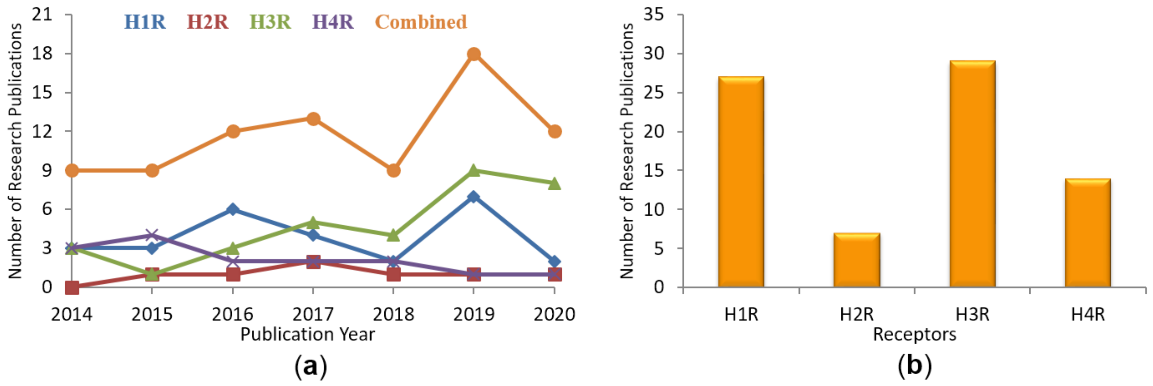
Figure 1. Number of histamine (H1–H4) receptor-related computational publications in the 2014–2020 time period: (a) Yearly on each HR; (b) Total publications on each HR.
Molecules 26 01778 g001
Figure 2. The most potent ligands targeting HRs discovered through diverse virtual screening and docking approaches in 2014–2020 and their biological activities.
Molecules 26 01778 g002
Figure 3. The areas of amino acids surrounding doxepin in the crystal structure of H1R: (red) the amine-binding region; (green) the upper aromatic region; (purple) the lower aromatic region; (yellow) the boundary amino acids participating in both amine-binding and aromatic regions. Doxepin is shown with its carbon atoms colored in orange. Side view of the receptor. TM helices are colored from blue (TM1) to red (TM7).
Molecules 26 01778 g003
Figure 4. Histamine molecule docked to the crystal structure of hH1R. The histamine is doubly positively charged. The amine group is bound to D3.32 and Y7.43, and the imidazole ring to S3.36 and Y6.51. The residue numbers are shown in Ballesteros-Weinstein numbering scheme. Histamine is shown with its carbon atoms colored in orange. View from extracellular side. TM helices are colored from blue (TM1) to red (TM7).
Molecules 26 01778 g004
Figure 5. Histamine molecule docked to the homology model of hH2R. The histamine is doubly positively charged. Both charged moieties of histamine are bound to D3.32. The imidazole ring is additionally bound to D5.42. The residue numbers are shown in Ballesteros-Weinstein numbering scheme. Histamine is shown with its carbon atoms colored in orange. View from extracellular side. TM helices are colored from blue (TM1) to red (TM7).
Molecules 26 01778 g005
Figure 6. Histamine molecule docked to the homology model of hH3R. The histamine is doubly positively charged. Both charged moieties of histamine are bound to D3.32. The imidazole ring is additionally bound to E5.46. The histamine hydrophobic linker is exposed toward hydrophobic residues F7.39 and L7.42. The residue numbers are shown in Ballesteros-Weinstein numbering scheme. Histamine is shown with its carbon atoms colored in orange. View from extracellular side. TM helices are colored from blue (TM1) to red (TM7).
Molecules 26 01778 g006
Figure 7. Histamine molecule docked to the homology model of hH4R. The histamine is doubly positively charged. The amine moiety of histamine is bound to D3.32 whereas the imidazole ring is bound to E5.46 and Q7.42. The histamine hydrophobic linker is exposed toward hydrophobic residues F7.39 and inversely rotated W7.43. The residue numbers are shown in Ballesteros-Weinstein numbering scheme. Histamine is shown with its carbon atoms colored in orange. View from extracellular side. TM helices are colored from blue (TM1) to red (TM7).
Molecules 26 01778 g007
Table 1. Overview of the drug design strategies for discovery of HR ligands. In parenthesis the PDB ids of templates used in homology modeling.
ReceptorTemplatesReceptor StateStrategyHit RateReference
H1Rβ2-adrenergic (2R4R)InactiveSBVS of Phytochemical inhibitors5 hits[48]
H1R3RZEInactiveIFP-based SBVS60.6% (20/33)[9]
PLANTS-based SBVS45.5% (15/33)
Combined IFP and PLANTS scoring based SBVS73.1% (19/26)
H3RH1R (3RZE)InactivePharmacophore-based virtual screening100% (5/5)[49]
H3RH1R (3RZE)InactiveProspective crystal structure-based pharmacophore virtual screening8% (6/76)[50]
H3RM3 muscarinic receptor (4DAJ)InactiveFP2 fingerprint/Electroshape / Spectrophores/LBVS50% (2/4)[51]
Hybrid VS100% (1/1)
H3RH1R (3RZE), M2 muscarinic (3UON), M3 muscarinic (4U15)InactivePharmacophore screening and redocking.25% (2/8)[52]
H4RH1R (3RZE)InactiveSingle Structure22% (11/50)[6]
Ensemble Docking16% (8/50)
Overlap hits27% (4/15)
H4RH1R (3RZE)InactiveLigand-based chemoinformatics: Intelligent Learning Engine/Iterative Stochastic Elimination/Extended connectivity fingerprint (ECFP4).11 hits[12]
H4RBovine rhodopsin (1F88)InactiveEnsemble docking5.3% (4/75)[25]
Ensemble docking followed by consensus scoring.15.4% (2/13)
H4Rβ2R (2RH1)InactiveProspective SBVS.26% (6/23)[53]
H1R (3RZE)InactiveProspective SBVS.21.4% (3/14)
H4RBovine rhodopsin (1L9H)InactiveHomology model refined by “scout screening”, VS using ECFP_4 fingerprint.23% (28/120)[22,23]
H4RH1R (3RZE)InactivePharmacophore-based virtual screening (Tanimoto similarity coefficient ≥0.9).1% (3/291)[26]
Table 2. Residues (in B-W numbering scheme) of HR subtypes involved in the binding of ligands studied in particular papers.
ResidueResiduesReference
H1RH2RH3RH4R
2.50 D80 [49,54]
2.61N84S75Y91Y72[26,55,56,57,58,59,60,61,62,63,64]
2.62I85 [31]
2.64Y87Y78Y94H75[55,60,63]
2.65L88 [55]
ECL1W93 [65]
3.28W103Y94W110W90[54,55,59,62,63,66,67,68]
3.29L104T95L111 [55,57,62,66]
3.32D107D98D114D94[6,9,12,22,23,24,25,26,31,48,49,51,52,53,54,55,56,57,58,59,60,62,63,64,66,69,70,71,72,73,74,75,76,77,78,79,80,81,82,83,84,85,86]
3.33Y108V99Y115Y95[6,12,22,24,26,31,51,52,55,56,57,59,60,62,63,64,66,68,69,70,71,72,75,76,77,78,79,80,82,83,84,86,87,88,89,90,91,92]
3.36S111C102C118C98[6,12,31,48,57,64,66,76,79,88,90]
3.37T112 T119T99[12,31,51,57,64,73,78,88]
3.40I115I106A122V102[31,55,57,64,76,78,88,89]
3.41F116 [31]
4.56W158L149L166V146[9,31,52,55,56,57,63,64,66,72,73,76,78,90]
4.57 Y167N147[6,12,53,93]
4.60 S153 M150[6,12,77]
4.61 I154 [66]
ECL2 N159E175 [49,66]
ECL2H167 [55]
ECL2R175 [65]
ECL2R176 [55,65,69]
ECL2 T171 [77]
ECL2D178 S162[12,55,83]
ECL2K179 H187E163[12,55,56,58,59,65,66,69,77,82,83]
ECL2C180C174 C164[12,55,66]
ECL2E181K175Y189E165[12,51,55,66,67,68,80,85,88,91,94]
ECL2T182V176A190P166[12,48,55,62,66,77,80,94,95]
ECL2D183Q177E191 [48,49,54,70,77,94,95]
ECL2F184V178F192F168[6,12,26,31,53,54,66,77,78,80,82,96]
ECL2Y185 F193F169[12,31,55,57,58,59,62,68,69,77,80,82,83,89]
ECL2 Y194 [62,94,95]
ECL2 W196 [62,94,95]
5.38F190Y182F198I174[6,12,31,51,57,59,63,66,88,90,94]
5.39K191G183L199L175[6,12,26,31,53,55,56,57,62,63,66,69,70,71,87]
5.42T194D186A202T178[6,12,56,57,63,64,66,75,77,79,82,85,87,90]
5.43A195G187S203S179[12,26,31,55,57,63,64,66,82,83,87]
5.46N198T190E206E182[6,12,23,24,25,26,31,49,50,51,52,53,57,58,59,60,61,63,64,65,66,67,68,69,75,76,77,79,80,81,82,83,85,86,87,88,89,90,91,92,93,94,95]
5.47F199 F207F183[9,12,26,31,55,64,67,72,76,90]
6.44F424 F367V184[66,72,76,82,90]
6.48W428W247W371W316[9,12,24,26,31,49,51,52,53,55,56,57,58,61,63,66,67,72,73,75,76,79,80,84,87,89,90,91,94,97]
6.51Y431Y250Y374Y319[6,12,22,24,31,48,49,51,52,53,55,56,57,59,60,61,63,65,66,67,69,70,71,72,73,75,76,77,79,80,81,82,83,85,86,87,88,89,91,92,94,95,98]
6.52F432F251T375S320[9,12,31,55,56,63,66,72,73,75,76,83,84,87]
6.55F435F254M378T323[9,12,31,53,55,56,57,66,72,75,76,83,84]
6.58I438 R381L326[12,55,61,62,65,80,89]
6.59A439 S327[12,55]
ECL3K442 S330[12,55]
ECL3N443 S331[12,55,65,69,83]
7.35H450E270Y394Y340[48,55,56,60,62,67,68,69,73,74,77,83,89]
7.39I454L274F398F344[6,23,24,51,52,53,55,57,59,60,62,63,66,68,72,76,84]
7.42G457G277L401Q347[6,23,24,26,31,52,53,57,58,63,79,80,82,86]
7.43Y458Y278W402W348[23,24,26,31,54,55,56,58,63,66,71,72,80,87]
Publisher’s Note: MDPI stays neutral with regard to jurisdictional claims in published maps and institutional affiliations.